Regulation of sertoli-germ cell adherens junction...
18
Title Regulation of sertoli-germ cell adherens junction dynamics via changes in protein-protein interactions of the N-cadherin-β- catenin protein complex which are possibly mediated by c-Src and myotubularin-related protein 2: An in vivo study using an androgen suppression model Author(s) Zhang, J; Wong, CH; Xia, W; Mruk, DD; Lee, NPY; Lee, WM; Cheng, CY Citation Endocrinology, 2005, v. 146 n. 3, p. 1268-1284 Issued Date 2005 URL http://hdl.handle.net/10722/48688 Rights This work is licensed under a Creative Commons Attribution- NonCommercial-NoDerivatives 4.0 International License.; Endocrinology. Copyright © The Endocrine Society.
Transcript of Regulation of sertoli-germ cell adherens junction...
Title
Regulation of sertoli-germ cell adherens junction dynamics viachanges in protein-protein interactions of the N-cadherin-β-catenin protein complex which are possibly mediated by c-Srcand myotubularin-related protein 2: An in vivo study using anandrogen suppression model
Author(s) Zhang, J; Wong, CH; Xia, W; Mruk, DD; Lee, NPY; Lee, WM;Cheng, CY
Citation Endocrinology, 2005, v. 146 n. 3, p. 1268-1284
Issued Date 2005
URL http://hdl.handle.net/10722/48688
RightsThis work is licensed under a Creative Commons Attribution-NonCommercial-NoDerivatives 4.0 International License.;Endocrinology. Copyright © The Endocrine Society.
This is a pre-published versionThis is a pre-published version
Regulation of Sertoli-Germ Cell Adherens JunctionDynamics via Changes in Protein-Protein Interactions ofthe N-Cadherin-�-Catenin Protein Complex which ArePossibly Mediated by c-Src and Myotubularin-RelatedProtein 2: An in Vivo Study Using an AndrogenSuppression Model
Jiayi Zhang, Ching-hang Wong, Weiliang Xia, Dolores D. Mruk, Nikki P. Y. Lee, Will M. Lee, andC. Yan Cheng
Population Council (J.Z., C.W., W.X., D.D.M., N.P.Y.L., C.Y.C.), Center for Biomedical Research, New York, New York10021; and Department of Zoology (W.M.L.), The University of Hong Kong, Hong Kong, China
Using a well characterized model of cell-cell actin-based ad-herens junction (AJ) disruption by suppressing the intrates-ticular testosterone level in adult rats with testosterone-estradiol implants, we have confirmed earlier findings thatSertoli-germ cell AJ dynamics are regulated by the activationof kinases via putative signaling pathways but with some un-expected findings as follows. First, the loss of germ cells fromthe seminiferous epithelium during androgen suppressionwas associated with a surge in myotubularin-related protein2 (MTMR2, a lipid phosphatase, in which adult MTMR2�/�
mice were recently shown to be azoospermic because of theloss of cell adhesion function between germ and Sertoli cells);kinases: phosphatidylinositol 3-kinase, c-Src, and C-terminalSrc kinase; adaptors: �-actinin, vinculin, afadin, and p130 Crk-associated protein; and AJ-integral membrane proteins at theectoplasmic specialization (ES, a testis-specific cell-cell actin-based AJ type) site: N-cadherin, �-catenin, integrin �1, andnectin 3. Second, MTMR2, instead of structurally interactingwith phosphatidylinositol 3-kinase, a protein and lipid kinase,was shown to associate only with c-Src, a nonreceptor proteintyrosine kinase, as demonstrated by both coimmunoprecipi-tation and fluorescent microscopy at the site of apical ES, but
none of the kinases, adaptors, and AJ-integral proteins thatwere examined. Collectively, these results suggest that theMTMR2/c-Src is an important phosphatase/kinase proteinpair in AJ dynamics in the testis. Because c-Src is known toassociate with the cadherin/catenin protein complex at the ESin the testis, we next sought to investigate any changes in theprotein-protein interactions of this protein complex duringandrogen suppression-induced germ cell loss. Indeed, therewas a loss of N-cadherin and �-catenin association, accompa-nied by a surge in Tyr phosphorylation of �-catenin, duringgerm cell loss from the epithelium. Third, and perhaps themost important of all, during natural recovery of the epithe-lium after removal of testosterone-estradiol implants whenspermatids were reattaching to Sertoli cells, an increase inN-cadherin and �-catenin association was detected with aconcomitant loss in the increased Tyr phosphorylation in�-catenin. In summary, these results illustrate that the cad-herin/catenin is a crucial cell adhesion complex that regulatesAJ dynamics in the testis, and its functionality is likely mod-ulated by the MTMR2/c-Src protein complex. (Endocrinology146: 1268–1284, 2005)
IN THE MAMMALIAN TESTIS, SUCH as in the rat, sper-matogenesis takes place in the seminiferous epithelium
in which one type A1 spermatogonium (diploid, 2n) dividesand differentiates into 256 spermatids (haploid, 1n). Thisevent is associated with extensive junction restructuring so
as to facilitate germ cell movement, because germ cells musttraverse the epithelium, translocating from the basal to theadluminal compartment. As such, fully developed sperma-tids can be released to the tubule lumen at spermiation (forreviews, see Refs. 1–3). If this event can be disrupted, sper-matogenesis will be compromised, leading to male infertility.Indeed, new approaches to disrupt tight junction and adhe-rens junction (AJ) dynamics are being widely investigated foridentifying novel male contraceptives (for reviews, see Refs.2, 3). 1-(2,4-Dichlorobenzyl)-indazole-3-carbohydrazide (AF-2364, also known as Adjudin, a molecule that induces Sertoli-germ cell AJ disruption) (3, 4) is one of these novel com-pounds that was synthesized based on the core structure ofindazole-3-carboxylic acid known to possess potent antisper-matagenic effects (for a review, see Ref. 5). Furthermore, thetoxic effects of AF-2364 were low when it was administeredonce weekly (6–8). Recent studies have shown that AF-2364-induced AJ disruption is a potential reversible male contra-
First Published Online December 9, 2004Abbreviations: AJ, Adherens junction; BW, body weight; CK, casein
kinase; CMT, Charcot-Marie-Tooth disease; CPA, cyproterone acetate;Csk, C-terminal Src kinase; DAPI, 4�,6-diamidino-2-phenylindole; Dlg1,disc large 1; E, estradiol-17�; ES, ectoplasmic specialization; FITC, flu-orescein isothiocyanate; IP, immunoprecipitation; MTM, myotubularin;MTMR2, myotubularin-related protein 2; NR, spontaneous natural re-covery; PI 3-K, phosphatidylinositol 3-kinase; PTP, protein tyrosinephosphatase, r, rat; T, testosterone; T, total gel concentration (g/100ml) � acrylamide � methylene bisacrylamide; TE, testosterone-estra-diol; ZO-1, zonula occludens 1.Endocrinology is published monthly by The Endocrine Society (http://www.endo-society.org), the foremost professional society serving theendocrine community.
balt3/zee-end/zee-end/zee00305/zee0730-05a hurtr S�12 1/21/05 7:52 4/Color Figure(s): 1,2,3,6,10 Art: 1195093 Input-tcm(v)
0013-7227/05/$15.00/0 Endocrinology 146(3):1268–1284Printed in U.S.A. Copyright © 2005 by The Endocrine Society
doi: 10.1210/en.2004-1194
1268
AQ: A
AQ: B
AQ: C
<zjs;Reproduction-Development> • <zjss;>
ceptive, which can also be used as an in vivo model to studyAJ dynamics in the testis (for reviews, see Refs. 2, 3, 9–11).Needless to say, earlier findings in which AF-2364 was usedto identify the signaling pathways that regulate AJ dynamicsin the testis could be the results of acute or chronic toxicity.We therefore sought to use another well characterizedmodel, in which cell adhesion function between Sertoli cellsand spermatids (step 8 and beyond) was compromised byintratesticular androgen suppression using testosterone-estradiol (TE) implants (for a review, see Ref. 12), to validateand expand these earlier observations. For instance, recentstudies have shown that the loss of germ cells from theepithelium after AF-2364 treatment is regulated, at least inpart, by a loss of protein-protein interactions in the ectoplas-mic specialization (ES)-associated AJ-protein complexes,such as the cadherin-catenin complex (for a review, see Ref.13). If such results can be reproduced in another in vivomodel, this will undoubtedly eliminate the possibility thatthese earlier observations were artifacts of nonspecific drugeffects. This would help to synthesize better compounds toperturb cell adhesion function conferred by these proteincomplexes, leading to the development of novel male pills.
Testosterone (T) is a male sex hormone that determinesmale phenotype and fertility. Its testicular level is approxi-mately 100-fold of that in serum (14). A reduction of testicularT level by increasing serum T and estradiol-17� (E) levels inmale rats using TE implants can suppress the developmentof step 8 spermatids and beyond, leading to their detachmentfrom the epithelium possibly by compromising ES function(for reviews, see Refs. 12, 15). After TE implants are removed,and if rats are permitted to recover in the presence of ex-cessive T or allowed to undergo spontaneous natural recov-ery (NR), a restoration of T level in the testes can be achieved,which is also accompanied by a reversal of spermatogenesis.Collectively, these observations illustrate that this is a novelmodel to study ES dynamics (for a review, see Ref. 12).
Myotubularin-related protein 2 (MTMR2) is a lipid phos-phatase at 3D site within the myotubularin (MTM) familyhaving a catalytic site found in protein tyrosine phosphatases(PTP) and dual specific phosphatases (16–18). Its mutationcauses the demyelinating neuropathy Charcot-Marie-Toothdisease type 4B1 (CMT 4B1) (16, 19). MTMR2 dephospho-rylates phosphatidylinositol 3-phosphate and phosphatidyl-inositol 3,5-bisphosphate, both in vivo and in vitro, and theproduct of the latter can act as a feedback regulator ofMTMR2 (20, 21). Interestingly, MTMR2 is expressed by Ser-toli and germ cells in the testis, and its level is induced duringtesticular maturation or when Sertoli-germ cell AJs are as-sembled in vitro (22, 23), implicating its role in AJ dynamics.A recent study has shown that MTMR2�/� mice displayednot only neuropathy but also azoospermia, accompanied by
a significant reduction in testis size and weight, implicatingthe pivotal role of MTMR2 in AJ function and spermatogen-esis (24). Besides forming homodimers between MTMR2(25), MTMR2 can interact with several proteins in other ep-ithelia such as MTMR5 (26). Moreover, neurofilament lightchain protein and disc large 1 (Dlg1) are also structurallyassociated with MTMR2 in the brain (24, 27). However, abinding partner of MTMR2 has yet to be found in the testis.We therefore sought to identify the putative interacting part-ner(s) of MTMR2 in the seminiferous epithelium and to as-sess whether MTMR2 interacts with any protein kinases atthe ES site to regulate cell adhesion function.
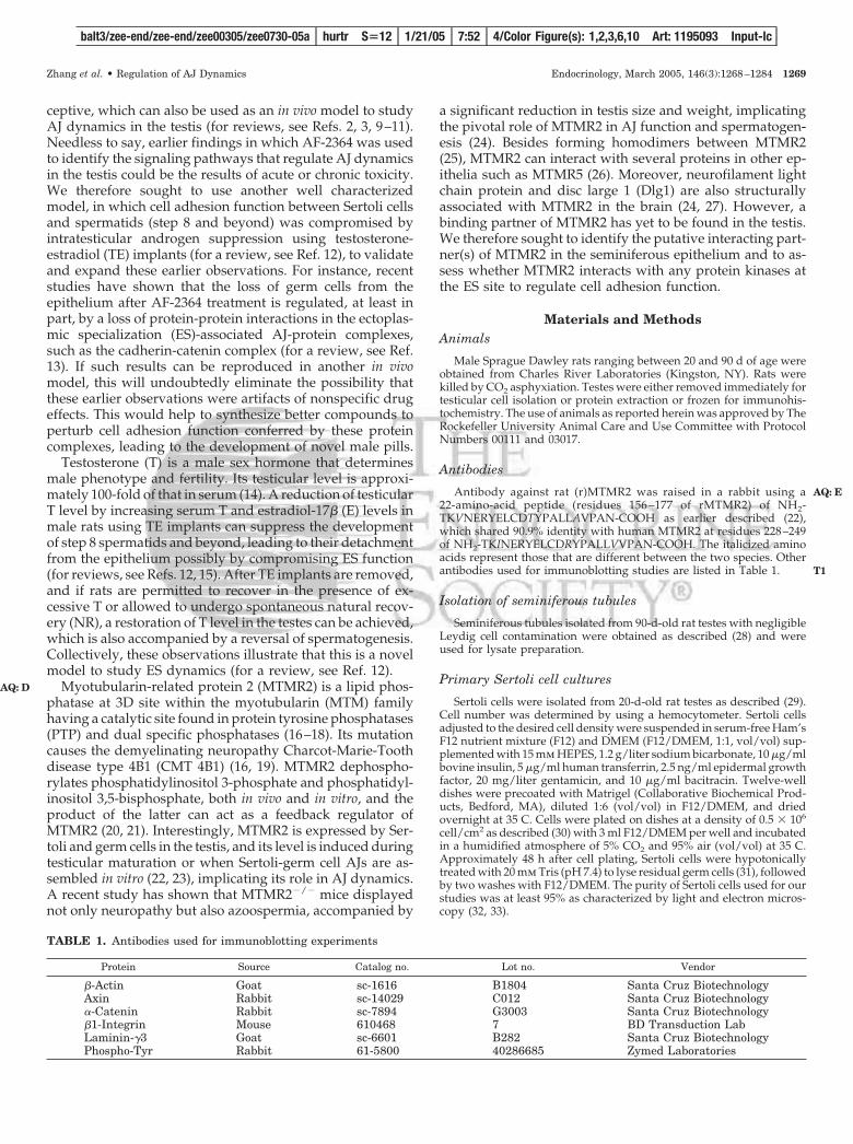
Materials and MethodsAnimals
Male Sprague Dawley rats ranging between 20 and 90 d of age wereobtained from Charles River Laboratories (Kingston, NY). Rats werekilled by CO2 asphyxiation. Testes were either removed immediately fortesticular cell isolation or protein extraction or frozen for immunohis-tochemistry. The use of animals as reported herein was approved by TheRockefeller University Animal Care and Use Committee with ProtocolNumbers 00111 and 03017.
Antibodies
Antibody against rat (r)MTMR2 was raised in a rabbit using a22-amino-acid peptide (residues 156 –177 of rMTMR2) of NH2-TKVNERYELCDTYPALLAVPAN-COOH as earlier described (22),which shared 90.9% identity with human MTMR2 at residues 228–249of NH2-TKINERYELCDRYPALLVVPAN-COOH. The italicized aminoacids represent those that are different between the two species. Otherantibodies used for immunoblotting studies are listed in Table 1.
Isolation of seminiferous tubules
Seminiferous tubules isolated from 90-d-old rat testes with negligibleLeydig cell contamination were obtained as described (28) and wereused for lysate preparation.
Primary Sertoli cell cultures
Sertoli cells were isolated from 20-d-old rat testes as described (29).Cell number was determined by using a hemocytometer. Sertoli cellsadjusted to the desired cell density were suspended in serum-free Ham’sF12 nutrient mixture (F12) and DMEM (F12/DMEM, 1:1, vol/vol) sup-plemented with 15 mm HEPES, 1.2 g/liter sodium bicarbonate, 10 �g/mlbovine insulin, 5 �g/ml human transferrin, 2.5 ng/ml epidermal growthfactor, 20 mg/liter gentamicin, and 10 �g/ml bacitracin. Twelve-welldishes were precoated with Matrigel (Collaborative Biochemical Prod-ucts, Bedford, MA), diluted 1:6 (vol/vol) in F12/DMEM, and driedovernight at 35 C. Cells were plated on dishes at a density of 0.5 � 106
cell/cm2 as described (30) with 3 ml F12/DMEM per well and incubatedin a humidified atmosphere of 5% CO2 and 95% air (vol/vol) at 35 C.Approximately 48 h after cell plating, Sertoli cells were hypotonicallytreated with 20 mm Tris (pH 7.4) to lyse residual germ cells (31), followedby two washes with F12/DMEM. The purity of Sertoli cells used for ourstudies was at least 95% as characterized by light and electron micros-copy (32, 33).
TABLE 1. Antibodies used for immunoblotting experiments
Protein Source Catalog no. Lot no. Vendor
�-Actin Goat sc-1616 B1804 Santa Cruz BiotechnologyAxin Rabbit sc-14029 C012 Santa Cruz Biotechnology�-Catenin Rabbit sc-7894 G3003 Santa Cruz Biotechnology�1-Integrin Mouse 610468 7 BD Transduction LabLaminin-�3 Goat sc-6601 B282 Santa Cruz BiotechnologyPhospho-Tyr Rabbit 61-5800 40286685 Zymed Laboratories
balt3/zee-end/zee-end/zee00305/zee0730-05a hurtr S�12 1/21/05 7:52 4/Color Figure(s): 1,2,3,6,10 Art: 1195093 Input-lc
Zhang et al. • Regulation of AJ Dynamics Endocrinology, March 2005, 146(3):1268–1284 1269
AQ: D
AQ: E
T1
Sertoli-germ cell cocultures
Germ cells were isolated from 90-d-old rat testes by a mechanicalprocedure using sequential filtrations as described (34). For germ celllysate preparation, cells were terminated within 3 h after their isolation.For cocultures, freshly isolated germ cells were suspended in F12/DMEM supplemented with 6 mm sodium lactate, 2 mm sodium pyru-vate, 20 mg/liter gentamicin, and 10 �g/ml bacitracin. Cell number wasdetermined by a hemocytometer. Germ cells at desired cell number werereconstituted in F12/DMEM in the absence or presence of T (2 � 10–8
or 2 � 10–7 m) (Sigma Chemical Co., St. Louis, MO) and cyproteroneacetate (1 � 10–6 m) (Sigma). Germ cells were cocultured with Sertolicells at a 1:1 ratio in which Sertoli cells had been cultured alone for 5 dat 0.5 � 106 cells/cm2 forming an intact epithelium (33, 35). Cocultureswere terminated at 24 and 48 h thereafter when functional anchoringjunctions, such as desmosome-like junctions and apical ES, were formed(3, 36, 37).
Immunohistochemistry
Testes isolated from normal rats and from animals of different treat-ment groups were frozen immediately in liquid nitrogen. Frozen testeswere embedded in OCT compounds (Miles Scientific, Elkhart, IN), and8-�m sections were cut using a microtome in a cryostat at �20 C andmounted on poly-l-lysine-coated slides. Histostain-SP kits obtainedfrom Zymed Laboratories (South San Francisco, CA) were used forimmunohistochemistry as described (38, 39). Sections were air dried atroom temperature, fixed in Bouin’s fixative (4% formaldehyde in picricacid) for 5 min, and washed with PBS [10 mm sodium phosphate, 0.15m sodium chloride (pH 7.4) at 22 C]. Sections were then treated with 3%hydrogen peroxide solution (vol/vol, in methanol) for 20 min to elim-inate endogenous peroxidase activity and blocked with a serum block-ing solution (10% nonimmune goat serum, Zymed). Rabbit anti-rMTMR2 serum (1:300, in 1% nonimmune goat serum) or preimmunerabbit serum (control, 1:300, in 1% nonimmune goat serum) was addedonto sections, and slides were incubated in a humidified chamber at 35C overnight. Thereafter, sections were rinsed in PBS and incubated withbiotinylated goat antirabbit IgG for 30 min, to be followed by strepta-vidin peroxidase conjugate for 10 min. Color development was per-formed using aminoethylcarbazole mixture. Sections were counter-stained using hematoxylin and mounted with Histomount (Zymed). Allsections from different rats within an experimental group were pro-cessed simultaneously in two to three microscopic slides (with two tofour sections per slide) to eliminate interexperimental variations. Mi-crographs were obtained using an Olympus DP70 12.5 MPa DigitalCamera interfaced to an HP Vectra VL800 Workstation and the QCap-ture Suite V2.60 software package from Quantitative Imaging Corp.(Burnaby, Canada). Digital images were subsequently processed usingAdobe Photoshop (version 7.0). All immunohistochemistry experimentswere repeated at least eight to 12 times over a period of 10 months usingeither frozen or paraffin sections from different rats, and the resultsdepicted in Figs. 1–3 are the representatives of these analyses.
Sample preparation and immunoblot analysis
Testicular cells, seminiferous tubules, testes, and brain previouslytrimmed into approximately 2 � 2-mm pieces, were sonicated for 15 secin an immunoprecipitation (IP) buffer [0.125 m Tris, containing 0.15 mNaCl, 5 mm sodium chloromercuribenzoate, 2 mm EDTA, 2 mm N-ethylmaleimide, 2 mm phenylmethylsulfonyl fluoride, 5 �g/ml pepsta-tin A, 1% Nonidet P-40 (vol/vol), 10% glycerol (vol/vol) (pH 7.4) at 22C] and centrifuged at 12,000 � g for 15 min to remove cell debris. Theclear supernatant was designated cell/tissue lysates and stored at �80C until used. Protein concentration was estimated by Coomassie bluedye-binding assay (40) using BSA as a standard. Equal amounts ofprotein (100 �g protein per lane) were resolved by SDS-PAGE onto 7.5%T SDS-polyacrylamide gels under reducing conditions as described (41).Proteins were electroblotted onto nitrocellulose membranes (0.45 �m,Schleicher and Schuell, Keene, NH). Target proteins in blots were de-tected by corresponding antibodies and visualized using an enhancedchemiluminescence kit (Amersham Pharmacia Biotech, Piscataway, NJ)as described (32, 38, 42). Each blot could be reused for up to five to sixtimes with different primary antibodies after the initial primary and
secondary antibodies were stripped using SDS as described (32, 43)without detectable protein loss when the level of �-actin was quantifiedat the beginning and the end of an experimental series.
Treatment of rats with AF-2364 to induce germ cell lossfrom the seminiferous epithelium
AF-2364 was synthesized as previously described (6) with a puritygreater than 99.8% when examined by elemental analysis. Male rats[250–300 g body weight (BW)] were fed with a single dose of AF-2364at 50 mg/kg BW as described (6, 7), which was shown to induce germcell loss from the epithelium, in particular round, elongating, and elon-gated spermatids (44), by perturbing cell adhesion function in the ep-ithelium (for reviews, see Refs. 2, 3). Rats at time 0 served as controls.Testes were removed at specific time points, frozen in liquid nitrogen,and stored at –80 C until used.
Induction of germ cell loss from the seminiferous epitheliumusing an in vivo androgen suppression model in adult ratswith steroid implants
Ethyl vinyl acetate (DuPont ELVAX 770) implants (3 or 4 cm in length)filled with T (Sigma) and 0.4-cm implants filled with E (Sigma) wereprepared as described (45). A single 3-cm T and a 0.4-cm E implant wereplaced under the skin along the dorsal surface of each male adult rat (n �3 rats for each specified time point) to suppress the endogenous T levelin the testis, which was shown to induce germ cell loss from the sem-iniferous epithelium (for reviews, see Refs. 12, 15). Rats were underanesthesia using ketamine HCl (�70 mg/kg BW) during the surgery.The surgical area (�2 cm2) was shaved to remove hair, cleansed with70% ethanol and Betadine (two times each). A small incision (�1.5 cm)was made using a sterile scalpel. Implants were carefully inserted underthe skin, and the surgical site was stitched using Ethilon PC-S sterile,nonabsorbable surgical suture with a 19 PC prime needle (Ethicon Inc.,Somerville, NJ). After 28 d, TE implants were removed from all ratsunder anesthesia as described above. In one group, rats (n � 3 rats foreach time point) received four 4-cm T implants (without E) to permitrapid T recovery and resumption of spermatogenesis, whereas in theother group, rats were allowed to have spontaneous (i.e. natural) re-covery without any steroid implants. Rats had free access to standardchow and water with a 12-h light, 12-h dark cycle. Rats at time 0 weredesignated controls. Testes were removed at specific time points, frozenin liquid nitrogen, and stored at –80 C until used.
Co-IP
Approximately 500 �g of protein from testis lysates, prepared asdescribed above from normal rats, rats that underwent spontaneousrecovery (NR) on d 42, or rats that received four 4-cm T implants toundergo rapid recovery on d 30, were used for IP. Samples were pre-treated with 2 �g rabbit IgG (or mouse or goat IgG, depending on thesource of the primary antibody) for 3 h, followed by an incubation with10 �l protein A/G plus agarose (Santa Cruz Biotechnology, Santa Cruz,CA) to eliminate nonspecific interactions between lysates and IgG. Aftercentrifugation at 1000 � g for 5 min to precipitate the IgG-nonspecificallybound protein complex, supernatants were collected for subsequentco-IP. In brief, 2 �g of an anti-rMTMR2 antibody, IgG against a targetprotein (e.g. c-Src), or normal rabbit IgG (negative control), was addedand incubated with the supernatant at 4 C overnight. The immunocom-plexes were then precipitated using 20 �l protein A/G plus agarose ona rotator (40 rpm) for 6 h. Immunoprecipitates were washed with 300 �lIP buffer (four times, by gentle resuspension of the pellet in the IP bufferfollowed by centrifugation at 1000 � g for 5 min). Proteins in the im-munocomplexes were extracted in SDS-sample buffer [0.125 m Tris (pH6.25) at 22 C containing 1% SDS (wt/vol), 1.6% 2-mercaptoethanol (vol/vol), and 20% glycerol (vol/vol)] at 100 C for 10 min, and samples wereresolved by SDS-PAGE onto 7.5% T SDS-polyacrylamide gels. Afterproteins were electroblotted onto nitrocellulose papers, immunoblottingwas performed as earlier described using different antibodies (38). Ly-sates from normal testes and seminiferous tubules served as positivecontrols to assess the specificity of the primary antibodies and theelectrophoretic mobility of the target proteins.
balt3/zee-end/zee-end/zee00305/zee0730-05a hurtr S�12 1/21/05 7:52 4/Color Figure(s): 1,2,3,6,10 Art: 1195093 Input-lc
1270 Endocrinology, March 2005, 146(3):1268–1284 Zhang et al. • Regulation of AJ Dynamics
AQ: F
Immunofluorescence microscopy
Immunofluorescence microscopy was performed as earlier described(32, 46) to examine whether rMTMR2 and c-Src colocalized to the samesite in the seminiferous epithelium, verifying results of IP that rMTMR2and c-Src are indeed part of a structural complex at the ES. Frozensections were fixed in Bouin’s fixative (4% formaldehyde in picric acid)for 5 min and washed with PBS [10 mm sodium phosphate, 0.15 msodium chloride (pH 7.4) at 22 C]. To eliminate interexperimental vari-ation, all sections obtained from testes in different treatment groupswithin an experimental set were processed simultaneously in two slidesbecause each slide holds at least three to four cross-sections of testes. Thisis necessary so that all sections within an experimental set can be stained,colorimetrically developed, and photographed simultaneously. Sectionswere blocked with 10% normal goat serum for 30 min. Sections were thenincubated with a rMTMR2 antibody at a dilution of 1:200 in PBS con-taining 1% goat serum and then with a p-Src-Tyr416 antibody (UpstateBiotechnology, Lake Placid, NY; catalog no. 05-677, lot 26419) at a con-centration of 20 �g/ml in PBS containing 1% goat serum, to be followedby 1:50 fluorescein isothiocyanate (FITC) goat-antirabbit IgG conjugate(Zymed, catalog no. 62-6111, lot 30677924) and 1:50 goat antimouseIgG-CY3 conjugate (Zymed, catalog no. 81-6515, lot 31079880R). Sectionswere then mounted by using Vectashield mounting medium with 4�,6-diamidino-2-phenylindole (DAPI) (Vector Laboratories, Inc., Burlin-game, CA; catalog no. H-1500, lot Q0220). Images were digitally cap-tured by using an Olympus BX40 microscope with UPlanF1 fluorescentoptics (Olympus Corp.) and an Olympus DP70 12.5 MPa digital camera.All data were analyzed using Adobe Photoshop (version 7.0). Controlsincluded the use of either IgG or 1% goat serum instead of the primaryantibodies, which failed to yield detectable fluorescence, illustrating thatthe staining was specific.
Statistical analysis
Statistical analysis was performed using the GB-STAT StatisticalAnalysis Software Package (version 7.0, Dynamic Microsystems, Inc.,Silver Spring, MD).
ResultsImmunohistochemical localization of rat MTMR2(rMTMR2) in the rat testis
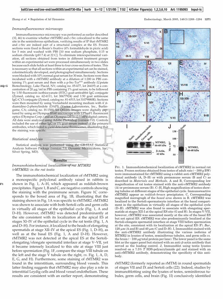
The immunohistochemical localization of rMTMR2 usinga monospecific polyclonal antibody raised in rabbits isshown in Fig. 1, A–H, which appeared as reddish-brownprecipitates. Figure 1, B and C, are negative controls showingthe staining with the preimmune serum. Figure 1C corre-sponds to the boxed area of Fig. 1B, illustrating that thestaining shown in Fig. 1A was specific to rMTMR2. rMTMR2was shown to associate with both Sertoli cells and germ cellsin virtually all stages of the epithelial cycle (Fig. 1, A andD–H). However, rMTMR2 was detected predominantly atthe site consistent with its localization at the apical ES atstages XI–IV of the epithelial cycle in the rat testis (Fig. 1, Aand D–H). For instance, it localized to the heads of elongatingspermatids at stage XII–IV at the apical ES (Fig. 1, D–H), aswell as at the basal ES (Fig. 1, A and D–H). However,rMTMR2 was not detected at the apical ES at the Sertoli-elongating/elongate spermatid interface at stage V–VII, yetit became intensely localized to this site at stage VIII justbefore spermiation (Fig. 1F, compare the stage VIII tubule onthe left and the stage V tubule on the right, vs. Fig. 1, A, D,E, G, and H). Furthermore, some staining of rMTMR2 wasfound in the interstitium, surrounding blood vessels. Thislatter finding suggests that rMTMR2 may also play a role ininterstitial Leydig cells and blood vessel endothelium. Theseresults are consistent with an earlier report, demonstrating
rMTMR2 (formerly reported as rMTM) in round spermatidsof stages VII and IX and Sertoli cells (22). Indeed, studies byimmunoblotting using the lysates of testes, seminiferous tu-bules, germ cells, and brain (Fig. 1I) conclusively identified
FIG. 1. Immunohistochemical localization of rMTMR2 in normal rattestes. Frozen sections obtained from testes of adult rats (�300 g BW)were immunostained for rMTMR2 using a rabbit anti-rMTMR2 poly-clonal antibody (A, D–H) or with preimmune serum (B and C) asdescribed in Materials and Methods. A and B, Corresponding lowmagnification of rat testes stained with the anti-rMTMR2 antibody(A) or preimmune serum (B). C–H, High magnification of testes show-ing tubules at different stages of the epithelial cycle. ImmunoreactiverMTMR2 appear as reddish-brown precipitates. C, Correspondingmagnified micrograph of the boxed area shown in B. rMTMR2 waslocalized to the Sertoli-spermatocyte interface at the basal compart-ment in the epithelium in virtually all stages of the epithelial cycle(D–H). rMTMR2 was also found to associate with elongating sper-matids at stages XII-I at the apical ES site (G and H). In stages V-VII,however, rMTMR2 was associated mostly at the site of the basal ESbut not apical ES. rMTMR2 was also predominantly localized at theSertoli-elongate spermatid interface at stage VIII before spermiationat the site, consistent with its localization at the apical ES (F). Bar,120 �m (A and B) and 40 �m (C and D–H). I, Immunoblot stained withthe anti-rMTMR2 antibody illustrating the various isoforms ofMTMR2 in lysates of testes, seminiferous tubules, and germ cells vs.the brain (�100 �g total protein per lane). The lower panel is the sameblot as the upper panel but stained with an anti-�-actin antibody thatserved as the loading control. J, Immunoblot using testis lysatesresolved on a 7.5% T SDS-polyacrylamide gel and stained with ananti-rMTMR2 antibody, demonstrating the specificity of this anti-body.
balt3/zee-end/zee-end/zee00305/zee0730-05a hurtr S�12 1/21/05 7:52 4/Color Figure(s): 1,2,3,6,10 Art: 1195093 Input-lc
Zhang et al. • Regulation of AJ Dynamics Endocrinology, March 2005, 146(3):1268–1284 1271
AQ: G
F1
rMTMR2 in these tissues/cells that exists as three isoformsranging between 66 and 68 kDa. Interestingly, germ cellswere shown to produce rMTMR2 (Fig. 1I) consistent with theimmunohistochemical data shown in Fig. 1, A–H. It is notedthat these germ cells had negligible somatic cell contamina-tions when assessed by RT-PCR using primer pairs specificto protein markers of Leydig, peritubular myoid, and Sertolicells (data not shown) as earlier reported (32, 43). The spec-ificity of this antibody was also shown by immunoblot anal-ysis (Fig. 1J).
Changes in the localization and protein level of rMTMR2during AF-2364-induced AJ disruption in theseminiferous epithelium
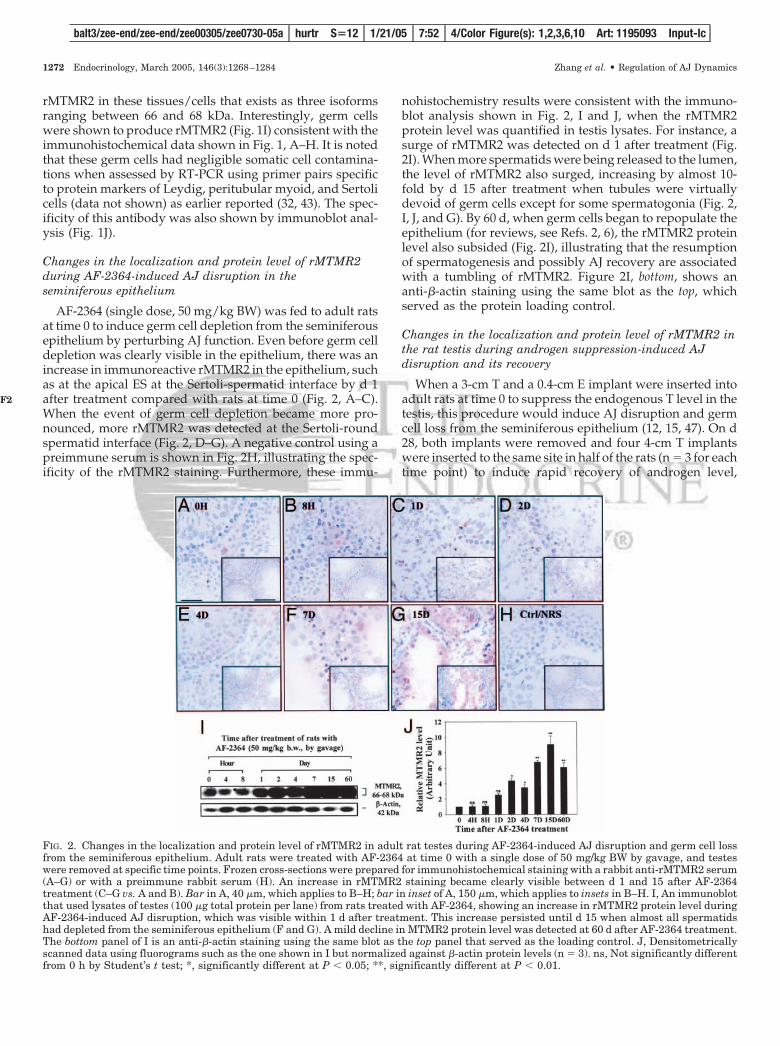
AF-2364 (single dose, 50 mg/kg BW) was fed to adult ratsat time 0 to induce germ cell depletion from the seminiferousepithelium by perturbing AJ function. Even before germ celldepletion was clearly visible in the epithelium, there was anincrease in immunoreactive rMTMR2 in the epithelium, suchas at the apical ES at the Sertoli-spermatid interface by d 1after treatment compared with rats at time 0 (Fig. 2, A–C).When the event of germ cell depletion became more pro-nounced, more rMTMR2 was detected at the Sertoli-roundspermatid interface (Fig. 2, D–G). A negative control using apreimmune serum is shown in Fig. 2H, illustrating the spec-ificity of the rMTMR2 staining. Furthermore, these immu-
nohistochemistry results were consistent with the immuno-blot analysis shown in Fig. 2, I and J, when the rMTMR2protein level was quantified in testis lysates. For instance, asurge of rMTMR2 was detected on d 1 after treatment (Fig.2I). When more spermatids were being released to the lumen,the level of rMTMR2 also surged, increasing by almost 10-fold by d 15 after treatment when tubules were virtuallydevoid of germ cells except for some spermatogonia (Fig. 2,I, J, and G). By 60 d, when germ cells began to repopulate theepithelium (for reviews, see Refs. 2, 6), the rMTMR2 proteinlevel also subsided (Fig. 2I), illustrating that the resumptionof spermatogenesis and possibly AJ recovery are associatedwith a tumbling of rMTMR2. Figure 2I, bottom, shows ananti-�-actin staining using the same blot as the top, whichserved as the protein loading control.
Changes in the localization and protein level of rMTMR2 inthe rat testis during androgen suppression-induced AJdisruption and its recovery
When a 3-cm T and a 0.4-cm E implant were inserted intoadult rats at time 0 to suppress the endogenous T level in thetestis, this procedure would induce AJ disruption and germcell loss from the seminiferous epithelium (12, 15, 47). On d28, both implants were removed and four 4-cm T implantswere inserted to the same site in half of the rats (n � 3 for eachtime point) to induce rapid recovery of androgen level,
FIG. 2. Changes in the localization and protein level of rMTMR2 in adult rat testes during AF-2364-induced AJ disruption and germ cell lossfrom the seminiferous epithelium. Adult rats were treated with AF-2364 at time 0 with a single dose of 50 mg/kg BW by gavage, and testeswere removed at specific time points. Frozen cross-sections were prepared for immunohistochemical staining with a rabbit anti-rMTMR2 serum(A–G) or with a preimmune rabbit serum (H). An increase in rMTMR2 staining became clearly visible between d 1 and 15 after AF-2364treatment (C–G vs. A and B). Bar in A, 40 �m, which applies to B–H; bar in inset of A, 150 �m, which applies to insets in B–H. I, An immunoblotthat used lysates of testes (100 �g total protein per lane) from rats treated with AF-2364, showing an increase in rMTMR2 protein level duringAF-2364-induced AJ disruption, which was visible within 1 d after treatment. This increase persisted until d 15 when almost all spermatidshad depleted from the seminiferous epithelium (F and G). A mild decline in MTMR2 protein level was detected at 60 d after AF-2364 treatment.The bottom panel of I is an anti-�-actin staining using the same blot as the top panel that served as the loading control. J, Densitometricallyscanned data using fluorograms such as the one shown in I but normalized against �-actin protein levels (n � 3). ns, Not significantly differentfrom 0 h by Student’s t test; *, significantly different at P � 0.05; **, significantly different at P � 0.01.
balt3/zee-end/zee-end/zee00305/zee0730-05a hurtr S�12 1/21/05 7:52 4/Color Figure(s): 1,2,3,6,10 Art: 1195093 Input-lc
1272 Endocrinology, March 2005, 146(3):1268–1284 Zhang et al. • Regulation of AJ Dynamics
F2
whereas the other half of the animals (n � 3 per time point)were allowed to undergo spontaneous recovery (NR) with-out any steroid implants (for a review, see Ref. 12). Similarto the AF-2364 model, there was a progressive surge inrMTMR2 staining in the epithelium during androgen
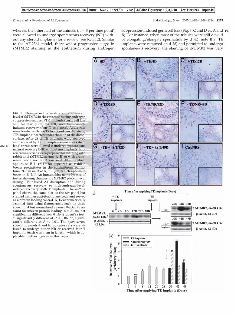
suppression-induced germ cell loss (Fig. 3, C and D vs. A andB). For instance, when most of the tubules were still devoidof elongating/elongate spermatids by d 42 (note that TEimplants were removed on d 28) and permitted to undergospontaneous recovery, the staining of rMTMR2 was very
FIG. 3. Changes in the localization and proteinlevel of rMTMR2 in the rat testis during androgensuppression-induced (TE implants) germ cell lossand AJ disruption, its NR, and high-dose-T-induced recovery (four T implants). Adult ratswere treated with one T (3-cm) and one E (0.4-cm)(TE) implant inserted under the skin at the dorsalsurface. After 28 d, TE implants were removedand replaced by four T implants (each was 4-cmlong) or rats were allowed to undergo spontaneousnatural recovery (NR) without any implants. Fro-zen cross-sections were prepared for staining withrabbit anti-rMTMR2 serum (A–H) or with preim-mune rabbit serum (I). Bar in A, 40 �m, whichapplies to B–I. rMTMR2 appeared as reddishbrown precipitates in the seminiferous epithe-lium. Bar in inset of A, 150 �m, which applies toinsets in B–I. J, An immunoblot using lysates oftestes showing changes in rMTMR2 protein levelduring TE-induced AJ disruption and duringspontaneous recovery or high-androgen-level-induced recovery with T implants. The bottompanel shows the same blot as the top panel butstained with an anti-�-actin antibody and servedas a protein loading control. K, Densitometricallyscanned data using fluorograms, such as thoseshown in J but normalized against �-actin to ac-count for uneven protein loading (n � 3). ns, notsignificantly different from 0 h by Student’s t test;*, significantly different at P � 0.05; **, signifi-cantly different at P � 0.01. The open arrowshown in panels J and K indicates rats were al-lowed to undergo either NR or received four Timplants (each was 4-cm in length), which is ap-plicable to other figures in this report.
balt3/zee-end/zee-end/zee00305/zee0730-05a hurtr S�12 1/21/05 7:52 4/Color Figure(s): 1,2,3,6,10 Art: 1195093 Input-lc
Zhang et al. • Regulation of AJ Dynamics Endocrinology, March 2005, 146(3):1268–1284 1273
AQ: U
F3
intense in the epithelium (Fig. 3E), yet rMTMR2 staining wasgreatly reduced when spermatids began to reappear in theepithelium in rats that either received four T implants orunderwent NR (Fig. 3, F–H vs. E). Based on the observationof such a rapid recovery of spermatids in the epithelium,within approximately 20 d, it is logical to conclude that mostspermatocytes were not depleted from the epithelium by thistreatment. Figure 3I shows a normal testis stained with pre-immune rabbit serum that served as a negative control. Suchchanges in immunohistochemical localization of rMTMR2during androgen suppression-induced spermatid loss andthe subsequent recovery were verified by immunoblottingshown in Fig. 3, J and K. For instance, rMTMR2 was induced
by d 8 after receiving TE implants and reached its peak by42 d in the NR group but peaked by 30 d in the four-T-implants group and declined gradually during the recoveryphase. The lower panels of Fig. 3J are the same blots as theupper panels but stained with an anti-�-actin antibody toillustrate equal protein loading.
Effects of androgen suppression-induced AJ disruption andits recovery on the protein levels of selected kinases andadaptors in the seminiferous epithelium in vivo
To investigate possible MTMR2 interacting protein part-ners, including kinases and adaptors, immunoblot analysis
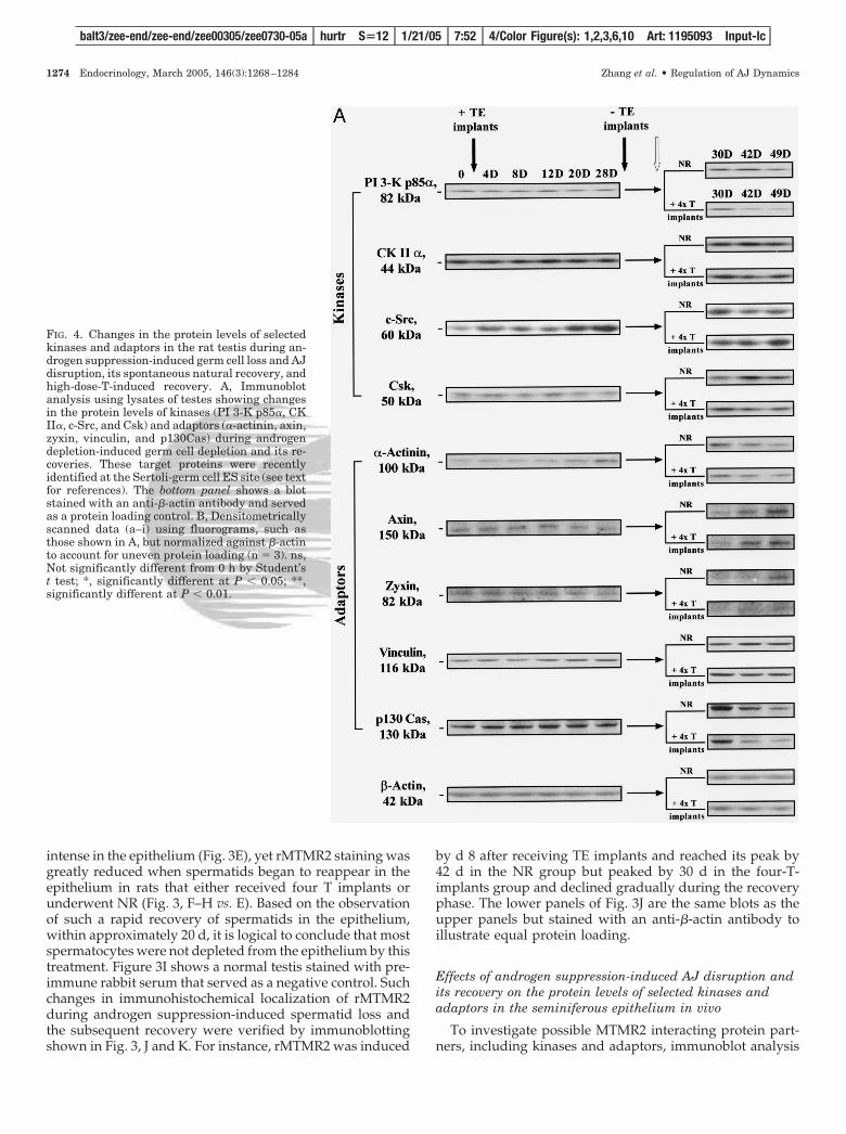
FIG. 4. Changes in the protein levels of selectedkinases and adaptors in the rat testis during an-drogen suppression-induced germ cell loss and AJdisruption, its spontaneous natural recovery, andhigh-dose-T-induced recovery. A, Immunoblotanalysis using lysates of testes showing changesin the protein levels of kinases (PI 3-K p85�, CKII�, c-Src, and Csk) and adaptors (�-actinin, axin,zyxin, vinculin, and p130Cas) during androgendepletion-induced germ cell depletion and its re-coveries. These target proteins were recentlyidentified at the Sertoli-germ cell ES site (see textfor references). The bottom panel shows a blotstained with an anti-�-actin antibody and servedas a protein loading control. B, Densitometricallyscanned data (a–i) using fluorograms, such asthose shown in A, but normalized against �-actinto account for uneven protein loading (n � 3). ns,Not significantly different from 0 h by Student’st test; *, significantly different at P � 0.05; **,significantly different at P � 0.01.
balt3/zee-end/zee-end/zee00305/zee0730-05a hurtr S�12 1/21/05 7:52 4/Color Figure(s): 1,2,3,6,10 Art: 1195093 Input-lc
1274 Endocrinology, March 2005, 146(3):1268–1284 Zhang et al. • Regulation of AJ Dynamics
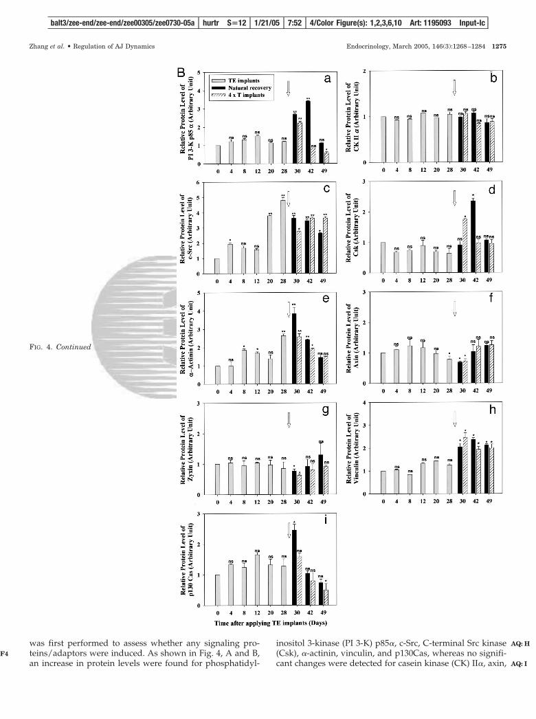
was first performed to assess whether any signaling pro-teins/adaptors were induced. As shown in Fig. 4, A and B,an increase in protein levels were found for phosphatidyl-
inositol 3-kinase (PI 3-K) p85�, c-Src, C-terminal Src kinase(Csk), �-actinin, vinculin, and p130Cas, whereas no signifi-cant changes were detected for casein kinase (CK) II�, axin,
FIG. 4. Continued
balt3/zee-end/zee-end/zee00305/zee0730-05a hurtr S�12 1/21/05 7:52 4/Color Figure(s): 1,2,3,6,10 Art: 1195093 Input-lc
Zhang et al. • Regulation of AJ Dynamics Endocrinology, March 2005, 146(3):1268–1284 1275
F4
AQ: H
AQ: I
and zyxin during androgen-induced AJ disruption. PI 3-Kp85�, Csk, and vinculin displayed a similar trend; their pro-tein levels were induced before the epithelium was fullyrecovered naturally on d 42 and on d 30 in the high-dose Trecovery group, but tumbled thereafter (Fig. 4, A and B),which was somewhat later than MTMR2 (Fig. 3J). c-Src, �-actinin, and p130Cas were also induced, yet their inductionwas detected earlier, such as by d 20–28 after TE implantswere placed to deplete androgen, and their recoveries werealso earlier (Fig. 4, A and B), similar to MTMR2 (Fig. 3J).These findings suggest that the expression of these twogroups of proteins may be regulated differently at the timeof androgen depletion-induced germ cell loss and its recov-eries. The bottom panel in Fig. 4A is the corresponding blotstained for �-actin to illustrate equal protein loading.
rMTMR2 is structurally associated with c-Src in theseminiferous epithelium during androgen suppression-induced germ cell loss
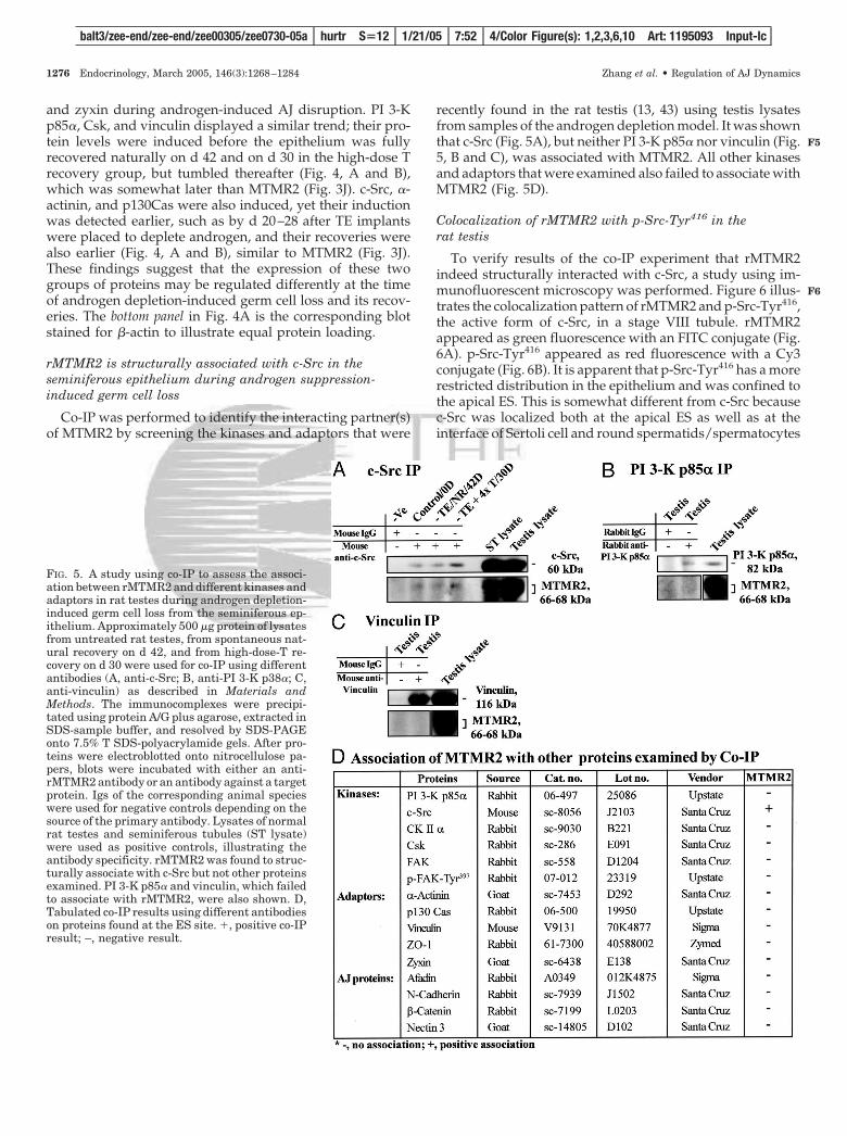
Co-IP was performed to identify the interacting partner(s)of MTMR2 by screening the kinases and adaptors that were
recently found in the rat testis (13, 43) using testis lysatesfrom samples of the androgen depletion model. It was shownthat c-Src (Fig. 5A), but neither PI 3-K p85� nor vinculin (Fig.5, B and C), was associated with MTMR2. All other kinasesand adaptors that were examined also failed to associate withMTMR2 (Fig. 5D).
Colocalization of rMTMR2 with p-Src-Tyr416 in therat testis
To verify results of the co-IP experiment that rMTMR2indeed structurally interacted with c-Src, a study using im-munofluorescent microscopy was performed. Figure 6 illus-trates the colocalization pattern of rMTMR2 and p-Src-Tyr416,the active form of c-Src, in a stage VIII tubule. rMTMR2appeared as green fluorescence with an FITC conjugate (Fig.6A). p-Src-Tyr416 appeared as red fluorescence with a Cy3conjugate (Fig. 6B). It is apparent that p-Src-Tyr416 has a morerestricted distribution in the epithelium and was confined tothe apical ES. This is somewhat different from c-Src becausec-Src was localized both at the apical ES as well as at theinterface of Sertoli cell and round spermatids/spermatocytes
FIG. 5. A study using co-IP to assess the associ-ation between rMTMR2 and different kinases andadaptors in rat testes during androgen depletion-induced germ cell loss from the seminiferous ep-ithelium. Approximately 500 �g protein of lysatesfrom untreated rat testes, from spontaneous nat-ural recovery on d 42, and from high-dose-T re-covery on d 30 were used for co-IP using differentantibodies (A, anti-c-Src; B, anti-PI 3-K p38�; C,anti-vinculin) as described in Materials andMethods. The immunocomplexes were precipi-tated using protein A/G plus agarose, extracted inSDS-sample buffer, and resolved by SDS-PAGEonto 7.5% T SDS-polyacrylamide gels. After pro-teins were electroblotted onto nitrocellulose pa-pers, blots were incubated with either an anti-rMTMR2 antibody or an antibody against a targetprotein. Igs of the corresponding animal specieswere used for negative controls depending on thesource of the primary antibody. Lysates of normalrat testes and seminiferous tubules (ST lysate)were used as positive controls, illustrating theantibody specificity. rMTMR2 was found to struc-turally associate with c-Src but not other proteinsexamined. PI 3-K p85� and vinculin, which failedto associate with rMTMR2, were also shown. D,Tabulated co-IP results using different antibodieson proteins found at the ES site. �, positive co-IPresult; –, negative result.
balt3/zee-end/zee-end/zee00305/zee0730-05a hurtr S�12 1/21/05 7:52 4/Color Figure(s): 1,2,3,6,10 Art: 1195093 Input-lc
1276 Endocrinology, March 2005, 146(3):1268–1284 Zhang et al. • Regulation of AJ Dynamics
F5
F6
at stages VI–VIII as recently reported (13). The merged imageof Fig. 6, A and B, is shown in Fig. 6C, illustrating rMTMR2and p-Src-Tyr416 were indeed colocalized to the same site atthe apical ES. Figure 6D is a DAPI staining.
Effects of androgen suppression-induced AJ disruption andits recovery on the protein levels of AJ structural proteincomplexes N-cadherin/�-catenin, �1-integrin/laminin-�3,and nectin 3/afadin in the rat testis
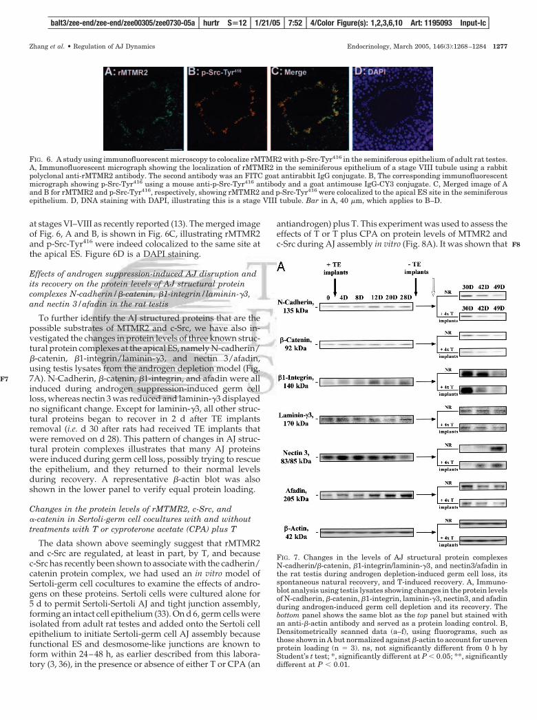
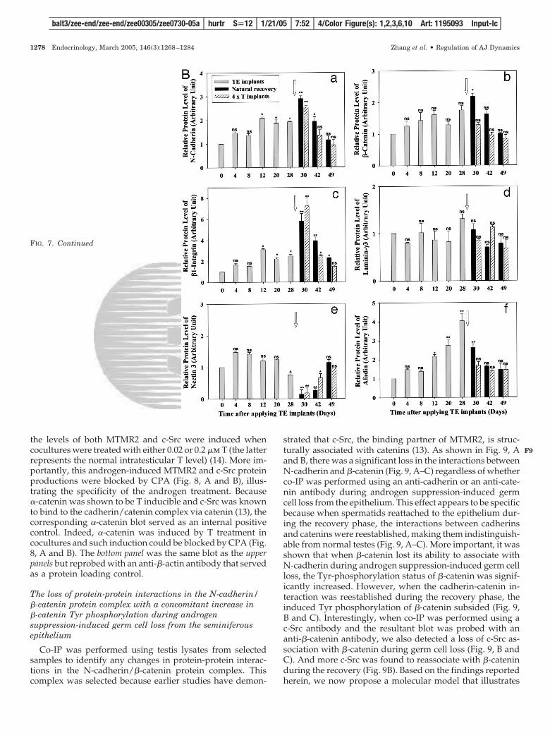
To further identify the AJ structured proteins that are thepossible substrates of MTMR2 and c-Src, we have also in-vestigated the changes in protein levels of three known struc-tural protein complexes at the apical ES, namely N-cadherin/�-catenin, �1-integrin/laminin-�3, and nectin 3/afadin,using testis lysates from the androgen depletion model (Fig.7A). N-Cadherin, �-catenin, �1-integrin, and afadin were allinduced during androgen suppression-induced germ cellloss, whereas nectin 3 was reduced and laminin-�3 displayedno significant change. Except for laminin-�3, all other struc-tural proteins began to recover in 2 d after TE implantsremoval (i.e. d 30 after rats had received TE implants thatwere removed on d 28). This pattern of changes in AJ struc-tural protein complexes illustrates that many AJ proteinswere induced during germ cell loss, possibly trying to rescuethe epithelium, and they returned to their normal levelsduring recovery. A representative �-actin blot was alsoshown in the lower panel to verify equal protein loading.
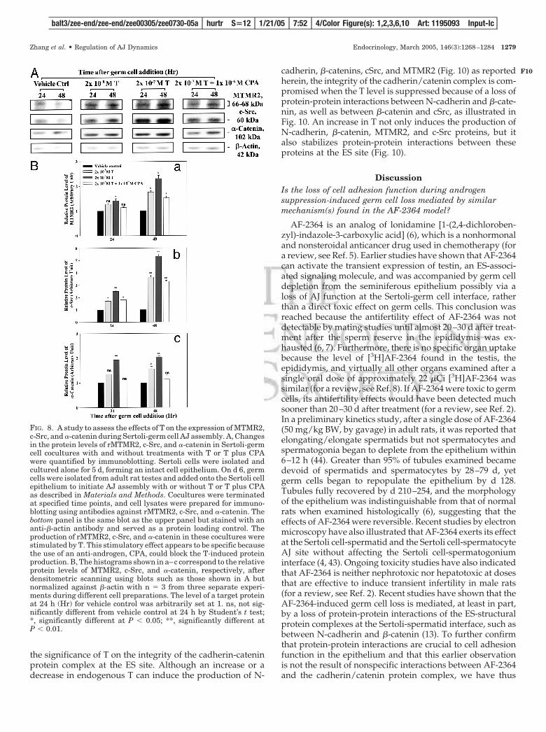
Changes in the protein levels of rMTMR2, c-Src, and�-catenin in Sertoli-germ cell cocultures with and withouttreatments with T or cyproterone acetate (CPA) plus T
The data shown above seemingly suggest that rMTMR2and c-Src are regulated, at least in part, by T, and becausec-Src has recently been shown to associate with the cadherin/catenin protein complex, we had used an in vitro model ofSertoli-germ cell cocultures to examine the effects of andro-gens on these proteins. Sertoli cells were cultured alone for5 d to permit Sertoli-Sertoli AJ and tight junction assembly,forming an intact cell epithelium (33). On d 6, germ cells wereisolated from adult rat testes and added onto the Sertoli cellepithelium to initiate Sertoli-germ cell AJ assembly becausefunctional ES and desmosome-like junctions are known toform within 24–48 h, as earlier described from this labora-tory (3, 36), in the presence or absence of either T or CPA (an
antiandrogen) plus T. This experiment was used to assess theeffects of T or T plus CPA on protein levels of MTMR2 andc-Src during AJ assembly in vitro (Fig. 8A). It was shown that
FIG. 6. A study using immunofluorescent microscopy to colocalize rMTMR2 with p-Src-Tyr416 in the seminiferous epithelium of adult rat testes.A, Immunofluorescent micrograph showing the localization of rMTMR2 in the seminiferous epithelium of a stage VIII tubule using a rabbitpolyclonal anti-rMTMR2 antibody. The second antibody was an FITC goat antirabbit IgG conjugate. B, The corresponding immunofluorescentmicrograph showing p-Src-Tyr416 using a mouse anti-p-Src-Tyr416 antibody and a goat antimouse IgG-CY3 conjugate. C, Merged image of Aand B for rMTMR2 and p-Src-Tyr416, respectively, showing rMTMR2 and p-Src-Tyr416 were colocalized to the apical ES site in the seminiferousepithelium. D, DNA staining with DAPI, illustrating this is a stage VIII tubule. Bar in A, 40 �m, which applies to B–D.
FIG. 7. Changes in the levels of AJ structural protein complexesN-cadherin/�-catenin, �1-integrin/laminin-�3, and nectin3/afadin inthe rat testis during androgen depletion-induced germ cell loss, itsspontaneous natural recovery, and T-induced recovery. A, Immuno-blot analysis using testis lysates showing changes in the protein levelsof N-cadherin, �-catenin, �1-integrin, laminin-�3, nectin3, and afadinduring androgen-induced germ cell depletion and its recovery. Thebottom panel shows the same blot as the top panel but stained withan anti-�-actin antibody and served as a protein loading control. B,Densitometrically scanned data (a–f), using fluorograms, such asthose shown in A but normalized against �-actin to account for unevenprotein loading (n � 3). ns, not significantly different from 0 h byStudent’s t test; *, significantly different at P � 0.05; **, significantlydifferent at P � 0.01.
balt3/zee-end/zee-end/zee00305/zee0730-05a hurtr S�12 1/21/05 7:52 4/Color Figure(s): 1,2,3,6,10 Art: 1195093 Input-lc
Zhang et al. • Regulation of AJ Dynamics Endocrinology, March 2005, 146(3):1268–1284 1277
F7
F8
the levels of both MTMR2 and c-Src were induced whencocultures were treated with either 0.02 or 0.2 �m T (the latterrepresents the normal intratesticular T level) (14). More im-portantly, this androgen-induced MTMR2 and c-Src proteinproductions were blocked by CPA (Fig. 8, A and B), illus-trating the specificity of the androgen treatment. Because�-catenin was shown to be T inducible and c-Src was knownto bind to the cadherin/catenin complex via catenin (13), thecorresponding �-catenin blot served as an internal positivecontrol. Indeed, �-catenin was induced by T treatment incocultures and such induction could be blocked by CPA (Fig.8, A and B). The bottom panel was the same blot as the upperpanels but reprobed with an anti-�-actin antibody that servedas a protein loading control.
The loss of protein-protein interactions in the N-cadherin/�-catenin protein complex with a concomitant increase in�-catenin Tyr phosphorylation during androgensuppression-induced germ cell loss from the seminiferousepithelium
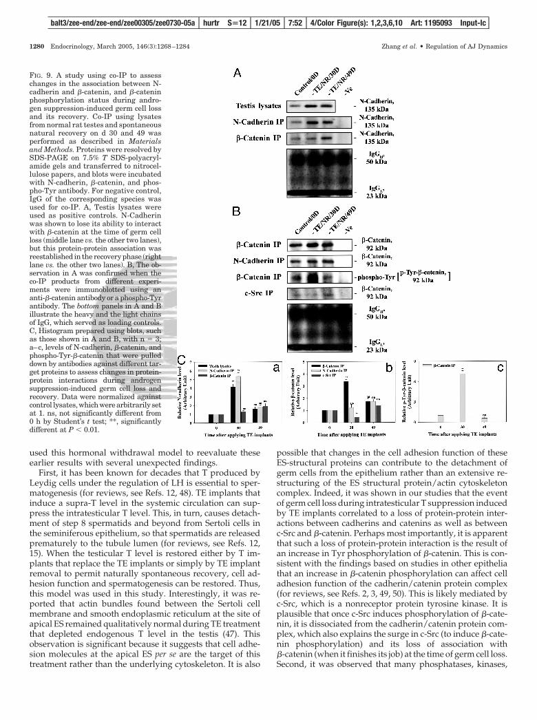
Co-IP was performed using testis lysates from selectedsamples to identify any changes in protein-protein interac-tions in the N-cadherin/�-catenin protein complex. Thiscomplex was selected because earlier studies have demon-
strated that c-Src, the binding partner of MTMR2, is struc-turally associated with catenins (13). As shown in Fig. 9, Aand B, there was a significant loss in the interactions betweenN-cadherin and �-catenin (Fig. 9, A–C) regardless of whetherco-IP was performed using an anti-cadherin or an anti-cate-nin antibody during androgen suppression-induced germcell loss from the epithelium. This effect appears to be specificbecause when spermatids reattached to the epithelium dur-ing the recovery phase, the interactions between cadherinsand catenins were reestablished, making them indistinguish-able from normal testes (Fig. 9, A–C). More important, it wasshown that when �-catenin lost its ability to associate withN-cadherin during androgen suppression-induced germ cellloss, the Tyr-phosphorylation status of �-catenin was signif-icantly increased. However, when the cadherin-catenin in-teraction was reestablished during the recovery phase, theinduced Tyr phosphorylation of �-catenin subsided (Fig. 9,B and C). Interestingly, when co-IP was performed using ac-Src antibody and the resultant blot was probed with ananti-�-catenin antibody, we also detected a loss of c-Src as-sociation with �-catenin during germ cell loss (Fig. 9, B andC). And more c-Src was found to reassociate with �-cateninduring the recovery (Fig. 9B). Based on the findings reportedherein, we now propose a molecular model that illustrates
FIG. 7. Continued
balt3/zee-end/zee-end/zee00305/zee0730-05a hurtr S�12 1/21/05 7:52 4/Color Figure(s): 1,2,3,6,10 Art: 1195093 Input-lc
1278 Endocrinology, March 2005, 146(3):1268–1284 Zhang et al. • Regulation of AJ Dynamics
F9
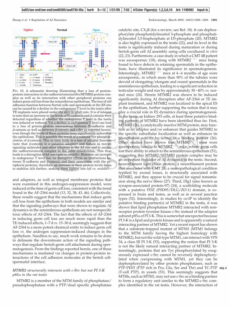
the significance of T on the integrity of the cadherin-cateninprotein complex at the ES site. Although an increase or adecrease in endogenous T can induce the production of N-
cadherin, �-catenins, cSrc, and MTMR2 (Fig. 10) as reportedherein, the integrity of the cadherin/catenin complex is com-promised when the T level is suppressed because of a loss ofprotein-protein interactions between N-cadherin and �-cate-nin, as well as between �-catenin and cSrc, as illustrated inFig. 10. An increase in T not only induces the production ofN-cadherin, �-catenin, MTMR2, and c-Src proteins, but italso stabilizes protein-protein interactions between theseproteins at the ES site (Fig. 10).
DiscussionIs the loss of cell adhesion function during androgensuppression-induced germ cell loss mediated by similarmechanism(s) found in the AF-2364 model?
AF-2364 is an analog of lonidamine [1-(2,4-dichloroben-zyl)-indazole-3-carboxylic acid] (6), which is a nonhormonaland nonsteroidal anticancer drug used in chemotherapy (fora review, see Ref. 5). Earlier studies have shown that AF-2364can activate the transient expression of testin, an ES-associ-ated signaling molecule, and was accompanied by germ celldepletion from the seminiferous epithelium possibly via aloss of AJ function at the Sertoli-germ cell interface, ratherthan a direct toxic effect on germ cells. This conclusion wasreached because the antifertility effect of AF-2364 was notdetectable by mating studies until almost 20–30 d after treat-ment after the sperm reserve in the epididymis was ex-hausted (6, 7). Furthermore, there is no specific organ uptakebecause the level of [3H]AF-2364 found in the testis, theepididymis, and virtually all other organs examined after asingle oral dose of approximately 22 �Ci [3H]AF-2364 wassimilar (for a review, see Ref. 8). If AF-2364 were toxic to germcells, its antifertility effects would have been detected muchsooner than 20–30 d after treatment (for a review, see Ref. 2).In a preliminary kinetics study, after a single dose of AF-2364(50 mg/kg BW, by gavage) in adult rats, it was reported thatelongating/elongate spermatids but not spermatocytes andspermatogonia began to deplete from the epithelium within6–12 h (44). Greater than 95% of tubules examined becamedevoid of spermatids and spermatocytes by 28–79 d, yetgerm cells began to repopulate the epithelium by d 128.Tubules fully recovered by d 210–254, and the morphologyof the epithelium was indistinguishable from that of normalrats when examined histologically (6), suggesting that theeffects of AF-2364 were reversible. Recent studies by electronmicroscopy have also illustrated that AF-2364 exerts its effectat the Sertoli cell-spermatid and the Sertoli cell-spermatocyteAJ site without affecting the Sertoli cell-spermatogoniuminterface (4, 43). Ongoing toxicity studies have also indicatedthat AF-2364 is neither nephrotoxic nor hepatotoxic at dosesthat are effective to induce transient infertility in male rats(for a review, see Ref. 2). Recent studies have shown that theAF-2364-induced germ cell loss is mediated, at least in part,by a loss of protein-protein interactions of the ES-structuralprotein complexes at the Sertoli-spermatid interface, such asbetween N-cadherin and �-catenin (13). To further confirmthat protein-protein interactions are crucial to cell adhesionfunction in the epithelium and that this earlier observationis not the result of nonspecific interactions between AF-2364and the cadherin/catenin protein complex, we have thus
FIG. 8. A study to assess the effects of T on the expression of MTMR2,c-Src, and �-catenin during Sertoli-germ cell AJ assembly. A, Changesin the protein levels of rMTMR2, c-Src, and �-catenin in Sertoli-germcell cocultures with and without treatments with T or T plus CPAwere quantified by immunoblotting. Sertoli cells were isolated andcultured alone for 5 d, forming an intact cell epithelium. On d 6, germcells were isolated from adult rat testes and added onto the Sertoli cellepithelium to initiate AJ assembly with or without T or T plus CPAas described in Materials and Methods. Cocultures were terminatedat specified time points, and cell lysates were prepared for immuno-blotting using antibodies against rMTMR2, c-Src, and �-catenin. Thebottom panel is the same blot as the upper panel but stained with ananti-�-actin antibody and served as a protein loading control. Theproduction of rMTMR2, c-Src, and �-catenin in these cocultures werestimulated by T. This stimulatory effect appears to be specific becausethe use of an anti-androgen, CPA, could block the T-induced proteinproduction. B, The histograms shown in a–c correspond to the relativeprotein levels of MTMR2, c-Src, and �-catenin, respectively, afterdensitometric scanning using blots such as those shown in A butnormalized against �-actin with n � 3 from three separate experi-ments during different cell preparations. The level of a target proteinat 24 h (Hr) for vehicle control was arbitrarily set at 1. ns, not sig-nificantly different from vehicle control at 24 h by Student’s t test;*, significantly different at P � 0.05; **, significantly different atP � 0.01.
balt3/zee-end/zee-end/zee00305/zee0730-05a hurtr S�12 1/21/05 7:52 4/Color Figure(s): 1,2,3,6,10 Art: 1195093 Input-lc
Zhang et al. • Regulation of AJ Dynamics Endocrinology, March 2005, 146(3):1268–1284 1279
F10
used this hormonal withdrawal model to reevaluate theseearlier results with several unexpected findings.
First, it has been known for decades that T produced byLeydig cells under the regulation of LH is essential to sper-matogenesis (for reviews, see Refs. 12, 48). TE implants thatinduce a supra-T level in the systemic circulation can sup-press the intratesticular T level. This, in turn, causes detach-ment of step 8 spermatids and beyond from Sertoli cells inthe seminiferous epithelium, so that spermatids are releasedprematurely to the tubule lumen (for reviews, see Refs. 12,15). When the testicular T level is restored either by T im-plants that replace the TE implants or simply by TE implantremoval to permit naturally spontaneous recovery, cell ad-hesion function and spermatogenesis can be restored. Thus,this model was used in this study. Interestingly, it was re-ported that actin bundles found between the Sertoli cellmembrane and smooth endoplasmic reticulum at the site ofapical ES remained qualitatively normal during TE treatmentthat depleted endogenous T level in the testis (47). Thisobservation is significant because it suggests that cell adhe-sion molecules at the apical ES per se are the target of thistreatment rather than the underlying cytoskeleton. It is also
possible that changes in the cell adhesion function of theseES-structural proteins can contribute to the detachment ofgerm cells from the epithelium rather than an extensive re-structuring of the ES structural protein/actin cytoskeletoncomplex. Indeed, it was shown in our studies that the eventof germ cell loss during intratesticular T suppression inducedby TE implants correlated to a loss of protein-protein inter-actions between cadherins and catenins as well as betweenc-Src and �-catenin. Perhaps most importantly, it is apparentthat such a loss of protein-protein interaction is the result ofan increase in Tyr phosphorylation of �-catenin. This is con-sistent with the findings based on studies in other epitheliathat an increase in �-catenin phosphorylation can affect celladhesion function of the cadherin/catenin protein complex(for reviews, see Refs. 2, 3, 49, 50). This is likely mediated byc-Src, which is a nonreceptor protein tyrosine kinase. It isplausible that once c-Src induces phosphorylation of �-cate-nin, it is dissociated from the cadherin/catenin protein com-plex, which also explains the surge in c-Src (to induce �-cate-nin phosphorylation) and its loss of association with�-catenin (when it finishes its job) at the time of germ cell loss.Second, it was observed that many phosphatases, kinases,
FIG. 9. A study using co-IP to assesschanges in the association between N-cadherin and �-catenin, and �-cateninphosphorylation status during andro-gen suppression-induced germ cell lossand its recovery. Co-IP using lysatesfrom normal rat testes and spontaneousnatural recovery on d 30 and 49 wasperformed as described in Materialsand Methods. Proteins were resolved bySDS-PAGE on 7.5% T SDS-polyacryl-amide gels and transferred to nitrocel-lulose papers, and blots were incubatedwith N-cadherin, �-catenin, and phos-pho-Tyr antibody. For negative control,IgG of the corresponding species wasused for co-IP. A, Testis lysates wereused as positive controls. N-Cadherinwas shown to lose its ability to interactwith �-catenin at the time of germ cellloss (middle lane vs. the other two lanes),but this protein-protein association wasreestablished in the recovery phase (rightlane vs. the other two lanes). B, The ob-servation in A was confirmed when theco-IP products from different experi-ments were immunoblotted using ananti-�-catenin antibody or a phospho-Tyrantibody. The bottom panels in A and Billustrate the heavy and the light chainsof IgG, which served as loading controls.C, Histogram prepared using blots, suchas those shown in A and B, with n � 3;a–c, levels of N-cadherin, �-catenin, andphospho-Tyr-�-catenin that were pulleddown by antibodies against different tar-get proteins to assess changes in protein-protein interactions during androgensuppression-induced germ cell loss andrecovery. Data were normalized againstcontrol lysates, which were arbitrarily setat 1. ns, not significantly different from0 h by Student’s t test; **, significantlydifferent at P � 0.01.
balt3/zee-end/zee-end/zee00305/zee0730-05a hurtr S�12 1/21/05 7:52 4/Color Figure(s): 1,2,3,6,10 Art: 1195093 Input-lc
1280 Endocrinology, March 2005, 146(3):1268–1284 Zhang et al. • Regulation of AJ Dynamics
and adaptors, as well as integral membrane proteins thatwere examined in this androgen-suppression model, wereinduced at the time of germ cell loss, consistent with the trendfound in the AF-2364 model (13, 32, 38, 43, 46). Collectively,these results suggest that the mechanisms that induce germcell loss from the epithelium in both models are similar andthat the signaling pathways that were shown to regulate AJdynamics in the seminiferous epithelium are not nonspecifictoxic effects of AF-2364. The fact that the effects of AF-2364in inducing germ cell loss are much more rapid than theTE-induced affects, 1–7 d vs. 15–30 d, seemingly suggests thatAF-2364 is a more potent chemical entity to induce germ cellloss vs. the androgen suppression-induced changes in theepithelium. Needless to say, much work remains to be doneto delineate the downstream action of the signaling path-ways that regulate Sertoli-germ cell attachment during sper-matogenesis. From the findings reported herein, one of thesemechanisms is mediated via changes in protein-protein in-teractions of the cell adhesion molecules at the Sertoli cell-spermatid interface.
MTMR2 structurally interacts with c-Src but not PI 3-Kp85� in the rat testis
MTMR2 is a member of the MTM family of phosphatase/pseudophosphatase with a PTP/dual specific phosphatase
catalytic site, CX5R (for a review, see Ref. 18). It can dephos-phorylate phosphatidylinositol 3-phosphate and phosphati-dylinositol 3,5-bisphosphate at D3-phosphate (20). MTMR2is also highly expressed in the testis (22), and its level in thetestis is significantly induced during maturation or duringSertoli-germ cell AJ assembly using cells cocultured in vitro(22, 23). Furthermore, a case study in which a CMT 4B patientwas azoospermic (18), along with MTMR2�/� mice beingfound to have defects in retaining spermatids in the epithe-lium, have illustrated its significance in spermatogenesis.Interestingly, MTMR2�/� mice at 4–6 months of age wereazoospermic, in which more than 90% of the tubules weredevoid of elongating/elongate and round spermatids in theseminiferous epithelium, leading to a significant reduction intesticular weight and size by approximately 30–40% vs. nor-mal mice (24). Herein MTMR2 was shown to be inducedsignificantly during AJ disruption after AF-2364 or TE im-plant treatment, and MTMR2 was localized to the apical ESin the epithelium, further supporting the notion that it mayplay a crucial role in ES dynamics during spermatogenesis.In the brain, or kidney 293 cells, at least three putative bind-ing partners of MTMR2 have been identified thus far. First,MTMR5 (26), a catalytically inactive MTM protein, may func-tion as an adaptor and/or enhancer that guides MTMR2 tothe specific subcellular localization as well as enhances itsphosphatase activity by acting as a binding partner (26).Other studies have shown that MTMR5�/� mice wereazoospermic, similar to MTMR2�/� mice, where germ cellslost their ability to attach to the seminiferous epithelium (51),suggesting this MTMR2/MTMR5 protein complex may bean important regulator of AJ dynamics in the testis. Second,neurofilament light chain protein, a neurofilament proteinthat associates with CMT 2E, a neuropathological conditiontypified by axonal losses, is structurally associated withMTMR2, and they appear to be crucial for signal transmis-sion along the nerve fibers (27). Third, Dlg1 (also known assynapse-associated protein-97) (24), a scaffolding moleculewith a putative PDZ (PSD95/DLG/ZO-1) domain, is ex-pressed in brain and testes, as well as in several junctiontypes (52). Interestingly, in studies by co-IP to identify theputative binding partner(s) of MTMR2 in the testis, it wasshown that lipid phosphatase MTMR2 interacted with non-receptor protein tyrosine kinase c-Src instead of the adaptorsubunit p85� of PI 3-K. This is somewhat unexpected becausePI 3-K is a lipid and protein kinase and is apparently a naturalinteracting partner of MTMR2. Yet a recent report has shownthat a substrate-trapped mutant of MTM1 (MTM1 belongsto the MTM family having the highest homology withMTMR2), but not the wild-type MTM1, can interact with VPS34, a class III PI 3-K (53), supporting the notion that PI 3-Kis not the likely natural interacting partner of MTMR2. In-terestingly, proteins that are Tyr phosphorylated by exog-enously expressed c-Src cannot be reversely dephosphory-lated when coexpressing with MTM1, yet they can bedephosphorylated by other protein phosphatases, such asPTP-PEST (PTP rich in Pro, Glu, Ser and Thr) and TC-PTP(T-cell PTP), in yeasts (53). This seemingly suggests thatMTMs, such as MTM1, may not use c-Src as a binding partnerto form a regulatory unit similar to the MTMR2-c-Src com-plex identified in the rat testis. However, the interaction of
FIG. 10. A schematic drawing illustrating that a loss of protein-protein interaction in the cadherin/catenin/cSrc/MTMR2 protein com-plex as well as its interaction with other peripheral proteins caninduce germ cell loss from the seminiferous epithelium. The loss of celladhesion function between Sertoli cells and spermatids at the ES sitecan be caused by a decline in the endogenous T level in the testis afterTE implants were placed under the skin in adult rats. It is of interestto note that an increase in the levels of N-cadherin and �-catenin weredetected regardless of whether the endogenous T level in the testiswas induced or reduced. Yet a decline in endogenous T level can leadto a loss of protein-protein interactions between N-cadherin and�-catenin as well as between �-catenin and c-Src as reported herein,even though the levels of these proteins were significantly induced inthe epithelium. This is possibly the result of a reduced Tyr phosphor-ylation of �-catenin. This in turn leads to a loss of adaptor function(note that �-catenin is a putative adaptor) and failure to recruitsignaling molecules and other adaptors to the AJ site and to anchorthe cadherin/catenin complex to the actin cytoskeleton. This thusleads to a disruption of AJ (see right vs. middle). However, an increasein endogenous T level has no disruptive effects on interactions be-tween N-cadherin and �-catenin and their association with the pe-ripheral proteins; therefore, an increase in their protein levels helpsto stabilize AJs further, making them tighter (see left vs. middle).
balt3/zee-end/zee-end/zee00305/zee0730-05a hurtr S�12 1/21/05 7:52 4/Color Figure(s): 1,2,3,6,10 Art: 1195093 Input-lc
Zhang et al. • Regulation of AJ Dynamics Endocrinology, March 2005, 146(3):1268–1284 1281
AQ: J
MTM and other kinases may be regulated either via a lipidsecond messenger, such as phosphatidylinositol 5-phos-phate, a product of MTMR2 that may increase the enzymaticactivity of MTMs (20) or a yet-to-be identified adaptor. An-other surprising finding is that because MTMR2 has a PDZ-binding domain near its C-terminus, and it can interact withthe PDZ domain of Dlg1 in COS-7 cells (24), it is expected tointeract with other proteins having the PDZ domain at the ES.Yet MTMR2 failed to interact with zonula occludens 1 (ZO-1), another PDZ-domain-containing protein, as reportedherein. Additionally, MTMR2 failed to interact with �-acti-nin, another lipid-associated adaptor and an actin-bindingand bundling protein. These findings also illustrate thatamong many adaptors and kinases that were identified in theapical ES, only c-Src physically interacts with MTMR2 in theseminiferous epithelium. This result obtained by co-IP wasalso confirmed by a colocalization study using fluorescentmicroscopy. Although MTMR2 failed to interact directlywith the known adaptors at the apical ES, including ZO-1,�-actinin, p130Cas, Wiskott-Aldrich syndrome protein(WASP), and others, but interacted only with c-Src, it mustbe noted that c-Src was recently shown to associate withsome of these adaptors such as Wiskott-Aldrich syndromeprotein and the cadherin-catenin protein complex (13). Assuch, MTMR2 can still indirectly interact with other adaptorsto affect cell adhesion function of the cadherin-catenin com-plex via c-Src, and this possibility must be vigorously inves-tigated in future studies.
The MTMR2/c-Src complex regulates ES dynamics likelyvia its interaction with the cadherin/cateninprotein complex
The cadherin/catenin, the integrin/laminin, and the nec-tin/afadin/ponsin protein complexes are the three currentlyknown structural protein complexes that confer cell adhesionfunction in the seminiferous epithelium at the apical ES site(for reviews, see Refs. 2, 10). Other studies have shown thatthe phosphorylation status of these protein complexes, inparticular the cadherin/catenin complex, is a determiningfactor to confer cell adhesion function (49). For instance, anincrease in Tyr phosphorylation of the cadherin/catenincomplex is associated with a loss of interaction betweencadherin, catenin, and the actin cytoskeleton, leading to a lossof cell adhesion function (49, 50, 54). In earlier studies, aninduction of N-cadherin and �-catenin was detected in theAF-2364 model (32, 43, 44) at the time of germ cell loss,similar to the results reported herein during TE treatment-induced germ cell loss. Yet it is not known from these earlierstudies whether any changes in the phosphorylation statusof these proteins indeed occurred. Furthermore, the cad-herin/catenin complex was localized to both the basal andapical ES sites (32, 43, 55, 56) and was recently shown tointeract with the c-Src/Csk/CK II kinase complex (13). Be-cause both MTMR2 and c-Src were induced during TE treat-ment at the time of germ cell loss and both MTMR2 and thephosphorylated form of Src (p-Src-Tyr416) were colocalizedto the apical ES and to a lesser extent to the basal ES at stageVIII, together with the fact that c-Src is a binding partner ofthe cadherin/catenin protein complex, it seems logical to
speculate that the cadherin/catenin protein complex may bethe target of the MTMR2/c-Src complex. We have nowshown that the N-cadherin/�-catenin protein complex wasdisassociated in vivo during androgen depletion-induced AJdisruption, accompanied by an increase in Tyr phosphory-lation of �-catenin. This result is consistent with earlier re-ports in other in vitro systems (e.g. Madin Darby caninekidney cells) that a loss of cell adhesion function of thecadherin-based complex is mediated by an increase in �-cate-nin phosphorylation (49, 54). Interestingly, a decrease in c-Srcassociation with �-catenin was also detected, suggesting thatc-Src was moving away from �-catenin once it phosphory-lated its substrate. It is obvious that the �6�1-integrin/lami-nin-�3 complex can also be another target of the MTMR2/c-Src complex. For instance, it was recently shown thatphospho-focal adhesion kinase-Tyr397 could bind to �1-integrin, vinculin, and c-Src (38, 46), suggesting c-Src may bethe linker that associates MTMR2 with �1-integrin indirectlyvia phospho-focal adhesion kinase. There are also reportsillustrating that Src and another lipid/protein phosphatase,PTEN (phosphatase and tensin homolog deleted on chro-mosome ten), can regulate integrin signaling (57, 58), sup-porting the notion that MTMR2/c-Src may regulate the in-tegrin/laminin complex at the apical ES. In short, results ofthis study have clearly demonstrated that c-Src is a putativebinding partner of MTMR2 in the testis, in particular at theapical ES, and the cadherin/catenin protein complex is oneof its targets.
MTMR2 and c-Src can be induced by T in Sertoli-germ cellcocultures in vitro
Results of the in vivo study presented herein suggest thatandrogen plays a crucial role in regulating both MTMR2 andc-Src in the testis. To further confirm this observation, thelevels of these proteins were quantified in Sertoli-germ cellscocultured in vitro. It was noted that both MTMR2 and c-Srcwere indeed induced by T in the cocultures, and this dose-dependent induction could be blocked by CPA, an antian-drogen (for a review, see Ref. 59). These data thus demon-strate unequivocally that both MTMR2 and c-Src, as well ascadherin as shown in an earlier study (32), were androgen-regulated proteins. However, much research is needed tounderstand how androgens regulate the interaction betweenthese proteins, adaptors, and the cadherin/catenin complex.
Issues on changes in levels of target proteins (e.g. kinases,phosphatases, and adaptors) in the seminiferous epitheliumas a result of germ cell loss
An interesting issue arises regarding results obtained fromthis androgen suppression model and the AF-2364 modelthat correlates changes in target proteins, such as MTMR2and c-Src, with the event of germ cell loss from the semi-niferous epithelium pertinent to AJ disruption. For instance,during the progressive loss of germ cells from the epithelium,the distribution of different cell types in the testis is alteredat different time points in both models. This may confoundour interpretation, at least to some extent, regarding changesin specific target proteins and/or genes by quantifying their
balt3/zee-end/zee-end/zee00305/zee0730-05a hurtr S�12 1/21/05 7:52 4/Color Figure(s): 1,2,3,6,10 Art: 1195093 Input-lc
1282 Endocrinology, March 2005, 146(3):1268–1284 Zhang et al. • Regulation of AJ Dynamics
AQ: K
AQ: L
AQ: M
AQ: N
AQ: O
steady-state protein and/or mRNA levels (for a review, seeRef. 60). For instance, if a protein, such as rMTMR2, is alsoproduced by germ cells in the seminiferous epithelium (22),an induction of this protein during germ cell loss as quan-tified by immunoblotting was likely an underestimate be-cause the cytosol that was being analyzed had been contrib-uted largely by Sertoli cells and other somatic cells (e.g.peritubular myoid and Leydig cells) instead of germ cells asin normal testes, even though the same amount of proteinwas being analyzed (and the blot was reprobed with ananti-actin antibody staining to assess equal protein loading).However, this is an inherent problem with these models,which is somewhat difficult to resolve. First, although wehad done some preliminary kinetic studies to assess the lossof different germ cell types (e.g. elongate, elongating, andround spermatids and spermatocytes), after AF-2364 treat-ment, which showed that elongate/elongating spermatidswere depleted from the epithelium much more rapidly thanround spermatids and spermatocytes (6.5 h vs. 3–6 d) (44), wedo not have detailed morphometric analysis results on thetime-dependent loss of specific germ cell types in both mod-els to correlate these changes to each treatment regimen ineither model. Even if these studies were completed, eachanimal would still respond differently, and there would havebeen variations between animals responding to the sametreatment. Second, although germ cells were depleted fromthe epithelium, they probably would still be included in theimmunoblot analysis because many germ cells would still bein the tubule lumen, in particular in the early phase of germcell loss. As such, it is important to use other approaches toconfirm changes of a specific target protein (e.g. MTMR2) inthe epithelium during germ cell loss, such as with the use ofimmunohistochemistry and fluorescent microscopy as re-ported herein.
Another concern related to the data interpretation usingthese models is that an induction of a target protein, eithera kinase, a phosphatase, or an adaptor, is in fact the result ofAJ disruption rather than a reflection of germ cell loss. Thebest argument against this possibility is the result of studiesusing inhibitors. For instance, it was shown that Sertoli-germcell AJ dynamics are regulated by the �1-integrin/RhoB/Rho-associated protein kinase/LIMK (Lin-11 Isl-1 Mec3kinase/cofilin signaling pathway (61) and the �1-integrin/laminin-�3/membrane-type 1 matrix metalloproteinase/matrix metalloproteinase 2 protein complex (46) becausethese proteins were significantly induced during AF-2364-mediated germ cell loss from the epithelium. Indeed, the useof either Y27632, a specific inhibitor for Rho-associated pro-tein kinase or 2R-2-[(4-biphenylylsulfonyl)amino]-3-phenyl-propionic acid, an inhibitor against matrix metalloproteinase2, could significantly delay the AF-2364-induced germ cellloss from the epithelium (46, 61), validating these interpre-tations. Likewise, an induction of the MTMR2/c-Src proteincomplex as reported herein can indeed induce an increase in�-catenin phosphorylation, which plausibly leads to a loss ofprotein-protein interactions between N-cadherin and �-catenin protein as confirmed by experiments using co-IP,thereby inducing germ cell loss from the epithelium.
Summary and concluding remarks
In summary, we have demonstrated that MTMR2 is astage-specific lipid phosphatase that is structurally associ-ated with c-Src and that both proteins are colocalized to theapical ES at stage VIII of the epithelial cycle before spermi-ation. It is likely that the MTMR2/c-Src is an importantprotein complex that regulates the cell adhesion function ofthe cadherin/catenin complex via a lipid second messengerand possibly other adaptor(s) and/or kinases(s) by alteringprotein-protein interactions of the structural protein com-plexes at the ES site, facilitating cell movement duringspermatogenesis.
Received September 7, 2004. Accepted December 1, 2004.Address all correspondence and requests for reprints to: C. Yan
Cheng, Ph.D., Population Council, 1230 York Avenue, New York, NewYork 10021. E-mail: [email protected].
This work was supported in part by grants from the National Insti-tutes of Health (National Institute of Child Health and Human Devel-opment, U01 HD045908 and U54 HD029990, Project 3, to C.Y.C.), theContraceptive Research and Development (CONRAD) Program (Con-sortium for Industrial Collaboration in Contraceptive Research, CICCRCIG 01-72 and CIG 96-05-B to C.Y.C. and CIG-01-74 to D.D.M.), andHong Kong Research Grant Council (HKU 7194/01M and 7413/04M toW.M.L. and C.Y.C.).
References
1. de Kretser DM, Loveland KL, Meinhardt A, Simorangkir D, Wreford N 1998Spermatogenesis. Hum Reprod 13:1–8
2. Cheng CY, Mruk DD 2002 Cell junction dynamics in the testis: Sertoli-germcell interactions and male contraceptive development. Physiol Rev 82:825–874
3. Mruk DD, Cheng CY 2004 Sertoli-Sertoli and Sertoli-germ cell interactions andtheir significance in germ cell movement in the seminiferous epithelium duringspermatogenesis. Endocr Rev 25:747–806
4. Mruk D, Cheng C 2004 Cell-cell interactions at the ectoplasmic specializationin the testis. Trends Endocrinol Metabol 15:439–447
5. Silvestrini B, Palazzo G, De Gregorio M 1984 Lonidamine and related com-pounds. Prog Med Chem 21:111–135
6. Cheng CY, Silvestrini B, Grima J, Mo MY, Zhu LJ, Johansson E, Saso L, LeoneMG, Palmery M, Mruk DD 2001 Two new male contraceptives exert theireffects by depleting germ cells prematurely from the testis. Biol Reprod 65:449–461
7. Grima J, Silvestrini B, Cheng CY 2001 Reversible inhibition of spermatogen-esis in rats using a new male contraceptive, 1-(2,4-dichlorobenzyl)-indazole-3-carbohydrazide. Biol Reprod 64:1500–1508
8. Cheng CY, Mruk DD, Silvestrini B, Bonanomi M, Wong CH, Siu MKY, LeeNPY, Mo MY, AF-2364 [1-(2,4-dichlorobenzyl)-indazole-3-carbohydrazide] isa potential male contraceptive in adult rats: a review of recent data. Contra-ception, in press
9. Siu MK, Cheng CY 2004 Dynamic cross-talk between cells and the extracel-lular matrix in the testis. Bioessays 26:978–992
10. Lee NPY, Cheng CY 2004 Ectoplasmic specialization, a testis-specific cell-cellactin-based adherens junction type: is this a potential target for male contra-ceptive development? Human Reprod Update 10:349–369
11. Lui WY, Mruk DD, Lee WM, Cheng CY 2003 Adherens junction dynamics inthe testis and spermatogenesis. J Androl 24:1–14
12. McLachlan RI, O’Donnell L, Meachem SJ, Stanton PG, de K, Pratis K,Robertson DM 2002 Hormonal regulation of spermatogenesis in primates andman: insights for development of the male hormonal contraceptive. J Androl23:149–162
13. Lee NPY, Cheng CY 2005 Protein kinases and adherens junction dynamics inthe seminiferous epithelium of the rat testis. J Cell Physiol 202:344–360
14. Turner TT, Jones CE, Howards SS, Ewing LL, Zegeye B, Gunsalus GL 1984On the androgen microenvironment of maturing spermatozoa. Endocrinology115:1925–1932
15. McLachlan RI, Wreford N, O’Donnell L, de Kretser DM, Robertson DM 1996The endocrine regulation of spermatogenesis: independent roles for testos-terone and FSH. J Endocrinol 148:1–9
16. Bolino A, Muglia M, Conforti FL, LeGuern E, Salih MA, Georgiou DM,Christodoulou K, Hausmanowa-Petrusewicz I, Mandich P, Schenone A,Gambardella A, Bono F, Quattrone A, Devoto M, Monaco AP 2000 Charcot-Marie-Tooth type 4B is caused by mutations in the gene encoding myotubu-larin-related protein-2. Nat Genet 25:17–19
17. Begley MJ, Taylor GS, Kim SA, Veine DM, Dixon JE, Stuckey JA 2003 Crystal
balt3/zee-end/zee-end/zee00305/zee0730-05a hurtr S�12 1/21/05 7:52 4/Color Figure(s): 1,2,3,6,10 Art: 1195093 Input-lc
Zhang et al. • Regulation of AJ Dynamics Endocrinology, March 2005, 146(3):1268–1284 1283
AQ: Q
AQ: R
AQ: S
AQ: T
structure of a phosphoinositide phosphatase, MTMR2: insights into myotu-bular myopathy and Charcot-Marie-Tooth syndrome. Mol Cell 12:1391–1402
18. Laporte J, Bedez F, Bolino A, Mandel JL 2003 Myotubularins, a large disease-associated family of cooperating catalytically active and inactive phosphoi-nositides phosphatases. Hum Mol Genet 12:R285–R292
19. Berger P, Bonneick S, Willi S, Wymann M, Suter U 2002 Loss of phosphataseactivity in myotubularin-related protein 2 is associated with Charcot-Marie-Tooth disease type 4B1. Hum Mol Genet 11:1569–1579
20. Schaletzky J, Dove SK, Short B, Lorenzo O, Clague MJ, Barr FA 2003 Phos-phatidylinositol-5-phosphate activation and conserved substrate specificity ofthe myotubularin phosphatidylinositol 3-phosphatases. Curr Biol 13:504–509
21. Nandurkar HH, Huysmans R 2002 The myotubularin family: novel phos-phoinositide regulators. IUBMB Life 53:37–43
22. Li JC, Samy ET, Grima J, Chung SS, Mruk DD, Lee WM, Silvestrini B, ChengCY 2000 Rat testicular myotubularin, a protein tyrosine phosphatase expressedby Sertoli and germ cells, is a potential marker for studying cell-cell interac-tions in the rat testis. J Cell Physiol 185:366–385
23. Li JC, Lee WM, Mruk DD, Cheng CY 2001 Regulation of Sertoli cell myo-tubularin (rMTM) expression by germ cells in vitro. J Androl 22:266–277
24. Bolino A, Bolis A, Previtali SC, Dina G, Bussini S, Dati G, Amadio S, CarroUD, Mruk DD, Feltri ML, Cheng CY, Quattrini A, Wrabetz L 2004 Disruptionof Mtmr2 produces CMT4B1-like neuropathy with myelin outfolding andimpaired spermatogenesis. J Cell Biol 167:711–721
25. Berger P, Schaffitzel C, Berger I, Ban N, Suter U 2003 Membrane associationof myotubularin-related protein 2 is mediated by a pleckstrin homology-GRAM domain and a coiled-coil dimerization module. Proc Natl Acad Sci USA100:12177–12182
26. Kim SA, Vacratsis PO, Firestein R, Cleary ML, Dixon JE 2003 Regulationof myotubularin-related (MTMR)2 phosphatidylinositol phosphatase byMTMR5, a catalytically inactive phosphatase. Proc Natl Acad Sci USA 100:4492–4497
27. Previtali SC, Zerega B, Sherman DL, Brophy PJ, Dina G, King RH, Salih MM,Feltri L, Quattrini A, Ravazzolo R, Wrabetz L, Monaco AP, Bolino A 2003Myotubularin-related 2 protein phosphatase and neurofilament light chainprotein, both mutated in CMT neuropathies, interact in peripheral nerve. HumMol Genet 12:1713–1723
28. Zwain IH, Cheng CY 1994 Rat seminiferous tubular culture medium containsa biological factor that inhibits Leydig cell steroidogenesis: its purification andmechanism of action. Mol Cell Endocrinol 104:213–227
29. Cheng CY, Mather JP, Byer AL, Bardin CW 1986 Identification of hormonallyresponsive proteins in primary Sertoli cell culture medium by anion-exchangehigh performance liquid chromatography. Endocrinology 118:480–488
30. Grima J, Pineau C, Bardin CW, Cheng CY 1992 Rat Sertoli cell clusterin,�2-macroglobulin, and testins: biosynthesis and differential regulation bygerm cells. Mol Cell Endocrinol 89:127–140
31. Galdieri M, Ziparo E, Palombi F, Russo MA, Stefanini M 1981 Pure Sertolicell cultures: a new model for the study of somatic-germ cell interactions. JAndrol 2:249–254
32. Lee NPY, Mruk DD, Lee WM, Cheng CY 2003 Is the cadherin/catenin com-plex a functional unit of cell-cell actin-based adherens junctions in the rat testis?Biol Reprod 68:489–508
33. Lee NPY, Cheng CY 2003 Regulation of Sertoli cell tight junction in the rat testisvia the nitric oxide synthase/soluble guanylate cyclase/cyclic guanosinemonophosphate/protein kinase G signaling pathway: an in vitro study. En-docrinology 144:3114–3129
34. Aravindan GR, Pineau CP, Bardin CW, Cheng CY 1996 Ability of trypsin inmimicking germ cell factors that affect Sertoli cell secretory function. J CellPhysiol 168:123–133
35. Wong CCS, Chung SSW, Grima J, Zhu LJ, Mruk DD, Lee WM, Cheng CY2000 Changes in the expression of junctional and nonjunctional complex com-ponent genes when inter-Sertoli tight junctions are formed in vitro. J Androl21:227–237
36. Lui WY, Mruk DD, Cheng CY 2005 Interactions among IQGAP1, Cdc42, andthe cadherin/catenin protein complex regulate Sertoli-germ cell adherensjunction dynamics in the testis. J Cell Physiol 202:49–66
37. Cameron DF, Muffly KE 1991 Hormonal regulation of spermatid binding.J Cell Sci 100:623–633
38. Siu MKY, Mruk DD, Lee WM, Cheng CY 2003 Adhering junction dynamics
in the testis are regulated by an interplay of �1-integrin and focal adhesioncomplex-associated proteins. Endocrinology 144:2141–2163
39. Grima J, Zhu LJ, Zong SD, Catterall JF, Bardin CW, Cheng CY 1995 Rat testinis a newly identified component of the junctional complexes in various tissueswhose mRNA is predominantly expressed in the testis and ovary. Biol Reprod52:340–355
40. Bradford MM 1976 A rapid and sensitive method for the quantitation ofmicrogram quantities of protein utilizing the principle of protein-dye binding.Anal Biochem 72:248–254
41. Laemmli UK 1970 Cleavage of structural proteins during the assembly of thehead of bacteriophage T4. Nature 227:680–685
42. Lui WY, Wong CH, Mruk DD, Cheng CY 2003 TGF-�3 regulates the blood-testis barrier dynamics via the p38 mitogen activated protein (MAP) kinasepathway: an in vivo study. Endocrinology 144:1139–1142
43. Lee NPY, Mruk DD, Conway AM, Cheng CY 2004 Zyxin, axin, and Wiskott-Aldrich syndrome protein are adaptors that link the cadherin/catenin proteincomplex to the cytoskeleton at adherens junctions in the seminiferous epithe-lium of the rat testis. J Androl 25:200–215
44. Chen YM, Lee NPY, Mruk DD, Lee WM, Cheng CY 2003 Fer kinase/FerT andadherens junction dynamics in the testis: an in vitro and in vivo study. BiolReprod 69:656–672
45. O’Donnell L, McLachlan RI, Wreford NG, Robertson DM 1994 Testosteronepromotes the conversion of round spermatids between stages VII and VIII ofthe rat spermatogenic cycle. Endocrinology 135:2608–2614
46. Siu MKY, Cheng CY 2004 Interactions of proteases, protease inhibitors, andthe �1 integrin/laminin �3 protein complex in the regulation of ectoplasmicspecialization dynamics in the rat testis. Biol Reprod 70:945–964
47. O’Donnell L, Stanton PG, Bartles JR, Robertson DM 2000 Sertoli cell ecto-plasmic specializations in the seminiferous epithelium of the testosterone-suppressed adult rat. Biol Reprod 63:99–108
48. Kamischke A, Nieschlag E 2004 Progress towards hormonal male contracep-tion. Trends Pharmacol Sci 25:49–57
49. Gumbiner BM 2000 Regulation of cadherin adhesive activity. J Cell Biol148:399–404
50. Daniel JM, Reynolds AB 1997 Tyrosine phosphorylation and cadherin/cate-nin function. Bioessays 19:883–891
51. Firestein R, Nagy PL, Daly M, Huie P, Conti M, Cleary ML 2002 Maleinfertility, impaired spermatogenesis, and azoospermia in mice deficient forthe pseudophosphatase Sbf1. J Clin Invest 109:1165–1172
52. Muller BM, Kistner U, Veh RW, Cases-Langhoff C, Becker B, GundelfingerED, Garner CC 1995 Molecular characterization and spatial distribution ofSAP97, a novel presynaptic protein homologous to SAP90 and the Drosophiladiscs-large tumor suppressor protein. J Neurosci 15:2354–2366
53. Blondeau F, Laporte J, Bodin S, Superti-Furga G, Payrastre B, Mandel JL 2000Myotubularin, a phosphatase deficient in myotubular myopathy, acts on phos-phatidylinositol 3-kinase and phosphatidylinositol 3-phosphate pathway.Hum Mol Genet 9:2223–2229
54. Gooding JM, Yap KL, Ikura M 2004 The cadherin-catenin complex as a focalpoint of cell adhesion and signalling: new insights from three-dimensionalstructures. Bioessays 26:497–511
55. Wine RN, Chapin RE 1999 Adhesion and signaling proteins spatiotemporallyassociated with spermiation in the rat. J Androl 20:198–213
56. Johnson KJ, Boekelheide K 2002 Dynamic testicular adhesion junctions areimmunologically unique. II. Localization of classic cadherins in rat testis. BiolReprod 66:992–1000
57. Tamura M, Gu J, Tran H, Yamada KM 1999 PTEN gene and integrin signalingin cancer. J Natl Cancer Inst 91:1820–1828
58. Felsenfeld DP, Schwartzberg PL, Venegas A, Tse R, Sheetz MP 1999 Selectiveregulation of integrin-cytoskeleton interactions by the tyrosine kinase Src.Nature Cell Biol 1:200–206
59. Berrevoets CA, Umar A, Brinkmann AO 2002 Antiandrogens: selective an-drogen receptor modulators. Mol Cell Endocrinol 198:97–103
60. Ivell R, Spiess A 2002 Analysing differential gene expression in the testis.Berlin: Springer-Verlag
61. Lui WY, Lee WM, Cheng CY 2003 Sertoli-germ cell adherens junction dy-namics in the testis are regulated by RhoB GTPase via the ROCK/LIMKsignaling pathway. Biol Reprod 68:2189–2206
Endocrinology is published monthly by The Endocrine Society (http://www.endo-society.org), the foremost professional society serving theendocrine community.
balt3/zee-end/zee-end/zee00305/zee0730-05a hurtr S�12 1/21/05 7:52 4/Color Figure(s): 1,2,3,6,10 Art: 1195093 Input-lc
1284 Endocrinology, March 2005, 146(3):1268–1284 Zhang et al. • Regulation of AJ Dynamics