Regeneration of the Pancreatic β Cell
8
Review The Journal of Clinical Investigation http://www.jci.org Volume 115 Number 1 January 2005 5 Regeneration of the pancreatic β cell Massimo Trucco Division of Immunogenetics, Department of Pediatrics, University of Pittsburgh School of Medicine, Children’ s Hospital of Pittsburgh, Pittsburgh, Pennsylvania, USA. Type 1 diabetes is the result of an autoimmune attack against the insulin-producing β cells of the endocrine pancre- as. Current treatment for patients with type 1 diabetes typically involves a rigorous and invasive regimen of testing blood glucose levels many times a day along with subcutaneous injections of recombinant DNA–derived insulin. Islet transplantation, even with its substantially improved outcome in recent years, is still not indicated for pediatric patients. However, in light of the fact that some regenerative capabilities of the e ndocrine pancreas have been docu- mented and recent research has shown that human ES cell lines can be derived in vitro, this review discusses whether it is practical or even possible to combine these lines of research to more effectively treat young diabetic patients. In vertebrates, the process of gastrulatio n takes place very early dur- ing the development of the embryo. This process reorganizes the embryo’s cells into 3 layers: ectoderm, endoderm, and mesoderm. The ectoderm forms the skin and the central nervous system; the mesoderm gives rise to the cells from which blood, bone, and muscle are derived; and the endoderm forms the respiratory and digestive tracts (1, 2). The embryonic endoderm, taking the shape of the prim- itive gut tube, serves as a template for the gastrointestinal tract from which the embryonic pancreas eventually buds. It has been shown that the branching morphogenesis of the pancreatic bud gives rise to the ducts and the acinar components of the gland. Endocrine progenitors, proliferating from the budding ducts, then form aggre- gates of differentiated cells known as the islets of Langerhans (Fig- ure 1). While the pancreatic acini, composed of cells dedicated to the secretion into the intestine of enzymes that will participate in the digestive process of the ingested food, constitute the exocrine component of the gland, the islets are made up of 4 cell popula- tions, organized in a stereotypical topological order, which consti- tute instead the endocrine component of the gland. In the islet, the α cells produce glucagon; the β cells, insulin; the γ cells, pancreatic polypeptide; and the δ cells, somatostatin (1, 2) (Figure 1). It is in large part due to this knowledge of pancreatic embryogenesis that residual pancreatic endocrine progenitors, able to guarantee islet homeostasis, were originally postulated to be still present in the pancreatic ducts of the adult gland. The identification and char- acterization of these putative progenitors is of paramount impor- tance, not only for a better understanding of endocrine pancreatic physiology and pathology, but also for the development of potential therapeutic approaches that their correct exploitation may offer. Type 1 diabetes is the clinical consequence of the destruction of the insulin-producing β cells of the pancreas, mediated by autoreactive T cells specifically directed against β cell determinants (3). The loss of the majority of the β cell population, evident at the onset of the disease, requires daily subcutaneous injections of quantities of insu- lin that should be proportional to the quantity of glucose present in the blood at each moment in time. The physical replacement of the β cell mass constitutes the rationale for which islet transplan- tation was originally proposed by Paul Lacy (4). Although it was recently demonstrated that islet cell transplants can be performed with greater chances of success than just a few years ago (5), the con- strains under which this is clinically possible are still too numerous to allow the broad application of this procedure to permanently cure the disease. The immunosuppressive drug regimen necessary to protect islets from a recurrent autoimmune response and allorejec- tion may, with time, irreversibly damage kidney function, while the process of islet isolation itself, even if drastically improved during the last few years, damages transplantable islets and, consequently, two to three donors are necessary in order to obtai n the minimal cell mass sufficient for transplantation into a single recipient (6). In light of recent discoveries showing the regenerative capabili- ties of the endocrine pancreas and the in vitro derivation of human ES cell lines, we have to consider possible, non–mutually exclusive alternatives to allogeneic islet transplantation. Does the elusive pancreatic stem cell exist? There exists, in humans, a stem cell committed to a specific lineage that is capable of giving rise to all types of differentiated cells and tissues, including extraembryonic tissues: the totipotent cell. The mammalian zygote perhaps should be considered the preeminent totipotent stem cell by antonomasia. However, in utero, this stem cell continues to divide and becomes an amalgam of similar, but not identical, daughter cells. We do not yet know how to distinguish among these daughter cells the few that continue to have the capac- ity to regenerate the whole, multivariate, final product — if these totipotent cells still exist at all. Consequently, we do not yet have spe- cific markers capable of characterizing the totipotent cell. Once the various tissues and organs begin to form, we do not know whether any totipotent cells are preserved within them and, if they are, how long they could continue to be functional. Intuitively, we can argue that precursors of some kind are present and active within our body throughout life, because even elderly people are able to repair dam- aged tissues, albeit with reduced efficiency. However, we still do not know where these hypothetical precursor cells may be hiding and which final differentiated cells they can in fact generate. Teleologically, our regenerative system should have developed according to the same rationale that underlies the stationing of firehouses throughout an entire city to allow each unit to be able to more rapidly reach the fire location and efficiently inter- vene. That is, it should have deployed into each organ not nec- essarily totipotent cells, but at least precursors with self-mainte- nance capabilities, as well as those necessary to replace worn-out Nonstandard abbreviations used: ALS, anti-lymphocyte serum; BMP4, bone mor- phogenetic protein 4; GIP, glucose-dependent insulinotropic polypeptide; MDC, muscle-derived cell; PMP, pancreas-derived multipotent precursor. Conflict of interest: The author has declared that no conflict of interest exists. Citation for this article: J. Clin. Invest. 115:5–12 (2005). doi:10.1172/JCI200423935.
-
Upload
stephaniedarda -
Category
Documents
-
view
234 -
download
0
Transcript of Regeneration of the Pancreatic β Cell
8/14/2019 Regeneration of the Pancreatic β Cell
http://slidepdf.com/reader/full/regeneration-of-the-pancreatic-cell 1/8
Review
The Journal of Clinical Investigation http://www.jci.org Volume 115 Number 1 January 2005 5
Regeneration of the pancreatic β cellMassimo Trucco
Division of Immunogenetics, Department of Pediatrics, University of Pittsburgh School of Medicine,
Children’s Hospital of Pittsburgh, Pittsburgh, Pennsylvania, USA.
Type 1 diabetes is the result of an autoimmune attack against the insulin-producingβ cells of the endocrine pancre-as. Current treatment for patients with type 1 diabetes typically involves a rigorous and invasive regimen of testingblood glucose levels many times a day along with subcutaneous injections of recombinant DNA–derived insulin.Islet transplantation, even with its substantially improved outcome in recent years, is still not indicated for pediatricpatients. However, in light of the fact that some regenerative capabilities of the endocrine pancreas have been docu-mented and recent research has shown that human ES cell lines can be derived in vitro, this review discusses whetherit is practical or even possible to combine these lines of research to more effectively treat young diabetic patients.
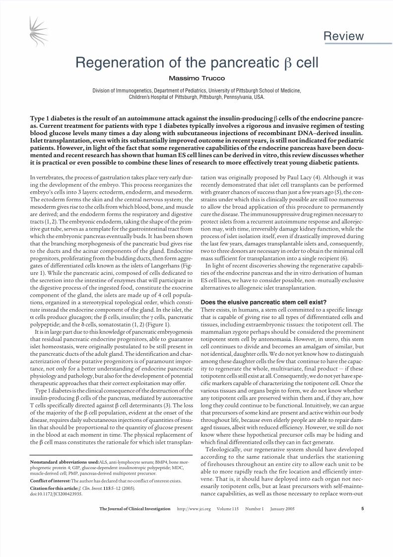
In vertebrates, the process of gastrulation takes place very early dur-ing the development of the embryo. This process reorganizes theembryo’s cells into 3 layers: ectoderm, endoderm, and mesoderm.The ectoderm forms the skin and the central nervous system; themesoderm gives rise to the cells from which blood, bone, and muscleare derived; and the endoderm forms the respiratory and digestivetracts (1, 2). The embryonic endoderm, taking the shape of the prim-itive gut tube, serves as a template for the gastrointestinal tract fromwhich the embryonic pancreas eventually buds. It has been shownthat the branching morphogenesis of the pancreatic bud gives riseto the ducts and the acinar components of the gland. Endocrineprogenitors, proliferating from the budding ducts, then form aggre-gates of differentiated cells known as the islets of Langerhans (Fig-ure 1). While the pancreatic acini, composed of cells dedicated tothe secretion into the intestine of enzymes that will participate inthe digestive process of the ingested food, constitute the exocrine
component of the gland, the islets are made up of 4 cell popula-tions, organized in a stereotypical topological order, which consti-tute instead the endocrine component of the gland. In the islet, theα cells produce glucagon; the β cells, insulin; the γ cells, pancreaticpolypeptide; and the δ cells, somatostatin (1, 2) (Figure 1).
It is in large part due to this knowledge of pancreatic embryogenesisthat residual pancreatic endocrine progenitors, able to guaranteeislet homeostasis, were originally postulated to be still present inthe pancreatic ducts of the adult gland. The identification and char-acterization of these putative progenitors is of paramount impor-tance, not only for a better understanding of endocrine pancreaticphysiology and pathology, but also for the development of potentialtherapeutic approaches that their correct exploitation may offer.
Type 1 diabetes is the clinical consequence of the destruction of theinsulin-producing β cells of the pancreas, mediated by autoreactiveT cells specifically directed against β cell determinants (3). The lossof the majority of the β cell population, evident at the onset of thedisease, requires daily subcutaneous injections of quantities of insu-lin that should be proportional to the quantity of glucose presentin the blood at each moment in time. The physical replacement of the β cell mass constitutes the rationale for which islet transplan-
tation was originally proposed by Paul Lacy (4). Although it wasrecently demonstrated that islet cell transplants can be performedwith greater chances of success than just a few years ago (5), the con-strains under which this is clinically possible are still too numerousto allow the broad application of this procedure to permanently cure the disease. The immunosuppressive drug regimen necessary toprotect islets from a recurrent autoimmune response and allorejec-tion may, with time, irreversibly damage kidney function, while theprocess of islet isolation itself, even if drastically improved duringthe last few years, damages transplantable islets and, consequently,two to three donors are necessary in order to obtain the minimal cellmass sufficient for transplantation into a single recipient (6).
In light of recent discoveries showing the regenerative capabili-ties of the endocrine pancreas and the in vitro derivation of humanES cell lines, we have to consider possible, non–mutually exclusivealternatives to allogeneic islet transplantation.
Does the elusive pancreatic stem cell exist?
There exists, in humans, a stem cell committed to a specific lineagethat is capable of giving rise to all types of differentiated cells andtissues, including extraembryonic tissues: the totipotent cell. Themammalian zygote perhaps should be considered the preeminenttotipotent stem cell by antonomasia. However, in utero, this stemcell continues to divide and becomes an amalgam of similar, butnot identical, daughter cells. We do not yet know how to distinguishamong these daughter cells the few that continue to have the capac-ity to regenerate the whole, multivariate, final product — if thesetotipotent cells still exist at all. Consequently, we do not yet have spe-cific markers capable of characterizing the totipotent cell. Once the
various tissues and organs begin to form, we do not know whetherany totipotent cells are preserved within them and, if they are, how long they could continue to be functional. Intuitively, we can arguethat precursors of some kind are present and active within our body throughout life, because even elderly people are able to repair dam-aged tissues, albeit with reduced efficiency. However, we still do notknow where these hypothetical precursor cells may be hiding andwhich final differentiated cells they can in fact generate.
Teleologically, our regenerative system should have developedaccording to the same rationale that underlies the stationingof firehouses throughout an entire city to allow each unit to beable to more rapidly reach the fire location and efficiently inter-
vene. That is, it should have deployed into each organ not nec-
essarily totipotent cells, but at least precursors with self-mainte-nance capabilities, as well as those necessary to replace worn-out
Nonstandard abbreviations used: ALS, anti-lymphocyte serum; BMP4, bone mor-phogenetic protein 4; GIP, glucose-dependent insulinotropic polypeptide; MDC,muscle-derived cell; PMP, pancreas-derived multipotent precursor.
Conflict of interest: The author has declared that no conflict of interest exists.
Citation for this article: J. Clin. Invest. 115:5–12 (2005).doi:10.1172/JCI200423935.
8/14/2019 Regeneration of the Pancreatic β Cell
http://slidepdf.com/reader/full/regeneration-of-the-pancreatic-cell 2/8
review
6 The Journal of Clinical Investigation http://www.jci.org Volume 115 Number 1 January 2005
cells. These precursors should be able to support a process that,while progressing relatively slowly in the maintenance of tissuehomeostasis, could be converted, in case of crisis, into a rapidly operating system. Consequently, this system would be highly effec-tive at responding promptly to a number of stimulations, promot-ed by nervous signaling, microarchitectural remnants of destroyedtissues, metabolites of energy generating processes, peptide growthfactors, etc. (7–9). Physiologically, in the endocrine pancreas, hor-
mone-producing cells that are at the end of their life span shouldbe continuously, albeit quite slowly, replaced by newly generatedcells. We need to learn more about this regenerative process if wewish to take advantage of it for therapeutic purposes.
In considering the utility of stem cells for the regeneration of theβ pancreatic cell, we currently face some major questions: in theadult endocrine pancreas, do multipotent progenitors still exist?If so, where exactly are they located? What are the best markers forrecognizing and isolating them? What are the stimuli able to acti-
vate their differentiation pathway(s)? How large is the time window in which they can still respond to the needs of an aging tissue? Inthe absence of pancreatic progenitors, are there adult pluripotentstem cells — cells not committed to a specific lineage that may dif-
ferentiate into all types of cells and tissues, with the exception of extraembryonic tissues — located elsewhere in the body that may
have the capability to regenerate β cell mass? If these cells are alllineage-committed precursor cells, can we instead use ES cell linesderived in vitro to substitute the lost β cells of the pancreas?
The road to regeneration
Increases in β cell mass may occur through increased β cell replica-tion, increased β cell size, decreased β cell death, and differentia-tion of possibly existing β cell progenitors (10). It has been shown
that occasional endocrine cells can be found embedded in normalpancreatic ducts. However, these cells are few and far between (11).The number of these duct-associated endocrine cells physiologically increases as the consequence of severe insulin resistance in obese indi-
viduals or during pregnancy (12, 13). Similar histological changes areobserved under conditions of tissue injury and repair after partialpancreatectomy, duct ligation, cellophane wrapping of the gland,or IFN-γ overexpression driven by the insulin promoter (14–17).Even then, within the ducts, only a small number of cells becomeinsulin positive. This suggests that even if some hypothetical precur-sors exist, the process of formation of endocrine cells out of the islet(neogenesis) would not be a frequently observed property of the ductepithelium. On the other hand, the fact that α and β cells develop
from a possibly common, non–hormone-expressing, yet Pdx1-posi-tive precursor (Pdx1 being a transcription factor required for pancre-
Figure 1Cross section of the pancreas. The pancreas houses 2 distinctly different tissues. Its bulk comprises exocrine tissue, which is made up of acinar
cells that secrete pancreatic enzymes delivered to the intestine to facilitate the digestion of food. Scattered throughout the exocrine tissue aremany thousands of clusters of endocrine cells known as islets of Langerhans. Within the islet, α cells produce glucagon; β cells, insulin; δ cells,
somatostatin; and γ cells, pancreatic polypeptide — all of which are delivered to the blood.
8/14/2019 Regeneration of the Pancreatic β Cell
http://slidepdf.com/reader/full/regeneration-of-the-pancreatic-cell 3/8
review
The Journal of Clinical Investigation http://www.jci.org Volume 115 Number 1 January 2005 7
atic development) suggests that all cell types found within the isletmay originate from a bona fide, common endocrine progenitor (18).These endocrine progenitors may be located close to the duct butmay not actually be components of the ductal epithelium (19). Theprogenitor cells could be mesenchymal in origin, or they could be
cells differentiated from an unknown cell type. If the number of theseprogenitors is extremely small, lineage analysis becomes very difficultbecause of the lack of known appropriate markers. Moreover, if thesecells are as rare as they appear to be, it becomes difficult to quantify their contribution to normal endocrine cell turnover.
These are some of the conclusions discussed by Weir and Bon-ner-Weir (20) in commenting on the study by Seaberg et al., inwhich it was shown that single murine adult pancreatic precursorcells can generate progeny with characteristics of pancreatic cells,including β cells (21). These rare (1 in 3,000–9,000 cells) pancreas-derived multipotent precursors (PMPs) do not seem to be bona fide pluripotent ES cells, since they lack, for example, the Oct4 andNanog markers that direct the propagation of undifferentiatedES cells; nor are these cells of clear ectodermal, mesodermal, orendodermal origin, since they failed to express other markers con-sidered specific for precursors of each of the embryonic cell types(20, 21). Because, surprisingly, these PMPs also lacked some β cellmarkers (e.g., HNF3β) as well as ductal epithelium markers (e.g.,cytokeratin), but were able to generate differentiation productswith neural characteristics along with α, β, δ, and acinar pancre-atic cells, the authors proposed the existence of a new and uniqueectodermal/endodermal precursor cell present during embryonicdevelopment that could persist in adult tissues (21).
These results support the conclusions of another recent study in which multipotent pancreatic progenitors were prospectively isolated using flow-cytometric cell sorting (22). The marker usedin this case was c-Met, the HGF receptor. The rationale for this
choice was the known signal exchange between epithelial andmesenchymal cells, promoting the interaction between c-Met andHGF, which plays an important role in the development of the pan-creas. The authors suggest that c-Met–HGF interaction is critically responsible for growth and differentiation of pancreatic stem andprogenitor cells not only during development but also in the adult,where they maintain homeostasis and promote regeneration. Colo-nies derived from single c-Met–positive cells, sorted from neonataland adult mouse pancreatic tissues, contained cells expressing sev-eral markers for endocrine, acinar, and ductal lineage cells. Whileneuroectodermal markers were not evaluated, the isolated pancre-atic stem cells in the study by Suzuki et al. (22) were also able togenerate offspring cells expressing hepatocyte and gastrointestinal
cell markers, possibly due to the selection marker used. Seaberg’sPMP-derived cells were grown instead in the serum-free mediumconditions normally used for neural stem cell culture (21).
All of these studies, even with their somewhat divergent outcomes,seem to support the conclusion that endocrine precursor cells of some kind exist in the pancreas. They are present not only in theduct, but also within the islets themselves, since both subpopulationswere independently used as the source of the isolated single cell pre-cursors (21). On the one hand, this conclusion supports the workinghypothesis of those who propose that pancreatic ductal cells cantransdifferentiate into β cells and that this is a physiologic processgenerally more efficiently activated by increased metabolic demandand tissue injuries (23); on the other hand, it may also accommo-
date the most recent results of Dor and colleagues (24), who proposeinstead that no β cell can arise from non–β cell progenitors, whether
in the normal adult pancreas or after pancreatectomy. As a directconsequence, the number of β cells should become virtually definedat a certain point, and, afterward, glycemia should be controlledonly by that defined cellular pool. Dor’s results were obtained by using a sophisticated Cre/lox system that, in transgenic mice, can be
induced by tamoxifen. This system labels fully differentiated β cells(defined as postnatal cells transcribing the insulin gene) that expressthe human alkaline phosphatase protein, which is in turn revealedby a histochemical stain. In a defined period of time, the “chase,”only the cells that are progeny of preexisting and labeled β cellsare newly labeled. New β cells derived from any non–β cell source,including stem cells, are not labeled. The frequency and distributionof labeledβ cells within pancreatic islets, at the end of the chase peri-od, should be inversely proportional to the number of new, nonla-beled cells present in the same structures. If the frequency of labeledβ cells does not change, as was observed, the number of cells derivedfrom the differentiation of non–insulin-producing precursors mustbe minimal or null, while terminally differentiated insulin-produc-ing β cells themselves should be the cells that actually proliferateand give rise to other insulin-producing β cells. While the results of Seaberg et al. (21) do not contest the proven yet limited ability of a β cell to divide, the failure of Dor et al. (24) to observe cells possibly differentiated from stem or precursor cells might actually be due toboth their extremely limited number (21) and technical issues. Forexample, the use of tamoxifen injected intraperitoneally or subcuta-neously twice a week for two and a half weeks (24) may have blockedneogenesis from precursor cells mainly in pancreatic ducts adjacentto involuted islets, as observed by Pelengaris et al. (25), and once thetamoxifen was withdrawn, these cells may not have had sufficienttime to differentiate.
Controlling the autoimmune response
On this basis, we can tentatively conclude that precursors of a per-haps unconventional type can be located both in close proximity to and inside the endocrine tissue and that they can be activatedby increased metabolic demand or by still-unknown secretedfactors, normally able to accelerate the process that guaranteeshomeostasis of islets of Langerhans under normal conditions.The physiologic equilibrium between lost and newly generatedcells can be altered by the action of β cell–specific, autoreactiveT cells in instances in which autoimmunity develops (3). OnceT cell killing activity overcomes the regenerative compensatory activity of the organ, the number of functional β cells progres-sively decreases until they become too few to maintain the gluco-homeostasis of the entire body. The time of transition over this
metabolic threshold becomes immediately evident with the pre-sentation of the characteristic signs of the clinical onset of type1 diabetes. During the course of disease, even if the regenerativeproperties of the pancreas remain functional, the continued pres-ence of diabetogenic, autoreactive T cells consistently nullifies thereparative effort. The fact that these autoreactive T cells remainpresent in the body of the diabetic patient for a long time is prov-en by experiments in which healthy islet cells transplanted intosyngeneic, long-term diabetic mice or humans were quickly killedby these same autoreactive T cells (26).
The autoimmune response is successfully averted in theNOD mouse either by directly eliminating the majority of theautoreactive T cells with anti–T cell antibodies or by substituting
all or part of the immunocompetent cell repertoire with bone mar-row cells obtained from diabetic-resistant donors.
8/14/2019 Regeneration of the Pancreatic β Cell
http://slidepdf.com/reader/full/regeneration-of-the-pancreatic-cell 4/8
review
8 The Journal of Clinical Investigation http://www.jci.org Volume 115 Number 1 January 2005
In a study by Ogawa et al., the treatment of overtly diabeticNOD mice with anti-lymphocyte serum (ALS) abrogated autoim-munity but achieved only partial clinical remission (27). Transienttreatment of overtly diabetic NOD mice with ALS and exendin-4,a potent insulinotropic hormone that promotes replication anddifferentiation of β cells in vitro and in vivo, achieved insteadcomplete remission of 88% of the treated animals within 75 days,accompanied by progressive normalization of glucose tolerance,
improved islet histology, increased insulin content in the pan-creas, and almost normal insulin release in response to a glucosechallenge. These results show that exendin-4 synergistically aug-ments the remission-inducing effect of ALS, possibly by promot-ing differentiation of β cell precursors (27).
Also, the successful induction of a mixed allogeneic chimerismobtained after transplanting bone marrow from a diabetes-resis-tant donor into a diabetic animal following a sublethal dose of irra-diation is sufficient to block and eventually also revert the system-atic invasion and inflammation of the islets by the autoreactive T
cells that result in insulitis (Figure 2) (28–30). Within the endocrinepancreas, once the insult of autoimmunity is abrogated, the physi-ologic process of regeneration can continue efficiently, eventually replenishing the population of insulin-producing cells to a numbersufficient to maintain euglycemia, thus curing the diabetic recipi-ent (Figure 2D) (31–33). While this process takes place — and itis still debatable whether this occurs over weeks (32) or months(33) — the recipient’s glycemia must be controlled by additional,independent measures. The most commonly used technique is totransplant into the recipient islets from the same marrow donor.However, the successful engraftment of the transplanted bone mar-row, or the establishment of a steady hematopoietic chimerism,would have to be maintained without the use of immunosuppres-sive agents. These potent drugs may kill not only the still-presentautoreactive T cells of the recipient, but also the β cells themselves,thereby defeating the purpose of the transplant (34–36). The useof immunosuppressive agents may also interfere with the observedrise of regulatory T cells, a possible explanation for the long-last-ing immunoregulatory cell–dominant condition observed in curedanimals. Adoptive transfer experiments in which both diabetogeniclymphocytes and splenocytes from ALS-treated, long-term diabe-tes–free NOD mice were transplanted in NOD/SCID mice with nosigns of diabetes induction support this hypothesis (27).
A subject of ongoing debate is whether either or both the trans-planted bone marrow and the cotransplanted β cells are necessary for promoting an efficient regenerative process, independent of theirability to block autoimmunity or preserve euglycemia, respectively.
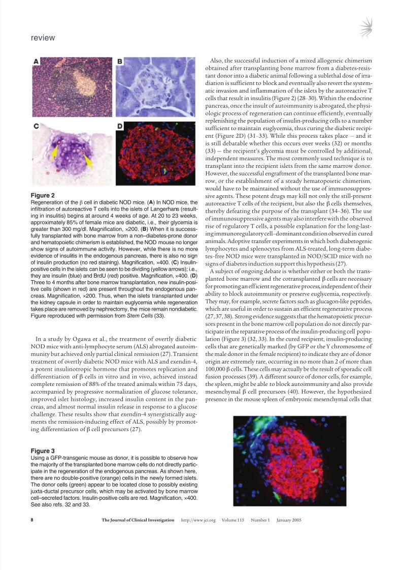
They may, for example, secrete factors such as glucagon-like peptides,which are useful in order to sustain an efficient regenerative process(27, 37, 38). Strong evidence suggests that the hematopoietic precur-sors present in the bone marrow cell population do not directly par-ticipate in the reparative process of the insulin-producing cell popu-lation (Figure 3) (32, 33). In the cured recipient, insulin-producingcells that are genetically marked (by GFP or the Y chromosome of the male donor in the female recipient) to indicate they are of donororigin are extremely rare, occurring in no more than 2 of more than100,000 β cells. These cells may actually be the result of sporadic cellfusion processes (39). A different source of donor cells, for example,the spleen, might be able to block autoimmunity and also providemesenchymal β cell precursors (40). However, the hypothesized
presence in the mouse spleen of embryonic mesenchymal cells that
Figure 3Using a GFP-transgenic mouse as donor, it is possible to observe how
the majority of the transplanted bone marrow cells do not directly partic-
ipate in the regeneration of the endogenous pancreas. As shown here,
there are no double-positive (orange) cells in the newly formed islets.
The donor cells (green) appear to be located close to possibly existing
juxta-ductal precursor cells, which may be activated by bone marrow
cell–secreted factors. Insulin-positive cells are red. Magnification, ×400.See also refs. 32 and 33.
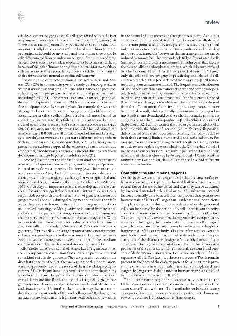
Figure 2Regeneration of the β cell in diabetic NOD mice. (A) In NOD mice, the
infiltration of autoreactive T cells into the islets of Langerhans (result-
ing in insulitis) begins at around 4 weeks of age. At 20 to 23 weeks,
approximately 85% of female mice are diabetic, i.e., their glycemia is
greater than 300 mg/dl. Magnification, ×200. (B) When it is success-
fully transplanted with bone marrow from a non–diabetes-prone donor
and hematopoietic chimerism is established, the NOD mouse no longer
show signs of autoimmune activity. However, while there is no more
evidence of insulitis in the endogenous pancreas, there is also no sign
of insulin production (no red staining). Magnification, ×400. (C) Insulin-
positive cells in the islets can be seen to be dividing (yellow arrows); i.e.,
they are insulin (blue) and BrdU (red) positive. Magnification, ×400. (D)
Three to 4 months after bone marrow transplantation, new insulin-posi-
tive cells (shown in red) are present throughout the endogenous pan-
creas. Magnification, ×200. Thus, when the islets transplanted under
the kidney capsule in order to maintain euglycemia while regeneration
takes place are removed by nephrectomy, the mice remain nondiabetic.
Figure reproduced with permission from Stem Cells (33).
8/14/2019 Regeneration of the Pancreatic β Cell
http://slidepdf.com/reader/full/regeneration-of-the-pancreatic-cell 5/8
review
The Journal of Clinical Investigation http://www.jci.org Volume 115 Number 1 January 2005 9
lack surface expression of CD45 and are able to differentiate intoendothelial and endodermal cells remains to be confirmed.
Further questions
Even when regeneration of theβ cells from precursor cells is defini-tively proven, issues still to be resolved will include the time frameand physiological conditions necessary for regeneration to occurand reach completion, as well as the circumstances that facilitate orlimit the regeneration process and the ability of clinicians to pro-mote or avoid these, to more efficiently achieve the desired thera-peutic results. Also, assuming the existence of β cell precursors, westill do not know whether these cells are immortal or subject tosenescence, a situation that would leave a narrow window of time
for intervention. This matter may be especially relevant in diabeticindividuals in whom the reparative process has been repressed by autoimmune surveillance for a long period of time. If successfulimmunoregulatory intervention cannot be initiated immediately after the clinical onset of the disease, a full recovery of the endocrinefunction of the gland via the physiologic regeneration route may become impossible. However, if the regenerative process is provento be irreversibly compromised at a certain point, it may still be pos-sible to transplant into diabetic patients functional precursor cellsfrom nondiabetic donors or to artificially convert the patient’s owncells from other tissues or lineages into insulin-producing β cells.
The potential for ES cells
Even if specific markers necessary to recognizeβ cell precursors wereavailable, the physical isolation of these cells from a patient’s pan-creas would not be an easy task. Increasing the number of possibleprecursors ex vivo while avoiding the activation of differentiationpathways would also be problematic, as would be eventually facilitat-ing their differentiation toward the final product, in this case, a func-tional β cell. Culturing and expanding β cells in vitro has also beenshown to be difficult and perhaps limited to a few proliferative cycles,since they are terminally differentiated cells. It has been postulatedthat it would be easier to derive precursor cells from the embryo anduse them to regenerate the damaged endocrine pancreas. Althoughhuman ES cell lines have been successfully derived (41, 42) andrecently made available to the scientific community (43), the need to
direct their differentiation toward a specific final product, in this casetheβ cell, still remains a major hurdle that must be overcome.
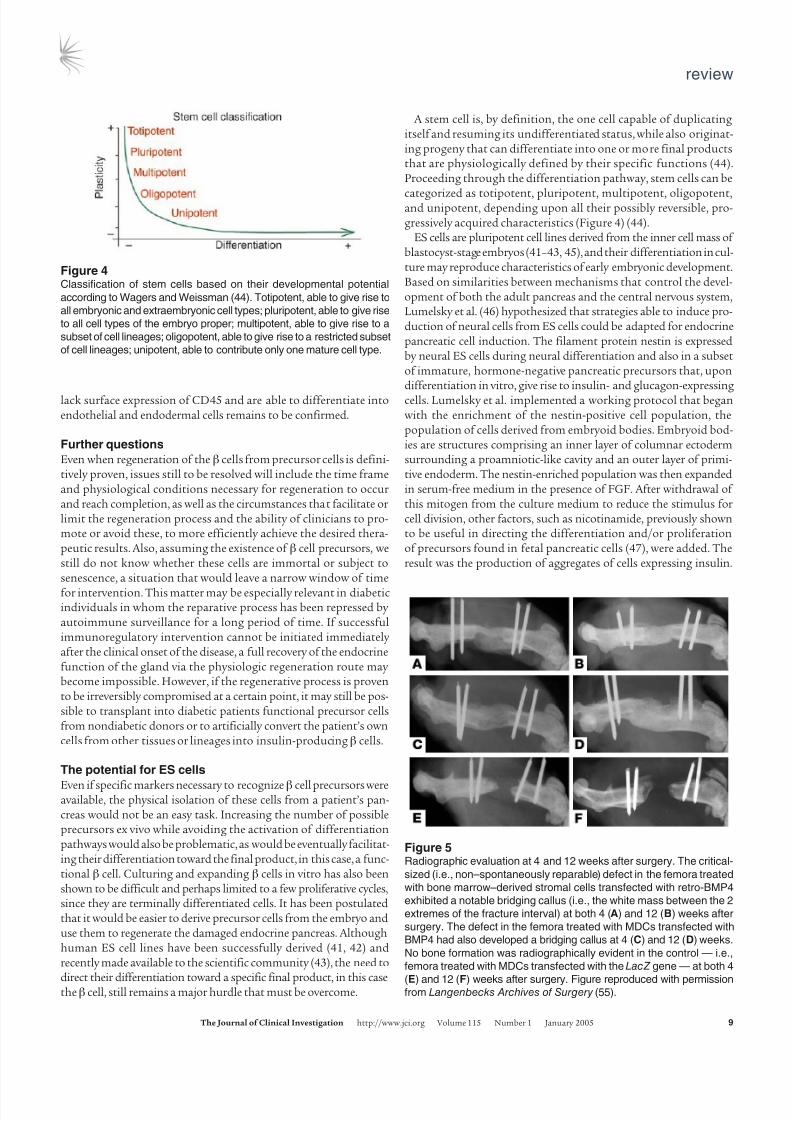
A stem cell is, by definition, the one cell capable of duplicatingitself and resuming its undifferentiated status, while also originat-ing progeny that can differentiate into one or more final productsthat are physiologically defined by their specific functions (44).Proceeding through the differentiation pathway, stem cells can be
categorized as totipotent, pluripotent, multipotent, oligopotent,and unipotent, depending upon all their possibly reversible, pro-gressively acquired characteristics (Figure 4) (44).
ES cells are pluripotent cell lines derived from the inner cell mass of blastocyst-stage embryos (41–43, 45), and their differentiation in cul-ture may reproduce characteristics of early embryonic development.Based on similarities between mechanisms that control the devel-opment of both the adult pancreas and the central nervous system,Lumelsky et al. (46) hypothesized that strategies able to induce pro-duction of neural cells from ES cells could be adapted for endocrinepancreatic cell induction. The filament protein nestin is expressedby neural ES cells during neural differentiation and also in a subsetof immature, hormone-negative pancreatic precursors that, upondifferentiation in vitro, give rise to insulin- and glucagon-expressingcells. Lumelsky et al. implemented a working protocol that beganwith the enrichment of the nestin-positive cell population, thepopulation of cells derived from embryoid bodies. Embryoid bod-ies are structures comprising an inner layer of columnar ectodermsurrounding a proamniotic-like cavity and an outer layer of primi-tive endoderm. The nestin-enriched population was then expandedin serum-free medium in the presence of FGF. After withdrawal of this mitogen from the culture medium to reduce the stimulus forcell division, other factors, such as nicotinamide, previously shownto be useful in directing the differentiation and/or proliferationof precursors found in fetal pancreatic cells (47), were added. Theresult was the production of aggregates of cells expressing insulin.
Figure 4Classification of stem cells based on their developmental potential
according to Wagers and Weissman (44). Totipotent, able to give rise to
all embryonic and extraembryonic cell types; pluripotent, able to give rise
to all cell types of the embryo proper; multipotent, able to give rise to a
subset of cell lineages; oligopotent, able to give rise to a restricted subset
of cell lineages; unipotent, able to contribute only one mature cell type.
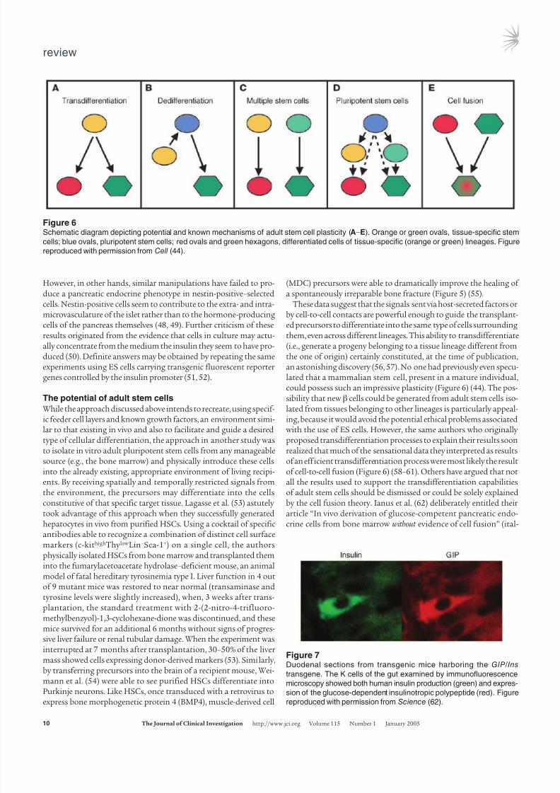
Figure 5Radiographic evaluation at 4 and 12 weeks after surgery. The critical-
sized (i.e., non–spontaneously reparable) defect in the femora treated
with bone marrow–derived stromal cells transfected with retro-BMP4
exhibited a notable bridging callus (i.e., the white mass between the 2
extremes of the fracture interval) at both 4 (A) and 12 (B) weeks after
surgery. The defect in the femora treated with MDCs transfected with
BMP4 had also developed a bridging callus at 4 (C) and 12 (D) weeks.
No bone formation was radiographically evident in the control — i.e.,
femora treated with MDCs transfected with the LacZ gene — at both 4
(E) and 12 (F) weeks after surgery. Figure reproduced with permissionfrom Langenbecks Archives of Surgery (55).
8/14/2019 Regeneration of the Pancreatic β Cell
http://slidepdf.com/reader/full/regeneration-of-the-pancreatic-cell 6/8
review
10 The Journal of Clinical Investigation http://www.jci.org Volume 115 Number 1 January 2005
However, in other hands, similar manipulations have failed to pro-duce a pancreatic endocrine phenotype in nestin-positive–selectedcells. Nestin-positive cells seem to contribute to the extra- and intra-microvasculature of the islet rather than to the hormone-producingcells of the pancreas themselves (48, 49). Further criticism of theseresults originated from the evidence that cells in culture may actu-ally concentrate from the medium the insulin they seem to have pro-duced (50). Definite answers may be obtained by repeating the sameexperiments using ES cells carrying transgenic fluorescent reportergenes controlled by the insulin promoter (51, 52).
The potential of adult stem cells
While the approach discussed above intends to recreate, using specif-
ic feeder cell layers and known growth factors, an environment simi-lar to that existing in vivo and also to facilitate and guide a desiredtype of cellular differentiation, the approach in another study wasto isolate in vitro adult pluripotent stem cells from any manageablesource (e.g., the bone marrow) and physically introduce these cellsinto the already existing, appropriate environment of living recipi-ents. By receiving spatially and temporally restricted signals fromthe environment, the precursors may differentiate into the cellsconstitutive of that specific target tissue. Lagasse et al. (53) astutely took advantage of this approach when they successfully generatedhepatocytes in vivo from purified HSCs. Using a cocktail of specificantibodies able to recognize a combination of distinct cell surfacemarkers (c-kithighThy low Lin–Sca-1+) on a single cell, the authors
physically isolated HSCs from bone marrow and transplanted theminto the fumarylacetoacetate hydrolase–deficient mouse, an animalmodel of fatal hereditary tyrosinemia type I. Liver function in 4 outof 9 mutant mice was restored to near normal (transaminase andtyrosine levels were slightly increased), when, 3 weeks after trans-plantation, the standard treatment with 2-(2-nitro-4-trifluoro-methylbenzyol)-1,3-cyclohexane-dione was discontinued, and thesemice survived for an additional 6 months without signs of progres-sive liver failure or renal tubular damage. When the experiment wasinterrupted at 7 months after transplantation, 30–50% of the livermass showed cells expressing donor-derived markers (53). Similarly,by transferring precursors into the brain of a recipient mouse, Wei-mann et al. (54) were able to see purified HSCs differentiate into
Purkinje neurons. Like HSCs, once transduced with a retrovirus toexpress bone morphogenetic protein 4 (BMP4), muscle-derived cell
(MDC) precursors were able to dramatically improve the healing of a spontaneously irreparable bone fracture (Figure 5) (55).
These data suggest that the signals sent via host-secreted factors orby cell-to-cell contacts are powerful enough to guide the transplant-ed precursors to differentiate into the same type of cells surroundingthem, even across different lineages. This ability to transdifferentiate(i.e., generate a progeny belonging to a tissue lineage different fromthe one of origin) certainly constituted, at the time of publication,an astonishing discovery (56, 57). No one had previously even specu-lated that a mammalian stem cell, present in a mature individual,could possess such an impressive plasticity (Figure 6) (44). The pos-sibility that new β cells could be generated from adult stem cells iso-lated from tissues belonging to other lineages is particularly appeal-
ing, because it would avoid the potential ethical problems associatedwith the use of ES cells. However, the same authors who originally proposed transdifferentiation processes to explain their results soonrealized that much of the sensational data they interpreted as resultsof an efficient transdifferentiation process were most likely the resultof cell-to-cell fusion (Figure 6) (58–61). Others have argued that notall the results used to support the transdifferentiation capabilitiesof adult stem cells should be dismissed or could be solely explainedby the cell fusion theory. Ianus et al. (62) deliberately entitled theirarticle “In vivo derivation of glucose-competent pancreatic endo-crine cells from bone marrow without evidence of cell fusion” (ital-
Figure 7Duodenal sections from transgenic mice harboring the GIP/Ins
transgene. The K cells of the gut examined by immunofluorescence
microscopy showed both human insulin production (green) and expres-
sion of the glucose-dependent insulinotropic polypeptide (red). Figurereproduced with permission from Science (62).
Figure 6Schematic diagram depicting potential and known mechanisms of adult stem cell plasticity (A–E). Orange or green ovals, tissue-specific stem
cells; blue ovals, pluripotent stem cells; red ovals and green hexagons, differentiated cells of tissue-specific (orange or green) lineages. Figure
reproduced with permission from Cell (44).
8/14/2019 Regeneration of the Pancreatic β Cell
http://slidepdf.com/reader/full/regeneration-of-the-pancreatic-cell 7/8
review
The Journal of Clinical Investigation http://www.jci.org Volume 115 Number 1 January 2005 11
ics added). Regardless, it is perhaps useful to instead consider thepossibility of actually forcing the fusion of transplanted precursorcells with the recipient damaged cells as a means of producing newly functional cells, even though the endocrine pancreas appears to bean environment difficult to physically reach in vivo.
The potential for gene therapy
Instead of promoting cell-to-cell fusion in an effort to restore certainphysiologic functions lost with the death of specific cells, it has beenconsidered perhaps more efficient to transfect cells belonging to a certain lineage with genes able to convert them into cells carrying thecharacteristics of those lost, even if they belong to a completely differ-ent lineage. In particular, in the field of diabetes research, gut K cellsof the mouse were induced to produce human insulin by transfectingthe human insulin gene linked to the 5′-regulatory region of the geneencoding glucose-dependent insulinotropic polypeptide (GIP) (Fig-ure 7) (63). Also, research demonstrating that hepatocytes transfectedwith the Pdx1 gene under the control of the rat insulin 1 promoterwere able to produce insulin (64, 65) attracted significant attentionand has inspired new hope. In these studies, sufficient levels of insu-lin were secreted to satisfy the needs of a diabetic mouse, which, whentreated, became and remained steadily euglycemic. These studies,however, have not yet been successfully repeated by other groups.
The future problems we face
The ability to clone human embryos and to derive from themhuman ES cell lines is already a reality (41, 43, 45). Even if tomor-row’s scientists could derive, by cloning, cell lines able to be dif-ferentiated toward cells with the phenotype of a β cell and insufficient numbers to satisfy the needs of the diabetic transplantrecipient, in order that they can be used for clinical purposes, thesecell lines should carry the diploid genome of somatic cell nuclei
derived from individuals unrelated to the oocyte donor, i.e., thoseof the recipient. Further, we would still need to isolate from eachpatient his or her own stem cell line or develop a bank of cell linesrepresenting the broad diversity of human MHC polymorphismsin order to avoid allorejection. Under these circumstances, howev-er, the presence of autoimmunity will remain a concern. If we optto avoid this demanding preparative effort to match donor andrecipient, it would be necessary to reduce the likelihood of allore-
jection by other means. It may be easier to achieve this goal by usingES cells rather than any other adult cell for transplant, since thealready-studied human ES cell lines appear able to downregulatethe expression of MHC antigens at their surface (66, 67). It shouldstill be noted, however, that rapidly dividing cells with this unique
phenotype have to spontaneously stop growing once a specific,predetermined, total population cell mass has been reached, sincethey appear not to be controlled by NK cells (66). Furthermore,even assuming that we could overcome all of these challenges, theuse of human ES cell lines tailored ad personam would constitutean extremely demanding and expensive proposition (68).
Perhaps equally demanding would be the use of means other thanbone marrow transplant to evade an autoimmune response. Howev-er, some promising strategies, possibly more suitable for therapeuticapplications than bone marrow transplant, have already been pro-
posed. Once refined, they may allow the reestablishment of a toler-ant immune state that would free the patient from the protractedattack of T cells against their β cell structures and consequently from the requirement for administration of powerful immuno-suppressive regimens even when transplanted with MHC-matched
insulin-producing cells. While very efficient in the NOD mouse, theuse of anti-CD3 monoclonal antibodies able to skew the responseof diabetogenic, autoreactive T cells from a Th1 to a Th2 program(69) seems only able to slow the progression of disease to permanentdiabetes in humans (70). The possible involvement of CD4+CD25+ T cells in the preservation of tolerance, postulated following resultsobtained in cured mice (71), is also the basis of the treatment pro-posed by Zheng and collaborators (72), which involves the adminis-tration of rapamycin, agonistic IL-2–Ig fusion protein, and a mutant,antagonist-type IL-15–related cytolytic Ig fusion protein to obtainlong-term engraftment and tolerance of allogeneic islets transplantedinto overtly diabetic NOD mice. The CD4+CD25+ T cell populationis also increased once CD40, CD80, and CD86 cell surface moleculesare specifically downregulated by ex vivo treatment of NOD miceDCs with a mixture of antisense oligonucleotides targeting CD40,CD80, and CD86 primary transcripts (73). The incidence of diabetesis significantly delayed by a single injection of the engineered NODmouse–derived DCs into syngeneic recipients. Assuming then thatautoimmunity could be overcome, the potential regeneration-basedtreatment would still require that we determine the length of timethe β cell regenerative potential could be exploited and the charac-terization of possible triggering factors that would allow quantitativeand chronological limits to be bypassed.
Conclusion
Our young diabetic patients must check their blood glucose levelsand be injected with insulin at least 4 times a day. Concurrently,
they live with the constant threat of incidents secondary to unpre-dictable acute hypoglycemic episodes and the ever-present worry of chronic complications. Although human ES cell research carrieswith it enormous scientific potential in the treatment and possiblecure of many diseases, in the near future, the advances in the realmof immunoregulation may precede those in the stem cell arena. Tome, finding safe ways to block autoimmunity seems to be the f irstgoal we should achieve in order to give our patients a reliable solu-tion to their heavy, lifelong burden, since it is a prerequisite for boththe efficient use of an ES cell–based therapy and the reestablishmentof euglycemia capitalizing on the pancreatic regenerative pathway.
Acknowledgments
I want to thank Len Harrison and the confidential invited peerreviewers for critically reading the paper; and Timothy Kieffer,
Johnny Huard, YiGang Chang, and Tatiana Zorina for allowing meto use their photographs.
Address correspondence to: Massimo Trucco, Division of Immunogenetics, Children’s Hospital of Pittsburgh, RangosResearch Center, 3460 5th Avenue, Pittsburgh, Pennsylvania 15213-3205, USA. Phone: (412) 692-6570; Fax: (412) 692-5809;E-mail: [email protected].
1. Wells, J.M. 2003. Genes expressed in the developingendocrine pancreas and their importance for stemcell and diabetes research. Diabetes Metab. Res. Rev.
19:191–201.2. Jensen, J. 2004. Gene regulatory factors in pancreatic
development. Dev. Dyn. 229:176–200.3. Bach, J.F. 1994. Insulin-dependent diabetes mellitus
as an autoimmune disease. Endocr. Rev. 15:516–542.
4. Lacy, P.E. 1982. Pancreatic transplantation as a meansof insulin delivery. Diabetes Care. 5(Suppl. 1):93–97.
5. Shapiro, A.M., et al. 2000. Islet transplantation inseven patients with type 1 diabetes mellitus using a glucocorticoid-free immunosuppressive regimen.
N. Eng. J. Med. 343:230–238.6. Ryan, E.A., et al. 2001. Clinical outcomes and insulin
8/14/2019 Regeneration of the Pancreatic β Cell
http://slidepdf.com/reader/full/regeneration-of-the-pancreatic-cell 8/8
review
12 The Journal of Clinical Investigation http://www.jci.org Volume 115 Number 1 January 2005
secretion after islet transplantation with the Edmon-ton protocol. Diabetes. 50:710–719.
7. Steffensen, I., Dulin, M.F., Walters, E.T., and Morris,C.E. 1995. Peripheral regeneration and central sprout-ing of sensory neurone axons in Aplysia californica fol-lowing nerve injury. J. Exp. Biol. 198:2067–2078.
8. Ramachandra, A.V., Swamy, M.S., and Kurup, A.K.
1996. Local and systemic alterations in cyclic 3’,5’ AMP phosphodiesterase activity in relation to tailregeneration under hypothyroidism and T4 replace-ment in the lizard, Mabuya carinata. Mol. Reprod. Dev. 45:48–51.
9. Stocum, D.L. 2002. A tail of transdifferentiation.Science. 298:1901–1903.
10. Lipsett, M., and Finegood, D.T. 2002. β-cell neogenesisduring prolonged hyperglycemia in rats. Diabetes. 51:1834–1841.
11. Gu, D., Lee, M.S., Krahl, T., and Sarvetnick, N. 1994.Transitional cells in the regenerating pancreas. Devel-opment. 120:1873–1881.
12. Bernard-Kargar, C., and Ktorza, A. 2001. Endocrinepancreas plasticity under physiological and patho-logical conditions. Diabetes. 50 (Suppl. 1):S30–S35.
13. Brelje, T.C., et al. 1993. Effect of homologous pla-
cental lactogens, prolactins, and growth hormoneson islet β-cell division and insulin secretion in rat,mouse, and human islets: implication for placentallactogen regulation of islet function during pregnan-cy. Endocrinology. 132:879–887.
14. Rosenberg, L. 1995. In vivo cell transformation:neogenesis of beta cells from pancreatic ductal cells.Cell Transplant. 4:371–383.
15. Bouwens, L. 1998. Transdifferentiation versus stemcell hypothesis for the regeneration of islet beta-cellsin the pancreas. Microsc. Res. Tech. 15:332–336.
16. Bonner-Weir, S., Deery, D., Leahy, J.L., and Weir, G.C.1989. Compensatory growth of pancreatic beta-cells in adult rats after short-term glucose infusion.
Diabetes. 38:49–53.17. Arnush, M., et al. 1996. Growth factors in the regener-
ating pancreas of gamma-interferon transgenic mice. Lab. Invest. 74:985–990.
18. Herrera, P.L. 2000. Adult insulin- and glucagon-pro-ducing cells differentiate from two independent celllineages. Development. 127:2317–2322.
19. Gu, G., Dubauskaite, J., and Melton, D.A. 2002. Directevidence for the pancreatic lineage: NGN3+ cells areislet progenitors and are distinct from duct progeni-tors. Development. 129:2447–2457.
20. Weir, G.C., and Bonner-Weir, S. 2004. Beta-cell pre-cursors — a work in progress. Nat. Biotechnol. 22:1–2.
21. Seaberg, R.M., et al. 2004. Clonal identification of multipotent precursors from adult mouse pancreasthat generate neural and pancreatic lineages. Nat. Bio-technol. 22:1115–1124.
22. Suzuki, A., Nakauchi, H., and Taniguchi, H. 2004.Prospective isolation of multipotent pancreatic pro-genitors using flow-cytometric cell sorting. Diabetes. 53:2143–2152.
23. Bonner-Wier, S., and Sharma, A. 2002. Pancreaticstem cells. J. Pathol. 197:519–526.
24. Dor, Y., Brown, J., Matinez, O.I., and Melton, D. 2004. Adult pancreatic beta-cells are formed by self-dupli-cation rather than stem-cell differentiation. Nature. 429:41–46.
25. Pelengaris, S., Khan, M., and Evan, G.I. 2002. Sup-pression of Myc-induced apoptosis in β cells exposesmultiple oncogenic properties of Myc and triggerscarcinogenic progression. Cell. 109:321–334.
26. Sutherland, D.E., et al. 1984. Twin-to-twin pancreastransplantation: reversal and reenactment of thepathogenesis of type I diabetes. Trans. Assoc. Am. Physi-cians. 97:80–87.
27. Ogawa, N., List, J.F., Habener, J.F., and Maki, T. 2004.Cure of overt diabetes in NOD mice by transient treat-ment with anti-lymphocyte serum and exendin-4.
Diabetes. 53:1700–1705.
28. Ikehara, S., et al. 1985. Prevention of type 1 diabetesin nonobese diabetic mice by allogeneic bone mar-row transplantation. Proc. Natl. Acad. Sci. U. S. A. 82:7743–7747.
29. Li, H., et al. 1996. Mixed allogeneic chimerism inducedby a sublethal approach prevents autoimmune diabe-tes and reverses insulitis in nonobese diabetic (NOD)
mice. J. Immunol. 156:380–388.30. Zorina, T.D., et al. 2002. Distinct characteristics and
features of allogeneic chimerism in the NOD mousemodel of autoimmune diabetes. Cell Transplant. 11:113–123.
31. Ryu, S., Kodama, S., Ryu, K., Schoenfeld, D.A., andFaustman, D.L. 2001. Reversal of established auto-immune diabetes by restoration of endogenous β cell function. J. Clin. Invest. 108:63–72. doi:10.1172/ JCI200112335.
32. Hess, D., et al. 2003. Bone marrow-derived stemcells initiate pancreatic regeneration. Nat. Biotechnol. 21:763–770.
33. Zorina, T.D., et al. 2003. Recovery of the endogenousbeta cell function in autoimmune diabetes. Stem Cells. 21:377–388.
34. Ricordi, C., et al. 1991. In vivo effect of FK506 on
human pancreatic islets. Transplantation. 52:519–526.35. Tamura, K., et al. 1995. Transcriptional inhibition of insulin by FK506 and possible involvement of FK506binding protein-12 in pancreatic β-cell. Transplanta-tion. 59:1606–1615.
36. Bell, E., et al. 2003. Rapamycin has a deleterious effecton MIN-6 cells and rat and human islets. Diabetes. 52:2731–2739.
37. Paris, M., Bernard-Kargar, C., Berthault, M.F., Bouw-ens, L., and Ktorza, A. 2003. Specific and combinedeffects of insulin and glucose on functional pancre-aticβ-cell mass in vivo and in adult rats. Endocrinology. 144:2717–2727.
38. Farilla, L., et al. 2002. Glucagon-like peptide-1 pro-motes islet cell growth and inhibits apoptosis inZucker diabetic rats. Endocrinology. 143:4397–4408.
39. Lechner, A., et al. 2004. No evidence for significanttransdifferentiation of bonemarrow into pancreatic
β-cells in vivo. Diabetes. 53:616–623.40. Kodama, S., Kühtreiber, W., Fujimura, S., Dale, E.A.,
and Faustman, D.L. 2003. Islet regeneration duringthe reversal of autoimmune diabetes in NOD mice.Science. 302:1223–1227.
41. Thomson, J.A., et al. 1998. Embryonic stem celllines derived from human blastocysts. Science. 282:1145–1147.
42. Shamblott, M.J., et al. 1998. Derivation of pluripotentstem cells from cultured human primordial germcells. Proc. Natl. Acad. Sci. U. S. A. 95:13726–13731.
43. Cowan, C.A., et al. 2004. Derivation of embryonicstem-cell lines from human blastocysts. N. Engl. J.
Med. 350:1353–1356.44. Wagers, A.J., and Weissman, I.L. 2004. Plasticity of
adult stem cells. Cell. 116:639–648.45. Hwang, W.S., et al. 2004. Evidence of a pluripotent
human embryonic stem cell line derived from a cloned blastocyst. Science. 303:1669–1674.
46. Lumelsky, N., et al. 2001. Differentiation of embry-onic stem cells to insulin-secreting structures similarto pancreatic islets. Science. 292:1389–1394.
47. Beattie, G.M., Rubin, J.S., Mally, M.I., Otonkoski, T.,and Hayek, A. 1996. Regulation of proliferation anddifferentiation of human fetal pancreatic islet cells by extracellular matrix, hepatocyte growth factor, andcell-cell contact. Diabetes. 45:1223–1228.
48. Selander, L., and Edlund, H. 2002. Nestin is expressedin mesenchymal and not epithelial cells of the devel-oping mouse pancreas. Mech. Dev. 113:189–192.
49. Treutelaar, M.K., et al. 2003. Nestin-lineage cells con-tribute to the microvasculature but not endocrinecells of the islet. Diabetes. 52:2503–2512.
50. Rajagopal, J., Anderson, W.J., Kume, S., Martinez,O.I., and Melton, D.A. 2003. Insulin staining of ES
cell progeny from insulin uptake. Science. 299:363.51. Hara, M., et al. 2003. Transgenic mice with green flu-
orescent protein-labeled pancreatic beta -cells. Am. J. Physiol. Endocrinol. Metab. 284:E177–E183.
52. Bertera, S., et al. 2003. Body window-enabled in vivomulticolor imaging of transplanted mouse isletsexpressing an insulin-timer fusion protein. BioTech-
niques. 35:718–722.53. Lagasse, E., et al. 2000. Purified hematopoietic stem
cells can differentiate into hepatocytes in vivo. Nat. Med. 6:1229–1234.
54. Weimann, J.M., Charlton, C.A., Brazelton, T.R., Hack-man, R.C., and Blau, H.M. 2003. Contribution of transplanted bone marrow cells to Purkinje neuronsin human adult brains. Proc. Natl. Acad. Sci. U. S. A. 100:2088–2093.
55. Rose, T., et al. 2003. The role of cell type in bone heal-ing mediated by ex vivo gene therapy. Langenbecks
Arch. Surg. 388:347–355.56. Kahn, A. 2000. Converting hepatocytes to β-cells — a
new approach for diabetes? (News & Views). Nat. Med. 6:505–506.
57. Perry, D. 2000. Patients’ voices: the powerful sound instem cell debate. Science. 287:1423.
58. Wagers, A.J., Sherwood, R.L., Christensen, J.L., andWeissman, I.L. 2002. Little evidence for developmen-tal plasticity of adult hematopoietic stem cells.Science. 297:2256–2259.
59. Wang, X., et al. 2003. Cell fusion is the principalsource of bone-marrow-derived hepatocytes. Nature. 422:897–901.
60. Choi, J.B., et al. 2003. Little evidence of trans-differentiation of bone marrow-derived cells intopancreatic beta cells. Diabetologia. 46:1366–1374.
61. Alvarez-Dolado, M., et al. 2003. Fusion of bone-mar-row-derived cells with Purkinje neurons, cardiomyo-cytes and hepatocytes. Nature. 425:968–973.
62. Ianus, A., Holz, G.G., Theise, N.D., and Hussain, M.A.2003. In vivo derivation of glucose-competent pan-creatic endocrine cells from bone marrow withoutevidence of cell fusion. J. Clin. Invest. 111:843–850.doi:10.1172/JCI200316502.
63. Cheung, A.T., et al. 2000. Glucose dependent insulinrelease from genetically engineered K cells. Science. 290:1959–1962.
64. Ferber, S., et al. 2000. Pancreatic and duodenalhomeobox gene 1 induces expression of insulingenes in liver and ameliorates streptozotocin-inducedhyperglycemia. Nat. Med. 6:568–572.
65. Ber, I., et al. 2003. Functional, persistent, and extend-ed liver to pancreas transdifferentiation. J. Biol. Chem. 278:31950–31957.
66. Drukker, M., et al. 2002. Characterization of theexpression of MHC proteins in human embryonicstem cells. Proc. Natl. Acad. Sci. U. S. A. 99:9864–9869.
67. Li, L., et al. 2004. Human embryonic stem cellspossess immune-privileged properties. Stem Cells. 22:448–456.
68. Kennedy, D. 2004. Ste m cells, redux. Science.
303:1581.69. Chatenoud, L., Primo, J., and Bach, J.-F. 1997. CD3
antibody-induced dominant self tolerance in overtautoimmunity in non-obese diabetic mice. J. Immunol. 158:2947–2954.
70. Herold, K.C., et al. 2002. Anti-CD3 monoclonal anti-body in new-onset type 1 diabetes mellitus. N. Engl. J.
Med. 346:1692–1698.71. Chatenoud, L. 2003. CD3-specific antibody-induced
active tolerance. Nat. Rev. Immunol. 3:123–132.72. Zheng, X.X., et al. 2003. Favorably tipping the bal-
ance between cytopathic and regulatory T cellsto create transplantation tolerance. Immunity. 19:503–514.
73. Machen, J., et al. 2004. Antisense oligonucleotidedownregulating co-stimulation confer diabetes-pre- ventive properties to NOD dendritic cells. J. Immunol. 173:4331–4341.
![uncoupling protein (UCP) activity in Drosophila insulin producing ... · β-pancreatic cell function, and aging [1-6]. Located in the inner membrane of mitochondria, these carriers](https://static.fdocument.org/doc/165x107/60821fc54ed0441d9a6788dc/uncoupling-protein-ucp-activity-in-drosophila-insulin-producing-pancreatic.jpg)

![Effects of age -dependent changes in cell size on ... · angiogenesis and organ regeneration (e.g., liver) in aged adults [31]. Deregulation of YAP1 signaling also contributes to](https://static.fdocument.org/doc/165x107/5ec35e349338be1cb63451fe/effects-of-age-dependent-changes-in-cell-size-on-angiogenesis-and-organ-regeneration.jpg)