Cartilage Repair Under OA Inammatory Stimuli α Through ERK ...
Page 1/27
E-cigarette-Induced Pulmonary In�ammation andDysregulated Repair are Mediated by nAChR α7Receptor: Role of nAChR α7 in ACE2 Covid-19receptor regulationQixin Wang
University of RochesterIsaac Sundar
University of RochesterDongmei Li
University of RochesterJoseph Lucas
University of RochesterThivanka Muthumalage
University of RochesterSamantha McDonough
University of RochesterIrfan Rahman ( [email protected] )
University of Rochester Medical Center
Research
Keywords: E-cig exposure, nAChR α7, In�ammation, Dysregulated repair
Posted Date: May 18th, 2020
DOI: https://doi.org/10.21203/rs.2.23829/v2
License: This work is licensed under a Creative Commons Attribution 4.0 International License. Read Full License
Version of Record: A version of this preprint was published on June 18th, 2020. See the published versionat https://doi.org/10.1186/s12931-020-01396-y.
Page 2/27
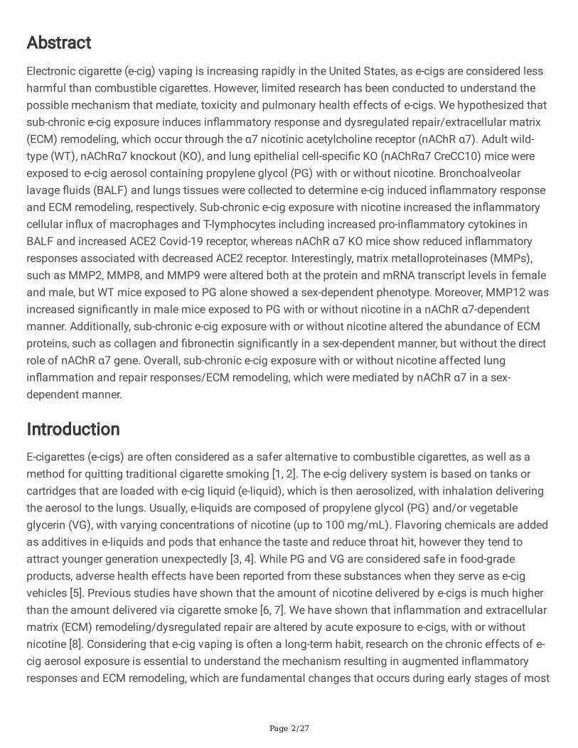
AbstractElectronic cigarette (e-cig) vaping is increasing rapidly in the United States, as e-cigs are considered lessharmful than combustible cigarettes. However, limited research has been conducted to understand thepossible mechanism that mediate, toxicity and pulmonary health effects of e-cigs. We hypothesized thatsub-chronic e-cig exposure induces in�ammatory response and dysregulated repair/extracellular matrix(ECM) remodeling, which occur through the α7 nicotinic acetylcholine receptor (nAChR α7). Adult wild-type (WT), nAChRα7 knockout (KO), and lung epithelial cell-speci�c KO (nAChRα7 CreCC10) mice wereexposed to e-cig aerosol containing propylene glycol (PG) with or without nicotine. Bronchoalveolarlavage �uids (BALF) and lungs tissues were collected to determine e-cig induced in�ammatory responseand ECM remodeling, respectively. Sub-chronic e-cig exposure with nicotine increased the in�ammatorycellular in�ux of macrophages and T-lymphocytes including increased pro-in�ammatory cytokines inBALF and increased ACE2 Covid-19 receptor, whereas nAChR α7 KO mice show reduced in�ammatoryresponses associated with decreased ACE2 receptor. Interestingly, matrix metalloproteinases (MMPs),such as MMP2, MMP8, and MMP9 were altered both at the protein and mRNA transcript levels in femaleand male, but WT mice exposed to PG alone showed a sex-dependent phenotype. Moreover, MMP12 wasincreased signi�cantly in male mice exposed to PG with or without nicotine in a nAChR α7-dependentmanner. Additionally, sub-chronic e-cig exposure with or without nicotine altered the abundance of ECMproteins, such as collagen and �bronectin signi�cantly in a sex-dependent manner, but without the directrole of nAChR α7 gene. Overall, sub-chronic e-cig exposure with or without nicotine affected lungin�ammation and repair responses/ECM remodeling, which were mediated by nAChR α7 in a sex-dependent manner.
IntroductionE-cigarettes (e-cigs) are often considered as a safer alternative to combustible cigarettes, as well as amethod for quitting traditional cigarette smoking [1, 2]. The e-cig delivery system is based on tanks orcartridges that are loaded with e-cig liquid (e-liquid), which is then aerosolized, with inhalation deliveringthe aerosol to the lungs. Usually, e-liquids are composed of propylene glycol (PG) and/or vegetableglycerin (VG), with varying concentrations of nicotine (up to 100 mg/mL). Flavoring chemicals are addedas additives in e-liquids and pods that enhance the taste and reduce throat hit, however they tend toattract younger generation unexpectedly [3, 4]. While PG and VG are considered safe in food-gradeproducts, adverse health effects have been reported from these substances when they serve as e-cigvehicles [5]. Previous studies have shown that the amount of nicotine delivered by e-cigs is much higherthan the amount delivered via cigarette smoke [6, 7]. We have shown that in�ammation and extracellularmatrix (ECM) remodeling/dysregulated repair are altered by acute exposure to e-cigs, with or withoutnicotine [8]. Considering that e-cig vaping is often a long-term habit, research on the chronic effects of e-cig aerosol exposure is essential to understand the mechanism resulting in augmented in�ammatoryresponses and ECM remodeling, which are fundamental changes that occurs during early stages of most
Page 3/27
chronic lung diseases, such as idiopathic pulmonary �brosis (IPF) and chronic obstructive pulmonarydisease (COPD) [9, 10].
In our previous studies, acute exposure to e-cig aerosol containing �avoring chemicals was shown tocause lung in�ammation, oxidative stress, and dysregulated repair [8, 11]. Further, nicotine exposurecould cause suppression of immune response resulting in augmented lung in�ammation and injuryfollowing viral infection [12]. Nicotinic Acetylcholine Receptors (nAChRs) are largely responsible foractivation of acetylcholine neurotransmitter signaling pathways in the central nervous system (CNS) [13,14]It has been postulated that angiotensin-converting enzyme (ACE2), a Covid-19 receptor is regulated bynAChRs. The nAChRs are widely distributed in the CNS, so they can easily be activated by nicotine,initiating and reinforcing a rewarding feedback loop which might induce nicotine addiction [15, 16].Interestingly, lung nAChRs activation could help inhibit the in�ammation caused by lipopolysaccharides(LPSs) and the receptor knockouts (KOs) that promote in�ammatory processes [17, 18]. However, as oneof the nAChR-agonists, nicotine could initiate receptor-related pathways, and nicotine aerosol inhalationalso induce in�ammation [11], and regulate ACE2 receptor via nAChR. Other chemicals may induceactivation of nAChRs, and they may also trigger feedback-loops, similar to �avoring chemicals [19-21].However, there are limited studies that directly describe about the role of dysregulated repair induced by e-cig vapors with or without nicotine in a nAChRα7-dependent manner.
A common vehicle used for nicotine delivery in e-cigs is PG and/or VG. PG (C3H8O2) is a colorless andodorless organic solvent that is an FDA-approved, food-grade chemical generally recognized as safe(GRAS). This is one of the reasons why people originally thought to use PG as a nicotine carrier in e-liquids. Previous study has shown that PG aerosols produce different byproducts, some of which mightlead to cancer [22]. Many e-cigs with or without nicotine are commercially available, but theunderstanding of respiratory health risks of e-cig inhalation are poorly investigated.
In this study, we hypothesized that e-cig aerosol induce lung in�ammation and dysregulated repair/ECMremodeling in a nAChR α7-dependent manner. Our results suggest that lung in�ammatory responses anddysregulated repair/ECM remodeling induced by e-cig aerosol containing nicotine could potentially berelated to the nAChR α7 signaling pathway. On the other hand, PG-induced lung dysregulated repair andin�ammatory response occurs in a nAChR α7-independent manner.
MethodsAnimals
Adult C57BL/6J (WT) mice and α7 nicotinic acetylcholine receptor knockout (nAChR α7 KO) mice wereboth purchased from Jackson Laboratory, weighing 25-35 grams and aged 3-4 months old. The nAChRα7 CreCC10 mice (clara/club-cell-speci�c nAChR α7 deletion) were generated by crossing nAChR α7�oxed mice (nAChR α7 �oxed mutant) from Jackson Laboratory (donated by Dr. Jerry Yakel, NIEHS/NIH)with mice have the Cre recombinase transgene controlled by the CC10 promoter (C57BL/6J; from TJ
Page 4/27
Mariani, University of Rochester, Rochester, NY). Prior to e-cig exposure, mice were housed in theinhalation core facility at the University of Rochester for one week. All experiments performed in thisstudy were in compliance with the standards set by the United States Animal Welfare Act. The AnimalResearch Committee (UCAR) approved the animal protocol at the University of Rochester Medical Center,Rochester, NY.
Blood gas and exercise ability measurement
Blood gas, including pH, pressure of CO2 and O2, concentration of HCO3, TCO2, glucose (Glu) andhemoglobin (Hb), percentage of O2 and hematocrit (Hct) were analyzed by i-STAT system (i-STAT CG8+cartridge, Cat# 03P88‐25; Vet scan i-STAT 1 analyzer; Abaxis Global Diagnostics), as described previously[23]. Exercise capacity measurement was done using a motorized animal treadmill (ColumbusInstruments) as described previously [24]. Running distance (meters) and running time (minutes) wererecorded to present exercise tolerance. Mice stayed on the treadmill for 5 min before start ofmeasurement. The treadmill began at 8.5 m/min with 0° incline for 9 min. Then, the speed was increasedto 10 m/min with 5° incline for 3 min. After that, the speed was increased to 2.5 m/min every 3 min, andincline was increased to 5° every 9 min until the mouse gets exhausted, judged by observation of failureof running and continuous contact with the electric grid. Following this exercise, blood was collected bysubmandibular venipuncture for blood gas and cotinine measurement.
E-cig device and e-liquid
The e-cig device used in this study was purchased from Joytech VTC mini (SciReq, Montreal, Canada).The atomizer/coil (0.15Ω) used for aerosolization of e-liquid was purchased from Kanger technology(Shenzhen, China). All the other components of the e-cig exposure chamber were purchased fromSCIREQ, and all the components were cleaned after exposure every day. The atomizer/Coil was replacedon a weekly basis to avoid overheating and generation of carbon monoxide. The e-liquids used in thisstudy that contains PG alone and PG with nicotine (25 mg/mL), were purchased from xtremevaping.com.
E-cig exposure
The e-cig exposure performed here has been described in our previous studies [25]. Brie�y, the in vivo e-cig exposure was setup inside a fume hood and based on the SCIREQ InExpose e-cig extension smokingsystem. The e-cig exposure pu�ng pro�le used was based on realistic topographical data from e-cigusers, with 3.3 sec/puff, 2 puffs/min and a 70mL puff volume [26]. The aerosolization of e-liquid wasperformed by using a 3rd generation e-cig device (Joytech eVIC VTCmini), which was controlled by theSCIREQ �exiware software (V8.0). The whole-body exposure was done for a total of 2 hrs/day, 5days/week, for 30 days. During e-cig exposures, temperature, humidity, oxygen and carbon dioxidepercentages were monitored along with carbon monoxide levels in the chamber. The e-cig aerosolgenerated was passed through the condensing chamber and pumped into the mixing chamber with a�owrate of 1.0 L/min. The vapor was diluted with air in the mixing chamber and then delivered into thewhole-body exposure chamber where mice were separated by dividers. Simultaneously, the e-cig aerosol
Page 5/27
in the exposure chamber was exhausted by another pump with a �owrate of 2.0 L/min. Both the pumpswere calibrated and adjusted each time before exposure. Pumps were cleaned at the end of eachexposure to minimize the effects of nicotine residues. Mice were divided into air (control), PG, and PGwith nicotine groups, each with an equal number of males and females, for both the WT and nAChR α7KO conditions. Air group mice stayed in the inhalation facility in a similar environment during the 30 dayexposure. Serum cotinine levels measured by ELISA (Calbiotech) were ~500 ng/mL for the PG withnicotine group, and ~20 ng/mL for the PG only group, due to nicotine residue in the pumps and exposurechambers which was di�cult to fully clean (Additional File 1: Figure. S1). As expected, the air groupshowed no cotinine.
Bronchoalveolar lavage �uid (BALF)
Mice were euthanized with Ketamine/Xylazine 24 hrs after the �nal exposure. Their tracheas werecannulated and their lungs were lavaged three times with 0.6 mL saline with 1% FBS (1.8 mL total). Therecovered �uids were collected and spun down at 1000 x g for 10 min at 4°C for harvesting of BALF cells.The supernatant was stored at -80°C for future analysis. The BALF cells were re-suspended in 1.0 mLsaline with 1% FBS and stained with acridine orange propidium iodide (AO/PI). Total cell counts per mLwere measured from AO/PI stained cells via cellometer.
In�ammatory cell count
The resuspended BALF cells were used for immune-in�ammatory cell counts with cell-type-speci�clabelled monoclonal antibodies. Total 1.0 x 10⁵ BALF cells were used for antibody labeling. Beforeantibody staining, all cells were blocked with puri�ed anti-mouse CD16/32 (Cat# 50-163-432, FisherScienti�c) to prevent non-speci�c binding, and washed with PBS once. The cells were stained with F4/80PE‐conjugated antibody for macrophages (Cat# 123109, BioLegend), LY6B.2 Alexa �uor488‐conjugatedantibody for neutrophils (Cat# NBP213077AF488, Novus Biologicals), PE-Cyanine7 antibody for CD4a+ T-lymphocytes (Cat# 25-0041-82, Fisher Scienti�c), and APC conjugated Monoclonal Antibody for CD8a+ T-Lymphocytes (Cat# 17-0081-82, Fisher Scienti�c). The absolute cell numbers of macrophages,neutrophils, and CD4a+/CD8a+ T-lymphocytes were determined by multiplying the percentage of cells bythe total cell counts. Flow cytometry was performed using the Guava® easyCyte™ �ow cytometer(Millipore Sigma) and analyzed using Guava® InCyte™ software.
Measurement of pro-in�ammatory cytokines by Luminex in BALF
To measure the pro-in�ammatory cytokines present in BALF, a Bio-Plex Pro mouse cytokine 23-pleximmunoassay kit (Cat#: M60009RDPD, BioRad) was used, according to the manufacturer’s instructions.Brie�y, the diluted magnetic beads were placed into the assay plate and rinsed with wash buffer. TheBALF samples and standards were then added into the wells, and shaken at 850 rpm at roomtemperature for 30 min. After sample incubation, the plates were washed with wash buffer 3 times, thenthe detection antibody was added and the plates incubated for 30 min at room temperature, shaking at850 rpm. Next, plates were washed with wash buffer 3 times, and SA-PE was added for 10 min at room
Page 6/27
temperature, shaking at 850 rpm. After this step, the plates were washed 3 times with wash buffer, andthe beads were resuspended in assay buffer for reading. Results are determined via the Luminex �exmap3d (Luminex Corp.)
Protein isolation
Lung tissues harvested from the animals during sacri�ce were stored at -80°C for future analysis. Frozenlungs were homogenized mechanically in RIPA buffer with protease inhibitor (Cat#: 78440, ThermoFisherScienti�c). After homogenization, lysates were kept on ice for 45 min, followed by centrifugation at15,000 x g for 30 min at 4°C. The supernatant was collected and protein concentration was quanti�edusing the Pierce BCA Protein Assay Kit (Cat#: 23227, ThermoFisher Scienti�c), based on themanufacturer's protocol.
Western blot
The protein samples from lung homogenates were running through SDS-polyacrylamide electrophoresisgels (SDS-PAGE: 10%) with 20 µg of protein in each lane. After electrophoresis, the gel was transferredonto a nitrocellulose membrane (Cat# 1620112, BioRad). The membrane was washed with tris-bufferedsaline containing 0.1% Tween 20 (TBS-T) for 15 min, then blocked with 5% non-fat dry milk for 1 hr atroom temperature. Then, the membranes were probed with primary antibodies 16 hr at 4°C: anti-MMP2(1:1000, ab92536, Abcam); anti-MMP9 (1:1000, ab38898, Abcam); anti-MMP8 (1:1000, ab81286, Abcam);anti-MMP12 (1:1000, NBP2-67344, Novus Biologicals); anti-Collagen 1α1 (1:1000, NBP1-30054, NovusBiologicals); anti-Collagen 1α2 (1:500, NBP1-57987, Novus Biologicals); anti-�bronectin (1:2000,ab45688, Abcam); and anti-PAI-1 (1:1000, ab182973, Abcam),anti-ACE2 (1:1000, ab15348, Abcam), andthe appropriate secondary antibody (Goat-Anti-Rabbit, 1:10000, Cat# 1706515, BioRad) for 1 hr at roomtemperature. After each antibody incubation, membrane was washed 4 times in TBS-T at roomtemperature, 15 min per wash. The luminescence signals were developed using chemiluminescencesubstrate (Perkin Elmer, Waltham, MA). The membrane exposure and band intensities were detected viathe Bio-Rad ChemiDoc MP imaging system (Bio-Rad Laboratories, Hercules, CA, USA). Proteinquanti�cation was done by densitometry analysis, and β-actin (1:2500, ab20272, Abcam) was used ashousekeeping control.
RNA isolation and Nanostring quanti�cation
Frozen lung tissues from -80ºC were used for RNA isolation. Lung tissue (~100 mg) per sample washomogenized in Trizol for 20s. The RNA was isolated using the Direct-zol™ RNA Miniprep Plus assay kit(Cat# R2073, Zymo Research). Isolated RNA was quanti�ed and checked for quality byspectrophotometer (ND-1000, NanoDrop Technologies, Wilmington, DE, USA). RNA samples werealiquoted and sent out to Nanostring facility for analysis of genes using nCounter Mouse Myeloid InnateImmunity v2 Panel (NanoString Technologies, Inc. Cat# XT-CSO-MMII2-12).
Statistical analysis
Page 7/27
The statistical analysis was done by either one-way ANOVA or student’s t-test, followed by Tukey’smultiple comparisons in GraphPad Prism software (Version 8.0, La Jolla, CA). Results were presented asthe mean ± SEM. P < 0.05 was considered statistically signi�cant.
ResultsSub-chronic e-cig exposure altered exercise capacity and blood-gas saturation
After 30 days e-cig exposure, WT (�oxed) mice showed no difference in exercise capacity among thegroups (Additional File 1: Figure S2). However, nAChR α7 de�cient (KO) mice showed less capacity to runwhen exposed to PG with nicotine compared to air control and PG alone groups. Similar results wereobserved in nAChR α7 lung epithelial cell-speci�c conditional KO mice exposed to PG with nicotine showreduced exercise capacity compared to air control and PG alone groups. E-cig aerosol with nicotineexposure in nAChR α7 KO show lowered exercise capacity indicating the possible role of nAChR α7 in thelung epithelium. Blood pH, O2 pressure, concentrations of HCO3 and CO2 were shown to be dysregulatedwhen WT mice were exposed to PG with nicotine compared to PG alone, while no difference wasobserved between WT and nAChR α7 KO mice exposed to PG with or without nicotine (Additional File 2:Table S1).
Sub-chronic e-cig exposure induces in�ammatory cellular in�ux in the mouse lung
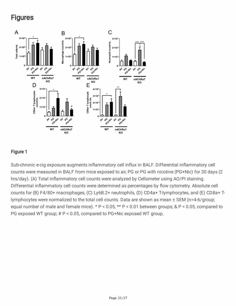
The total cell counts were increased in WT mice exposed to PG with or without nicotine relative torespective air group, and total cell counts in WT mice exposed to PG with nicotine increased signi�cantlycompared to air and PG alone groups (Figure 1A). The nAChR α7 KO mice showed no change in total cellcounts when exposed to PG with nicotine (Figure 1A). However, the total cell counts were increased butnot signi�cant compared to respective control group (Figure 1A). Macrophage counts followed a similartrend as observed in total cell counts (Figure 1B). WT and nAChR α7 KO mice exposed to PG with nicotineshowed no change in neutrophil counts in BALF (Figure 1C). Additionally, CD4a+ and CD8a+ T-lymphocytecounts were signi�cantly increased when WT mice were exposed to PG with nicotine, and loss of nAChRα7 prevents increase in T-lymphocytes (Figure 1D-E). PG-exposed nAChR α7 KO mice showed higherlevels of neutrophils and CD4a+/CD8a+ T-lymphocytes than PG-exposed WT mice (Figure 1C-E).
Sub-chronic e-cig exposure augments in�ux of pro-in�ammatory cytokines in BALF
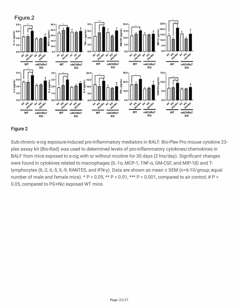
We next determined the level of pro-in�ammatory cytokines in BALF after sub-chronic e-cig exposure.Most of the pro-in�ammatory cytokines in BALF were signi�cantly increased in WT mice exposed to PGwith nicotine compared to PG alone and air control (Figure 2 and Additional File 2: Table S2). We havenoticed that macrophage mediated pro-in�ammatory cytokines: IL-1α, MCP-1, and GM-CSF weresigni�cantly increased in WT mice exposed to PG with nicotine, and nAChR α7 KO blocks the release ofthose mediators (Figure 2). Similarly, lymphocytes-related cytokines: IL-2, IL-9, IFNγ, and RANTES showednAChR α7-dependent reduction in cytokine levels (Figure 2). Additionally, TNFα, MIP-1β, IL-1β and IL-5were increased in PG with nicotine in WT mice, but not in nAChR α7 KO mice exposed to PG with nicotine.
Page 8/27
The remaining cytokines IL-3, IL-4, IL-10, IL-12p40, IL-17A, G-CSF, and MIP-1α were not affected amongany of the exposure groups in WT and nAChR α7 KO mice compared to respective air control (AdditionalFile 2: Table S2).
Sub-chronic e-cig exposure alters mRNA transcript levels of in�ammation and dysregulated repairresponse genes in WT and nAChR α7 KO mice
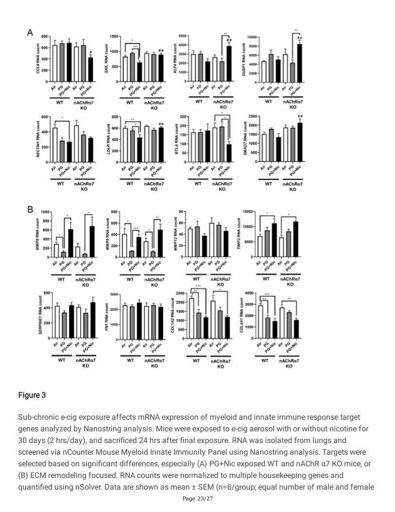
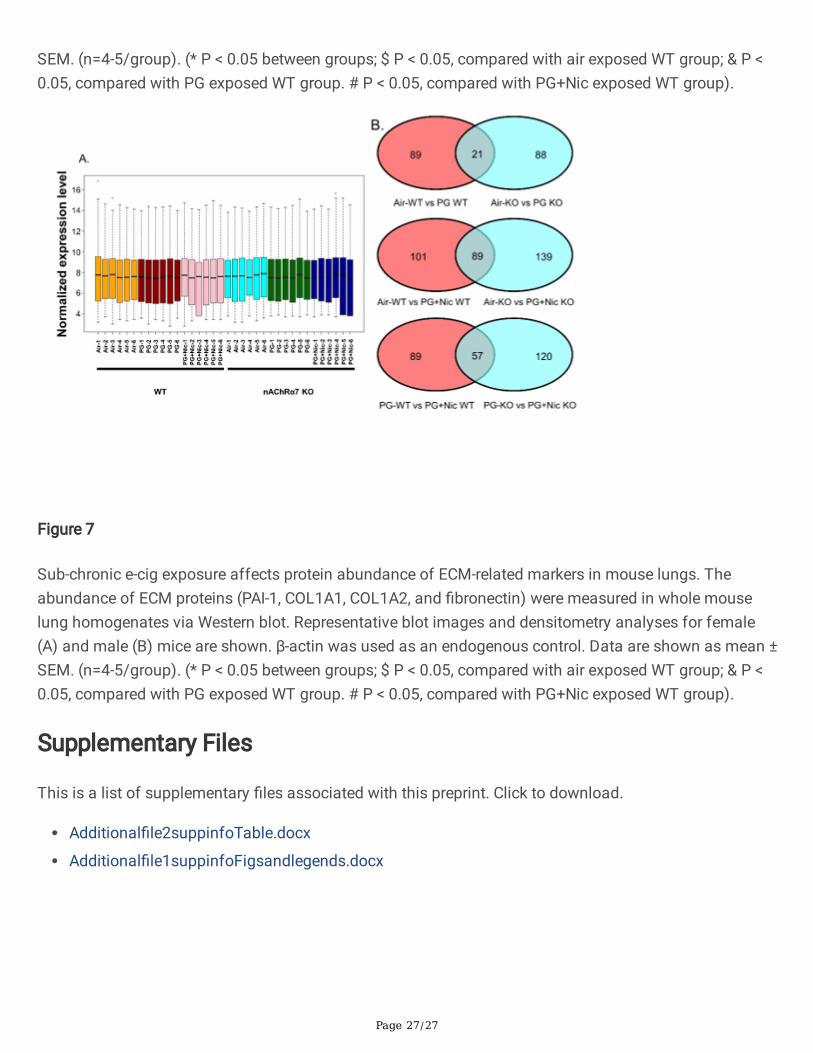
To further determine whether sub-chronic e-cig exposure mediated in�ammatory responses anddysregulated repair/ECM remodeling was caused by altered expression of myeloid innate immuneresponse target genes (~734 genes), we performed gene expression analysis using nCounter MouseMyeloid Innate Immunity Panel (Nanostring Technologies, Inc.). The pairwise comparison between PGalone exposed WT and nAChR α7 KO mice, revealed 21 genes were differentially expressed compared torespective air control (Figure 7B). Additionally, pairwise comparison analysis among PG with nicotineexposed WT and nAChR α7 KO mice revealed 89 genes differentially expressed when compared torespective air control (Figure 7B). Furthermore, pairwise comparison between PG with nicotine and PGalone groups in WT and nAChR α7 KO mice revealed 57 genes differentially expressed among them(Figure 7B).
WT mice showed signi�cantly higher RNA counts indicative of increased mRNA transcript levels formultiple targets in PG with or without nicotine exposed groups, compared to air control (Additional File 1:Figure S3). When we compared WT mice exposed to PG alone, air control or PG with nicotine showedaltered gene expression of in�ammatory and ECM remodeling targets such as matrix metalloproteases(MMP9, and MMP8), S100A8, and collagens (COL17A1 and COL14A1) (Additional File 1: Figure S3A).When WT mice exposed to PG with nicotine were compared with air control, we found signi�cantdifference in transcript levels of in�ammatory targets such as ARG1 and LPL (Additional File 1: FigureS3A). PG with nicotine exposure caused signi�cant alterations in the expression of several target genessuch as SMAD7, KLF4, CDH1, COL4A2, ICAM1, LDLR, IL1B, TLR5, NFKBIA, and CXCL2 among WT andnAChR α7 KO mice that belong to the broad categories in�ammation and dysregulated repaired/ECMremodeling (Additional File 1: Figure S3B). Based on the above-mentioned gene expression analysis byNanostring, we choose speci�c target genes for further analysis (Figure. 3).
Based on the Nanostring nCounter analysis, transcript levels of target genes that showed signi�cantdifferences between WT and nAChR α7 KO mice following PG with nicotine exposure were selectivelyrepresented in Figure 3A. The gene expression levels of SKIL (Ski-like protein) and LDLR (Low-densitylipoprotein receptor) were signi�cantly decreased in WT mice exposed to PG with nicotine, but not nAChRα7 KO mice. However, the gene expression levels of CCL9 (Chemokine ligand 9), KLF4 (Kruppel-like factor4), DUSP1 (Dual Speci�city Phosphatase 1), BTLA (B and T Lymphocyte Associated), and SMAD7 (SMADFamily Member 7) were signi�cantly increased only in nAChR α7 KO mice exposed to PG with nicotinecompared to WT mice (Figure 3A). Finally, NECTIN1 gene expression in WT and nAChR α7 KO miceshowed a decreased trend, but only WT mice exposed to PG with nicotine showed a signi�cant reductionin the expression of NECTIN1 transcript compared to air control (Figure 3A).
Page 9/27
Sub-chronic e-cig exposure with nicotine in nAChR α7 de�cient mice prevents dysregulation of p50/p105
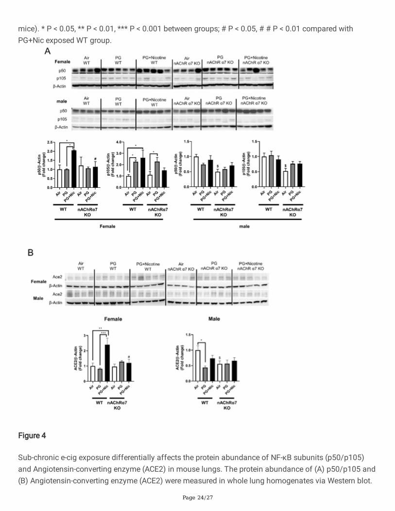
In order to determine the role of target molecules that contribute to e-cig exposure induced in�ammatoryresponse at the protein level, we measured the protein abundance of a NF-κB subunit (nuclear factorkappa-light-chain-enhancer; p50/p105). We have observed that the protein levels of both p50 and p105were upregulated in WT female mice exposed to PG with nicotine, while nAChR α7 KO showedattenuation of p50 and p105 expression in the lungs (Figure 4A). However, there was no signi�cantdifference in male mice exposed to PG with or without nicotine compared to air control in both WT andnAChR α7 KO mice (Figure 4A). WT or nAChR α7 KO female mice exposed to PG alone showed similarupregulation of p105, indicating that PG alone could induce pro-in�ammatory responses in a nAChR α7-independent manner (Figure 4A).
Sub-chronic e-cig with nicotine exposure induced up-regulation of Angiotensin-converting enzyme (ACE2)in mouse lung
Recently, it has been shown that ACE2 (Covid-19 receptor) is a key point in migration of �broblast toinjured locations in lungs, and ACE2 is involved in lung ECM remodeling. We determined the proteinabundance of ACE2 (Figure 4B). We have noticed that PG with nicotine has increased the proteinabundance, whereas nAChR α7 deletion attenuated the dysregulation in female. While in male, there wasno difference between PG with nicotine exposure and air control, PG exposure decreased the ACE2 proteinlevel, and nAChR α7 knockdown lowered the baseline abundance of ACE2 in lungs.
Sub-chronic e-cig exposure with or without nicotine induces dysregulated repair/ECM remodeling in anAChR α7-independent manner
In our previous study, we have identi�ed that acute e-cig exposure causes dysregulated repair/ECMremodeling in mouse lungs [8]. In this study, we were interested to investigate the role of dysregulatedrepair/ECM remodeling in sub-chronic e-cig exposed WT and nAChR α7 KO mice. We were speci�callyinterested in selected MMPs and ECM remodeling markers to determine their effects following sub-chronic e-cig exposure both at the gene expression level (Figures 3B), and protein abundance (Figure 5-6).
PG exposed WT and nAChR α7 KO mice showed decreased expression of MMP8 and MMP9 compared toair control or PG with nicotine group (Figure 3B). WT and nAChR α7 KO mice when exposed to PG withnicotine showed signi�cant increase in TIMP3, which is an inhibitor of MMPs (Figure 3B). ECM targetgenes �bronectin (FN1) or Plasminogen activator inhibitor-1 (PAI-1, gene code: SERPINE1) remainunaffected, while genes that modulate collagen expression (COL1A2 and COL4A1) was signi�cantdecreases in PG with or without nicotine exposed WT and nAChR α7 KO mice.
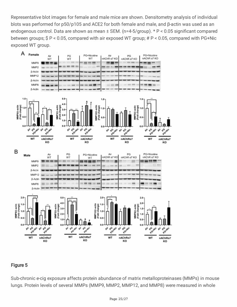
Based on the gene expression changes in dysregulated repair markers, we measured the proteinabundances of the same in e-cig exposed WT and nAChR α7 KO mice. Results from immunoblot analysiscorrelated with gene expression of MMP9 and MMP8 in both female and male mice (Figure 5A-B).Decreased MMP8 and MMP9 were also observed in PG-exposed WT and nAChR α7 KO mice compared to
Page 10/27
PG with nicotine and air control (Figure 5A-B). MMP2 protein abundance was increased in WT male andfemale mice exposed to PG alone, compared to air control and PG with nicotine-exposed mice. Moreover,nAChR α7 KO female mice show increased MMP2 protein abundance at baseline, while no change wasobserved in male mice (Figure 5A-B). The protein levels of MMP9, MMP2, and MMP12 were increased inWT male mice exposed to PG with nicotine, while nAChR α7 KO inhibits protein abundance of MMPs(Figure 5B). MMP8 protein levels were comparable in female and male mice and nAChR α7 KO showeddecreased protein abundance of MMP8 at baseline (Figure 5A-B).
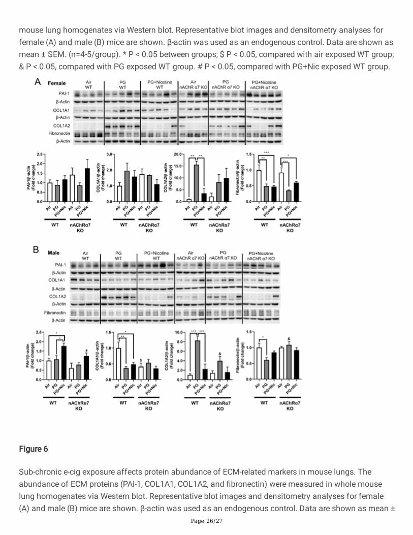
ECM proteins/regulator such as collagens, �bronectin, and PAI-1, at the protein levels were measured inWT and nAChR α7 KO mouse lungs (Figure 6). The protein abundance of PAI-1 showed no differenceamong air, PG alone and PG with nicotine exposed WT and nAChR α7 KO female mice (Figure 6A).Whereas, PAI-1 was increased in WT male mice exposed to PG with nicotine, but was reduced in nAChRα7 KO male mice (Figure 6B). Furthermore, two different subunits of type 1 collagens (COL1A1 andCOL1A2) were differentially expressed at the protein level among the experimental groups. WT andnAChR α7 KO female and male mice showed increased expression of COL1A2 when exposed to PG alone(Figure 6). However, COL1A1 protein abundance was reduced in WT male mice exposed to PG with orwithout nicotine, and nAChR α7 de�ciency further decreased the baseline of COL1A1 levels in male mice.There is no change in the protein levels of COL1A1 among PG alone and PG with nicotine exposure in WTand nAChR α7 KO mice. PG with or without nicotine exposure reduced the protein abundance of�bronectin in WT female and nAChR α7 KO mice compared to air control (Figure 6A). However, decreased�bronectin protein level was observed in the PG-exposed WT male mice, and there was no alteration innAChR α7 KO male mice among all the different exposure conditions (Figure 6B).
Sub-chronic e-cig exposure in lung epithelial cell-speci�c nAChR α7 deletion protects againstin�ammation
Considering the in�ammatory responses seen in WT and nAChR α7 KO mice, we exposed nAChR α7 lungepithelial cell-speci�c KO mice to e-cig with or without nicotine to determine the role of nAChR α7 in thelung epithelium. Surprisingly, we did not see any signi�cant changes in pro-in�ammatory cytokinesfollowing PG with nicotine exposure in nAChR α7 epithelial cell-speci�c KO mice. However, IL-5, MCP-1,KC, Eotaxin, GM-CSF, and G-CSF levels were signi�cantly increased in PG alone group when compared toair control, and these cytokines were inhibited in nAChR α7 epithelial cell-speci�c KO mice (Additional File1: Figure S4A). The rest of the cytokines from BALF showed no changes among all the different exposuregroups in nAChR α7 epithelial cell-speci�c KO mice (Additional File 1: Figure S4B-C).
DiscussionThe popularity of e-cig vaping has been rising recently in the United States, and hence the healthconcerns about vaping have recently attracted public attention [27]. Our previous studies have shown thatacute exposure to e-cigarettes could cause in�ammatory responses, oxidative stress, and ECMremodeling [8, 11, 28]. Although in past, we have shown the health risks caused by acute e-cig exposures
Page 11/27
in mice [8, 11], sub-chronic or chronic effects of e-cig exposure-induced toxicity and respiratory healtheffects remain elusive. Based on our knowledge, no study has yet shown that long-term exposure of e-cigaerosol can result in persistent dysregulated repair/ECM remodeling and in�ammatory responsesimplicating the role of nAChR α7 receptor in the lung.
Similar to our acute exposure results, e-cig aerosol with nicotine induced in�ammatory cell counts inBALF compared to air control, especially increase in macrophages and CD4a+/CD8a+ T-lymphocytes.Previous report has demonstrated that two weeks of e-cig (with nicotine) exposure increased the numberof macrophages but not neutrophils, which supports our �ndings [29]. Another study reported that acuteexposure of e-cig aerosol increased total cell and macrophage counts; even the vehicle exposure aloneshowed a trend towards increase in total cells and macrophages [30]. Based on our results, e-cigexposure with or without nicotine causes increased in�ammatory cellular in�ux into the lungs (BALF). PGalone (vehicle) exposure was considered safe by e-cig users, but our data reveals that sub-chronic e-cigexposure with PG (without nicotine) induced lung in�ammation. The genetic ablation of nAChR α7protects against e-cig exposure induced increased in macrophages and CD4a+/CD8a+ T-lymphocytes.Probably, e-cig exposure containing PG with nicotine causes lung in�ammation/remodeling response viathe nAChR α7 receptor mediated signaling pathway, and blocking the receptor could attenuate nicotine-dependent effects in the lungs. Prior reports show that activation of nAChR can also inhibit in�ammation[31], and we have observed increased neutrophils and CD4a+/CD8a+ T-lymphocytes when exposed to PGalone in nAChR α7 KO mice. The nAChR deletion may promote in�ammation independent of nicotineexposure in the lungs. The chemical derived from e-cig aerosols containing PG, such as formaldehyde,acetaldehyde, and methylglyoxal, in nicotine-free aerosol [32], may result in increased in�ammation. Asreported previously, both e-cig with or without nicotine caused in�ltration of in�ammatory cells and pro-in�ammatory mediators that are key players for causing lung in�ammatory response [8, 11, 30]. As anagonist of nAChR α7, nicotine might be pose both pro-in�ammatory and anti-in�ammatory roles, andfurther research is needed to study the relationship between nicotine and nicotinic receptors in the contextof sub-chronic e-cig exposure.
Apart from the in�ammatory cells, some pro-in�ammatory mediators in BALF were signi�cantly increasedfollowing e-cig exposure with nicotine. Speci�c macrophage-mediated pro-in�ammatory cytokines suchas IL-1α, MCP-1, and GM-CSF, were increased in BALF after PG with nicotine exposure in a nAChR α7-dependent manner. However, a few other macrophage-driven cytokines, IL-1β, TNF-α, and MIP-1β, doesnot show nAChR α7 dependency. Our results paralleled reports that e-cig aerosol containing nicotineinduced cytotoxicity in alveolar macrophage and signi�cant release of in�ammatory mediators (TNF-α,CXCL-8, MCP-1, and IL-6) [33]. Additional studies have shown speci�c T-lymphocytes-relatedcytokines/chemokines (IL-2, IL-9, IFN-γ, and RANTES) and their role in in�ammatory signaling [34-37], andour results indicated that these altered cytokines response were nAChR-dependent in e-cig exposedmouse lungs. Mice exposed to PG with nicotine show increased IL-5 release in a nAChR independentmanner. Another report shows increased IL-4, IL-5, and IL-13 levels following e-cig containing nicotinewhen challenged to ovalbumin treated mice resulting in exacerbation of allergic airway in�ammation [38].
Page 12/27
The increased IFN-γ production has been related to cytotoxicity and in�ltration of CD8a+ T-cells [39, 40].These altered pro-in�ammatory cytokines response and cellular in�ux in the lung are inter-connected toeach other partly due to nAChR α7 mediated signaling. It is possible that nAChR α7 deletion couldattenuate the in�ammatory responses induced by e-cig aerosol contained nicotine in the lungs. However,PG-induced increase in CD4a+/CD8a+ T-lymphocytes does not corroborate with the cytokine levels. Futurestudies will address immune cell-type speci�c role in pulmonary toxicity of e-cig with and without nicotineduring long-term exposure in mice.
We measured the mRNA expression using the mouse myeloid innate immunity panel from Nanostringtechnology to screen the potentially affected target genes. Our results showed altered expression of SKILand LDLR in PG with nicotine-exposed WT mice, but the same genes remain unaffected in nAChR α7 KOexposed groups. These target genes have been shown to play vital role in TGFβ/SMAD canonicalsignaling and TGFβ-induced epithelial–mesenchymal transition (EMT) may be inhibited by SKIL bindingto SMAD [41, 42]In this study, we found increased protein abundance of PAI-1 in WT mice exposed to PGwith nicotine, but PAI-1 was further reduced in nAChR α7 KO mice. Our results suggest that e-cig exposurewith PG nicotine may affect SKIL-TGFβ-PAI-1 axis in the lung possibly through nAChR α7 activation. Theother important target signi�cantly altered by PG with nicotine is LDLR gene. Overexpression of LDLR iscapable of promoting macrophage differentiation and exaggerated in�ammatory response and silencingLDLR results in reduced in�ammatory responses in THP-1 cells [43]. In our study, LDLR was reduced inPG with nicotine exposed WT mice, while LDLR transcript levels remain unchanged in both PG with andwithout nicotine exposed nAChR α7 KO groups. These �ndings suggest that nAChR α7 may be animportant gateway that affects speci�c in�ammatory response/ECM remodeling target genes during sub-chronic e-cig exposure with nicotine.
To further study the in�ammatory responses, we measured the protein abundance of NF-κB (p50/p105)in both female and male mice. The activation of NF-κB is well documented in different lung injury models[44] including cigarette smoke-induced in�ammation, wheras research on e-cig aerosol induced NF-κBactivation remain less explored [45-48]. Although, the anti-in�ammatory effects (i.e. inhibition of NF-κBsignaling) via activation of nAChRs is well-known and has been studied previously, nicotine has beenshown induce in�ammation through activation of the NF-κB signaling pathway [31, 49]. Our �ndingssuggest that e-cig aerosol containing nicotine increased p50 and p105 protein abundance in female micein a nAChR α7-dependent manner. We believe nicotine is a potent inducer of altered lung in�ammation viaactivated nAChR α7 compared to the anti-in�ammatory effects regulated by nAChR-related signaling. Wefound lack of nAChR α7 showing attenuation of in�ammatory response following e-cig exposure withnicotine. However, nicotine-free e-cig exposure induced p105 protein levels, which suggest this subunitmay act through an alternative pathway that may be independent of nAChR α7 signaling. We for the �rsttime show that lung in�ammation caused by e-cig aerosol containing nicotine is nAChR α7-dependent,preferentially in a sex-speci�c manner. However, more detailed studies are needed to further illustrate theexact mechanism by which sex-differences in lung in�ammatory responses occurs following e-cigexposure through cell-type speci�c nAChR α7-mediated signaling.
Page 13/27
As we recently reported, acute exposure of e-cig aerosol with or without nicotine resulted in dysregulatedrepair and ECM remodeling [8]. We noticed similar effects in our sub-chronic exposure model as well. E-cig aerosol with or without nicotine alters MMPs and ECM remodeling proteins in a sex-dependentmanner, which are nAChR α7 receptor independent. It is known that nicotine exposure causes harmfulrespiratory effects, such as airway remodeling and airspace enlargement [50, 51], which is a key featureof emphysema and COPD [52-54]. However, dysregulation of MMPs serves not only as a factor for ECMremodeling, but also as a marker of in�ammation mediated by dysregulated macrophages. Based on ourresults, nicotine-free e-cig aerosol induced augmented MMPs dysregulation than e-cig aerosol withnicotine. Increased MMP2/MMP12, along with decreased MMP9/MMP8, might suggest a compensatoryfeedback loop [55]. We observed that ACE2 is induced by e-cig with nicotine exposure, whereas the levelsis decreased in nAChR α7-de�cient micesuggesting that ACE2 upregulation by nicotine is mediated bynAChR α7. It is thought that ACE2 is upregulated by smoking, as well as its expression level is increasedin smokers and COPD patients [56, 57], as well as served as the entry gate for the novel coronavirus(SARS-CoV-2) [58]. Hence, e-cig with nicotine inhalation would promote SARS-CoV-2 infection, and nAChRα7 deletion and downregulation of ACE2 may have some rami�cations on the virus infection.
One of the major substrates of MMPs is ECM related proteins. From our previous study, mice exposed toe-cig aerosols for 3 days showed dysregulated repair/ECM remodeling both at the mRNA and protein levelin a sex-dependent manner [8]. The current study shows that sub-chronic e-cig exposure with or withoutnicotine affects ECM remodeling to a certain extent dependent on nAChR α7 with some sex-speci�cphenotypes. We have observed increased PAI-1 only in WT male mice exposed to PG with nicotine in anAChR α7-dependent manner, and PAI-1 is a primary ECM regulator and pro-�brotic marker [59].Therefore, increased PAI-1 following inhalation of e-cig aerosol with nicotine indicates that e-cig vaporcontaining nicotine could increase risk of chronic �brotic disease, implicating crucial role of nAChR α7-related signaling. Some other ECM proteins, such as type 1 collagen and �bronectin were altered afterexposure of e-cig aerosol with or without nicotine. ECM proteins affected by e-cig exposure areassociated with dysregulated wound healing and �brotic disease [60]. The observed increase in COL1A2following e-cig with PG alone exposure in both male and female mice demonstrates the signi�cant healthrisk imposed by PG alone, in addition to the risks associated with PG nicotine. As mentioned above,nicotine might not be the only target that can activate nAChR α7, and PG alone via other indirectlymechanisms could activate nicotinic receptors, which may be in agreement with our previous study [8,19]. It is well known that collagen and �bronectin are required when the wound healing process happensand delayed wound healing were observed when epithelial cells were unable to synthesize �bronectin[61]. We found down-regulation of collagen and �bronectin following e-cig exposure, which will makelungs more vulnerable later in life when exposed to other noxious gases and environmental toxicants.Further studies are required to understand the mechanisms underlying how e-cig exposures may inducedamaging responses, leading to lung disease and injury.
Based on our �ndings, long-term studies are needed to provide further insights on how nAChRα7dependency plays an important cell-type speci�c role in the lung (epithelial and �broblast) during e-cigexposure, and its involvement in ACE2 regulation. We know that several factors, such as duration of
Page 14/27
exposure, route of exposure, dose, e-liquid composition, setting used for delivering the e-cig aerosols,concentration of PG mixture and nicotine, including mouse strain, background, sex of mice may equallycontribute to the difference and phenotype observed in the measure outcomes. Chronic e-cig exposure areunderway that will help us to understand how these signaling mechanisms altered by e-cig exposure withand without nicotine may contribute to the respiratory toxicity observed in mice could translate to clinicalrelevance in human e-cig users. Ongoing human studies are being carried out to identify potential targetsand toxicity biomarkers that may relates to human respiratory health effects of e-cigarettes.
ConclusionIn conclusion, nAChR α7 ablation attenuates the in�ammation induced by sub-chronic e-cig exposuremay be partly due to e-cig induced ECM remodeling/dysregulated repair response in the lung. In a sub-chronic e-cig exposure, nAChR α7 plays a novel role in an anti-in�ammatory response induced bynicotine. We show a signi�cant difference in terms of altered genes that belong to in�ammation,dysregulated repair and ECM remodeling in WT mice compared to nAChR α7 KO mice when exposed toPG with nicotine. The differential in�ammatory cell counts and pro-in�ammatory cytokines in BALF werecomparable to our previous �ndings. Hence, nAChR α7 may play a vital role in the in�ammatoryresponses induced by nicotine-containing e-cig aerosols. Our data suggest that ECMremodeling/dysregulated repair caused by e-cig exposure is mediated by nAChR α7, and occurs in a sex-dependent manner. Overall, e-cig aerosol with nicotine causes lung in�ammation due to altereddysregulated repair response and ECM remodeling mediated by nAChR α7. Deletion of nAChR α7 maypossibly protect against lung in�ammation and injury by attenuating associated signaling targets forECM remodeling. Hence, nAChR α7 is considered a valid target for in�ammation induced by e-cig aerosolwith nicotine in the lung.
DeclarationsEthical approval
This study was performed according to the standards from the United States Animal Welfare Act,National Institutes of Health (NIH). All the animal experiments were followed the protocol approved byThe Animal Research Committee of the University of Rochester (UCAR).
Human participants, human data and human tissue: Not applicable
Human ethical approval: Not applicable
Consent for publication: Not applicable.
Competing interests
The authors have declared that no competing interest.
Page 15/27
Availability of data and material: All data and materials are described in the manuscript.
Funding
This study was supported by the National Institutes of Health (NIH) 1R01HL135613 andNIH 1R01HL085613. The funding body has no role in design of the study, data collection, analysis, andinterpretation of data and in writing the manuscript.
Competing interests: None
Author Contributions
QW, IKS, IR conceived and designed the experiments. QW, DL, JHL, TM, SRM conducted the experiments.QW, IKS, DL, TM, SRM analyzed the data. QW, IKS, SRM, IR wrote and revised the manuscript.
Acknowledgments
The authors thank Dr. Ye, Dongxia, for technical help of running ELISA/Luminex assays.
AbbreviationsE-cig: Electronic cigarette
ECM: extracellular matrix
nAChRs: Nicotinic Acetylcholine Receptors
WT: wild-type
nAChRα7 KO: nAChRα7 knockout
nAChRα7 CreCC10: lung epithelial-cell-speci�c nAChRα7 KO
PG: propylene glycol
VG: vegetable glycerin
BALF: Bronchoalveolar lavage �uids
MMPs: matrix metalloproteinases
IPF: idiopathic pulmonary �brosis
COPD: chronic obstructive pulmonary disease
CNS: central nervous system
Page 16/27
LPSs: lipopolysaccharides
Glu: glucose
Hb: hemoglobin
Hct: hematocrit
AO/PI: acridine orange propidium iodide
CCL9: Chemokine ligand 9
KLF4: Kruppel-like factor 4
DUSP1: Dual Speci�city Phosphatase 1
BTLA: B and T Lymphocyte Associated
SMAD7: SMAD Family Member 7
NF-κB: nuclear factor kappa-light-chain-enhancer of activated B cells
PAI-1: Plasminogen activator inhibitor-1
COL1A1: type 1 collagens chain 1
COL1A2: type 1 collagens chain 2
ACE2: Angiotensin-converting enzyme 2
SARS-CoV2: Severe acute respiratory syndrome coronavirus 2
References1. Farsalinos KE, Polosa R: Safety evaluation and risk assessment of electronic cigarettes as tobacco
cigarette substitutes: a systematic review.Ther Adv Drug Saf 2014, 5:67-86.
2. Javed F, Kellesarian SV, Sundar IK, Romanos GE, Rahman I: Recent updates on electronic cigaretteaerosol and inhaled nicotine effects on periodontal and pulmonary tissues.Oral Dis 2017, 23:1052-1057.
3. Kaur G, Muthumalage T, Rahman I: Mechanisms of toxicity and biomarkers of �avoring and �avorenhancing chemicals in emerging tobacco and non-tobacco products.Toxicol Lett 2018, 288:143-155.
4. Lawyer GR, Jackson M, Prinz M, Lamb T, Wang Q, Muthumalage T, Rahman I: Classi�cation of�avors in cigarillos and little cigars and their variable cellular and acellular oxidative and cytotoxicresponses.PLOS ONE 2019, 14:e0226066.
Page 17/27
5. Madison MC, Landers CT, Gu B-H, Chang C-Y, Tung H-Y, You R, Hong MJ, Baghaei N, Song L-Z, PorterPJTJoci: Electronic cigarettes disrupt lung lipid homeostasis and innate immunity independent ofnicotine. 2019, 129.
�. Goniewicz ML, Kuma T, Gawron M, Knysak J, Kosmider L: Nicotine Levels in ElectronicCigarettes.Nicotine Tobacco Res 2012, 15:158-166.
7. Grana RA, Popova L, Ling PM: A Longitudinal Analysis of Electronic Cigarette Use and SmokingCessationElectronic Cigarette Use and Smoking CessationLetters.JAMA Internal Medicine 2014,174:812-813.
�. Wang Q, Ahmad Khan N, Muthumalage T, Lawyer GR, McDonough SR, Chuang T-D, Gong M, SundarIK, Rehan VK, Rahman I: Dysregulated repair and in�ammatory responses by e-cigarette-derivedinhaled nicotine and humectant propylene glycol in a sex-dependent manner in mouse lung.FASEBBioAdvances 2019, 1:609-623.
9. Burgstaller G, Oehrle B, Gerckens M, White ES, Schiller HB, Eickelberg O: The instructive extracellularmatrix of the lung: basic composition and alterations in chronic lung disease.Eur Respir J 2017, 50.
10. Wight TN, Frevert CW, Debley JS, Reeves SR, Parks WC, Ziegler SF: Interplay of extracellular matrixand leukocytes in lung in�ammation.Cell Immunol 2017, 312:1-14.
11. Lerner CA, Sundar IK, Yao H, Gerloff J, Ossip DJ, McIntosh S, Robinson R, Rahman I: Vaporsproduced by electronic cigarettes and e-juices with �avorings induce toxicity, oxidative stress, andin�ammatory response in lung epithelial cells and in mouse lung.PLoS One 2015, 10:e0116732.
12. Wu Q, Jiang D, Minor M, Chu HW: Electronic cigarette liquid increases in�ammation and virusinfection in primary human airway epithelial cells.PLoS One 2014, 9:e108342.
13. Zia S, Ndoye A, Nguyen VT, Grando SA: Nicotine enhances expression of the alpha 3, alpha 4, alpha5, and alpha 7 nicotinic receptors modulating calcium metabolism and regulating adhesion andmotility of respiratory epithelial cells.Res Commun Mol Pathol Pharmacol 1997, 97:243-262.
14. Jensen K, Nizamutdinov D, Guerrier M, Afroze S, Dostal D, Glaser S: General mechanisms of nicotine-induced �brogenesis.FASEB J 2012, 26:4778-4787.
15. Dani JA, De Biasi MJPB, Behavior: Cellular mechanisms of nicotine addiction. 2001, 70:439-446.
1�. Benowitz NLJNEJoM: Nicotine addiction. 2010, 362:2295-2303.
17. Mabley J, Gordon S, Pacher PJI: Nicotine exerts an anti-in�ammatory effect in a murine model ofacute lung injury. 2011, 34:231-237.
1�. Su X, Matthay MA, Malik ABJTjoi: Requisite role of the cholinergic α7 nicotinic acetylcholine receptorpathway in suppressing gram-negative sepsis-induced acute lung in�ammatory injury. 2010,184:401-410.
19. Avelar AJ, Akers AT, Baumgard ZJ, Cooper SY, Casinelli GP, Henderson BJJN: Why �avored vapeproducts may be attractive: Green apple tobacco �avor elicits reward-related behavior, upregulatesnAChRs on VTA dopamine neurons, and alters midbrain dopamine and GABA neuron function. 2019,158:107729.
Page 18/27
20. Henderson BJ, Wall TR, Henley BM, Kim CH, Nichols WA, Moaddel R, Xiao C, Lester HAJJoN: Mentholalone upregulates midbrain nAChRs, alters nAChR subtype stoichiometry, alters dopamine neuron�ring frequency, and prevents nicotine reward. 2016, 36:2957-2974.
21. Henderson BJ, Wall TR, Henley BM, Kim CH, McKinney S, Lester HAJN: Menthol enhances nicotinereward-related behavior by potentiating nicotine-induced changes in nAChR function, nAChRupregulation, and DA neuron excitability. 2017, 42:2285.
22. Jensen RP, Strongin RM, Peyton DHJSr: Solvent chemistry in the electronic cigarette reaction vessel.2017, 7:42549.
23. Khan NA, Sundar IK, Rahman I: Strain- and sex-dependent pulmonary toxicity of waterpipe smoke inmouse.Physiological Reports 2018, 6:e13579.
24. Yao H, Arunachalam G, Hwang JW, Chung S, Sundar IK, Kinnula VL, Crapo JD, Rahman I:Extracellular superoxide dismutase protects against pulmonary emphysema by attenuating oxidativefragmentation of ECM.Proc Natl Acad Sci U S A 2010, 107:15571-15576.
25. Khan NA, Yogeswaran S, Wang Q, Muthumalage T, Sundar IK, Rahman I: Waterpipe smoke and e-cigarette vapor differentially affect circadian molecular clock gene expression in mouse lungs.PLOSONE 2019, 14:e0211645.
2�. Behar RZ, Hua M, Talbot P: Pu�ng topography and nicotine intake of electronic cigarette users.PLoSOne 2015, 10.
27. Tegin G, Mekala HM, Sarai SK, Lippmann S: E-Cigarette Toxicity?South Med J 2018, 111:35-38.
2�. Gerloff J, Sundar IK, Freter R, Sekera ER, Friedman AE, Robinson R, Pagano T, Rahman I:In�ammatory Response and Barrier Dysfunction by Different e-Cigarette Flavoring ChemicalsIdenti�ed by Gas Chromatography-Mass Spectrometry in e-Liquids and e-Vapors on Human LungEpithelial Cells and Fibroblasts.Appl In Vitro Toxicol 2017, 3:28-40.
29. Sussan TE, Gajghate S, Thimmulappa RK, Ma J, Kim JH, Sudini K, Consolini N, Cormier SA, LomnickiS, Hasan F, et al: Exposure to electronic cigarettes impairs pulmonary anti-bacterial and anti-viraldefenses in a mouse model.PLoS One 2015, 10:e0116861.
30. Glynos C, Bibli S-I, Katsaounou P, Pavlidou A, Magkou C, Karavana V, Topouzis S, Kalomenidis I,Zakynthinos S, Papapetropoulos A: Comparison of the effects of e-cigarette vapor with cigarettesmoke on lung function and in�ammation in mice.Am J Physiol Lung Cell Mol Physiol 2018,315:L662-L672.
31. Patel H, McIntire J, Ryan S, Dunah A, Loring R: Anti-in�ammatory effects of astroglial alpha7nicotinic acetylcholine receptors are mediated by inhibition of the NF-kappaB pathway and activationof the Nrf2 pathway.J Neuroin�ammation 2017, 14:192.
32. Uchiyama S, Noguchi M, Sato A, Ishitsuka M, Inaba Y, Kunugita N: Determination of ThermalDecomposition Products Generated from E-cigarettes.Chemical Research in Toxicology 2020.
33. Scott A, Lugg ST, Aldridge K, Lewis KE, Bowden A, Mahida RY, Grudzinska FS, Dosanjh D, Parekh D,Foronjy R, et al: Pro-in�ammatory effects of e-cigarette vapour condensate on human alveolarmacrophages. 2018, 73:1161-1169.
Page 19/27
34. Crawford A, Angelosanto JM, Nadwodny KL, Blackburn SD, Wherry EJ: A role for the chemokineRANTES in regulating CD8 T cell responses during chronic viral infection.PLoS Pathog 2011,7:e1002098.
35. Ross SH, Cantrell DA: Signaling and Function of Interleukin-2 in T Lymphocytes.Annu Rev Immunol2018, 36:411-433.
3�. Li H, Nourbakhsh B, Cullimore M, Zhang GX, Rostami A: IL-9 is important for T-cell activation anddifferentiation in autoimmune in�ammation of the central nervous system.Eur J Immunol 2011,41:2197-2206.
37. De Cunto G, Lunghi B, Bartalesi B, Cavarra E, Fineschi S, Ulivieri C, Lungarella G, Lucattelli M: SevereReduction in Number and Function of Peripheral T Cells Does Not Afford Protection towardEmphysema and Bronchial Remodeling Induced in Mice by Cigarette Smoke.Am J Pathol 2016,186:1814-1824.
3�. Lim HB, Kim SH: Inhallation of e-Cigarette Cartridge Solution Aggravates Allergen-induced AirwayIn�ammation and Hyper-responsiveness in Mice.Toxicol Res 2014, 30:13-18.
39. Bhat P, Leggatt G, Waterhouse N, Frazer IH: Interferon-gamma derived from cytotoxic lymphocytesdirectly enhances their motility and cytotoxicity.Cell Death Dis 2017, 8:e2836.
40. Whitmire JK, Tan JT, Whitton JL: Interferon-gamma acts directly on CD8+ T cells to increase theirabundance during virus infection.J Exp Med 2005, 201:1053-1059.
41. Tecalco-Cruz AC, Rios-Lopez DG, Vazquez-Victorio G, Rosales-Alvarez RE, Macias-Silva M:Transcriptional cofactors Ski and SnoN are major regulators of the TGF-beta/Smad signalingpathway in health and disease.Signal Transduct Target Ther 2018, 3:15.
42. Yang H, Zhan L, Yang T, Wang L, Li C, Zhao J, Lei Z, Li X, Zhang HT: Ski prevents TGF-beta-inducedEMT and cell invasion by repressing SMAD-dependent signaling in non-small cell lung cancer.OncolRep 2015, 34:87-94.
43. Ricci C, Ruscica M, Camera M, Rossetti L, Macchi C, Colciago A, Zanotti I, Lupo MG, Adorni MP, CiceroAFG, et al: PCSK9 induces a pro-in�ammatory response in macrophages.Scienti�c Reports 2018,8:2267.
44. Wang J, Liu Y-T, Xiao L, Zhu L, Wang Q, Yan T: Anti-in�ammatory effects of apigenin inlipopolysaccharide-induced in�ammatory in acute lung injury by suppressing COX-2 and NF-kBpathway.In�ammation 2014, 37:2085-2090.
45. Zhang C, Qin S, Qin L, Liu L, Sun W, Li X, Li N, Wu R, Wang X: Cigarette smoke extract-induced p120-mediated NF-κB activation in human epithelial cells is dependent on the RhoA/ROCK pathway.SciRep 2016, 6:23131.
4�. Tartell HE: Are E-Cigarettes a Safe Alternative to Smoking? Using Airway Cell Cultures to Assess EarlyIndicators of COPD Progression. In Inquiries Journal/Student Pulse, vol. 7; 2015.
47. Hasnis E, Bar-Shai M, Burbea Z, Reznick AZ: Mechanisms underlying cigarette smoke-induced NF-kappaB activation in human lymphocytes: the role of reactive nitrogen species.J Physiol Pharmacol2007, 58 Suppl 5:275-287.
Page 20/27
4�. Liu X, Togo S, Al-Mugotir M, Kim H, Fang Q, Kobayashi T, Wang X, Mao L, Bitterman P, Rennard S: NF-kappaB mediates the survival of human bronchial epithelial cells exposed to cigarette smokeextract.Respiratory Research 2008, 9:66.
49. Iho S, Tanaka Y, Takauji R, Kobayashi C, Muramatsu I, Iwasaki H, Nakamura K, Sasaki Y, Nakao K,Takahashi T: Nicotine induces human neutrophils to produce IL-8 through the generation ofperoxynitrite and subsequent activation of NF-kappaB.J Leukoc Biol 2003, 74:942-951.
50. Hahn HL, Lang M, Bleicher S, Zwerenz S, Rausch C: Nicotine-induced airway smooth musclecontraction: neural mechanisms involving the airway epithelium. Functional and histologic studies invitro.Clin Investig 1992, 70:252-262.
51. Garcia-Arcos I, Geraghty P, Baumlin N, Campos M, Dabo AJ, Jundi B, Cummins N, Eden E, Grosche A,Salathe M: Chronic electronic cigarette exposure in mice induces features of COPD in a nicotine-dependent manner.Thorax 2016, 71:1119-1129.
52. McDonough JE, Yuan R, Suzuki M, Seyednejad N, Elliott WM, Sanchez PG, Wright AC, Gefter WB,Litzky L, Coxson HO, et al: Small-airway obstruction and emphysema in chronic obstructivepulmonary disease.N Engl J Med 2011, 365:1567-1575.
53. Garcia-Arcos I, Geraghty P, Baumlin N, Campos M, Dabo AJ, Jundi B, Cummins N, Eden E, Grosche A,Salathe M, Foronjy R: Chronic electronic cigarette exposure in mice induces features of COPD in anicotine-dependent manner.Thorax 2016, 71:1119-1129.
54. Shapiro SD: Animal models for COPD.Chest 2000, 117:223S-227S.
55. Kato H, Duarte S, Liu D, Busuttil RW, Coito AJ: Matrix Metalloproteinase-2 (MMP-2) Gene DeletionEnhances MMP-9 Activity, Impairs PARP-1 Degradation, and Exacerbates Hepatic Ischemia andReperfusion Injury in Mice.PLoS One 2015, 10:e0137642.
5�. Leung JM, Yang CX, Tam A, Shaipanich T, Hackett TL, Singhera GK, Dorscheid DR, Sin DD: ACE-2Expression in the Small Airway Epithelia of Smokers and COPD Patients: Implications for COVID-19.Eur Respir J 2020.
57. Brake SJ, Barnsley K, Lu W, McAlinden KD, Eapen MS, Sohal SS: Smoking Upregulates Angiotensin-Converting Enzyme-2 Receptor: A Potential Adhesion Site for Novel Coronavirus SARS-CoV-2 (Covid-19).J Clin Med 2020, 9.
5�. Hoffmann M, Kleine-Weber H, Schroeder S, Kruger N, Herrler T, Erichsen S, Schiergens TS, Herrler G,Wu NH, Nitsche A, et al: SARS-CoV-2 Cell Entry Depends on ACE2 and TMPRSS2 and Is Blocked by aClinically Proven Protease Inhibitor.Cell 2020, 181:271-280.e278.
59. Rabieian R, Boshtam M, Zareei M, Kouhpayeh S, Masoudifar A, Mirzaei H: Plasminogen ActivatorInhibitor Type-1 as a Regulator of Fibrosis.J Cell Biochem 2018, 119:17-27.
�0. Xue M, Jackson CJ: Extracellular Matrix Reorganization During Wound Healing and Its Impact onAbnormal Scarring.Adv Wound Care (New Rochelle) 2015, 4:119-136.
�1. Kicic A, Hallstrand TS, Sutanto EN, Stevens PT, Kobor MS, Taplin C, Pare PD, Beyer RP, Stick SM,Knight DA: Decreased �bronectin production signi�cantly contributes to dysregulated repair ofasthmatic epithelium.Am J Respir Crit Care Med 2010, 181:889-898.
Page 21/27
Figures
Figure 1
Sub-chronic e-cig exposure augments in�ammatory cell in�ux in BALF. Differential in�ammatory cellcounts were measured in BALF from mice exposed to air, PG or PG with nicotine (PG+Nic) for 30 days (2hrs/day). (A) Total in�ammatory cell counts were analyzed by Cellometer using AO/PI staining.Differential in�ammatory cell counts were determined as percentages by �ow cytometry. Absolute cellcounts for (B) F4/80+ macrophages, (C) Ly6B.2+ neutrophils, (D) CD4a+ T-lymphocytes, and (E) CD8a+ T-lymphocytes were normalized to the total cell counts. Data are shown as mean ± SEM (n=4-6/group;equal number of male and female mice). * P < 0.05, ** P < 0.01 between groups; & P < 0.05, compared toPG exposed WT group; # P < 0.05, compared to PG+Nic exposed WT group.
Page 22/27
Figure 2
Sub-chronic e-cig exposure-induced pro-in�ammatory mediators in BALF. Bio-Plex Pro mouse cytokine 23-plex assay kit (Bio-Rad) was used to determined levels of pro-in�ammatory cytokines/chemokines inBALF from mice exposed to e-cig with or without nicotine for 30 days (2 hrs/day). Signi�cant changeswere found in cytokines related to macrophages (IL-1α, MCP-1, TNF-α, GM-CSF, and MIP-1β) and T-lymphocytes (IL-2, IL-5, IL-9, RANTES, and IFN-γ). Data are shown as mean ± SEM (n=6-10/group; equalnumber of male and female mice). * P < 0.05, ** P < 0.01, *** P < 0.001, compared to air control; # P <0.05, compared to PG+Nic exposed WT mice.
Page 23/27
Figure 3
Sub-chronic e-cig exposure affects mRNA expression of myeloid and innate immune response targetgenes analyzed by Nanostring analysis. Mice were exposed to e-cig aerosol with or without nicotine for30 days (2 hrs/day), and sacri�ced 24 hrs after �nal exposure. RNA was isolated from lungs andscreened via nCounter Mouse Myeloid Innate Immunity Panel using Nanostring analysis. Targets wereselected based on signi�cant differences, especially (A) PG+Nic exposed WT and nAChR α7 KO mice, or(B) ECM remodeling focused. RNA counts were normalized to multiple housekeeping genes andquanti�ed using nSolver. Data are shown as mean ± SEM (n=6/group; equal number of male and female
Page 24/27
mice). * P < 0.05, ** P < 0.01, *** P < 0.001 between groups; # P < 0.05, # # P < 0.01 compared withPG+Nic exposed WT group.
Figure 4
Sub-chronic e-cig exposure differentially affects the protein abundance of NF-κB subunits (p50/p105)and Angiotensin-converting enzyme (ACE2) in mouse lungs. The protein abundance of (A) p50/p105 and(B) Angiotensin-converting enzyme (ACE2) were measured in whole lung homogenates via Western blot.
Page 25/27
Representative blot images for female and male mice are shown. Densitometry analysis of individualblots was performed for p50/p105 and ACE2 for both female and male, and β-actin was used as anendogenous control. Data are shown as mean ± SEM. (n=4-5/group). * P < 0.05 signi�cant comparedbetween groups; $ P < 0.05, compared with air exposed WT group; # P < 0.05, compared with PG+Nicexposed WT group.
Figure 5
Sub-chronic e-cig exposure affects protein abundance of matrix metalloproteinases (MMPs) in mouselungs. Protein levels of several MMPs (MMP9, MMP2, MMP12, and MMP8) were measured in whole
Page 26/27
mouse lung homogenates via Western blot. Representative blot images and densitometry analyses forfemale (A) and male (B) mice are shown. β-actin was used as an endogenous control. Data are shown asmean ± SEM. (n=4-5/group). * P < 0.05 between groups; $ P < 0.05, compared with air exposed WT group;& P < 0.05, compared with PG exposed WT group. # P < 0.05, compared with PG+Nic exposed WT group.
Figure 6
Sub-chronic e-cig exposure affects protein abundance of ECM-related markers in mouse lungs. Theabundance of ECM proteins (PAI-1, COL1A1, COL1A2, and �bronectin) were measured in whole mouselung homogenates via Western blot. Representative blot images and densitometry analyses for female(A) and male (B) mice are shown. β-actin was used as an endogenous control. Data are shown as mean ±
Page 27/27
SEM. (n=4-5/group). (* P < 0.05 between groups; $ P < 0.05, compared with air exposed WT group; & P <0.05, compared with PG exposed WT group. # P < 0.05, compared with PG+Nic exposed WT group).
Figure 7
Sub-chronic e-cig exposure affects protein abundance of ECM-related markers in mouse lungs. Theabundance of ECM proteins (PAI-1, COL1A1, COL1A2, and �bronectin) were measured in whole mouselung homogenates via Western blot. Representative blot images and densitometry analyses for female(A) and male (B) mice are shown. β-actin was used as an endogenous control. Data are shown as mean ±SEM. (n=4-5/group). (* P < 0.05 between groups; $ P < 0.05, compared with air exposed WT group; & P <0.05, compared with PG exposed WT group. # P < 0.05, compared with PG+Nic exposed WT group).
Supplementary Files
This is a list of supplementary �les associated with this preprint. Click to download.
Additional�le2suppinfoTable.docx
Additional�le1suppinfoFigsandlegends.docx