Original Article Repair of intervertebral disc ...ijcem.com/files/ijcem0048059.pdf · role in the...
11
Int J Clin Exp Med 2017;10(5):7457-7467 www.ijcem.com /ISSN:1940-5901/IJCEM0048059 Original Article Repair of intervertebral disc degeneration using KLD-12/TGF-β1 fiber gel combined with BMSCs in a rabbit model Xiangchen Li, Hui Shao, Bin Wu, Yang Yu, Yan Du, Shaozheng Liu, Jianhua Sun Department of Orthopedics, The First Affiliated Hospital, Shihezi University College of Medicine, Shihezi, China Received January 4, 2017; Accepted January 27, 2017; Epub May 15, 2017; Published May 30, 2017 Abstract: The purpose of this study was to examine the repairing effect of KLD-12 polypeptide/TGF-β1 nanofiber gel combined with BMSCs in a rabbit intervertebral disc degeneration model. The rabbit models were divided into four groups after 2 weeks: KLD-12 polypeptide/TGF-β/BMSCs gel (A), normal saline (B), KLD-12 polypeptide/BMSCs gel (C), and no treatment (D). X-ray examination revealed that the %DHI (disc height index) of group A increased from 4 weeks to 24 weeks and that these values were significantly higher than those of groups B and C (P<0.05). A similar trend was observed for the intensity of MRI T2-weighted signal. We observed newborn nucleus pulposus tissue in group A, grayish yellow fibrous hyperplasia in group B, white fibrous hyperplasia in group C; and translucent jelly-like tissue in group D. HE staining scores for groups A, B, C, and D were at levels 1-2, 4-5, 3-4, and 0, respectively. The nucleus pulposus cells in groups A and D were deep dyed with toluidine blue. The gray values for aggrecan and col- lagen II of group A showed slight increases from 4 weeks to 24 weeks, and these values were significantly higher than those of groups B and C (P<0.05). KLD-12 polypeptide/TGF-β1 nanofiber gel combined with BMSCs plays a role in the repair of degenerative intervertebral disc and promotes the regeneration of the nucleus pulposus tissue in the intervertebral disc in rabbits. Keywords: KLD-12 polypeptide, transforming growth factor β1, mesenchymal stem cells, intervertebral disc de- generation Introduction Intervertebral disc degeneration is one of the main causes of low back pain and has a high prevalence [1]. Currently, the main methods of treatment include spinal fusion and nucleus pulposus removal. However, these treatment methods can only alleviate the symptoms, but cannot solve the fundamental problem of in- tervertebral disc degeneration [2]. A previous study showed that the main reason for the degeneration of intervertebral disc is the reduc- tion of the water content in the extracellular matrix of the nucleus pulposus and nucleus pulposus cells and reduced amounts of extra- cellular matrix components such as protein polysaccharide and type II collagen [3]. There is no effective way to treat early stage degene- ration of intervertebral discs and the residual cavity after the filling of the nucleus pulposus. Increased numbers of intervertebral disc cells can promote the formation of nucleus pulposus cells and the extracellular matrix to repair the structure and function of degenerated inter- vertebral discs. Tissue engineering technology in the treatment of intervertebral disc degeneration shows a good application prospect [4-6]. In recent years, tissue engineering has provided new ideas for the treatment of intervertebral disc degeneration and for the filling the residual cavity after nucleus pulposus. The three core elements of tissue engineering are seed cells, biological materials, and growth factors. The simple application of cytokines cannot over- come the decrease in the number of interver- tebral disc cells. BMSCs as seed cells have therefore been used in studies on interverte- bral disc regeneration. BMSCs are relatively primitive mononuclear cells in the bone mar- row, which are characterized by high prolifera- tion and differentiation potential and low im- munity. Yim RL et al. showed that BMSCs are
Transcript of Original Article Repair of intervertebral disc ...ijcem.com/files/ijcem0048059.pdf · role in the...

Int J Clin Exp Med 2017;10(5):7457-7467www.ijcem.com /ISSN:1940-5901/IJCEM0048059
Original ArticleRepair of intervertebral disc degeneration using KLD-12/TGF-β1 fiber gel combined with BMSCs in a rabbit model
Xiangchen Li, Hui Shao, Bin Wu, Yang Yu, Yan Du, Shaozheng Liu, Jianhua Sun
Department of Orthopedics, The First Affiliated Hospital, Shihezi University College of Medicine, Shihezi, China
Received January 4, 2017; Accepted January 27, 2017; Epub May 15, 2017; Published May 30, 2017
Abstract: The purpose of this study was to examine the repairing effect of KLD-12 polypeptide/TGF-β1 nanofiber gel combined with BMSCs in a rabbit intervertebral disc degeneration model. The rabbit models were divided into four groups after 2 weeks: KLD-12 polypeptide/TGF-β/BMSCs gel (A), normal saline (B), KLD-12 polypeptide/BMSCs gel (C), and no treatment (D). X-ray examination revealed that the %DHI (disc height index) of group A increased from 4 weeks to 24 weeks and that these values were significantly higher than those of groups B and C (P<0.05). A similar trend was observed for the intensity of MRI T2-weighted signal. We observed newborn nucleus pulposus tissue in group A, grayish yellow fibrous hyperplasia in group B, white fibrous hyperplasia in group C; and translucent jelly-like tissue in group D. HE staining scores for groups A, B, C, and D were at levels 1-2, 4-5, 3-4, and 0, respectively. The nucleus pulposus cells in groups A and D were deep dyed with toluidine blue. The gray values for aggrecan and col-lagen II of group A showed slight increases from 4 weeks to 24 weeks, and these values were significantly higher than those of groups B and C (P<0.05). KLD-12 polypeptide/TGF-β1 nanofiber gel combined with BMSCs plays a role in the repair of degenerative intervertebral disc and promotes the regeneration of the nucleus pulposus tissue in the intervertebral disc in rabbits.
Keywords: KLD-12 polypeptide, transforming growth factor β1, mesenchymal stem cells, intervertebral disc de-generation
Introduction
Intervertebral disc degeneration is one of the main causes of low back pain and has a high prevalence [1]. Currently, the main methods of treatment include spinal fusion and nucleus pulposus removal. However, these treatment methods can only alleviate the symptoms, but cannot solve the fundamental problem of in- tervertebral disc degeneration [2]. A previous study showed that the main reason for the degeneration of intervertebral disc is the reduc-tion of the water content in the extracellular matrix of the nucleus pulposus and nucleus pulposus cells and reduced amounts of extra-cellular matrix components such as protein polysaccharide and type II collagen [3]. There is no effective way to treat early stage degene- ration of intervertebral discs and the residual cavity after the filling of the nucleus pulposus. Increased numbers of intervertebral disc cells can promote the formation of nucleus pulposus
cells and the extracellular matrix to repair the structure and function of degenerated inter- vertebral discs.
Tissue engineering technology in the treatment of intervertebral disc degeneration shows a good application prospect [4-6]. In recent years, tissue engineering has provided new ideas for the treatment of intervertebral disc degeneration and for the filling the residual cavity after nucleus pulposus. The three core elements of tissue engineering are seed cells, biological materials, and growth factors. The simple application of cytokines cannot over-come the decrease in the number of interver- tebral disc cells. BMSCs as seed cells have therefore been used in studies on interverte-bral disc regeneration. BMSCs are relatively primitive mononuclear cells in the bone mar-row, which are characterized by high prolifera-tion and differentiation potential and low im- munity. Yim RL et al. showed that BMSCs are
Repair effect of KLD-12 polypeptide/TGF-β1 nanofiber gel and BMSCs in rabbits
7458 Int J Clin Exp Med 2017;10(5):7457-7467
safe and effective for intervertebral disc re- generation [7]. Ryan JM et al. confirmed that BMSCs have the potential to self-renew and differentiate into a variety of functional cells, which can escape from recognition by homolo-gous antigens [8]. Wei A et al. demonstrated that human BMSCs in rat intervertebral disk memory live for 6 weeks and express type II collagen and protein polysaccharide [9].
With regard to the biological material, the KLD-12 polypeptide is a novel self-assembling poly-peptide, which has been shown to be able to assemble to form a complete structure of the hydrogel and the nanofiber network structure by itself [10, 11]. Oxalate is another option for the biological materials, but it has poor me- chanical properties and cannot be degraded as a cell scaffold. In a previous study, Sun JH et al. showed that KLD-12 and its degradation products do not induce host immunologic re- jection reactions or hemolytic reactions, do not affect the biological activity of interverte-bral disc seed cells (BMSCs), and have good biocompatibility [11]. Bian Z et al. showed that KLD-12 peptide shows a sustained and slow release of TGF-β1 [12], and the KLD-12 pepti- de complex TGF-β1 may induce the differentia-tion of BMSCs into nucleus pulposus-like cells.
Growth factors such as transforming growth factor (TGF) play an important role in cell growth, differentiation, and secretion. Mem- bers of the TGF-β family, including the TGF- β1, TGF-β2, and TGF-β3 subtypes, which are encoded by different genes with different tis-sue distribution patterns, play a role in differ- ent biological activities in vivo [13]. TGF-β1 is the most widely used growth factor in interver-tebral disc tissue engineering. Previously, Peng et al. showed that the level of TGF-β1 in de- generated intervertebral discs is significantly higher than that in normal intervertebral disc tissues [14]. Steck E found that TGF-β1 could induce BMSCs to synthesize type II collagen, protein polysaccharide, and other matrix com-ponents [15]. Risbud MV et al. showed that TGF-β1 could induce rat BMSCs to differentiate into nucleus pulposus-like cells in sodium algi-nate scaffolds under hypoxic conditions [16].
Currently, the application of tissue engineering for the treatment of intervertebral disc degen-eration is still in the experimental stages. Most importantly, there is no uniform protocol for establishing animal models of intervertebral
disc degeneration. Disc degeneration is mostly induced by fiber ring puncture, and a disc de- generation model can mimic discogenic pain, which has a high correlation with lumbar inter-vertebral disc degeneration [17].
In this study, we used KLD-12 polypeptide/TGF-β1 nanofiber gels combined with BMSCs in rab-bit models of intervertebral disc degeneration. Our study shows the potential for the applica-tion of KLD-12 polypeptide/TGF-β1 nanofiber gels combined with BMSCs in the treatment of intervertebral disc degeneration.
Materials and methods
Animals
Procedures involving animals and their care were conducted in conformity with NIH guide-lines (NIH Pub. No. 85-23, revised 1996) and was approved by the Animal Care and Use Committee of Shihezi University. Thirty-two healthy New Zealand white rabbits (age: 10 months, weight: 3±0.3 kg) were obtained from the Laboratory Animal Center of the School of Medicine, Shihezi University. The animals were divided into groups A, B, C, and D, with each group receiving different treatments.
Materials
KLD-12 polypeptide (AcN-KLDLKLDLKLDL-CN- H2, purity >98%) was purchased from Bio En- gineering Ltd by Share Ltd. (Shanghai), TGF-1 was purchased from PeproTech (USA), RNA reverse transcription Kit was obtained from Thermo (USA), PCR primer and 5X TBE were obtained from Bio Engineering Ltd by Share Ltd. (Shanghai), agarose was obtained from Biowest (Spain), diethylpyrocarbonate (DEPC) was obtained from Sigma-Aldrich (Germany), and ISO alcohol was obtained from Rich Yu Fine Chemical Co., Ltd. (Tianjin).
Isolation, culture, and passage of rabbit BMSCs
The rabbits were anesthetized by injecting chlo-ral hydrate (10%) intravenously in the ear vein, and approximately 4 mL of bone marrow was extracted under sterile conditions from each of the rabbits. Cells were then extracted from the bone marrow samples using the density gradi-ent method. LDMEM containing 10% FBS was used as the culture medium, and the cells were

Repair effect of KLD-12 polypeptide/TGF-β1 nanofiber gel and BMSCs in rabbits
7459 Int J Clin Exp Med 2017;10(5):7457-7467
cultured at 37°C in 5% CO2. Half the medium was replaced with fresh medium after 24 h, and again after 48 h, and the whole medium was replaced every 3 days. When the cells re- ached 80-90% confluency, 0.25% trypsin was added for cell splitting.
KLD-12 polypeptide/TGF-β1/BMSCs
Freeze-dried powder of KLD-12 polypeptide (10 mg) was dissolved in 10% sucrose solution con-taining 5 μL of TGF-β1 (0.01 μg/μL) to prepare a 0.56% polypeptide (w/v) solution. One millili-ter of the cell suspension containing 10 × 106 cells/mL with 0.25% trypsin was prepared. The cell suspension was then mixed with the KLD-12 polypeptide/TGF-β1 solution and then sub-jected to oscillating mixing. The KLD-12 po- lypeptide/TGF-β1/BMSC suspension complex containing a final concentration of 0.5% KLD-
the skin on the left side (Figure 1A). The sub- cutaneous tissue and the muscle were then separated layer-by-layer through the retroperi-toneal approach until the L3-4, L4-5, and L5-6 fiber rings were exposed (Figure 1B). Next, the fiber ring was punctured using a 16G puncture needle and the nucleus pulposus was aspirat-ed (Figure 1C). The incision was then sutured layer by layer and bandaged by sterile dressing. Penicillin was injected intramuscularly after the operation to prevent infection. The dress- ing was changed regularly. The animals were placed in different cages and fed.
Implantation of the KLD-12 polypeptide/TGF-β1/BMSCs
After 2 weeks, the fiber rings were exposed and the graft was injected using a microsyringe. Group A received 40 µL of KLD-12 polypeptide/
Figure 1. A. Incision (between twelve ribs and the crista iliaca). B. Exposed disc space (white indicates the outer annular fibrosus). C. Nucleus pulposus drawn out.
Table 1. Histological grading of the intervertebral discs (AF grading)Grading Histological changes0 NormalI Minor rupture of fiber ringII Medium rupture of fiber ringIII Serious rupture of the fibrous ring and slight changes in histological profileIV Serious damage of fiber ring structureV Fiber ring is not clear
Table 2. Primer sequences for PCRGenes Sequences of primers bpβ-actin Forward TGCTTCTAGGCGGACTGTTA 314
Reverse CGTCACATGGCATCTCACGACollagen II Forward CTGTCCTGTGCGACGACATA 140
Reverse TCCTTTCTGCCCCTTTGGTCAggrecan Forward GCTACGGAGACAAGGATGAGTTC 114
Reverse CGTAAAAGACCTCACCCTCCAT
12 polypeptide and 100 ng/mL TGF-β1 was thus prepared in vitro.
Preparation of the rabbit model of lumbar intervertebral disc degeneration
The rabbits were placed in the right lateral decubitus position, and the skin of the operation area was prepared and disin-fected with iodophor and covered by the towel. A longitudinal incision was made on
Repair effect of KLD-12 polypeptide/TGF-β1 nanofiber gel and BMSCs in rabbits
7460 Int J Clin Exp Med 2017;10(5):7457-7467
TGF-β1/BMSCs gel, group B received only saline (40 μL), group C received 40 µL of the KLD-12 polypeptide/TGF-β1 gel, and group D did not undergo any treatment (control group).
Plain radiographic imaging
The vertebral body and the intervertebral space height were assessed using X-rays at 4,8,12, and 24 weeks after implantation. The intervertebral disc height index (DHI) was cal- culated as described previously by Lu et al. [18]; %DHI indicates the change in the height of the intervertebral disc caused due to the operation (%DHI=postoperative DHI/preopera-tive DHI×100%).
MRI
The animals were subjected to MRI at 4, 8, 12 and 24 weeks after implantation, and the sig-nal of the T2-weighted images was measured.
Macroscopic examination
At 4, 8, 12 and 24 weeks after implantation, two New Zealand white rabbits were killed by
sodium overdose. The lumbar spine was re- moved and fixed using 4% polyformaldehyde and incised at the upper and lower cartilage plate-bonding department. Next, the L3-4, L4-5, and L5-L6 intervertebral discs were re- moved and subjected to gross observation.
HE staining
The L3-4, L4-5, and L5-L6 intervertebral disc specimens were collected, fixed in 4% poly-formaldehyde, dehydrated using a series of alcohols (anhydrous alcohol 1-2 grade), embed-ded in paraffin, and sliced in ~5-μm thick sec-tions. The sections were then stained using HE stain, and histological grading was perform- ed according to the standards described by Nishimura and Mochida in 1998 [19] (Table 1).
Toluidine blue staining
For toluidine blue staining, the intervertebral disc tissue specimens were fixed for 24 h in 10% polyformaldehyde and rapidly decalcified for 48 h. The tissue samples were then embed-ded in paraffin, sliced into ~5-μm thick sec-tions, and stained with toluidine blue.
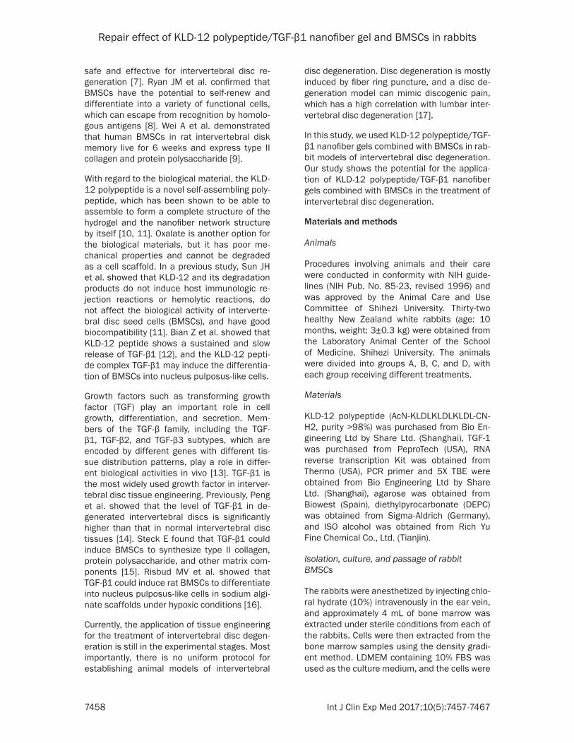
Figure 2. X-ray at 12 weeks after transplantation. Group A: The disc height was restored; group B: The height of the intervertebral disc stenosis; group C: Intervertebral disc height recovery is not obvious; group D: The disc height shows no obvious change.

Repair effect of KLD-12 polypeptide/TGF-β1 nanofiber gel and BMSCs in rabbits
7461 Int J Clin Exp Med 2017;10(5):7457-7467
RT-PCR
The cryopreserved tissue samples were then taken out from the liquid nitrogen. Total RNA was extracted using Trizol reagent extraction and reverse transcribed RNA to cDNA using First Stand cDNA Synthesis. RT-PCR was then performed using the cDNA as the template. The primer sequences used for the PCR are shown in Table 2.
Statistical analysis
Experimental data was processed and ana-lyzed using SPSS 17.0 statistical software.
ratio of the PCR-amplified bands were shown as means ± standard deviation and were ana-lyzed and compared through variance. P<0.05 was considered to indicate statistical signifi- cance.
Results
X-ray examination
The %DHI values in group A were 87.01± 2.40%, 92.25±2.14%, 93.97±2.49%, and 94.98±1.91% at 4, 8, 12, and 24 weeks, respectively. The height of the intervertebral discs in group A showed obvious recovery at 4
Figure 3. Percentage of intervertebral disc height at various time points.
Table 3. %DHI comparison of the four groups of animals at different time points (
_x±sDH)
Group n%DHI
4 weeks 8 weeks 12 weeks 24 weeksA 6 87.01±2.40 92.25±2.14 93.97±2.49 94.98±1.91B 6 66.61±3.79a 61.65±3.92a 57.48±4.77a 48.74±5.46a
C 6 73.95±7.38a,b 79.31±3.53ab 83.94±2.77ab 84.80±4.34a,b
D 6 100.20±2.27a,b,c 100.46±1.53a,b,c 100.43±0.94a,b,c 100.06±3.08a,b,c
Note: compared with group A, aP<0.05; compared with group B, a,bP<0.05; compared with group C, a,b,cP<0.05.
Variance analysis was used to compare the intervertebral disc hei- ght and the express- ion of aggrecan and Collagen II between groups A, B, C, and D after the normal state, and homogeneity of variance was satisfied using the levene test. The %DHI and the gray
Repair effect of KLD-12 polypeptide/TGF-β1 nanofiber gel and BMSCs in rabbits
7462 Int J Clin Exp Med 2017;10(5):7457-7467
and 8 weeks and tended to be stable at 12 and 24 weeks. The height of the interverte- bral discs in group B decreased significantly at 4 and 8 weeks and further showed a slight decrease at 12 and 24 weeks. In group C, no obvious recovery in the height of the interver- tebral disc was observed. The intervertebral disc height in group D also did not show any obvious changes (Figures 2 and 3). Compared with group B, C, and D, the %DHI in group A showed a statistically significant difference (P<0.05) (Table 3).
MRI
The intensity of the MRI T2-weighted signal in group A at 4, 8, 12, and 24 weeks was
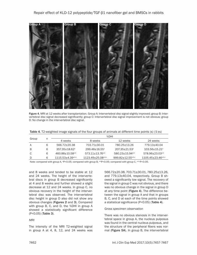
566.72±20.38, 703.71±30.01, 780.25±13.26, and 779.13±40.04, respectively. Group B sh- owed a significantly low signal. The recovery of the signal in group C was not obvious, and there was no obvious change in the signal in group D at any time point (Figure 4). The difference be- tween the signal in group A and that in groups B, C, and D at each of the time points showed a statistical significance (P<0.05) (Table 4).
Gross specimen observation
There was no obvious stenosis in the interver-tebral space in group A, the nucleus pulposus was found in the central nucleus pulposus, and the structure of the peripheral fibers was nor-mal (Figure 5A). In group B, the intervertebral
Figure 4. MRI at 12 weeks after transplantation. Group A: Intervertebral disc signal slightly improved; group B: Inter-vertebral disc signal decreased significantly; group C: Intervertebral disc signal improvement is not obvious; group D: No change in the intervertebral disc signal.
Table 4. T2-weighted image signals of the four groups of animals at different time points (s) (_x±s)
Group n%DHI
4 weeks 8 weeks 12 weeks 24 weeksA 6 566.72±20.38 703.71±30.01 780.25±13.26 779.13±40.04B 6 357.35±18.62a 299.48±16.55a 207.35±21.53a 103.56±15.21a
C 6 460.88±10.58a,b 573.11±13.76a,b 580.23±15.94a,b 578.96±23.03a,b
D 6 1115.53±4.39a,b,c 1123.49±25.08a,b,c 999.82±12.55a,b,c 1105.45±23.46a,b,c
Note: compared with group A, aP<0.05; compared with group B, a,bP<0.05; compared with group C, a,b,cP<0.05.

Repair effect of KLD-12 polypeptide/TGF-β1 nanofiber gel and BMSCs in rabbits
7463 Int J Clin Exp Med 2017;10(5):7457-7467
space was narrow, and the central nucleus pulposus was absent (Figure 5B). In group C, there was a slight gap in the intervertebral space, and white fibrous tissue was found in the central nucleus pulposus area (Figure 5C). The intervertebral space in group D was not narrow (Figure 5D).
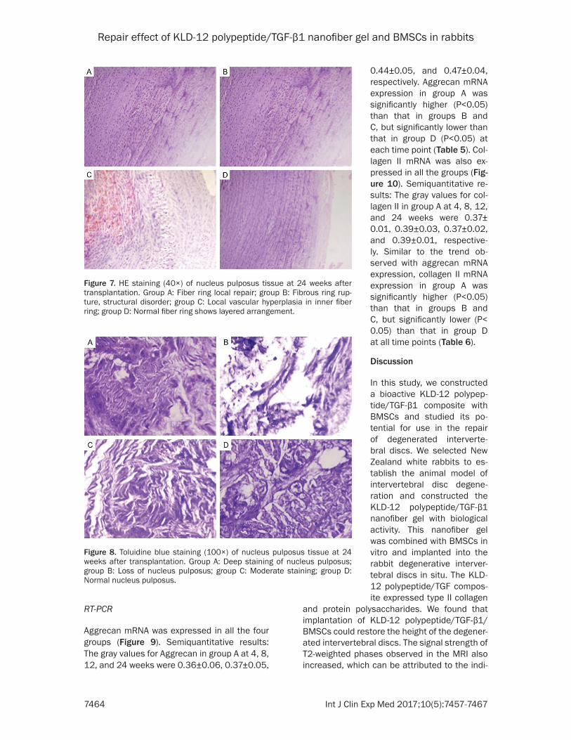
Toluidine blue staining showed that the nu- cleus pulposus in group A was deeply stained (Figure 8A) and that in group B showed loss of the nucleus pulposus and light local staining (Figure 8B). The nucleus pulposus in group C was moderately stained (Figure 8C), and that in group D was deeply stained (Figure 8D).
Figure 5. Gross specimens at 12 weeks after transplantation. Group A: New pith nucleus-like; group B: Fiber hyperplasia yellow; group C: White fiber hy-perplasia; group D: Translucent jelly-like.
Figure 6. HE staining (100×) of nucleus pulposus tissue at 24 weeks af-ter transplantation. Group A: A large number of nucleus pulposus-like cells; group B: The nucleus pulposus cells significantly decreased, fiber hyperpla-sia; group C: Cartilage like-cells, hyperplasia; group D: Normal nucleus pulpo-sus cells scattered.
HE staining
In group A, the intervertebral disc structure was normal, and nucleus pulposus-like cells were seen in the cen- tral nucleus pulposus (Figure 6A); the peripheral fiber ring array layer was slightly disor-dered (Figure 7A). In group B, the intervertebral disc struc-ture was obviously damaged, and the number of nucleus pulposus cells in the cen- tral nucleus pulposus area was significantly decreased; fibrous hyperplasia was also observed (Figure 6B). More- over, the peripheral fiber ring structure was obviously disor-dered and fractured (Figure 7B). The intervertebral disc structure in group C was not clear, and fiber cartilage-like cells were observed in the central nucleus pulposus area (Figure 6C). The inner layer of the fiber ring structure was slightly disordered and the blood vessels showed local proliferation (Figure 7C). In group D, the nucleus pulpo-sus area showed a large num-ber of nucleus pulposus cells, and the intervertebral disc structure was normal (Figure 6D); the peripheral fiber ring was in a layer of neat arrange-ment (Figure 7D). The inter-vertebral disc degeneration in the four groups showed the following grades: group A: 1- 2, group B: 4-5, group C: 3- 4, and group D: -0.
Toluidine blue staining
Repair effect of KLD-12 polypeptide/TGF-β1 nanofiber gel and BMSCs in rabbits
7464 Int J Clin Exp Med 2017;10(5):7457-7467
RT-PCR
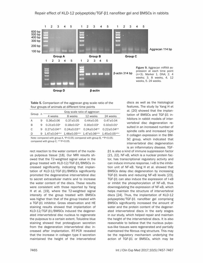
Aggrecan mRNA was expressed in all the four groups (Figure 9). Semiquantitative results: The gray values for Aggrecan in group A at 4, 8, 12, and 24 weeks were 0.36±0.06, 0.37±0.05,
and protein polysaccharides. We found that implantation of KLD-12 polypeptide/TGF-β1/BMSCs could restore the height of the degener-ated intervertebral discs. The signal strength of T2-weighted phases observed in the MRI also increased, which can be attributed to the indi-
Figure 7. HE staining (40×) of nucleus pulposus tissue at 24 weeks after transplantation. Group A: Fiber ring local repair; group B: Fibrous ring rup-ture, structural disorder; group C: Local vascular hyperplasia in inner fiber ring; group D: Normal fiber ring shows layered arrangement.
Figure 8. Toluidine blue staining (100×) of nucleus pulposus tissue at 24 weeks after transplantation. Group A: Deep staining of nucleus pulposus; group B: Loss of nucleus pulposus; group C: Moderate staining; group D: Normal nucleus pulposus.
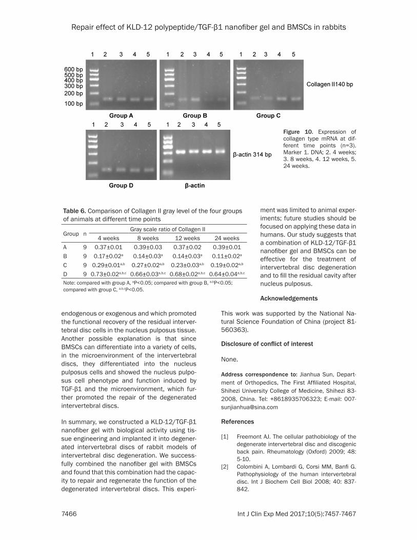
0.44±0.05, and 0.47±0.04, respectively. Aggrecan mRNA expression in group A was significantly higher (P<0.05) than that in groups B and C, but significantly lower than that in group D (P<0.05) at each time point (Table 5). Col- lagen II mRNA was also ex- pressed in all the groups (Fig- ure 10). Semiquantitative re- sults: The gray values for col-lagen II in group A at 4, 8, 12, and 24 weeks were 0.37± 0.01, 0.39±0.03, 0.37±0.02, and 0.39±0.01, respective- ly. Similar to the trend ob- served with aggrecan mRNA expression, collagen II mRNA expression in group A was significantly higher (P<0.05) than that in groups B and C, but significantly lower (P< 0.05) than that in group D at all time points (Table 6).
Discussion
In this study, we constructed a bioactive KLD-12 polypep-tide/TGF-β1 composite with BMSCs and studied its po- tential for use in the repair of degenerated interverte- bral discs. We selected New Zealand white rabbits to es- tablish the animal model of intervertebral disc degene- ration and constructed the KLD-12 polypeptide/TGF-β1 nanofiber gel with biological activity. This nanofiber gel was combined with BMSCs in vitro and implanted into the rabbit degenerative interver-tebral discs in situ. The KLD-12 polypeptide/TGF compos-ite expressed type II collagen
Repair effect of KLD-12 polypeptide/TGF-β1 nanofiber gel and BMSCs in rabbits
7465 Int J Clin Exp Med 2017;10(5):7457-7467
rect reaction to the water content of the nucle-us pulposus tissue [18]. Our MRI results sh- owed that the T2-weighted signal value in the group treated with KLD-12/TGF-β1/BMSCs in- creased significantly, indicating that implan- tation of KLD-12/TGF-β1/BMSCs significantly promoted the degenerative intervertebral disc to secret extracellular matrix and to increase the water content of the discs. These results were consistent with those reported by Yang H et al. [19], where the T2-weighted signal intensity of the group treated with BMSCs was higher than that of the group treated with a TGF-β1 inhibitor. Gross observation and HE staining results showed that implantation of KLD-12/TGF-β1/BMSCs induced the degener-ated intervertebral disc nucleus to regenerate the pulposus to a certain extent. Toluidine blue staining showed that proteoglycan secretion from the degeneration intervertebral disc in- creased after implantation. RT-PCR revealed that the increase in collagen type II secretion maintained the height of the intervertebral
β1 is also a kind of immune suppression factor [21, 22]. NF-κB, which is a nuclear protein fac-tor, has transcriptional regulatory activity and can induce immune response. I-κB is the inhibi-tion unit of NF-κB. Yang H et al. showed that BMSCs delay disc degeneration by increasing TGF-β1 levels and reducing NF-κB levels [23]. TGF-β1 can also induce the expression of I-κB or inhibit the phosphorylation of NF-κB, thus downregulating the expression of NF-κB, which helps maintain the structure of intervertebral discs [24]. Thus, the implantation of KLD-12 polypeptide/TGF-β1 nanofiber gel comprising BMSCs significantly increased the amount of water and the protein content of the degener-ated intervertebral discs in the early stages in our study, which helped repair and maintain the height of the intervertebral discs. It is also reasonable to believe that the nucleus pulpo-sus-like tissues were regenerated and partially maintained the fibrous ring structure. This may be the regulatory mechanism underlying the action of TGF-β1 or BMSCs, which may be
Figure 9. Aggrecan mRNA ex-pression at each time point (n=3). Marker 1. DNA; 2. 4 weeks; 3. 8 weeks, 4. 12 weeks, 5. 24 weeks.
Table 5. Comparison of the aggrecan gray scale ratio of the four groups of animals at different time points
Group nGray scale ratio of aggrecan
4 weeks 8 weeks 12 weeks 24 weeksA 9 0.36±0.06 0.37±0.05 0.44±0.05 0.47±0.04B 9 0.21±0.03a 0.18±0.02a 0.16±0.03a 0.10±0.01a
C 9 0.27±0.04a,b 0.24±0.03a,b 0.24±0.04a,b 0.22±0.04a,b
D 9 1.47±0.04a,b,c 1.48±0.05a,b,c 1.47±0.06a,b,c 1.45±0.05a,b,c
Note: compared with group A, aP<0.05; compared with group B, a,bP<0.05; compared with group C, a,b,cP<0.05.
discs as well as the histological features. The study by Yang H et al. [20] showed that the implan- tation of BMSCs and TGF-β1 in- hibitors in rabbit models of inter-vertebral disc degeneration re- sulted in an increased number of spindle cells and increased type II collagen expression in the BM- SC group, which indicated that intervertebral disc degeneration is an inflammatory disease. TGF-
Repair effect of KLD-12 polypeptide/TGF-β1 nanofiber gel and BMSCs in rabbits
7466 Int J Clin Exp Med 2017;10(5):7457-7467
endogenous or exogenous and which promoted the functional recovery of the residual interver-tebral disc cells in the nucleus pulposus tissue. Another possible explanation is that since BMSCs can differentiate into a variety of cells, in the microenvironment of the intervertebral discs, they differentiated into the nucleus pulposus cells and showed the nucleus pulpo-sus cell phenotype and function induced by TGF-β1 and the microenvironment, which fur-ther promoted the repair of the degenerated intervertebral discs.
In summary, we constructed a KLD-12/TGF-β1 nanofiber gel with biological activity using tis-sue engineering and implanted it into degener-ated intervertebral discs of rabbit models of intervertebral disc degeneration. We success-fully combined the nanofiber gel with BMSCs and found that this combination had the capac-ity to repair and regenerate the function of the degenerated intervertebral discs. This experi-
This work was supported by the National Na- tural Science Foundation of China (project 81- 560363).
Disclosure of conflict of interest
None.
Address correspondence to: Jianhua Sun, Depart- ment of Orthopedics, The First Affiliated Hospital, Shihezi University College of Medicine, Shihezi 83- 2008, China. Tel: +8618935706323; E-mail: 007- [email protected]
References
[1] Freemont AJ. The cellular pathobiology of the degenerate intervertebral disc and discogenic back pain. Rheumatology (Oxford) 2009; 48: 5-10.
[2] Colombini A, Lombardi G, Corsi MM, Banfi G. Pathophysiology of the human intervertebral disc. Int J Biochem Cell Biol 2008; 40: 837-842.
Figure 10. Expression of collagen type mRNA at dif-ferent time points (n=3). Marker 1. DNA; 2. 4 weeks; 3. 8 weeks, 4. 12 weeks, 5. 24 weeks.
Table 6. Comparison of Collagen II gray level of the four groups of animals at different time points
Group nGray scale ratio of Collagen II
4 weeks 8 weeks 12 weeks 24 weeksA 9 0.37±0.01 0.39±0.03 0.37±0.02 0.39±0.01B 9 0.17±0.02a 0.14±0.03a 0.14±0.03a 0.11±0.02a
C 9 0.29±0.01a,b 0.27±0.02a,b 0.23±0.03a,b 0.19±0.02a,b
D 9 0.73±0.02a,b,c 0.66±0.03a,b,c 0.68±0.02a,b,c 0.64±0.04a,b,c
Note: compared with group A, aP<0.05; compared with group B, a,bP<0.05; compared with group C, a,b,cP<0.05.
ment was limited to animal exper-iments; future studies should be focused on applying these data in humans. Our study suggests that a combination of KLD-12/TGF-β1 nanofiber gel and BMSCs can be effective for the treatment of intervertebral disc degeneration and to fill the residual cavity after nucleus pulposus.
Acknowledgements
Repair effect of KLD-12 polypeptide/TGF-β1 nanofiber gel and BMSCs in rabbits
7467 Int J Clin Exp Med 2017;10(5):7457-7467
[3] Jandial R, Aryan HE, Park J, Taylor WT, Snyder EY. Stem cell mediated regeneration of the in-tervertebral disc: cellular and molecular chal-lenge. Neurosurg Focus 2008; 21: 3-4.
[4] Jandial R, Aryan HE, Park J, Taylor WT, Snyder EY. Stem cell mediated regeneration of the in-tervertebral disc: cellular and molecular chal-lenge. Neurosurg Focus 2008; 21: 3-4.
[5] Sebastine IM, Williams DJ. Current develop-ments in tissue engineering of nucleus pulpo-sus for the treatment of intervertebral disc de-generation. Conf Proc IEEE Eng Med Biol Soc 2007; 2007: 6401-6406.
[6] Richardson SM and Hoyland JA. Stem cell re-generation of degenerated intervertebral dis- cs: current status. Curr Pain Headache Rep 2008; 12: 83-88.
[7] Yim RL, Lee JT, Bow CH, Meij B, Leung V, Cheung KM, Vavken P, Samartzis D. A system-atic review of the safety and efficacy of mesen-chymal stem cells for disc degeneration: in-sights and future directions for regenerative therapeutics. Stem Cells Dev 2014; 23: 2553-2567.
[8] Ryan JM, Barry FP, Murphy JM, Mahon BP. Mesenchymal stem cells avoid allogeneic re-jection. J Inflamm (Lond) 2005; 2: 8.
[9] Wei A, Shen B, Williams L, Diwan A. Mesen- chymal stem cells: potential application in in-tervertebral disc regeneration. Transl Pediatr 2014; 3: 71-90.
[10] Sun JH, Zheng QX, Wu YC, Liu Y, Guo X, Wu W. Culture of nucleus pulposus cells from inter-vertebral disc on self-assembling KLD-12 pep-tide hydrogel scaffold. Mat Sci Eng C 2010; 975-980.
[11] Sun JH and Zheng QX. Experimental study on self-assembly of KLD-12 peptide hydrogel and 3-D culture of MSC encapsulated within hydro-gel in vitro. J Huazhong Univ Sci Technol [Med Sci] 2009; 29: 645-648.
[12] Bian Z and Sun J. Development of a KLD-12 polypeptide/TGF-β1-tissue scaffold promoting the differentiation of mesenchymal stem cell into nucleus pulposus-like cells for treatment of intervertebral disc degeneration. Int J Clin Exp Pathol 2015; 8: 1093-1103.
[13] Blobe GC, Schiemann WP, Lodishm HF. Role of transforming growth factor beta in human dis-ease. N Engl J Med 2000; 342: 1350-1358.
[14] Peng B, Hao J, Hou S, Wu W, Jiang D, Fu X, Yand Y. Possible pathogenesis of painful in- tervertebral disc degeneration. Spine (Phila Pa 1976) 2006; 31: 560-566.
[15] Steck E, Bertram H, Abel R, Chen B, Winter A, Richter W. Induction of intervertebral disc-like cells from adult mesenchymal stem cells. Stem Cells 2005; 23: 403-411.
[16] Risbud MV, Albert TJ, Guttapalli A, Vresilovic EJ, Hillibrand AS, Vaccaro AR, Shapiro IM. Differen- tiation of mesenchymal stem cells towards a nucleus pulposus-like phenotype in vitro: impli-cations for cell-based transplantation therapy. Spine (Phila Pa 1976) 2004; 29: 2627-2632.
[17] Qi Q, Kang R, Xie L. Research progress of es-tablishment of animal model for lumbar in- tervertebral disc degeneration. J Liaoning Uni Traditional Chin Med 2011; 13: 79-81.
[18] Lu DS, Shono Y, Odal I, Abumi K, Kaneda K. Effects of chondroitinase ABC and chymopa- pain on spinal motion segment biomechanics. An in vivo biomechanical, radiologic, and his- tologic canine study. Spine (Phila Pa 1976) 1997; 22: 1828-1834.
[19] Nishimura K and Mochida J. Percutaneous re-insertion of the nucleus pulposus. An experi-mental study. Spine (Phila Pa 1976) 1998; 23: 1531-1538.
[20] Luoma K, Vehmas T, Riihimaki H, Raininko R. Disc height and signal intensity of the nucleus pulposus on magnetic resonance imaging as indicators of lumbar disc degeneration. Spine (Phila Pa 1976) 2001; 26: 680-686.
[21] Yang H, Cao C, Wu C, Yuan C, Gu Q, Shi Q, Zou J. TGF-β1 suppresses inflammation in cell ther-apy for intervertebral disc degeneration. Sci Rep 2015; 5: 13254.
[22] Cao C, Zou J, Liu X, Shapiro A, Moral M, Luo Z, Shi Q, Liu J, Yang H, Ebraheim N. Bone marrow mesenchymal stem cells slow intervertebral disc degeneration through the NF-κB pathway. Spine J 2015; 15: 530-538.
[23] Cavin LG, Romieu-Mourez R, Panta GR, Sun J, Factor VM, Thorgeirsson SS, Sonenshein GE, Arsura M. Inhibition of CK2 activity by TGF-beta1 promotes IkappaB-alpha protein stabili-zation and apoptosis of immortalized hepato-cytes. Hepatology 2003; 38: 1540-1551.
[24] Azuma M, Motegi K, Aota K, Yamashita T, Yoshida H, Sato M. TGF-beta1 inhibits NF-kappaB activity through induction of IkappaB-alpha expression in human salivary gland cells: a possible mechanism of growth sup-pression by TGF-beta1. Exp Cell Res 1999; 250: 213-222.


![Impact of NF-κB pathway on the intervertebral disc inflammation … · 2021. 2. 2. · ing collagen II and aggrecan expression [6–9]. Interleukin 1 beta (IL-1β) has similar functions](https://static.fdocument.org/doc/165x107/60d96dfa1e56e6593e770a5e/impact-of-nf-b-pathway-on-the-intervertebral-disc-inflammation-2021-2-2-ing.jpg)
![GPF12DU Chassis - TV Repair Tips - TV Repair Help · GPF12DU Chassis. 2 1 Safety Precautions 1.1. General Guidelines 1. ... This TV supports [HDAVI Control 4] function. PC D-SUB 15PIN:](https://static.fdocument.org/doc/165x107/5adb28017f8b9a6d7e8d9e31/gpf12du-chassis-tv-repair-tips-tv-repair-help-chassis-2-1-safety-precautions.jpg)