Dysregulated Sonic hedgehog signaling and...
9
The Journal of Clinical Investigation | August 2003 | Volume 112 | Number 4 535 Introduction The type I IFNs (IFN-α and IFN-β) bind to a com- mon receptor and are crucial effector cytokines in many antiviral, antiproliferative, and immune re- sponses (1, 2). However, these cytokines are also implicated in the pathogenesis of infectious (3), genetic (4), and autoimmune (5) disorders. The cel- lular actions of the type I IFNs result primarily from their activation of the Janus kinase/signal transduc- er and activator of transcription (JAK/STAT) signal- ing pathway (6, 7). Type I IFN receptor (IFNAR) activation triggers the JAK-mediated tyrosine phos- phorylation of STAT2 and STAT1. These activated STAT molecules form heterodimers that translocate to the nucleus and associate with an additional fac- tor, IFN regulatory factor-9 (IRF-9), forming the complex termed IFN-stimulated gene factor-3 (ISGF3). ISGF3 then interacts with the IFN-stimulated response element (ISRE) to modulate the transcrip- tion of over 300 genes (8). The importance of the JAK/STAT pathway in medi- ating the actions of the type I IFNs is underscored by the observations that mice lacking either STAT1 (9, 10) or STAT2 (11) have impaired type I IFN respons- es and are highly susceptible to viral infection. Never- theless, other findings in STAT1-null mice showing that antiviral responses occur to type I IFNs (12), and that the neuropathogenic actions of IFN-α are markedly exacerbated (13), point to alternative, but as-yet unidentified, signaling pathways that can medi- ate both physiological and pathophysiological actions of these cytokines. STAT1 is a key factor not only in signal transduction coupled to IFNAR but also in that coupled to the type II IFN (i.e., IFN-γ) receptor (6). In addition, activation of STAT1 can occur in response to other cytokines, such as IL-6 (14). By contrast, STAT2 is implicated in only IFNAR-coupled signal transduction (6). However, it is unknown whether type I IFNs can signal and alter any cellular functions in the absence of STAT2 and what the physiological consequences of this alternative signaling might be. To study these issues, here we developed glial fibrillary acidic protein–IFN-α (GFAP–IFN-α; hereafter called Dysregulated Sonic hedgehog signaling and medulloblastoma consequent to IFN-α–stimulated STAT2-independent production of IFN-γ in the brain Jianping Wang, 1 Ngan Pham-Mitchell, 1 Christian Schindler, 2 and Iain L. Campbell 1 1 Department of Neuropharmacology, The Scripps Research Institute, La Jolla, California, USA 2 Department of Microbiology, Columbia University, New York, New York, USA The type I IFNs (IFN-α and IFN-β), which are crucial in antiviral defense and immune regulation, sig- nal via the Janus kinase/signal transducer and activator of transcription (JAK/STAT) pathway with activation of STAT1 and STAT2. Here, the function of STAT2 was studied in transgenic mice (termed GIFN/STAT2 –/– ) with CNS production of IFN-α. Surprisingly, GIFN/STAT2 –/– , but not GIFN/STAT1- null, transgenic mice, with CNS production of IFN-α, died prematurely with medulloblastoma. An immune response also induced in the brain of the GIFN/STAT2 –/– mice was associated with IFN-γ gene expression by CD3 + T cells and the activation of the STAT1, STAT3, STAT4, and STAT5 molecules. Expression of the Sonic hedgehog (Shh) and the downstream transcriptional factor Gli-1 genes, impli- cated in the pathogenesis of medulloblastoma, was found to be significantly increased and cotran- scribed in cerebellar granule neurons of the GIFN/STAT2 –/– mice. IFN-γ, but not IFN-α, induced STAT1-dependent expression of the Shh gene in cultured cerebellar granule neurons. Thus, there is an unexpected and extraordinarily adverse biological potency of IFN-α in the CNS when the primary sig- nal transduction molecule STAT2 is absent. Moreover, a hitherto unknown role is indicated for the immune system in the pathogenesis of developmental disorders and tumorigenesis of the CNS via dys- regulated Shh signaling mediated by IFN-γ. J. Clin. Invest. 112:535–543 (2003). doi:10.1172/JCI200318637. Received for publication April 14, 2003, and accepted in revised form June 24, 2003. Address correspondence to: Iain L. Campbell, The Scripps Research Institute, SP315, 10550 N. Torrey Pines Road, La Jolla, California 92037, USA. Phone: (858) 784-9306; Fax: (858) 784-9544; E-mail: [email protected]. Conflict of interest: The authors have declared that no conflict of interest exists. Nonstandard abbreviations used: Janus kinase (JAK); signal transducer and activator of transcription (STAT); type I IFN receptor (IFNAR); IFN regulatory factor (IRF); IFN-stimulated gene factor-3 (ISGF3); IFN-stimulated response element (ISRE); glial fibrillary acidic protein (GFAP); GFAP-IFN-α (GIFN); RNase protection assay (RPA); Sonic hedgehog (Shh); proliferative cell nuclear antigen (PCNA); external germinal layer (EGL); γ-activated sequence (GAS).
Transcript of Dysregulated Sonic hedgehog signaling and...

The Journal of Clinical Investigation | August 2003 | Volume 112 | Number 4 535
IntroductionThe type I IFNs (IFN-α and IFN-β) bind to a com-mon receptor and are crucial effector cytokines inmany antiviral, antiproliferative, and immune re-sponses (1, 2). However, these cytokines are alsoimplicated in the pathogenesis of infectious (3),genetic (4), and autoimmune (5) disorders. The cel-lular actions of the type I IFNs result primarily fromtheir activation of the Janus kinase/signal transduc-er and activator of transcription (JAK/STAT) signal-ing pathway (6, 7). Type I IFN receptor (IFNAR)activation triggers the JAK-mediated tyrosine phos-phorylation of STAT2 and STAT1. These activatedSTAT molecules form heterodimers that translocate
to the nucleus and associate with an additional fac-tor, IFN regulatory factor-9 (IRF-9), forming thecomplex termed IFN-stimulated gene factor-3 (ISGF3).ISGF3 then interacts with the IFN-stimulatedresponse element (ISRE) to modulate the transcrip-tion of over 300 genes (8).
The importance of the JAK/STAT pathway in medi-ating the actions of the type I IFNs is underscored bythe observations that mice lacking either STAT1 (9,10) or STAT2 (11) have impaired type I IFN respons-es and are highly susceptible to viral infection. Never-theless, other findings in STAT1-null mice showingthat antiviral responses occur to type I IFNs (12), andthat the neuropathogenic actions of IFN-α aremarkedly exacerbated (13), point to alternative, butas-yet unidentified, signaling pathways that can medi-ate both physiological and pathophysiological actionsof these cytokines. STAT1 is a key factor not only insignal transduction coupled to IFNAR but also in thatcoupled to the type II IFN (i.e., IFN-γ) receptor (6). Inaddition, activation of STAT1 can occur in responseto other cytokines, such as IL-6 (14). By contrast,STAT2 is implicated in only IFNAR-coupled signaltransduction (6). However, it is unknown whethertype I IFNs can signal and alter any cellular functionsin the absence of STAT2 and what the physiologicalconsequences of this alternative signaling might be.To study these issues, here we developed glial fibrillaryacidic protein–IFN-α (GFAP–IFN-α; hereafter called
Dysregulated Sonic hedgehog signaling and medulloblastoma consequent to IFN-α–stimulated STAT2-independent production of IFN-γ in the brain
Jianping Wang,1 Ngan Pham-Mitchell,1 Christian Schindler,2 and Iain L. Campbell1
1Department of Neuropharmacology, The Scripps Research Institute, La Jolla, California, USA2Department of Microbiology, Columbia University, New York, New York, USA
The type I IFNs (IFN-α and IFN-β), which are crucial in antiviral defense and immune regulation, sig-nal via the Janus kinase/signal transducer and activator of transcription (JAK/STAT) pathway withactivation of STAT1 and STAT2. Here, the function of STAT2 was studied in transgenic mice (termedGIFN/STAT2–/–) with CNS production of IFN-α. Surprisingly, GIFN/STAT2–/–, but not GIFN/STAT1-null, transgenic mice, with CNS production of IFN-α, died prematurely with medulloblastoma. Animmune response also induced in the brain of the GIFN/STAT2–/– mice was associated with IFN-γgeneexpression by CD3+ T cells and the activation of the STAT1, STAT3, STAT4, and STAT5 molecules.Expression of the Sonic hedgehog (Shh) and the downstream transcriptional factor Gli-1 genes, impli-cated in the pathogenesis of medulloblastoma, was found to be significantly increased and cotran-scribed in cerebellar granule neurons of the GIFN/STAT2–/– mice. IFN-γ, but not IFN-α, inducedSTAT1-dependent expression of the Shh gene in cultured cerebellar granule neurons. Thus, there is anunexpected and extraordinarily adverse biological potency of IFN-α in the CNS when the primary sig-nal transduction molecule STAT2 is absent. Moreover, a hitherto unknown role is indicated for theimmune system in the pathogenesis of developmental disorders and tumorigenesis of the CNS via dys-regulated Shh signaling mediated by IFN-γ.
J. Clin. Invest. 112:535–543 (2003). doi:10.1172/JCI200318637.
Received for publication April 14, 2003, and accepted in revised formJune 24, 2003.
Address correspondence to: Iain L. Campbell, The ScrippsResearch Institute, SP315, 10550 N. Torrey Pines Road, La Jolla,California 92037, USA. Phone: (858) 784-9306; Fax: (858) 784-9544; E-mail: [email protected] of interest: The authors have declared that no conflictof interest exists.Nonstandard abbreviations used: Janus kinase (JAK); signaltransducer and activator of transcription (STAT); type I IFNreceptor (IFNAR); IFN regulatory factor (IRF); IFN-stimulatedgene factor-3 (ISGF3); IFN-stimulated response element (ISRE);glial fibrillary acidic protein (GFAP); GFAP-IFN-α (GIFN);RNase protection assay (RPA); Sonic hedgehog (Shh);proliferative cell nuclear antigen (PCNA); external germinallayer (EGL); γ-activated sequence (GAS).
536 The Journal of Clinical Investigation | August 2003 | Volume 112 | Number 4
GIFN) transgenic mice with CNS-specific astrocyteproduction of IFN-α1 (15, 16) that were null for theSTAT2 gene (11).
MethodsAnimals. Previously characterized GIFN transgenicmice (GIFN12 line; CB6 strain) (15), STAT1–/– mice(9), and STAT2–/– (11) mice (both 129/Sv background)were used. GIFN×STAT null mice were produced byinterbreeding of GIFN transgenic mice with therespective STAT-null mice, and the genotype of prog-eny was verified by PCR analysis of tail DNA. Handl-ing of mice and experimental procedures were con-ducted in accordance with the NIH guidelines foranimal care and use.
RNase protection assay. Total RNA and poly(A)+ RNAwere isolated from freshly dissected snap-frozen brainor cerebellum using TRIZOL reagent (Life Technolo-gies Inc., Gaithersburg, Maryland, USA) and oligo-dT(Ambion Inc., Austin, Texas, USA), respectively, asdescribed previously (17). RNase protection assays(RPAs) were performed and RNA levels were quanti-fied from autoradiographs by densitometry usingNIH Image software (version 1.31) as described previ-ously (18). RPA probes for Sonic hedgehog (Shh) andGli-1 were cloned by PCR using cDNA templates gen-erously provided by Matthew Scott (Stanford Univer-sity, San Jose, California, USA) and Alexandra L. Joyn-er (The Skirball Institute, New York University Schoolof Medicine, New York, New York, USA), respectively.The Patched1 (Ptch1) RPA probe was synthesized byRT-PCR using total RNA extracted from mouse brainas substrate and cloned as described previously (18).The targeted sequences for Shh, Gli-1, and Ptch1 ribo-probes were 121–446 (GenBank X76290), 2041–2331(GenBank NM_010296), and 3601–3831 (GenBankU46155), respectively. Other multiprobe sets for thedifferent STATs, IFN-regulated genes, and IFNs weredescribed previously (17, 18).
In situ hybridization and immunocytochemistry. Brainswere removed, and one hemibrain was fixedovernight in ice-cold 4% paraformaldehyde in PBS(pH 7.4). Paraffin-embedded sagittal sections (8 µm)were prepared for in situ hybridization. 33P-labeledcRNA probes transcribed from the linearized Shh,Gli-1, or IFN-γ RPA plasmids described above wereused for in situ hybridization performed as describedpreviously (17, 18). For dual labeling, hybridized sec-tions were incubated with the following antibodies:rabbit anti–human CD3 (DAKO Corp., Carpinteria,California, USA), rabbit anti–cow GFAP (DAKOCorp.), mouse anti–human neurofilament (Stern-berger Monoclonals Inc., Lutherville, Maryland,USA), or biotinylated lectin from Lycopersicon esculen-tum (Sigma-Aldrich, St. Louis, Missouri, USA), toidentify T lymphocytes, astrocytes, neurons, or cellsof the monocytic lineage, respectively. Subsequentimmunoperoxidase staining was performed usingVectastain ABC kits (Vector Laboratories Inc.,
Burlingame, California, USA) and 3′,5′-diaminoben-zidine/H2O2 reagent as substrate. For additionalimmunohistochemistry, the other brain hemispherewas embedded in OCT compound (Sakura FinetekUSA Inc., Torrance, California, USA) and snap-frozen.Sagittal sections (10 µm) were cut with a cryostatmicrotome. Mouse anti–proliferative cell nuclearantigen (anti-PCNA) mAb (BD PharMingen, SanDiego, California, USA) was used to label proliferat-ing cells. The following rat mAb’s were used forimmunophenotyping of frozen sections: anti–mousepan-leukocyte (CD45), anti–mouse CD4, andanti–mouse CD8, all from BD PharMingen. All anti-bodies were used at a final concentration of 5 µg/mldiluted in blocking buffer. Bound antibody wasdetected using Vectastain ABC kits (Vector Laborato-ries Inc.). Prior to mounting, sections were counter-stained with Mayer’s hematoxylin, and dehydrated ingraded ethanols.
Western blot. Total-protein lysates of cerebellumwere prepared and analyzed by SDS-PAGE followedby immunoblotting as described previously (13).Antibodies against Shh-N, Shh, STAT1, STAT4,STAT5, and actin were purchased from Santa CruzBiotechnology Inc. (Santa Cruz, California, USA).Anti–phosphotyrosine-STAT1, -STAT3, and -STAT5antibodies were obtained from Cell Signaling Tech-nology Inc. (Beverly, Massachusetts, USA). Anti-STAT3 and anti–phosphotyrosine-STAT4 antibod-ies were obtained from BioSource International Inc.(Camarillo, California, USA) and Zymed Laborato-ries Inc. (South San Francisco, California, USA),respectively. Bound antibodies were visualized usingperoxidase-conjugated secondary antibody and thendetected using an ECL kit (Pierce Chemical Co.,Rockford, Illinois, USA). The membrane wasstripped in buffer (62.5 mM Tris-HCl, pH 6.8, with2% SDS and 100 mM β-mercaptoethanol) for 30minutes at 50°C before reblotting.
Granule neuron culture. Cerebellum from postnatalday 5 WT, STAT1–/–, or STAT2–/– mice was dissectedand digested at 37°C for 10 minutes in HBSS with0.25% trypsin without Ca++ and Mg++ (InvitrogenCorp., San Diego, California, USA). The digestionmixture was then treated with 0.004% DNase (RocheDiagnostics Corp., Indianapolis, Indiana, USA) and0.03% soybean trypsin inhibitor (Sigma-Aldrich).After trituration with a fire-polished Pasteur pipetteand washing with HBSS, the single-cell suspensionwas resuspended in Neurobasal medium with 2%FCS and 1% B-27 supplement (all from InvitrogenCorp.), counted by trypan blue exclusion analysis,and plated on a 75-ml culture flask at a density of9.0 × 105 cells/ml. After an initial 48 hours of incu-bation (5% CO2 at 37°C), the culture medium wasreplaced with the same medium but no serum andincubated for another 24 hours. Finally, murine IFN-α(250 IU/ml; Roche Diagnostics Corp.) or IFN-γ (50IU/ml; R&D Systems Inc., Minneapolis, Minnesota,
USA) was added to the culture and incubated for 4hours at 37°C. Thereafter, the total RNA was extract-ed using TRIZOL reagent.
Splenic-leukocyte analysis. Erythrocyte-free spleencell suspensions were prepared from 2- to 3-month-old WT, STAT1–/–, or STAT2–/– mice. The splenicleukocytes (2 × 10⁷ viable cells) were resuspended inRPMI-1640 medium (Invitrogen Corp.) containing1% BSA with or without murine IFN-α (500 IU/ml;Roche Diagnostics Corp.) and placed at 37°C for 2hours. Thereafter, total RNA was extracted usingTRIZOL reagent. For immunoblot analysis, splenic
leukocytes were treated with IFN-α for 30 minutes,and total-protein lysates were prepared and ana-lyzed as described above.
Statistical analysis. All data are given as the mean ± SEM.Data were analyzed by two-tailed unpaired Student’s ttest with a significance level of 0.05.
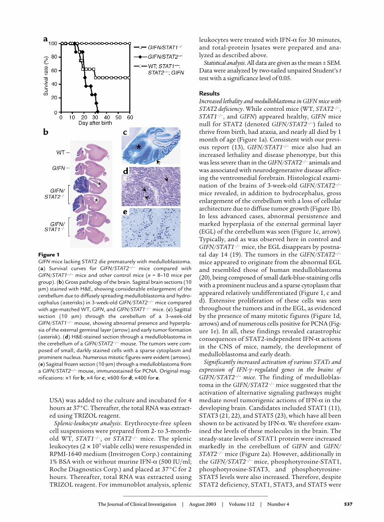
ResultsIncreased lethality and medulloblastoma in GIFN mice withSTAT2 deficiency. While control mice (WT, STAT2–/–,STAT1–/–, and GIFN) appeared healthy, GIFN micenull for STAT2 (denoted GIFN/STAT2–/–) failed tothrive from birth, had ataxia, and nearly all died by 1month of age (Figure 1a). Consistent with our previ-ous report (13), GIFN/STAT1–/– mice also had anincreased lethality and disease phenotype, but thiswas less severe than in the GIFN/STAT2–/– animals andwas associated with neurodegenerative disease affect-ing the ventromedial forebrain. Histological exami-nation of the brains of 3-week-old GIFN/STAT2–/–
mice revealed, in addition to hydrocephalus, grossenlargement of the cerebellum with a loss of cellulararchitecture due to diffuse tumor growth (Figure 1b).In less advanced cases, abnormal persistence andmarked hyperplasia of the external germinal layer(EGL) of the cerebellum was seen (Figure 1c, arrow).Typically, and as was observed here in control andGIFN/STAT1–/– mice, the EGL disappears by postna-tal day 14 (19). The tumors in the GIFN/STAT2–/–
mice appeared to originate from the abnormal EGLand resembled those of human medulloblastoma(20), being composed of small dark-blue-staining cellswith a prominent nucleus and a sparse cytoplasm thatappeared relatively undifferentiated (Figure 1, c andd). Extensive proliferation of these cells was seenthroughout the tumors and in the EGL, as evidencedby the presence of many mitotic figures (Figure 1d,arrows) and of numerous cells positive for PCNA (Fig-ure 1e). In all, these findings revealed catastrophicconsequences of STAT2-independent IFN-α actionsin the CNS of mice, namely, the development ofmedulloblastoma and early death.
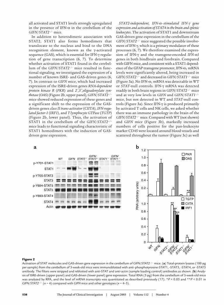
Significantly increased activation of various STATs andexpression of IFN-γ–regulated genes in the brains ofGIFN/STAT2–/– mice. The finding of medulloblas-toma in the GIFN/STAT2–/– mice suggested that theactivation of alternative signaling pathways mightmediate novel tumorigenic actions of IFN-α in thedeveloping brain. Candidates included STAT1 (11),STAT3 (21, 22), and STAT5 (23), which have all beenshown to be activated by IFN-α. We therefore exam-ined the levels of these molecules in the brain. Thesteady-state levels of STAT1 protein were increasedmarkedly in the cerebellum of GIFN and GIFN/STAT2–/– mice (Figure 2a). However, additionally inthe GIFN/STAT2–/– mice, phosphotyrosine-STAT1,phosphotyrosine-STAT3, and phosphotyrosine-STAT5 levels were also increased. Therefore, despiteSTAT2 deficiency, STAT1, STAT3, and STAT5 were
The Journal of Clinical Investigation | August 2003 | Volume 112 | Number 4 537
Figure 1GIFN mice lacking STAT2 die prematurely with medulloblastoma.(a) Survival curves for GIFN/STAT2–/– mice compared withGIFN/STAT1–/– mice and other control mice (n = 8–10 mice pergroup). (b) Gross pathology of the brain. Sagittal brain sections (10µm) stained with H&E, showing considerable enlargement of thecerebellum due to diffusely spreading medulloblastoma and hydro-cephalus (asterisks) in 3-week-old GIFN/STAT2–/– mice comparedwith age-matched WT, GIFN, and GIFN/STAT1–/– mice. (c) Sagittalsection (10 µm) through the cerebellum of a 3-week-oldGIFN/STAT1–/– mouse, showing abnormal presence and hyperpla-sia of the external germinal layer (arrow) and early tumor formation(asterisk). (d) H&E-stained section through a medulloblastoma inthe cerebellum of a GIFN/STAT2–/– mouse. The tumors were com-posed of small, darkly stained cells with a sparse cytoplasm andprominent nucleus. Numerous mitotic figures were evident (arrows).(e) Sagittal frozen section (10 µm) through a medulloblastoma froma GIFN/STAT2–/– mouse, immunostained for PCNA. Original mag-nifications: ×1 for b; ×4 for c; ×600 for d; ×400 for e.
538 The Journal of Clinical Investigation | August 2003 | Volume 112 | Number 4
all activated and STAT1 levels strongly upregulatedin the presence of IFN-α in the cerebellum of theGIFN/STAT2–/– mice.
In addition to heterodimeric association withSTAT2, STAT1 also forms homodimers thattranslocate to the nucleus and bind to the DNArecognition element, known as the γ-activatedsequence (GAS), which is essential for IFN-γ regula-tion of gene transcription (6, 7). To determinewhether activation of STAT1 found in the cerebel-lum of the GIFN/STAT2–/– mice resulted in func-tional signaling, we investigated the expression of anumber of known ISRE- and GAS-driven genes (6,7). In contrast to GIFN mice, which had increasedexpression of the ISRE-driven genes RNA-dependentprotein kinase R (PKR) and 2′,5′,oligoadenylate syn-thetase (OAS) (Figure 2b, upper panel), GIFN/STAT2–/–
mice showed reduced expression of these genes anda significant shift to the expression of the GAS-driven genes class II trans-activator (CIITA), IFN-regu-lated factor-1 (IRF1), and T lymphocyte GTPase (TGTP)(Figure 2b, lower panel). Thus, the activation ofSTAT1 in the cerebellum of the GIFN/STAT2–/–
mice leads to functional signaling characteristic ofSTAT1 homodimers with the induction of GAS-driven gene expression.
STAT2-independent, IFN-α–stimulated IFN-γ geneexpression and activation of STAT4 in the brain and splenicleukocytes. The activation of STAT1 and downstreamGAS-driven gene expression in the cerebellum of theGIFN/STAT2–/– mice suggested the possible involve-ment of IFN-γ, which is a primary modulator of theseprocesses (6, 7). We therefore examined the expres-sion of IFN-γ and the transgene-encoded IFN-α1genes in both hindbrain and forebrain. Comparedwith GIFN mice, and consistent with a STAT1 depend-ence of the GFAP transgene promoter, IFN-α1 mRNAlevels were significantly altered, being increased inGIFN/STAT2–/– and decreased in GIFN/STAT1–/– mice(Figure 3a). No IFN-α1 mRNA was detectable in WTor STAT-null controls. IFN-γ mRNA was detectedreadily in both brain regions in GIFN/STAT2–/– miceand at very low levels in GIFN and GIFN/STAT1–/–
mice, but not detected in WT and STAT-null con-trols (Figure 3a). Since IFN-γ is produced primarilyby activated T cells and NK cells, we asked whetherthere was an immune pathology in the brain of theGIFN/STAT2–/– mice. Compared with WT (not shown)and GIFN mice (Figure 3b), markedly increasednumbers of cells positive for the pan-leukocytemarker CD45 were located around blood vessels andscattered throughout the tumor (Figure 3c) as well
Figure 2Activation of STAT molecules and GAS-driven gene expression in the cerebellum of GIFN/STAT2–/– mice. (a) Total-protein lysates (100 µgper sample) from the cerebellum of 3-week-old mice were immunoblotted with anti–phosphotyrosine-STAT1, -STAT3, -STAT4, or -STAT5antibody. The filters were stripped and reblotted with anti-STAT and anti-actin (sample loading control) antibodies as shown. (b) Analy-sis of ISRE-driven (upper panel) and GAS-driven (lower panel) gene expression. Total RNA (5 µg) from the cerebellum of 3-week-old micewas analyzed by RPA, and the level of mRNA transcripts was quantitated as described previously (17). *P < 0.05 and **P < 0.01 inGIFN/STAT2–/– (n = 4) compared with GIFN mice and other genotypes (n = 4–5).
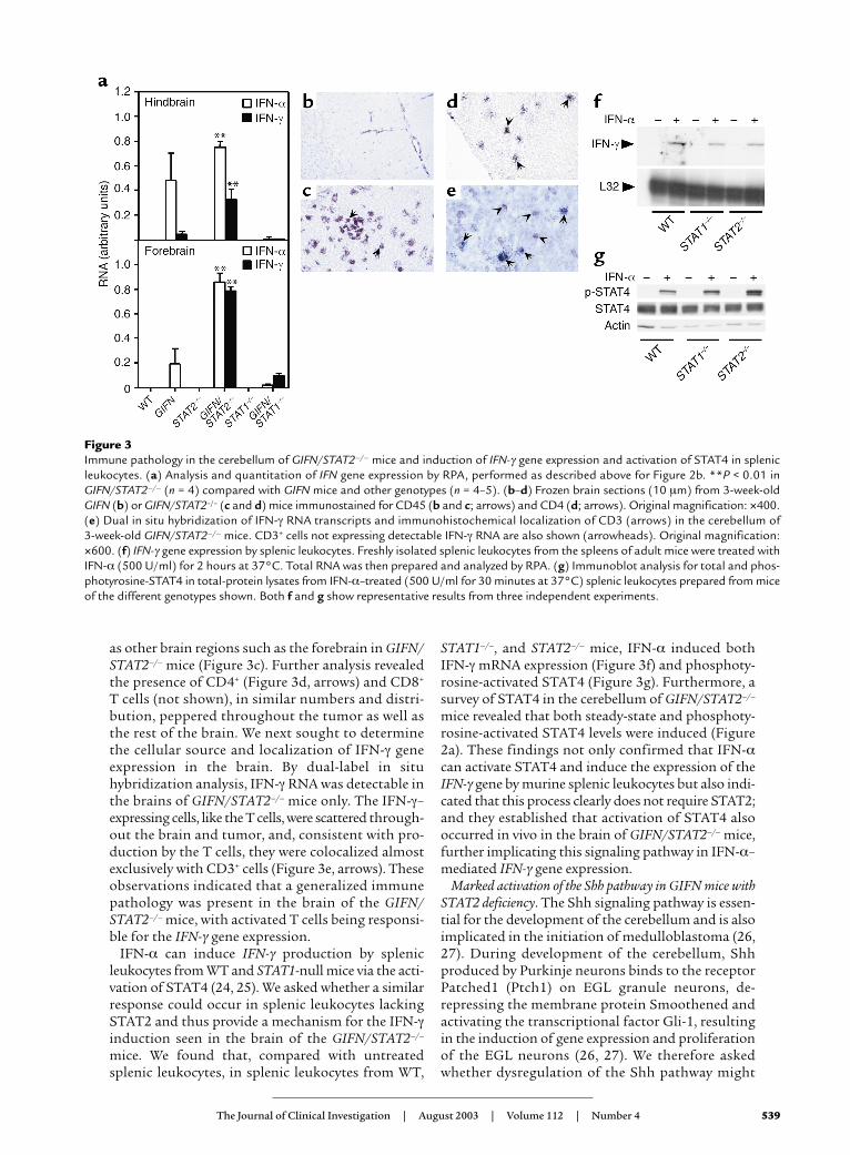
as other brain regions such as the forebrain in GIFN/STAT2–/– mice (Figure 3c). Further analysis revealedthe presence of CD4+ (Figure 3d, arrows) and CD8+
T cells (not shown), in similar numbers and distri-bution, peppered throughout the tumor as well asthe rest of the brain. We next sought to determinethe cellular source and localization of IFN-γ geneexpression in the brain. By dual-label in situhybridization analysis, IFN-γ RNA was detectable inthe brains of GIFN/STAT2–/– mice only. The IFN-γ–expressing cells, like the T cells, were scattered through-out the brain and tumor, and, consistent with pro-duction by the T cells, they were colocalized almostexclusively with CD3+ cells (Figure 3e, arrows). Theseobservations indicated that a generalized immunepathology was present in the brain of the GIFN/STAT2–/– mice, with activated T cells being responsi-ble for the IFN-γ gene expression.
IFN-α can induce IFN-γ production by splenicleukocytes from WT and STAT1-null mice via the acti-vation of STAT4 (24, 25). We asked whether a similarresponse could occur in splenic leukocytes lackingSTAT2 and thus provide a mechanism for the IFN-γinduction seen in the brain of the GIFN/STAT2–/–
mice. We found that, compared with untreatedsplenic leukocytes, in splenic leukocytes from WT,
STAT1–/–, and STAT2–/– mice, IFN-α induced bothIFN-γ mRNA expression (Figure 3f) and phosphoty-rosine-activated STAT4 (Figure 3g). Furthermore, asurvey of STAT4 in the cerebellum of GIFN/STAT2–/–
mice revealed that both steady-state and phosphoty-rosine-activated STAT4 levels were induced (Figure2a). These findings not only confirmed that IFN-αcan activate STAT4 and induce the expression of theIFN-γ gene by murine splenic leukocytes but also indi-cated that this process clearly does not require STAT2;and they established that activation of STAT4 alsooccurred in vivo in the brain of GIFN/STAT2–/– mice,further implicating this signaling pathway in IFN-α–mediated IFN-γ gene expression.
Marked activation of the Shh pathway in GIFN mice withSTAT2 deficiency. The Shh signaling pathway is essen-tial for the development of the cerebellum and is alsoimplicated in the initiation of medulloblastoma (26,27). During development of the cerebellum, Shhproduced by Purkinje neurons binds to the receptorPatched1 (Ptch1) on EGL granule neurons, de-repressing the membrane protein Smoothened andactivating the transcriptional factor Gli-1, resultingin the induction of gene expression and proliferationof the EGL neurons (26, 27). We therefore askedwhether dysregulation of the Shh pathway might
The Journal of Clinical Investigation | August 2003 | Volume 112 | Number 4 539
Figure 3Immune pathology in the cerebellum of GIFN/STAT2–/– mice and induction of IFN-γ gene expression and activation of STAT4 in splenicleukocytes. (a) Analysis and quantitation of IFN gene expression by RPA, performed as described above for Figure 2b. **P < 0.01 inGIFN/STAT2–/– (n = 4) compared with GIFN mice and other genotypes (n = 4–5). (b–d) Frozen brain sections (10 µm) from 3-week-oldGIFN (b) or GIFN/STAT2–/– (c and d) mice immunostained for CD45 (b and c; arrows) and CD4 (d; arrows). Original magnification: ×400.(e) Dual in situ hybridization of IFN-γ RNA transcripts and immunohistochemical localization of CD3 (arrows) in the cerebellum of 3-week-old GIFN/STAT2–/– mice. CD3+ cells not expressing detectable IFN-γ RNA are also shown (arrowheads). Original magnification:×600. (f) IFN-γ gene expression by splenic leukocytes. Freshly isolated splenic leukocytes from the spleens of adult mice were treated withIFN-α (500 U/ml) for 2 hours at 37°C. Total RNA was then prepared and analyzed by RPA. (g) Immunoblot analysis for total and phos-photyrosine-STAT4 in total-protein lysates from IFN-α–treated (500 U/ml for 30 minutes at 37°C) splenic leukocytes prepared from miceof the different genotypes shown. Both f and g show representative results from three independent experiments.
540 The Journal of Clinical Investigation | August 2003 | Volume 112 | Number 4
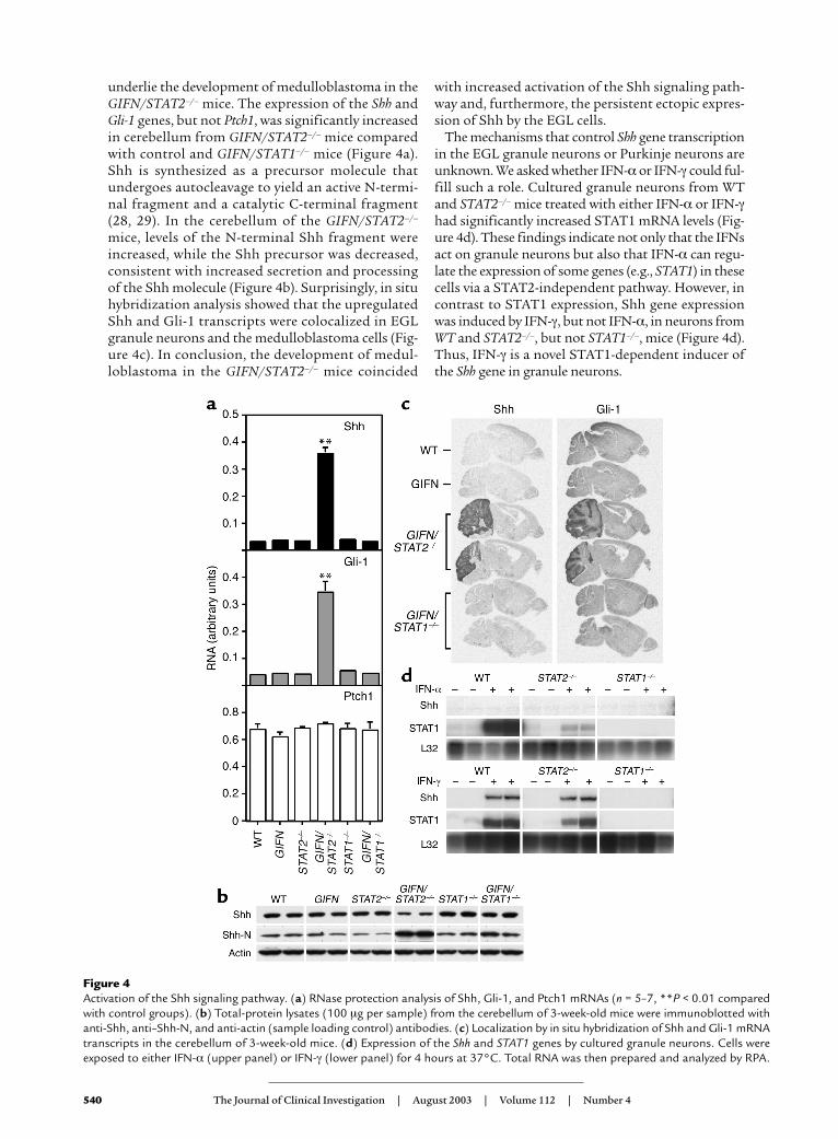
with increased activation of the Shh signaling path-way and, furthermore, the persistent ectopic expres-sion of Shh by the EGL cells.
The mechanisms that control Shh gene transcriptionin the EGL granule neurons or Purkinje neurons areunknown. We asked whether IFN-α or IFN-γ could ful-fill such a role. Cultured granule neurons from WTand STAT2–/– mice treated with either IFN-α or IFN-γhad significantly increased STAT1 mRNA levels (Fig-ure 4d). These findings indicate not only that the IFNsact on granule neurons but also that IFN-α can regu-late the expression of some genes (e.g., STAT1) in thesecells via a STAT2-independent pathway. However, incontrast to STAT1 expression, Shh gene expressionwas induced by IFN-γ, but not IFN-α, in neurons fromWT and STAT2–/–, but not STAT1–/–, mice (Figure 4d).Thus, IFN-γ is a novel STAT1-dependent inducer ofthe Shh gene in granule neurons.
underlie the development of medulloblastoma in theGIFN/STAT2–/– mice. The expression of the Shh andGli-1 genes, but not Ptch1, was significantly increasedin cerebellum from GIFN/STAT2–/– mice comparedwith control and GIFN/STAT1–/– mice (Figure 4a).Shh is synthesized as a precursor molecule thatundergoes autocleavage to yield an active N-termi-nal fragment and a catalytic C-terminal fragment(28, 29). In the cerebellum of the GIFN/STAT2–/–
mice, levels of the N-terminal Shh fragment wereincreased, while the Shh precursor was decreased,consistent with increased secretion and processingof the Shh molecule (Figure 4b). Surprisingly, in situhybridization analysis showed that the upregulatedShh and Gli-1 transcripts were colocalized in EGLgranule neurons and the medulloblastoma cells (Fig-ure 4c). In conclusion, the development of medul-loblastoma in the GIFN/STAT2–/– mice coincided
Figure 4Activation of the Shh signaling pathway. (a) RNase protection analysis of Shh, Gli-1, and Ptch1 mRNAs (n = 5–7, **P < 0.01 comparedwith control groups). (b) Total-protein lysates (100 µg per sample) from the cerebellum of 3-week-old mice were immunoblotted withanti-Shh, anti–Shh-N, and anti-actin (sample loading control) antibodies. (c) Localization by in situ hybridization of Shh and Gli-1 mRNAtranscripts in the cerebellum of 3-week-old mice. (d) Expression of the Shh and STAT1 genes by cultured granule neurons. Cells wereexposed to either IFN-α (upper panel) or IFN-γ (lower panel) for 4 hours at 37°C. Total RNA was then prepared and analyzed by RPA.
DiscussionActivated STAT1 and STAT2 form heterodimers that,together with IRF-9, combine to make the transcrip-tionally active complex ISGF3, which is responsiblefor mediating many key actions of the type I IFNs.However, recent studies in STAT1-null mice and cellsindicate that type I IFNs maintain some biologicaland pathobiological functions, pointing to the exis-tence of alternative IFNAR-coupled signaling path-ways (12, 13, 24). We now show that despite theabsence of STAT2, a primary signaling moleculelinked to IFNAR activation, IFN-α in the CNS of miceinduces an unexpectedly profound neurological dis-order characterized by medulloblastoma, inflamma-tion, and early death. Although GIFN mice deficientin STAT1 also have an accelerated and more severeneurological disease phenotype, the pathological hall-marks of this disorder are quite distinct, consisting ofcalcification and neurodegeneraton (13). Thus, notonly do our findings reveal an extraordinarily adversebiological potency of IFN-α in the CNS when eitherof the primary signal transduction molecules STAT1and STAT2 is deficient, but they highlight the pointthat fundamentally unique roles may exist for thesetranscriptional factors in the cellular response fol-lowing IFNAR activation. A further possibility is thatalternative signaling pathways are engaged by theIFNAR that are independent of both STAT1 andSTAT2. Recent published reports, together with ourstudies described here, offer support for all threemechanisms. Thus, IFN-α can stimulate both (a) theSTAT1-independent nuclear translocation of STAT2,which binds to Brg-1, forming a transcriptionallyactive complex that can induce the expression of spe-cific genes (30), and (b) the STAT2-independent acti-vation (11) and increased expression (see Figure 4d) ofSTAT1. Finally, IFN-α activation of the unique tran-scriptional factor STAT4 is both STAT1 independent(25) and STAT2 independent (see Figure 3g).
Despite initial findings to the contrary (31–33), it isnow evident that in mice, like in humans, IFN-α has asignificant role in promoting the development of theTh1 immune response, bridging innate and adaptiveimmunity (24, 25). Consistent with this role, IFN-α,in concert with T cell receptor signaling, induces IFN-γproduction by T cells — an effect mediated by directIFN-α activation of STAT4 (25). Although theseactions of IFN-α are STAT1 independent (24, 25),whether they might also occur in the absence ofSTAT2 was unclear and had been questioned (33).However, our findings clearly showed that, both in thebrain and in splenic leukocytes, IFN-α stimulated theactivation of STAT4 and IFN-γ gene expression in theabsence of STAT2. Moreover, our findings indicatedthat the induction of IFN-γ gene expression in thebrain under these circumstances was due to infiltrat-ing CD3+ T cells. Thus, the ability of IFN-α to pro-mote Th1 immune responses is not compromised butenhanced by the absence of STAT2.
In addition to STAT1 (11), other signal transduc-tion molecules can be activated in certain cell types bytype I IFNs. These include the STAT transcriptionalfactors STAT3 (21, 22) and STAT5 (23). Whether theactivation of alternative signaling pathways involvingactivated STAT3 and STAT5 in response to IFNrequires STAT2 is unclear. Here we found that bothSTAT3 and STAT5 were activated in the cerebellum ofGIFN mice deficient in STAT2. Therefore, one possi-bility is that the activation of STAT3 and STAT5 inthe brains of the GIFN/STAT2–null mice is mediateddirectly by the action of IFN-α on target cells. Howev-er, this interpretation is complicated by the compleximmunopathological and tumorigenic phenotype ofthese mice. Further studies are now required to deter-mine the regulation, distribution, and cellular local-ization of the activated STAT3 and STAT5 in thebrain of the GIFN/STAT2-null mice. Moreover, thebiological consequences of STAT3 and STAT5 activa-tion in the CNS remain unknown, although it istempting to speculate that either or both of thesemolecules might contribute to the development ofmedulloblastoma. Thus, in the case of both STAT3and STAT5, constitutive activation has been demon-strated in a wide range of human primary tumors(34), including medulloblastoma (35), while STAT3activation is required for oncogenic transformation ofsome cell lines in vitro (34).
The unexpected development of medulloblastomain the GIFN mice null for STAT2 was linked to a sig-nificant increase in the activity of the developmentalsignaling pathway involving Shh. During normaldevelopment, Shh produced by Purkinje neuronsstimulates the proliferation of granule neuron pro-genitor cells in the EGL (36–38). Shh signalinginduces the activation and increased gene transcrip-tion of Gli-1 (39). Dysregulation of this pathway, forexample, via mutations producing deficiency inPatch1 in mice and humans (26) or by overexpressionof Shh in the mouse brain (40), predisposes to thedevelopment of medulloblastoma. Although the Shhgene is expressed transiently by early EGL neurons inthe cerebellum of mice at postnatal day 2 (36), ourfindings revealed persistent expression of this gene byEGL neurons in the brains of GIFN mice null forSTAT2. The increased expression of Gli-1 correlatedclosely with that of Shh, consistent with active sig-naling by the Shh pathway in these cells. These find-ings suggest a novel mechanism whereby sustainedproliferation of the EGL neurons could occur inresponse to autocrine growth stimulation by Shh,eventually leading to their neoplastic transformation,giving rise to medulloblastoma.
The identity of factors that control the transcrip-tion of the Shh gene remains largely unknown. How-ever, our finding that ectopic expression of Shhoccurred in the presence of a strong inflammatoryprocess in the cerebellum of GIFN mice null forSTAT2 suggested that factors involved in this process
The Journal of Clinical Investigation | August 2003 | Volume 112 | Number 4 541
542 The Journal of Clinical Investigation | August 2003 | Volume 112 | Number 4
might also be responsible for regulating Shh geneexpression. Our studies identified one factor, IFN-γ,as the most likely candidate, since this cytokine wascapable of inducing Shh gene expression by culturedcerebellar granule neurons. Interestingly, like IFN-γ,IFN-α upregulated STAT1 gene expression by thesecells, but did not influence Shh gene expression, indi-cating that the effect of IFN-γ was specific and likelymediated by GAS-responsive elements within the Shhgene promoter. Previous studies in transgenic micewith CNS-targeted IFN-γ production found abnor-mal persistence and dysplasia of the EGL, althoughmedulloblastoma was not found (41, 42). These stud-ies offer additional support for a role of IFN-γ in per-turbing cerebellar EGL development, which, based onour findings here, we suggest occurs via the directmodulation of Shh signaling by this cytokine. How-ever, further progression to medulloblastoma mightdepend on the loss of STAT2, which could functionas a tumor repressor (30) and/or involve other mech-anisms such as activation of STAT3- and STAT5-dependent signaling (see above).
Our findings raise the question of their relevanceto the pathogenesis of human neurological disease.A role for viruses in the etiology of human medul-loblastoma is suspected (43, 44). It is conceivablethat an antiviral immune response directed againstsuch a viral infection might lead to IFN-γ produc-tion in the brain during crucial developmentalstages that expose the EGL to this cytokine. Also,various agents are known to inhibit or depleteSTAT2 and/or STAT1, possibly producing dysregu-lated IFN signaling and altered biological responses.These include (a) viruses that have the capacity toblock the activation or induction of these STATs byIFNs (45–47); (b) physiological inhibitors of STATS,e.g., the suppressors of cytokine signaling (SOCS)(48); and (c) the recently identified germline muta-tions in human STAT1 that cause defective IFN sig-naling (49, 50). This last point raises the possibilitythat mutations that similarly affect STAT2 mightexist in the human population.
In conclusion, we have identified alternative IFN-α–signaling pathways in the brain that provoke aSTAT2-independent, type I–like immune responsewith localized IFN-γ production. We propose thatIFN-γ mediates the STAT1-dependent induction ofShh in early granule neurons of the EGL, resulting inthe upregulation of the transcriptional factor Gli-1and the perpetual growth and eventual neoplastictransformation of these cells to medulloblastoma. Inaddition to medulloblastoma, a highly malignanttumor with unknown etiology and pathophysiologythat accounts for almost 20% of all brain tumors inchildren (51, 52), abnormal Shh signaling is found ina number of other human developmental tumors,including rhabdomyosarcoma and basal cell carcino-ma (26, 27). Therefore, our study points to a hithertounknown role for the immune system in the patho-
genesis of developmental disorders and tumorigene-sis of the CNS and perhaps other organs, involvingdysregulated Shh signaling mediated by IFN-γ.
AcknowledgmentsWe thank Heather J. Weinkauf and Mary Wagner fortheir technical support, Matthew P. Scott (StanfordUniversity) and Alexandra L. Joyner (New York Uni-versity School of Medicine) for providing cDNAclones for murine Shh and Gli-1, respectively, andRobert Schreiber (Washington University School ofMedicine) for providing STAT1–/– mice. We are grate-ful to Henry C. Powell and Eliezer Masliah (Universi-ty of California at San Diego) for helpful discussion.This study was supported by NIH grants MH-62231(to I.L. Campbell), MH-62261 (to I.L. Campbell), andGM-54686 (to C. Schindler) and National MultipleSclerosis Society Research Grant RG 3237-A-1 (to C.Schindler). This is manuscript 15449-NP from TheScripps Research Institute.
1. Sen, G.C., and Ransohoff, R.M. 1993. Interferon-induced antiviralactions and their regulation. Adv. Virus Res. 42:57–102.
2. Belardelli, F., and Gresser, I. 1996. The neglected role of type 1 inter-feron in the T-cell response: implications for its clinical use. Immunol.Today. 17:369–372.
3. Krivine, A., et al. 1999. Measuring HIV-1 RNA and interferon-α in thecerebrospinal fluid of AIDS patients: insights into the pathogenesis ofAIDS Dementia Complex. J. Neurovirol. 5:500–506.
4. Lebon, P., et al. 1988. Intrathecal synthesis of interferon-alpha in infantswith progressive familial encephalopathy. J. Neurol. Sci. 84:201–208.
5. Shiozawa, S., Kuroki, Y., Kim, M., Hirohata, S., and Ogino, T. 1992.Interferon-α in lupus psychosis. Arthritis Rheum. 35:417–422.
6. Stark, G.R., Kerr, I.M., Williams, B.R.G., Silverman, R.H., and Schreiber,R.D. 1998. How cells respond to interferons. Annu. Rev. Biochem.67:227–264.
7. Schindler, C., and Brutsaert, S. 1999. Interferons as a paradigm forcytokine signal transduction. Cell. Mol. Life Sci. 55:1509–1522.
8. Der, S.D., Zhou, A., Williams, B.R.G., and Silverman, R.H. 1998. Iden-tification of genes differentially regulated by interferon α, β, or γ usingoligonucleotide arrays. Proc. Natl. Acad. Sci. U. S. A. 95:15623–15628.
9. Meraz, M.A., et al. 1996. Targeted disruption of the Stat1 gene in micereveals unexpected physiologic specificity in the JAK-STAT signalingpathway. Cell. 84:431–442.
10. Durbin, J.E., Hackenmiller, R., Simon, M.C., and Levy, D.E. 1996. Tar-geted disruption of the mouse Stat1 gene results in compromisedinnate immunity to viral disease. Cell. 84:443–450.
11. Park, C., Li, S., Cha, E., and Schindler, C. 2000. Immune response inStat2 knockout mice. Immunity. 13:795–804.
12. Gil, M.P., et al. 2001. Biologic consequences of Stat1-independent IFNsignaling. Proc. Natl. Acad. Sci. U. S. A. 98:6680–6685.
13. Wang, J., Schreiber, R.D., and Campbell, I.L. 2002. STAT1 deficiencyunexpectedly and markedly exacerbates the pathophysiological actionsof IFN-α in the central nervous system. Proc. Natl. Acad. Sci. U. S. A.99:16209–16214.
14. Schindler, C., and Strehlow, I. 2000. Cytokines and STAT signaling.Adv. Pharmacol. 47:113–174.
15. Akwa, Y., et al. 1998. Transgenic expression of IFN-α in the centralnervous system of mice protects against lethal neurotropic viral infec-tion but induces inflammation and neurodegeneration. J. Immunol.161:5016–5026.
16. Campbell, I.L., et al. 1999. Structural and functional neuropathologyin transgenic mice with CNS expression of IFN-α. Brain Res. 835:46–61.
17. Asensio, V.C., Kincaid, C., and Campbell, I.L. 1999. Chemokines andthe inflammatory response to viral infection in the central nervous sys-tem with a focus on lymphocytic choriomeningitis virus. J. Neurovirol.5:65–75.
18. Maier, J., Kincaid, C., Pagenstecher, A., and Campbell, I.L. 2002. Regu-lation of signal transducer and activator of transcription and sup-pressor of cytokine signaling gene expression in the brain of mice withastrocyte-targeted production of IL-12 and experimental autoimmuneencephalomyelitis. Am. J. Pathol. 160:271–288.
19. Miale, I., and Sidman, R.L. 1961. An autoradiographic analysis of his-togenesis in the mouse cerebellum. Exp. Neurol. 4:277–296.
The Journal of Clinical Investigation | August 2003 | Volume 112 | Number 4 543
20. Kleihues, P., et al. 2002. The WHO classification of tumors of the nerv-ous system. J. Neuropathol. Exp. Neurol. 61:215–225.
21. Yang, C.-H., Murti, A., and Pfeffer, L.M. 1998. STAT3 complementsdefects in an interferon-resistant cell line: evidence for an essential rolefor STAT3 in interferon signaling and biological activities. Proc. Natl.Acad. Sci. U. S. A. 95:5568–5572.
22. Rani, M.R.S., et al. 1999. Catalytically active TYK2 is essential for inter-feron-β-mediated phosphorylatin of STAT3 and interferon-α recep-tor-1 (IFNAR-1) but not for activation of phosphoinositol 3-kinase. J. Biol. Chem. 274:32507–32511.
23. Fasler-Kan, E., et al. 1998. Interferon-α activates signal transducers andactivators of transcription 5 and 6 in Daudi cells. Eur. J. Biochem.254:514–519.
24. Nguyen, K.B., et al. 2000. Interferon α/β-mediated inhibition and pro-motion of interferon-γ: STAT1 resolves a paradox. Nat. Immunol. 1:70–76.
25. Nguyen, K.B., et al. 2002. Critical role for STAT4 activation by type Iinterferons in the interferon-γ response to viral infection. Science.297:2063–2066.
26. Wechsler-Reya, R., and Scott, M.P. 2001. The developmental biology ofbrain tumors. Annu. Rev. Neurosci. 24:385–428.
27. Ruiz i Altaba, A., Sanchez, P., and Dahmane, N. 2002. Gli and hedge-hog in cancer: tumours, embryos and stem cells. Nat. Rev. Cancer.2:361–372.
28. Bumcrot, D.A., Takada, R., and McMahon, A.P. 1995. Proteolytic pro-cessing yields two secreted forms of sonic hedgehog. Mol. Cell. Biol.15:2294–2303.
29. Porter, J.A., et al. 1995. The product of hedgehog autoproteolytic cleav-age active in local and long-range signalling. Nature. 374:363–366.
30. Huang, M., et al. 2002. Chromatin-remodelling factor BRG1 selective-ly activates a subset of interferon-α-inducible genes. Nat. Cell Biol.4:774–781.
31. Rogge, L., et al. 1998. The role of Stat4 in species-specific regulation ofTh cell development by type I IFNs. J. Immunol. 161:6567–6574.
32. Wenner, C.A., Guler, M.L., Macatonia, S.E., O’Garra, A., and Murphy,K.M. 1996. Roles of IFN-γ and IFN-α in IL-12-induced T helper cell-1development. J. Immunol. 156:1442–1447.
33. Farrar, J.D., et al. 2000. Selective loss of type I interferon-inducedSTAT4 activation caused by a minisatellite insertion in mouse Stat2.Nat. Immunol. 1:65–69.
34. Bromberg, J. 2002. Stat proteins and oncogenesis. J. Clin. Invest.109:1139–1142. doi:10.1172/JCI200215617.
35. Schaefer, L.K., Ren, Z., Fuller, G.N., and Schaefer, T.S. 2002. Constitu-tive activation of Stat3alpha in brain tumors: localization to tumorendothelial cells and activation by the endothelial tyrosine kinasereceptor (VEGFR-2). Oncogene. 21:2058–2065.
36. Dahmane, N., and Ruiz i Altaba, A. 1999. Sonic hedgehog regulatesthe growth and patterning of the cerebellum. Development.126:3089–3100.
37. Wallace, V.A. 1999. Purkinje-cell-derived Sonic hedgehog regulatesgranule neuron precursor cell proliferation in the developing mousecerebellum. Curr. Biol. 9:445–448.
38. Wechsler-Reya, R.J., and Scott, M.P. 1999. Control of neuronal precur-sor proliferation in the cerebellum by Sonic Hedgehog. Neuron.22:103–114.
39. Matise, M.P., and Joyner, A.L. 1999. Gli genes in development and can-cer. Oncogene. 18:7852–7859.
40. Weiner, H.L., et al. 2002. Induction of medulloblastomas in mice bysonic hedgehog, independent of Gli1. Cancer Res. 62:6385–6389.
41. Corbin, J.G., et al. 1996. Targeted CNS expression of interferon-γ intransgenic mice leads to hypomyelination, reactive gliosis, and abnor-mal cerebellar development. Mol. Cell. Neurosci. 7:354–370.
42. LaFerla, F.M., Sugarman, M.C., Lane, T.E., and Leissring, M.A. 2000.Regional hypomyelination and dysplasia in transgenic mice with astro-cyte-targeted expression of interferon-γ. J. Mol. Neurosci. 15:45–59.
43. Del Valle, L., et al. 2002. Expression of human neurotropic poly-omavirus JCV late gene product agnoprotein in human medulloblas-toma. J. Natl. Cancer Inst. 94:267–273.
44. Fine, H.A. 2002. Polyomavirus and medulloblastoma: a smoking gunor guilt by association? J. Natl. Cancer Inst. 94:240–241.
45. Cebulla, C.M., Miller, D.M., and Sedmak, D.D. 1999. Viral inhibitionof interferon signal transduction. Intervirology. 42:325–330.
46. Hariya, Y., Yokosawa, N., Yonekura, N., Kohama, G., and Fuji, N. 2000.Mumps virus can suppress the effective augmentation of HPC-inducedapoptosis by IFN-gamma through disruption of IFN signaling inU3937 cells. Microbiol. Immunol. 44:537–541.
47. Parisien, J.P., Lau, J.F., and Horvath, C.M. 2002. STAT2 acts as a hostrange determinant for species-specific paramyxovirus interferon antag-onism and simian virus 5 replication. J. Virol. 76:6435–6441.
48. Starr, R., and Hilton, D.J. 1999. Negative regulation of the JAK/STATpathway. Bioessays. 21:47–52.
49. Dupuis, S., et al. 2001. Impairment of mycobacterial but not viralimmunity by a germline human STAT1 mutation. Science.293:300–303.
50. Dupuis, S., et al. 2003. Impaired response to interferon-α/β and lethalviral disease in human STAT1 deficiency. Nat. Genet. 33:388–391.
51. Friedman, H.S., Oakes, W.J., Bigner, S.H., Wikstrand, C.J., and Bigner,D.D. 1991. Medulloblastoma: tumor biological and clinical perspec-tives. J. Neurooncol. 11:1–15.
52. Packer, R.J., Cogen, P., Vezina, G., and Rorke, L.B. 1999. Medulloblas-toma: clinical and biologic aspects. Neuro-oncol. 1:232–250.