
Reactivity of hydride bridges in a high-spin [Fe -H) … · containers, and sealed with screw caps...
28
S1 Supporting Information for Reactivity of hydride bridges in a high-spin [Fe 3 (μ-H) 3 ] 3+ cluster: reversible H 2 /CO exchange and Fe–H/B–F bond metathesis Kevin J. Anderton a , Brian J. Knight a , Arnold L. Rheingold c , Khalil A. Abboud b , Ricardo García-Serres d and Leslie J. Murray a, * a Center for Catalysis and b Department of Chemistry, University of Florida, Gainesville, Florida 32611, United States c Department of Chemistry and Biochemistry, University of California, San Diego, La Jolla, California 92093, United States d Univ. Grenoble Alpes, LCBM/PMB and CEA, IRTSV/CBM/PMB and CNRS, LCBM UMR 5249, PMB 38000 Grenoble, France Table of Contents Experimental details S2 Figure S1 1 H NMR Spectrum of Fe 3 H 3 L (1) S5 Figure S2 1 H NMR Spectrum of Fe 3 D 3 L (1-D 3 ) S6 Figure S3 1 H NMR spectrum of (FeCO) 2 Fe(μ 3 -H)L (2) S7 Figure S4 1 H NMR Spectrum of Fe 3 F 3 L (3) S8 Figure S5 1 H NMR spectrum of attempted H/D exchange reaction between 1-D 3 and H 2 S9 Figure S6 Portion of the 1 H NMR spectrum of the reaction of 1 with CO showing generation of H 2 S10 Figure S7 Portion of the 1 H NMR spectrum of the reaction of a mixture of 1 and 1-D 3 with CO showing generation of H 2 but not HD S11 Figure S8 1 H NMR spectrum of the reaction of 2 with H 2 S12 Figure S9 11 B NMR spectrum of the reaction of 1 with BF 3 •OEt 2 after addition of NEt 3 S13 Figure S10 IR spectrum of (FeCO) 2 Fe(μ 3 -H)L (2) S14 Figure S11 IR spectrum of Fe 3 F 3 L (3) S15 Figure S12 IR spectrum of (Fe 13 CO) 2 Fe(μ 3 -H)L (2- 13 CO) S16 Figure S13 Powder X-band EPR spectrum of 2 in parallel mode at 5 K S17 Figure S14 UV-vis spectrum of (FeCO) 2 Fe(μ 3 -H)L (2) S18 Figure S15 UV-vis spectrum of Fe 3 F 3 L (3) S19 Figure S16 Plot of C-O distance versus Fe-C distance for 2 compared with complexes in the CSD featuring the monocarbonyliron motif S20 Figure S17 Plot of C-O distance versus Fe-C distance for 2 compared with iron carbonyl complexes in the CSD featuring Fe···CO semi-bridging interactions similar to those in 2 S21 Figure S18 Top-down view of the solid state structure of (FeCO) 2 Fe(μ 3 -H)L (2) showing the minor occupied site for Fe1 S22 Figure S19 4.8 K Mössbauer spectra of 2 measured in different parallel applied magnetic fields S23 Table S1 Simulation parameters for the Mössbauer spectrum of 2 measured under zero applied field at 80 K S24 Table S2 Selected bond distances and angles of Fe 3 H 3 L (1) and Fe 3 F 3 L (3) and comparison with other iron β-diketiminates S25 Table S3 Crystal data and structure refinement for 2 S26 Table S4 Crystal data and structure refinement for 3 S27 References S28 Electronic Supplementary Material (ESI) for Chemical Science. This journal is © The Royal Society of Chemistry 2017
Transcript of Reactivity of hydride bridges in a high-spin [Fe -H) … · containers, and sealed with screw caps...
S1
Supporting Information for
Reactivity of hydride bridges in a high-spin [Fe3(μ-H)3]3+ cluster: reversible
H2/CO exchange and Fe–H/B–F bond metathesis Kevin J. Andertona, Brian J. Knighta, Arnold L. Rheingoldc, Khalil A. Abboudb, Ricardo García-Serresd and Leslie J. Murraya,* a Center for Catalysis and bDepartment of Chemistry, University of Florida, Gainesville, Florida 32611, United States c Department of Chemistry and Biochemistry, University of California, San Diego, La Jolla, California 92093, United States
d Univ. Grenoble Alpes, LCBM/PMB and CEA, IRTSV/CBM/PMB and CNRS, LCBM UMR 5249, PMB 38000 Grenoble, France
Table of Contents Experimental details S2
Figure S1 1H NMR Spectrum of Fe3H3L (1) S5
Figure S2 1H NMR Spectrum of Fe3D3L (1-D3) S6
Figure S3 1H NMR spectrum of (FeCO)2Fe(μ3-H)L (2) S7
Figure S4 1H NMR Spectrum of Fe3F3L (3) S8
Figure S5 1H NMR spectrum of attempted H/D exchange reaction between 1-D3 and H2 S9
Figure S6 Portion of the 1H NMR spectrum of the reaction of 1 with CO showing generation of H2 S10
Figure S7 Portion of the 1H NMR spectrum of the reaction of a mixture of 1 and 1-D3 with CO showing generation of H2 but not HD
S11
Figure S8 1H NMR spectrum of the reaction of 2 with H2 S12
Figure S9 11B NMR spectrum of the reaction of 1 with BF3•OEt2 after addition of NEt3 S13
Figure S10 IR spectrum of (FeCO)2Fe(μ3-H)L (2) S14
Figure S11 IR spectrum of Fe3F3L (3) S15
Figure S12 IR spectrum of (Fe 13CO)2Fe(μ3-H)L (2-13CO) S16
Figure S13 Powder X-band EPR spectrum of 2 in parallel mode at 5 K S17
Figure S14 UV-vis spectrum of (FeCO)2Fe(μ3-H)L (2) S18 Figure S15 UV-vis spectrum of Fe3F3L (3) S19 Figure S16 Plot of C-O distance versus Fe-C distance for 2 compared with complexes in the CSD
featuring the monocarbonyliron motif S20
Figure S17 Plot of C-O distance versus Fe-C distance for 2 compared with iron carbonyl complexes in the CSD featuring Fe···CO semi-bridging interactions similar to those in 2
S21
Figure S18 Top-down view of the solid state structure of (FeCO)2Fe(μ3-H)L (2) showing the minor occupied site for Fe1
S22
Figure S19 4.8 K Mössbauer spectra of 2 measured in different parallel applied magnetic fields S23
Table S1 Simulation parameters for the Mössbauer spectrum of 2 measured under zero applied field at 80 K
S24
Table S2 Selected bond distances and angles of Fe3H3L (1) and Fe3F3L (3) and comparison with other iron β-diketiminates
S25
Table S3 Crystal data and structure refinement for 2 S26
Table S4 Crystal data and structure refinement for 3 S27
References S28
Electronic Supplementary Material (ESI) for Chemical Science.This journal is © The Royal Society of Chemistry 2017
S2
General considerations. Unless specified otherwise, all operations were performed under a dry, air-free atmosphere using an argon- or dinitrogen-filled MBraun Unilab glovebox or standard Schlenk techniques. 1H NMR spectra were recorded on a Varian Unity Inova 500 MHz spectrometer. Chemical shifts were referenced to solvent resonances at δH = 1.73 and 3.58 ppm for THF-d8, at δH = 2.08, 6.97, 7.01, and 7.09 ppm for toluene-d8, and at δH = 7.27 for CDCl3. Protio impurities from the NMR solvent are marked with asterisks in the spectra. Solution magnetic susceptibilities were determined by the Evans method1. Infrared spectra were recorded in a nitrogen-filled glovebox as solids on a Bruker Alpha FTIR with an ATR diamond crystal stage using the Opus 7.0 software package or on a Cary 630 1B FTIR with an ATR diamond crystal using the Agilent Microlab software package. EPR samples were loaded in quartz tubes (4 x 5mm i.d. x o.d.) in an Ar-filled glovebox and sealed with gas-tight screw caps. EPR spectra were collected on a Bruker ELEXSYS E500 spectrometer fitted with a Bruker DM4116 dual-mode resonator, and temperature was maintained using an Oxford Instruments ESR900 continuous flow helium cryostat fitted with a Lakeshore Cryotronics Cernox sensor and monitored with a Lakeshore Model 336 Temperature Controller. Samples for UV/visible spectroscopy were prepared under an Ar atmosphere using air-free anhydrous THF, transferred to air-tight cuvettes (Starna Cells, Atascadero, CA, USA), and spectra recorded using an Agilent Cary 50 spectrophotometer. Mössbauer samples were ground, placed in Delrin sample containers, and sealed with screw caps in an Ar-filled glovebox. Mössbauer spectra were measured either on a low-field Mössbauer spectrometer equipped with a closed-cycle SHI-850-5 cryostat from Janis and SHI or an Oxford Instruments Spectromag 4000 cryostat containing an 8T split-pair superconducting magnet. Both spectrometers were operated in constant acceleration mode in transmission geometry. The isomer shifts are referenced against a room temperature metallic iron foil, and analysis of the data was performed using the program WMOSS (WEB research). Searches of the Cambridge Structure Database (CSD) were performed with Cambridge Crystallographic Data Center ConQuest software package using CSD version 5.37 Update 2 (Feb 2016). Significant outliers from the plots of the data generated were manually inspected and removed in cases of disorder. Tetrahydrofuran (THF), toluene, benzene, pentane, and dichloromethane were purified using either a Glass-Contour or Innovative Technologies solvent purification system and stored over 3 Å molecular sieves prior to use. The water content of each solvent was measured using a Mettler Toledo C20 Coulometric Karl Fischer Titrator prior to use and was below 1 ppm in all cases. Celite and 3 Å molecular sieves were dried at 220 °C under vacuum overnight. Fe3H3L (1)2 and LiBEt3D
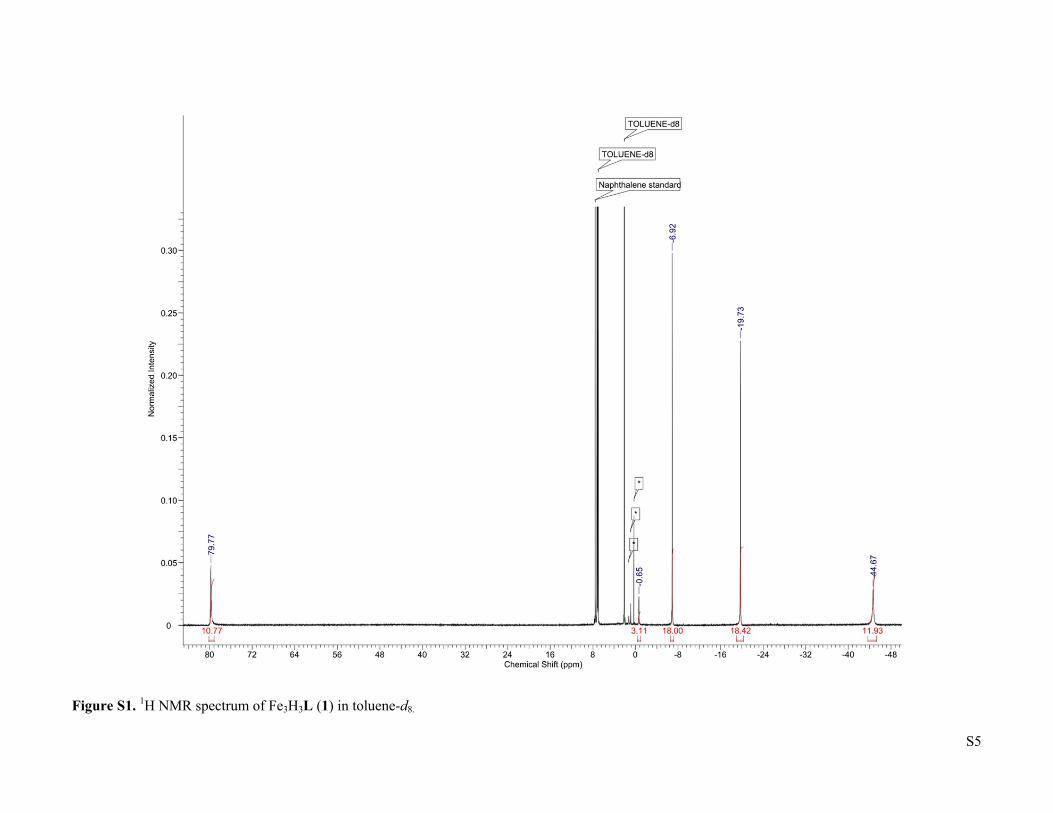
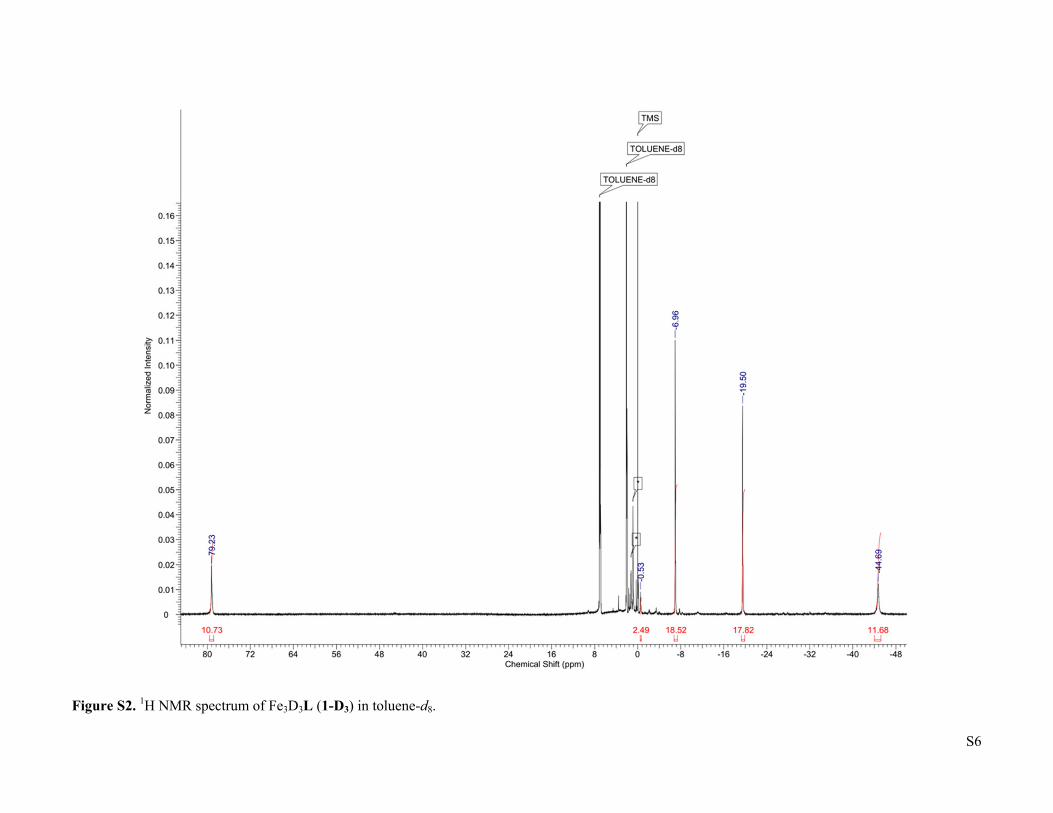
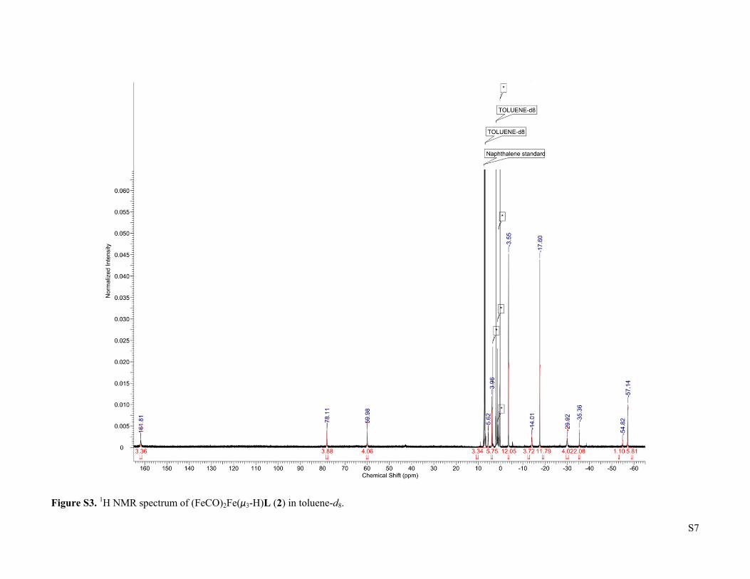
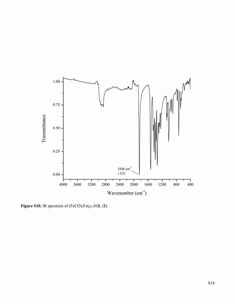
3 were prepared according to previous reports. CO (UHP grade, 99.9%) and H2 (UHP grade, 99.999%) were purchased from Airgas, Inc. and purified by passage through two cold traps (LN2/i-PrOH). 13CO was purchased from Sigma-Aldrich and used as received. Deuterated solvents were purchased from Sigma-Aldrich or Cambridge Isotope Laboratories, dried using standard methods, and stored over 3 Å molecular sieves. All other reagents were purchased from Sigma-Aldrich and used without further purification. 1H NMR of Fe3H3L (1). δ (toluene-d8) = -44.67 (12H), -19.73 (18H), -6.92 (18H), -0.65 (3H), 79.77 (12H). Fe3D3L (1-D). The synthesis followed the reported procedure for Fe3H3L (1)2, but with LiBEt3D instead of KBEt3H. 1H NMR (toluene-d8): δ = -44.69 (12H), -19.50 (18H), -6.96 (18H), -0.53 (3H), 79.23 (12H). No characteristic Fe-D vibration was observed in the IR spectrum. (FeCO)2Fe(μ3-H)L (2). A 200 mL Schlenk flask was charged with Fe3H3L (466.0 mg, 0.543 mmol), a Teflon-coated stir bar, and THF (65 mL). The solution was degassed by the freeze-pump-thaw method, then exposed to a slow flow of CO for 5 minutes with stirring. The flask was closed and the reaction was stirred for 2 hours, with a color change from dark red-orange to very dark yellow-green occurring over the first 20 minutes. After this time the reaction was evaporated and the residue was dissolved in boiling THF (65 mL). Cooling the solution to -35 oC yielded dark yellow-green crystals (342.3 mg, 0.375 mmol, 69 %) after 2 d. Integration of the 1H NMR signal of the crude reaction product relative to an internal naphthalene standard indicates that the product forms in quantitative spectroscopic yield, such that the recrystallization step can be omitted without substantially affecting the purity. X-ray quality crystals were obtained by slow evaporation of a saturated THF solution in the presence of benzene. 1H NMR (toluene-d8): δ = -57.14 (6H), -54.82 (1H), -35.36 (2H), -29.92 (4H), -17.60 (12H), -14.01 (4H), -3.55 (12H), 3.96 (6H), 5.62 (4H), 59.98 (4H), 78.11 (4H), 161.81 (4H). IR (cm-1): 1846 (ν CO), 1526, 1429, 1398, 1371, 1339, 1016. μeff (toluene-d8, 298 K) = 5.6 μB. Anal. Found (calcd) for C47H64N6O2Fe3: C, 61.85 (61.86); H, 7.08 (7.07); N, 8.71 (9.21).
S3
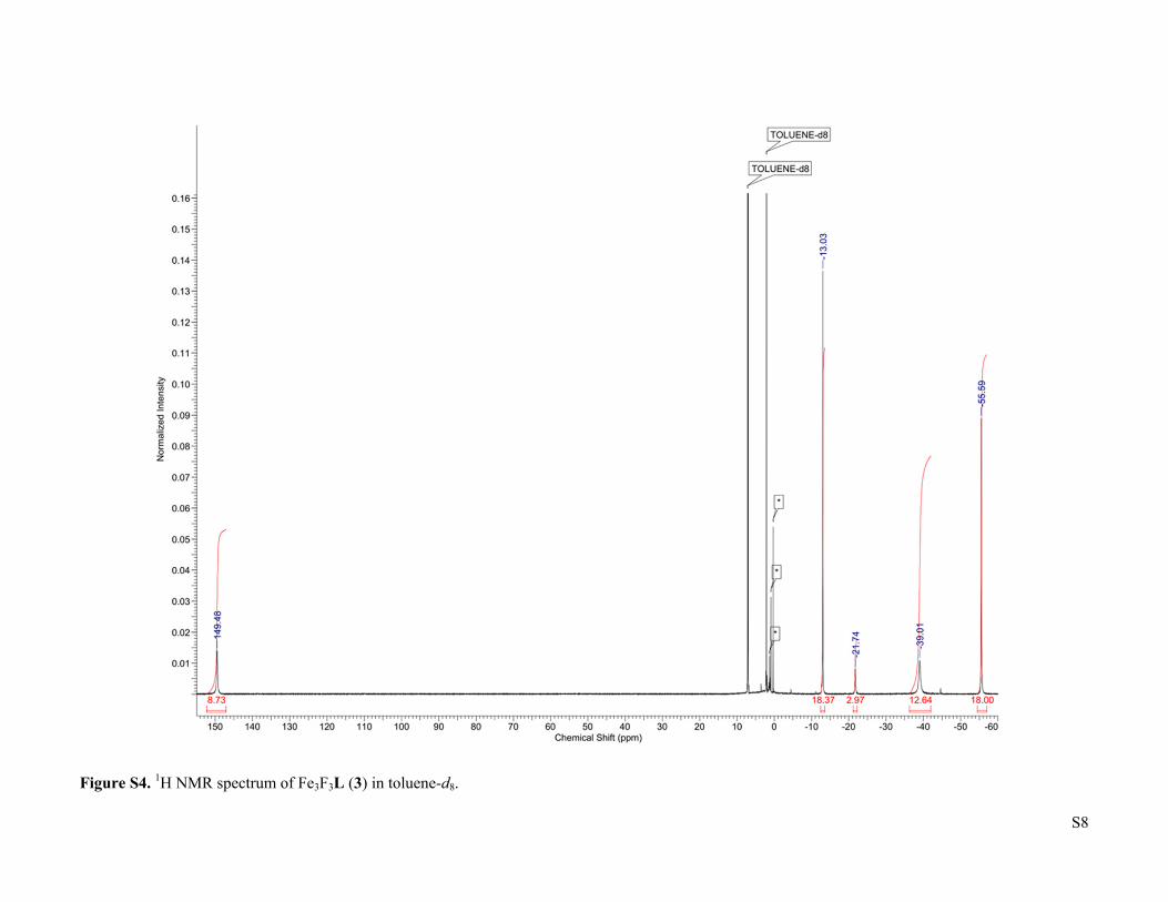
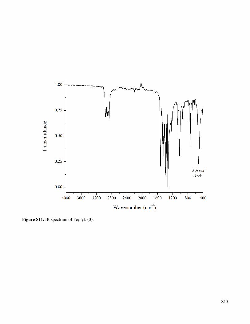
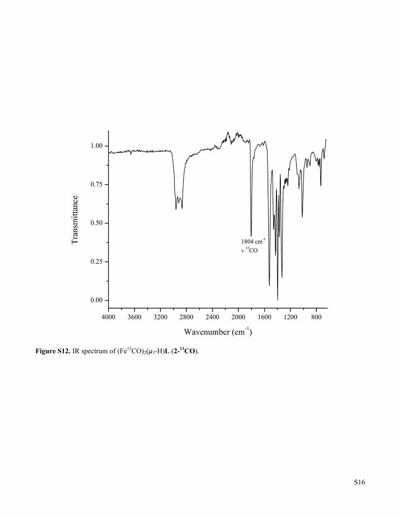
(Fe13CO)2Fe(μ3-H)L (2-13CO). The synthesis was the same as for 2 except a static atmosphere of 13CO instead of a slow flow of 12CO was used. IR (cm-1): 1804 (ν 13CO). No resonances were visible in the 13C NMR. Fe3F3L (3). A scintillation vial was charged with Fe3H3L (60.0 mg, 0.0699 mmol), a Teflon-coated stir bar, and toluene (17 mL), followed by BF3•OEt2 (9.06 µL, 0.0734 mmol). The vial was then sealed with a Teflon-lined cap and stirred at ambient temperature. The red suspension progressively converted to a bright yellow solution. After 1.5 h, the reaction was passed through a toluene-rinsed celite plug and the resulting solution was evaporated. The bright yellow residue was then dissolved in boiling benzene (4 mL). Cooling the solution to ambient temperature yielded a bright yellow powder (36.4 mg, 0.0399 mmol, 57% yield) after 2 d. Integration of the 1H NMR signal of the crude reaction product relative to an internal naphthalene standard indicates that the product forms in quantitative spectroscopic yield, such that the recrystallization step can be omitted without substantially affecting the purity. X-ray quality crystals were obtained by slow evaporation of a saturated dichloromethane solution in the presence of pentane. 1H NMR (toluene-d8): δ = -55.60 (18H), -39.01 (12H), -21.76 (3H), -13.04 (18H), 149.46 (12H). 19F NMR showed no resonances between 500 and -500 ppm. IR (cm-1): 1518, 1429, 1393, 1373, 1326, 1018, 516 (ν Fe-F). μeff (toluene-d8, 298 K) = 7.4 μB. HRMS (ESI+) m/z calcd for (M)+ [C45H63N6F3Fe3]
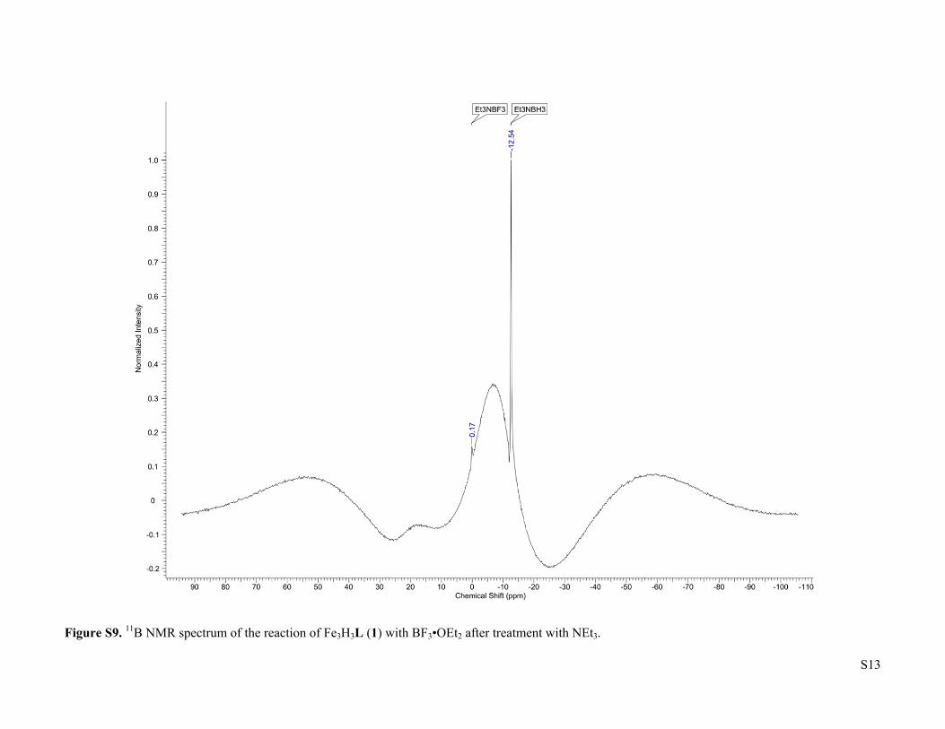
+: 912.3115, found 912.3123. Anal. Found (calcd) for C45H63N6F3Fe3(CH2Cl2)0.5(C5H12): C, 59.69 (59.38); H, 7.60 (7.37); N, 8.38 (8.48). Reaction of Fe3D3L (1-D3) with H2. A J. Young NMR tube was charged with a solution of Fe3D3L (2.0 mg, 2.3 μmol) in toluene-d8 (400 μL). The tube was degassed by the freeze-pump-thaw method, allowed to warm to r.t., then filled with H2. The tube was then closed, inverted, shaken, and refilled with H2 three times to ensure saturation. A 1H NMR spectrum was recorded after heating the reaction at 80 oC for 20 h. No shift in the resonances due to the PIECS effect was observed, consistent with the absence of exchange with H2. Detection of H2 in the reaction of Fe3H3L (1) with CO. A J. Young NMR tube was charged with a solution of Fe3H3L (2.1 mg, 2.4 μmol) and naphthalene (2.0 mg, 16 μmol) in THF-d8 (400 μL). The tube was degassed by the freeze-pump-thaw method, allowed to warm to r.t., then filled with CO. The tube was then closed, inverted, shaken, and refilled with CO three times to ensure saturation. During this process, a rapid color change to dark yellow-green occurred. A 1H NMR spectrum was recorded after 25 min. H2 and 2 were both observed in 76(6) % spectroscopic yield relative to the naphthalene standard. The non-quantitative yield results from the decomposition of the product under sub-atmospheric pressures, which cannot be avoided due to the difficulty of rapidly and completely saturating the solution with gas in a J. Young tube. Detection of H2 in the reaction of Fe3H3L (1) and Fe3H3L (1-D) with CO. A J. Young NMR tube was charged with a solution of Fe3H3L (1.6 mg, 1.9 μmol) and Fe3D3L (1.6 mg, 1.9 μmol) in THF-d8 (400 μL). The tube was degassed by the freeze-pump-thaw method, allowed to warm to 0 °C., then filled with CO. The tube was then closed, inverted, shaken, and refilled with CO three times to ensure saturation. During this process, a rapid color change to dark yellow-green occurred. A 1H NMR spectrum was recorded after 30 min at 0 °C. H2, 2, and 2-D were observed, while HD was not detected. Reaction of (FeCO)2Fe(μ3-H)L (2) with H2. A Parr bomb was charged with 2 (15.0 mg, 16.4 µmol), a Teflon-coated stir bar, and toluene (10 mL) under an Ar environment and sealed. The inlet connection to the vessel was purged for 2 min with H2 before pressurizing the vessel to 20 bar. The reaction was heated at 80 °C with stirring for 2 h to afford a red solution, then depressurized and evaporated under reduced pressure. A 1H NMR spectrum was recorded; 1 was observed in 75±7% spectroscopic yield relative to a naphthalene standard. Detection of BH3 in the reaction of Fe3H3L (1) with BF3•OEt2. A J. Young NMR tube was charged with a solution of Fe3H3L (2.1 mg, 2.45 µmol) in toluene (200 µL) and then a solution of BF3•OEt2 (200 µL, 12.9 mM in toluene, 2.57 µmol). The tube was closed, inverted, and shaken. The mixture changed from a dark red to a bright yellow within 10 minutes. The cap was removed after 1 h and the solution was rapidly charged with Et3N (4.29 µL, 30.8 µmol). The tube was closed, inverted, and shaken again. A 11B NMR spectrum was recorded, and the only boron containing compounds observed in the mixture were Et3NBF3 and Et3NBH3.
S4
Synthesis of Et3NBF3 as an NMR internal standard. To a stirring solution of triethylamine (204 mg, 2.02 mmol) in THF (2.0 mL) was added BF3•OEt2 (274 µL, 2.22 mmol). The resulting light amber suspension was stirred at 23 °C for 1 h. The mixture was then concentrated by rotary evaporation, washed with cold Et2O (2 x 1 mL, 0 °C), and the concentrated under reduced pressure to afford Et3NBF3 (191 mg, 57% yield) as an amber oil. 1H NMR (CDCl3): δ = 3.25-3.16 (comp. m, 6H), 1.39-1.30 (comp. m, 9H). 13C NMR (CDCl3): δ = 47.0, 8.8. 11B NMR (toluene-d8): δ = 0.17 (q, 1JB–F = 17.8 Hz). 19F NMR (CDCl3): δ = -151.5. IR (cm-1): 1475, 1401, 1009, 762. LRMS (ESI+) m/z calc’d for (M + H – BF3)
+ [169.1 + H – BF3]+: 102.1, 102.1 found.
Synthesis of Et3NBH3 as an NMR internal standard. To stirring triethylamine (222 mg, 2.20 mmol) was added a solution of BH3•THF (2.42 mL, 1 M in THF, 2.42 mmol) at ambient temperature. The clear solution was stirred for 1 h and then filtered through an ether-rinsed celite plug and concentrated by rotary evaporation to afford Et3NBH3 (253 mg, >99% yield) as a colorless oil. 1H NMR (CDCl3): δ = 2.78 (q, J = 7.5 Hz, 6 H), 1.42 (q, 1JB–H = 158.0 Hz, 3H) 1.19 (t, J = 7.5 Hz, 9H). 13C NMR (CDCl3): δ = 52.5, 8.7. 11B NMR (toluene-d8): δ = -12.58. IR (cm-1): 2381, 2326, 1163, 767. LRMS (ESI+) m/z calc’d for (M + H – BH3)
+ [115.1 + H – BH3]+: 102.1, 102.1
found. X-ray crystallography. (FeCO)2Fe(μ3-H)L (2). X-Ray Intensity data were collected at 100 K on a Bruker DUO diffractometer using MoKα radiation (λ = 0.71073 Å) and an APEXII CCD area detector.
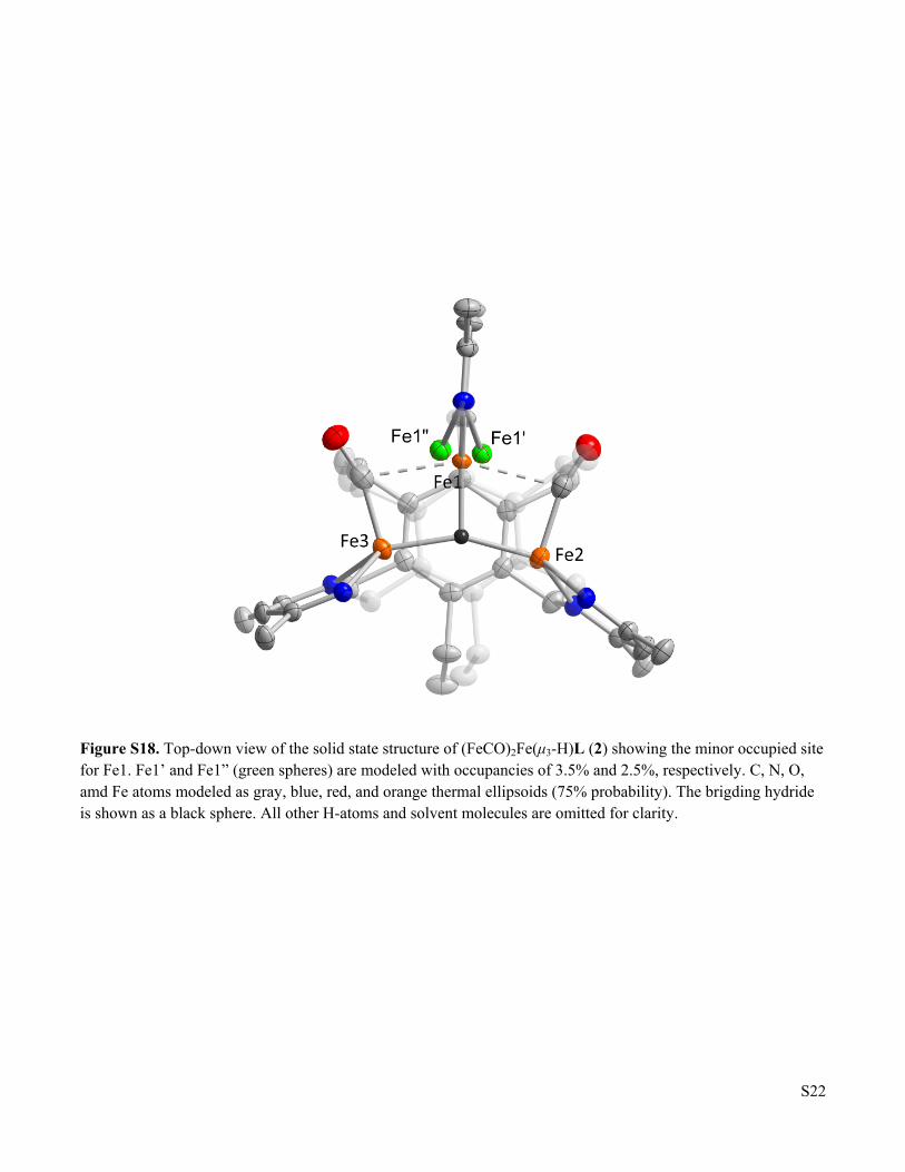
Raw data frames were read by program SAINT4 and integrated using 3D profiling algorithms. The resulting data were reduced to produce hkl reflections and their intensities and estimated standard deviations. The data were corrected for Lorentz and polarization effects and numerical absorption corrections were applied based on indexed and measured faces. The structure was solved and refined in SHELXTL20145, using full-matrix least-squares refinement. The non-H atoms were refined with anisotropic thermal parameters and all of the H atoms were calculated in idealized positions and refined riding on their parent atoms. The asymmetric unit consists of the Fe3 complex, one benzene molecule in general position and two half benzene molecules (with each located on an inversion center). Thus, the ratio of complex-to-solvent is 1-to-2. In the core of the complex, there are three Fe centers with one of them, Fe1 disordered over three positions and is refined in these three positions namely Fe1, Fe1’ and Fe1”. Fe1’ and Fe1” are present only in trace amounts. Their site occupation factors, refined in the early stages of refinement, were fixed at 3.5% and 2.5% for Fe1’ and Fe1”, respectively. The three Fe centers are connected by two CO ligands and a hydride ligand. The hydride refined with negative displacement parameter indicating that there is more than 1 electron density in the position. The proton, H1, which is refined at 96% occupancy, is sharing the same position with traces of a hydroxyl oxygen, O1, of 4%. The proton of the 4% O1 hydroxyl ligand was not included in the final refinement model. Ligands H1/O1 link the Fe2 and Fe3 centers while the Fe1 is linked to Fe2 by C2’-O2, and to Fe3 by C3’-O3. There are no other disorders in the structure. In the final cycle of refinement, 12038 reflections (of which 9908 are observed with I > 2σ(I)) were used to refine 655 parameters and the resulting R1, wR2 and S (goodness of fit) were 3.04%, 8.20% and 1.085, respectively. The refinement was carried out by minimizing the wR2 function using F2 rather than F values. R1 is calculated to provide a reference to the conventional R value but its function is not minimized. Fe3F3L (3). A brown crystal was mounted on a Cryo-Loop with Paratone-N. Data were collected on a Bruker Ultra diffractometer equipped with a rotating-anode source and micro-focus optics at 100K. All aspects of the data collection and subsequent processing of the data employed standard practices using software contained in the ApexIII and Olex2 libraries of programs. The diffuse contributions of unidentified solvent totaling 108e-/unit cell in four equal voids were removed using the SQUEEZE routine in the Platon library of programs. ADDSYM, also part of the Platon library, confirmed that space-group assignment.
S5
Figure S1. 1H NMR spectrum of Fe3H3L (1) in toluene-d8.
S6
Figure S2. 1H NMR spectrum of Fe3D3L (1-D3) in toluene-d8.
S7
Figure S3. 1H NMR spectrum of (FeCO)2Fe(μ3-H)L (2) in toluene-d8.
S8
Figure S4. 1H NMR spectrum of Fe3F3L (3) in toluene-d8.
S9
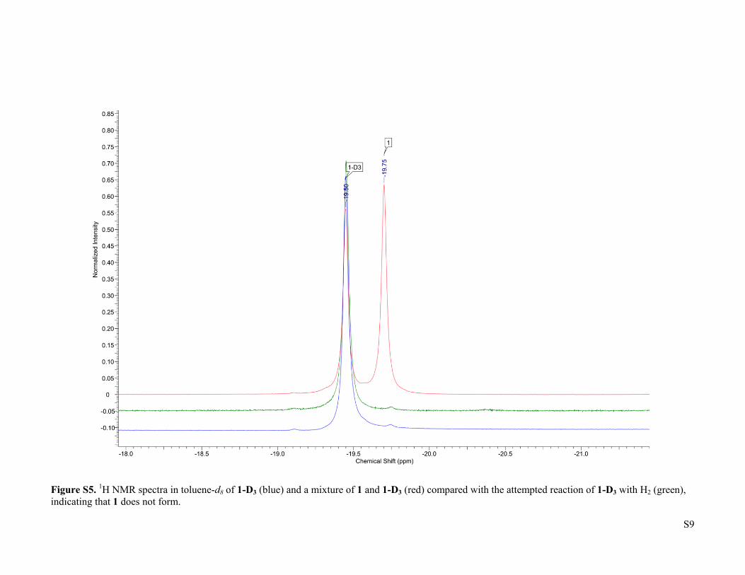
Figure S5. 1H NMR spectra in toluene-d8 of 1-D3 (blue) and a mixture of 1 and 1-D3 (red) compared with the attempted reaction of 1-D3 with H2 (green), indicating that 1 does not form.
S10
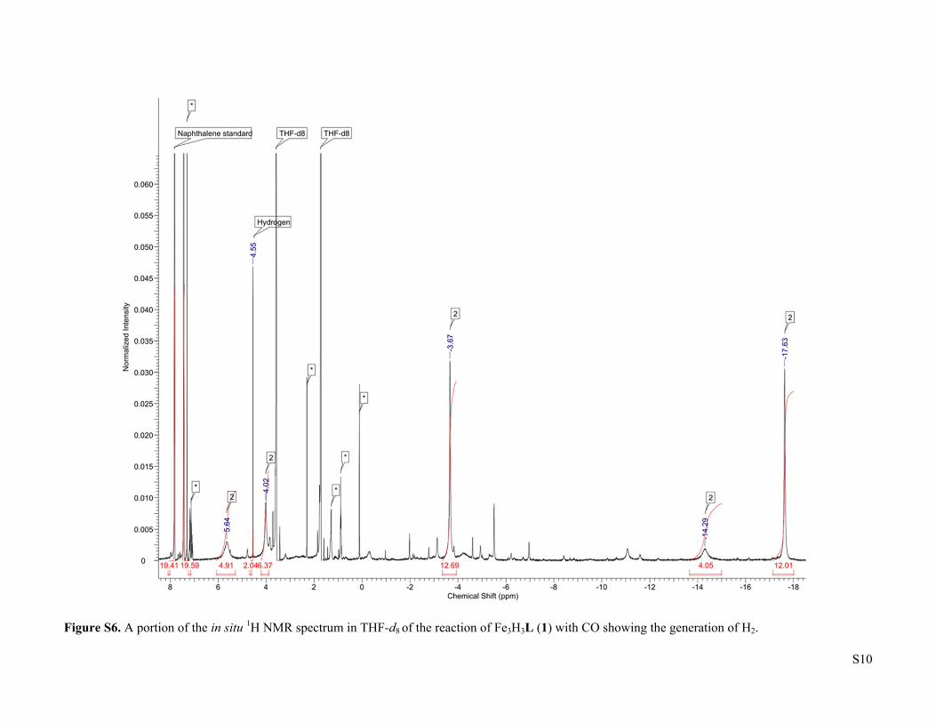
Figure S6. A portion of the in situ 1H NMR spectrum in THF-d8 of the reaction of Fe3H3L (1) with CO showing the generation of H2.
S11
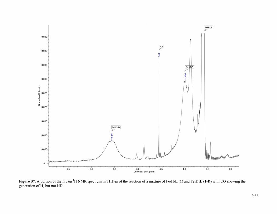
Figure S7. A portion of the in situ 1H NMR spectrum in THF-d8 of the reaction of a mixture of Fe3H3L (1) and Fe3D3L (1-D) with CO showing the generation of H2 but not HD.
S12
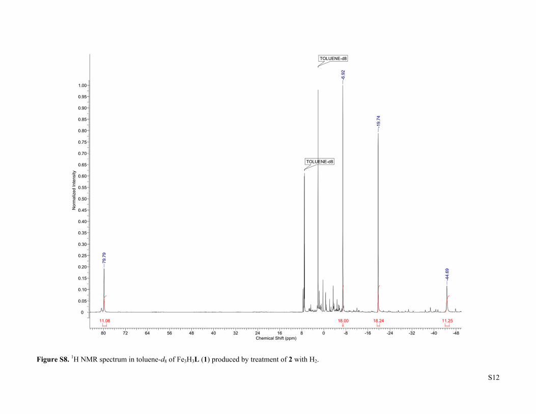
Figure S8. 1H NMR spectrum in toluene-d8 of Fe3H3L (1) produced by treatment of 2 with H2.
S13
Figure S9. 11B NMR spectrum of the reaction of Fe3H3L (1) with BF3•OEt2 after treatment with NEt3.
S14
Figure S10. IR spectrum of (FeCO)2Fe(μ3-H)L (2).
S15
Figure S11. IR spectrum of Fe3F3L (3).
S16
Figure S12. IR spectrum of (Fe13CO)2(μ3-H)L (2-13CO).
S17
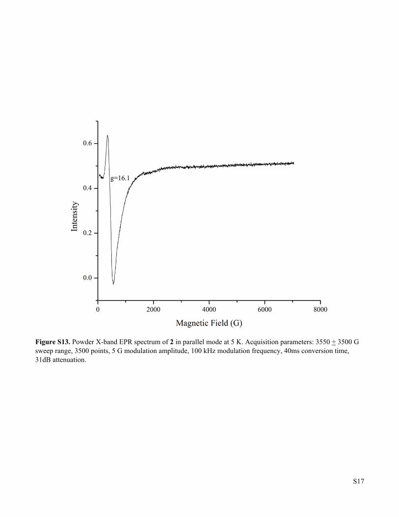
Figure S13. Powder X-band EPR spectrum of 2 in parallel mode at 5 K. Acquisition parameters: 3550 + 3500 G sweep range, 3500 points, 5 G modulation amplitude, 100 kHz modulation frequency, 40ms conversion time, 31dB attenuation.
S18
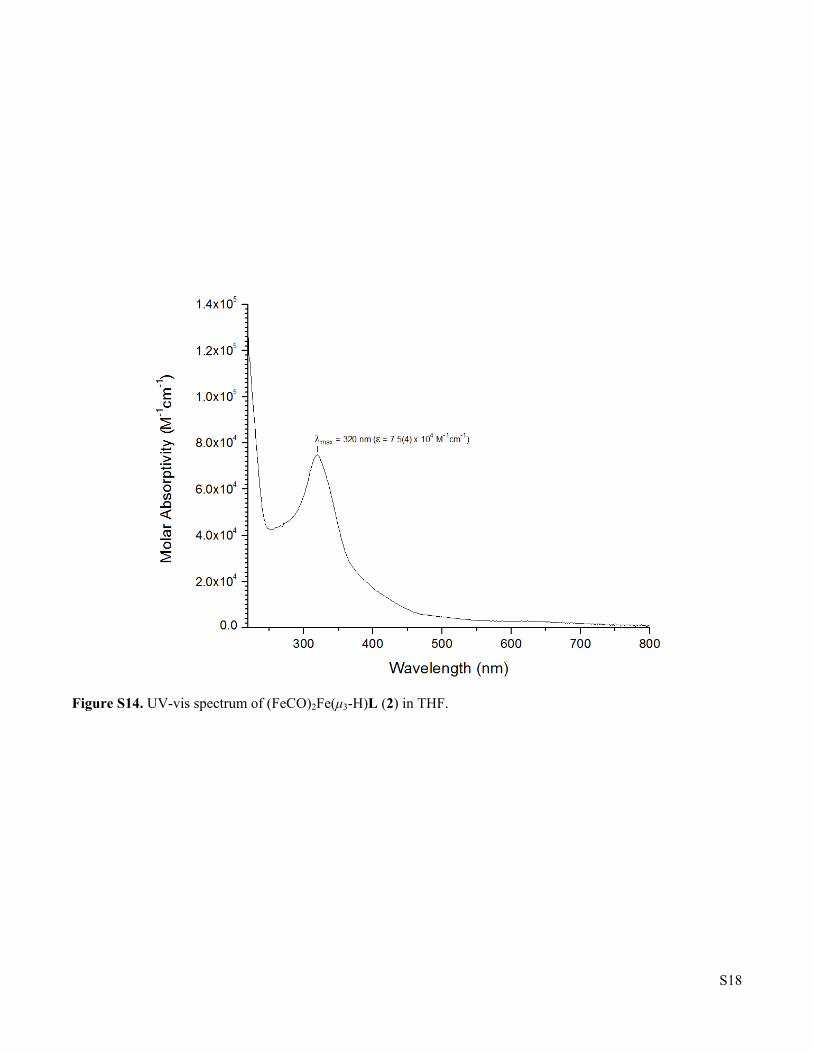
Figure S14. UV-vis spectrum of (FeCO)2Fe(μ3-H)L (2) in THF.
S19
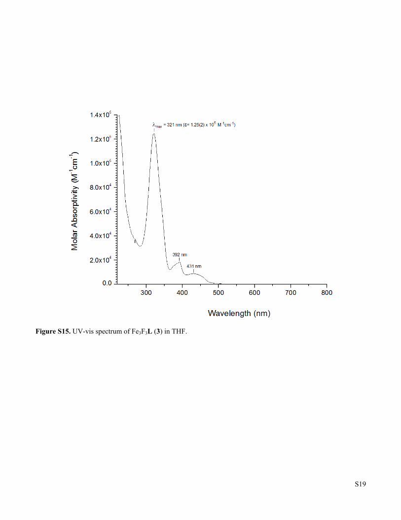
Figure S15. UV-vis spectrum of Fe3F3L (3) in THF.
S20
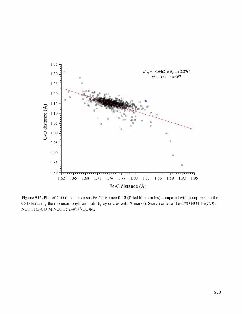
Figure S16. Plot of C-O distance versus Fe-C distance for 2 (filled blue circles) compared with complexes in the CSD featuring the monocarbonyliron motif (gray circles with X marks). Search criteria: Fe-C≡O NOT Fe(CO)2 NOT Fe(μ-CO)M NOT Fe(μ-η1:η1-CO)M.
S21
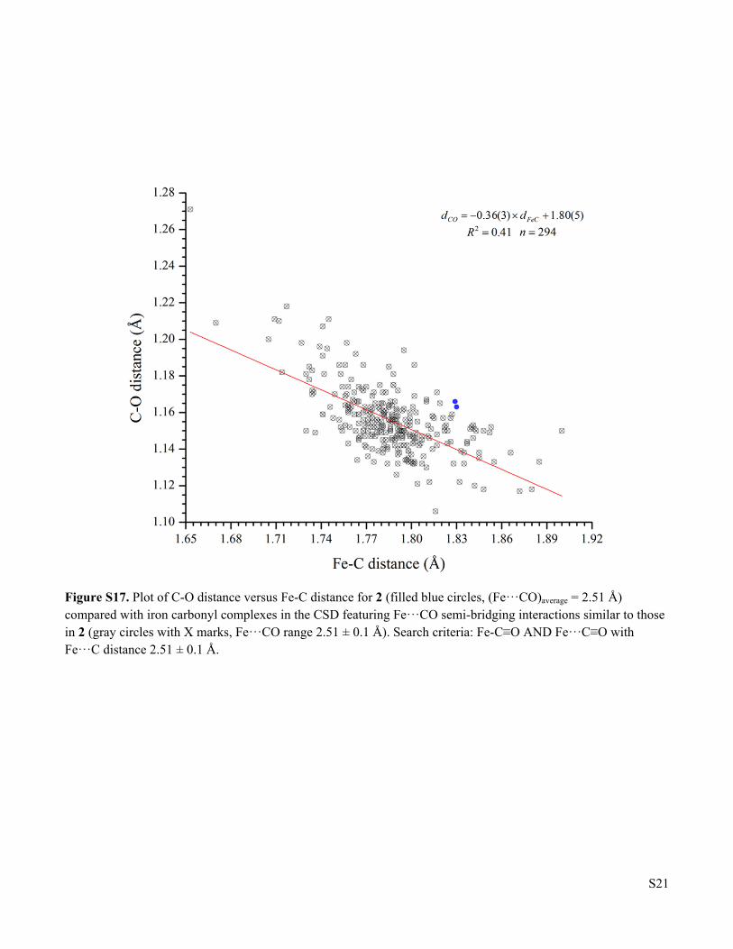
Figure S17. Plot of C-O distance versus Fe-C distance for 2 (filled blue circles, (Fe···CO)average = 2.51 Å) compared with iron carbonyl complexes in the CSD featuring Fe···CO semi-bridging interactions similar to those in 2 (gray circles with X marks, Fe···CO range 2.51 ± 0.1 Å). Search criteria: Fe-C≡O AND Fe···C≡O with Fe···C distance 2.51 ± 0.1 Å.
S22
Figure S18. Top-down view of the solid state structure of (FeCO)2Fe(μ3-H)L (2) showing the minor occupied site for Fe1. Fe1’ and Fe1” (green spheres) are modeled with occupancies of 3.5% and 2.5%, respectively. C, N, O, amd Fe atoms modeled as gray, blue, red, and orange thermal ellipsoids (75% probability). The brigding hydride is shown as a black sphere. All other H-atoms and solvent molecules are omitted for clarity.
S23
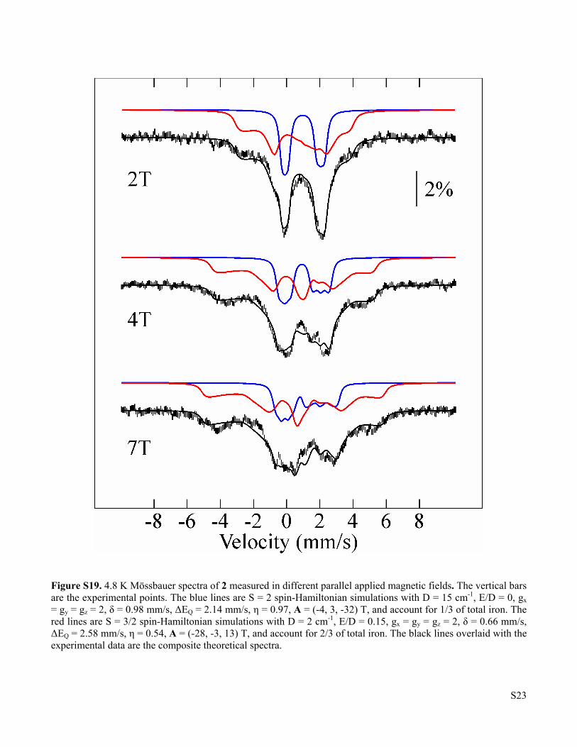
Figure S19. 4.8 K Mössbauer spectra of 2 measured in different parallel applied magnetic fields. The vertical bars are the experimental points. The blue lines are S = 2 spin-Hamiltonian simulations with D = 15 cm-1, E/D = 0, gx = gy = gz = 2, δ = 0.98 mm/s, ΔEQ = 2.14 mm/s, η = 0.97, A = (-4, 3, -32) T, and account for 1/3 of total iron. The red lines are S = 3/2 spin-Hamiltonian simulations with D = 2 cm-1, E/D = 0.15, gx = gy = gz = 2, δ = 0.66 mm/s, ΔEQ = 2.58 mm/s, η = 0.54, A = (-28, -3, 13) T, and account for 2/3 of total iron. The black lines overlaid with the experimental data are the composite theoretical spectra.
S24
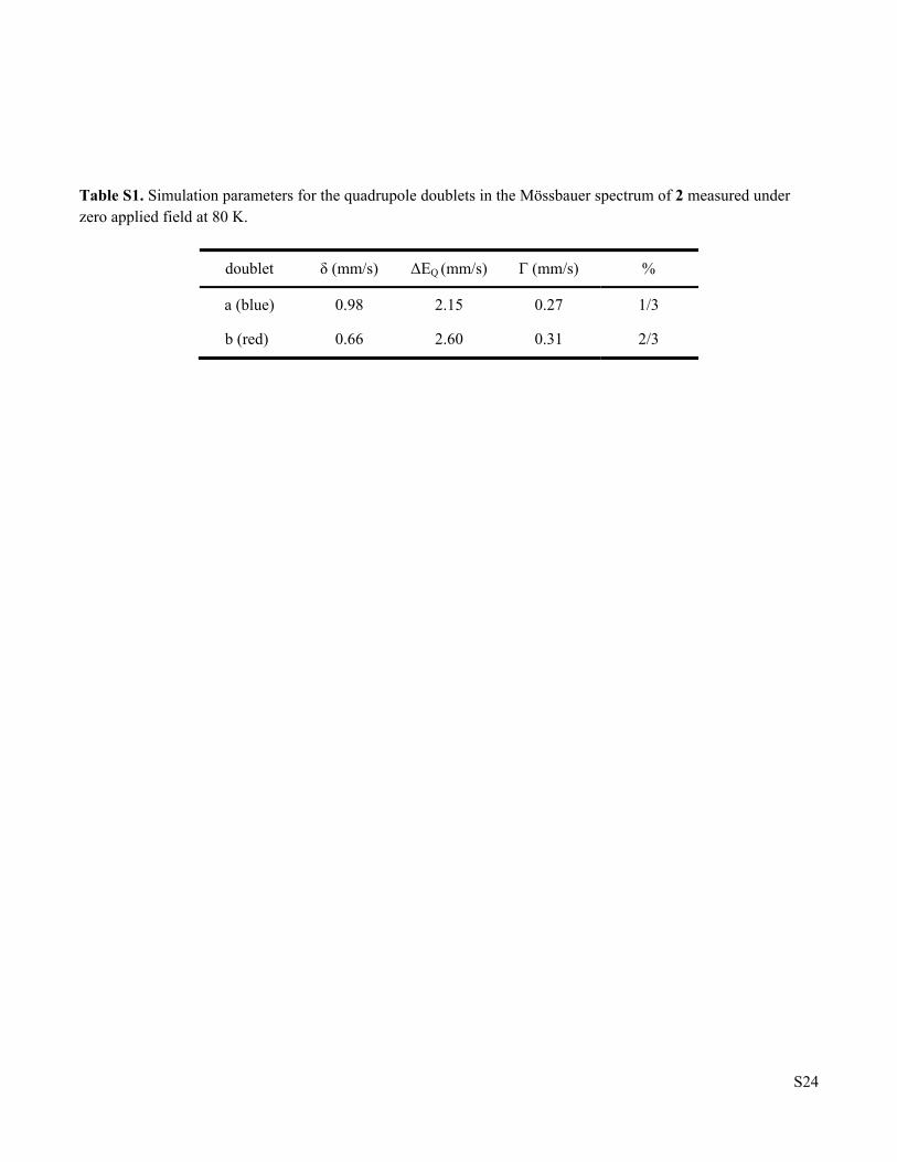
Table S1. Simulation parameters for the quadrupole doublets in the Mössbauer spectrum of 2 measured under zero applied field at 80 K.
doublet δ (mm/s) ΔEQ (mm/s) Γ (mm/s) %
a (blue) 0.98 2.15 0.27 1/3
b (red) 0.66 2.60 0.31 2/3
S25
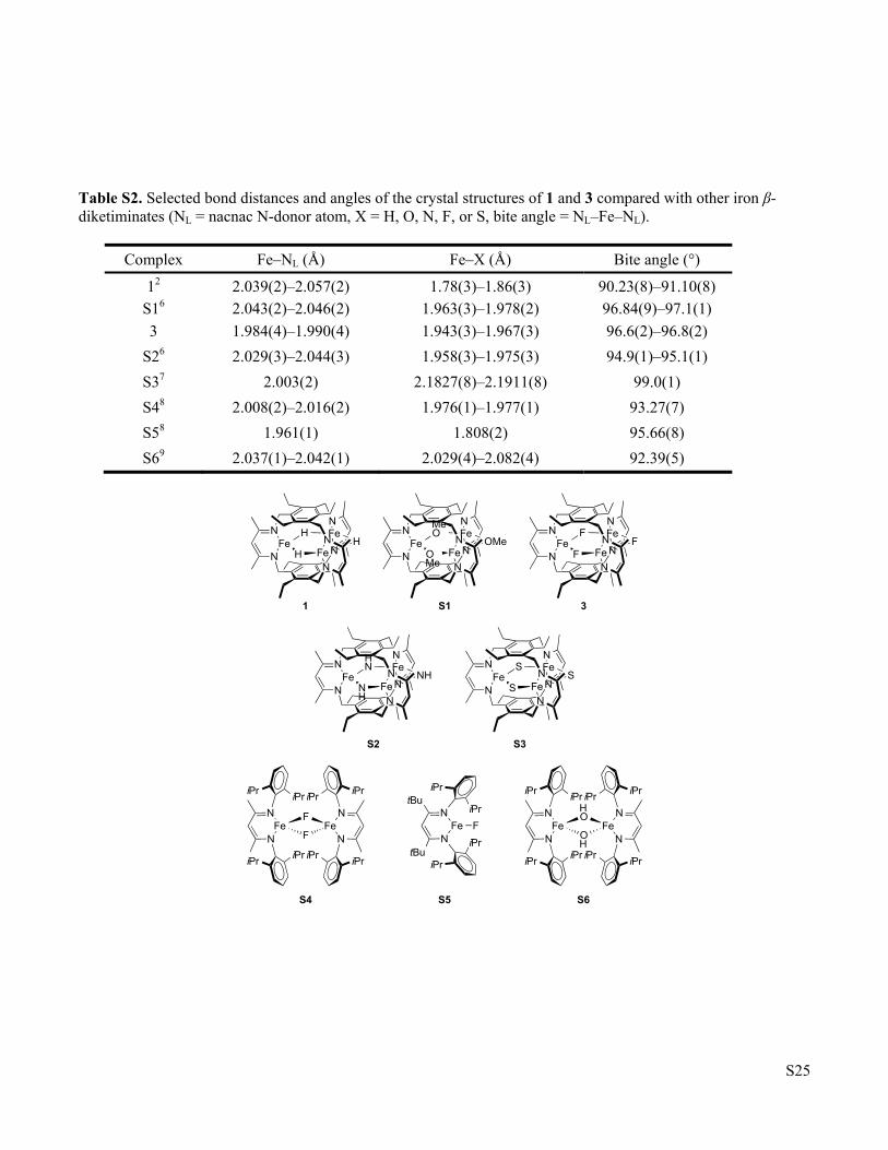
Table S2. Selected bond distances and angles of the crystal structures of 1 and 3 compared with other iron β-diketiminates (NL = nacnac N-donor atom, X = H, O, N, F, or S, bite angle = NL–Fe–NL).
Complex Fe–NL (Å) Fe–X (Å) Bite angle (°)
12 2.039(2)–2.057(2) 1.78(3)–1.86(3) 90.23(8)–91.10(8)
S16 2.043(2)–2.046(2) 1.963(3)–1.978(2) 96.84(9)–97.1(1)
3 1.984(4)–1.990(4) 1.943(3)–1.967(3) 96.6(2)–96.8(2)
S26 2.029(3)–2.044(3) 1.958(3)–1.975(3) 94.9(1)–95.1(1)
S37 2.003(2) 2.1827(8)–2.1911(8) 99.0(1)
S48 2.008(2)–2.016(2) 1.976(1)–1.977(1) 93.27(7)
S58 1.961(1) 1.808(2) 95.66(8)
S69 2.037(1)–2.042(1) 2.029(4)–2.082(4) 92.39(5)
NFe
NFe
HN
N
N
N
FeH
H
NFe
NFe
MeON
N
N
N
FeOMe
OMe
NFe
NFe
FN
N
N
N
FeF
F
NFe
NFe
HNN
N
N
N
FeNH
NH
NFe
NFe
SN
N
N
N
FeS
S
N
NFe
F
F
iPriPr
iPriPr
N
NFe
iPriPr
iPriPr
N
NFe
OH
HO
iPriPr
iPriPr
N
NFe
iPriPr
iPriPr
NtBu
tBuNFe
iPr
iPr
F
iPr
iPr
1 S1 3
S2 S3
S4 S5 S6
S26
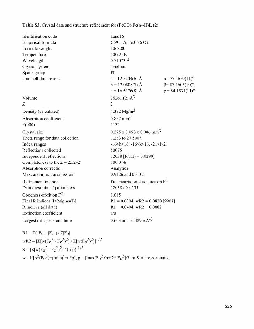
Table S3. Crystal data and structure refinement for (FeCO)2Fe(μ3-H)L (2). Identification code kand16 Empirical formula C59 H76 Fe3 N6 O2 Formula weight 1068.80 Temperature 100(2) K Wavelength 0.71073 Å Crystal system Triclinic Space group Pī Unit cell dimensions a = 12.5204(6) Å α= 77.1659(11)°. b = 13.0808(7) Å β= 87.1605(10)°. c = 16.5376(8) Å γ = 84.1531(11)°.
Volume 2626.1(2) Å3 Z 2
Density (calculated) 1.352 Mg/m3
Absorption coefficient 0.867 mm-1 F(000) 1132
Crystal size 0.275 x 0.098 x 0.086 mm3 Theta range for data collection 1.263 to 27.500°. Index ranges -16≤h≤16, -16≤k≤16, -21≤l≤21 Reflections collected 50075 Independent reflections 12038 [R(int) = 0.0290] Completeness to theta = 25.242° 100.0 % Absorption correction Analytical Max. and min. transmission 0.9426 and 0.8105
Refinement method Full-matrix least-squares on F2 Data / restraints / parameters 12038 / 0 / 655
Goodness-of-fit on F2 1.085 Final R indices [I>2sigma(I)] R1 = 0.0304, wR2 = 0.0820 [9908] R indices (all data) R1 = 0.0404, wR2 = 0.0882 Extinction coefficient n/a
Largest diff. peak and hole 0.603 and -0.489 e.Å-3 R1 = Σ(||Fo| - |Fc||) / Σ|Fo|
wR2 = [Σ[w(Fo2 - Fc2)2] / Σ[w(Fo2)2]]1/2
S = [Σ[w(Fo2 - Fc2)2] / (n-p)]1/2
w= 1/[σ2(Fo2)+(m*p)2+n*p], p = [max(Fo2,0)+ 2* Fc2]/3, m & n are constants.
S27
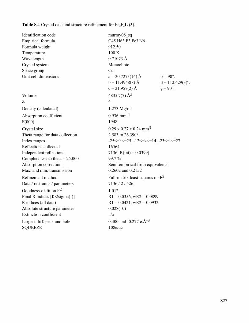
Table S4. Crystal data and structure refinement for Fe3F3L (3). Identification code murray08_sq Empirical formula C45 H63 F3 Fe3 N6 Formula weight 912.50 Temperature 100 K Wavelength 0.71073 Å Crystal system Monoclinic Space group Cc Unit cell dimensions a = 20.7273(14) Å α = 90°. b = 11.4948(8) Å β = 112.429(3)°. c = 21.957(2) Å γ = 90°.
Volume 4835.7(7) Å3 Z 4
Density (calculated) 1.273 Mg/m3
Absorption coefficient 0.936 mm-1 F(000) 1948
Crystal size 0.29 x 0.27 x 0.24 mm3 Theta range for data collection 2.583 to 26.390°. Index ranges -25<=h<=25, -12<=k<=14, -23<=l<=27 Reflections collected 16564 Independent reflections 7136 [R(int) = 0.0399] Completeness to theta = 25.000° 99.7 % Absorption correction Semi-empirical from equivalents Max. and min. transmission 0.2602 and 0.2152
Refinement method Full-matrix least-squares on F2 Data / restraints / parameters 7136 / 2 / 526
Goodness-of-fit on F2 1.012 Final R indices [I>2sigma(I)] R1 = 0.0356, wR2 = 0.0899 R indices (all data) R1 = 0.0421, wR2 = 0.0932 Absolute structure parameter 0.028(10) Extinction coefficient n/a
Largest diff. peak and hole 0.400 and -0.277 e.Å-3 SQUEEZE 108e/uc
S28
References. 1. (a) Evans, D. F. J. Chem. Soc. 1959, 2003-2005. (b) Bain, G. A.; Berry, J. F. J. Chem. Educ. 2008, 85,
532-536. (c) Sur, S. K. J. Magn. Reson. 1989, 82, 169-173. (d) Schubert, E. M. J. Chem. Educ. 1992, 69, 62.
2. Lee, Y.; Anderton, K. J.; Sloane, F. T.; Ermert, D. M.; Abboud, K. A.; García-Serres, R.; Murray, L. J. J. Am. Chem. Soc. 2015, 137, 10610-10617.
3. Brown, H.C.; Khuri, A.; Kim, S.C. Inorg. Chem. 1977, 16, 2229-2233. 4. SHELXTL2014 (2014). Bruker-AXS, Madison, Wisconsin, USA. 5. G. M. Sheldrick, SHELXTL-Plus Structure Determination Software Programs (2014). 6. Lee, Y.; Sloane, F. T.; Blondin, G.; Abboud, K. A.; García-Serres, R.; Murray, L. J. Angew. Chem. Int.
Ed. 2015, 54, 1499-1503. 7. Lee, Y.; Jeon, I.-R.; Abboud, K. A.; García-Serres, R.; Shearer, J.; Murray, L. J. Chem. Commun. 2016,
52, 1174-1177. 8. Vela, J.; Smith, J. M.; Yu, Y.; Ketterer, N. A.; Flaschenriem, C. J.; Lachicotte, R. J.; Holland, P. L. J. Am.
Chem. Soc. 2005, 127, 7857-7870. 9. Yu, Y.; Sadique, A. R.; Smith, J. M.; Dugan, T. R.; Cowley, R. E.; Brennessel, W. W.; Flaschenriem, C.
J.; Bill, E.; Cundari, T.; Holland, P. J. Am. Chem. Soc. 2008, 130, 6624-6638.


![Thesis title goes here - University of Toronto T-Space · PDF filedba dibenzalacetone DBU 1,8-diazabicyclo[5.4.0]undec-7-ene DIBAL diisobutylaluminum hydride DMF N,N-dimethylformamide](https://static.fdocument.org/doc/165x107/5aafc3be7f8b9a190d8dc089/thesis-title-goes-here-university-of-toronto-t-space-dibenzalacetone-dbu-18-diazabicyclo540undec-7-ene.jpg)