Radiographic features of plasma cell leukemia in the ... · Radiographic features of plasma cell...
6
- 273 - Imaging Science in Dentistry 2016; 46: 273-8 https://doi.org/10.5624/isd.2016.46.4.273 Plasma cell leukemia (PCL) is considered a symptoma- tic version of multiple myeloma, where more than 2,000 plasma cells/μL are found in the circulatory system, plas ma cells constitute more than 20% of the differential white count, and there is frequent extramedullary involvement. Symptoms include, but are not limited to, osteolytic le- sions, bone pain, hypercalcemia, anemia, and renal in- sufficiency. PCL can either be primary, where the disease manifests without a prior diagnosis of multiple myeloma, or secondary, where the disease progresses naturally from a previous diagnosis of multiple myeloma. 1-7 The causes of PCL are similar to those of multiple my- eloma. A series of genetic alterations during the develop- ment of a plasma cell may lead to the uncontrolled growth of the cell. However, what induces these alterations is not fully known. Risk factors, such as age and exposure to in- dustrial and environmental elements, are thought to play important roles. On a molecular level, PCL can be differ- entiated from multiple myeloma by fluorescent in situ hy- bridization (FISH) analysis looking for IgH translocations and chromosome 13q deletions. 4 The prognosis of primary and secondary PCL is report- ed to be poor, with a median survival of 6-8 months. This poor prognosis is most likely a result of the biologically aggressive nature of the disease and the sequelae of multi- ple myeloma treatments in patients with secondary PCL. 8 Magnetic resonance imaging is advised if neurologic symptoms exist, to evaluate for spinal cord compres- sion. 9-11 Leptomeningeal enhancement is the most common feature seen. In the case of extramedullary myelomas, like PCL, epidural soft tissue masses are reported to be homogenously hypointense on T1-weighted images and hyperintense on T2-weighted images. Other reports of radiographic features of PCL include multi-detector com- puted tomography (CT) of the abdominal region exhibit- ing masses attached the chest wall 12 or focal lesions of metastatic infiltration to the liver. Positron emission to- mography/CT (PET/CT) imaging features have also been reported with increased glucose uptake in the vertebrae, ribs, and pelvis. 10 However, our literature review did not reveal any exclusive studies about the oral and maxillo- facial manifestations of plasma cell leukemia using com- mon dental imaging techniques. Radiographic features of plasma cell leukemia in the maxilla: A case report Phillip Wong 1 , Deeba Kashtwari 1 , Madhu K. Nair 1, * 1 Division of Oral and Maxillofacial Radiology, Oral and Maxillofacial Diagnostic Sciences/Radiology, Colleges of Dentistry/Medicine, University of Florida, Gainesville, FL, USA ABSTRACT Plasma cell leukemia (PCL) is an aggressive form of multiple myeloma where there is hematogenous spread of abnormal plasma cells into the periphery. This is opposed to multiple myeloma, where the abnormal plasma cells stay in the bone marrow. PCL is more common in males than females, and is also more common in African- Americans than Caucasians. Signs and symptoms of PCL include, but are not limited to, renal insufficiency, hypercalcemia, anemia, lytic bone lesions, thrombocytopenia, hepatomegaly, and splenomegaly. Here, we discussed a case of a 71-year-old Caucasian female recently diagnosed with primary PCL with radiographic features of this disease throughout the body, with an emphasis on the maxillofacial skeleton and relevance from a dental standpoint. (Imaging Sci Dent 2016; 46: 273-8) KEY WORDS: Leukemia, Plasma Cell; Radiology; Pathology Copyright ⓒ 2016 by Korean Academy of Oral and Maxillofacial Radiology This is an Open Access article distributed under the terms of the Creative Commons Attribution Non-Commercial License (http://creativecommons.org/licenses/by-nc/3.0) which permits unrestricted non-commercial use, distribution, and reproduction in any medium, provided the original work is properly cited. Imaging Science in Dentistry·pISSN 2233-7822 eISSN 2233-7830 Received June 14, 2016; Revised September 6, 2016; Accepted September 20, 2016 *Correspondence to : Prof. Madhu K. Nair Division of Oral and Maxillofacial Radiology, Oral and Maxillofacial Diagnostic Sciences/Radiology, Colleges of Dentistry/Medicine, University of Florida, 1395 Center Dr., Room D8-6, Gainesville, FL 32610, USA Tel) 1-352-273-6690, Fax) 1-352-294-5321, E-mail) [email protected]
Transcript of Radiographic features of plasma cell leukemia in the ... · Radiographic features of plasma cell...
- 273 -
Imaging Science in Dentistry 2016; 46: 273-8https://doi.org/10.5624/isd.2016.46.4.273
Plasma cell leukemia (PCL) is considered a symptomatic version of multiple myeloma, where more than 2,000 plasma cells/μL are found in the circulatory system, plasma cells constitute more than 20% of the differential white count, and there is frequent extramedullary involvement. Symptoms include, but are not limited to, osteolytic lesions, bone pain, hypercalcemia, anemia, and renal insufficiency. PCL can either be primary, where the disease manifests without a prior diagnosis of multiple myeloma, or secondary, where the disease progresses naturally from a previous diagnosis of multiple myeloma.17
The causes of PCL are similar to those of multiple myeloma. A series of genetic alterations during the development of a plasma cell may lead to the uncontrolled growth of the cell. However, what induces these alterations is not fully known. Risk factors, such as age and exposure to industrial and environmental elements, are thought to play important roles. On a molecular level, PCL can be differentiated from multiple myeloma by fluorescent in situ hy
bridization (FISH) analysis looking for IgH translocations and chromosome 13q deletions.4
The prognosis of primary and secondary PCL is reported to be poor, with a median survival of 68 months. This poor prognosis is most likely a result of the biologically aggressive nature of the disease and the sequelae of multiple myeloma treatments in patients with secondary PCL.8
Magnetic resonance imaging is advised if neurologic symptoms exist, to evaluate for spinal cord compression.911 Leptomeningeal enhancement is the most common feature seen. In the case of extramedullary myelomas, like PCL, epidural soft tissue masses are reported to be homogenously hypointense on T1weighted images and hyperintense on T2weighted images. Other reports of radiographic features of PCL include multidetector computed tomography (CT) of the abdominal region exhibiting masses attached the chest wall12 or focal lesions of metastatic infiltration to the liver. Positron emission tomography/CT (PET/CT) imaging features have also been reported with increased glucose uptake in the vertebrae, ribs, and pelvis.10 However, our literature review did not reveal any exclusive studies about the oral and maxillofacial manifestations of plasma cell leukemia using common dental imaging techniques.
Radiographic features of plasma cell leukemia in the maxilla: A case report
Phillip Wong1, Deeba Kashtwari1, Madhu K. Nair1,*1Division of Oral and Maxillofacial Radiology, Oral and Maxillofacial Diagnostic Sciences/Radiology, Colleges of Dentistry/Medicine, University of Florida, Gainesville, FL, USA
AbstRAct
Plasma cell leukemia (PCL) is an aggressive form of multiple myeloma where there is hematogenous spread of abnormal plasma cells into the periphery. This is opposed to multiple myeloma, where the abnormal plasma cells stay in the bone marrow. PCL is more common in males than females, and is also more common in AfricanAmericans than Caucasians. Signs and symptoms of PCL include, but are not limited to, renal insufficiency, hypercalcemia, anemia, lytic bone lesions, thrombocytopenia, hepatomegaly, and splenomegaly. Here, we discussed a case of a 71yearold Caucasian female recently diagnosed with primary PCL with radiographic features of this disease throughout the body, with an emphasis on the maxillofacial skeleton and relevance from a dental standpoint.
(Imaging Sci Dent 2016; 46: 273-8)
Key woRds: Leukemia, Plasma Cell; Radiology; Pathology
Copyright ⓒ 2016 by Korean Academy of Oral and Maxillofacial RadiologyThis is an Open Access article distributed under the terms of the Creative Commons Attribution NonCommercial License (http://creativecommons.org/licenses/bync/3.0)
which permits unrestricted noncommercial use, distribution, and reproduction in any medium, provided the original work is properly cited.Imaging Science in Dentistry·pISSN 22337822 eISSN 22337830
Received June 14, 2016; Revised September 6, 2016; Accepted September 20, 2016*Correspondence to : Prof. Madhu K. NairDivision of Oral and Maxillofacial Radiology, Oral and Maxillofacial Diagnostic Sciences/Radiology, Colleges of Dentistry/Medicine, University of Florida, 1395 Center Dr., Room D86, Gainesville, FL 32610, USATel) 13522736690, Fax) 13522945321, Email) [email protected]
Radiographic features of plasma cell leukemia in the maxilla: A case report
- 274 -
case ReportA 71yearold female presented to her primary care
physician in late 2014 complaining of fatigue, generalized weakness, and dyspnea upon minimal exertion. Routine laboratory values of major significance revealed hemoglobin levels of about 5.4 g/dL (reference 1116) and her platelets count at 85,000/mL (reference 150,000450,000/mL).
The patient was referred to the emergency department at the University of Florida Health, Shands Hospital for
bone marrow biopsy and a hematology consult. Upon admission, it was noted that her serum calcium levels were elevated to 11.2 mg/dL (reference 8.510.2) and IgG levels were elevated to 2638 mg/dL (reference 6201400) with a kappa:lambda ratio of 1047 : 27 (reference 3 : 1). A peripheral blood smear revealed abnormal plasma cells and a 41% distribution of lymphocytes, with 20% being plasma cells.
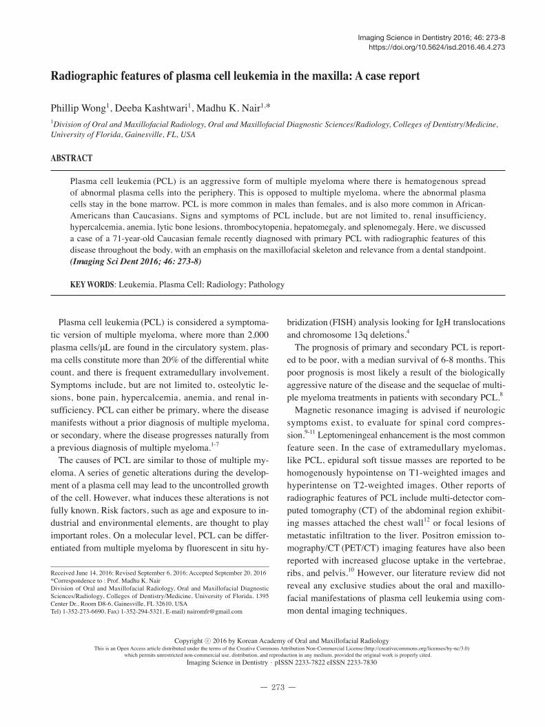
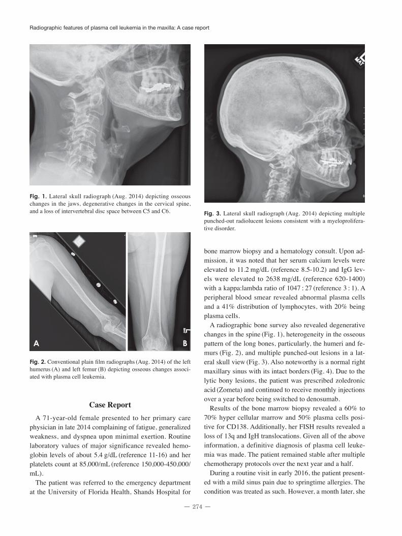
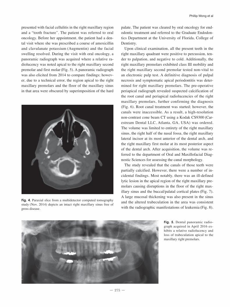
A radiographic bone survey also revealed degenerative changes in the spine (Fig. 1), heterogeneity in the osseous pattern of the long bones, particularly, the humeri and femurs (Fig. 2), and multiple punchedout lesions in a lateral skull view (Fig. 3). Also noteworthy is a normal right maxillary sinus with its intact borders (Fig. 4). Due to the lytic bony lesions, the patient was prescribed zoledronic acid (Zometa) and continued to receive monthly injections over a year before being switched to denosumab.
Results of the bone marrow biopsy revealed a 60% to 70% hyper cellular marrow and 50% plasma cells positive for CD138. Additionally, her FISH results revealed a loss of 13q and IgH translocations. Given all of the above information, a definitive diagnosis of plasma cell leukemia was made. The patient remained stable after multiple chemotherapy protocols over the next year and a half.
During a routine visit in early 2016, the patient presented with a mild sinus pain due to springtime allergies. The condition was treated as such. However, a month later, she
Fig. 1. Lateral skull radiograph (Aug. 2014) depicting osseous changes in the jaws, degenerative changes in the cervical spine, and a loss of intervertebral disc space between C5 and C6. Fig. 3. Lateral skull radiograph (Aug. 2014) depicting multiple
punchedout radiolucent lesions consistent with a myeloproliferative disorder.
Fig. 2. Conventional plain film radiographs (Aug. 2014) of the left humerus (A) and left femur (B) depicting osseous changes associated with plasma cell leukemia.
A B
- 275 -
Phillip Wong et al
presented with facial cellulitis in the right maxillary region and a “tooth fracture”. The patient was referred to oral oncology. Before her appointment, the patient had a dental visit where she was prescribed a course of amoxicillin and clavulanate potassium (Augmentin) and the facial swelling resolved. During the visit with oral oncology, a panoramic radiograph was acquired where a relative radiolucency was noted apical to the right maxillary second premolar and first molar (Fig. 5). A panoramic radiograph was also elicited from 2014 to compare findings; however, due to a technical error, the region apical to the right maxillary premolars and the floor of the maxillary sinus in that area were obscured by superimposition of the hard
palate. The patient was cleared by oral oncology for endodontic treatment and referred to the Graduate Endodontics Department at the University of Florida, College of Dentistry.
Upon clinical examination, all the present teeth in the right maxillary quadrant were positive to percussion, tender to palpation, and negative to cold. Additionally, the right maxillary premolars exhibited class III mobility and the right maxillary second premolar tested nonvital to an electronic pulp test. A definitive diagnosis of pulpal necrosis and symptomatic apical periodontitis was determined for right maxillary premolars. The preoperative periapical radiograph revealed suspected calcification of the root canal and periapical radiolucencies of the right maxillary premolars, further confirming the diagnosis
(Fig. 6). Root canal treatment was started; however, the canals were inaccessible. As a result, a highresolution noncontrast cone beam CT using a Kodak CS9300 (Carestream Dental LLC, Atlanta, GA, USA) was ordered. The volume was limited to entirety of the right maxillary sinus, the right half of the nasal fossa, the right maxillary lateral incisor at its most anterior of the dental arch, and the right maxillary first molar at its most posterior aspect of the dental arch. After acquisition, the volume was referred to the department of Oral and Maxillofacial Diagnostic Sciences for assessing the canal morphology.
The study revealed that the canals of those teeth were partially calcified. However, there were a number of incidental findings. Most notably, there was an illdefined lytic lesion in the apical region of the right maxillary premolars causing disruptions in the floor of the right maxillary sinus and the buccal/palatal cortical plates (Fig. 7). A large mucosal thickening was also present in the sinus and the altered trabeculation in the area was consistent with the radiographic manifestations of leukemia (Fig. 8).
Fig. 5. Dental panoramic radiograph acquired in April 2016 exhibits a relative radiolucency and loss of trabeculation apical to the maxillary right premolars.
Fig. 4. Paraxial slice from a multidetector computed tomography study (Nov. 2014) depicts an intact right maxillary sinus free of gross disease.
Radiographic features of plasma cell leukemia in the maxilla: A case report
- 276 -
In the absence of a medical history, these imaging features could also represent other malignant lesions such as squamous cell carcinoma, or an inflammatory process like a medicationinduced osteonecrosis. However, these disease processes are not mutually exclusive and a histopathological examination and an immediate oncology consultation were recommended.
Temporary restorations were placed and the patient agreed that the questionable teeth would be extracted if they cause any further problems. An incisional biopsy
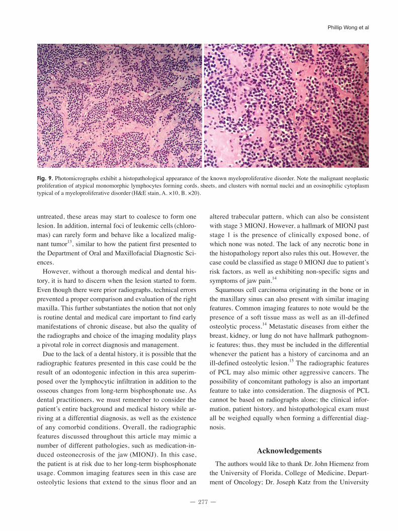
from the right maxilla approximately 1.5 cm apical to the premolars with no identifiable bone was submitted to the Division of Oral Pathology for histopathologic examination. Under further analysis, a malignant neoplastic proliferation of atypical lymphocytes was found exhibiting abnormal mitosis, pleomorphism, and anaplasia. These cells were forming cords, sheets, and clusters with normal nuclei and an eosinophilic cytoplasm (Fig. 9). There was also evidence of perineural and lymphovascular spread with a sclerotic fibrous connective tissue present in the background. Overall, the histomorphology was deemed to be compatible with the patient’s history of plasma cell leukemia and no additional stains were needed. The patient was referred to the HematologyOncology Department in the College of Medicine for hematopathologic workup.
discussionAlthough osseous changes of the jawbones in leukemia
are rare, the practitioner must keep them in consideration, especially in patients with a history of the disease. It is frequently localized around the periapical region of teeth and can mimic the signs and symptoms of rarefying osteitis. There may also be generalized illdefined radiolucent lesions due to the enlarged marrow spaces and, if left
Fig. 8. Parasagittal slice from a cone beam computed tomography study (Apr. 2016) shows the altered trabeculation in the periapical region of the premolars, an oroantral communication, and the reactive mucositis of the right maxillary sinus.
Fig. 7. Paraxial slice from a cone beam computed tomography study (Apr. 2016) reveals the loss of the buccal and lingual cortical borders in the region of the premolars.
Fig. 6. Periapical radiograph (Apr. 2016). Note the characteristic widening of the periodontal ligament spaces and sparse trabeculation in the periapical region surrounding the premolars.
- 277 -
Phillip Wong et al
untreated, these areas may start to coalesce to form one lesion. In addition, internal foci of leukemic cells (chloromas) can rarely form and behave like a localized malignant tumor13, similar to how the patient first presented to the Department of Oral and Maxillofacial Diagnostic Sciences.
However, without a thorough medical and dental history, it is hard to discern when the lesion started to form. Even though there were prior radiographs, technical errors prevented a proper comparison and evaluation of the right maxilla. This further substantiates the notion that not only is routine dental and medical care important to find early manifestations of chronic disease, but also the quality of the radiographs and choice of the imaging modality plays a pivotal role in correct diagnosis and management.
Due to the lack of a dental history, it is possible that the radiographic features presented in this case could be the result of an odontogenic infection in this area superimposed over the lymphocytic infiltration in addition to the osseous changes from longterm bisphosphonate use. As dental practitioners, we must remember to consider the patient’s entire background and medical history while arriving at a differential diagnosis, as well as the existence of any comorbid conditions. Overall, the radiographic features discussed throughout this article may mimic a number of different pathologies, such as medicationinduced osteonecrosis of the jaw (MIONJ). In this case, the patient is at risk due to her longterm bisphosphonate usage. Common imaging features seen in this case are osteolytic lesions that extend to the sinus floor and an
altered trabecular pattern, which can also be consistent with stage 3 MIONJ. However, a hallmark of MIONJ past stage 1 is the presence of clinically exposed bone, of which none was noted. The lack of any necrotic bone in the histopathology report also rules this out. However, the case could be classified as stage 0 MIONJ due to patient’s risk factors, as well as exhibiting nonspecific signs and symptoms of jaw pain.14
Squamous cell carcinoma originating in the bone or in the maxillary sinus can also present with similar imaging features. Common imaging features to note would be the presence of a soft tissue mass as well as an illdefined osteolytic process.14 Metastatic diseases from either the breast, kidney, or lung do not have hallmark pathognomic features; thus, they must be included in the differential whenever the patient has a history of carcinoma and an illdefined osteolytic lesion.15 The radiographic features of PCL may also mimic other aggressive cancers. The possibility of concomitant pathology is also an important feature to take into consideration. The diagnosis of PCL cannot be based on radiographs alone; the clinical information, patient history, and histopathological exam must all be weighed equally when forming a differential diagnosis.
AcknowledgementsThe authors would like to thank Dr. John Hiemenz from
the University of Florida, College of Medicine, Department of Oncology; Dr. Joseph Katz from the University
Fig. 9. Photomicrographs exhibit a histopathological appearance of the known myeloproliferative disorder. Note the malignant neoplastic proliferation of atypical monomorphic lymphocytes forming cords, sheets, and clusters with normal nuclei and an eosinophilic cytoplasm typical of a myeloproliferative disorder (H&E stain, A. ×10, B. ×20).

Radiographic features of plasma cell leukemia in the maxilla: A case report
- 278 -
of Florida, College of Dentistry, Department of Oral Medicine; Drs. Hope Johnson, Craig Nixon, Ernest Rillman, John Zongker from the University of Florida, College of Dentistry, Department of Endodontics; Drs. Matthew Holman, Melvin Dolwick, and Benjamin Schlott from the University of Florida, College of Dentistry, Department of Oral Surgery; and Dr. Indraneel Bhattacharyya from the University of Florida, College of Dentistry, Department of Oral and Maxillofacial Diagnostic Sciences for their contributions and care provided to the patient.
References 1. JimenezZepeda VH, Dominguez VJ. Plasma cell leukemia: a
rare condition. Ann Hematol 2006; 85: 2637. 2. Yamamoto JF, Goodman MT. Patterns of leukemia incidence
in the United States by subtype and demographic characteristics, 19972002. Cancer Causes Control 2008; 19: 37990.
3. Tiedemann RE, GonzalezPaz N, Kyle RA, SantanaDavila R, PriceTroska T, Van Wier SA, et al. Genetic aberrations and survival in plasma cell leukemia. Leukemia 2008; 22: 104452.
4. Albarracin F, Fonseca R. Plasma cell leukemia. Blood Rev 2011; 25: 10712.
5. Sher T, Miller KC, Deeb G, Lee K, ChananKhan A. Plasma cell leukaemia and other aggressive plasma cell malignancies. Br J Haematol 2010; 150: 41827.
6. Han X, Kilfoy B, Zheng T, Holford TR, Zhu C, Zhu Y, et al. Lymphoma survival patterns by WHO subtype in the United
States, 19732003. Cancer Causes Control 2008; 19: 84158. 7. Campo E, Swerdlow SH, Harris NL, Pileri S, Stein H, Jaffe
ES. The 2008 WHO classification of lymphoid neoplasms and beyond: evolving concepts and practical applications. Blood 2011; 117: 501932.
8. Hayman SR, Fonseca R. Plasma cell leukemia. Curr Treat Options Oncol 2001; 2: 20516.
9. Keraliya AR, Krajewski KM, Giardino AA, Tirumani SH, Shinagare AB, Ramaiya NH, et al. Imaging of nervous system involvement in hematologic malignancies: what radiologists need to know. AJR Am J Roentgenol 2015; 205: 60417.
10. Fernandez de Larrea C, Kyle RA, Durie BG, Ludwig H, Usmani S, Vesole DH, et al. Plasma cell leukemia: consensus statement on diagnostic requirements, response criteria and treatment recommendations by the International Myeloma Working Group. Leukemia 2013; 27: 78091.
11. Moulopoulos LA, Dimopoulos MA. Magnetic resonance imaging of the bone marrow in hematologic malignancies. Blood 1997; 90: 212747.
12. Ali A, Paul Y, Nwabudike SM, Ogbonna O, Grantham M, TaddesseHeath L. Plasma cell leukemia presenting as a chest wall mass: a case report. Case Rep Oncol 2016; 9: 33843.
13. Epstein JB, Voss NJ, StevensonMoore P. Maxillofacial manifestations of multiple myeloma. An unusual case and review of the literature. Oral Surg Oral Med Oral Pathol 1984; 57: 26771.
14. Ruggiero SL. Diagnosis and staging of medicationrelated osteonecrosis of the jaw. Oral Maxillofac Surg Clin North Am 2015; 27: 47987.
15. Kumar G, Manjunatha B. Metastatic tumors to the jaws and oral cavity. J Oral Maxillofac Pathol 2013; 17: 715.