Protein kinase C β inhibits autophagy and sensitizes cervical … · cervical cancer Hela cells to...
12
Bioscience Reports (2017) 37 BSR20160445 DOI: 10.1042/BSR20160445 Received: 08 October 2016 Revised: 21 February 2017 Accepted: 24 February 2017 Accepted Manuscript Online: 28 February 2017 Version of Record published: 27 March 2017 Research Article Protein kinase C β inhibits autophagy and sensitizes cervical cancer Hela cells to cisplatin Na Li and Wei Zhang Department of Gynaecology and Obstetrics, Zhongnan Hospital of Wuhan University, Wuhan 430071, China Correspondence: Wei Zhang ([email protected]) Recently, autophagy has been indicated to play an essential role in various biological events, such as the response of cervical cancer cells to chemotherapy. However, the exact signalling mechanism that regulates autophagy during chemotherapy remains unclear. In the present study, we investigated the regulation by cisplatin on protein kinase C β (PKC β), on B-cell lymphoma 2 (Bcl-2) and on apoptosis in cervical cancer Hela cells. And then we examined the regulation by cisplatin on autophagy and the role of autophagy on the chemotherapy in Hela cells. In addition, the regulation of the PKC β on the autophagy was also investigated. Our results indicated that cisplatin promoted PKC β in Hela cells. The PKC β inhibitor re- duced the cisplatin-induced apoptosis, whereas increased the cisplatin-induced autophagy in Hela cells. On the other side, the PKC β overexpression aggravated the cisplatin-induced apoptosis, whereas down-regulated the cisplatin-induced autophagy. Taken together, our study firstly recognized the involvement of PKC β in the cytotoxicity of cisplatin via inhibit- ing autophagy in cervical cancer cells. We propose that PKC β would sensitize cervical cancer cells to chemotherapy via reducing the chemotherapy induced autophagy in cancer cells. Introduction Cisplatin is an effective chemotherapeutic agent that is widely applied in treating solid tumours such as bladder, head and neck, lung, ovarian and testicular cancers [1]. Besides its side effects, acquired resis- tance to the cisplatin of tumour emerges during the course of treatment limits its application [2-4], how- ever, cisplatin combination chemotherapy is still the basis of chemotherapy against many cancers. To our knowledge, the proposed mechanisms of chemotherapy resistance include changes in cellular uptake and release of agent, improved biotransformation, antiapoptotic mechanisms and autophagy [5-8]. Autophagy is a type of non-apoptotic cell death that degrades unnecessary or dysfunctional cellular components through the actions of lysosomes, and is essential for survival when cells faced a harsh en- vironment such as radiation and chemotherapy [9]. During the autophagy, targeted cytoplasmic con- stituents are packed into a double-membraned vesicle (autophagosome), which is fused with lysosomes to generate autolysosomes and is degraded consequently[10]. I t was found that chemotherapy activates autophagy in tumour cells, which has been considered to enhance cancer cell death or play a role in chemotherapy resistance [11,12]. The protein kinase C (PKC) family consists of at least 12 kinases with distinct roles in cell proliferation, differentiation and apoptosis [13].PCK familyplays a key role in tumour promotion and progression and it is an ideal target for cancer therapy [14].PCK β activation blocks insulin-induced endothelial nitric ox- ide synthase (eNOS) stimulation, whereas inhibition of PKC β restores vascular function in animal models [15]. Inaddition, the improved PKC β activity has also been found implicating in activating endothelial cell inflammatory [16]. Cisplatin activates several signal transduction pathways mediated by ROS, DNA damage, p53, TNFα andMAPKs [17], yet the specific pathways involved in autophagy relating to cisplatin are unknown, the c 2017 The Author(s). This is an open access article published by Portland Press Limited on behalf of the Biochemical Society and distributed under the Creative Commons Attribution Licence 4.0 (CC BY). 1
-
Upload
vuongduong -
Category
Documents
-
view
214 -
download
0
Transcript of Protein kinase C β inhibits autophagy and sensitizes cervical … · cervical cancer Hela cells to...
Bioscience Reports (2017) 37 BSR20160445DOI: 10.1042/BSR20160445
Received: 08 October 2016Revised: 21 February 2017Accepted: 24 February 2017
Accepted Manuscript Online:28 February 2017Version of Record published:27 March 2017
Research Article
Protein kinase C β inhibits autophagy and sensitizescervical cancer Hela cells to cisplatinNa Li and Wei ZhangDepartment of Gynaecology and Obstetrics, Zhongnan Hospital of Wuhan University, Wuhan 430071, China
Correspondence: Wei Zhang ([email protected])
Recently, autophagy has been indicated to play an essential role in various biological events,such as the response of cervical cancer cells to chemotherapy. However, the exact signallingmechanism that regulates autophagy during chemotherapy remains unclear. In the presentstudy, we investigated the regulation by cisplatin on protein kinase C β (PKC β), on B-celllymphoma 2 (Bcl-2) and on apoptosis in cervical cancer Hela cells. And then we examinedthe regulation by cisplatin on autophagy and the role of autophagy on the chemotherapy inHela cells. In addition, the regulation of the PKC β on the autophagy was also investigated.Our results indicated that cisplatin promoted PKC β in Hela cells. The PKC β inhibitor re-duced the cisplatin-induced apoptosis, whereas increased the cisplatin-induced autophagyin Hela cells. On the other side, the PKC β overexpression aggravated the cisplatin-inducedapoptosis, whereas down-regulated the cisplatin-induced autophagy. Taken together, ourstudy firstly recognized the involvement of PKC β in the cytotoxicity of cisplatin via inhibit-ing autophagy in cervical cancer cells. We propose that PKC β would sensitize cervicalcancer cells to chemotherapy via reducing the chemotherapy induced autophagy in cancercells.
IntroductionCisplatin is an effective chemotherapeutic agent that is widely applied in treating solid tumours such asbladder, head and neck, lung, ovarian and testicular cancers [1]. Besides its side effects, acquired resis-tance to the cisplatin of tumour emerges during the course of treatment limits its application [2-4], how-ever, cisplatin combination chemotherapy is still the basis of chemotherapy against many cancers. To ourknowledge, the proposed mechanisms of chemotherapy resistance include changes in cellular uptake andrelease of agent, improved biotransformation, antiapoptotic mechanisms and autophagy [5-8].
Autophagy is a type of non-apoptotic cell death that degrades unnecessary or dysfunctional cellularcomponents through the actions of lysosomes, and is essential for survival when cells faced a harsh en-vironment such as radiation and chemotherapy [9]. During the autophagy, targeted cytoplasmic con-stituents are packed into a double-membraned vesicle (autophagosome), which is fused with lysosomesto generate autolysosomes and is degraded consequently [10]. It was found that chemotherapy activatesautophagy in tumour cells, which has been considered to enhance cancer cell death or play a role inchemotherapy resistance [11,12].
The protein kinase C (PKC) family consists of at least 12 kinases with distinct roles in cell proliferation,differentiation and apoptosis [13]. PCK family plays a key role in tumour promotion and progression andit is an ideal target for cancer therapy [14].PCK β activation blocks insulin-induced endothelial nitric ox-ide synthase (eNOS) stimulation, whereas inhibition of PKCβ restores vascular function in animal models[15]. In addition, the improved PKC β activity has also been found implicating in activating endothelialcell inflammatory [16].
Cisplatin activates several signal transduction pathways mediated by ROS, DNA damage, p53, TNFαand MAPKs [17], yet the specific pathways involved in autophagy relating to cisplatin are unknown, the
c© 2017 The Author(s). This is an open access article published by Portland Press Limited on behalf of the Biochemical Society and distributed under the Creative Commons AttributionLicence 4.0 (CC BY).
1
Bioscience Reports (2017) 37 BSR20160445DOI: 10.1042/BSR20160445
relationship between autophagy and apoptosis in cancer cells are still unclear and the signalling mechanism thatregulates autophagy during chemotherapy is to be explored. In this research, we investigated the regulation by cisplatinon PKC β, on B-cell lymphoma 2 (Bcl-2) and on apoptosis in cervical cancer Hela cells, examined the regulation bycisplatin on autophagy and the role of autophagy on the chemotherapy and investigated the regulation of the PKC β
on the autophagy.
Materials and methodsCell cultureThe cervical cancer Hela cell was purchased from ATCC. All cell lines were cultured in Dulbecco’s modified Eagle’smedium (DMEM) containing 10% FBS (Gibco, U.S.A.) in the humidified air with 5% CO2 at 37◦C. Cisplatin was pur-chased from Sigma–Aldrich (St. Louis, U.S.A.) and was dissolved in DMEM with a storage concentration of 1 mM.Enzastaurin (Sigma–Aldrich, U.S.A.) was utilized to inhibit PKC β. Rapamycin (Rapa) (Thermo Fisher Scientific,U.S.A.) and 3-Methyladenine (3-MA) (Sigma-Aldrich, U.S.A.) were utilized as positive and negative autophagy acti-vators respectively. To overexpress PCK β, the PCK β-pcDNA3.1(+) (Sino Biological, Beijing, China) was transfectedwith Lipofectamine 2000 (Invitrogen, Carlsbad, CA, U.S.A.) into Hela cells.
Assessment of cellular viability using MTT assayHela cells (2 × 10³) in the log-growth phase were seeded in a 96-well plate. After overnight growth, the cells weretreated with different doses of cisplatin/enzastaurin/PKC β. MTT (20 μl) in the final concentration of 5 mg/ml wasadded and the cells were incubated for 4 h at 37◦C. After the supernatant was carefully removed, 200 μl of DMSOwas added into each well and mixed. The plate was put into a 37◦C incubator to dissolve air bubbles and OD570 valueof each well was measured at 570 nm wavelength using a microplate reader (Thermo Scientific, U.S.A.). The resultswere expressed as (A570 of control wells – A570 of treated wells)/(A570 of control wells – A570 of blank wells) × 100%.
Western blot analysisTreated cells were lysed with lysis buffer (Invitrogen, U.S.A.) on ice for 20 min. The cell lysates were centrifugated at13000 × g at 4◦C for 30min, the supernatant was collected as the total cellular protein extract. Protein concentrationwas determined using the BCA Protein Assay Kit (Kangwei Shiji Company, Beijing). Samples of total cellular proteinwere loaded on to 10% SDS/PAGE. The separated proteins were electrophoretically transferred to PVDF membranes(Bio-Rad, U.S.A.). The membrane was blocked overnight in blocking buffer containing PBS-T and 5% non-fattymilk. Then the membrane was incubated with primary antibody against different antigens for 1 h separately and waswashed with PBST for four times subsequently. Following incubating with the secondary HRP-conjugated antibodyfor 1 h, the PVDF membrane was washed for four times and was treated with ECL reagent and exposed to X-ray film.Each band was quantified using Image software. The protein level of each molecule was calculated according to itsband intensity to glyceraldehyde-3-phosphate dehydrogenase (GAPDH) band intensity and was averaged for threeindependent experiments.
Apoptosis assayCells apoptotic percentage was performed by AnnexinV-FITC Apoptosis Detection Kit (Merk Company, U.S.A.).Briefly, the treated cells were centrifugated at 1000 × g for 5 min and washed twice with PBS, then the cells weresuspended in the 400 μl 1× binding buffer at a concentration of 1 × 10⁵ cells/ml, then 5 μl of Annexin V-FITCand propidium iodide was added in turn and mixed, the treated cells were placed in the dark at RT for 5–15 min toperform flow cytometry analysis.
Detection of autophagic punctas by fluorescence microscopyDetection of autophagic punctas was performed as previously described [18]. Briefly, treated Hela cells were seeded onsterile coverslips and incubated under the conditions mentioned above. The growth medium was removed and cellswere washed with cold PBS for two times when the cells’ confluence reached 80%. Then, fresh growth medium wassupplemented subsequently and cells were incubated for an additional 12 h. Detection of GFP–LC3 was performedusing FlowCellectTM GFP–LC3 Reporter Autophagy Assay kit (Millipore, U.S.A.). Briefly, 10 μl autophagy reagentA was added and cells were incubated in a humidified incubator at 37◦C with 5% CO2 for 2 h. Post the removal ofmedium, cells were washed with 5 ml 1× HBSS, were added with 100 ml 1× autophagy reagent B, and then werecultured with cells for 5 min, followed by washing with assay buffer for the autophagy reagent removal. Last, the
2 c© 2017 The Author(s). This is an open access article published by Portland Press Limited on behalf of the Biochemical Society and distributed under the Creative Commons AttributionLicence 4.0 (CC BY).
Bioscience Reports (2017) 37 BSR20160445DOI: 10.1042/BSR20160445
coverslips were covered by sterile slides and the slides were observed under an Olympus fluorescence microscope(BX51, Olympus Corporation, Japan).
Statistical analysisData are depicted as the mean +− S.D. For a comparison between the two groups, Student’s t test was used. For mul-tiple comparisons among three or more groups, one-way ANOVA was used. P<0.05 was considered as a statisticallysignificant difference.
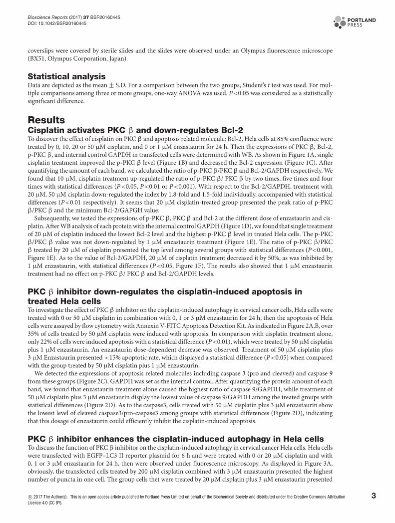
ResultsCisplatin activates PKC β and down-regulates Bcl-2To discover the effect of cisplatin on PKC β and apoptosis related molecule: Bcl-2, Hela cells at 85% confluence weretreated by 0, 10, 20 or 50 μM cisplatin, and 0 or 1 μM enzastaurin for 24 h. Then the expressions of PKC β, Bcl-2,p-PKC β, and internal control GAPDH in transfected cells were determined with WB. As shown in Figure 1A, singlecisplatin treatment improved the p-PKC β level (Figure 1B) and decreased the Bcl-2 expression (Figure 1C). Afterquantifying the amount of each band, we calculated the ratio of p-PKC β/PKC β and Bcl-2/GAPDH respectively. Wefound that 10 μM, cisplatin treatment up-regulated the ratio of p-PKC β/ PKC β by two times, five times and fourtimes with statistical differences (P<0.05, P<0.01 or P<0.001). With respect to the Bcl-2/GAPDH, treatment with20 μM, 50 μM cisplatin down-regulated the index by 1.8-fold and 1.5-fold individually, accompanied with statisticaldifferences (P<0.01 respectively). It seems that 20 μM cisplatin-treated group presented the peak ratio of p-PKCβ/PKC β and the minimum Bcl-2/GAPGH value.
Subsequently, we tested the expressions of p-PKC β, PKC β and Bcl-2 at the different dose of enzastaurin and cis-platin. After WB analysis of each protein with the internal control GAPDH (Figure 1D), we found that single treatmentof 20 μM of cisplatin induced the lowest Bcl-2 level and the highest p-PKC β level in treated Hela cells. The p-PKCβ/PKC β value was not down-regulated by 1 μM enzastaurin treatment (Figure 1E). The ratio of p-PKC β/PKCβ treated by 20 μM of cisplatin presented the top level among several groups with statistical differences (P<0.001,Figure 1E). As to the value of Bcl-2/GAPDH, 20 μM of cisplatin treatment decreased it by 50%, as was inhibited by1 μM enzastaurin, with statistical differences (P<0.05, Figure 1F). The results also showed that 1 μM enzastaurintreatment had no effect on p-PKC β/ PKC β and Bcl-2/GAPDH levels.
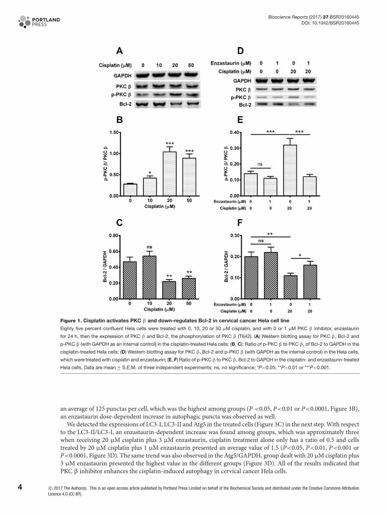
PKC β inhibitor down-regulates the cisplatin-induced apoptosis intreated Hela cellsTo investigate the effect of PKCβ inhibitor on the cisplatin-induced autophagy in cervical cancer cells, Hela cells weretreated with 0 or 50 μM cisplatin in combination with 0, 1 or 3 μM enzastaurin for 24 h, then the apoptosis of Helacells were assayed by flow cytometry with Annexin V-FITC Apoptosis Detection Kit. As indicated in Figure 2A,B, over35% of cells treated by 50 μM cisplatin were induced with apoptosis. In comparison with cisplatin treatment alone,only 22% of cells were induced apoptosis with a statistical difference (P<0.01), which were treated by 50 μM cisplatinplus 1 μM enzastaurin. An enzastaurin dose-dependent decrease was observed. Treatment of 50 μM cisplatin plus3 μM Enzastaurin presented <15% apoptotic rate, which displayed a statistical difference (P<0.05) when comparedwith the group treated by 50 μM cisplatin plus 1 μM enzastaurin.
We detected the expressions of apoptosis related molecules including caspase 3 (pro and cleaved) and caspase 9from these groups (Figure 2C), GAPDH was set as the internal control. After quantifying the protein amount of eachband, we found that enzastaurin treatment alone caused the highest ratio of caspase 9/GAPDH, while treatment of50 μM cisplatin plus 3 μM enzastaurin display the lowest value of caspase 9/GAPDH among the treated groups withstatistical differences (Figure 2D). As to the caspase3, cells treated with 50 μM cisplatin plus 3 μM enzastaurin showthe lowest level of cleaved caspase3/pro-caspase3 among groups with statistical differences (Figure 2D), indicatingthat this dosage of enzastaurin could efficiently inhibit the cisplatin-induced apoptosis.
PKC β inhibitor enhances the cisplatin-induced autophagy in Hela cellsTo discuss the function of PKC β inhibitor on the cisplatin-induced autophagy in cervical cancer Hela cells. Hela cellswere transfected with EGFP–LC3 II reporter plasmid for 6 h and were treated with 0 or 20 μM cisplatin and with0, 1 or 3 μM enzastaurin for 24 h, then were observed under fluorescence microscopy. As displayed in Figure 3A,obviously, the transfected cells treated by 200 μM cisplatin combined with 3 μM enzastaurin presented the highestnumber of puncta in one cell. The group cells thet were treated by 20 μM cisplatin plus 3 μM enzastaurin presented
c© 2017 The Author(s). This is an open access article published by Portland Press Limited on behalf of the Biochemical Society and distributed under the Creative Commons AttributionLicence 4.0 (CC BY).
3
Bioscience Reports (2017) 37 BSR20160445DOI: 10.1042/BSR20160445
Figure 1. Cisplatin activates PKC β and down-regulates Bcl-2 in cervical cancer Hela cell line
Eighty five percent confluent Hela cells were treated with 0, 10, 20 or 50 μM cisplatin, and with 0 or 1 μM PKC β inhibitor, enzastaurin
for 24 h, then the expression of PKC β and Bcl-2, the phosphorylation of PKC β (T642). (A) Western blotting assay for PKC β, Bcl-2 and
p-PKC β (with GAPDH as an internal control) in the cisplatin-treated Hela cells; (B, C): Ratio of p-PKC β to PKC β, of Bcl-2 to GAPDH in the
cisplatin-treated Hela cells; (D) Western blotting assay for PKC β, Bcl-2 and p-PKC β (with GAPDH as the internal control) in the Hela cells,
which were treated with cisplatin and enzastaurin; (E, F) Ratio of p-PKC β to PKC β, Bcl-2 to GAPDH in the cisplatin- and enzastaurin-treated
Hela cells. Data are mean +− S.E.M. of three independent experiments; ns, no significance; *P<0.05; **P<0.01 or ***P<0.001.
an average of 125 punctas per cell, which was the highest among groups (P <0.05, P<0.01 or P<0.0001, Figure 3B),an enzastaurin dose-dependent increase in autophagic puncta was observed as well.
We detected the expressions of LC3-I, LC3-II and Atg5 in the treated cells (Figure 3C) in the next step. With respectto the LC3-II/LC3-I, an enzastaurin-dependent increase was found among groups, which was approximately threewhen receiving 20 μM cisplatin plus 3 μM enzastaurin, cisplatin treatment alone only has a ratio of 0.5 and cellstreated by 20 μM cisplatin plus 1 μM enzastaurin presented an average value of 1.5 (P<0.05, P<0.01, P<0.001 orP<0.0001, Figure 3D). The same trend was also observed in the Atg5/GAPDH, group dealt with 20 μM cisplatin plus3 μM enzastaurin presented the highest value in the different groups (Figure 3D). All of the results indicated thatPKC β inhibitor enhances the cisplatin-induced autophagy in cervical cancer Hela cells.
4 c© 2017 The Author(s). This is an open access article published by Portland Press Limited on behalf of the Biochemical Society and distributed under the Creative Commons AttributionLicence 4.0 (CC BY).
Bioscience Reports (2017) 37 BSR20160445DOI: 10.1042/BSR20160445
Figure 2. PKC β inhibitor down-regulates the cisplatin-induced apoptosis in Hela cells
Hela cells were treated with 0 or 50 μM cisplatin, with 0, 1 or 3 μM PKC β inhibitor, enzastaurin for 24 h, then the apoptosis of Hela cells
were assayed by flow cytometry with Annexin V-FITC Apoptosis Detection Kit (A) and was presented as a percentage of apoptotic cells
to total cells (B), the apoptosis-related markers were examined by Western blotting method (C) and were presented as a relative levels to
GAPDH (D). Experiments were performed independently in triplicate; *P<0.05, **P<0.01 or ***P<0.001.
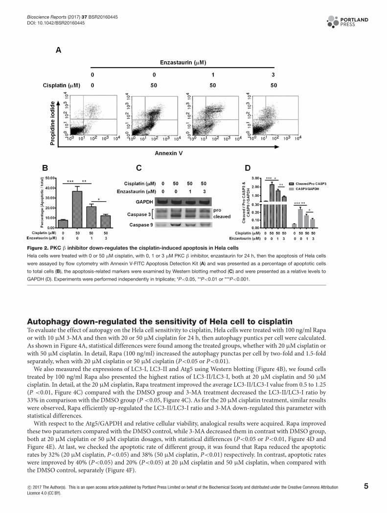
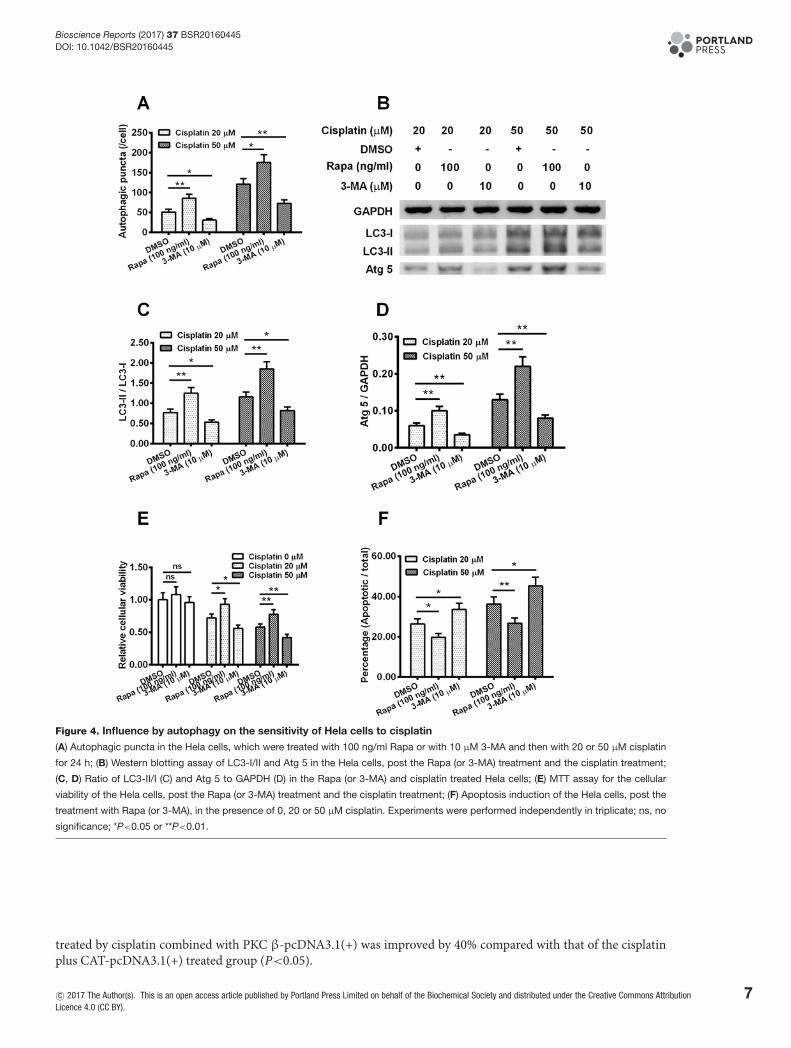
Autophagy down-regulated the sensitivity of Hela cell to cisplatinTo evaluate the effect of autopagy on the Hela cell sensitivity to cisplatin, Hela cells were treated with 100 ng/ml Rapaor with 10 μM 3-MA and then with 20 or 50 μM cisplatin for 24 h, then autophagy puntics per cell were calculated.As shown in Figure 4A, statistical differences were found among the treated groups, whether with 20 μM cisplatin orwith 50 μM cisplatin. In detail, Rapa (100 ng/ml) increased the autophagy punctas per cell by two-fold and 1.5-foldseparately, when with 20 μM cisplatin or 50 μM cisplatin (P<0.05 or P<0.01).
We also measured the expressions of LC3-I, LC3-II and Atg5 using Western blotting (Figure 4B), we found cellstreated by 100 ng/ml Rapa also presented the highest ratios of LC3-II/LC3-I, both at 20 μM cisplatin and 50 μMcisplatin. In detail, at the 20 μM cisplatin, Rapa treatment improved the average LC3-II/LC3-I value from 0.5 to 1.25(P <0.01, Figure 4C) compared with the DMSO group and 3-MA treatment decreased the LC3-II/LC3-I ratio by33% in comparison with the DMSO group (P <0.05, Figure 4C). As for the 20 μM cisplatin treatment, similar resultswere observed, Rapa efficiently up-regulated the LC3-II/LC3-I ratio and 3-MA down-regulated this parameter withstatistical differences.
With respect to the Atg5/GAPDH and relative cellular viability, analogical results were acquired. Rapa improvedthese two parameters compared with the DMSO control, while 3-MA decreased them in contrast with DMSO group,both at 20 μM cisplatin or 50 μM cisplatin dosages, with statistical differences (P<0.05 or P<0.01, Figure 4D andFigure 4E). At last, we checked the apoptotic rate of different group, it was found that Rapa reduced the apoptoticrates by 32% (20 μM cisplatin, P<0.05) and 38% (50 μM cisplatin, P<0.01) respectively. In contrast, apoptotic rateswere improved by 40% (P<0.05) and 20% (P<0.05) at 20 μM cisplatin and 50 μM cisplatin, when compared withthe DMSO control, separately (Figure 4F).
c© 2017 The Author(s). This is an open access article published by Portland Press Limited on behalf of the Biochemical Society and distributed under the Creative Commons AttributionLicence 4.0 (CC BY).
5
Bioscience Reports (2017) 37 BSR20160445DOI: 10.1042/BSR20160445
Figure 3. PKC β inhibitor enhances the cisplatin-induced autophagy in cervical cancer Hela cells
Hela cells were transfected with EGFP–LC3 II reporter plasmid for 6 h and were treated with 0 or 20 μM cisplatin and with 0, 1 or 3 μM
enzastaurin for 24 h, then Hela cells were observed under fluorescence microscopy. The image (A) and the number (B) of autophagic
puncta were presented/counted respectively. And the autophagy-associated markers, such as microtubule-associated protein 1 light chain
3 (LC3)-I/II and autophagy protein 5 (ATG 5) were analysed by Western blotting assay (C) and were presented as ratio (D) of LC3-II/I and Atg
5 to GAPDH. Experiments were performed independently in triplicate; ns, no significance; * P<0.05, **P<0.01, *** P<0.001 or ****P<0.0001.
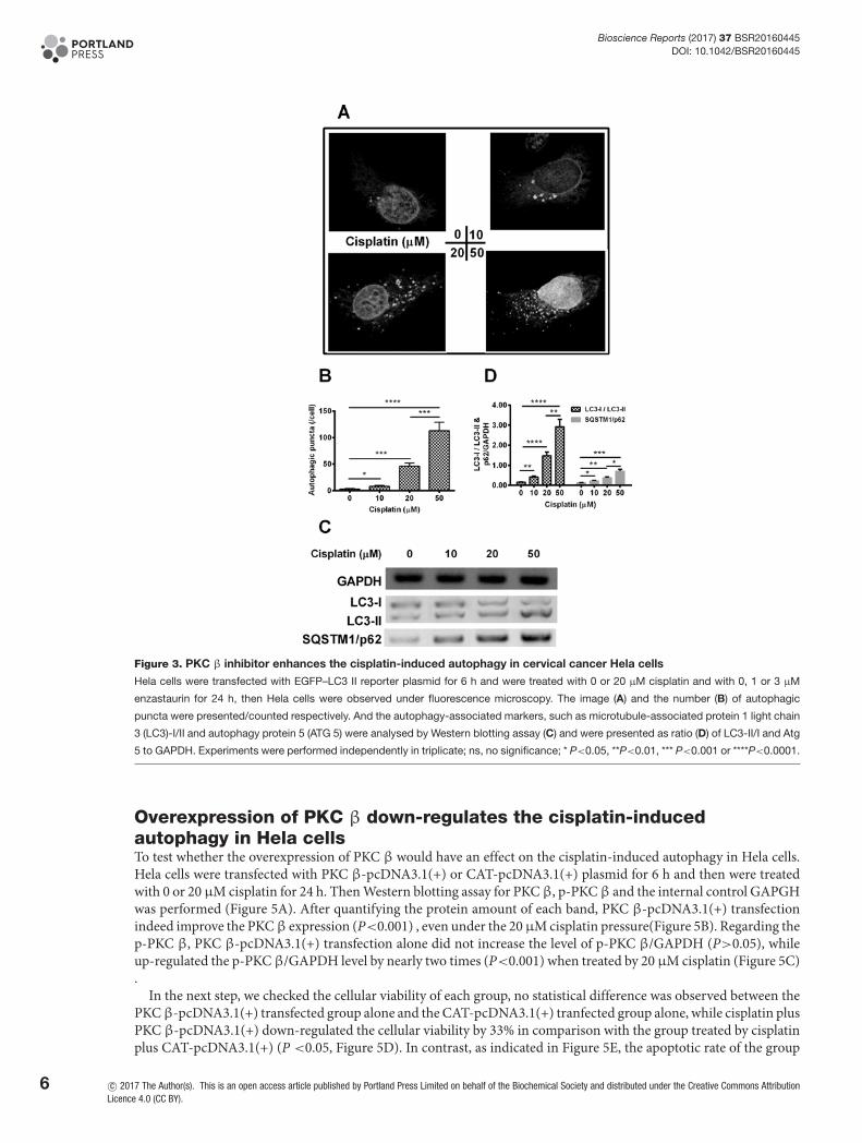
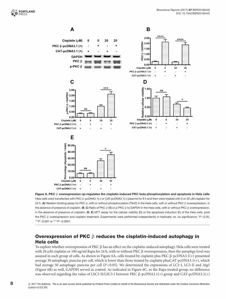
Overexpression of PKC β down-regulates the cisplatin-inducedautophagy in Hela cellsTo test whether the overexpression of PKC β would have an effect on the cisplatin-induced autophagy in Hela cells.Hela cells were transfected with PKC β-pcDNA3.1(+) or CAT-pcDNA3.1(+) plasmid for 6 h and then were treatedwith 0 or 20 μM cisplatin for 24 h. Then Western blotting assay for PKC β, p-PKC β and the internal control GAPGHwas performed (Figure 5A). After quantifying the protein amount of each band, PKC β-pcDNA3.1(+) transfectionindeed improve the PKCβ expression (P<0.001) , even under the 20μM cisplatin pressure(Figure 5B). Regarding thep-PKC β, PKC β-pcDNA3.1(+) transfection alone did not increase the level of p-PKC β/GAPDH (P>0.05), whileup-regulated the p-PKC β/GAPDH level by nearly two times (P<0.001) when treated by 20 μM cisplatin (Figure 5C).
In the next step, we checked the cellular viability of each group, no statistical difference was observed between thePKCβ-pcDNA3.1(+) transfected group alone and the CAT-pcDNA3.1(+) tranfected group alone, while cisplatin plusPKC β-pcDNA3.1(+) down-regulated the cellular viability by 33% in comparison with the group treated by cisplatinplus CAT-pcDNA3.1(+) (P <0.05, Figure 5D). In contrast, as indicated in Figure 5E, the apoptotic rate of the group
6 c© 2017 The Author(s). This is an open access article published by Portland Press Limited on behalf of the Biochemical Society and distributed under the Creative Commons AttributionLicence 4.0 (CC BY).
Bioscience Reports (2017) 37 BSR20160445DOI: 10.1042/BSR20160445
Figure 4. Influence by autophagy on the sensitivity of Hela cells to cisplatin
(A) Autophagic puncta in the Hela cells, which were treated with 100 ng/ml Rapa or with 10 μM 3-MA and then with 20 or 50 μM cisplatin
for 24 h; (B) Western blotting assay of LC3-I/II and Atg 5 in the Hela cells, post the Rapa (or 3-MA) treatment and the cisplatin treatment;
(C, D) Ratio of LC3-II/I (C) and Atg 5 to GAPDH (D) in the Rapa (or 3-MA) and cisplatin treated Hela cells; (E) MTT assay for the cellular
viability of the Hela cells, post the Rapa (or 3-MA) treatment and the cisplatin treatment; (F) Apoptosis induction of the Hela cells, post the
treatment with Rapa (or 3-MA), in the presence of 0, 20 or 50 μM cisplatin. Experiments were performed independently in triplicate; ns, no
significance; *P<0.05 or **P<0.01.
treated by cisplatin combined with PKC β-pcDNA3.1(+) was improved by 40% compared with that of the cisplatinplus CAT-pcDNA3.1(+) treated group (P<0.05).
c© 2017 The Author(s). This is an open access article published by Portland Press Limited on behalf of the Biochemical Society and distributed under the Creative Commons AttributionLicence 4.0 (CC BY).
7
Bioscience Reports (2017) 37 BSR20160445DOI: 10.1042/BSR20160445
Figure 5. PKC β overexpression up-regulates the cisplatin-induced PKC beta phosphorylation and apoptosis in Hela cells
Hela cells were transfected with PKC β-pcDNA3.1(+) or CAT-pcDNA3.1(+) plasmid for 6 h and then were treated with 0 or 20 μM cisplatin for
24 h. (A) Western blotting assay for PKC β, with or without phosphorylation (T642) in the Hela cells, with or without PKC β overexpression, in
the absence of presence of cisplatin. (B, C) Ratio of PKC β (B) or p-PKC β to GAPDH in the Hela cells, with or without PKC β overexpression,
in the absence of presence of cisplatin; (D, E) MTT assay for the cellular viability (D) or the apoptosis induction (E) of the Hela cells, post
the PKC β overexpression and cisplatin treatment. Experiments were performed independently in triplicate; ns, no significance; *P<0.05,
***P<0.001 or ****P<0.0001.
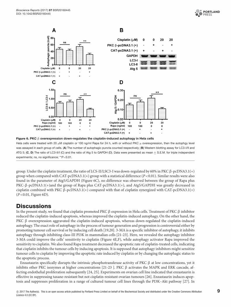
Overexpression of PKC β reduces the cisplatin-induced autophagy inHela cellsTo explore whether overexpression of PKC β has an effect on the cisplatin-induced autophagy. Hela cells were treatedwith 20 μM cisplatin or 100 ng/ml Rapa for 24 h, with or without PKC β overexpression, then the autophgy level wasassayed in each group of cells. As shown in Figure 6A, cells treated by cisplatin plus PKC β-pcDNA3.1(+) presentedaverage 30 autophagic punctas per cell, which is lower than those treated by cisplatin plusCAT-pcDNA3.1(+), whichhad average 50 autophagic punctas per cell (P<0.05). We determined the expressions of LC3-I, LC3-II and Atg5(Figure 6B) as well, GAPDH served as control. As indicated in Figure 6C, in the Rapa treated group, no differencewas observed regarding the value of LSC3-II/LSC3-I between PKC β-pcDNA3.1(+) group and CAT-pcDNA3.1(+)
8 c© 2017 The Author(s). This is an open access article published by Portland Press Limited on behalf of the Biochemical Society and distributed under the Creative Commons AttributionLicence 4.0 (CC BY).
Bioscience Reports (2017) 37 BSR20160445DOI: 10.1042/BSR20160445
Figure 6. PKC β overexpression down-regulates the cisplatin-induced autophagy in Hela cells
Hela cells were treated with 20 μM cisplatin or 100 ng/ml Rapa for 24 h, with or without PKC μ overexpression, then the autophgy level
was assayed in each group of cells. (A) The number of autophagic puncta counted respectively; (B) Western blotting assay for LC3-I/II and
ATG 5; (C, D) The ratio of LC3-II/I (C) and the ratio of Atg 5 to GAPDH (D). Data were presented as mean +− S.E.M. for triple independent
experiments; ns, no significance; **P<0.01.
group. Under the cisplatin treatment, the ratio of LCS-II/LSC3-I was down-regulated by 60% in PKCβ-pcDNA3.1(+)group when compared with CAT-pcDNA3.1(+) group with a statistical difference (P<0.01). Similar results were alsofound in the parameter of Atg5/GAPDH (Figure 6C), no difference was observed between the group of Rapa plusPKC-β-pcDNA3.1(+)and the group of Rapa plus CAT-pcDNA3.1(+), and Atg5/GAPDH was greatly decreased incisplatin combined with PKC β-pcDNA3.1(+) compared with that of cisplatin synergized with CAT-pcDNA3.1(+)(P<0.01, Figure 6D).
DiscussionsIn the present study, we found that cisplatin promoted PKC β expression in Hela cells. Treatment of PKC β inhibitorreduced the cisplatin-induced apoptosis, whereas improved the cisplatin-induced autophagy. On the other hand, thePKC β overexpression aggravated the cisplatin-induced apoptosis, whereas down-regulated the cisplatin-inducedautophagy. The exact role of autophagy in the process of tumour generation and progression is controversial either bypromoting tumour cell survival or by inducing cell death [19,20]. 3-MA is a specific inhibitor of autophagy, it inhibitsautophagy through inhibiting class-III PI3K in mammalian cells [21-23]. Here, we revealed the autophagy inhibitor3-MA could improve the cells’ sensitivity to cisplatin (Figure 4E,F), while autophagy activator Rapa improved thesensitivity to cisplatin. We also found Rapa treatment decreased the apoptotic rate of cisplatin-treated cells, indicatingthat cisplatin inhibits the tumour cells by inducing apoptosis. It is supposed that autophagy inhibitors might sensitizetumour cells to cisplatin by improving the apoptotic rate induced by cisplatin or by changing the autophagic status tothe apoptotic process.
Enzastaurin specifically disrupts the intrinsic phosphotransferase activity of PKC β at low concentrations, yet itinhibits other PKC isozymes at higher concentrations [21-23 ]. PKC β activates the MAPK and ERK cascade, af-fecting endothelial proliferation subsequently [24, 25]. Experiments on ovarian cell line indicated that enzastaurin iseffective in suppressing taxane-resistant but not cisplatin-resistant ovarian tumours [26]. Enzastaurin induces apop-tosis and suppresses proliferation in a range of cultured tumour cell lines through the PI3K–Akt pathway [27]. In
c© 2017 The Author(s). This is an open access article published by Portland Press Limited on behalf of the Biochemical Society and distributed under the Creative Commons AttributionLicence 4.0 (CC BY).
9
Bioscience Reports (2017) 37 BSR20160445DOI: 10.1042/BSR20160445
the present study, we found that enzastaurin treatment presented a dose-dependent suppression of the apoptotic rateof cisplatin-treated cells. Above all, we also found that enzastaurin improved the autophagic puncta per cell in thecisplatin-treated cells with a dose-dependent increase. Key molecules Atg5 and LC3-II were also improved. Theseresults indicate that enzastaurin efficiently induces the autophagy in treated cells and this should be noted in theclinical therapy.
Autophagy not only represents a survival mechanism to anticancer drugs but also can potentially lead to tumour celldeath, which is also called type II programmed cell death (PCD) when autophagy exceeds a certain threshold [28,29].It has been reported that Rapa exerts antitumour effect on malignant glioma cells by inducing autophagy, with amechanism through inhibiting mTOR-signalling pathways [30,31]. Our data suggest that the PKCβ inhibitor reducedthe cisplatin-induced apoptosis, whereas increased the cisplatin-induced autophagy in Hela cells. In contrast, the PKCβ overexpression aggravated the cisplatin-induced apoptosis, but down-regulated the cisplatin-induced autophagy.From these results, we can conclude that PKCβ is ideal to sensitize cervical cancer cells to chemotherapy via reducingthe chemotherapy-induced autophagy. Whether PKC β overexpression at a higher level could directly suppress Helacells proliferation by inducing autophagy is to be explored.
In conclusion, our study firstly explored the involvement of PKC β in the cytotoxicity of cisplatin via inhibitingautophagy in cervical cancer cells. We propose that PKC β would sensitize cervical cancer cells to chemotherapy viadecreasing the chemotherapy-induced autophagy in cancer cells. It will be a promising candidate for developing novelcisplatin-based chemotherapy against different kinds of tumours.
FundingThe authors declare that there are no sources of funding to be acknowledged.
Competing interestsThe authors declare that there are no competing interests associated with the manuscript.
Author contributionN.L. and W.Z. designed the study, performed the experiments, conceived and drafted the manuscript, and performed the statisti-cal analysis. Both the authors read and approved the final manuscript.
Abbreviations3-MA, 3-methyladenine; ATG 5, autophagy protein 5; Bcl-2, B-cell lymphoma 2; DMEM, Dulbecco’s modified Eagle’s medium;GAPDH, glyceraldehyde-3-phosphate dehydrogenase; LC3, light chain 3; PKC , protein kinase C; Rapa, rapamycin; mTOR, themammalian target of rapamyin; MAPK, mitogen-activated protein kinase; ERK, extracellular regulated protein kinase; PI3K-Akt,phosphoinositide-3-kinase-RAC-alpha serine/threonine-protein kinase; ROS, reactive oxygen species; TNFalpha, tumor necrosisfactor alpha; WB, western blotting.
References1 Dasari, S. and Tchounwou, P.B. (2014) Cisplatin in cancer therapy: molecular mechanisms of action. Eur. J. Pharmacol. 740, 364–3782 Loehrer, P.J. and Einhorn, L.H. (1984) Drugs five years later. Cisplatin. Ann. Intern. Med. 100, 704–7133 Reedijk, J. (2003) New clues for platinum antitumor chemistry: kinetically controlled metal binding to DNA. Proc. Natl. Acad. Sci. U.S.A. 100,
3611–36164 Wozniak, K. and Blasiak, J. (2002) Recognition and repair of DNA-cisplatin adducts. Acta Biochim. Pol. 49, 583–5965 Wang, H., Zhang, G., Zhang, H., Zhang, F., Zhou, B., Ning, F. et al. (2014) Acquisition of epithelial-mesenchymal transition phenotype and cancer stem
cell-like properties in cisplatin-resistant lung cancer cells through AKT/β-catenin/Snail signaling pathway. Eur. J. Pharmacol. 723, 156–1666 Basu, A. and Krishnamurthy, S. (2010) Cellular responses to cisplatin-induced DNA damage. J. Nucleic Acids 20107 Ma, B., Liang, L.Z., Liao, G.Q., Liang, Y.J., Liu, H.C., Zheng, G.S. et al. (2013) Inhibition of autophagy enhances cisplatin cytotoxicity in human adenoid
cystic carcinoma cells of salivary glands. J. Oral Pathol. Med. 42, 774–7808 Xu, Y., Yu, H., Qin, H., Kang, J., Yu, C., Zhong, J. et al. (2012) Inhibition of autophagy enhances cisplatin cytotoxicity through endoplasmic reticulum
stress in human cervical cancer cells. Cancer Lett. 314, 232–2439 Levine, B. and Klionsky, D.J. (2004) Development by self-digestion: molecular mechanisms and biological functions of autophagy. Dev. Cell 6, 463–47710 Klionsky, D.J. and Emr, S.D. (2000) Autophagy as a regulated pathway of cellular degradation. Science 290, 1717–172111 Katayama, M., Kawaguchi, T., Berger, M.S. and Pieper, R.O. (2007) DNA damaging agent-induced autophagy produces a cytoprotective adenosine
triphosphate surge in malignant glioma cells. Cell Death Differ. 14, 548–55812 Carew, J.S., Nawrocki, S.T. and Cleveland, J.L. (2007) Modulating autophagy for therapeutic benefit. Autophagy 3, 464–46713 Blobe, G.C., Obeid, L.M. and Hannun, Y.A. (1994) Regulation of protein kinase C and role in cancer biology. Cancer Metastasis Rev. 13, 411–43114 Maioli, E. and Fortino, V. (2004) Protein kinase C: a target for anticancer drugs? Endocr. Relat. Cancer 11, 161–162
10 c© 2017 The Author(s). This is an open access article published by Portland Press Limited on behalf of the Biochemical Society and distributed under the Creative CommonsAttribution Licence 4.0 (CC BY).
Bioscience Reports (2017) 37 BSR20160445DOI: 10.1042/BSR20160445
15 Kouroedov, A., Eto, M., Joch, H., Volpe, M., Luscher, T.F. and Cosentino, F. (2004) Selective inhibition of protein kinase Cbeta2 prevents acute effects ofhigh glucose on vascular cell adhesion molecule-1 expression in human endothelial cells. Circulation 110, 91–96
16 Ishii, H., Jirousek, M.R., Koya, D., Takagi, C., Xia, P., Clermont, A. et al. (1996) Amelioration of vascular dysfunctions in diabetic rats by an oral PKC betainhibitor. Science 272, 728–731
17 Kaushal, G.P., Kaushal, V., Herzog, C. and Yang, C. (2008) Autophagy delays apoptosis in renal tubular epithelial cells in cisplatin cytotoxicity. Autophagy4, 710–712
18 Burman, C. and Ktistakis, N.T. (2010) Autophagosome formation in mammalian cells. Semin. Immunopathol. 32, 397–41319 Gatti, L., Cossa, G., Tinelli, S., Carenini, N., Arrighetti, N., Pennati, M. et al. (2014) Improved apoptotic cell death in drug-resistant non-small-cell lung
cancer cells by tumor necrosis factor-related apoptosis-inducing ligand-based treatment. J. Pharmacol. Exp. Ther. 348, 360–37120 Maiuri, M.C., Tasdemir, E., Criollo, A., Morselli, E., Vicencio, J.M., Carnuccio, R. et al. (2009) Control of autophagy by oncogenes and tumor suppressor
genes. Cell Death Differ. 16, 87–9321 Lum, J.J., Bauer, D.E., Kong, M., Harris, M.H., Li, C., Lindsten, T. et al. (2005) Growth factor regulation of autophagy and cell survival in the absence of
apoptosis. Cell 120, 237–24822 Takasaka, N., Araya, J., Hara, H., Ito, S., Kobayashi, K., Kurita, Y. et al. (2014) Autophagy induction by SIRT6 through attenuation of insulin-like growth
factor signaling is involved in the regulation of human bronchial epithelial cell senescence. J. Immunol. 192, 958–96823 Seglen, P.O. and Gordon, P.B. (1982) 3-Methyladenine: specific inhibitor of autophagic/lysosomal protein degradation in isolated rat hepatocytes. Proc.
Natl. Acad. Sci. U.S.A. 79, 1889–189224 Teicher, B.A., Alvarez, E., Menon, K., Esterman, M.A., Considine, E., Shih, C. et al. (2002) Antiangiogenic effects of a protein kinase Cbeta-selective
small molecule. Cancer Chemother. Pharmacol. 49, 69–7725 Mackay, H.J. and Twelves, C.J. (2007) Targeting the protein kinase C family: are we there yet? Nat. Rev. Cancer 7, 554–56226 Cadron, I., Van Gorp, T., Mihalyi, A., Luyten, C., Drijkoningen, K., Amant, F. et al. (2010) The impact of enzastaurin (LY317615.HCl) on CA125
biosynthesis and shedding in ovarian cancer cells. Gynecol. Oncol. 118, 64–6827 Willey, C.D., Xiao, D., Tu, T., Kim, K.W., Moretti, L., Niermann, K.J. et al. (2010) Enzastaurin (LY317615), a protein kinase C beta selective inhibitor,
enhances antiangiogenic effect of radiation. Int. J. Radiat. Oncol. Biol. Phys. 77, 1518–152628 Chen, N. and Karantza-Wadsworth, V. (2009) Role and regulation of autophagy in cancer. Biochim. Biophys. Acta 1793, 1516–152329 Liu, D., Yang, Y., Liu, Q. and Wang, J. (2011) Inhibition of autophagy by 3-MA potentiates cisplatin-induced apoptosis in esophageal squamous cell
carcinoma cells. Med. Oncol. 28, 105–11130 Iwamaru, A., Kondo, Y., Iwado, E., Aoki, H., Fujiwara, K., Yokoyama, T. et al. (2007) Silencing mammalian target of rapamycin signaling by small
interfering RNA enhances rapamycin-induced autophagy in malignant glioma cells. Oncogene 26, 1840–185131 Takeuchi, H., Kondo, Y., Fujiwara, K., Kanzawa, T., Aoki, H., Mills, G.B. et al. (2005) Synergistic augmentation of rapamycin-induced autophagy in
malignant glioma cells by phosphatidylinositol 3-kinase/protein kinase B inhibitors. Cancer Res. 65, 3336–3346
c© 2017 The Author(s). This is an open access article published by Portland Press Limited on behalf of the Biochemical Society and distributed under the Creative Commons AttributionLicence 4.0 (CC BY).
11