Lithium inhibits NF-κB nuclear translocation and modulate ...
Int J Clin Exp Med 2017;10(2):2826-2833www.ijcem.com /ISSN:1940-5901/IJCEM0036869
Original ArticleOrmeloxifene inhibits the proliferation of cervical cancer cells by suppressing Wnt/β-catenin signaling
Lihua Zhang1*, Yanhua Du2*, Caiying Zhu2, Weiwei Yu1, Aidi Xu3
1Department of Radiation Oncology, Shanghai Jiao Tong University Affiliated Sixth People’s Hospital, Shanghai 200233, P. R. China; 2Obstetrics and Gynecology Hospital of Fudan University, Shanghai 200011, P. R. China; 3Shanghai Hongkou District Health and Family Planning Commission, Shanghai 200086, P. R. China. *Equal con-tributors and co-first authors.
Received July 29, 2016; Accepted November 20, 2016; Epub February 15, 2017; Published February 28, 2017
Abstract: Background: Ormeloxifene, a non-steroidal selective estrogen receptor modulator used widely as an oral contraceptive, which was recently reported to play anti-cancer activity in various types of cancer. This study aimed to investigate the effect of Ormeloxifene on human cervical cancer HeLa cells and related mechanism. Material and methods: After treated with different doses of Ormeloxifene, cell viability was determined by MTT method, the cell cycle distribution and apoptosis were observed by flow cytometry (FCM), the expression of Wingless-type (Wnt), β-catenin, glycogen synthase kinase (GSK)-3β and CyclinD1 was measured by Quantitative real-time PCR (QPCR) and Western blot analysis. Results: Ormeloxifene treatment significantly inhibited the proliferation, induced G1 arrest and apoptosis in human cervical cancer HeLa cells. More importantly, QPCR and Western blot showed that Ormeloxifene markedly down-regulated the expression levels of Wnt, β-catenin, and cyclinD1, up-regulated the expression of GSK-3β in HeLa cells. Conclusion: Together, the results of this study reveal that Ormeloxifene signifi-cantly inhibits the proliferation of HeLa cell and is a potential drug for the treatment of cervical cancer.
Keywords: Ormeloxifene, cervical cancer, HeLa cells, Wnt, β-catenin, proliferation
Introduction
According the data from GLOBOCAN 2012, there were an estimated 527,600 new cervical cancer cases worldwide, ranks the fourth most common cancer in women worldwide, and the second in developing regions [1]. Nowadays, cervical cytology screening is useful to reduce the mortality of this cancer, however there were still 265,700 people died of cervical cancer worldwide in 2012 [1, 2]. Current treatment options of cervical cancer including surgery, chemotherapy and radiotherapy. However, sur-gical treatment is limited to the young patients with early stage; chemotherapy and radiothera-py often lead to drug resistance and multiple severe side effects, such as organ toxicity and immunotoxicity [3]. Therefore, an increasing number of investigators have become interest-ed in reposition established drugs as anti-can-cer agents, such as cyclooxygenase-2 inhibi-tors [4], lenalidomide [5, 6], as well as metformin [7], and medroxyprogesterone acetate [8].
Ormeloxifene (C30H35O3N HCl; [3,4-trans-2,2-dimethyl-3-phenyl-4-p-(b-pyrrolidinoethoxy) phenyl-7-methoxy chroman], also known as Centchroman, is a non-steroidal selective estro-gen receptor modulator which is used as an oral contraceptive in many countries including India, Thailand and Russia [9]. Interestingly, previous study have demonstrated the anti-cancer activities of Ormeloxifene in several can-cers including breast, head and neck cancer, prostate cancer, and ovarian cancer [10]. For example, it has been reported that Ormeloxifene induces G0/G1 arrest and apoptosis in MCF-7 and MDA MB-231 breast cancer cells [11]. Ormeloxifene has been demonstrated to dis-play anti-proliferative activity against head and neck squamous cell carcinoma via the modula-tion of phosphatidylinositol-3’-kinase (PI3K)/ mechanistic target of rapamycin (mTOR) path-way [12]. Moreover, Ormeloxifene effectively inhibited the growth of cisplatin resistant ovari-an cancer cells by inducing cell cycle arrest and apoptosis [13]. In vivo study showed that Or-
Ormeloxifene inhibits the proliferation of cervical cancer cells
2827 Int J Clin Exp Med 2017;10(2):2826-2833
meloxifene enhanced sensitivity of Gemcita- bine in pancreatic cancer [14]. These studies indicate that Ormeloxifene exerts potent anti-cancer activities. In addition, Ormeloxifene was reported safe for chronic administration. Re- cently, Neeraj et al reported that Ormeloxifene significantly inhibited cervical cancer cell gro- wth and decreased mitochondrial membrane potential [15]. Yet little is known about the effect and molecular mechanism of Ormelo- xifene on HeLa cells.
Therefore, this present study aimed to investi-gate the potential anti-cancer effects of Orme- loxifene on human cervical cancer HeLa cells, and to explore the underlying molecular mecha-nisms involved in this process.
Material and methods
Reagents
Polyvinylidenedifluoride (PVDF) membranes and enhanced chemi-luminiscence (ECL), were purchased from Merck Millipore (Millipore, Bi- llerica, MA, USA). Antibodies of Wingless-type (Wnt), glycogen synthase kinase (GSK)-3β, pho- sphor (p)-GSK-3β, β-catenin, CyclinD1 as well as glyceraldehyde-3-phosphate (GAPDH) were obtained from Santa Cruz (Santa Cruz, CA). Dulbecco’s Modified Eagle’s Medium (DMEM) and Fetal Bovine Serum (FBS) were purchased from Gibco, Life technologies (Carlsbad, CA), 3-(4,5-Dimethylthiazol-2-yl)-2,5-diphenyltetra-zolium bromide (MTT) was purchased from Serva (Heidelberg, Germany) and was dissolved in PBS at a concentration of 5 mg/ml. Propidium iodide and the Annexin-V FITC apoptosis detec-tion kits were purchased from Beyotime (Sha- nghai, P. R. China). Ormeloxifene was obtained from Sigma-Aldrich Shanghai Trading Co., Ltd. (Shanghai, P. R. China) and prepared in Dimethyl Sulphoxide (DMSO) and stored at -20°C.
Cell culture
Human cervical cancer cells HeLa were pur-chased from Shanghai Cell Bank (Shanghai, P.
Cell proliferation and viability assays
HeLa cell proliferation was evaluated using a MTT assay according to the manufacturer’s protocol. Briefly, cells were seeded in 96-well plates at the concentration of 5×10⁴ cells/well and added various doses of Ormeloxifene at the final concentrations of 5, 10, 20, or 40 μM. After incubation for 48 h, 20 μL of MTT was added to each well, followed by an incubation of the plates at 37°C for 4 h. The medium was then removed and 150 µl of 10% DMSO was added to each well. Absorbance at 450 nm was measured by a 96 well plate reader (Bio-Tek, Burleigh, QLD, Australia). Cell viability was cal-culated as: cell viability (%) = (corrected treated sample OD/corrected control sample OD) ×100%.
Cell apoptosis assays
Hela cell apoptosis was measured by using an Annexin-V FITC Apoptosis Detection Kit (Beyo- time, Shanghai, P. R. China) following the manu-facturer’s instruction. Briefly, Hela cells at a concentration of 3×10⁶ cells per in 6-well plates were cultured in DMEM medium with 10 or 20 μM Ormeloxifene for 48 h, respectively. The medium was removed and the cells were tryp-sinized, then centrifuged at 1,000× g for 5 min to remove trypsin. Next, 50 µg/mL of Annexin-V-FITC and 10 µL of propidium iodide from the detection kit were added to each well and the plate was incubated at room temperature for 10 min in the dark, followed immediately by flow cytometric analysis.
Cell cycle assays
After treatment with and without Ormeloxifene for 48 h, Hela cells were trypsinized and centri-fuged at 1,000× g for 5 min. The supernatant was removed and the cells were washed with phosphate buffered saline (PBS) and fixed with 70% ethanol at 4°C for overnight. The fixed cells were washed and stained with 50 μg/mL
Table 1. Sequences of primers used in the experimentGene name Forward primer Reverse primerWnt 5’-CTGTCCTGCCTCCTCATC-3’ 5’-GGACCCAGCACAATAAATAGTT-3’β-Catenin 5’-TGCTGAAGGTGCTGTCTGTC-3’ 5’-TCGCTGACTTGGGTCTGTC-3’GSK-3β 5’-CACCTCTGCCTACCATCCTTA-3’ 5’-ATTATTGGTCTGTCCACGGTC-3’CyclinD1 5’-TCGGGAGAGGATTAGGTTCC-3’ 5’-GTCACTGGATGGTTTGTTGG-3’GAPDH 5’-TGTGTCCGTCGTGGATCTGA-3’ 5’-TTGCTGTTGAAGTCGCAGGAG-3’
R. China). HeLa cells were cultured in DMEM supple-mented with 10% heat-inactivated FBS, 100 U/ml penicillin, 100 μg/ml streptomycin. Cells were grown at 37°C in a humid-ified atmosphere contain-ing 5% CO2.
Ormeloxifene inhibits the proliferation of cervical cancer cells
2828 Int J Clin Exp Med 2017;10(2):2826-2833
of PI, 50 mg/mL of RNase A at 37°C for 30 min. The cellular DNA content and cell cycle phases were analyzed with flow cytometer (Becton Dickinson, SanJosé, CA).
Quantitative real-time PCR
Total RNA was extracted from cultured cells with TRIzol reagent (Invitrogen, Carsbad, CA) and DNA was removed by DNase (Promega) according to the manufacturer’s instructions and mRNA expression levels were measured by Quantitative real-time PCR (QPCR) using an iQ5 multicolor real-time PCR Detection System (Bio-Rad) with SYBR Green I staining (Takara). Reverse transcription was performed with the PrimeScript RT reagent Kit (Perfect Real Time; Takara) according to the manufacturer’s ins- tructions. For mRNA analysis, GAPDH mRNA
levels were used as internal normalization con-trol. Fold changes were calculated and normal-ized using the CT method. Primers used were listed in Table 1.
Western blot assay
Cells were collected and suspended in 250 μl of lysis buffer (50 mM Tirs, 150 mM NaCl, 1% NP-40, 0.5% sodium deoxycholate, 0.1% SDS). Equal amounts of protein (20 µg) from different samples were separated by 10% SDS-polyac- rylamide gel electrophoresis (PAGE), and then transferred onto PVDF membranes (Millipore, Billerica, MA, USA) for Western blot analysis. After transfer, the membranes were blocked in non-fat milk (5% in Tris-buffered saline with TWEEN-20, TBST) and then incubated with anti-bodies against Wnt (1:500 dilution), β-caten-
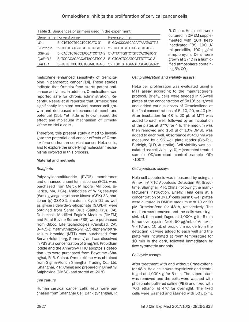
Figure 1. Effects of Ormeloxifene on proliferation of HeLa cells. Cells were incubated with 0, 10, 20 μM of Ormel-oxifene for 48 h (A-C) and MTT assay (D) was used for the measurement of cell viabilities. Data were expressed as mean ± SD. #, P<0.05, compared with the control group. Control, control group; ORM, Ormeloxifene-treated group.
Ormeloxifene inhibits the proliferation of cervical cancer cells
2829 Int J Clin Exp Med 2017;10(2):2826-2833
in (1:500 dilution), CyclinD1 (1:500 dilution), p-GSK-3β (1:500 dilution), GSK-3β (1:500 dilu-tion) and GAPDH (1:500 dilution) at 4°C over-night. After two washes with TBST, the mem-branes were incubated with horseradish pero- xidase-conjugated anti-rabbit or anti-mouse IgG (Thermo Fisher Scientific, New York, NY, USA). Then the bands were detected by using an enhanced chemiluminescence reagent (Mi- llipore, MA, USA) and exposed by autoradiogra-phy. GAPDH was used as an internal loading control.
Statistical analysis
Data was analyzed with Graph pad Prism soft-ware (Graph pad version 6.01). Statistical dif-
ferences between groups were determined using one-way ANOVA followed by the post-hoc LSD. Statistically significant differences were considered for p values <0.05.
Results
Ormeloxifene inhibits proliferation of HeLa cells
The potential of Ormeloxifene to inhibit the pro-liferation of HeLa cells was evaluated by MTT assay. As showed in Figure 1, after treatment with Ormeloxifene for 48 h, the cell viability of Hela cells was significantly decreased (10 and 20 μM: P<0.05) in comparison of control group, suggesting that Ormeloxifene was effective in inhibiting the proliferation of Hela cells.
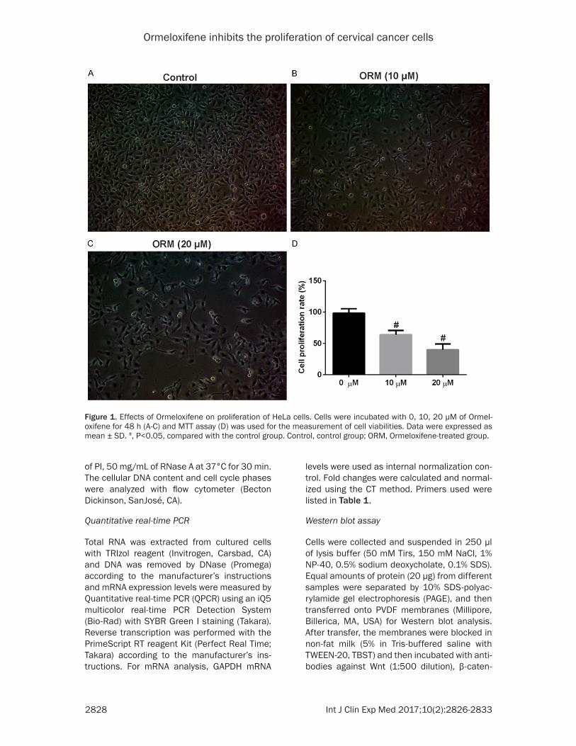
Figure 2. Effects of Ormeloxifene on cell-cycle phase distribution of HeLa cells. Cells were incubated with 0, 10, 20 μM of Ormeloxifene for 48 h and Cell cycles were determined using PI staining of the nuclei. A: Control; B: Hela cells were treated with 10 μM Ormeloxifene; C: Hela cells were treated with 20 μM Ormeloxifene; D: G1 phase rate of control and sample. Data were expressed as mean ± SD. #, P<0.05, compared with the control group. Control, control group; ORM, Ormeloxifene-treated group.
Ormeloxifene inhibits the proliferation of cervical cancer cells
2830 Int J Clin Exp Med 2017;10(2):2826-2833
Ormeloxifene induced G1 phase arrest in HeLa cells
In order to determine whether the cell-growth suppressive effect of Ormeloxifene attributed to inhibit proliferation, the cell cycle distribu-tion was detected by flow cytometric analysis. As illustrated in Figure 2, Ormeloxifene treat-ment obviously increased the percentage of cells at the G1 phase compared with the con-trol group (10 and 20 μM: P<0.05).
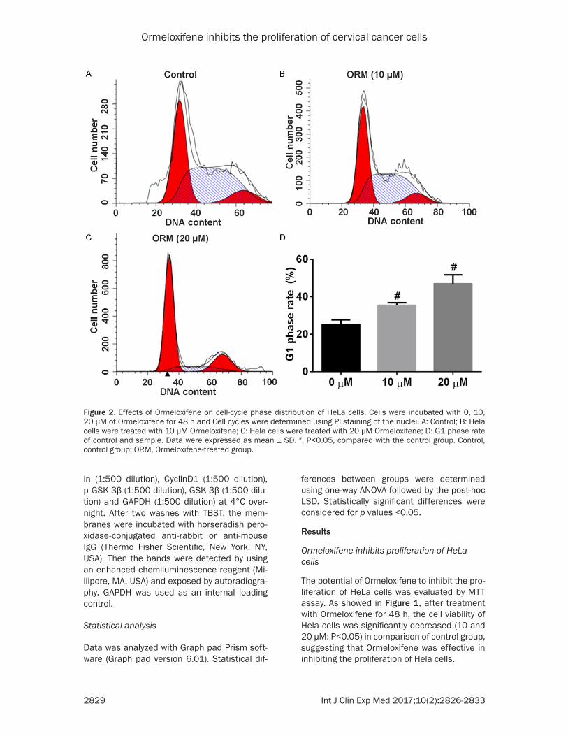
Ormeloxifene induces apoptosis of HeLa cells
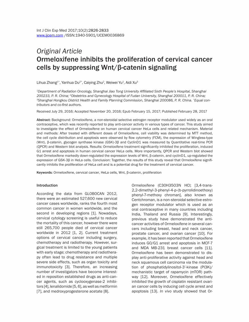
We then investigated the effects of Ormeloxifene on cell apoptosis by using an Annexin-V FITC Apoptosis Detection Kit. Cells were treated with 10, 20 μM of Ormeloxifene for 48 h, Figure 3 shows the representative results of flow cytom-etry. Apoptosis rate was significantly higher in the Ormeloxifene treatment groups compared to the control group (10 and 20 μM: P<0.05). These results above indicated that Ormeloxifene could induce the apoptosis of HeLa cells.
Figure 3. Effect of Ormeloxifene on the apoptosis of HeLa cells. Apoptosis analysis of HeLa cells induced by Ormel-oxifene for 48 h, using a flow cytometer with Annexin V-FITC/PI binding assay. A: Control; B: HeLa cells were treated with 10 μM Ormeloxifene; C: HeLa cells were treated with 20 μM Ormeloxifene; D: Apoptosis rate of control and samples. Data were expressed as mean ± SD. #, P<0.05, compared with the control group. Control, control group; ORM, Ormeloxifene-treated group.
Ormeloxifene inhibits the proliferation of cervical cancer cells
2831 Int J Clin Exp Med 2017;10(2):2826-2833
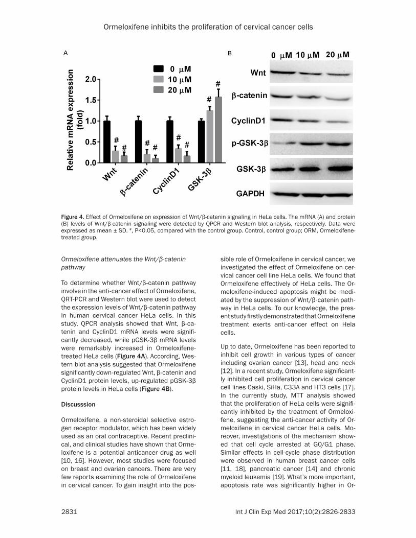
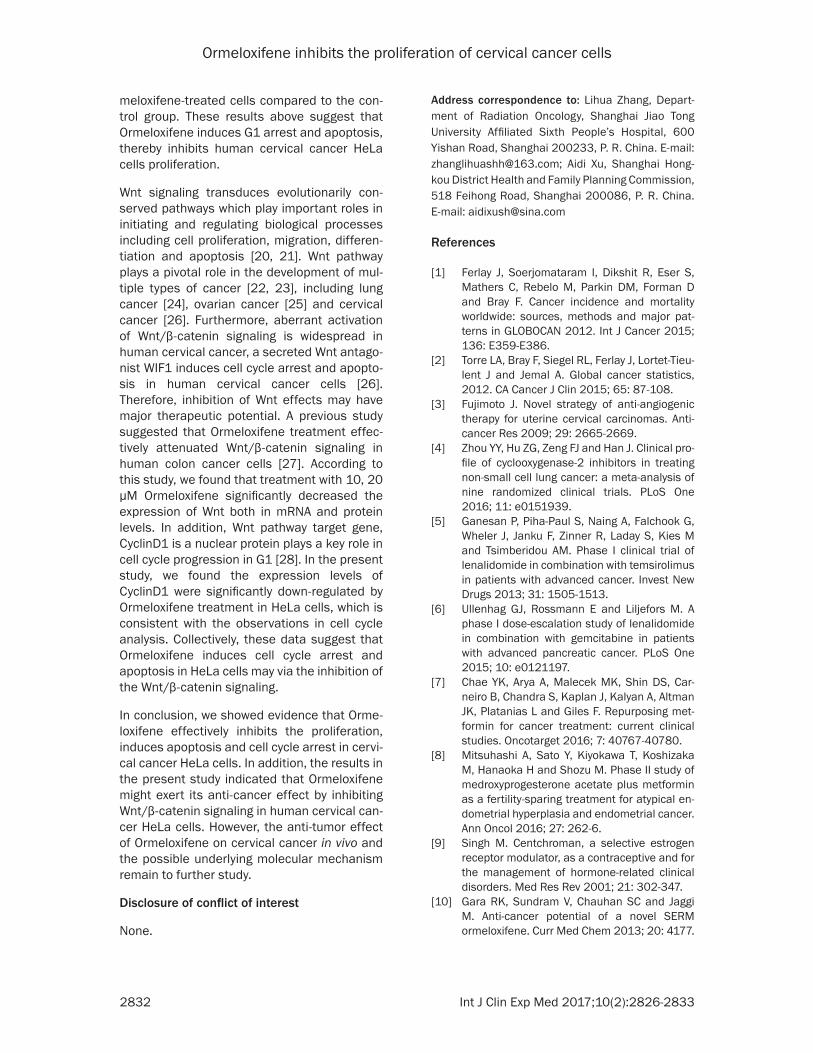
Ormeloxifene attenuates the Wnt/β-catenin pathway
To determine whether Wnt/β-catenin pathway involve in the anti-cancer effect of Ormeloxifene, QRT-PCR and Western blot were used to detect the expression levels of Wnt/β-catenin pathway in human cervical cancer HeLa cells. In this study, QPCR analysis showed that Wnt, β-ca- tenin and CyclinD1 mRNA levels were signifi-cantly decreased, while pGSK-3β mRNA levels were remarkably increased in Ormeloxifene-treated HeLa cells (Figure 4A). According, Wes- tern blot analysis suggested that Ormeloxifene significantly down-regulated Wnt, β-catenin and CyclinD1 protein levels, up-regulated pGSK-3β protein levels in HeLa cells (Figure 4B).
Discusssion
Ormeloxifene, a non-steroidal selective estro-gen receptor modulator, which has been widely used as an oral contraceptive. Recent preclini-cal, and clinical studies have shown that Orme- loxifene is a potential anticancer drug as well [10, 16]. However, most studies were focused on breast and ovarian cancers. There are very few reports examining the role of Ormeloxifene in cervical cancer. To gain insight into the pos-
sible role of Ormeloxifene in cervical cancer, we investigated the effect of Ormeloxifene on cer-vical cancer cell line HeLa cells. We found that Ormeloxifene effectively of HeLa cells. The Or- meloxifene-induced apoptosis might be medi-ated by the suppression of Wnt/β-catenin path-way in HeLa cells. To our knowledge, the pres-ent study firstly demonstrated that Ormeloxifene treatment exerts anti-cancer effect on Hela cells.
Up to date, Ormeloxifene has been reported to inhibit cell growth in various types of cancer including ovarian cancer [13], head and neck [12]. In a recent study, Ormeloxifene significant-ly inhibited cell proliferation in cervical cancer cell lines Caski, SiHa, C33A and HT3 cells [17]. In the currently study, MTT analysis showed that the proliferation of HeLa cells were signifi-cantly inhibited by the treatment of Ormeloxi- fene, suggesting the anti-cancer activity of Or- meloxifene in cervical cancer HeLa cells. Mo- reover, investigations of the mechanism show- ed that cell cycle arrested at G0/G1 phase. Similar effects in cell-cycle phase distribution were observed in human breast cancer cells [11, 18], pancreatic cancer [14] and chronic myeloid leukemia [19]. What’s more important, apoptosis rate was significantly higher in Or-
Figure 4. Effect of Ormeloxifene on expression of Wnt/β-catenin signaling in HeLa cells. The mRNA (A) and protein (B) levels of Wnt/β-catenin signaling were detected by QPCR and Western blot analysis, respectively. Data were expressed as mean ± SD. #, P<0.05, compared with the control group. Control, control group; ORM, Ormeloxifene-treated group.
Ormeloxifene inhibits the proliferation of cervical cancer cells
2832 Int J Clin Exp Med 2017;10(2):2826-2833
meloxifene-treated cells compared to the con-trol group. These results above suggest that Ormeloxifene induces G1 arrest and apoptosis, thereby inhibits human cervical cancer HeLa cells proliferation.
Wnt signaling transduces evolutionarily con-served pathways which play important roles in initiating and regulating biological processes including cell proliferation, migration, differen-tiation and apoptosis [20, 21]. Wnt pathway plays a pivotal role in the development of mul-tiple types of cancer [22, 23], including lung cancer [24], ovarian cancer [25] and cervical cancer [26]. Furthermore, aberrant activation of Wnt/β-catenin signaling is widespread in human cervical cancer, a secreted Wnt antago-nist WIF1 induces cell cycle arrest and apopto-sis in human cervical cancer cells [26]. Therefore, inhibition of Wnt effects may have major therapeutic potential. A previous study suggested that Ormeloxifene treatment effec-tively attenuated Wnt/β-catenin signaling in human colon cancer cells [27]. According to this study, we found that treatment with 10, 20 μM Ormeloxifene significantly decreased the expression of Wnt both in mRNA and protein levels. In addition, Wnt pathway target gene, CyclinD1 is a nuclear protein plays a key role in cell cycle progression in G1 [28]. In the present study, we found the expression levels of CyclinD1 were significantly down-regulated by Ormeloxifene treatment in HeLa cells, which is consistent with the observations in cell cycle analysis. Collectively, these data suggest that Ormeloxifene induces cell cycle arrest and apoptosis in HeLa cells may via the inhibition of the Wnt/β-catenin signaling.
In conclusion, we showed evidence that Orme- loxifene effectively inhibits the proliferation, induces apoptosis and cell cycle arrest in cervi-cal cancer HeLa cells. In addition, the results in the present study indicated that Ormeloxifene might exert its anti-cancer effect by inhibiting Wnt/β-catenin signaling in human cervical can-cer HeLa cells. However, the anti-tumor effect of Ormeloxifene on cervical cancer in vivo and the possible underlying molecular mechanism remain to further study.
Disclosure of conflict of interest
None.
Address correspondence to: Lihua Zhang, Depart- ment of Radiation Oncology, Shanghai Jiao Tong University Affiliated Sixth People’s Hospital, 600 Yishan Road, Shanghai 200233, P. R. China. E-mail: [email protected]; Aidi Xu, Shanghai Hong- kou District Health and Family Planning Commission, 518 Feihong Road, Shanghai 200086, P. R. China. E-mail: [email protected]
References
[1] Ferlay J, Soerjomataram I, Dikshit R, Eser S, Mathers C, Rebelo M, Parkin DM, Forman D and Bray F. Cancer incidence and mortality worldwide: sources, methods and major pat-terns in GLOBOCAN 2012. Int J Cancer 2015; 136: E359-E386.
[2] Torre LA, Bray F, Siegel RL, Ferlay J, Lortet-Tieu-lent J and Jemal A. Global cancer statistics, 2012. CA Cancer J Clin 2015; 65: 87-108.
[3] Fujimoto J. Novel strategy of anti-angiogenic therapy for uterine cervical carcinomas. Anti-cancer Res 2009; 29: 2665-2669.
[4] Zhou YY, Hu ZG, Zeng FJ and Han J. Clinical pro-file of cyclooxygenase-2 inhibitors in treating non-small cell lung cancer: a meta-analysis of nine randomized clinical trials. PLoS One 2016; 11: e0151939.
[5] Ganesan P, Piha-Paul S, Naing A, Falchook G, Wheler J, Janku F, Zinner R, Laday S, Kies M and Tsimberidou AM. Phase I clinical trial of lenalidomide in combination with temsirolimus in patients with advanced cancer. Invest New Drugs 2013; 31: 1505-1513.
[6] Ullenhag GJ, Rossmann E and Liljefors M. A phase I dose-escalation study of lenalidomide in combination with gemcitabine in patients with advanced pancreatic cancer. PLoS One 2015; 10: e0121197.
[7] Chae YK, Arya A, Malecek MK, Shin DS, Car-neiro B, Chandra S, Kaplan J, Kalyan A, Altman JK, Platanias L and Giles F. Repurposing met-formin for cancer treatment: current clinical studies. Oncotarget 2016; 7: 40767-40780.
[8] Mitsuhashi A, Sato Y, Kiyokawa T, Koshizaka M, Hanaoka H and Shozu M. Phase II study of medroxyprogesterone acetate plus metformin as a fertility-sparing treatment for atypical en-dometrial hyperplasia and endometrial cancer. Ann Oncol 2016; 27: 262-6.
[9] Singh M. Centchroman, a selective estrogen receptor modulator, as a contraceptive and for the management of hormone-related clinical disorders. Med Res Rev 2001; 21: 302-347.
[10] Gara RK, Sundram V, Chauhan SC and Jaggi M. Anti-cancer potential of a novel SERM ormeloxifene. Curr Med Chem 2013; 20: 4177.
Ormeloxifene inhibits the proliferation of cervical cancer cells
2833 Int J Clin Exp Med 2017;10(2):2826-2833
[11] Nigam M, Ranjan V, Srivastava S, Sharma R and Balapure AK. Centchroman induces G 0/G 1 arrest and caspase-dependent apoptosis in-volving mitochondrial membrane depolariza-tion in MCF-7 and MDA MB-231 human breast cancer cells. Life Sci 2008; 82: 577-590.
[12] Srivastava VK, Gara RK, Bhatt M, Sahu D and Mishra DP. Centchroman inhibits proliferation of head and neck cancer cells through the modulation of PI3K/mTOR pathway. Biochem Biophys Res Commun 2011; 404: 40-45.
[13] Maher DM, Khan S, Nordquist JL, Ebeling MC, Bauer NA, Kopel L, Singh MM, Halaweish F, Bell MC and Jaggi M. Ormeloxifene efficiently inhibits ovarian cancer growth. Cancer Lett 2015; 356: 606-612.
[14] Khan S, Ebeling MC, Chauhan N, Thompson PA, Gara RK, Ganju A, Yallapu MM, Behrman SW, Zhao H and Zafar N. Ormeloxifene Sup-presses Desmoplasia and Enhances Sensitivi-ty of Gemcitabine in Pancreatic Cancer. Can-cer Res 2015; 75: 2292-304.
[15] Chauhan N, Zaman MS, Yallapu MM, Maher DM, Ebeling MC, Chauhan SC and Jaggi M. Ormeloxifene inhibits cervical cancer cell growth through intrinsic apoptotic pathway. Cancer Res 2014; 74: 2753-2753.
[16] Misra N, Nigam P, Gupta R, Agarwal A and Kamboj V. Centchroman-a non-steroidal anti-cancer agent for advanced breast cancer: Phase-II study. Int J Cancer 1989; 43: 781-783.
[17] Chauhan N, Maher DM, Yallapu MM, Ebeling MC, Jaggi M and Chauhan SC. Therapeutic ef-fects of ormeloxifene on cervical cancer. Can-cer Res 2012; 72: 2882-2882.
[18] Nigam M, Singh N, Ranjan V, Zaidi D, Sharma R, Nigam D, Gupta DK, Sundaram S and Bala-pure AK. Centchroman mediated apoptosis in-volves cross-talk between extrinsic/intrinsic pathways and oxidative regulation. Life Sci 2010; 87: 750-758.
[19] Pal P, Kanaujiya JK, Lochab S, Tripathi SB, Bhatt ML, Singh PK, Sanyal S and Trivedi AK. 2-D gel electrophoresis-based proteomic anal-ysis reveals that ormeloxifen induces G0-G1 growth arrest and ERK-mediated apoptosis in chronic myeloid leukemia cells K562. Pro-teomics 2011; 11: 1517-1529.
[20] Freese JL, Pino D and Pleasure SJ. Wnt signal-ing in development and disease. Neurobiol Dis 2010; 38: 148-153.
[21] Luo J, Chen J, Deng ZL, Luo X, Song WX, Sharff KA, Tang N, Haydon RC, Luu HH and He TC. Wnt signaling and human diseases: what are the therapeutic implications? Lab Invest 2007; 87: 97-103.
[22] Anastas JN and Moon RT. WNT signalling path-ways as therapeutic targets in cancer. Nat Rev Cancer 2013; 13: 11-26.
[23] Polakis P. Wnt signaling in cancer. Csh Per-spect Biol 2012; 4: a008052.
[24] Stewart DJ. Wnt signaling pathway in non-small cell lung cancer. J Natl Cancer Inst 2014; 106: djt356.
[25] Arend RC, Londoño-Joshi AI, Straughn JM and Buchsbaum DJ. The Wnt/β-catenin pathway in ovarian cancer: a review. Gynecol Oncol 2013; 131: 772-779.
[26] Ramachandran I, Thavathiru E, Ramalingam S, Natarajan G, Mills W, Benbrook DM, Zuna R, Lightfoot S, Reis A and Anant S. Wnt inhibitory factor 1 induces apoptosis and inhibits cervi-cal cancer growth, invasion and angiogenesis in vivo. Oncogene 2012; 31: 2725-2737.
[27] Ganju A, Gara R, Kumari S, Singh MM, Chau-han S and Jaggi M. Ormeloxifene attenuates wnt/β-catenin signaling in colon cancer cells by modulation of PKD1 and glycolytic path-ways. Cancer Res 2015; 75: 2072-2072.
[28] Chaves-Perez A, Mack B, Maetzel D, Kremling H, Eggert C, Harreus U and Gires O. EpCAM regulates cell cycle progression via control of cyclinD1 expression. Oncogene 2013; 32: 641-650.