Proline-glutamate chimera’s side chain conformation directs the type of β-hairpin structure
10
ORIGINAL ARTICLE Proline-glutamate chimera’s side chain conformation directs the type of b-hairpin structure Jyotirmoy Maity • Ulla I. M. Gerling • Stella Vukelic ´ • Andreas Scha ¨fer • Beate Koksch Received: 22 June 2013 / Accepted: 19 October 2013 / Published online: 13 November 2013 Ó Springer-Verlag Wien 2013 Abstract Our aim was to study the impact of two proline chimeras, containing a glutamic acid side chain in cis- or trans-configuration, on secondary structure formation. We further investigated to what extent the configuration of the side chain contributes to the overall peptide conformation. We used a 10 residue peptide (IYSNPDGTWT) that forms a b-hairpin in water. The turn-forming proline was substituted with either a cis- or trans-proline-glutamic acid chimera, resulting in the peptides IYSNP cis-E DGTWT (P1_P cis-E ) and IYSNP trans-E DGTWT (P1_P trans-E ). We studied the conformation of the modified peptides by cir- cular dichroism (CD) and NMR-spectroscopy, and SEC/ static light scattering (SLS) analysis. NMR analysis reveals that the modified peptides maintain the b-hairpin confor- mation in aqueous solution. At 5 °C and pH 4.3, the pep- tide (P1_P cis-E ) was found to adopt two coexisting b- hairpin conformations (2:2 b-hairpin, and 3:5 b-hairpin). In contrast to that, the peptide (P1_P trans-E ) adopts a 2:2 b- hairpin that exists in equilibrium with a 4:4 b-hairpin conformation. The adoption of ordered b-hairpin structures for both modified peptides could be confirmed by CD spectroscopy, while SEC/SLS analysis showed a mono- meric oligomerization state for all three investigated pep- tides. With the combination of several NMR methods, we were able to elucidate that even small alterations in the side chain conformation of the proline-glutamate chimera (cis or trans) can significantly influence the conformation of the adopted b-hairpin. Keywords Proline chimera b-Hairpin conformation b-Turn Conformational analysis CD NMR Abbreviations CD Circular dichroism HPLC High performance liquid chromatography Fmoc Fluorenylmethoxy carbonyl AU Analytical ultracentrifugation SEC Size exclusion chromatography MD Molecular dynamics DIC Diisopropylcarbodiimide HOBT 1-Hydroxybenzotriazole HOAT 1-Hydroxy-7-azabenzotriazole TFA Trifluoroacetic acid TIS Triisopropylsilane P cis-E cis-Proline glutamate chimera P trans-E trans-proline glutamate chimera NMR Nuclear magnetic resonance 1 H NMR Proton nuclear magnetic resonance COSY Correlation spectroscopy TOCSY Total correlation spectroscopy NOE Nuclear Overhauser effect NOESY Nuclear Overhauser effect spectroscopy ROESY Rotating frame nuclear Overhauser effect spectroscopy SPPS Solid phase peptide synthesis J. Maity and U. I. M. Gerling contributed equally. Electronic supplementary material The online version of this article (doi:10.1007/s00726-013-1610-1) contains supplementary material, which is available to authorized users. J. Maity U. I. M. Gerling S. Vukelic ´ B. Koksch (&) Institut fu ¨r Chemie und Biochemie, Freie Universita ¨t Berlin, Takustrasse 3, 14195 Berlin, Germany e-mail: [email protected] A. Scha ¨fer Gera ¨tezentrum BioSupraMol, Freie Universita ¨t Berlin, Takustrasse 3, 14195 Berlin, Germany 123 Amino Acids (2014) 46:177–186 DOI 10.1007/s00726-013-1610-1
Transcript of Proline-glutamate chimera’s side chain conformation directs the type of β-hairpin structure
ORIGINAL ARTICLE
Proline-glutamate chimera’s side chain conformation directsthe type of b-hairpin structure
Jyotirmoy Maity • Ulla I. M. Gerling •
Stella Vukelic • Andreas Schafer • Beate Koksch
Received: 22 June 2013 / Accepted: 19 October 2013 / Published online: 13 November 2013
� Springer-Verlag Wien 2013
Abstract Our aim was to study the impact of two proline
chimeras, containing a glutamic acid side chain in cis- or
trans-configuration, on secondary structure formation. We
further investigated to what extent the configuration of the
side chain contributes to the overall peptide conformation.
We used a 10 residue peptide (IYSNPDGTWT) that forms
a b-hairpin in water. The turn-forming proline was
substituted with either a cis- or trans-proline-glutamic acid
chimera, resulting in the peptides IYSNPcis-EDGTWT
(P1_Pcis-E) and IYSNPtrans-EDGTWT (P1_Ptrans-E). We
studied the conformation of the modified peptides by cir-
cular dichroism (CD) and NMR-spectroscopy, and SEC/
static light scattering (SLS) analysis. NMR analysis reveals
that the modified peptides maintain the b-hairpin confor-
mation in aqueous solution. At 5 �C and pH 4.3, the pep-
tide (P1_Pcis-E) was found to adopt two coexisting b-
hairpin conformations (2:2 b-hairpin, and 3:5 b-hairpin). In
contrast to that, the peptide (P1_Ptrans-E) adopts a 2:2 b-
hairpin that exists in equilibrium with a 4:4 b-hairpin
conformation. The adoption of ordered b-hairpin structures
for both modified peptides could be confirmed by CD
spectroscopy, while SEC/SLS analysis showed a mono-
meric oligomerization state for all three investigated pep-
tides. With the combination of several NMR methods, we
were able to elucidate that even small alterations in the side
chain conformation of the proline-glutamate chimera (cis
or trans) can significantly influence the conformation of
the adopted b-hairpin.
Keywords Proline chimera � b-Hairpin
conformation � b-Turn � Conformational analysis �CD � NMR
Abbreviations
CD Circular dichroism
HPLC High performance liquid chromatography
Fmoc Fluorenylmethoxy carbonyl
AU Analytical ultracentrifugation
SEC Size exclusion chromatography
MD Molecular dynamics
DIC Diisopropylcarbodiimide
HOBT 1-Hydroxybenzotriazole
HOAT 1-Hydroxy-7-azabenzotriazole
TFA Trifluoroacetic acid
TIS Triisopropylsilane
Pcis-E cis-Proline glutamate chimera
Ptrans-E trans-proline glutamate chimera
NMR Nuclear magnetic resonance1H NMR Proton nuclear magnetic resonance
COSY Correlation spectroscopy
TOCSY Total correlation spectroscopy
NOE Nuclear Overhauser effect
NOESY Nuclear Overhauser effect spectroscopy
ROESY Rotating frame nuclear Overhauser effect
spectroscopy
SPPS Solid phase peptide synthesis
J. Maity and U. I. M. Gerling contributed equally.
Electronic supplementary material The online version of thisarticle (doi:10.1007/s00726-013-1610-1) contains supplementarymaterial, which is available to authorized users.
J. Maity � U. I. M. Gerling � S. Vukelic � B. Koksch (&)
Institut fur Chemie und Biochemie, Freie Universitat Berlin,
Takustrasse 3, 14195 Berlin, Germany
e-mail: [email protected]
A. Schafer
Geratezentrum BioSupraMol, Freie Universitat Berlin,
Takustrasse 3, 14195 Berlin, Germany
123
Amino Acids (2014) 46:177–186
DOI 10.1007/s00726-013-1610-1
Introduction
Secondary structure formation plays a crucial role in protein
folding (Kim and Baldwin 1990; Dyson and Wright 1991,
1993; Munoz et al. 1997). To better understand this process,
short segments of proteins have been studied to explore
their ability to form a-helical (Ishikawa et al. 1996; Pagel
et al. 2006; Horne et al. 2007, 2009; Price et al. 2010;
Rezaei Araghi et al. 2011) or b-hairpin structures (Blanco
et al. 1993, 1994; Searle et al. 1995; Ramirez-Alvarado
et al. 1996; de Alba et al. 1997; Robinson 2008) in aqueous
solution. Among the secondary structures that naturally
occurring proteins can adopt, b-hairpin conformations are
important folding motifs that play a key role in biomolec-
ular recognition. For example, antibodies and T cell
receptors contain b-hairpin motifs. Numerous short pep-
tides that form b-hairpin structures in aqueous solution have
been investigated, in the last two decades. Scaffolds of b-
hairpin-like folding motifs have been investigated by
mimetic design (Robinson 2008). With the aim of designing
an ideal b-hairpin model the conformational properties of
non-natural amino acids have been studied in b-hairpin
conformations (Haque et al. 1994; Haque et al. 1996). Also
non-peptidic scaffolds (Nesloney and Kelly 1996; Wu et al.
2008; Loughlin et al. 2010), and a/b peptides (Lengyel et al.
2011) have been investigated. Especially, non-natural
building blocks based on proline have been incorporated
into turn regions of b-hairpins (Mothes et al. 2010; Guitot
et al. 2011). Due to their rigid backbone, proline residues
induce turns or bends within peptide structures. Thus, pro-
lines occur often at tight turn conformations of b-hairpins,
and have been incorporated into sequences that usually do
not contain this residue, to stabilize the b-turn region of the
structures (Wilmot and Thornton 1988; MacArthur and
Thornton 1991). Proline was also used in systematic scans
to verify the internal peptide architecture of amyloid-
forming peptides (Williams et al. 2004; Gerling et al. 2010).
Diproline segments are useful templates to devise well-
structured peptide sequences, whereas heterochiral dipro-
line segments have been found to adopt b-turn conforma-
tions (Aubry et al. 1985; Chatterjee et al. 2008).
To combine the conformational rigidity of proline with
the functionality of other natural amino acids, several
substituted proline derivatives have been synthesized
(Karoyan et al. 2003; Oba et al. 2009; Delaye et al. 2010;
Fatas et al. 2012). In a previous study, we reported the
synthesis of two 3-substituted proline glutamic acid chi-
meras. Starting from trans-4-hydroxy proline, a straight-
forward synthesis yielded two enantiomerically pure forms,
of Fmoc protected 3-substituted cis- and trans-proline-
glutamic acid, which were suitable for incorporation into
peptide sequences via solid phase peptide synthesis (SPPS)
(Maity et al. 2012).
Here, we present the incorporation of both proline-glu-
tamic acid chimeras into a short b-hairpin sequence and the
comprehensive study of the structural alteration that occur
upon the substitution of a turn-contributing proline residue.
The utilized short b-hairpin contains a proline residue as
one of the four turn-contributing residues. Our aim was to
study the impact of an additional side chain on the con-
formation of the proline ring. Furthermore, we wanted to
investigate to what extent the configuration of the proline
side chain (cis or trans) influences the overall conformation
of the hairpin. Following the maximum probability of the
four residue sequence NPDG (Asn-Pro-Asp-Gly) to form a
regular tight b-turn, the natural sequences of the a-amylase
inhibitor Tendamistat were modified and found to form a
2:2 b-hairpin type in aqueous solution (Blanco et al. 1993).
Introducing the same four residue sequence into a short
linear peptide derived from the N-terminal sequence of
ubiquitin led to a non-native b-hairpin structure (Searle
et al. 1995). Fully designed peptides that contain the
NPDG-turn sequence and differ only with regard to one
residue in the sequence have been found to adopt different
b-hairpin structures that exist in equilibrium with one
another in aqueous solution. Altered turn conformations of
the studied sequences result in different H-bond patterns in
the several types of b-hairpin adopted (Fig. 1) (de Alba
et al. 1997). The nomenclature of b-hairpins according to
the turn conformation they adopt was introduced by (Si-
banda et al. 1989). The turn type is defined by the number
of H-bonds that connect turn-contributing residues and
differentiates residues of the turn sequence that form
H-bonds with the opposing b-strand. The model peptide
IYSNPDGTWT was found to adopt two types of b-hairpins
which coexist in aqueous solution (de Alba et al. 1996).
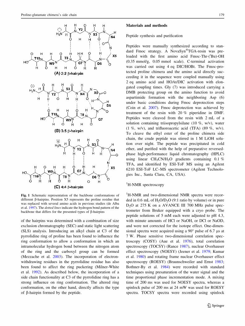
The predominant form (3:5 b-hairpin) contains a type I b-
turn, while the less abundant conformation (4:4 b-hairpin)
features a G1 b-bulge (Fig. 1). Substitution of the turn-
inducing Pro residue by Ser (P5-S) does not significantly
influence the conformation; the same ratio of b-hairpin
conformations (3:5 b-hairpin and 4:4 b-hairpin) was
observed for the Ser-containing peptide.
The Pro-containing sequence was used as a model
peptide for the current conformational investigations. Pro
5 of the sequence IYSNPDGTWT (P1_P) is part of the
turn moiety and was replaced by either cis-proline-glu-
tamic acid chimera (Pcis-E) or trans-proline-glutamic acid
chimera (Ptrans-E). The consequences of the substitution on
the turn conformation were studied with a combination of
several analytical techniques. The peptides (P1_P,
P1_Pcis-E, and P1_Ptrans-E) were manually synthesized
using the Fmoc strategy. The conformations of the
resulting hairpins were studied with the help of one-
dimensional- and two-dimensional- NMR analysis and
circular dichroism (CD) spectroscopy. The oligomerization
178 J. Maity et al.
123
of the hairpins was determined with a combination of size
exclusion chromatography (SEC) and static light scattering
(SLS) analysis. Introducing an alkyl chain at C3 of the
pyrrolidine ring of proline has been found to influence the
ring conformation to allow a conformation in which an
intramolecular hydrogen bond between the nitrogen atom
of the ring and the carboxyl group can be formed
(Mezzache et al. 2003). The incorporation of electron-
withdrawing residues in the pyrrolidine residue has also
been found to affect the ring puckering (Milner-White
et al. 1992). As described below, the incorporation of a
side chain functionality at C3 of the pyrrolidine ring has a
strong influence on ring conformation. The altered ring
conformation, on the other hand, directly affects the type
of b-hairpin formed by the peptide.
Materials and methods
Peptide synthesis and purification
Peptides were manually synthesized according to stan-
dard Fmoc strategy. A NovaSyn�TGA-resin was pre-
loaded with the first amino acid Fmoc-Thr-(tBu)-OH
(0.35 mmol/g, 0.05 mmol scale). C-terminal activation
was carried out using 4 eq DIC/HOBt. The Fmoc-pro-
tected proline chimera and the amino acid directly suc-
ceeding it in the sequence were coupled manually using
2 eq amino acid and HOAt/DIC activation with elon-
gated coupling times. Gly (7) was introduced carrying a
DMB protecting group on the amino function to avoid
aspartimide formation with the neighboring Asp (6)
under basic conditions during Fmoc deprotection steps
(Coin et al. 2007). Fmoc deprotection was achieved by
treatment of the resin with 20 % piperidine in DMF.
Peptides were cleaved from the resin with 2 mL of a
solution containing triisopropylsilane (10 %, w/v), water
(1 %, w/v), and trifluoroacetic acid (TFA) (89 %, w/v).
To cleave the ethyl ester of the proline chimera side
chain, the crude peptide was stirred in 1 M LiOH solu-
tion over night. The peptide was precipitated in cold
ether, and purified with the help of preparative reversed-
phase high-performance liquid chromatography (HPLC)
using linear CH3CN/H2O gradients containing 0.1 %
TFA, and identified by ESI-ToF MS using an Agilent
6210 ESI-ToF LC–MS spectrometer (Agilent Technolo-
gies Inc., Santa Clara, CA, USA).
1H-NMR spectroscopy
1H-NMR and two-dimensional NMR spectra were recor-
ded in 0.6 mL of H2O/D2O (9:1 ratio by volume) or in pure
D2O at 275 K on a AVANCE III 700 MHz pulse spec-
trometer from Bruker equipped with a cryo probe. The
peptide solutions of 5 mM each were adjusted to pH 4.3,
with minute amounts of HCl or NaOH, or DCl or NaOD,
and were not corrected for the isotope effect. One-dimen-
sional spectra were acquired using a 90� pulse of 6.7 ls at
7 W. Phase sensitive two-dimensional correlation spec-
troscopy (COSY) (Aue et al. 1976), total correlation
spectroscopy (TOCSY) (Rance 1987), nuclear Overhauser
effect spectroscopy (NOESY) (Jeener et al. 1979; Kumar
et al. 1980) and rotating frame nuclear Overhauser effect
spectroscopy (ROESY) (Braunschweiler and Ernst 1983;
Bothner-By et al. 1984) were recorded with standard
techniques using presaturation of the water signal and the
time proportional phase incrementation mode. A mixing
time of 200 ms was used for NOESY spectra, whereas a
spinlock pulse of 200 ms at 24 mW was used for ROESY
spectra. TOCSY spectra were recorded using spinlock
Fig. 1 Schematic representation of the backbone conformations of
different b-hairpins. Position X5 represents the proline residue that
was replaced with several amino acids in previous studies (de Alba
et al. 1997). The dotted lines indicate the hydrogen bond pattern of the
backbone that differs for the presented types of b-hairpins
Proline-glutamate chimera’s side chain 179
123
pulses of 400 ms. Additional NOESY and ROESY exper-
iments were performed for peptide samples in pure D2O to
facilitate the observation of the Ha–Ha NOE cross peaks
close to the water signal. Obtained data were processed
using Bruker TOPSPIIN software.
Circular dichroism spectroscopy
The lyophilized peptide was dissolved in H2O/D2O (9:1
ratio by volume) and the pH was adjusted to 4.3 to main-
tain the conditions used during the NMR-measurements.
The concentration of peptide stock solutions was deter-
mined according to the tyrosine and tryptophan absorbance
in 6 M guanidine hydrochloride (eTyr;280 nm =
1,200 mol-1cm-1, eTrp;280 nm = 5,560 mol-1cm-1) using
a Varian Cary 50 spectrophotometer (Varian Medical
Systems, Palo Alto, CA, USA) and PMMA cuvettes
(10 mm path length, 1.5 mL, Plastibrand�, VWR Interna-
tional GmbH, Darmstadt, Germany). Stock solutions of
1 mM concentration were prepared and subsequently
diluted to give solutions with final concentrations of 100,
300, and 500 lM. CD spectra of the peptide variants were
recorded using a Jasco J-810 spectropolarimeter at 5 �C.
Quartz cells (0.2 mm path length for 300 and 500 lM
samples, and 1.0 mm path length for 100 lM samples)
were used throughout. The denaturation experiments were
conducted using 1.0 mm Quartz cells. The spectra were the
average of three scans, obtained by collecting data from
240 to 195 nm (240–200 nm for the denaturation experi-
ments) at 0.5-nm intervals, 2-nm bandwidth, and 2 s
response times. Spectra were background-corrected by
subtracting the corresponding H2O/D2O spectra.
Size exclusion chromatography/static light scattering
The oligomerization state was determined by applying a
combined SEC/SLS analysis. SEC analysis was per-
formed using a WTC-015S5 column (5 lm, 150 A,
7.8 9 300 mm, Wyatt Technology) connected to a HPLC
workstation (La Chrom, VWR, Hitachi, L-2130). The
separation was carried out at a velocity of 0.3 ml/min at
room temperature in an aqueous buffer consisting of
10 mM sodium phosphate and 150 mM sodium chloride at
pH 7.3. Elution of peptide was monitored by UV detection
(VWR, Hitachi, L-2400) at 280 nm. The concentration of
all injected samples was 150 lM and the sample volume
was 100 ll in each case. SLS experiments were performed
at room temperature using a Dawn Heleos eight light
scattering photometer and an Optilab rEX refractive index
detector (Wyatt Technology). Molecular weights were
calculated using a dn/dc value of 0.185 mL/g. All data
were analyzed with ASTRA software version 5.3.4.20
(Wyatt Technology).
Results and discussion
Peptide design
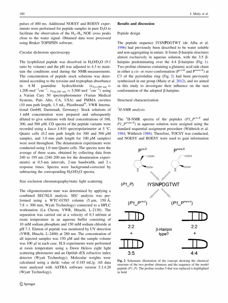
The peptide sequence IYSNPDGTWT (de Alba et al.
1996) had previously been described to be water soluble
and non-aggregating in nature. It forms b-hairpin structures
almost exclusively in aqueous solution, with the 3:5 b-
hairpins predominating over the 4:4 b-hairpins (Fig. 1).
Two proline chimeras containing a glutamic acid side chain
in either a cis- or trans-conformation (Pcis-E and Ptrans-E) at
C3 of the pyrrolidine ring (Fig. 2) had been previously
synthesized in our group (Maity et al. 2012), and we aimed
in this study to investigate their influence on the turn
conformation of the adopted b-hairpins.
Structural characterization
1H-NMR analysis
The 1H-NMR spectra of the peptides (P1_Pcis-E and
P1_Ptrans-E) in aqueous solution were assigned using the
standard sequential assignment procedure (Wuthrich et al.
1984; Wuthrich 1986). Therefore, TOCSY was conducted,
and NOESY and ROESY were used to gain information
Fig. 2 Schematic illustration of the concept showing the chemical
structure of the two proline chimeras and the sequence of the model
peptide (P1_P). The proline residue 5 that was replaced is highlighted
in bold
180 J. Maity et al.
123
about the proximity of the protons. NMR analysis of the
three peptides revealed that the substitution of Pro in the
reference peptide with the two proline chimeras resulted in
altered conformations of the formed b-hairpins. The ste-
reochemistry of the glutamic acid side chain at the C3 of
the proline chimera is the only distinguishing factor
between the peptides (P1_Pcis-E) and (P1_Ptrans-E) found to
have a crucial impact on the shape of the ring conformation
and thus on the type of b-hairpin that is formed.
The peak corresponding to CdH3 of the model peptide
(P1_P) has been used as the reference peak in the 1H-NMR
spectra for the peptides containing the proline chimera. The1H-NMR spectra of the peptides (P1_Pcis-E and P1_Ptrans-
E) show peaks arising from the cis/trans equilibrium of the
proline imide bond due to the presence of the proline
chimera moiety in these peptides (Kang and Young Choi
2004; Thunecke et al. 1996; Sugawara et al. 2001; Ivanova
et al. 2010). We assume that the trans form of the peptides
is the predominating species due to the similar conforma-
tion of the X-proline chimera bond with the X-proline bond
in the peptides (Zimmerman and Scheraga 1976). The
chemical shifts of the proton resonances for the peptides
P1_Pcis-E and P1_Ptrans-E in aqueous solution at 5 �C and
pH 4.3 are given in Tables 1 and 2, respectively.
The conformational shift of the Ca protons (Ha) can be
used to gain insight into the secondary structures of pro-
teins (Wishart et al. 1991; Wuthrich 1986). The deviation
of chemical shift [(DdHa) = dHa (observed) - dHa (ran-
dom coil)] of the Ha proton from the random coil values is
negative in turn or helical regions and positive in b-strand
regions. This type of analysis was conducted for all resi-
dues of the peptides containing the proline chimeras.
Except for the proline chimera moieties, all residues of the
peptides followed the same trend in DdHa values. In the
case of proline chimera residues, we found that the con-
formational shift for Ha is positive for the Pcis-E moiety,
and negative for the Ptrans-E moiety (Fig. 3). Since a neg-
ative DdHa value is more suitable for residues at turn
regions of b-hairpins, we infer that the negative DdHa
value of the Ptrans-E residue at the turn position could be
linked to the pre-organization of the peptide, which pro-
motes folding into a b-hairpin structure. Although both
proline chimera moieties have been found to be suitable for
a b-turn position of the hairpin forming peptides, this result
indicates that the Ptrans-E moiety is presumably better
accommodated in the b-turn position than the Pcis-E moiety.
Table 1 Chemical shift assignments of peptide (P1_Pcis-E)
NH Ha Hb Other
Ile 1 3.80 1.91 CcH 1.42, 1.14; Cc’H3 0.93; CdH3
0.86
Tyr 2 8.50 4.65 2.89,
2.78
CdH 7.07; CeH 6.78
Ser 3 8.18 4.26 3.62,
3.62
Asn 4 8.44 4.72 2.81,
2.57
NdH2 7.62, 7.00
Procis-
E 5
4.44 2.79 CcH 2.15, 1.72; Cc’H 2.44, 2.37;
CdH 3.87, 3.66
Asp 6 8.78 4.59 2.98,
2.91
Gly 7 7.98 3.85,
3.81
Thr 8 7.96 4.28 4.11 CcH3 1.08
Trp 9 8.35 4.78 3.30,
3.20
Ne1H 10.09; Cd1H 7.19; Ce3H 7.57;
Cf3H 7.09; Cg2H 7.17; Cf2H 7.44
Thr 10 7.99 4.24 4.20 CcH3 1.06
Table 2 Chemical shift assignments of peptide (P1_Ptrans-E)
NH Ha Hb Other
Ile 1 3.82 1.93 CcH 1.43, 1.14; Cc’H3 0.94;
CdH3 0.86
Tyr 2 8.52 4.63 2.89,
2.89
CdH 7.07; CeH 6.78
Ser 3 8.16 4.30 3.57,
3.57
Asn 4 8.49 4.79 2.79,
2.64
NdH2 7.64, 7.02
Protrans-
E 5
4.03 2.62 CcH 2.23, 1.78; Cc’H 2.60,
2.48; CdH 3.96, 3.73
Asp 6 8.76 4.61 2.98,
2.92
Gly 7 8.21 3.93,
3.88
Thr 8 7.97 4.31 4.14 CcH3 1.10
Trp 9 8.40 4.77 3.31,
3.20
Ne1H 10.11; Cd1H 7.21; Ce3H
7.55; Cf3H 7.09; Cg2H 7.19;
Cf2H 7.44
Thr 10 8.06 4.29 4.22 CcH3 1.08
Fig. 3 Histograms of the conformational shift values of the Ha
(DdHa) relative to random coil values in the peptides P1_Pcis-E and
P1_Ptrans-E at pH 4.3 and 5 �C in aqueous solution
Proline-glutamate chimera’s side chain 181
123
Several NMR parameters have been used to identify the
b-hairpin structures adopted by P1_Pcis-E and P1_Ptrans-E.
The observed NOE patterns of both peptides give infor-
mation about sequentially non-adjacent residues that are in
close proximity, such as interacting residues of opposing b-
strands. ROESY spectra were obtained under the same
experimental conditions that were used for the NOESY
spectra (pH 4.3 and 5 �C) to obtain identical NOE patterns
(Supporting information). The cross-strand NOEs (NH–
NH, NH–Ha, Ha–Ha and involved side chains) that were
observed for P1_Pcis-E and P1_Ptrans-E are illustrated in
Figs. 4, 5, respectively.
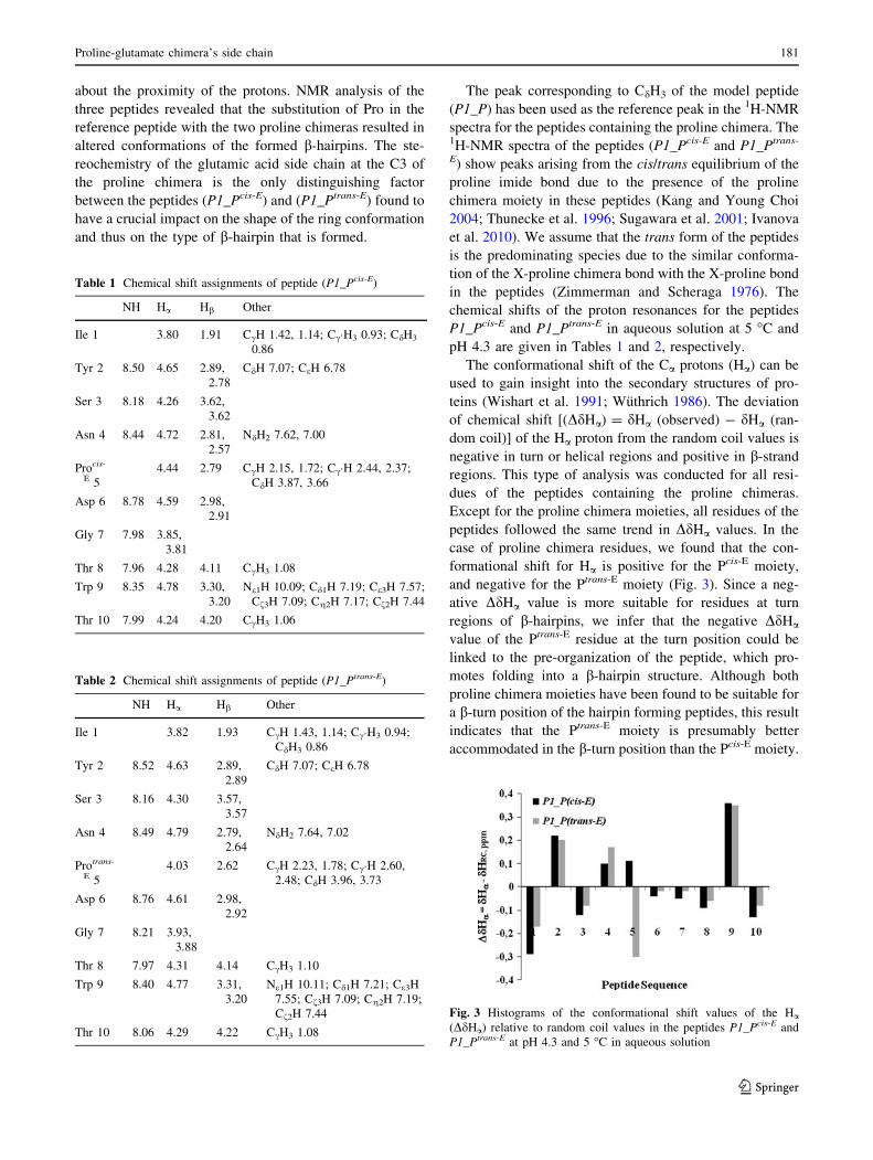
In the peptide P1_Pcis-E, long range NOEs relating the
backbone protons, NH(Y2)–NH(T10), Ha(S3)–NH(T10)
and Ha(S3)–Ha(W9) have been found, indicating the for-
mation of a 3:5 b-hairpin. For this type of b-hairpin, the
residues (Y2–S3 and W9–T10) face towards each other. In
addition to the backbone NOEs, strong NH–NH interac-
tions of S3 and D6 were observed, which indicate the
formation of a second conformation, a 2:2 b-hairpin.
However, the expected Ha(Y2)–Ha(G7) interaction was not
observed. The described interactions reveal that the peptide
(P1_Pcis-E) adopts two different types of b-hairpin in
aqueous solution, which are in equilibrium with each other.
Additional long range NOEs involving side chain protons
were also found for peptide P1_Pcis-E (Fig. 4).
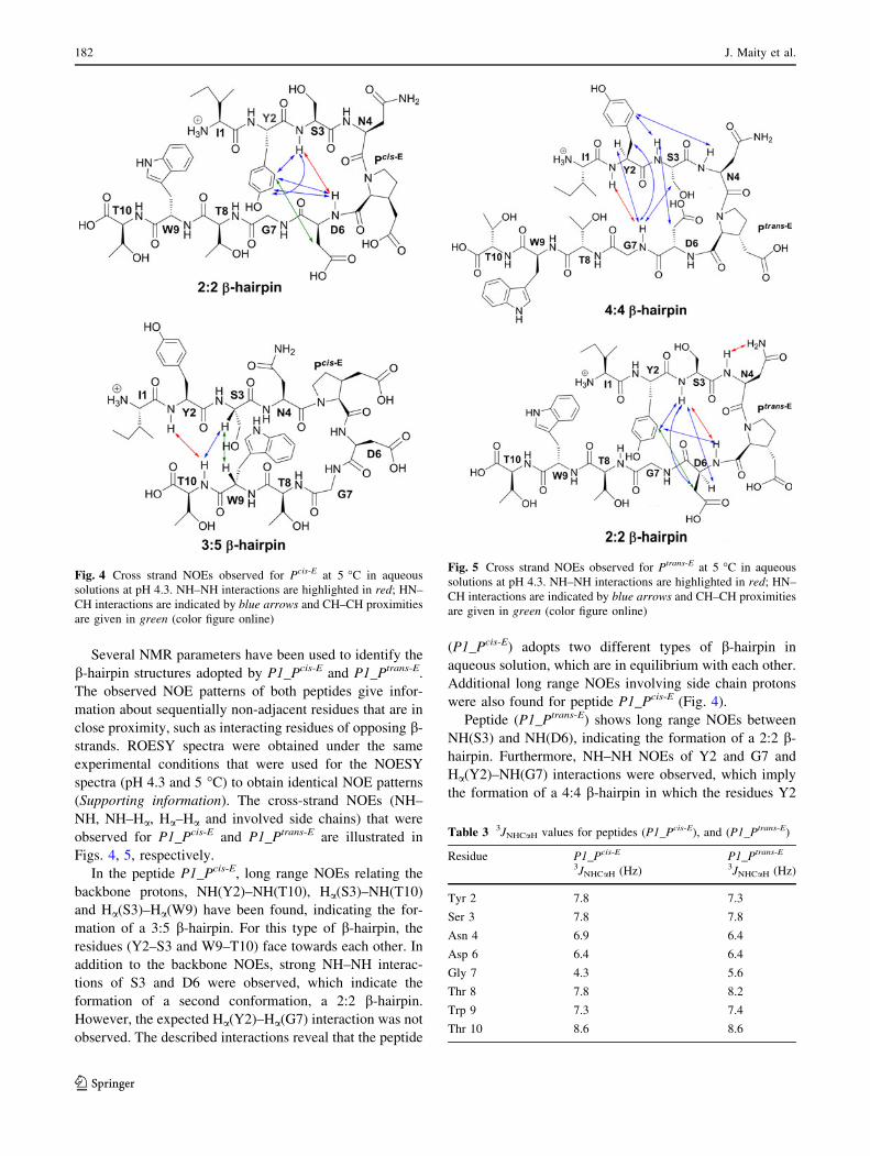
Peptide (P1_Ptrans-E) shows long range NOEs between
NH(S3) and NH(D6), indicating the formation of a 2:2 b-
hairpin. Furthermore, NH–NH NOEs of Y2 and G7 and
Ha(Y2)–NH(G7) interactions were observed, which imply
the formation of a 4:4 b-hairpin in which the residues Y2
Fig. 4 Cross strand NOEs observed for Pcis-E at 5 �C in aqueous
solutions at pH 4.3. NH–NH interactions are highlighted in red; HN–
CH interactions are indicated by blue arrows and CH–CH proximities
are given in green (color figure online)
Fig. 5 Cross strand NOEs observed for Ptrans-E at 5 �C in aqueous
solutions at pH 4.3. NH–NH interactions are highlighted in red; HN–
CH interactions are indicated by blue arrows and CH–CH proximities
are given in green (color figure online)
Table 3 3JNHCaH values for peptides (P1_Pcis-E), and (P1_Ptrans-E)
Residue P1_Pcis-E
3JNHCaH (Hz)
P1_Ptrans-E
3JNHCaH (Hz)
Tyr 2 7.8 7.3
Ser 3 7.8 7.8
Asn 4 6.9 6.4
Asp 6 6.4 6.4
Gly 7 4.3 5.6
Thr 8 7.8 8.2
Trp 9 7.3 7.4
Thr 10 8.6 8.6
182 J. Maity et al.
123
and S3 face towards the residues D6 and G7 (Fig. 5). Thus,
the existence of two different b-hairpin structures was also
observed for this peptide. However, the 4:4 b-hairpin
formed by P1_Ptrans-E is not exactly the same type that the
reference peptide (P1_P) adopts. In the case of P1_P, the
distal strand residues are S3 and T8 and the four turn
participating residues are N4, P5, D6 and G7 (de Alba et al.
1996). For P1_Ptrans-E, however, the distal strand residues
are Y2 and G7 and the four residues forming the turn are
S3, N4, P5 and D6. Side chain protons were also found to
be involved in cross-correlation NOEs (Fig. 5). The
reference peptide P1_P exists in equilibrium between a 3:5
b-hairpin and a 4:4 b-hairpin. P1_Pcis-E adopts a 2:2 b-
hairpin coexisting with a 3:5 b-hairpin, while P1_Ptrans-E
occurs as a mixture of 2:2 b-hairpin and 4:4 b-hairpin.
Vicinal coupling constant values (3JNHCaH) were cal-
culated to verify b-sheet formation by the peptides
(Table 3). Values larger than 7 Hz are characteristic for a
b-strand structure.
Almost all residues show values larger than 7 Hz,
indicating their existence in b-strand conformations.
However, the residues Asn 4, Asp 6, and Gly 7 have
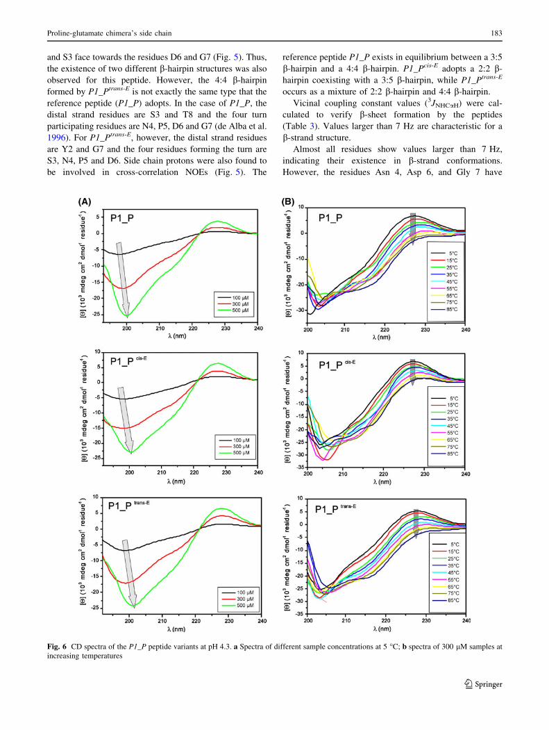
Fig. 6 CD spectra of the P1_P peptide variants at pH 4.3. a Spectra of different sample concentrations at 5 �C; b spectra of 300 lM samples at
increasing temperatures
Proline-glutamate chimera’s side chain 183
123
smaller coupling constants because they are present within
the turn structure. For residues near the C- and N-terminus
of the peptide, larger 3JNHCaH values were found, indicat-
ing that these residues also adopt b-strand structures.
Circular dichroism
In addition to the NMR analysis, the conformations of the
three peptides were investigated by CD spectroscopy. Due
to the short sequence of only ten residues, no characteristic
b-sheet structures containing a minimum at around 216 nm
have been observed. However, some characteristic signa-
tures for secondary structures were observed. With regard
to the assumed b-hairpin conformations, elucidated during
NMR analysis, only three residues are expected to form b-
strands. Thus, the spectra resemble more a random coil
structure, although a small minimum at around 216 nm was
observed. With increasing peptide concentration, the min-
imum at 198 nm, being characteristic for random coil
structures, is slightly shifted towards 201 nm (Fig. 6a).
This indicates a higher amount of order in the structure
with increasing peptide concentration. Also, the maxima
between 220 and 230 nm observed in all spectra indicate
that the aromatic residues (Trp, Tyr) participate in ordered
structures (Fernandez-Escamilla et al. 2006; Lakshmina-
rayanan et al. 2009). This assumption is further supported
by the systematic decrease in this maximum with increas-
ing temperature (Fig. 6b). The obtained results, which are
in good agreement with previously published CD spectra
(de Alba et al. 1996; Fernandez-Escamilla et al. 2006),
show that all three peptides adopt similar structures in
aqueous solution, and indications for b-hairpin conforma-
tion were found in the CD spectra. However, a distinction
between the different b-hairpin types that were found with
NMR cannot be made with CD spectroscopy alone.
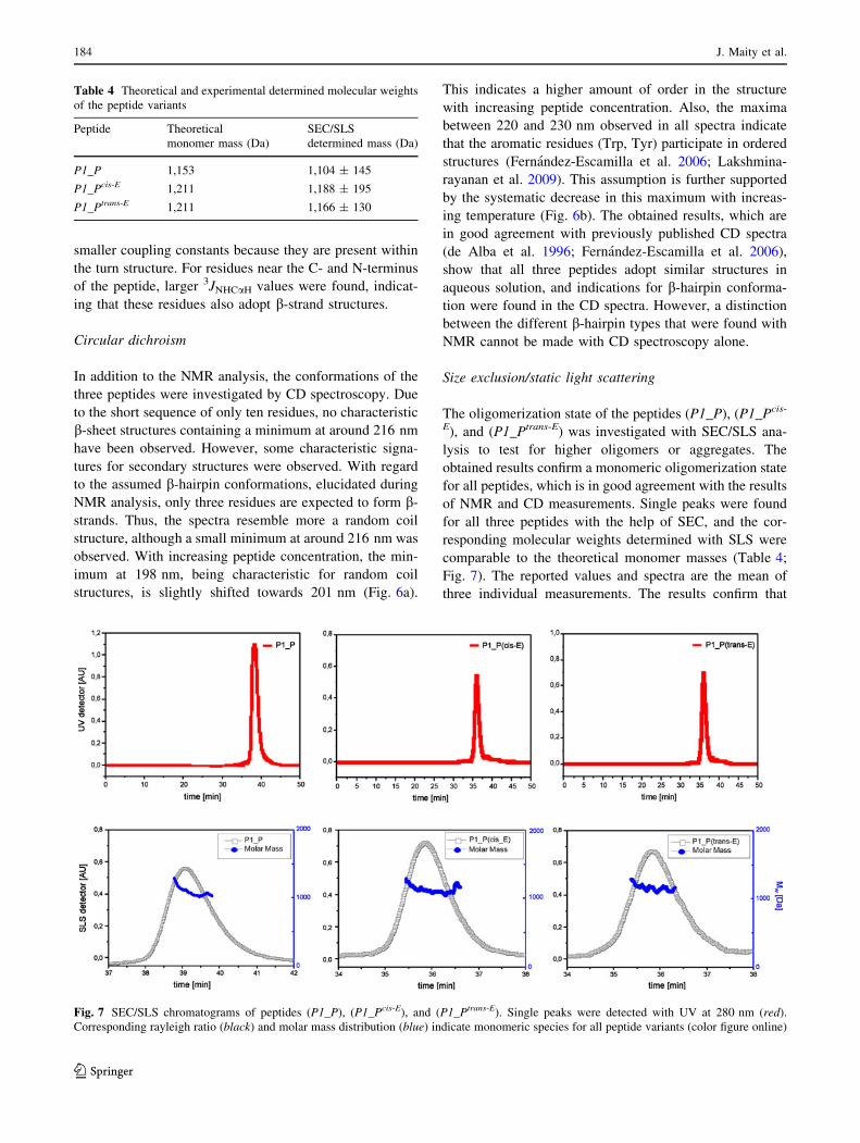
Size exclusion/static light scattering
The oligomerization state of the peptides (P1_P), (P1_Pcis-
E), and (P1_Ptrans-E) was investigated with SEC/SLS ana-
lysis to test for higher oligomers or aggregates. The
obtained results confirm a monomeric oligomerization state
for all peptides, which is in good agreement with the results
of NMR and CD measurements. Single peaks were found
for all three peptides with the help of SEC, and the cor-
responding molecular weights determined with SLS were
comparable to the theoretical monomer masses (Table 4;
Fig. 7). The reported values and spectra are the mean of
three individual measurements. The results confirm that
Table 4 Theoretical and experimental determined molecular weights
of the peptide variants
Peptide Theoretical
monomer mass (Da)
SEC/SLS
determined mass (Da)
P1_P 1,153 1,104 ± 145
P1_Pcis-E 1,211 1,188 ± 195
P1_Ptrans-E 1,211 1,166 ± 130
Fig. 7 SEC/SLS chromatograms of peptides (P1_P), (P1_Pcis-E), and (P1_Ptrans-E). Single peaks were detected with UV at 280 nm (red).
Corresponding rayleigh ratio (black) and molar mass distribution (blue) indicate monomeric species for all peptide variants (color figure online)
184 J. Maity et al.
123
single peptide strands form the observed b-hairpin struc-
tures and that the interactions found in the NMR-experi-
ments are of intramolecular nature rather than occurring
between two individual hairpins.
Conclusion
The conformational analysis of short b-hairpins containing
Pcis-E and Ptrans-E in the turn region clearly demonstrates
the importance of the proline ring conformation for the
formation of b-hairpins. Even small alterations in the
sensitive turn region can have large consequences for the
whole peptide conformation as the turn type dictates the
type of b-hairpin that is formed. The reference peptide
(P1_P) contains L-proline at position 5, and forms a 3:5 b-
hairpin that coexists with a 4:4 b-hairpin in aqueous
solution. Substitution of their turn-contributing Pro residue
with Pcis-E results in the formation of a 2:2 b-hairpin and a
3:5 b-hairpin. The incorporation of Ptrans-E yields an
equilibrium between a 2:2 b-hairpin and a 4:4 b-hairpin.
These results clearly show the importance of the ring
puckering of proline as a key factor in determining the type
of b-hairpin that will be formed by the peptide. With two
proline chimeras that differ only at a single chiral center,
we were able to alter the b-hairpin structures of the cor-
responding peptides. Our findings show that such proline
chimeras can be used to fine-tune the conformation of b-
hairpin structures and are therefore likely to become
interesting tools for applications in medicinal chemistry
and materials sciences.
Acknowledgments The authors thank the Center for International
Cooperation and the Dahlem Research School of Freie Universitat
Berlin for funding.
Conflict of interest The authors declare that they have no conflict
of interest.
References
Aubry A, Vitoux B, Marraud M (1985) Conformational properties of
Pro–Pro sequences. I. Crystal structures of two dipeptides with L-
Pro-L-Pro and L-Pro-D-Pro sequences. Biopolymers
24:1089–1100
Aue WP, Bartholdi E, Ernst RR (1976) Two-dimensional spectros-
copy. Application to nuclear magnetic resonance. J Chem Phys
64:2229–2246
Blanco FJ, Jimenez MA, Herranz J, Rico M, Santoro J, Nieto JL (1993)
NMR evidence of a short linear peptide that folds into a b-hairpin
in aqueous solution. J Am Chem Soc 115:5887–5888
Blanco FJ, Rivas G, Serrano L (1994) A short linear peptide that folds
into a native stable b-hairpin in aqueous solution. Nat Struct Biol
1:584–590
Bothner-By AA, Stephens RL, Lee J, Warren CD, Jeanloz RW (1984)
Structure determination of a tetrasaccharide: transient nuclear
Overhauser effects in the rotating frame. J Am Chem Soc
106:811–813
Braunschweiler L, Ernst RR (1983) Coherence transfer by isotropic
mixing: application to proton correlation spectroscopy. J Magn
Reson 53:521–528
Chatterjee B, Saha I, Raghothama S, Aravinda S, Rai R, Shamala N,
Balaram P (2008) Designed peptides with homochiral and
heterochiral diproline templates as conformational constraints.
Chem Eur J 14:6192–6204
Coin I, Beyermann M, Bienert M (2007) Solid-phase peptide
synthesis: from standard procedures to the synthesis of difficult
sequences. Nat Protoc 2:3247–3256
de Alba Ed, Jimenez MA, Rico M, Nieto JL (1996) Conformational
investigation of designed short linear peptides able to fold into b-
hairpin structures in aqueous solution. Fold Des 1:133–144
de Alba E, Jimenez MA, Rico M (1997) Turn residue sequence
determines b-Hairpin conformation in designed peptides. J Am
Chem Soc 119:175–183
Delaye PO, Vasse JL, Szymoniak J (2010) Asymmetric synthesis of
proline-based conformationally constrained tryptophan mimetic.
Org Biomol Chem 8:3635–3637
Dyson HJ, Wright PE (1991) Defining solution conformations of
small linear peptides. Annu Rev Biophys Biophys Chem
20:519–538
Dyson HJ, Wright PE (1993) Peptide conformation and protein
folding. Curr Opin Struct Biol 3:60–65
Fatas P, Jimenez AI, Calaza MI, Cativiela C (2012) b-Phenylproline:
the high b-turn forming propensity of proline combined with an
aromatic side chain. Org Biomol Chem 10:640–651
Fernandez-Escamilla AM, Ventura S, Serrano L, Jimenez MA (2006)
Design and NMR conformational study of a b-sheet peptide
based on Betanova and WW domains. Protein Sci 15:2278–2289
Gerling UIM, Brandenburg E, Berlepsch Hv, Pagel K, Koksch B
(2010) Structure analysis of an amyloid-forming model peptide
by a systematic glycine and proline scan. Biomacromolecules
12:2988–2996
Guitot K, Larregola M, Pradhan TK, Vasse JL, Lavielle S, Bertus P,
Szymoniak J, Lequin O, Karoyan P (2011) The combination of
prolinoamino acids and cyclopropylamino acids leads to fully
functionalized, stable b-turns in water. Chembiochem
12:1039–1042
Haque TS, Little JC, Gellman SH (1994) ‘‘Mirror image’’ reverse
turns promote b-hairpin formation. J Am Chem Soc 116:
4105–4106
Haque TS, Little JC, Gellman SH (1996) Stereochemical require-
ments for b-hairpin formation: model studies with four-residue
peptides and depsipeptides. J Am Chem Soc 118:6975–6985
Horne WS, Price JL, Keck JL, Gellman SH (2007) Helix bundle
quaternary structure from a/b-peptide foldamers. J Am Chem
Soc 129:4178–4180
Horne WS, Johnson LM, Ketas TJ, Klasse PJ, Lu M, Moore JP,
Gellman SH (2009) Structural and biological mimicry of protein
surface recognition by a/b-peptide foldamers. Proc Nat Acad Sci
USA 106:14751–14756. doi:10.1073/pnas.0902663106
Ishikawa Y, Oka M, Hayashi T, Nishinaga A (1996) Theoretical
analysis of a-helix hairpin structures constructed by two right-
handed a-helices. Polym J 28:86–90
Ivanova G, Yakimova B, Angelova S, Stoineva I, Enchev V (2010)
Influence of pH on the cis-trans isomerization of valine-proline
dipeptide: an integrated NMR and theoretical investigation.
J Mol Struct 975:330–334
Jeener J, Meier BH, Bachmann P, Ernst RR (1979) Investigation of
exchange processes by two-dimensional NMR spectroscopy.
J Chem Phys 71:4546–4553
Kang YK, Young Choi H (2004) Cis-trans isomerization and
puckering of proline residue. Biophys Chem 111:135–142
Proline-glutamate chimera’s side chain 185
123
Karoyan P, Quancard J, Vaissermann J, Chassaing Gr (2003) Amino-
zinc-enolate carbometalation reactions: application to ring
closure of terminally substituted olefin for the asymmetric
synthesis of cis- and trans-3-prolinoleucine. J Org Chem 68:
2256–2265
Kim PS, Baldwin RL (1990) Intermediates in the Folding Reactions
of Small Proteins. Annu Rev Biochem 59:631–660
Kumar A, Ernst RR, Wuthrich K (1980) A two-dimensional nuclear
Overhauser enhancement (2D NOE) experiment for the eluci-
dation of complete proton–proton cross-relaxation networks in
biological macromolecules. Biochem Biophys Res Commun
95:1–6
Lakshminarayanan R, Yoon I, Hegde BG, Fan D, Du C, Moradian-
Oldak J (2009) Analysis of secondary structure and self-
assembly of amelogenin by variable temperature circular
dichroism and isothermal titration calorimetry. Proteins: Struct
Funct Bioinf 76:560–569
Lengyel GA, Frank RC, Horne WS (2011) Hairpin folding behavior
of mixed a/b-peptides in aqueous solution. J Am Chem Soc
133:4246–4249
Loughlin WA, Tyndall JDA, Glenn MP, Hill TA, Fairlie DP (2010)
Update 1 of: b-strand mimetics. Chem Rev 110:32–69
MacArthur MW, Thornton JM (1991) Influence of proline residues on
protein conformation. J Mol Biol 218:397–412
Maity J, Saha P, Gerling UIM, Lentz D, Koksch B (2012) An
approach for simultaneous synthesis of cis- and trans-3-substi-
tuted proline-glutamic acid chimeras. Synthesis 44:3063–3070
Mezzache S, Afonso C, Pepe C, Karoyan P, Fournier F, Tabet JC
(2003) Proton affinity of proline and modified prolines using the
kinetic method: role of the conformation investigated by ab initio
calculations. Rapid Commun Mass Spectrom 17:1626–1632
Milner-White EJ, Bell LH, Maccallum PH (1992) Pyrrolidine ring
puckering in cis and trans-proline residues in proteins and
polypeptides. Different puckers are favoured in certain situations.
J Mol Biol 228:725–734
Mothes C, Larregola M, Quancard J, Goasdoue N, Lavielle S,
Chassaing G, Lequin O, Karoyan P (2010) Prolinoamino acids as
tools to build bifunctionalized, stable b-turns in water. Chem-
biochem 11:55–58
Munoz V, Thompson PA, Hofrichter J, Eaton WA (1997) Folding
dynamics and mechanism of b-hairpin formation. Nature
390:196–199
Nesloney CL, Kelly JW (1996) A 2,30-substituted biphenyl-based
amino acid facilitates the formation of a monomeric b-hairpin-
like structure in aqueous solution at elevated temperature. J Am
Chem Soc 118:5836–5845
Oba M, Saegusa T, Nishiyama N, Nishiyama K (2009) Synthesis of
non-proteinogenic amino acids using Michael addition to
unsaturated orthopyroglutamate derivative. Tetrahedron 65:
128–133
Pagel K, Wagner SC, Samedov K, von Berlepsch H, Bottcher C,
Koksch B (2006) Random coils, b-sheet ribbons, and a-helical
fibers: one peptide adopting three different secondary structures
at will. J Am Chem Soc 128:2196–2197
Price JL, Horne WS, Gellman SH (2010) Structural consequences of
b-amino acid preorganization in a self-assembling a/b-peptide:
fundamental studies of foldameric helix bundles. J Am Chem
Soc 132:12378–12387
Ramirez-Alvarado M, Blanco FJ, Serrano L (1996) De novo design
and structural analysis of a model b-hairpin peptide system. Nat
Struct Biol 3:604–612
Rance M (1987) Improved techniques for homonuclear rotating-frame
and isotropic mixing experiments. J Magn Reson 74:557–564
Rezaei Araghi R, Baldauf C, Gerling UIM, Cadicamo CD, Koksch B
(2011) A systematic study of fundamentals in a-helical coiled
coil mimicry by alternating sequences of b- and c-amino acids.
Amino Acids 41:733–742
Robinson JA (2008) b-Hairpin peptidomimetics: design, structures
and biological activities. Acc Chem Res 41:1278–1288
Searle MS, Williams DH, Packman LC (1995) A short linear peptide
derived from the N-terminal sequence of ubiquitin folds into a
water-stable non-native b-hairpin. Nat Struct Biol 2:999–1006
Sibanda BL, Blundell TL, Thornton JM (1989) Conformation of b-
hairpins in protein structures. A systematic classification with
applications to modelling by homology, electron density fitting
and protein engineering. J Mol Biol 206:759–777
Sugawara M, Tonan K, Ikawa S-i (2001) Effect of solvent on the cis-
trans conformational equilibrium of a proline imide bond of
short model peptides in solution. Spectrochim Acta A Mol
Biomol Spectrosc 57:1305–1316
Thunecke F, Kalman A, Kalman F, Ma S, Rathore AS, Horvath C
(1996) Kinetic study on the cis-trans isomerization of peptidyl-
proline dipeptides. J Chromatogr 744:259–272
Williams AD, Portelius E, Kheterpal I, Guo JT, Cook KD, Xu Y,
Wetzel R (2004) Mapping ab amyloid fibril secondary structure
using scanning proline mutagenesis. J Mol Biol 335:833–842
Wilmot CM, Thornton JM (1988) Analysis and prediction of the
different types of b-turn in proteins. J Mol Biol 203:221–232
Wishart DS, Sykes BD, Richards FM (1991) Relationship between
nuclear magnetic resonance chemical shift and protein secondary
structure. J Mol Biol 222:311–333
Wu YD, Han W, Wang DP, Gao Y, Zhao Y-L (2008) Theoretical
Analysis of Secondary Structures of b-Peptides. Acc Chem Res
41:1418–1427
Wuthrich K (1986) NMR of proteins and nucleic acids. Wiley, New
York
Wuthrich K, Billeter M, Braun W (1984) Polypeptide secondary
structure determination by nuclear magnetic resonance observa-
tion of short proton–proton distances. J Mol Biol 180:715–740
Zimmerman SS, Scheraga HA (1976) Stability of cis, trans, and
nonplanar peptide groups. Macromolecules 9:408–416
186 J. Maity et al.
123