Glycogen synthase kinase 3β-dependent Snail degradation directs … · Snail degradation was...
6
Glycogen synthase kinase 3β-dependent Snail degradation directs hepatocyte proliferation in normal liver regeneration Sayaka Sekiya a and Atsushi Suzuki a,b,1 a Division of Organogenesis and Regeneration, Medical Institute of Bioregulation, Kyushu University, Higashi-ku, Fukuoka, Fukuoka 812-8582, Japan; and b Precursory Research for Embryonic Science and Technology (PRESTO), Japan Science and Technology Agency, Kawaguchi, Saitama 332-0012, Japan Edited by James E. Darnell, The Rockefeller University, New York, NY, and approved May 25, 2011 (received for review October 27, 2010) Liver regeneration proceeds under the well-orchestrated control of multiple transcription factors that lead hepatocytes to reenter the cell cycle, proliferate, and renew quiescence. Here, we found an important role of the zinc-finger transcription factor Snail in liver regeneration. Snail was typically expressed in quiescent adult hepatocytes, but was rapidly degraded when the liver needed to regenerate itself. Decreased levels of Snail induced DNA synthesis in hepatocytes through up-regulation of cell cycle-related proteins. Snail degradation was dependent on phosphorylation by glycogen synthase kinase (GSK)-3β, whose quantity and activity were imme- diately increased after loss of liver mass or hepatic injury. Inacti- vation of GSK-3β resulted in suppression of Snail degradation and DNA synthesis in hepatocytes, leading to impaired liver growth during regeneration. This GSK-3β–dependent Snail degradation occurred as a result of cytokine, growth factor, and bile acid sig- nals that are known to drive liver regeneration. Thus, GSK-3β– dependent Snail degradation acts as a fundamental cue for the initiation of hepatocyte proliferation in liver regeneration. liver development | cell signaling | hydrodynamic gene delivery L iver regeneration is a fundamental response to hepatic damage, and has been favorably studied since the two-thirds partial hepatectomy (PH) model in rodents was described (1). In this system, quiescent mature hepatocytes rapidly reenter the cell cycle after PH and proliferate to restore the original liver mass. The priming of hepatocyte proliferation depends on the cellular responses to external stimulation by specific humoral factors, such as cytokines (e.g., IL-6) and growth factors [e.g., hepatocyte growth factor (HGF)], that are increased in the liver after PH (2). In most cases, these key effectors for liver regeneration are secreted by nonparenchymal cells and lead to the proliferation and survival of hepatocytes via activation of particular intra- cellular signaling pathways (3). In parallel with the effects of the cytokines and growth factors on liver growth, the remnant hep- atocytes after PH are also responsive to relatively increased bile acid flux and thereby initiate proliferation. This bile acid signaling pathway depends on nuclear bile acid receptors, and therefore hepatocytes lacking the bile acid receptor FXR suffer severe defects in proliferation after PH (4). These humoral factor signals alter the gene expression pattern in hepatocytes by activating transcription factors, including signal transducer and activator of transcription (STAT)-3, AP1, NF-κB, CCAAT-enhancer-binding protein (C/EBP)β, c-Myc, and forkhead box (Fox) m1b, and fi- nally induce DNA synthesis in hepatocytes through regulation of the cell cycle machinery (5). Consequently, the cell-extrinsic and -intrinsic mechanisms underlying the induction of hepatocyte proliferation have been studied intensively. However, it is still incompletely understood how such dynamic changes in the cel- lular proliferation state occur during liver regeneration. Here, we investigated the role of Snail, a zinc-finger tran- scription factor, in mouse liver regeneration. Snail is involved in many aspects of cellular function, including cell motility, differen- tiation, proliferation, and survival, during embryonic development and tumor progression (6). Until now, however, there has been no evidence regarding the contribution of Snail to normal liver regeneration. The amount of Snail in cells is controlled by gly- cogen synthase kinase (GSK)-3β, which binds to and phosphor- ylates Snail in the nucleus and cytoplasm of cells and eventually leads to β-Trcp–mediated ubiquitination and subsequent pro- teasomal degradation of Snail (7). Thus, in this study, we also investigated the role of GSK-3β in the regulation of Snail during liver regeneration. Results Snail Is Expressed in Quiescent Adult Hepatocytes, but Is Rapidly Degraded After Loss of Liver Mass or Hepatic Injury. Western blot analyses revealed that both Snail and GSK-3β were expressed in quiescent adult mouse hepatocytes and present in the nucleus and cytoplasm (Fig. 1A). Intriguingly, the amount of Snail in the liver was significantly decreased within 24 h after PH, although it returned to its normal level by 4 d after PH (Fig. 1B and Fig. S1A). In contrast, the amount of GSK-3β in the liver was in- creased at 1 d after PH, but returned to its normal level by 7 d after PH (Fig. 1B and Fig. S1A), whereas there was no significant change in the amount of serine 9-phosphorylated GSK-3β in the liver after PH (Fig. S1 B and C). Following such drastic changes of the amounts of Snail and GSK-3β, the amount of proliferating cell nuclear antigen (PCNA), which is expressed in the nuclei of cells during DNA synthesis, was increased in the liver at 2 d after PH and then gradually decreased to its normal level (Fig. 1B and Fig. S1A). According to a decrease in Snail after PH, the mRNA expression of E-cadherin, a transcriptional target of Snail for suppression, was increased in the liver (Fig. S1D). In a detailed analysis, a decrease in Snail and an increase in GSK-3β in the liver was first observed at 12 and 6 h after PH, respectively (Fig. 1C and Fig. S1E). To determine why Snail in the liver was de- creased after PH, we examined the protein degradation and changes in mRNA expression of Snail in the regenerating liver. When we directly injected the proteasome inhibitor MG132 into adult mice after PH to block proteasomal degradation of ubiq- uitinated proteins, ubiquitinated Snail had accumulated in the liver by 1 d after PH (Fig. 1D). However, quantitative PCR (qPCR) analyses revealed that, even after PH, Snail mRNA was stably expressed (Fig. 1E). Thus, the immediate decrease in Snail after PH was caused by degradation of ubiquitinated Snail. Moreover, as in the case of PH, the amounts of Snail and GSK- 3β in the liver were also decreased and increased, respectively, at 2 d after s.c. injection of carbon tetrachloride (CCl 4 ), a hep- Author contributions: A.S. designed research; S.S. and A.S. performed research; S.S. and A.S. analyzed data; and A.S. wrote the paper. The authors declare no conflict of interest. This article is a PNAS Direct Submission. 1 To whom correspondence should be addressed. E-mail: [email protected]. This article contains supporting information online at www.pnas.org/lookup/suppl/doi:10. 1073/pnas.1016122108/-/DCSupplemental. www.pnas.org/cgi/doi/10.1073/pnas.1016122108 PNAS Early Edition | 1 of 6 MEDICAL SCIENCES Downloaded by guest on June 5, 2020
Transcript of Glycogen synthase kinase 3β-dependent Snail degradation directs … · Snail degradation was...
Glycogen synthase kinase 3β-dependent Snaildegradation directs hepatocyte proliferationin normal liver regenerationSayaka Sekiyaa and Atsushi Suzukia,b,1
aDivision of Organogenesis and Regeneration, Medical Institute of Bioregulation, Kyushu University, Higashi-ku, Fukuoka, Fukuoka 812-8582, Japan; andbPrecursory Research for Embryonic Science and Technology (PRESTO), Japan Science and Technology Agency, Kawaguchi, Saitama 332-0012, Japan
Edited by James E. Darnell, The Rockefeller University, New York, NY, and approved May 25, 2011 (received for review October 27, 2010)
Liver regeneration proceeds under the well-orchestrated controlof multiple transcription factors that lead hepatocytes to reenterthe cell cycle, proliferate, and renew quiescence. Here, we foundan important role of the zinc-finger transcription factor Snail inliver regeneration. Snail was typically expressed in quiescent adulthepatocytes, but was rapidly degraded when the liver needed toregenerate itself. Decreased levels of Snail induced DNA synthesisin hepatocytes through up-regulation of cell cycle-related proteins.Snail degradation was dependent on phosphorylation by glycogensynthase kinase (GSK)-3β, whose quantity and activity were imme-diately increased after loss of liver mass or hepatic injury. Inacti-vation of GSK-3β resulted in suppression of Snail degradation andDNA synthesis in hepatocytes, leading to impaired liver growthduring regeneration. This GSK-3β–dependent Snail degradationoccurred as a result of cytokine, growth factor, and bile acid sig-nals that are known to drive liver regeneration. Thus, GSK-3β–dependent Snail degradation acts as a fundamental cue for theinitiation of hepatocyte proliferation in liver regeneration.
liver development | cell signaling | hydrodynamic gene delivery
Liver regeneration is a fundamental response to hepaticdamage, and has been favorably studied since the two-thirds
partial hepatectomy (PH) model in rodents was described (1). Inthis system, quiescent mature hepatocytes rapidly reenter the cellcycle after PH and proliferate to restore the original liver mass.The priming of hepatocyte proliferation depends on the cellularresponses to external stimulation by specific humoral factors,such as cytokines (e.g., IL-6) and growth factors [e.g., hepatocytegrowth factor (HGF)], that are increased in the liver after PH(2). In most cases, these key effectors for liver regeneration aresecreted by nonparenchymal cells and lead to the proliferationand survival of hepatocytes via activation of particular intra-cellular signaling pathways (3). In parallel with the effects of thecytokines and growth factors on liver growth, the remnant hep-atocytes after PH are also responsive to relatively increased bileacid flux and thereby initiate proliferation. This bile acid signalingpathway depends on nuclear bile acid receptors, and thereforehepatocytes lacking the bile acid receptor FXR suffer severedefects in proliferation after PH (4). These humoral factor signalsalter the gene expression pattern in hepatocytes by activatingtranscription factors, including signal transducer and activator oftranscription (STAT)-3, AP1, NF-κB, CCAAT-enhancer-bindingprotein (C/EBP)β, c-Myc, and forkhead box (Fox) m1b, and fi-nally induce DNA synthesis in hepatocytes through regulation ofthe cell cycle machinery (5). Consequently, the cell-extrinsic and-intrinsic mechanisms underlying the induction of hepatocyteproliferation have been studied intensively. However, it is stillincompletely understood how such dynamic changes in the cel-lular proliferation state occur during liver regeneration.Here, we investigated the role of Snail, a zinc-finger tran-
scription factor, in mouse liver regeneration. Snail is involved inmany aspects of cellular function, including cell motility, differen-tiation, proliferation, and survival, during embryonic development
and tumor progression (6). Until now, however, there has beenno evidence regarding the contribution of Snail to normal liverregeneration. The amount of Snail in cells is controlled by gly-cogen synthase kinase (GSK)-3β, which binds to and phosphor-ylates Snail in the nucleus and cytoplasm of cells and eventuallyleads to β-Trcp–mediated ubiquitination and subsequent pro-teasomal degradation of Snail (7). Thus, in this study, we alsoinvestigated the role of GSK-3β in the regulation of Snail duringliver regeneration.
ResultsSnail Is Expressed in Quiescent Adult Hepatocytes, but Is RapidlyDegraded After Loss of Liver Mass or Hepatic Injury. Western blotanalyses revealed that both Snail and GSK-3β were expressed inquiescent adult mouse hepatocytes and present in the nucleusand cytoplasm (Fig. 1A). Intriguingly, the amount of Snail in theliver was significantly decreased within 24 h after PH, althoughit returned to its normal level by 4 d after PH (Fig. 1B and Fig.S1A). In contrast, the amount of GSK-3β in the liver was in-creased at 1 d after PH, but returned to its normal level by 7 dafter PH (Fig. 1B and Fig. S1A), whereas there was no significantchange in the amount of serine 9-phosphorylated GSK-3β in theliver after PH (Fig. S1 B and C). Following such drastic changesof the amounts of Snail and GSK-3β, the amount of proliferatingcell nuclear antigen (PCNA), which is expressed in the nuclei ofcells during DNA synthesis, was increased in the liver at 2 d afterPH and then gradually decreased to its normal level (Fig. 1B andFig. S1A). According to a decrease in Snail after PH, the mRNAexpression of E-cadherin, a transcriptional target of Snail forsuppression, was increased in the liver (Fig. S1D). In a detailedanalysis, a decrease in Snail and an increase in GSK-3β in theliver was first observed at 12 and 6 h after PH, respectively (Fig.1C and Fig. S1E). To determine why Snail in the liver was de-creased after PH, we examined the protein degradation andchanges in mRNA expression of Snail in the regenerating liver.When we directly injected the proteasome inhibitor MG132 intoadult mice after PH to block proteasomal degradation of ubiq-uitinated proteins, ubiquitinated Snail had accumulated in theliver by 1 d after PH (Fig. 1D). However, quantitative PCR(qPCR) analyses revealed that, even after PH, Snail mRNA wasstably expressed (Fig. 1E). Thus, the immediate decrease in Snailafter PH was caused by degradation of ubiquitinated Snail.Moreover, as in the case of PH, the amounts of Snail and GSK-3β in the liver were also decreased and increased, respectively, at2 d after s.c. injection of carbon tetrachloride (CCl4), a hep-
Author contributions: A.S. designed research; S.S. and A.S. performed research; S.S. andA.S. analyzed data; and A.S. wrote the paper.
The authors declare no conflict of interest.
This article is a PNAS Direct Submission.1To whom correspondence should be addressed. E-mail: [email protected].
This article contains supporting information online at www.pnas.org/lookup/suppl/doi:10.1073/pnas.1016122108/-/DCSupplemental.
www.pnas.org/cgi/doi/10.1073/pnas.1016122108 PNAS Early Edition | 1 of 6
MED
ICALSC
IENCE
S
Dow
nloa
ded
by g
uest
on
June
5, 2
020
atotoxin that leads to fulminant hepatic failure and subsequentliver regeneration (Fig. 1F and Fig. S1F). Following this, theamount of PCNA in the liver was increased at 4 d after CCl4injection (Fig. 1F and Fig. S1F). However, Snail mRNA ex-pression was not decreased in the liver after CCl4 injection (Fig.1G). Thus, Snail residing in quiescent adult hepatocytes is de-graded in the early stages of liver regeneration, which is followedby activation of DNA synthesis in hepatocytes, suggesting that animmediate decrease in Snail in the damaged liver is important toinduce the proliferation of hepatocytes.
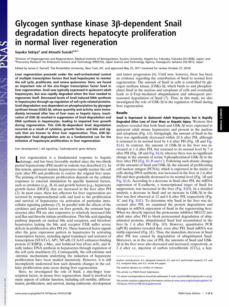
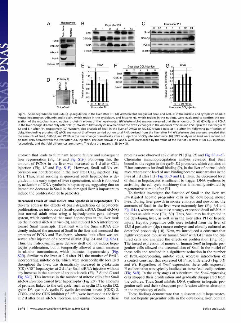
Decreased Levels of Snail Induce DNA Synthesis in Hepatocytes. Todirectly address the effects of Snail degradation on hepatocyteproliferation, we introduced a Snail-specific siRNA (Snail siRNA)into normal adult mice using a hydrodynamic gene deliverysystem, which confirmed that most hepatocytes in the liver tookup the injected siRNA in vivo (8), and induced RNA interferencetoward Snail transcripts. Treatment with the Snail siRNA effi-ciently reduced the amount of Snail in the liver and increased theamounts of PCNA and E-cadherin, whereas little effect was ob-served after injection of a control siRNA (Fig. 2A and Fig. S2A).Thus, the hydrodynamic gene delivery itself did not induce hepa-tocyte proliferation, but it temporally allowed a small increasein alanine transaminase, which indicates hepatotoxicity (Fig.S2B). Similar to the liver at 2 d after PH, the number of BrdU-incorporating mitotic cells, which were nonspecifically localizedthroughout the liver, was dramatically increased in cytokeratin(CK) 8/18+ hepatocytes at 2 d after Snail siRNA injection withoutany increase in the number of apoptotic cells (Fig. 2 B and C andFig. S2C). This increase in the number of mitotic cells after SnailsiRNA injection caused liver hypertrophy (Fig. 2D). The amountsof proteins linked to the cell cycle, such as cyclin D1, cyclin D2,cyclin D3, cyclin A, cyclin E, cyclin-dependent kinase (CDK) 2,CDK4, and the CDK inhibitor p21CIP1, were increased in the liverat 2 d after Snail siRNA injection, and similar increases in these
proteins were observed at 2 d after PH (Fig. 2E and Fig. S3 A–C).Chromatin immunoprecipitation analysis revealed that Snailbound to the region in the cyclin D2 promoter, which contains anE-box consensus for Snail binding (9), in the liver of normal adultmice, whereas the level of such binding becamemuchweaker in theliver at 1 d after PH (Fig. S3 D and E). Thus, the decreased levelof Snail in hepatocytes is sufficient to trigger DNA synthesis byactivating the cell cycle machinery that is normally activated byregenerative stimuli after PH.To further investigate the function of Snail in the liver, we
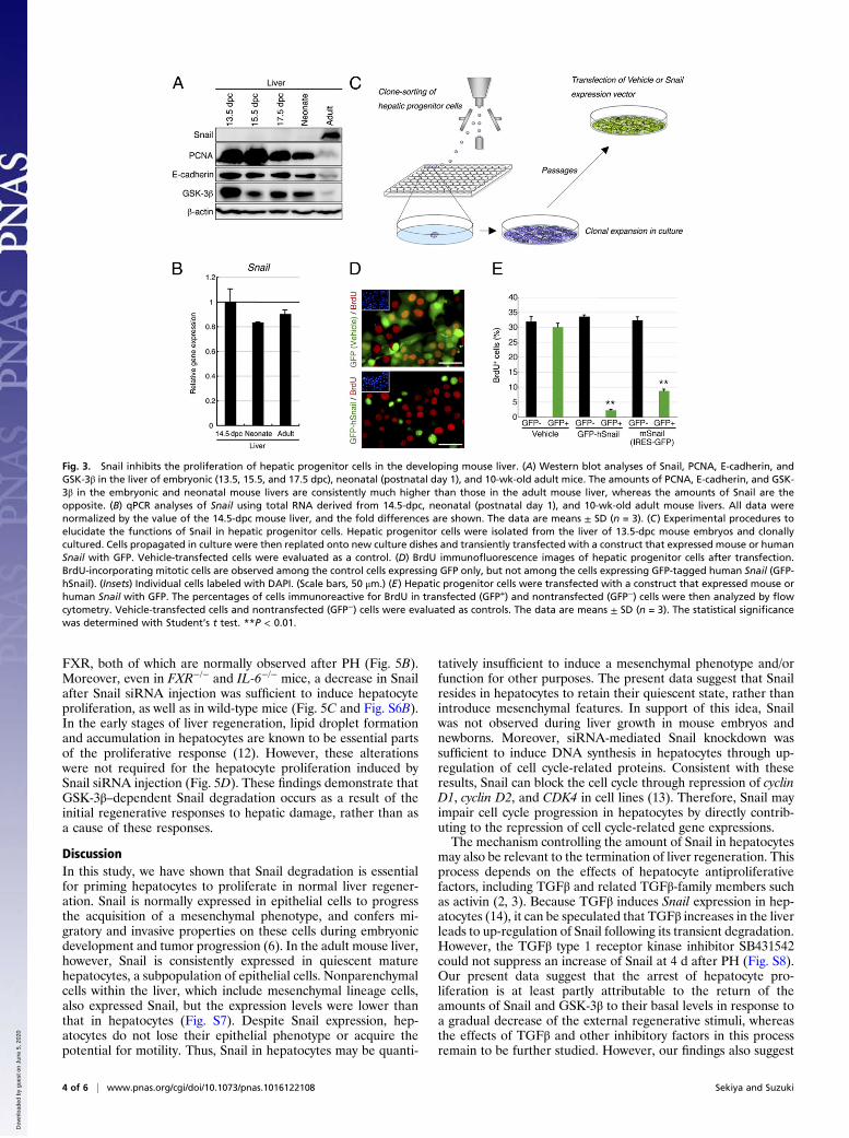
investigated hepatic progenitor cells in the developing mouseliver. During liver growth in mouse embryos and newborns, theamounts of Snail in the liver were extremely low (Fig. 3A andFig. S4A), whereas these mice strongly expressed Snail mRNA inthe liver as adult mice (Fig. 3B). Thus, Snail may be degraded inthe developing liver, as well as in the liver after PH or hepaticinjury. Hepatic progenitor cells were isolated from the liver of13.5-d postcoitum (dpc) mouse embryos and clonally cultured asdescribed previously (10). Next, we introduced a construct thathighly expressed mouse or human Snail with GFP into the cul-tured cells and analyzed the effects on proliferation (Fig. 3C).The forced expression of mouse or human Snail in hepatic pro-genitor cells allowed the accumulation of Snail in the nuclei ofthese cells and resulted in a significant reduction in the numberof BrdU-incorporating mitotic cells, whereas introduction ofa control construct that expressed GFP had little effect (Fig. 3 Dand E). Regardless of Snail expression, these cells expressedE-cadherin that was typically localized at sites of cell–cell junctions(Fig. S4B). In the early stages of subculture, the Snail-expressingcells stopped their proliferation and gradually disappeared fromthe cultures. Thus, Snail inhibits DNA synthesis in hepatic pro-genitor cells and their subsequent proliferation without alterationin the morphology of cells.These findings demonstrate that quiescent adult hepatocytes,
but not hepatic progenitor cells in the developing liver, contain
Fig. 1. Snail degradation and GSK-3β up-regulation in the liver after PH. (A) Western blot analyses of Snail and GSK-3β in the nucleus and cytoplasm of adultmouse hepatocytes. Albumin and β-actin, which reside in the cytoplasm, and histone H3, which resides in the nucleus, were evaluated to confirm the sep-aration of the cytoplasmic and nuclear protein fractions of the hepatocytes. (B) Western blot analyses revealed that the amounts of Snail, GSK-3β, and PCNAin the liver change dramatically after PH. (C) Western blot analyses revealed that the drastic changes in the amounts of Snail and GSK-3β in the liver begin at12 and 6 h after PH, respectively. (D) Western blot analysis of Snail in the liver of DMSO or MG132-treated mice at 1 d after PH, following purification ofubiquitin-binding proteins. (E) qPCR analyses of Snail were carried out on total RNA derived from the liver after PH. (F) Western blot analyses revealed thatthe amounts of Snail, GSK-3β, and PCNA in the liver change dramatically after s.c. injection of CCl4 into adult mice. (G) qPCR analyses of Snail were carried outon total RNA derived from the liver after CCl4 injection. The data shown in E and G were normalized by the value of the liver at 0 h after PH or CCl4 injection,respectively, and the fold differences are shown. The data are means ± SD (n = 3).
2 of 6 | www.pnas.org/cgi/doi/10.1073/pnas.1016122108 Sekiya and Suzuki
Dow
nloa
ded
by g
uest
on
June
5, 2
020
Snail to block proliferation, and that a transient decrease in Snailin the damaged liver immediately induces DNA synthesis inhepatocytes for proliferation.
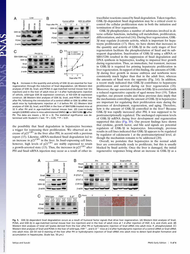
Increases in the Quantity and Activity of GSK-3β in the Damaged LiverInduce Snail Degradation and DNA Synthesis in Hepatocytes. Next,we sought to determine why Snail degradation occurs after PH.As described above, the amount of GSK-3β in the liver began toincrease at 6 h after PH and peaked at 24 h, followed by deg-radation of ubiquitinated Snail in the liver. It is known that GSK-3β–mediated phosphorylation of Snail leads to ubiquitinationand subsequent proteasomal degradation of Snail (7). Thus, animmediate increase in GSK-3β in the damaged liver may beimportant for inducing Snail degradation. To address this issue,we introduced a construct that highly expressed wild-type or ki-nase-dead (KD) GSK-3β (11) into normal adult mice by hydro-dynamic injection. The forced expression of these genesincreased the amount of GSK-3β in the liver at 1 d after injection(Fig. 4A and Fig. S5A). However, a decrease in Snail and anincrease in PCNA only occurred when wild-type GSK-3β wasoverexpressed (Fig. 4A and Fig. S5A). In addition, with anincrease in GSK-3β, the amount of phosphorylated serine resi-dues within Snail, which are known to undergo phosphorylationby GSK-3β (7), was increased at 8 h after injection of the wild-type GSK-3β expression construct (Fig. S5B). Thus, an increasein GSK-3β in hepatocytes is sufficient to cause Snail degradationand induce cell proliferation through GSK-3β–mediated phos-phorylation of Snail. To examine whether increased GSK-3β isimportant for liver regeneration to proceed, we introduceda GSK-3β–specific siRNA (GSK-3β siRNA) into adult mice by
hydrodynamic injection at 1 d before PH. This GSK-3β siRNAefficiently reduced the amount of GSK-3β and increased theamount of Snail upon injection into the liver (Fig. S5 C and D).Treatment with the GSK-3β siRNA was effective for suppressingnot only the increase in GSK-3β but also the decrease in Snailand increase in PCNA in the liver at 1 and 2 d after PH (Fig. 4Band Fig. S5E). Moreover, the GSK-3β inhibitor SB415286, whichwas i.p. injected into adult mice after PH, also dose-dependentlysuppressed the decrease in Snail and increase in PCNA in theliver and strongly inhibited liver growth in the early stages ofregeneration (Fig. 4 C and D and Fig. S5F). Thus, increases inthe quantity and activity of GSK-3β in the damaged liver areessential for liver regeneration through the induction of Snaildegradation.
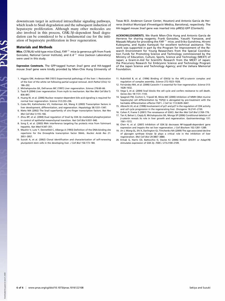
GSK-3β–Dependent Snail Degradation Occurs as a Result of the InitialRegenerative Responses to Hepatic Damage. Our findings demon-strate that GSK-3β–dependent Snail degradation directs hepa-tocyte proliferation in normal liver regeneration, but do notaddress the relationship between this event and the well-definedregenerative responses to PH. To clarify this issue, we directlyinjected IL-6, HGF, and cholic acid into adult mice and exam-ined their impact on Snail and GSK-3β in the liver. Concomitantwith an increase in PCNA, i.p. injection of these regenerativefactors induced a decrease in Snail and an increase in GSK-3β inthe liver (Fig. 5A and Fig. S6A). However, treatment with theSnail siRNA was capable of inducing hepatocyte proliferationwithout phosphorylation of STAT-3, a downstream transcriptionfactor in the IL-6 signaling pathway, and a decrease in choles-terol 7a hydroxylase (CYP7A1), a downstream negative target of
Fig. 2. Decreased levels of Snail induce DNA synthesis in hepatocytes through activation of the cell cycle machinery. (A) Western blot analyses of Snail, PCNA,and E-cadherin in age-matched normal mouse liver (no injection) and in the liver of adult mice at 2 d after hydrodynamic injection of a control siRNA or SnailsiRNA. The Snail siRNA efficiently reduces the amount of Snail and increases the amounts of PCNA and E-cadherin in the liver. (B) Coimmunofluorescencestaining of BrdU with glutamine synthetase (GS; Left) or CK8/18 (Right) in livers obtained at 2 d after injection of a control siRNA or Snail siRNA and at 2 dafter PH. GS marks hepatocytes surrounding the central veins of the liver, indicating that the BrdU-incorporating cells after Snail siRNA injection and PH arenonspecifically localized throughout the liver. DNA was stained with DAPI. (Scale bars, 100 μm.) (C) The percentages of cells immunoreactive for BrdU in 2,090 ±121 or 1,879 ± 124 CK8/18+ cells in the liver at 2 d after injection of a control siRNA or Snail siRNA, respectively, and in 1,977 ± 123 CK8/18+ cells in the liver at2 d after PH. The data are means ± SD (n = 3). (D) Liver-to-body weight (LW/BW) ratios in age-matched normal mice (no injection) and in mice at 2 d afterinjection of a control siRNA or Snail siRNA. The data are means ± SD (n = 3). (E) Western blot analyses revealed up-regulation of cell cycle-related proteins,except for p27KIP1, in the liver at 2 d after Snail siRNA injection or PH.
Sekiya and Suzuki PNAS Early Edition | 3 of 6
MED
ICALSC
IENCE
S
Dow
nloa
ded
by g
uest
on
June
5, 2
020
FXR, both of which are normally observed after PH (Fig. 5B).Moreover, even in FXR−/− and IL-6−/− mice, a decrease in Snailafter Snail siRNA injection was sufficient to induce hepatocyteproliferation, as well as in wild-type mice (Fig. 5C and Fig. S6B).In the early stages of liver regeneration, lipid droplet formationand accumulation in hepatocytes are known to be essential partsof the proliferative response (12). However, these alterationswere not required for the hepatocyte proliferation induced bySnail siRNA injection (Fig. 5D). These findings demonstrate thatGSK-3β–dependent Snail degradation occurs as a result of theinitial regenerative responses to hepatic damage, rather than asa cause of these responses.
DiscussionIn this study, we have shown that Snail degradation is essentialfor priming hepatocytes to proliferate in normal liver regener-ation. Snail is normally expressed in epithelial cells to progressthe acquisition of a mesenchymal phenotype, and confers mi-gratory and invasive properties on these cells during embryonicdevelopment and tumor progression (6). In the adult mouse liver,however, Snail is consistently expressed in quiescent maturehepatocytes, a subpopulation of epithelial cells. Nonparenchymalcells within the liver, which include mesenchymal lineage cells,also expressed Snail, but the expression levels were lower thanthat in hepatocytes (Fig. S7). Despite Snail expression, hep-atocytes do not lose their epithelial phenotype or acquire thepotential for motility. Thus, Snail in hepatocytes may be quanti-
tatively insufficient to induce a mesenchymal phenotype and/orfunction for other purposes. The present data suggest that Snailresides in hepatocytes to retain their quiescent state, rather thanintroduce mesenchymal features. In support of this idea, Snailwas not observed during liver growth in mouse embryos andnewborns. Moreover, siRNA-mediated Snail knockdown wassufficient to induce DNA synthesis in hepatocytes through up-regulation of cell cycle-related proteins. Consistent with theseresults, Snail can block the cell cycle through repression of cyclinD1, cyclin D2, and CDK4 in cell lines (13). Therefore, Snail mayimpair cell cycle progression in hepatocytes by directly contrib-uting to the repression of cell cycle-related gene expressions.The mechanism controlling the amount of Snail in hepatocytes
may also be relevant to the termination of liver regeneration. Thisprocess depends on the effects of hepatocyte antiproliferativefactors, including TGFβ and related TGFβ-family members suchas activin (2, 3). Because TGFβ induces Snail expression in hep-atocytes (14), it can be speculated that TGFβ increases in the liverleads to up-regulation of Snail following its transient degradation.However, the TGFβ type 1 receptor kinase inhibitor SB431542could not suppress an increase of Snail at 4 d after PH (Fig. S8).Our present data suggest that the arrest of hepatocyte pro-liferation is at least partly attributable to the return of theamounts of Snail and GSK-3β to their basal levels in response toa gradual decrease of the external regenerative stimuli, whereasthe effects of TGFβ and other inhibitory factors in this processremain to be further studied. However, our findings also suggest
Fig. 3. Snail inhibits the proliferation of hepatic progenitor cells in the developing mouse liver. (A) Western blot analyses of Snail, PCNA, E-cadherin, andGSK-3β in the liver of embryonic (13.5, 15.5, and 17.5 dpc), neonatal (postnatal day 1), and 10-wk-old adult mice. The amounts of PCNA, E-cadherin, and GSK-3β in the embryonic and neonatal mouse livers are consistently much higher than those in the adult mouse liver, whereas the amounts of Snail are theopposite. (B) qPCR analyses of Snail using total RNA derived from 14.5-dpc, neonatal (postnatal day 1), and 10-wk-old adult mouse livers. All data werenormalized by the value of the 14.5-dpc mouse liver, and the fold differences are shown. The data are means ± SD (n = 3). (C) Experimental procedures toelucidate the functions of Snail in hepatic progenitor cells. Hepatic progenitor cells were isolated from the liver of 13.5-dpc mouse embryos and clonallycultured. Cells propagated in culture were then replated onto new culture dishes and transiently transfected with a construct that expressed mouse or humanSnail with GFP. Vehicle-transfected cells were evaluated as a control. (D) BrdU immunofluorescence images of hepatic progenitor cells after transfection.BrdU-incorporating mitotic cells are observed among the control cells expressing GFP only, but not among the cells expressing GFP-tagged human Snail (GFP-hSnail). (Insets) Individual cells labeled with DAPI. (Scale bars, 50 μm.) (E) Hepatic progenitor cells were transfected with a construct that expressed mouse orhuman Snail with GFP. The percentages of cells immunoreactive for BrdU in transfected (GFP+) and nontransfected (GFP−) cells were then analyzed by flowcytometry. Vehicle-transfected cells and nontransfected (GFP−) cells were evaluated as controls. The data are means ± SD (n = 3). The statistical significancewas determined with Student’s t test. **P < 0.01.
4 of 6 | www.pnas.org/cgi/doi/10.1073/pnas.1016122108 Sekiya and Suzuki
Dow
nloa
ded
by g
uest
on
June
5, 2
020
the possibility that Snail degradation in hepatocytes becomesa trigger for repressing their proliferation. We observed an in-crease of p21CIP1 in the liver after PH, in accord with a previousreport (15). Likewise, siRNA-mediated Snail degradation led toan increase in p21CIP1 in the liver. In Snail-expressing cell lines,however, high levels of p21CIP1 are stably expressed to retaina growth-arrested state (13). Thus, the increases in p21CIP1 afterPH and Snail siRNA injection may occur as a result of other in-
tracellular reactions caused by Snail degradation. Taken together,GSK-3β–dependent Snail degradation may be a critical event tocontrol the cellular proliferation state in both the initiation andtermination of liver regeneration.GSK-3β phosphorylates a number of substrates involved in di-
verse cellular functions, including cell metabolism, proliferation,differentiation, and survival (16). During liver regeneration, GSK-3β may regulate β-catenin activity, which is important for hepa-tocyte proliferation (17). Here, we have shown that increases inthe quantity and activity of GSK-3β in the early stages of liverregeneration facilitate the phosphorylation of Snail and its sub-sequent degradation. Inactivation of GSK-3β using a siRNA orSB415286 resulted in the suppression of Snail degradation andDNA synthesis in hepatocytes, leading to impaired liver growthduring regeneration. Thus, an immediate, but transient, increasein GSK-3β is required for priming hepatocyte proliferation inliver regeneration. In support of this finding, the amounts of GSK-3β during liver growth in mouse embryos and newborns wereconsistently much higher than that in the adult liver, whereasthe amounts of Snail were the opposite (Fig. 3A). In addition,a recent study indicated that GSK-3β contributes to hepatocyteproliferation and survival during rat liver regeneration (18).Moreover, the age-associated decline inGSK-3β is correlated witha reduced regenerative capacity of aged mouse liver (19). Takentogether, our present results and these previous data imply thatthe mechanisms controlling the amount of GSK-3β in hepatocytesare important for regulating their proliferation state during theprocesses of development, regeneration, and aging. Therefore,how is the amount of GSK-3β controlled in the liver? BecauseGSK-3β was rapidly increased after PH, it was supposed to beposttranscriptionally regulated. The unchanged expression levelsof GSK-3β mRNA during liver development and regenerationsupported this idea (Fig. S9). Our present findings have shownthat cytokine, growth factor, and bile acid signals are potentialregulators of GSK-3β in hepatocytes. Interestingly, previousresults in cell lines indicated that GSK-3β appears to be regulatedby regulator of calcineurin 1 at the posttranscriptional level, al-though the mechanism remains to be addressed (20).Overall, we postulate that hepatocytes in the normal adult
liver are conventionally ready to proliferate, but this is usuallyblocked by Snail activity. Once the liver is damaged, the initialregenerative responses bring about an increase in GSK-3β as a
Fig. 4. Increases in the quantity and activity of GSK-3β are essential for liverregeneration through the induction of Snail degradation. (A) Western blotanalyses of GSK-3β, Snail, and PCNA in age-matched normal mouse liver (noinjection) and in the liver of adult mice at 1 d after hydrodynamic injectionof vehicle, wild-type GSK-3β expression construct, or KD-GSK-3β expressionconstruct. (B) Western blot analyses of GSK-3β, Snail, and PCNA in the liverafter PH, following the introduction of a control siRNA or GSK-3β siRNA intoadult mice by hydrodynamic injection at 1 d before PH. (C) Western blotanalyses of GSK-3β, Snail, and PCNA in the liver of SB415286-treated mice at32 h after PH and in age-matched normal mouse liver. (D) Liver-to-bodyweight (LW/BW) ratios in mice administered DMSO (●) or SB415286 (■) afterPH. The data are means ± SD (n = 3). The statistical significance was de-termined with Student’s t test. *P < 0.05, **P < 0.01.
Fig. 5. GSK-3β–dependent Snail degradation occurs as a result of humoral factor signals that drive liver regeneration. (A) Western blot analyses of Snail,PCNA, and GSK-3β in age-matched normal mouse liver (no injection) and in the liver of adult mice at 1 d after injection of HGF, IL-6, and cholic acid. (B)Western blot analyses of total cell lysates derived from the liver after PH or hydrodynamic injection of Snail siRNA into adult mice. P, phosphorylated. (C)Western blot analyses of Snail and PCNA in the liver of wild-type, FXR−/−, and IL-6−/− mice at 2 d after hydrodynamic injection of a control siRNA or Snail siRNAinto adult mice. (D) Oil red O staining of the liver after PH or hydrodynamic injection of Snail siRNA into adult mice to detect lipid droplet formation andaccumulation in hepatocytes. (Scale bar, 50 μm.)
Sekiya and Suzuki PNAS Early Edition | 5 of 6
MED
ICALSC
IENCE
S
Dow
nloa
ded
by g
uest
on
June
5, 2
020
downstream target in activated intracellular signaling pathways,which leads to Snail degradation and the subsequent induction ofhepatocyte proliferation. Although many other molecules arealso involved in this process, GSK-3β–dependent Snail degra-dation can be considered to be a fundamental cue for the initi-ation of hepatocyte proliferation in liver regeneration.
Materials and MethodsMice. C57BL/6J wild-type mice (Clea), FXR−/− mice (a generous gift from FrankGonzalez, National Cancer Institute), and IL-6−/− mice (Jackson Laboratory)were used in this study.
Expression Constructs. The GFP-tagged human Snail gene and HA-taggedmouse Snail gene were kindly provided by Mien-Chie Hung (University of
Texas M.D. Anderson Cancer Center, Houston) and Antonio García de Her-reros (Institut Municipal d’Investigació Mèdica, Barcelona), respectively. TheHA-tagged mouse Snail gene was inserted into pIRES2-GFP (Clontech).
ACKNOWLEDGMENTS. We thank Mien-Chie Hung and Antonio García deHerreros for sharing reagents; Frank Gonzalez, Yasushi Yamazoe, andMasaaki Miyata for providing the FXR−/− mice; and Eriko Gunshima, HiromiKuboyama, and Ayako Kaneyuki for excellent technical assistance. Thiswork was supported in part by the Program for Improvement of the Re-search Environment for Young Researchers from the Special Coordina-tion Funds for Promoting Science and Technology commissioned by theMinistry of Education, Culture, Sports, Science and Technology (MEXT) ofJapan; a Grant-in-Aid for Scientific Research from the MEXT of Japan;the Precursory Research for Embryonic Science and Technology Programof the Japan Science and Technology Agency; and the Uehara MemorialFoundation.
1. Higgins GM, Anderson RM (1931) Experimental pathology of the liver: I. Restoration
of the liver of the white rat following partial surgical removal. Arch Pathol (Chic) 12:
186–202.2. Michalopoulos GK, DeFrances MC (1997) Liver regeneration. Science 276:60–66.3. Taub R (2004) Liver regeneration: From myth to mechanism. Nat Rev Mol Cell Biol 5:
836–847.4. Huang W, et al. (2006) Nuclear receptor-dependent bile acid signaling is required for
normal liver regeneration. Science 312:233–236.5. Costa RH, Kalinichenko VV, Holterman AX, Wang X (2003) Transcription factors in
liver development, differentiation, and regeneration. Hepatology 38:1331–1347.6. Nieto MA (2002) The Snail superfamily of zinc-finger transcription factors. Nat Rev
Mol Cell Biol 3:155–166.7. Zhou BP, et al. (2004) Dual regulation of Snail by GSK-3β–mediated phosphorylation
in control of epithelial-mesenchymal transition. Nat Cell Biol 6:931–940.8. Song E, et al. (2003) RNA interference targeting Fas protects mice from fulminant
hepatitis. Nat Med 9:347–351.9. Mauhin V, Lutz Y, Dennefeld C, Alberga A (1993) Definition of the DNA-binding site
repertoire for the Drosophila transcription factor SNAIL. Nucleic Acids Res 21:
3951–3957.10. Suzuki A, et al. (2002) Clonal identification and characterization of self-renewing
pluripotent stem cells in the developing liver. J Cell Biol 156:173–184.
11. Rubinfeld B, et al. (1996) Binding of GSK3β to the APC-β-catenin complex andregulation of complex assembly. Science 272:1023–1026.
12. Fernández MA, et al. (2006) Caveolin-1 is essential for liver regeneration. Science 313:1628–1632.
13. Vega S, et al. (2004) Snail blocks the cell cycle and confers resistance to cell death.Genes Dev 18:1131–1143.
14. Spagnoli FM, Cicchini C, Tripodi M, Weiss MC (2000) Inhibition of MMH (Met murinehepatocyte) cell differentiation by TGF(β) is abrogated by pre-treatment with theheritable differentiation effector FGF1. J Cell Sci 113:3639–3647.
15. Albrecht JH, et al. (1998) Involvement of p21 and p27 in the regulation of CDK activityand cell cycle progression in the regenerating liver. Oncogene 16:2141–2150.
16. Cohen P, Frame S (2001) The renaissance of GSK3. Nat Rev Mol Cell Biol 2:769–776.17. Tan X, Behari J, Cieply B, Michalopoulos GK, Monga SP (2006) Conditional deletion of
β-catenin reveals its role in liver growth and regeneration. Gastroenterology 131:1561–1572.
18. Chen H, et al. (2007) Inhibition of GSK-3β decreases NF-kappaB-dependent geneexpression and impairs the rat liver regeneration. J Cell Biochem 102:1281–1289.
19. Jin J, Wang GL, Shi X, Darlington GJ, Timchenko NA (2009) The age-associated declineof glycogen synthase kinase 3β plays a critical role in the inhibition of liverregeneration. Mol Cell Biol 29:3867–3880.
20. Ermak G, Harris CD, Battocchio D, Davies KJ (2006) RCAN1 (DSCR1 or Adapt78)stimulates expression of GSK-3β. FEBS J 273:2100–2109.
6 of 6 | www.pnas.org/cgi/doi/10.1073/pnas.1016122108 Sekiya and Suzuki
Dow
nloa
ded
by g
uest
on
June
5, 2
020