
Perioperative digital pupillometry-the future?
17
The Greek E-Journal of Perioperative Medicine 2015; 13(b): 24-40 (ISSN 1109-6888) www.e-journal.gr/ Ελληνικό Περιοδικό Περιεγχειρητικής Ιατρικής 2015; 13(b): 24-40 (ISSN 1109-6888) www.e-journal.gr/ 24 ©2015 Society of Anesthesiology and Intensive Medicine of Northern Greece ©2015ΕταιρείαΑναισθησιολογίαςκαιΕντατικήςΙατρικήςΒορείουΕλλάδος 1 Mobile Intensive Care Unit, National Center of Emergency Care, Thessaloniki Dep., Greece 2 Neurologist, Kozani, Greece Perioperative digital pupillometry-the future? AslanidisTh 1 MD, Kontogounis G 2 MD ABSTRACT Perioperative digital pupillometry-the future? AslanidisTh, Kontogounis G. Clinical evaluation of pupils is considered as an essential part of neurological examination. The pu- pillary response to light is controlled by the autonomic nervous system. Numerous factors affect pupils dynamics, like e.g. luminance, visual field area, pain, drug administration, age, the functional integrity of anatomical structures involved, e.t.c. Moreover, pupillometry card method and examina- tion of pupil reaction with the use of a penlight is subjective to a lot of bias. Portable infrared pupil- lometry allows a more objective and detail evaluation of pupil’s dynamics. That’s why it has al- ready found applications in various clinical areas, like e.g. neurology, psychology, ophthalmology, endocrinology, anesthesia, pain management, intensive care, emergency medicine. This review fo- cuses on physiology of pupil’s dynamics and on applications of infrared pupillometry in periopera- tive setting. INTRODUCTION Archimedes was the first to report observa- tions about the size and the symmetry of eye’s pupil and Galileo is believed to be the first who attempted to measure its diameter 1 . Pupil- lary reflex dilation was originally described by Budge in 1852 as a sympathetic spinal reflex that dilated the pupil after noxious stimula- tion 2 . In 1885 Bellerminov published static records of pupil’s shape under side light 3 . In 1942 Lowenstein presented dynamic records of pupil’s changes under ultraviolet and infra- red light emission. Later, Matsunaga (1973) and Jones (1983) developed recording systems of pupil’s movement; but the latter were cum- bersome and thus, impossible to be used in clinical setting 4-6 . Despite the difficulties of an objective bedside measurement, manual (performed using a pen- light or ophthalmoscope) pupil evaluation re-
Transcript of Perioperative digital pupillometry-the future?
The Greek E-Journal of Perioperative Medicine 2015; 13(b): 24-40 (ISSN 1109-6888) www.e-journal.gr/ Ελληνικό Περιοδικό Περιεγχειρητικής Ιατρικής 2015; 13(b): 24-40 (ISSN 1109-6888) www.e-journal.gr/
24
©2015 Society of Anesthesiology and Intensive Medicine of Northern Greece
©2015ΕταιρείαΑναισθησιολογίαςκαιΕντατικήςΙατρικήςΒορείουΕλλάδος
1Mobile Intensive Care Unit, National Center of
Emergency Care, Thessaloniki Dep., Greece
2Neurologist, Kozani, Greece
Perioperative digital pupillometry-the future?
AslanidisTh1
MD, Kontogounis G2
MD
ABSTRACT
Perioperative digital pupillometry-the future?
AslanidisTh, Kontogounis G.
Clinical evaluation of pupils is considered as an essential part of neurological examination. The pu-
pillary response to light is controlled by the autonomic nervous system. Numerous factors affect
pupils dynamics, like e.g. luminance, visual field area, pain, drug administration, age, the functional
integrity of anatomical structures involved, e.t.c. Moreover, pupillometry card method and examina-
tion of pupil reaction with the use of a penlight is subjective to a lot of bias. Portable infrared pupil-
lometry allows a more objective and detail evaluation of pupil’s dynamics. That’s why it has al-
ready found applications in various clinical areas, like e.g. neurology, psychology, ophthalmology,
endocrinology, anesthesia, pain management, intensive care, emergency medicine. This review fo-
cuses on physiology of pupil’s dynamics and on applications of infrared pupillometry in periopera-
tive setting.
INTRODUCTION
Archimedes was the first to report observa-
tions about the size and the symmetry of eye’s
pupil and Galileo is believed to be the first
who attempted to measure its diameter1. Pupil-
lary reflex dilation was originally described by
Budge in 1852 as a sympathetic spinal reflex
that dilated the pupil after noxious stimula-
tion2. In 1885 Bellerminov published static
records of pupil’s shape under side light3. In
1942 Lowenstein presented dynamic records
of pupil’s changes under ultraviolet and infra-
red light emission. Later, Matsunaga (1973)
and Jones (1983) developed recording systems
of pupil’s movement; but the latter were cum-
bersome and thus, impossible to be used in
clinical setting4-6
.
Despite the difficulties of an objective bedside
measurement, manual (performed using a pen-
light or ophthalmoscope) pupil evaluation re-

The Greek E-Journal of Perioperative Medicine 2015; 13(b): 24-40 (ISSN 1109-6888) www. e-journal.gr/ Ελληνικό Περιοδικό Περιεγχειρητικής Ιατρικής2015; 13(b): 24-40 (ISSN 1109-6888) www. e-journal.gr/
25
©2015 Society of Anesthesiology and Intensive Medicine of Northern Greece
©2015ΕταιρείαΑναισθησιολογίαςκαιΕντατικήςΙατρικήςΒορείουΕλλάδος
mains an essential part of neurological clinical
examination for over a century now1.
The recent emergence of new technologies of-
fered us a new perspective as they introduced
us to the era of digital pupil evaluation. The
present article focuses on digital portable pu-
pillometry and it reviews the possible applica-
tions of this technology in perioperative field.
ANATOMICAL AND PHYSIOLOGICAL
BASE OF PUPIL’S DYNAMIC
The pupils are dilated in excitement, in the
dark, and with mydriatic agents like atropine;
conversely they are constricted in neonates,
during sleep, with ocular convergence as part
of the near reflex, with use of the miotic agents
like acetylcholine and pilocarpine, and also
they are constricted with light directed toward
the eye. In fact, the most impart factor for the
pupillary size is the amount of ambient light.
The size of the pupil of the eye is determined
by the balance between the tone of two mus-
cles, the constrictor (sphincter, the circular
muscle) and the dilator (the radial muscle).
The light reflex (PLR)is the simultaneous and
equal constriction of the pupilin response to
illumination of one (direct) or the other eye
(indirect or consensual). The reflex is multi-
synaptic with pulse speed 160 m/sec and trig-
gering time about 200msec. The afferent pupil-
lary pathway is controlled by the sympathetic
autonomous nerve system (ANS) and origi-
nates in the retina. The axons of retinal gangli-
on cells pass into the optic nerve, partially (on-
ly the 2nd
, 3rd
, 5th
and 6th
laminae) decussate in
the chiasm and continue (optic tract) to the
midbrain (lateral geniculate body), while some
pupillary fibers bypass the ciliospinal center
and continue cephalad to the pretectal nuclei
at the level of the superior colliculus (thala-
mus). Optical cortex include Brodmann areas
17,18 and 19 (occipital lobe) and inter-
connection with frontal lobe (Brodmann area
8) and several nuclei of cranial nerves (oculo-
motor, trochlear and abducens), (Fig. 1).
Figure 1.a.b.The neural efferent optical path-
way.(Fromhttp://en.wikipedia.org/wiki/Pupilla
ry_light_reflex)
a
The Greek E-Journal of Perioperative Medicine 2015; 13(b): 24-40 (ISSN 1109-6888) www. e-journal.gr/ Ελληνικό Περιοδικό Περιεγχειρητικής Ιατρικής2015; 13(b): 24-40 (ISSN 1109-6888) www. e-journal.gr/
26
©2015 Society of Anesthesiology and Intensive Medicine of Northern Greece
©2015ΕταιρείαΑναισθησιολογίαςκαιΕντατικήςΙατρικήςΒορείουΕλλάδος
b
Considering the efferent arc of the light reflex,
the visceral nuclei of the oculomotor nucleus,
the nuclei that are believed to contain cell bod-
ies of preganglionic, parasympathetic neurons
that project to the ciliary ganglion and synapse
with neurons that eventually produce both pu-
pillary constriction and accommodation, are
located in the dorsal midbrain. These consist
of the Edinger-Westphal nuclei (also called the
dorsal visceral cell columns), the anterior me-
dian nuclei, and the nucleus of Perlia. Pregan-
glionic parasympathetic fibers run in the ocu-
lomotor nerve as it leaves the brain stem. The
fibers pass downward to lie inferiorly in the
inferior division of the third nerve as it enters
the orbit. These fibers synapse in the ciliary
ganglion and give rise to postganglionic para-
sympathetic myelinated short ciliary nerves,
about 3%-5% of which are pupillomotor. The
rest are designated for the ciliary muscle and
are concerned with the near reflex, (Fig. 2a and
2b).
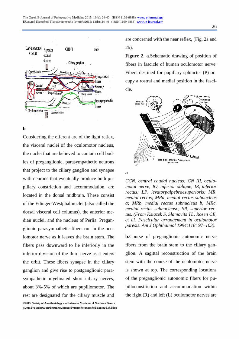
Figure 2. a.Schematic drawing of position of
fibers in fascicle of human oculomotor nerve.
Fibers destined for pupillary sphincter (P) oc-
cupy a rostral and medial position in the fasci-
cle.
a
CCN, central caudal nucleus; CN III, oculo-
motor nerve; IO, inferior oblique; IR, inferior
rectus; LP, levatorpalpebraesuperioris; MR,
medial rectus; MRa, medial rectus subnucleus
a; MRb, medial rectus subnucleus b; MRc,
medial rectus subnucleusc; SR, superior rec-
tus. (From Ksiazek S, Slamovits TL, Rosen CE,
et al. Fascicular arrangement in oculomotor
paresis. Am J Ophthalmol 1994;118: 97–103).
b.Course of preganglionic autonomic nerve
fibers from the brain stem to the ciliary gan-
glion. A sagittal reconstruction of the brain
stem with the course of the oculomotor nerve
is shown at top. The corresponding locations
of the preganglionic autonomic fibers for pu-
pilloconstriction and accommodation within
the right (R) and left (L) oculomotor nerves are
The Greek E-Journal of Perioperative Medicine 2015; 13(b): 24-40 (ISSN 1109-6888) www. e-journal.gr/ Ελληνικό Περιοδικό Περιεγχειρητικής Ιατρικής2015; 13(b): 24-40 (ISSN 1109-6888) www. e-journal.gr/
27
©2015 Society of Anesthesiology and Intensive Medicine of Northern Greece
©2015ΕταιρείαΑναισθησιολογίαςκαιΕντατικήςΙατρικήςΒορείουΕλλάδος
shown in black in coronal sections through
slices at 1 (emergence from the brain stem), 2
(midpoint in the subarachnoid space), 3 (at the
point where the third nerve enters the dura),
and 4 (in the anterior cavernous sinus where
the fibers have entered the anatomic inferior
division of the third nerve). The autonomic
fibers are located superiorly as the oculomotor
nerve exits the brain stem and then come to lie
more medially as the oculomotor nerve passes
toward the orbit.
b
A, dorsal and B, ventral side of brain stem. P,
pons; M, medulla; EW, Edinger-Westphal nu-
cleus; IIIn, somatic portion of third nerve nu-
cleus; III, third nerve;ID, inferior division of
third nerve; SD, superior division of third
nerve; NCilV, nasociliary branch of fifth
nerve; CG, ciliary ganglion; Sym, sympathetic
route; m, medial; l, lateral. (From Kerr FWL,
Hollowell OW. Location of pupillomotor and
accommodation fibres in the oculomotor
nerve: experimental observations on paralytic
mydriasis. J NeurolNeurosurg Psychia-
try1964; 27:473.).
FACTORS THAT DETERMINE THE
PUPILLARY CHARACTERISTICS. THE
DIFFICULTY OF OBJECTIVE
MEASUREMENT.
In a young observer, pupil diameter may vary
between 2-8 mm (i.e. a 16-fold change) with
variations in light level. In the majority of cas-
es both pupils are equal. However, about 20%
of individuals have noticeably different diame-
ters in their two pupils; a phenomenon known
as physiologic (also simple or essen-
tial)anisocoria.
Pupil size is affected by diverse intrinsic and
extrinsic factors (table 1). The variations in
pupil diameter trigger changes in retinal stimu-
lation, relevant to retinal function: changes in
retinal illuminance, the ratio of rod/cone
stimulation, spectral sensitivity and spatial
resolution7
(Fig. 3).
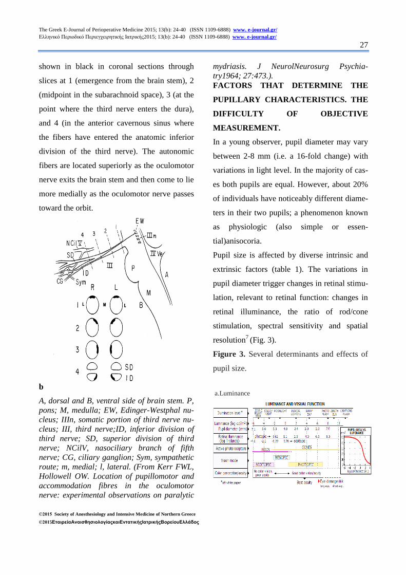
Figure 3. Several determinants and effects of
pupil size.
a.Luminance
The Greek E-Journal of Perioperative Medicine 2015; 13(b): 24-40 (ISSN 1109-6888) www. e-journal.gr/ Ελληνικό Περιοδικό Περιεγχειρητικής Ιατρικής2015; 13(b): 24-40 (ISSN 1109-6888) www. e-journal.gr/
28
©2015 Society of Anesthesiology and Intensive Medicine of Northern Greece
©2015ΕταιρείαΑναισθησιολογίαςκαιΕντατικήςΙατρικήςΒορείουΕλλάδος
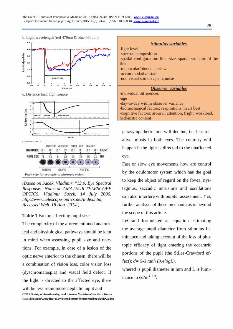
b. Light wavelength (red 470nm & blue 660 nm)
c. Distance form light source
(Based on Sacek, Vladimir. "13.9. Eye Spectral
Response." Notes on AMATEUR TELESCOPE
OPTICS. Vladimir Sacek, 14 July 2006.
http://www.telescope-optics.net/index.htm,
Accessed Web. 18 Aug. 2014.)
Table 1.Factors affecting pupil size.
The complexity of the aforementioned anatom-
ical and physiological pathways should be kept
in mind when assessing pupil size and reac-
tions. For example, in case of a lesion of the
optic nerve anterior to the chiasm, there will be
a combination of vision loss, color vision loss
(dyschromatospia) and visual field defect. If
the light is directed to the affected eye, there
will be less retinomesencephalic input and
parasympathetic tone will decline, i.e. less rel-
ative miosis in both eyes. The contrary will
happen if the light is directed to the unaffected
eye.
Fast or slow eye movements how are control
by the oculomotor system which has the goal
to keep the object of regard on the fovea, nys-
tagmus, saccadic intrusions and oscillations
can also interfere with pupils’ assessment. Yet,
further analysis of these mechanisms is beyond
the scope of this article.
LeGrand formulated an equation estimating
the average pupil diameter from stimulus lu-
minance and taking account of the loss of pho-
topic efficacy of light entering the eccentric
portions of the pupil (the Stiles-Crawford ef-
fect): d= 5-3 tanh (0.4logL),
whered is pupil diameter in mm and L is lumi-
nance in cd/m2 7-8
.
Stimulus variables -light level
-spectral composition
-spatial configuration: field size, spatial structure of the
field
-monocular/binocular view
-accommodative state
-non visual stimuli : pain, noise
Observer variables -individual differences
-age
-day-to-day within observer variance
-biomechanical factors: respirations, heart beat
-cognitive factors: arousal, attention, fright, workload,
hedonistic content
The Greek E-Journal of Perioperative Medicine 2015; 13(b): 24-40 (ISSN 1109-6888) www. e-journal.gr/ Ελληνικό Περιοδικό Περιεγχειρητικής Ιατρικής2015; 13(b): 24-40 (ISSN 1109-6888) www. e-journal.gr/
29
©2015 Society of Anesthesiology and Intensive Medicine of Northern Greece
©2015ΕταιρείαΑναισθησιολογίαςκαιΕντατικήςΙατρικήςΒορείουΕλλάδος
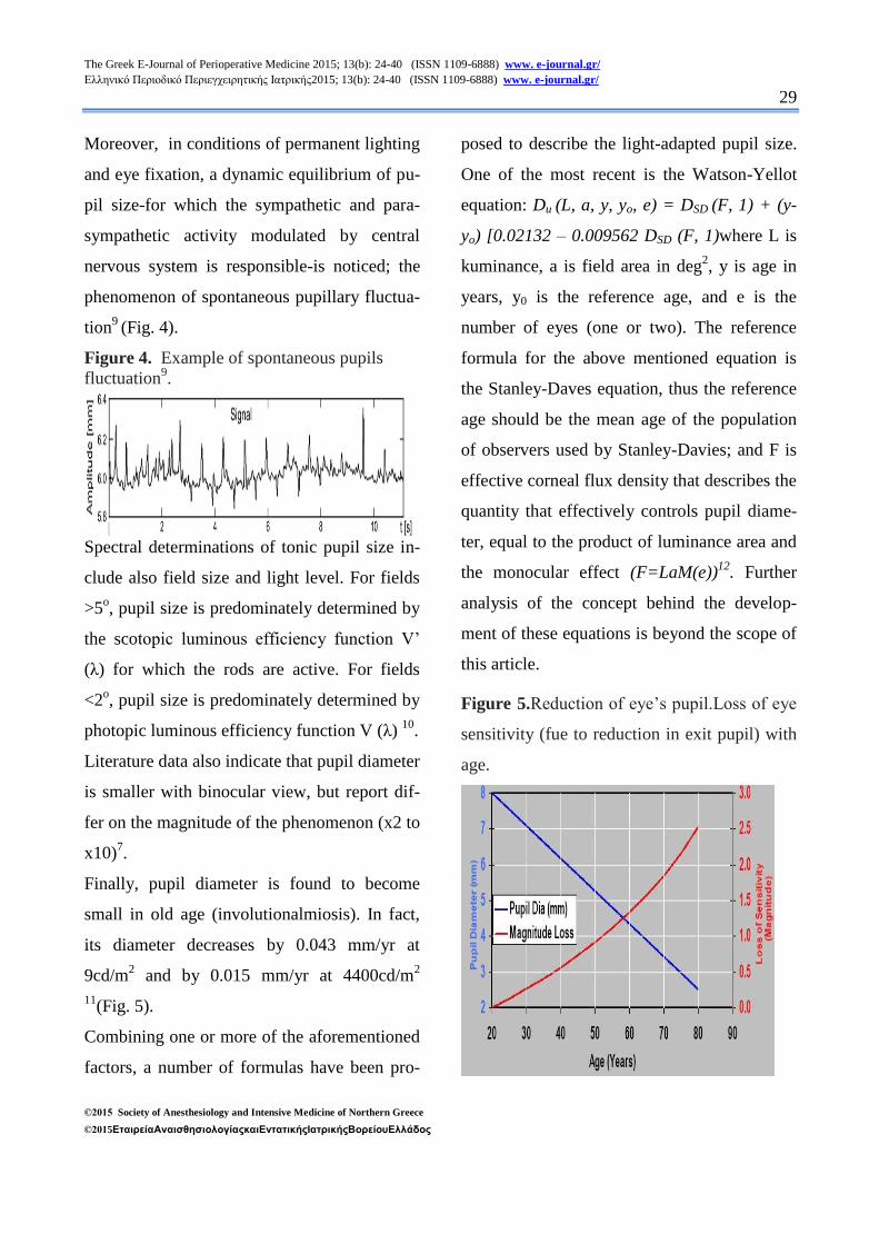
Moreover, in conditions of permanent lighting
and eye fixation, a dynamic equilibrium of pu-
pil size-for which the sympathetic and para-
sympathetic activity modulated by central
nervous system is responsible-is noticed; the
phenomenon of spontaneous pupillary fluctua-
tion9
(Fig. 4).
Figure 4. Example of spontaneous pupils
fluctuation9.
Spectral determinations of tonic pupil size in-
clude also field size and light level. For fields
>5o, pupil size is predominately determined by
the scotopic luminous efficiency function V’
(λ) for which the rods are active. For fields
<2o, pupil size is predominately determined by
photopic luminous efficiency function V (λ) 10
.
Literature data also indicate that pupil diameter
is smaller with binocular view, but report dif-
fer on the magnitude of the phenomenon (x2 to
x10)7.
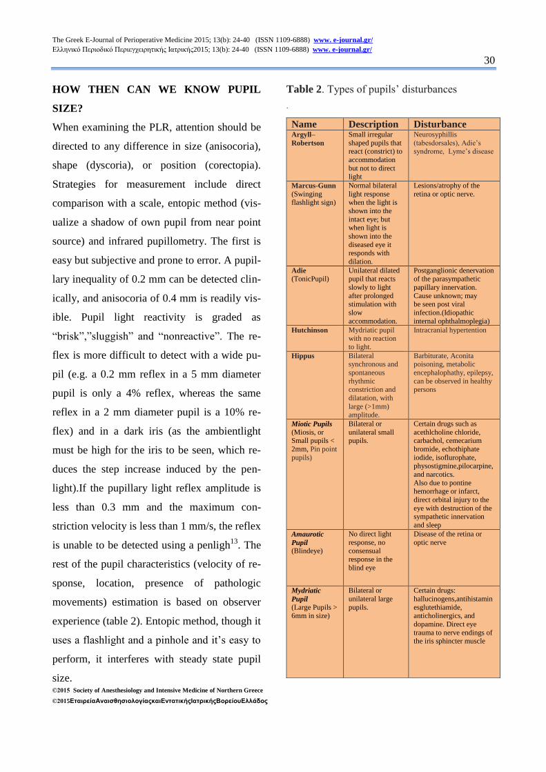
Finally, pupil diameter is found to become
small in old age (involutionalmiosis). In fact,
its diameter decreases by 0.043 mm/yr at
9cd/m2 and by 0.015 mm/yr at 4400cd/m
2
11(Fig. 5).
Combining one or more of the aforementioned
factors, a number of formulas have been pro-
posed to describe the light-adapted pupil size.
One of the most recent is the Watson-Yellot
equation: Du (L, a, y, yo, e) = DSD (F, 1) + (y-
yo) [0.02132 – 0.009562 DSD (F, 1)where L is
kuminance, a is field area in deg2, y is age in
years, y0 is the reference age, and e is the
number of eyes (one or two). The reference
formula for the above mentioned equation is
the Stanley-Daves equation, thus the reference
age should be the mean age of the population
of observers used by Stanley-Davies; and F is
effective corneal flux density that describes the
quantity that effectively controls pupil diame-
ter, equal to the product of luminance area and
the monocular effect (F=LaM(e))12
. Further
analysis of the concept behind the develop-
ment of these equations is beyond the scope of
this article.
Figure 5.Reduction of eye’s pupil.Loss of eye
sensitivity (fue to reduction in exit pupil) with
age.
The Greek E-Journal of Perioperative Medicine 2015; 13(b): 24-40 (ISSN 1109-6888) www. e-journal.gr/ Ελληνικό Περιοδικό Περιεγχειρητικής Ιατρικής2015; 13(b): 24-40 (ISSN 1109-6888) www. e-journal.gr/
30
©2015 Society of Anesthesiology and Intensive Medicine of Northern Greece
©2015ΕταιρείαΑναισθησιολογίαςκαιΕντατικήςΙατρικήςΒορείουΕλλάδος
HOW THEN CAN WE KNOW PUPIL
SIZE?
When examining the PLR, attention should be
directed to any difference in size (anisocoria),
shape (dyscoria), or position (corectopia).
Strategies for measurement include direct
comparison with a scale, entopic method (vis-
ualize a shadow of own pupil from near point
source) and infrared pupillometry. The first is
easy but subjective and prone to error. A pupil-
lary inequality of 0.2 mm can be detected clin-
ically, and anisocoria of 0.4 mm is readily vis-
ible. Pupil light reactivity is graded as
“brisk”,”sluggish” and “nonreactive”. The re-
flex is more difficult to detect with a wide pu-
pil (e.g. a 0.2 mm reflex in a 5 mm diameter
pupil is only a 4% reflex, whereas the same
reflex in a 2 mm diameter pupil is a 10% re-
flex) and in a dark iris (as the ambientlight
must be high for the iris to be seen, which re-
duces the step increase induced by the pen-
light).If the pupillary light reflex amplitude is
less than 0.3 mm and the maximum con-
striction velocity is less than 1 mm/s, the reflex
is unable to be detected using a penligh13
. The
rest of the pupil characteristics (velocity of re-
sponse, location, presence of pathologic
movements) estimation is based on observer
experience (table 2). Entopic method, though it
uses a flashlight and a pinhole and it’s easy to
perform, it interferes with steady state pupil
size.
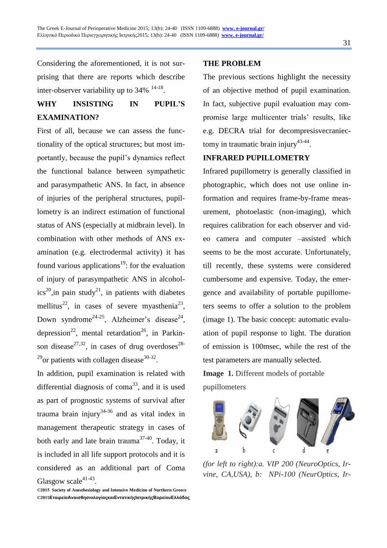
Table 2. Types of pupils’ disturbances
.
Name Description Disturbance Argyll–
Robertson
Small irregular
shaped pupils that react (constrict) to
accommodation
but not to direct light
Neurosyphillis
(tabesdorsales), Adie’s syndrome, Lyme’s disease
Marcus-Gunn
(Swinging flashlight sign)
Normal bilateral
light response when the light is
shown into the
intact eye; but when light is
shown into the
diseased eye it responds with
dilation.
Lesions/atrophy of the
retina or optic nerve.
Adie
(TonicPupil) Unilateral dilated pupil that reacts
slowly to light
after prolonged stimulation with
slow
accommodation.
Postganglionic denervation of the parasympathetic
papillary innervation.
Cause unknown; may be seen post viral
infection.(Idiopathic
internal ophthalmoplegia)
Hutchinson Mydriatic pupil with no reaction
to light.
Intracranial hypertention
Hippus Bilateral synchronous and
spontaneous
rhythmic constriction and
dilatation, with
large (>1mm) amplitude.
Barbiturate, Aconita poisoning, metabolic
encephalophathy, epilepsy,
can be observed in healthy persons
Miotic Pupils
(Miosis, or Small pupils <
2mm, Pin point
pupils)
Bilateral or
unilateral small pupils.
Certain drugs such as
acethlcholine chloride, carbachol, cemecarium
bromide, echothiphate
iodide, isoflurophate, physostigmine,pilocarpine,
and narcotics.
Also due to pontine hemorrhage or infarct,
direct orbital injury to the
eye with destruction of the sympathetic innervation
and sleep
Amaurotic
Pupil (Blindeye)
No direct light
response, no consensual
response in the
blind eye
Disease of the retina or
optic nerve
Mydriatic
Pupil
(Large Pupils > 6mm in size)
Bilateral or
unilateral large
pupils.
Certain drugs:
hallucinogens,antihistamin
esglutethiamide, anticholinergics, and
dopamine. Direct eye
trauma to nerve endings of the iris sphincter muscle
The Greek E-Journal of Perioperative Medicine 2015; 13(b): 24-40 (ISSN 1109-6888) www. e-journal.gr/ Ελληνικό Περιοδικό Περιεγχειρητικής Ιατρικής2015; 13(b): 24-40 (ISSN 1109-6888) www. e-journal.gr/
31
©2015 Society of Anesthesiology and Intensive Medicine of Northern Greece
©2015ΕταιρείαΑναισθησιολογίαςκαιΕντατικήςΙατρικήςΒορείουΕλλάδος
Considering the aforementioned, it is not sur-
prising that there are reports which describe
inter-observer variability up to 34% 14-18
.
WHY INSISTING IN PUPIL’S
EXAMINATION?
First of all, because we can assess the func-
tionality of the optical structures; but most im-
portantly, because the pupil’s dynamics reflect
the functional balance between sympathetic
and parasympathetic ANS. In fact, in absence
of injuries of the peripheral structures, pupil-
lometry is an indirect estimation of functional
status of ANS (especially at midbrain level). In
combination with other methods of ANS ex-
amination (e.g. electrodermal activity) it has
found various applications19
: for the evaluation
of injury of parasympathetic ANS in alcohol-
ics20
,in pain study21
, in patients with diabetes
mellitus22
, in cases of severe myasthenia23
,
Down syndrome24-25
, Alzheimer’s disease24
,
depression22
, mental retardation26
, in Parkin-
son disease27,32
, in cases of drug overdoses28-
29or patients with collagen disease
30-32.
In addition, pupil examination is related with
differential diagnosis of coma33
, and it is used
as part of prognostic systems of survival after
trauma brain injury34-36
and as vital index in
management therapeutic strategy in cases of
both early and late brain trauma37-40
. Today, it
is included in all life support protocols and it is
considered as an additional part of Coma
Glasgow scale41-43
.
THE PROBLEM
The previous sections highlight the necessity
of an objective method of pupil examination.
In fact, subjective pupil evaluation may com-
promise large multicenter trials’ results, like
e.g. DECRA trial for decompresisvecraniec-
tomy in traumatic brain injury43-44
.
INFRARED PUPILLOMETRY
Infrared pupillometry is generally classified in
photographic, which does not use online in-
formation and requires frame-by-frame meas-
urement, photoelastic (non-imaging), which
requires calibration for each observer and vid-
eo camera and computer –assisted which
seems to be the most accurate. Unfortunately,
till recently, these systems were considered
cumbersome and expensive. Today, the emer-
gence and availability of portable pupillome-
ters seems to offer a solution to the problem
(image 1). The basic concept: automatic evalu-
ation of pupil response to light. The duration
of emission is 100msec, while the rest of the
test parameters are manually selected.
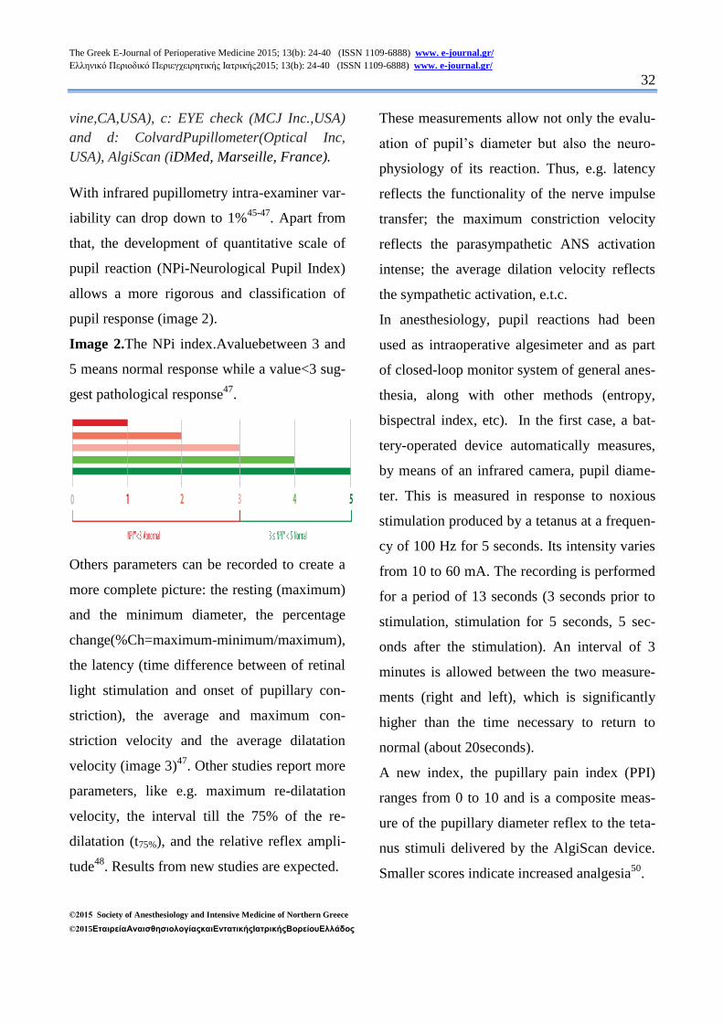
Image 1. Different models of portable
pupillometers
(for left to right):a. VIP 200 (NeuroOptics, Ir-
vine, CA,USA), b: NPi-100 (NeurOptics, Ir-
The Greek E-Journal of Perioperative Medicine 2015; 13(b): 24-40 (ISSN 1109-6888) www. e-journal.gr/ Ελληνικό Περιοδικό Περιεγχειρητικής Ιατρικής2015; 13(b): 24-40 (ISSN 1109-6888) www. e-journal.gr/
32
©2015 Society of Anesthesiology and Intensive Medicine of Northern Greece
©2015ΕταιρείαΑναισθησιολογίαςκαιΕντατικήςΙατρικήςΒορείουΕλλάδος
vine,CA,USA), c: EYE check (MCJ Inc.,USA)
and d: ColvardPupillometer(Optical Inc,
USA), AlgiScan (iDMed, Marseille, France).
With infrared pupillometry intra-examiner var-
iability can drop down to 1%45-47
. Apart from
that, the development of quantitative scale of
pupil reaction (NPi-Neurological Pupil Index)
allows a more rigorous and classification of
pupil response (image 2).
Image 2.The NPi index.Avaluebetween 3 and
5 means normal response while a value<3 sug-
gest pathological response47
.
Others parameters can be recorded to create a
more complete picture: the resting (maximum)
and the minimum diameter, the percentage
change(%Ch=maximum-minimum/maximum),
the latency (time difference between of retinal
light stimulation and onset of pupillary con-
striction), the average and maximum con-
striction velocity and the average dilatation
velocity (image 3)47
. Other studies report more
parameters, like e.g. maximum re-dilatation
velocity, the interval till the 75% of the re-
dilatation (t75%), and the relative reflex ampli-
tude48
. Results from new studies are expected.
These measurements allow not only the evalu-
ation of pupil’s diameter but also the neuro-
physiology of its reaction. Thus, e.g. latency
reflects the functionality of the nerve impulse
transfer; the maximum constriction velocity
reflects the parasympathetic ANS activation
intense; the average dilation velocity reflects
the sympathetic activation, e.t.c.
In anesthesiology, pupil reactions had been
used as intraoperative algesimeter and as part
of closed-loop monitor system of general anes-
thesia, along with other methods (entropy,
bispectral index, etc). In the first case, a bat-
tery-operated device automatically measures,
by means of an infrared camera, pupil diame-
ter. This is measured in response to noxious
stimulation produced by a tetanus at a frequen-
cy of 100 Hz for 5 seconds. Its intensity varies
from 10 to 60 mA. The recording is performed
for a period of 13 seconds (3 seconds prior to
stimulation, stimulation for 5 seconds, 5 sec-
onds after the stimulation). An interval of 3
minutes is allowed between the two measure-
ments (right and left), which is significantly
higher than the time necessary to return to
normal (about 20seconds).
A new index, the pupillary pain index (PPI)
ranges from 0 to 10 and is a composite meas-
ure of the pupillary diameter reflex to the teta-
nus stimuli delivered by the AlgiScan device.
Smaller scores indicate increased analgesia50
.
The Greek E-Journal of Perioperative Medicine 2015; 13(b): 24-40 (ISSN 1109-6888) www. e-journal.gr/ Ελληνικό Περιοδικό Περιεγχειρητικής Ιατρικής2015; 13(b): 24-40 (ISSN 1109-6888) www. e-journal.gr/
33
©2015 Society of Anesthesiology and Intensive Medicine of Northern Greece
©2015ΕταιρείαΑναισθησιολογίαςκαιΕντατικήςΙατρικήςΒορείουΕλλάδος
The overall use of these devices is easy and
can be employed by both medical and para-
medical personnel.
Limitations of the method include the relative
high cost, the need for an open eye during the
examination, ophthalmic diseases or opera-
tions and the lack of global collective experi-
ence and database.
APPLICATIONS IN PERIOPERATIVE
SETTING
Among the first application were the conduc-
tion of studies for the drugs effect on pupil
light reflex (Image 4), creating databases
which can be used for noninvasive detection of
drugs, even those administered epiduraly28, 49,
50.
Pupillometry seems more relevant than para-
sympathetic component of heart rate variabil-
ity to assess analgesia during general anesthe-
sia50
. It had been with promising results used
both for general and local anesthesia, both in
adults and children51-54
. Changes in PLR
brought about by a uterine contraction may be
used as a tool to assess analgesia in noncom-
municating obstetrical patients as well55
. Re-
sults from new studies (i.g. ALGISCAN Trial)
both for intraoperative condition and postan-
esthesia setting are expected56-57
.
Pupillometry has also been used for prediction
outcome after cardiac arrests: it seems that ear-
ly detection of PLR is associated with better
outcome; moreover, PLR and has comparable
prognostic accuracy than electroencephalo-
gram (EEG) and somato-sensory evoked po-
tentials (SSEP) .in predicting outcome of post-
anoxic coma, irrespective of temperature and
sedation58-63
. Also in post-resuscitation non-
brain dead critically ill patients with ‘absent’
pupillary reflexes, the reflex has been demon-
strated using a portable infrared pupillome-
ter58
.
Likewise, there is growing literature about
cases with “reversible fixed pupils” as it is re-
alized that some of these conditions might be
associated with ‘clinically undetectable’ rather
than ‘absent’ pupillary light reflexes64
. Other
phenomena like, e.g. pupillary hippus have
been associated with increased hospital mortal-
ity65
and with nonconvulsive status epilepticus
in critically ill patients65, 66
.
In neurosurgical critical care patients, there are
several reports claiming that pupillometry can
detect an early increase in intracranial pressure
(ICP). A rise of ICP above the level of
20mmHg decrease the constriction velocity of
ipsilateral to the injury pupil in values < 0.6
mm/sec (normal range 1.48 ± 0.33 mm/sec),
while changes in NPi can be detected up to
15.9 hours before the ICP increase1,64,67-69
.
Apart from that, PLR measurement has been
used with good results as analgesia index in
critically ill70-73
.
The Greek E-Journal of Perioperative Medicine 2015; 13(b): 24-40 (ISSN 1109-6888) www. e-journal.gr/ Ελληνικό Περιοδικό Περιεγχειρητικής Ιατρικής2015; 13(b): 24-40 (ISSN 1109-6888) www. e-journal.gr/
34
©2015 Society of Anesthesiology and Intensive Medicine of Northern Greece
©2015ΕταιρείαΑναισθησιολογίαςκαιΕντατικήςΙατρικήςΒορείουΕλλάδος
CONLCUSION–FUTURE
PERSPECTIVES
Infrared pupillometry complete an armamen-
tarium of monitor tools for central neural sys-
tem like transcranial Doppler, brain tissue
monitoring, microdialysis, several specific
(e.g. S-100 protein) biomarkers and other im-
aging examination (CT,MRI,fMRI,SPECT).
New studies are already investigating these
fields74
. Hence, a new era of neuromonitoring
has begun, and it bring along great changes in
future perception of conditions like anesthesia,
sleep, coma, locked-in syndrome and brain
mapping.
REFERENCES
1. Fountas KN, Kapsalaki EZ,
MachinisTG, et al. Clinical
implications of quantitative infrared
pupillometry in neurosurgical patients.
Neurocrit. Care 2005; 5: 55-60.
2. Budge J. Uber den Einflus des nerven
systems suf die Bewegung des
Iris.ArchPhiolHeilk 1852; 2:773–9.
3. Bellarminov L,Anwendung der
graphischenMethodezurUntersuchungd
erPupillen-Bewegung.Pflugers
Arch.Ges. Phusiol. 1885; 37:7.
4. Lowenstein O, Lowenfeld E.
Electronic PupillographyElectronic
Pupillography, A. M. A. Arch.
Ophthal.,1958; 59: 352-363
5. Matsunaga K. A new Binocular
Electronic Scanning Pupillometer,
Physiologia 1973; 16: 115-120.
6. Jones DP, Smith RA. A New Solid
State Dynamic Pupillometer Using A
Self-Scanning Photodiode Array. J
Phys E. 1983; 16: 1169-72.
7. Pokorny J, Smith VC. How much light
reach the retina? In Color Vision
Deficiencies XIII, Cavonious CR(ed.),
Klower Acad. publishers, Dodreht,
UK, 1997:pp. 491-511.
8. LeGrand Y. Light, Color,
Vision.2nd
Ed.Chapman& Hall, London
1968.pp.106.
9. Nowa W, Hachol A, Kasprzak H.
Time-frequency analysis of
spontaneous fluctuation of the pupil
size of the human eye. OpticaApplicata
2008; 43:469-80.
10. Berman SM, Fein G, Jewett J, et al.
Spectral determinants of steady-state
pupil size with full field of view. J.
Illum. Eng. Soc. 1992; 21: 12-33.
11. Winn B, Whitaker D, Elliott DB, et al.
Factors affecting light-adapted pupil
size in normal human subjects. Invest.
Ophthal. Vis. Sci. 1994; 35: 1132-7.
12. Watson, AB, Yellott, JI. A unified
formula for light-adapted pupil size.
Journal of Vision, 2012; 12:1–16.
The Greek E-Journal of Perioperative Medicine 2015; 13(b): 24-40 (ISSN 1109-6888) www. e-journal.gr/ Ελληνικό Περιοδικό Περιεγχειρητικής Ιατρικής2015; 13(b): 24-40 (ISSN 1109-6888) www. e-journal.gr/
35
©2015 Society of Anesthesiology and Intensive Medicine of Northern Greece
©2015ΕταιρείαΑναισθησιολογίαςκαιΕντατικήςΙατρικήςΒορείουΕλλάδος
13. Larson MD, Muhiudeen I.
Pupillometric analysis of the ‘absent
light reflex’. Arch Neurol 1995;
52:369-72.
14. Clark A, Clarke TN, Gregson B,
Hooker PN, Chambers IR.Variability
in pupil size estimation. Emerg Med J.
2006; 23: 440-1.
15. Litvan I, Saposnik G, Mauriño J, et al.
Pupillary diameter assessment: need
for a graded scale. Neurology. 2000;
54:530-1.
16. Wilson SF, Amling JK, Floyd SD, et
al. Determining interrater reliability of
nurses' assessments of pupillary size
and reaction. J NeurosciNurs 1988;
20:189-92.
17. Witting MD. Validity of simple
measurement to diagnose pupillary
dilation. Am J Emerg Med. 2005;
23:155-8.
18. Pop M, Payette Y, Santoriello
E.Comparison of the pupil card and
pupillometer in measuring pupilsize. J
CataractRefractSurg.2002 ;28 :283-8.
19. Bremner F. Pupil evaluation as a test
for autonomic disorders.ClinAuton
Res. 2009; 19: 88-101.
20. Tan ET, Johnson RH, Lambie DG, et
al. Alcoholic vagal neuropathy:
recovery following prolonged
abstinence.J NeurolNeurosurg
Psychiatry. 1984; 47:1335-7.
21. Chapman CR, Oka S, Bradshaw DH, et
al. Phasic pupil dilation response to
noxious stimulation in normal
volunteers: relationship to brain evoked
potentials and pain
report.Psychophysiology. 1999;36: 44-
52.
22. Lee H, Kim Y, Park J. Pupil cycle time
and contrast sensitivity in type II
diabetes mellitus patients: a pilot
study.Indian J Ophthalmol. 2011; 59:
201-5.
23. Tsiptsios D, Fotiou DF, Haidich AB, et
al. Evaluation of pupil mobility in
patients with myasthenia
gravis.ElectromyogrClinNeurophysiol.
2008; 48: 209-18.
24. Kono K, Miyao M, Ishihara S, et al.
Hypersensitivity in the pupil dilation
response to a cholinergic antagonist in
patients with Alzheimer's disease and
Down's syndrome.Nihon Ronen
IgakkaiZasshi. 1996;33:829-34.
25. Fotiou DF, Brozou CG, Haidich AB, et
al.Pupil reaction to light in Alzheimer's
disease: evaluation of pupil size
changes and mobility.Aging ClinExp
Res. 2007; 19: 364-71.
26. Fountoulakis KN, Siamouli M,
Kaprinis G, et al. Changes in the pupil
The Greek E-Journal of Perioperative Medicine 2015; 13(b): 24-40 (ISSN 1109-6888) www. e-journal.gr/ Ελληνικό Περιοδικό Περιεγχειρητικής Ιατρικής2015; 13(b): 24-40 (ISSN 1109-6888) www. e-journal.gr/
36
©2015 Society of Anesthesiology and Intensive Medicine of Northern Greece
©2015ΕταιρείαΑναισθησιολογίαςκαιΕντατικήςΙατρικήςΒορείουΕλλάδος
reflex arc in depressive patients.J
Affect Disord. 2005; 87: 341-2.
27. Ionescu S. Psychophysiology of mental
deficiency: evaluation of studies
involving the recording of autonomic
indices. PsychiatrEnfant. 1985;28:39-
134.
28. Richman JE, McAndrew KG, Decker
D, Mullaney SC. An evaluation of
pupil size standards used by police
officers for detecting drug
impairment.Optometry. 2004; 75: 175-
82.
29. Yanagawa Y, Miyazaki M, Sakamoto
T. Relationship between abnormal
pupillary reactivity and the outcome of
a psychotropic drug overdose.Am J
EmergMed. 2010; 28: 703-7.
30. Vermersch, P, Dufourd-Delalande, S,
Defoort-Dhellemmes S, et al. Tonic
pupils in Sjogren's syndrome, Rev
Neurol 2005; 161: 963-6.
31. Surakka J, Ruutiainen J, Romberg A, et
al. Pupillary function in early multiple
sclerosis.ClinAuton Res. 2008; 18:
150-4.
32. Armstrong RA. Visual signs and
symptoms of Parkinson's
disease.ClinExpOptom. 2008; 91: 129-
38.
33. Tokuda Y, Nakazato N, Stein GH.
Pupillary evaluation for differential
diagnosis of coma. Postgrad Med J
2003;79:49–51.
34. Clusmann H, Schaller C, Schramm J.
Fixed and dilated pupils after trauma,
stroke, and previous intracranial
surgery: management and outcome. J
NeurolNeurosurg Psychiatry 2001; 71:
175-81.
35. Chesnut RM, Ghajar J, Maas AI, et al.
Management and prognosis of severe
traumatic brain injury. Part II: Early
indicators of prognosis in severe
traumatic brain injury. Brain Trauma
Foundation, American Association of
Neurological Surgeons, Joint section
on neurotrauma and critical care.
2000;41:54.
36. Marmarou A, Lu J, Butcher I, et al
.Prognostic Value of the Glasgow
Coma Scale and Pupil Reactivity in
Traumatic Brain Injury Assessed Pre-
Hospital and on Enrollment: An
IMPACT Analysis. J Neurotrauma,
2007; 24: 270-80.
37. Shamim MS, Qadeer M, Murtaza G, et
al.Emergency department predictors of
tracheostomy in patients with isolated
traumatic brain injury requiring
emergency cranial decompression.J
Neurosurg. 2011; 115 :1007-12.
38. Al-Jishi A, Saluja RS, Al-Jehani H, et
al. Primary or secondary
The Greek E-Journal of Perioperative Medicine 2015; 13(b): 24-40 (ISSN 1109-6888) www. e-journal.gr/ Ελληνικό Περιοδικό Περιεγχειρητικής Ιατρικής2015; 13(b): 24-40 (ISSN 1109-6888) www. e-journal.gr/
37
©2015 Society of Anesthesiology and Intensive Medicine of Northern Greece
©2015ΕταιρείαΑναισθησιολογίαςκαιΕντατικήςΙατρικήςΒορείουΕλλάδος
decompressivecraniectomy: different
indication and outcome.Can J Neurol
Sci. 2011; 38: 612-20.
39. Kim YJ.A systematic review of factors
contributing to outcomes in patients
with traumatic brain injury.J ClinNurs.
2011 ;20 :1518-32.
40. Tien HC, Cunha JRF, Wu SN, et al.
Do Trauma Patients with a Glasgow
Coma Scale Score of 3 and Bilateral
Fixed and Dilated Pupils Have Any
Chance of Survival? J Trauma, 2006;
60: 274-8.
41. Hoffmann M, Lefering R, Rueger JM,
et al. Trauma Registry of the German
Society for Trauma Surgery. Pupil
evaluation in addition to Glasgow
Coma Scale components in prediction
of traumatic brain injury and
mortality.Br J Surg. 2012 Jan;99:122-
30.
42. Franschman G, Peerdeman SM,
Andriessen TM, et al. Analysis of
Results and Methods--Traumatic Brain
Injury (ALARM-TBI) Investigator.
Effect of secondary prehospital risk
factors on outcome in severe traumatic
brain injury in the context of fast
access to trauma care.J Trauma.
2011;71:826-32.
43. Cooper DJ, Rosenfeld JV, Murray L, et
al. Decompressivecraniectomy in
diffuse traumatic brain injury.N Engl J
Med. 2011; 364: 1493-1502.
44. Chi JH.Craniectomy for traumatic
brain injury: results from the DECRA
trial.Neurosurgery. 2011 ;68 :N19-20.
45. Rose D, Meeker M, Bacchetti P, et al.
Evaluation of the portable infrared
pupillometer. Neurosurgery 2005; 57:
198-203.
46. Taylor WR, Chen JW, Meltzer H, et al.
Quantitative pupillometry, a new
technology: normative data and
preliminary observations in patients
with acute head injury. J Neurosurgery,
2003; 98: 205-13.
47. www.neuroptics.org./pi_faq_110721.p
df
48. Surakka J, Ruutiainen J, Romberg A, et
al. Pupillary function in early multiple
sclerosis. ClinAuton Res. 2008;18
:150-4.
49. Matouskova O, Slanar O, Chytil L, et
al. Pupillometry in healthy volunteers
as a biomarker of tramadol efficacy.J
ClinPharmTher. 2011 ;36:513-7.
50. Larson MD, Berry PD. Supraspinal
pupillary effects of intravenous and
epidural fentanyl during
isofluraneanesthesia.RegAnesth Pain
Med. 2000; 25:60-6.
51. Charier D,lZantour D, Pichot V, et al.
Evaluation of Analgesia During
The Greek E-Journal of Perioperative Medicine 2015; 13(b): 24-40 (ISSN 1109-6888) www. e-journal.gr/ Ελληνικό Περιοδικό Περιεγχειρητικής Ιατρικής2015; 13(b): 24-40 (ISSN 1109-6888) www. e-journal.gr/
38
©2015 Society of Anesthesiology and Intensive Medicine of Northern Greece
©2015ΕταιρείαΑναισθησιολογίαςκαιΕντατικήςΙατρικήςΒορείουΕλλάδος
General Anesthesia: Pupillometry
Versus Heart Rate Variability. ASA
meeting abstracts 2012; A187.
52. Constant I, Nghe MC, Boudet L, et al.
Reflex pupillary dilatation in response
to skin incision and alfentanil in
children anaesthetized with
sevoflurane: a more sensitive measure
of noxious stimulation than the
commonly used variables. Br J
Anaesth. 2006; 96:614-9.
53. Bourgeois E, Sabourdin N, Louvet N,
et al.Minimal alveolar concentration of
sevoflurane inhibiting the reflex
pupillary dilatation after noxious
stimulation in children and young
adults.Br J Anaesth. 2012;108:648-54.
54. Merlin S, Larson D, Berry PD, et al.
Latenchy of papillary reflex during
general anesthesia. J ApplPhysiol
2004; 97:725-30.
55. Guglielminotti J, Mentré F, Gaillard J,
et al. Assessment of pain during labor
with pupillometry: a prospective
observational study. Anesth Analg.
2013;116:1057-62.
56. Kantor E, Montravers P, Longrois D, et
al. Pain assessment in the
postanaesthesia care unit using
pupillometry: A cross-sectional study
after standard anaesthetic care.Eur J
Anaesthesiol. 2014 ;31:91-7.
57. http://clinicaltrials.gov/show/NCT0168
5645
58. Behrends M, Niemann CU, Larson
MD. Infrared pupillometry to detect the
light reflex during cardiopulmonary
resuscitation: A case
series.Resuscitation. 2012 ;83 :1223-8.
59. Breckwoldt J, Arntz HR. Infrared
pupillometry during cardiopulmonary
resuscitation for prognostication—A
new tool on the horizon?
Resuscitation.2012 ;83:1181-2.
60. Okada K, Ohde S, Otani N, et
al.Prediction protocol for neurological
outcome for survivors of out-of-
hospital cardiac arrest treated with
targeted temperature
management.Resuscitation.2012;83:73
4-9.
61. Suys T, Bouzat P, Marques-Vidal P, et
al. Automated Quantitative
Pupillometry for the Prognostication of
Coma After Cardiac Arrest. NeuroCrit.
Care 2014; [Epub ahead of print]
62. Wijdicks EF, Hijdra A, Young GB, et
al. Practice parameter: prediction of
outcome in comatose survivors after
cardiopulmonary resuscitation (an
evidence-based review): report of the
Quality Standards Subcommittee of the
American Academy of Neurology.
Neurology 2006;67:203-10.
The Greek E-Journal of Perioperative Medicine 2015; 13(b): 24-40 (ISSN 1109-6888) www. e-journal.gr/ Ελληνικό Περιοδικό Περιεγχειρητικής Ιατρικής2015; 13(b): 24-40 (ISSN 1109-6888) www. e-journal.gr/
39
©2015 Society of Anesthesiology and Intensive Medicine of Northern Greece
©2015ΕταιρείαΑναισθησιολογίαςκαιΕντατικήςΙατρικήςΒορείουΕλλάδος
63. Denny JC, Arndt FV, Dupont WD, et
al. Increased hospital mortality in
patients with bedside hippus.Am. J
Med. 2008; 121:239-45.
64. Chaudhuri K, Malhma GM, Rosenfield
JV. Survival of patients with coma and
bilateral fixed pupils. Injury 2009 ;40
:28-32.
65. Schnell D, Arnaud L, Lemial V, et al.
Pupillary hippus in nonconvulsive
status epilepticus. Epileptic Disord.
2012; 14:310-2.
66. Legriel S, Azoulay E, Resche-Rigon
M, et al. Functional outcome after
convulsive status epilepticus. Crit Care
Med 2010; 38: 2295-303.
67. Chen JW, Gombart ZJ, Rogers S, et al.
Pupillary reactivity as an early
indicator of increased intracranial
pressure: The introduction of the
neurological pupil index.
SurgNeurolInt 2011; 2:82.
68. Kuo JR, Lo CJ, Lo CL, et al.
Prognostic predictor of outcome in an
operative series in trauma brain injury.
J Formos Med Assoc 2011; 110:258-
64.
69. Hautin E, Cour M, Illinger M, et al.
Intérêt de l’ examen pupillair
eautomatiséen réanimation.
Réanimation 2010; 19 (Suppl 1) :S40.
70. Bader MK. Gizmos and Gadgets for
the Neuroscience Intensive Care Unit. J
NeurosciNurs. 2006; 38:248-60.
71. Paulus J, Roquilly A, Beloeil H, et al.
Pupillary reflex measurement predicts
insufficient analgesia before
endotracheal suction in critically ill
patients. Crit. Care 2013; 17:R161.
72. Payen JF, Isnardon S, Lavolaine J, et
al. Pupillometry in anesthesia and
critical care.Ann Fr AnesthReanim.
2012; 31:155-9.
73. Κοντογούνης Γεώργιος: Μελετη των
διαγνωστικών δυνατοτήτων της
κορημετρίας σε νευρολογικούς
ασθενείς στη μονάδα εντατικής
θεραπείας. Ιατρική σχολή ΑΠΘ.
Διδακτορική διατριβή (τρέχουσα).
Επιβλέπων: καθ. Τάσκος Νικόλαος
74. Murphy P, O’Connel R, O’Sullivan M,
et al. Pupil diameter covaries with bold
activity in human locus ceruleus. Hum
Brain Mapp.2014;35:4140-54.
Key words:pupillometry, intensive care unit, perioperative medicine.
The Greek E-Journal of Perioperative Medicine 2015; 13(b): 24-40 (ISSN 1109-6888) www. e-journal.gr/ Ελληνικό Περιοδικό Περιεγχειρητικής Ιατρικής2015; 13(b): 24-40 (ISSN 1109-6888) www. e-journal.gr/
40
©2015 Society of Anesthesiology and Intensive Medicine of Northern Greece
©2015ΕταιρείαΑναισθησιολογίαςκαιΕντατικήςΙατρικήςΒορείουΕλλάδος
Author Disclosures:
Authors AslanidisTh, Kontogounis Ghave noconflicts of interest or financial ties to disclose.
Corresponding author:
TheodorosAslanidis,
4 Doridos Street 54633,
Thessaloniki, Greece,
tel.: +306972477166,
e-mail:[email protected]