p38 MAPK Antibody p38 MAPK Antibody. Phospho-p38 MAP Kinase(Thr180/Tyr182) p38 MAP Kinase 0 0.5 18...
2
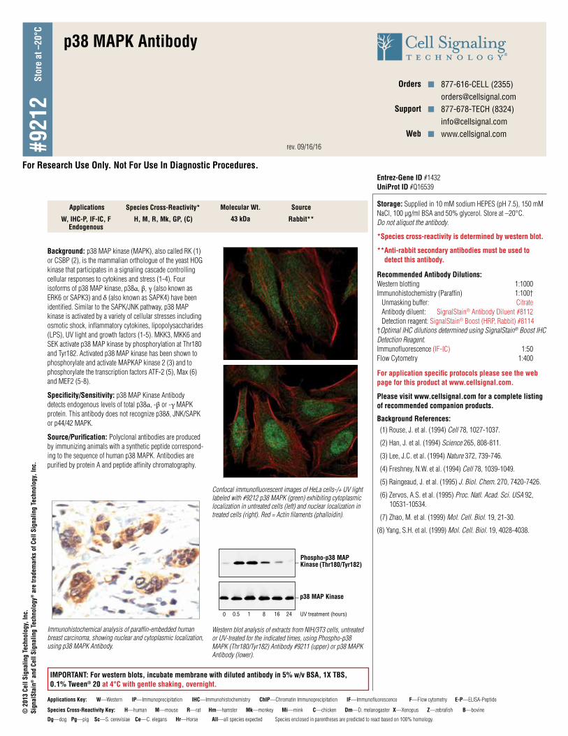
#9212 Store at –20°C p38 MAPK Antibody W, IHC-P, IF-IC, F Endogenous H, M, R, Mk, GP, (C) 43 kDa Rabbit** Background: p38 MAP kinase (MAPK), also called RK (1) or CSBP (2), is the mammalian orthologue of the yeast HOG kinase that participates in a signaling cascade controlling cellular responses to cytokines and stress (1-4). Four isoforms of p38 MAP kinase, p38α, β, γ (also known as ERK6 or SAPK3) and δ (also known as SAPK4) have been identified. Similar to the SAPK/JNK pathway, p38 MAP kinase is activated by a variety of cellular stresses including osmotic shock, inflammatory cytokines, lipopolysaccharides (LPS), UV light and growth factors (1-5). MKK3, MKK6 and SEK activate p38 MAP kinase by phosphorylation at Thr180 and Tyr182. Activated p38 MAP kinase has been shown to phosphorylate and activate MAPKAP kinase 2 (3) and to phosphorylate the transcription factors ATF-2 (5), Max (6) and MEF2 (5-8). Specificity/Sensitivity: p38 MAP Kinase Antibody detects endogenous levels of total p38α, -β or -γ MAPK protein. This antibody does not recognize p38δ, JNK/SAPK or p44/42 MAPK. Source/Purification: Polyclonal antibodies are produced by immunizing animals with a synthetic peptide correspond- ing to the sequence of human p38 MAPK. Antibodies are purified by protein A and peptide affinity chromatography. Storage: Supplied in 10 mM sodium HEPES (pH 7.5), 150 mM NaCl, 100 µg/ml BSA and 50% glycerol. Store at –20°C. Do not aliquot the antibody. *Species cross-reactivity is determined by western blot. **Anti-rabbit secondary antibodies must be used to detect this antibody. Recommended Antibody Dilutions: Western blotting 1:1000 Immunohistochemistry (Paraffin) 1:100† Unmasking buffer: Citrate Antibody diluent: SignalStain ® Antibody Diluent #8112 Detection reagent: SignalStain ® Boost (HRP, Rabbit) #8114 †Optimal IHC dilutions determined using SignalStain ® Boost IHC Detection Reagent. Immunofluorescence (IF-IC) 1:50 Flow Cytometry 1:400 For application specific protocols please see the web page for this product at www.cellsignal.com. Please visit www.cellsignal.com for a complete listing of recommended companion products. Background References: (1) Rouse, J. et al. (1994) Cell 78, 1027-1037. (2) Han, J. et al. (1994) Science 265, 808-811. (3) Lee, J.C. et al. (1994) Nature 372, 739-746. (4) Freshney, N.W. et al. (1994) Cell 78, 1039-1049. (5) Raingeaud, J. et al. (1995) J. Biol. Chem. 270, 7420-7426. (6) Zervos, A.S. et al. (1995) Proc. Natl. Acad. Sci. USA 92, 10531-10534. (7) Zhao, M. et al. (1999) Mol. Cell. Biol. 19, 21-30. (8) Yang, S.H. et al. (1999) Mol. Cell. Biol. 19, 4028-4038. Orders n 877-616-CELL (2355) [email protected] Support n 877-678-TECH (8324) [email protected] Web n www.cellsignal.com Applications Species Cross-Reactivity* Molecular Wt. Source IMPORTANT: For western blots, incubate membrane with diluted antibody in 5% w/v BSA, 1X TBS, 0.1% Tween ® 20 at 4°C with gentle shaking, overnight. Immunohistochemical analysis of paraffin-embedded human breast carcinoma, showing nuclear and cytoplasmic localization, using p38 MAPK Antibody. Phospho-p38 MAP Kinase (Thr180/Tyr182) p38 MAP Kinase 0 0.5 1 8 16 24 UV treatment (hours) Western blot analysis of extracts from NIH/3T3 cells, untreated or UV-treated for the indicated times, using Phospho-p38 MAPK (Thr180/Tyr182) Antibody #9211 (upper) or p38 MAPK Antibody (lower). Confocal immunofluorescent images of HeLa cells-/+ UV light labeled with #9212 p38 MAPK (green) exhibiting cytoplasmic localization in untreated cells (left) and nuclear localization in treated cells (right). Red = Actin filaments (phalloidin). Entrez-Gene ID #1432 UniProt ID #Q16539 Species Cross-Reactivity Key: H—human M—mouse R—rat Hm—hamster Mk—monkey Mi—mink C—chicken Dm—D. melanogaster X—Xenopus Z—zebrafish B—bovine Dg—dog Pg—pig Sc—S. cerevisiae Ce—C. elegans Hr—Horse All—all species expected Species enclosed in parentheses are predicted to react based on 100% homology. Applications Key: W—Western IP—Immunoprecipitation IHC—Immunohistochemistry ChIP—Chromatin Immunoprecipitation IF—Immunofluorescence F—Flow cytometry E-P—ELISA-Peptide © 2013 Cell Signaling Technology, Inc. SignalStain ® and Cell Signaling Technology ® are trademarks of Cell Signaling Technology, Inc. rev. 09/16/16 For Research Use Only. Not For Use In Diagnostic Procedures.
Transcript of p38 MAPK Antibody p38 MAPK Antibody. Phospho-p38 MAP Kinase(Thr180/Tyr182) p38 MAP Kinase 0 0.5 18...
#921
2St
ore
at –
20°C
p38 MAPK Antibody
W, IHC-P, IF-IC, FEndogenous
H, M, R, Mk, GP, (C) 43 kDa Rabbit**
Background: p38 MAP kinase (MAPK), also called RK (1) or CSBP (2), is the mammalian orthologue of the yeast HOG kinase that participates in a signaling cascade controlling cellular responses to cytokines and stress (1-4). Four isoforms of p38 MAP kinase, p38α, β, γ (also known as ERK6 or SAPK3) and δ (also known as SAPK4) have been identified. Similar to the SAPK/JNK pathway, p38 MAP kinase is activated by a variety of cellular stresses including osmotic shock, inflammatory cytokines, lipopolysaccharides (LPS), UV light and growth factors (1-5). MKK3, MKK6 and SEK activate p38 MAP kinase by phosphorylation at Thr180 and Tyr182. Activated p38 MAP kinase has been shown to phosphorylate and activate MAPKAP kinase 2 (3) and to phosphorylate the transcription factors ATF-2 (5), Max (6) and MEF2 (5-8).
Specificity/Sensitivity: p38 MAP Kinase Antibody detects endogenous levels of total p38α, -β or -γ MAPK protein. This antibody does not recognize p38δ, JNK/SAPK or p44/42 MAPK.
Source/Purification: Polyclonal antibodies are produced by immunizing animals with a synthetic peptide correspond-ing to the sequence of human p38 MAPK. Antibodies are purified by protein A and peptide affinity chromatography.
Storage: Supplied in 10 mM sodium HEPES (pH 7.5), 150 mM NaCl, 100 µg/ml BSA and 50% glycerol. Store at –20°C. Do not aliquot the antibody.
*Species cross-reactivity is determined by western blot.
** Anti-rabbit secondary antibodies must be used to detect this antibody.
Recommended Antibody Dilutions: Western blotting 1:1000 Immunohistochemistry (Paraffin) 1:100† Unmasking buffer: Citrate Antibody diluent: SignalStain® Antibody Diluent #8112 Detection reagent: SignalStain® Boost (HRP, Rabbit) #8114 †Optimal IHC dilutions determined using SignalStain® Boost IHC Detection Reagent. Immunofluorescence (IF-IC) 1:50 Flow Cytometry 1:400
For application specific protocols please see the web page for this product at www.cellsignal.com.
Please visit www.cellsignal.com for a complete listing of recommended companion products.
Background References: (1) Rouse, J. et al. (1994) Cell 78, 1027-1037.
(2) Han, J. et al. (1994) Science 265, 808-811.
(3) Lee, J.C. et al. (1994) Nature 372, 739-746.
(4) Freshney, N.W. et al. (1994) Cell 78, 1039-1049.
(5) Raingeaud, J. et al. (1995) J. Biol. Chem. 270, 7420-7426.
(6) Zervos, A.S. et al. (1995) Proc. Natl. Acad. Sci. USA 92, 10531-10534.
(7) Zhao, M. et al. (1999) Mol. Cell. Biol. 19, 21-30.
(8) Yang, S.H. et al. (1999) Mol. Cell. Biol. 19, 4028-4038.
Orders n 877-616-CELL (2355)[email protected]
Support n 877-678-TECH (8324)[email protected]
Web n www.cellsignal.com
Applications Species Cross-Reactivity* Molecular Wt. Source
IMPORTANT: For western blots, incubate membrane with diluted antibody in 5% w/v BSA, 1X TBS, 0.1% Tween® 20 at 4°C with gentle shaking, overnight.
Immunohistochemical analysis of paraffin-embedded human breast carcinoma, showing nuclear and cytoplasmic localization, using p38 MAPK Antibody.
Phospho-p38 MAP Kinase (Thr180/Tyr182)
p38 MAP Kinase
0 0.5 1 8 16 24 UV treatment (hours)
Western blot analysis of extracts from NIH/3T3 cells, untreated or UV-treated for the indicated times, using Phospho-p38 MAPK (Thr180/Tyr182) Antibody #9211 (upper) or p38 MAPK Antibody (lower).
Confocal immunofluorescent images of HeLa cells-/+ UV light labeled with #9212 p38 MAPK (green) exhibiting cytoplasmic localization in untreated cells (left) and nuclear localization in treated cells (right). Red = Actin filaments (phalloidin).
Entrez-Gene ID #1432 UniProt ID #Q16539
Species Cross-Reactivity Key: H—human M—mouse R—rat Hm—hamster Mk—monkey Mi—mink C—chicken Dm—D. melanogaster X—Xenopus Z—zebrafish B—bovine
Dg—dog Pg—pig Sc—S. cerevisiae Ce—C. elegans Hr—Horse All—all species expected Species enclosed in parentheses are predicted to react based on 100% homology.
Applications Key: W—Western IP—Immunoprecipitation IHC—Immunohistochemistry ChIP—Chromatin Immunoprecipitation IF—Immunofluorescence F—Flow cytometry E-P—ELISA-Peptide
© 2
013
Cell
Sign
alin
g Te
chno
logy
, Inc
.Si
gnal
Stai
n® a
nd C
ell S
igna
ling
Tech
nolo
gy® a
re tr
adem
arks
of C
ell S
igna
ling
Tech
nolo
gy, I
nc.
rev. 09/16/16
For Research Use Only. Not For Use In Diagnostic Procedures.
Orders n 877-616-CELL (2355) [email protected] Support n 877-678-TECH (8324) [email protected] Web n www.cellsignal.com© 2
010
Cell
Sign
alin
g Te
chno
logy
, Inc
.
p38 MAPK
Even
ts
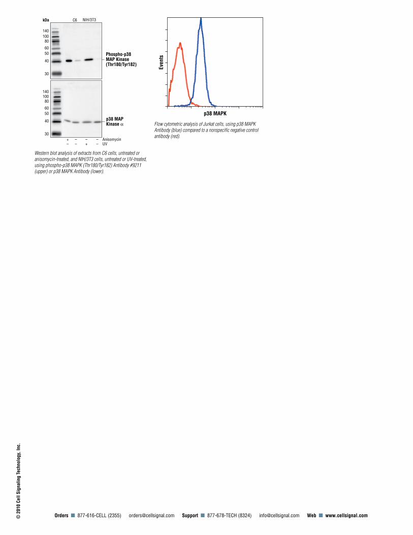
Flow cytometric analysis of Jurkat cells, using p38 MAPK Antibody (blue) compared to a nonspecific negative control antibody (red).
kDa
Phospho-p38 MAP Kinase (Thr180/Tyr182)
C6
140 100 80
60 50
40
30
140 100 80
60 50
40
30
NIH/3T3
p38 MAP Kinase α
Anisomycin UV
– – – + – + – –
Western blot analysis of extracts from C6 cells, untreated or anisomycin-treated, and NIH/3T3 cells, untreated or UV-treated, using phospho-p38 MAPK (Thr180/Tyr182) Antibody #9211 (upper) or p38 MAPK Antibody (lower).
![Diacylglycerol kinase ζ generates dipalmitoyl-phosphatidic ... · kinase C [6], and p21 activated protein kinase 1 [7,8].PAasan intracellular signaling lipid is generated by phosphorylation](https://static.fdocument.org/doc/165x107/5fe275ed0f93ac2b35696d07/diacylglycerol-kinase-generates-dipalmitoyl-phosphatidic-kinase-c-6-and.jpg)


![BMC Gastroenterology BioMed Central · 2017. 8. 28. · BMC Gastroenterology Research article ... MAP kinase [33], and AMP-activated protein kinase [34]. Further-more, several different](https://static.fdocument.org/doc/165x107/609f415b38f68d540772e0a3/bmc-gastroenterology-biomed-central-2017-8-28-bmc-gastroenterology-research.jpg)