p38 mitogen-activated protein kinase regulates...
10
3598 Research Article Introduction Wnt signaling is critical for normal embryonic development, patterning, cellular proliferation and homeostasis, and its aberrant activity is linked to many forms of prevalent human carcinomas (Logan and Nusse, 2004; Moon et al., 2002; Moon et al., 2004; Polakis, 2000). Wnt ligands initiate intracellular signaling pathways by binding to the G-protein-coupled receptors Frizzleds (Fzs) (Bhanot et al., 1996; Liu et al., 2001). The Wnt-sensitive pathways include the canonical (Wnt–β-catenin), and the non-canonical (planar cell polarity and Wnt-cGMP-Ca 2+ ) pathways (Bhanot et al., 1996; Liu et al., 1999). In the canonical pathway, in the absence of Wnt, cellular β-catenin is subjected to proteasome-mediated degradation by the destruction complex, which includes among other proteins Axin and the product of the adenomatous polyposis coli gene (APC). These proteins facilitate the phosphorylation of β- catenin by the Ser/Thr protein kinase glycogen synthase kinase 3β (GSK3β). This ultimately leads to ubiquitylation and proteasome- mediated degradation of β-catenin. Wnt3a binding to Fz1 leads to activation of the G-proteins Gα o and Gα q and of the phosphoprotein Dishevelled, resulting in reduced GSK3β activity and increased accumulation of β-catenin. Nuclear accumulation of β-catenin follows the stimulatory activation of lymphoid-enhancer factor/T- cell factor (Lef/Tcf)-sensitive transcription of developmentally related genes (Behrens et al., 1996; Molenaar et al., 1996). Aberrant accumulation of β-catenin contributes to tumorigenesis and therefore requires strict regulation. Recent advances suggest a possible intersection of other major signaling pathways, including mitogen-activated protein kinase (MAPK) pathways with the Wnt–β-catenin signaling pathway (Bikkavilli et al., 2008; Caverzasio and Manen, 2007; Gao et al., 2002; Hildesheim et al., 2005; Keren et al., 2005). In NIH3T3 fibroblasts, Wnt3a activates the ERK pathway through Ras, Raf and MEK and plays an important role in cellular proliferation (Yun et al., 2005). In mesenchymal stem cells (MSCs), Wnt4-mediated activation of p38 MAPK was found to be crucial for enhancing osteogenic differentiation of MSCs (Chang et al., 2007). Similarly, in C3H10T1/2 mesenchymal cells, Wnt3a also induced transient activation of p38 and ERK, which regulate alkaline phosphatase activity and nodule mineralization, suggesting an important role for the MAPKs in the development of mesenchymal cells into osteoprogenitors (Caverzasio and Manen, 2007). Therefore, in addition to the canonical β-catenin pathway and non-canonical pathways, Wnts also activate MAPK pathways that play important roles in proliferation, myogenesis and osteogenesis (Caverzasio and Manen, 2007; Chang et al., 2007; Keren et al., 2005). Our earlier studies highlighted a probable role for p38 MAPK in canonical Wnt–β-catenin signaling (Bikkavilli et al., 2008) but the mechanism remains unclear. Here, we reveal that p38 MAPK is activated upon stimulation by Wnt3a and is crucial for Wnt3a- induced accumulation of β-catenin, Lef/Tcf-sensitive gene activation and primitive endoderm formation. We also show that Wnt3a- induced activation of p38 MAPK is sensitive to depletion of Gα q , Gα s or Dishevelled3 (Dvl3) and reveal an important role for p38 MAPK in regulating GSK3β activity. Results Wnt3a activates p38 MAPK in mouse F9 cells Totipotent mouse embryonic carcinoma F9 cells transfected to express rat Frizzled-1 (Rfz1 or FZD1) provide an optimal system for the biochemical analysis of Wnt/Fz1 signaling (Malbon, 2005). Upon Wnt3a stimulation, F9 cells differentiate into primitive endoderm, which is characteristic of early mouse development (Liu The Wnt–β-catenin canonical signaling pathway is crucial for normal embryonic development, and aberrant expression of components of this pathway results in oncogenesis. Upon scanning for the mitogen-activated protein kinase (MAPK) pathways that might intersect with the canonical Wnt–β- catenin signaling pathway in response to Wnt3a, we observed a strong activation of p38 MAPK in mouse F9 teratocarcinoma cells. Wnt3a-induced p38 MAPK activation was sensitive to siRNAs against Gα q or Gα s , but not against either Gα o or Gα 11 . Activation of p38 MAPK is critical for canonical Wnt–β- catenin signaling. Chemical inhibitors of p38 MAPK (SB203580 or SB239063) and expression of a dominant negative-version of p38 MAPK attenuate Wnt3a-induced accumulation of β- catenin, Lef/Tcf-sensitive gene activation, and primitive endoderm formation. Furthermore, epistasis experiments pinpoint p38 MAPK as operating downstream of Dishevelleds. We also demonstrate that chemical inhibition of p38 MAPK restores Wnt3a-attenuated GSK3β kinase activity. We demonstrate the involvement of G-proteins and Dishevelleds in Wnt3a-induced p38 MAPK activation, highlighting a critical role for p38 MAPK in canonical Wnt–β-catenin signaling. Key words: Wnt, β-catenin, p38 MAPK, Frizzled, Dishevelled, Canonical pathway, Lef/Tcf, Primitive endoderm, G-protein Summary p38 mitogen-activated protein kinase regulates canonical Wnt–β-catenin signaling by inactivation of GSK3β Rama Kamesh Bikkavilli*, Michael E. Feigin and Craig C. Malbon Department of Pharmacology, Health Sciences Center, State University of New York at Stony Brook, Stony Brook, NY 11794-8651, USA *Author for correspondence (e-mail: [email protected]) Accepted 5 August 2008 Journal of Cell Science 121, 3598-3607 Published by The Company of Biologists 2008 doi:10.1242/jcs.032854 Journal of Cell Science
Transcript of p38 mitogen-activated protein kinase regulates...
3598 Research Article
IntroductionWnt signaling is critical for normal embryonic development,patterning, cellular proliferation and homeostasis, and its aberrantactivity is linked to many forms of prevalent human carcinomas(Logan and Nusse, 2004; Moon et al., 2002; Moon et al., 2004;Polakis, 2000). Wnt ligands initiate intracellular signaling pathwaysby binding to the G-protein-coupled receptors Frizzleds (Fzs)(Bhanot et al., 1996; Liu et al., 2001). The Wnt-sensitive pathwaysinclude the canonical (Wnt–β-catenin), and the non-canonical(planar cell polarity and Wnt-cGMP-Ca2+) pathways (Bhanot et al.,1996; Liu et al., 1999). In the canonical pathway, in the absence ofWnt, cellular β-catenin is subjected to proteasome-mediateddegradation by the destruction complex, which includes among otherproteins Axin and the product of the adenomatous polyposis coligene (APC). These proteins facilitate the phosphorylation of β-catenin by the Ser/Thr protein kinase glycogen synthase kinase 3β(GSK3β). This ultimately leads to ubiquitylation and proteasome-mediated degradation of β-catenin. Wnt3a binding to Fz1 leads toactivation of the G-proteins Gαo and Gαq and of the phosphoproteinDishevelled, resulting in reduced GSK3β activity and increasedaccumulation of β-catenin. Nuclear accumulation of β-cateninfollows the stimulatory activation of lymphoid-enhancer factor/T-cell factor (Lef/Tcf)-sensitive transcription of developmentallyrelated genes (Behrens et al., 1996; Molenaar et al., 1996). Aberrantaccumulation of β-catenin contributes to tumorigenesis and thereforerequires strict regulation.
Recent advances suggest a possible intersection of other majorsignaling pathways, including mitogen-activated protein kinase(MAPK) pathways with the Wnt–β-catenin signaling pathway(Bikkavilli et al., 2008; Caverzasio and Manen, 2007; Gao et al.,2002; Hildesheim et al., 2005; Keren et al., 2005). In NIH3T3
fibroblasts, Wnt3a activates the ERK pathway through Ras, Rafand MEK and plays an important role in cellular proliferation (Yunet al., 2005). In mesenchymal stem cells (MSCs), Wnt4-mediatedactivation of p38 MAPK was found to be crucial for enhancingosteogenic differentiation of MSCs (Chang et al., 2007). Similarly,in C3H10T1/2 mesenchymal cells, Wnt3a also induced transientactivation of p38 and ERK, which regulate alkaline phosphataseactivity and nodule mineralization, suggesting an important role forthe MAPKs in the development of mesenchymal cells intoosteoprogenitors (Caverzasio and Manen, 2007). Therefore, inaddition to the canonical β-catenin pathway and non-canonicalpathways, Wnts also activate MAPK pathways that play importantroles in proliferation, myogenesis and osteogenesis (Caverzasio andManen, 2007; Chang et al., 2007; Keren et al., 2005).
Our earlier studies highlighted a probable role for p38 MAPKin canonical Wnt–β-catenin signaling (Bikkavilli et al., 2008) butthe mechanism remains unclear. Here, we reveal that p38 MAPKis activated upon stimulation by Wnt3a and is crucial for Wnt3a-induced accumulation of β-catenin, Lef/Tcf-sensitive gene activationand primitive endoderm formation. We also show that Wnt3a-induced activation of p38 MAPK is sensitive to depletion of Gαq,Gαs or Dishevelled3 (Dvl3) and reveal an important role for p38MAPK in regulating GSK3β activity.
ResultsWnt3a activates p38 MAPK in mouse F9 cellsTotipotent mouse embryonic carcinoma F9 cells transfected toexpress rat Frizzled-1 (Rfz1 or FZD1) provide an optimal systemfor the biochemical analysis of Wnt/Fz1 signaling (Malbon, 2005).Upon Wnt3a stimulation, F9 cells differentiate into primitiveendoderm, which is characteristic of early mouse development (Liu
The Wnt–β-catenin canonical signaling pathway is crucial fornormal embryonic development, and aberrant expression ofcomponents of this pathway results in oncogenesis. Uponscanning for the mitogen-activated protein kinase (MAPK)pathways that might intersect with the canonical Wnt–β-catenin signaling pathway in response to Wnt3a, we observeda strong activation of p38 MAPK in mouse F9 teratocarcinomacells. Wnt3a-induced p38 MAPK activation was sensitive tosiRNAs against Gαq or Gαs, but not against either Gαo or Gα11.Activation of p38 MAPK is critical for canonical Wnt–β-catenin signaling. Chemical inhibitors of p38 MAPK (SB203580or SB239063) and expression of a dominant negative-version
of p38 MAPK attenuate Wnt3a-induced accumulation of β-catenin, Lef/Tcf-sensitive gene activation, and primitiveendoderm formation. Furthermore, epistasis experimentspinpoint p38 MAPK as operating downstream of Dishevelleds.We also demonstrate that chemical inhibition of p38 MAPKrestores Wnt3a-attenuated GSK3β kinase activity. Wedemonstrate the involvement of G-proteins and Dishevelleds inWnt3a-induced p38 MAPK activation, highlighting a criticalrole for p38 MAPK in canonical Wnt–β-catenin signaling.
Key words: Wnt, β-catenin, p38 MAPK, Frizzled, Dishevelled,Canonical pathway, Lef/Tcf, Primitive endoderm, G-protein
Summary
p38 mitogen-activated protein kinase regulatescanonical Wnt–β-catenin signaling by inactivation ofGSK3βRama Kamesh Bikkavilli*, Michael E. Feigin and Craig C. MalbonDepartment of Pharmacology, Health Sciences Center, State University of New York at Stony Brook, Stony Brook, NY 11794-8651, USA*Author for correspondence (e-mail: [email protected])
Accepted 5 August 2008Journal of Cell Science 121, 3598-3607 Published by The Company of Biologists 2008doi:10.1242/jcs.032854
Jour
nal o
f Cel
l Sci
ence
3599p38 regulates Wnt–β-catenin signaling
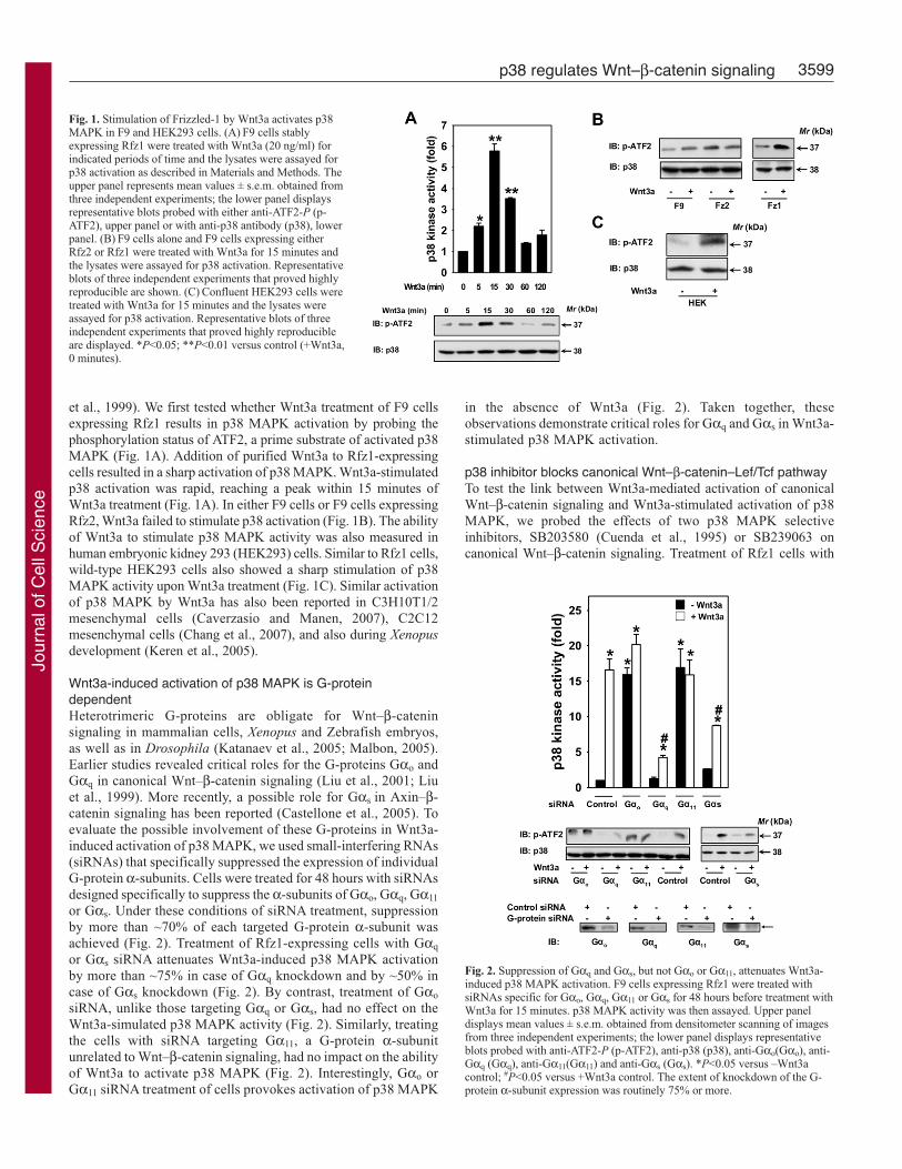
et al., 1999). We first tested whether Wnt3a treatment of F9 cellsexpressing Rfz1 results in p38 MAPK activation by probing thephosphorylation status of ATF2, a prime substrate of activated p38MAPK (Fig. 1A). Addition of purified Wnt3a to Rfz1-expressingcells resulted in a sharp activation of p38 MAPK. Wnt3a-stimulatedp38 activation was rapid, reaching a peak within 15 minutes ofWnt3a treatment (Fig. 1A). In either F9 cells or F9 cells expressingRfz2, Wnt3a failed to stimulate p38 activation (Fig. 1B). The abilityof Wnt3a to stimulate p38 MAPK activity was also measured inhuman embryonic kidney 293 (HEK293) cells. Similar to Rfz1 cells,wild-type HEK293 cells also showed a sharp stimulation of p38MAPK activity upon Wnt3a treatment (Fig. 1C). Similar activationof p38 MAPK by Wnt3a has also been reported in C3H10T1/2mesenchymal cells (Caverzasio and Manen, 2007), C2C12mesenchymal cells (Chang et al., 2007), and also during Xenopusdevelopment (Keren et al., 2005).
Wnt3a-induced activation of p38 MAPK is G-proteindependentHeterotrimeric G-proteins are obligate for Wnt–β-cateninsignaling in mammalian cells, Xenopus and Zebrafish embryos,as well as in Drosophila (Katanaev et al., 2005; Malbon, 2005).Earlier studies revealed critical roles for the G-proteins Gαo andGαq in canonical Wnt–β-catenin signaling (Liu et al., 2001; Liuet al., 1999). More recently, a possible role for Gαs in Axin–β-catenin signaling has been reported (Castellone et al., 2005). Toevaluate the possible involvement of these G-proteins in Wnt3a-induced activation of p38 MAPK, we used small-interfering RNAs(siRNAs) that specifically suppressed the expression of individualG-protein α-subunits. Cells were treated for 48 hours with siRNAsdesigned specifically to suppress the α-subunits of Gαo, Gαq, Gα11
or Gαs. Under these conditions of siRNA treatment, suppressionby more than ~70% of each targeted G-protein α-subunit wasachieved (Fig. 2). Treatment of Rfz1-expressing cells with Gαq
or Gαs siRNA attenuates Wnt3a-induced p38 MAPK activationby more than ~75% in case of Gαq knockdown and by ~50% incase of Gαs knockdown (Fig. 2). By contrast, treatment of Gαo
siRNA, unlike those targeting Gαq or Gαs, had no effect on theWnt3a-simulated p38 MAPK activity (Fig. 2). Similarly, treatingthe cells with siRNA targeting Gα11, a G-protein α-subunitunrelated to Wnt–β-catenin signaling, had no impact on the abilityof Wnt3a to activate p38 MAPK (Fig. 2). Interestingly, Gαo orGα11 siRNA treatment of cells provokes activation of p38 MAPK
in the absence of Wnt3a (Fig. 2). Taken together, theseobservations demonstrate critical roles for Gαq and Gαs in Wnt3a-stimulated p38 MAPK activation.
p38 inhibitor blocks canonical Wnt–β-catenin–Lef/Tcf pathwayTo test the link between Wnt3a-mediated activation of canonicalWnt–β-catenin signaling and Wnt3a-stimulated activation of p38MAPK, we probed the effects of two p38 MAPK selectiveinhibitors, SB203580 (Cuenda et al., 1995) or SB239063 oncanonical Wnt–β-catenin signaling. Treatment of Rfz1 cells with
Fig. 1. Stimulation of Frizzled-1 by Wnt3a activates p38MAPK in F9 and HEK293 cells. (A) F9 cells stablyexpressing Rfz1 were treated with Wnt3a (20 ng/ml) forindicated periods of time and the lysates were assayed forp38 activation as described in Materials and Methods. Theupper panel represents mean values ± s.e.m. obtained fromthree independent experiments; the lower panel displaysrepresentative blots probed with either anti-ATF2-P (p-ATF2), upper panel or with anti-p38 antibody (p38), lowerpanel. (B) F9 cells alone and F9 cells expressing eitherRfz2 or Rfz1 were treated with Wnt3a for 15 minutes andthe lysates were assayed for p38 activation. Representativeblots of three independent experiments that proved highlyreproducible are shown. (C) Confluent HEK293 cells weretreated with Wnt3a for 15 minutes and the lysates wereassayed for p38 activation. Representative blots of threeindependent experiments that proved highly reproducibleare displayed. *P<0.05; **P<0.01 versus control (+Wnt3a,0 minutes).
Fig. 2. Suppression of Gαq and Gαs, but not Gαo or Gα11, attenuates Wnt3a-induced p38 MAPK activation. F9 cells expressing Rfz1 were treated withsiRNAs specific for Gαo, Gαq, Gα11 or Gαs for 48 hours before treatment withWnt3a for 15 minutes. p38 MAPK activity was then assayed. Upper paneldisplays mean values ± s.e.m. obtained from densitometer scanning of imagesfrom three independent experiments; the lower panel displays representativeblots probed with anti-ATF2-P (p-ATF2), anti-p38 (p38), anti-Gαo(Gαo), anti-Gαq (Gαq), anti-Gα11(Gα11) and anti-Gαs (Gαs). *P<0.05 versus –Wnt3acontrol; #P<0.05 versus +Wnt3a control. The extent of knockdown of the G-protein α-subunit expression was routinely 75% or more.
Jour
nal o
f Cel
l Sci
ence

3600
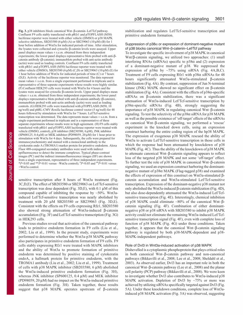
Wnt3a displayed a time-dependent accumulation of β-catenin (Fig.3A,B). Consistently, a strong β-catenin accumulation was observedafter 2 hours of Wnt3a treatment, with an ~8- to 12-fold increasein free β-catenin levels after 4 hours of Wnt3a treatment (Fig. 3A,B).Interestingly, Wnt3a-induced β-catenin accumulation was markedlyattenuated by SB203580 (6 μM) or SB239063 (10 μM) (Fig. 3A,B),suggesting a probable role for p38 MAPK pathway in canonicalWnt–β-catenin signaling. The time delay in the ability of SB203580-treated cells to attenuate β-catenin accumulation in response to
Journal of Cell Science 121 (21)
Wnt3a at the 1 hour time point may reflect the presence of distinct,p38-dependent and independent pathways, or incomplete p38MAPK inactivation (Fig. 3A).
We next tested for the effects of inhibition of p38 MAPK activityon Wnt3a-stimulated Lef/Tcf-sensitive transcription in Rfz1 cells(Fig. 3C,D,E). Consistent with the effects on β-cateninaccumulation, SB203580 or SB239063 dramatically attenuated theLef/Tcf-sensitive transcription (Fig. 3C,D,E). Treatment withSB203580 (6 μM) resulted in a more than 75% decrease in Lef/Tcf-
Fig. 3. See next page for legend.
Jour
nal o
f Cel
l Sci
ence
3601p38 regulates Wnt–β-catenin signaling
sensitive transcription after 8 hours of Wnt3a treatment (Fig.3C,D,E). The effect of SB203580 or SB239063 on Lef/Tcf-sensitivetranscription was dose dependent (Fig. 3D,E), with 0.1 μM of thiscompound capable of inhibiting the response by ~50%. Wnt3a-induced Lef/Tcf-sensitive transcription was nearly abolished bytreatment with 20 μM SB203580 or SB239063 (Fig. 3D,E).Consistent with the effects on F9 cells expressing Rfz1, SB203580also showed strong attenuation of Wnt3a-induced β-cateninaccumulation (Fig. 3F) and Lef/Tcf-sensitive transcription (Fig. 3G)in HEK293 cells.
Previous studies reveal that activation of the canonical pathwayleads to primitive endoderm formation in F9 cells (Liu et al.,2002; Liu et al., 1999). In the present study, experiments wereperformed to determine whether the Wnt3a-p38 MAPK pathwayalso participates in primitive endoderm formation of F9 cells. F9cells stably expressing Rfz1 were treated with MAPK inhibitorsand the ability of Wnt3a to promote formation of primitiveendoderm was determined by positive staining of cytokeratinendoA, a hallmark protein for primitive endoderm, with theTROMA1 antibody (Liu et al., 2002; Liu et al., 1999). Treatmentof cells with p38 MAPK inhibitor (SB203580, 6 μM) abolishedthe Wnt3a-induced primitive endoderm formation (Fig. 3H),whereas JNK inhibitor (SP600125, 0.4 μM) and MEK inhibitor(PD98059, 20 μM) had no impact on the Wnt3a-induced primitiveendoderm formation (Fig. 3H). Taken together, these resultssuggest that p38 MAPK operates upstream of β-catenin
stabilization and regulates Lef/Tcf-sensitive transcription andprimitive endoderm formation.
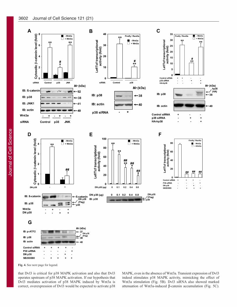
Suppression of p38α or expression of dominant-negative mutantof p38 blocks canonical Wnt–β-catenin–Lef/Tcf pathwayTo investigate the specific involvement of p38 MAPK in canonicalWnt/β-catenin signaling, we utilized two approaches: (1) smallinterfering RNAs (siRNAs) specific to p38α and (2) expressionof a dominant-negative mutant of p38. We suppressed theexpression of p38α by ~75% using siRNA (Fig. 4A,B,C).Treatment of F9 cells expressing Rfz1 with p38α siRNAs for 48hours significantly attenuated Wnt3a-stimulated β-cateninstabilization (Fig. 4A). By contrast, suppression of Jun N-terminalkinase (JNK) MAPK showed no significant effect on β-cateninstabilization (Fig. 4A). Consistent with the effects of p38α-specificsiRNAs on β-catenin stabilization, there was a significantattenuation of Wnt3a-induced Lef/Tcf-sensitive transcription byp38α-specific siRNAs (Fig. 4B), strongly suggesting theimportance of p38 MAPK in regulating canonical Wnt–β-cateninsignaling. To test the selectivity of the p38α siRNA for p38 MAPKas well as the possible existence of ‘off-target’ effects of the siRNAson canonical Wnt–β-catenin signaling, we performed a ‘rescue’experiment in the knocked down cells, using an expressionconstruct harboring the entire coding region of the hp38 MAPK.The expression of exogenous p38 MAPK rescued the ability ofWnt3a to activate Lef/Tcf-sensitive transcription in those cells inwhich the response had been attenuated by knockdown of p38MAPK (Fig. 4C). Thus the ability of the knockdown of p38 MAPKto attenuate canonical Wnt–β-catenin signaling appears to reflectloss of the targeted p38 MAPK and not some ‘off-target’ effect.To further test the role of p38 MAPK in canonical Wnt–β-cateninsignaling, we used an expression construct harboring the dominant-negative mutant of p38α MAPK (Flag-tagged p38) and examinedthe effects of expression of this construct on Wnt3a-stimulated β-catenin accumulation and Wnt3a-stimulated Lef/Tcf-sensitivetranscription. Expression of the dominant-negative p38 mutant notonly abolished the Wnt3a-induced β-catenin stabilization (Fig. 4D),but also dose-dependently attenuated the Wnt3a-induced Lef/Tcf-sensitive transcription (Fig. 4E). Interestingly, chemical inhibitionof p38 MAPK could eliminate ~80% of the canonical Wnt–β-catenin signaling (Fig. 4F). Combination of either dominant-negative p38 or p38 siRNA with SB203580 to inhibit p38 MAPKactivity could not eliminate the remaining Wnt3a-induced Lef/Tcf-sensitive transcription signal (Fig. 4F), even with complete loss offunction of p38 MAPK (Fig. 4G) under those conditions. Takentogether, it appears that the canonical Wnt–β-catenin signalingpathway is regulated by both p38-MAPK-dependent and p38-MAPK-independent pathways.
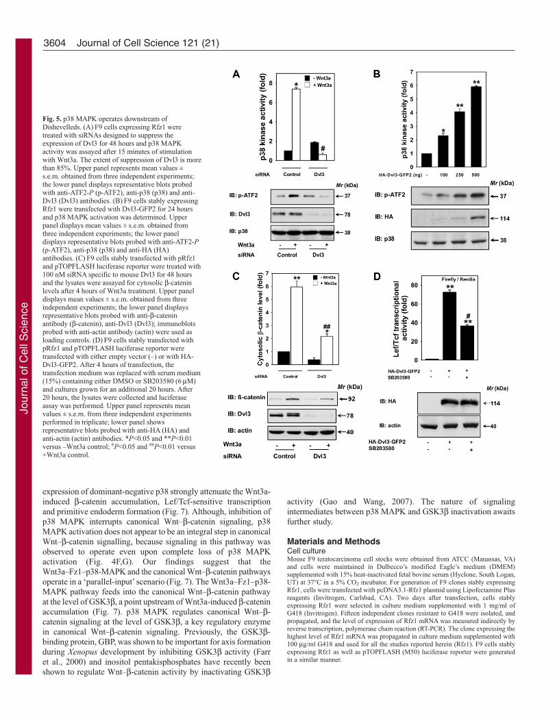
Role of Dvl3 in Wnt3a-induced activation of p38 MAPKDishevelled is a cytoplasmic phosphoprotein that plays critical rolesin both canonical Wnt–β-catenin pathway and non-canonicalpathways (Bikkavilli et al., 2008; Lee et al., 2008; Sheldahl et al.,2003). As observed earlier, Dvl3 has an important role in both thecanonical Wnt–β-catenin pathway (Lee et al., 2008) and the planarcell polarity (PCP) pathway (Bikkavilli et al., 2008). We were keento investigate whether Dvl3 also contributes to Wnt3a-induced p38MAPK activation. Depletion of Dvl3 by ~75% or more wasachieved by utilizing siRNAs specifically targeted against Dvl3 (Fig.5A). Under these knockdown conditions, complete loss of Wnt3a-induced p38 MAPK activation (Fig. 5A) was observed, suggesting
Fig. 3. p38 inhibitors block canonical Wnt–β-catenin–Lef/Tcf pathway.Confluent F9 cells stably transfected with pRfz1 and pTOPFLASH (M50)luciferase reporter were treated with either vehicle (DMSO) or p38 MAPKselective inhibitors, SB203580 (6μM) (A) or SB239063 (10μM) (B) for 1hour before addition of Wnt3a for indicated periods of time. After stimulation,the lysates were collected and cytosolic β-catenin levels were assayed. Upperpanel displays mean values ± s.e.m. obtained from three independentexperiments; the lower panel displays representative blots probed with anti-β-catenin antibody (β-catenin); immunoblots probed with anti-actin antibody(actin) were used as loading controls. Confluent F9 cells stably transfectedwith pRfz1 and pTOPFLASH (M50) luciferase reporter were treated witheither vehicle (DMSO), SB203580 (6μM) (C,D) or SB239063 (10μM) (E) for1 hour before addition of Wnt3a for indicated periods of time (C) or 7 hours(D,E). Activity of the luciferase reporter was monitored. The data representmean values ± s.e.m. from a single experiment performed in triplicate and isrepresentative of three separate experiments whose results were highly similar.(F) Confluent HEK293 cells were treated with Wnt3a for 4 hours and thelysates were assayed for cytosolic β-catenin levels. Upper panel displays meanvalues ± s.e.m. obtained from three independent experiments; the lower paneldisplays representative blots probed with anti-β-catenin antibody (β-catenin),immunoblots probed with anti-actin antibody (actin) were used as loadingcontrols. (G) HEK293 cells were transfected with pTOPFLASH (M50, 10ng/well) and phRL-CMV Renilla luciferase control vector (5 ng/well) for 48hours followed by stimulation with Wnt3a for 7 hours. Lef/Tcf-sensitivetranscription was determined. The data represents mean values ± s.e.m. from asingle experiment performed in triplicate and is a representative of threeseparate experiments whose results were in high agreement. (H) F9 cells stablytransfected with pRfz1 and pTOPFLASH luciferase reporter were treated withvehicle (DMSO, control), p38 inhibitor (SB230580, 6μM), JNK inhibitor(SP600125, 0.4μM) or MEK inhibitor (PD98059, 20μM) for 1 hour prior tostimulation with Wnt3a for 4 days. Subsequently, the cells were prepared forimmunocytochemistry and stained with a monoclonal antibody to thecytokeratin endo A (TROMA1) marker protein for primitive endoderm. AlexaFluor 488-conjugated secondary antibodies were used with indirectepifluorescence to detect the immune complexes. Typical phase-contrastimages (PC) and the indirect immunofluorescence images (IIF) are shownfrom a single experiment, representative of three independent experiments.*P<0.05 and **P<0.01 versus –Wnt3a control); #P<0.05 and ##P<0.01 versus+Wnt3a control.
Jour
nal o
f Cel
l Sci
ence
3602
that Dvl3 is critical for p38 MAPK activation and also that Dvl3operates upstream of p38 MAPK activation. If our hypothesis thatDvl3 mediates activation of p38 MAPK induced by Wnt3a iscorrect, overexpression of Dvl3 would be expected to activate p38
Journal of Cell Science 121 (21)
MAPK, even in the absence of Wnt3a. Transient expression of Dvl3indeed stimulates p38 MAPK activity, mimicking the effect ofWnt3a stimulation (Fig. 5B). Dvl3 siRNA also showed markedattenuation of Wnt3a-induced β-catenin accumulation (Fig. 5C).
Fig. 4. See next page for legend.
Jour
nal o
f Cel
l Sci
ence
3603p38 regulates Wnt–β-catenin signaling
Overexpression of Dvl3 induces Lef/Tcf-sensitive transcription (Leeet al., 2008). To confirm that p38 MAPK operates downstream ofDvl3 in Wnt–β-catenin signalling, we compared the ability ofoverexpressed HA-Dvl3-GFP2 to activate Lef/Tcf-sensitivetranscription in the absence versus the presence of the p38 MAPKinhibitor (SB203580) (Fig. 5D). Overexpression of HA-Dvl3-GFP2 induced a robust stimulation of Lef/Tcf-sensitive transcription(~70-fold), which was attenuated more than twofold in the presenceof SB203580 (Fig. 5D). Taken together, these observations suggestthat in the canonical Wnt–β-catenin signaling pathway, Dvl3 iscritical for Wnt3a-induced p38 MAPK activation and p38 MAPKoperates downstream of Dishevelleds.
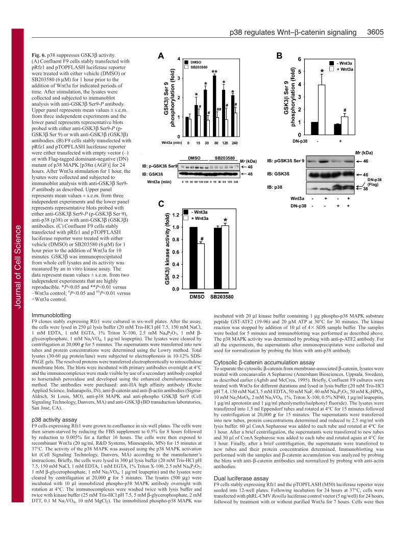
p38 MAPK suppresses GSK3β activityTo identify the likely targets of p38 MAPK in regulating canonicalWnt–β-catenin signaling, we explored the effects of SB203580 anddominant-negative p38 on GSK3β activity. GSK3β is the keyenzyme that regulates canonical Wnt–β-catenin signaling byphosphorylating β-catenin, ultimately leading to proteasome-mediateddegradation. We tested the possible regulation of GSK3β by p38MAPK experimentally by using two approaches: (1) probing Wnt3a-induced GSK3β Ser9 phosphorylation in the absence or presence ofSB203580. (2) In vitro GSK3β kinase assay in the absence or presenceof SB203580. GSK3β is highly active under basal conditions. Akt-mediated phosphorylation of GSK3β at Ser9 leads to GSK3βinactivation (Cross et al., 1995). Whether Ser9 phosphorylation isalso a major mechanism of GSK3β inactivation in Wnt–β-cateninsignaling is still under debate. However, earlier studies demonstratean increase in GSK3β Ser9 phosphorylation upon Wnt stimulation
(Cook et al., 1996; Yokoyama et al., 2007). Therefore, we tested theeffects of p38 MAPK inhibitor, SB203580, on Wnt3a-induced Ser9phosphorylation of GSK3β (Fig. 6A). Treatment of Rfz1 cells withWnt3a stimulated a sharp increase in GSK3β Ser9 phosphorylation,as revealed by immunoblotting with antibodies specific to GSK3βSer9-P (Fig. 6A). A time-dependent increase in GSK3β Ser9phosphorylation was observed, with a peak activity at ~1 hour afterWnt3a treatment (Fig. 6A). Interestingly, Wnt3a-induced Ser9phosphorylation was abolished by SB203580 (Fig. 6A). Similar tothe effects with SB203580, overexpression of dominant-negative p38also abolished Wnt3a-induced GSK3β Ser9 phosphorylation (Fig.6B). To validate our finding, we performed an in vitro GSK3β kinaseassay. Treatment of Rfz1 cells with Wnt3a suppressed the GSK3βkinase activity (Fig. 6C). Treatment of cells with p38 MAPKinhibitor, SB203580, prior to stimulation with Wnt3a abolished theability of Wnt3a to inhibit GSK3β kinase activity (Fig. 6C), similarto the effects of this inhibitor on β-catenin stabilization and Lef/Tcf-sensitive transcription. Taken together, these data suggest that p38MAPK regulates canonical Wnt–β-catenin signaling by inhibitingGSK3β activity.
DiscussionThe p38 MAPKs are a MAPK subfamily that is conserved fromyeast to mammals. They are activated in response to manyextracellular stimuli, including growth factors, cytokines andenvironmental stress (Martin-Blanco, 2000). Recently, the p38MAPK pathway has been shown to be important for mineralizationand development of osteoprogenitors (Caverzasio and Manen,2007), bone regeneration of mesenchymal stem cells (Chang et al.,2007) and in myogenesis during Xenopus development (Keren etal., 2005). In the present study, we demonstrate for the first timethat p38 MAPK is transiently activated upon Wnt3a stimulationand this activation is dependent on both G-protein and Dishevelleds(Fig. 7). The activation of Wnt3a/Fz1, which signal through G-proteins, leads to activation of the Wnt–β-catenin pathway(Katanaev et al., 2005; Liu et al., 2001; Liu et al., 1999; Malbon,2005). By utilizing siRNA, we show that Gαq and Gαs are obligatefor Wnt3a-induced p38 MAPK activation. By contrast, Gαo, whichis obligate for Wnt3a-induced JNK activation, was found to bedispensable for Wnt3a–Fz1–p38-MAPK signaling (Bikkavilli et al.,2008). Thus, there is a divergence between G-protein function inthe Wnt3a–Fz1–p38-MAPK pathway compared with that in theplanar cell polarity pathways.
Epistasis experiments reveal that Dishevelleds operatedownstream of G-proteins in the fly (Katanaev et al., 2005), as wellas in mammalian cells (Bikkavilli et al., 2008). Dishevelleds arecytosolic phosphoproteins that play crucial roles in canonicalWnt–β-catenin pathways (Lee et al., 2008) and also in the planarcell polarity pathway (Bikkavilli et al., 2008). We demonstrate thatDvl3 is obligate for Wnt3a-stimulated p38 MAPK activation.Overexpression of Dvl3 stimulates Lef/Tcf-sensitive transcription,mimicking the effect of Wnt3a (Lee et al., 2008). Furthermore, theDvl3-stimulated activation of Lef/Tcf-sensitive transcription wasfound to be sensitive to SB203580. This ability of SB203580 toattenuate the response to Dvl3, positions p38 MAPK downstreamof Dishevelleds in the Wnt3a-Fz1-p38 MAPK pathway. Thus Wnt3abinding to Fz1 activates Gαq and Dishevelled, which in turn leadsto p38 MAPK activation (Fig. 7).
The current study reveals a novel role for p38 MAPK incanonical Wnt–β-catenin signaling. The p38 MAPK specificinhibitors (SB203580 or SB239063), p38 siRNA, as well as
Fig. 4. Suppression of p38α by siRNA treatment or expression of dominant-negative mutant of p38 blocks canonical Wnt–β-catenin–Lef/Tcf pathway.(A) F9 cells stably transfected with pRfz1 and pTOPFLASH luciferasereporter were treated with 100 nM siRNA specific to mouse p38α or JNK for48 hours, and the lysates were assayed for cytosolic β-catenin levels. Upperpanel displays mean values ± s.e.m. obtained from three independentexperiments; the lower panel displays representative blots probed with anti-β-catenin antibody (β-catenin); immunoblots probed with anti-actin antibody(actin) were used as loading controls. (B) F9 cells stably transfected withpRfz1 and pTOPFLASH luciferase reporter were treated with 100 nM siRNAspecific to mouse p38α for 48 hours followed by stimulation with Wnt3a for 7hours. Lef/Tcf-sensitive transcription was determined. The data representmean values ± s.e.m. from a single experiment performed in triplicate and isrepresentative of three separate experiments whose results were in highagreement. (C) Rescue experiment performed by transfection of hp38 into F9cells stably transfected with pRfz1 and pTOPFLASH luciferase reporter inwhich p38 MAPK was knocked down by siRNA treatment. Lef/Tcf-sensitivetranscription was determined. The data represents mean values ± s.e.m. from asingle experiment performed in triplicate and is representative of threeseparate experiments whose results were in high agreement. (D<E) F9 cellsstably transfected with pRfz1 and pTOPFLASH luciferase reporter were eithertransfected with empty vector (–) or with indicated amounts of Flag-taggeddominant-negative mutant (DN) of p38 MAPK [p38α (AGF)] for 24 hoursand the lysates were assayed either for β-catenin stabilization after 4 hours ofWnt3a treatment (D) or luciferase reporter activity after 7 hours of Wnt3atreatment (E). Upper panel displays mean values ± s.e.m. obtained from threeindependent experiments; the lower panel displays representative blots probedwith anti-β-catenin antibody (β-catenin), anti-p38 antibody (p38) and theimmunoblots with anti-actin antibody (actin) were used as loading controls.(F,G) F9 cells stably transfected with pRfz1 and pTOPFLASH luciferasereporter were treated with either 100 nM siRNA specific to mouse p38α ordominant-negative mutant (DN) of p38 MAPK for 48 hours. The cells werethen treated with SB203580 (6 μM) for 1 hour followed by stimulation withWnt3a for 7 hours. Lef/Tcf-sensitive transcription (F) and p38 MAPKactivation (G) was determined as described in Materials and Methods.*P<0.05 and **P<0.01 versus –Wnt3a control; #P<0.05 and ##P<0.01 versus+Wnt3a control.
Jour
nal o
f Cel
l Sci
ence
3604
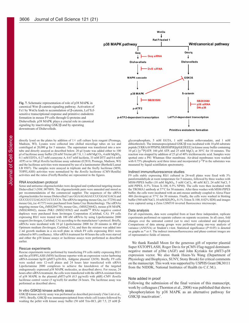
expression of dominant-negative p38 strongly attenuate the Wnt3a-induced β-catenin accumulation, Lef/Tcf-sensitive transcriptionand primitive endoderm formation (Fig. 7). Although, inhibition ofp38 MAPK interrupts canonical Wnt–β-catenin signaling, p38MAPK activation does not appear to be an integral step in canonicalWnt–β-catenin signalling, because signaling in this pathway wasobserved to operate even upon complete loss of p38 MAPKactivation (Fig. 4F,G). Our findings suggest that theWnt3a–Fz1–p38-MAPK and the canonical Wnt–β-catenin pathwaysoperate in a ‘parallel-input’ scenario (Fig. 7). The Wnt3a–Fz1–p38-MAPK pathway feeds into the canonical Wnt–β-catenin pathwayat the level of GSK3β, a point upstream of Wnt3a-induced β-cateninaccumulation (Fig. 7). p38 MAPK regulates canonical Wnt–β-catenin signaling at the level of GSK3β, a key regulatory enzymein canonical Wnt–β-catenin signaling. Previously, the GSK3β-binding protein, GBP, was shown to be important for axis formationduring Xenopus development by inhibiting GSK3β activity (Farret al., 2000) and inositol pentakisphosphates have recently beenshown to regulate Wnt–β-catenin activity by inactivating GSK3β
Journal of Cell Science 121 (21)
activity (Gao and Wang, 2007). The nature of signalingintermediates between p38 MAPK and GSK3β inactivation awaitsfurther study.
Materials and MethodsCell cultureMouse F9 teratocarcinoma cell stocks were obtained from ATCC (Manassas, VA)and cells were maintained in Dulbecco’s modified Eagle’s medium (DMEM)supplemented with 15% heat-inactivated fetal bovine serum (Hyclone, South Logan,UT) at 37°C in a 5% CO2 incubator. For generation of F9 clones stably expressingRfz1, cells were transfected with pcDNA3.1-Rfz1 plasmid using Lipofectamine Plusreagents (Invitrogen, Carlsbad, CA). Two days after transfection, cells stablyexpressing Rfz1 were selected in culture medium supplemented with 1 mg/ml ofG418 (Invitrogen). Fifteen independent clones resistant to G418 were isolated, andpropagated, and the level of expression of Rfz1 mRNA was measured indirectly byreverse transcription, polymerase chain reaction (RT-PCR). The clone expressing thehighest level of Rfz1 mRNA was propagated in culture medium supplemented with100 μg/ml G418 and used for all the studies reported herein (Rfz1). F9 cells stablyexpressing Rfz1 as well as pTOPFLASH (M50) luciferase reporter were generatedin a similar manner.
Fig. 5. p38 MAPK operates downstream ofDishevelleds. (A) F9 cells expressing Rfz1 weretreated with siRNAs designed to suppress theexpression of Dvl3 for 48 hours and p38 MAPKactivity was assayed after 15 minutes of stimulationwith Wnt3a. The extent of suppression of Dvl3 is morethan 85%. Upper panel represents mean values ±s.e.m. obtained from three independent experiments;the lower panel displays representative blots probedwith anti-ATF2-P (p-ATF2), anti-p38 (p38) and anti-Dvl3 (Dvl3) antibodies. (B) F9 cells stably expressingRfz1 were transfected with Dvl3-GFP2 for 24 hoursand p38 MAPK activation was determined. Upperpanel displays mean values ± s.e.m. obtained fromthree independent experiments; the lower paneldisplays representative blots probed with anti-ATF2-P(p-ATF2), anti-p38 (p38) and anti-HA (HA)antibodies. (C) F9 cells stably transfected with pRfz1and pTOPFLASH luciferase reporter were treated with100 nM siRNA specific to mouse Dvl3 for 48 hoursand the lysates were assayed for cytosolic β-cateninlevels after 4 hours of Wnt3a treatment. Upper paneldisplays mean values ± s.e.m. obtained from threeindependent experiments; the lower panel displaysrepresentative blots probed with anti-β-cateninantibody (β-catenin), anti-Dvl3 (Dvl3); immunoblotsprobed with anti-actin antibody (actin) were used asloading controls. (D) F9 cells stably transfected withpRfz1 and pTOPFLASH luciferase reporter weretransfected with either empty vector (–) or with HA-Dvl3-GFP2. After 4 hours of transfection, thetransfection medium was replaced with serum medium(15%) containing either DMSO or SB203580 (6 μM)and cultures grown for an additional 20 hours. After20 hours, the lysates were collected and luciferaseassay was performed. Upper panel represents meanvalues ± s.e.m. from three independent experimentsperformed in triplicate; lower panel showsrepresentative blots probed with anti-HA (HA) andanti-actin (actin) antibodies. *P<0.05 and **P<0.01versus –Wnt3a control; #P<0.05 and ##P<0.01 versus+Wnt3a control.
Jour
nal o
f Cel
l Sci
ence
3605p38 regulates Wnt–β-catenin signaling
ImmunoblottingF9 clones stably expressing Rfz1 were cultured in six-well plates. After the assay,the cells were lysed in 250 μl lysis buffer (20 mM Tris-HCl pH 7.5, 150 mM NaCl,1 mM EDTA, 1 mM EGTA, 1% Triton X-100, 2.5 mM Na4P2O7, 1 mM β-glycerophosphate, 1 mM Na3VO4, 1 μg/ml leupeptin). The lysates were cleared bycentrifugation at 20,000 g for 5 minutes. The supernatants were transferred into newtubes and protein concentrations were determined using the Lowry method. Totallysates (30-60 μg protein/lane) were subjected to electrophoresis in 10-12% SDS-PAGE gels. The resolved proteins were transferred electrophoretically to nitrocellulosemembrane blots. The blots were incubated with primary antibodies overnight at 4°Cand the immunocomplexes were made visible by use of a secondary antibody coupledto horseradish peroxidase and developed using the enhanced chemiluminescencemethod. The antibodies were purchased: anti-HA high affinity antibody (RocheApplied Science, Indianapolis, IN), anti-β-catenin and anti-β-actin antibodies (Sigma-Aldrich, St Louis, MO), anti-p38 MAPK and anti-phospho GSK3β Ser9 (CellSignaling Technology, Danvers, MA) and anti-GSK3β (BD transduction laboratories,San Jose, CA).
p38 activity assayF9 cells expressing Rfz1 were grown to confluence in six-well plates. The cells werethen serum-starved by reducing the FBS supplement to 0.5% for 8 hours followedby reduction to 0.005% for a further 16 hours. The cells were then exposed torecombinant Wnt3a (20 ng/ml, R&D Systems, Minneapolis, MN) for 15 minutes at37°C. The activity of the p38 MAPK was assayed using the p38 MAPK activationkit (Cell Signaling Technology, Danvers, MA) according to the manufacturer’sinstructions. Briefly, the cells were lysed in 300 μl lysis buffer (20 mM Tris-HCl pH7.5, 150 mM NaCl, 1 mM EDTA, 1 mM EGTA, 1% Triton X-100, 2.5 mM Na4P2O7,1 mM β-glycerophosphate, 1 mM Na3VO4, 1 μg/ml leupeptin) and the lysates werecleared by centrifugation at 20,000 g for 5 minutes. The lysates (500 μg) wereincubated with 10 μl immobilized phospho-p38 MAPK antibody overnight withrotation at 4°C. The immunocomplexes were washed twice with lysis buffer andtwice with kinase buffer (25 mM Tris-HCl pH 7.5, 5 mM β-glycerophosphate, 2 mMDTT, 0.1 M Na3VO4, 10 mM MgCl2). The immobilized phospho-p38 MAPK was
incubated with 20 μl kinase buffer containing 1 μg phospho-p38 MAPK substratepeptide GST-ATF2 (19-96) and 20 μM ATP at 30°C for 30 minutes. The kinasereaction was stopped by addition of 10 μl of 4� SDS sample buffer. The sampleswere boiled for 5 minutes and immunoblotting was performed as described above.The p38 MAPK activity was determined by probing with anti-p-ATF2 antibody. Forall the experiments, the supernatants after immunoprecipitates were collected andused for normalization by probing the blots with anti-p38 antibody.
Cytosolic β-catenin accumulation assayTo separate the cytosolic β-catenin from membrane-associated β-catenin, lysates weretreated with concanavalin A Sepharose (Amersham Biosciences, Uppsala, Sweden),as described earlier (Aghib and McCrea, 1995). Briefly, Confluent F9 cultures weretreated with Wnt3a for different durations and lysed in lysis buffer (20 mM Tris-HClpH 7.4, 150 mM NaCl, 5 mM EDTA, 50 mM NaF, 40 mM Na4P2O7, 50 mM K2HPO4,10 mM Na2MoO4, 2 mM Na3VO4, 1%, Triton X-100, 0.5% NP40, 1 μg/ml leupeptin,1 μg/ml aprotonin and 1 μg/ml phenlymethylsulphonyl fluoride). The lysates weretransferred into 1.5 ml Eppendorf tubes and rotated at 4°C for 15 minutes followedby centrifugation at 20,000 g for 15 minutes. The supernatants were transferredinto new tubes, protein concentrations determined and reduced to 2.5 mg/ml withlysis buffer. 60 μl ConA Sepharose was added to each tube and rotated at 4°C for1 hour. After a brief centrifugation, the supernatants were transferred to new tubesand 30 μl of ConA Sepharose was added to each tube and rotated again at 4°C for1 hour. Finally, after a brief centrifugation, the supernatants were transferred tonew tubes and their protein concentration determined. Immunoblotting wasperformed with the samples and β-catenin accumulation was analyzed by probingthe blots with anti-β-catenin antibodies and normalized by probing with anti-actinantibodies.
Dual luciferase assayF9 cells stably expressing Rfz1 and the pTOPFLASH (M50) luciferase reporter wereseeded into 12-well plates. Following incubation for 24 hours at 37°C, cells weretransfected with phRL-CMV Renilla luciferase control vector (5 ng/well) for 24 hours,followed by treatment with or without purified Wnt3a for 7 hours. Cells were then
Fig. 6. p38 suppresses GSK3β activity.(A) Confluent F9 cells stably transfected withpRfz1 and pTOPFLASH luciferase reporterwere treated with either vehicle (DMSO) orSB203580 (6 μM) for 1 hour prior to theaddition of Wnt3a for indicated periods oftime. After stimulation, the lysates werecollected and subjected to immunoblotanalysis with anti-GSK3β Ser9-P antibody.Upper panel represents mean values ± s.e.m.from three independent experiments and thelower panel represents representative blotsprobed with either anti-GSK3β Ser9-P (p-GSK3β Ser 9) or with anti-GSK3β (GSK3β)antibodies. (B) F9 cells stably transfected withpRfz1 and pTOPFLASH luciferase reporterwere either transfected with empty vector (–)or with Flag-tagged dominant-negative (DN)mutant of p38 MAPK [p38α (AGF)] for 24hours. After Wnt3a stimulation for 1 hour, thelysates were collected and subjected toimmunoblot analysis with anti-GSK3β Ser9-P antibody as described. Upper panelrepresents mean values ± s.e.m. from threeindependent experiments and the lower panelrepresents representative blots probed witheither anti-GSK3β Ser9-P (p-GSK3β Ser 9),anti-p38 (p38) or with anti-GSK3β (GSK3β)antibodies. (C) Confluent F9 cells stablytransfected with pRfz1 and pTOPFLASHluciferase reporter were treated with eithervehicle (DMSO) or SB203580 (6 μM) for 1hour prior to the addition of Wnt3a for 10minutes. GSK3β was immunoprecipitatedfrom whole cell lysates and its activity wasmeasured by an in vitro kinase assay. Thedata represent mean values ± s.e.m. from twoindependent experiments that are highlyreproducible. *P<0.05 and **P<0.01 versus–Wnt3a control; #P<0.05 and ##P<0.01 versus+Wnt3a control.
Jour
nal o
f Cel
l Sci
ence
3606
directly lysed on the plates by addition of 1� cell culture lysis reagent (Promega,Madison, WI). Lysates were collected into chilled microfuge tubes on ice andcentrifuged at 20,000 g for 5 minutes. The supernatant was transferred into a newtube and directly assayed as described below. 20 μl lysate was added either to 100μl of luciferase assay buffer (20 mM Tricine pH 7.8, 1.1 mM MgCO3, 4 mM MgSO4,0.1 mM EDTA, 0.27 mM coenzyme A, 0.67 mM luciferin, 33 mM DTT and 0.6 mMATP) or to 100 μl Renilla luciferase assay substrate (E2810, Promega, Madison, WI)and the luciferase activities were measured by use of a luminometer (Berthold LumatLB 9507). The samples were assayed in triplicate and the firefly luciferase (M50,TOPFLASH) activities were normalized by the Renilla luciferase (CMV-Renilla)activities and the ratios (Firefly/Renilla) are represented in the figures.
RNA knockdown protocolSense and antisense oligonucleotides were designed and synthesized targeting mouseDishevelled 3 (NM_007889). The oligonucleotide pairs were annealed and stored asper recommendations of the commercial supplier. The sequences of the siRNAoligonucleotides for mouse Dishevelled 3 are GGAAGAGAUCUCGGACGACtt andGUCGUCCGAGAUCUCUUCCtt. The siRNAs targeting mouse Gαo (sc-37256) andmouse Gαs (sc-41757) were purchased from Santa Cruz Biotechnology. The siRNAstargeting mouse Gαq (MSS204770), mouse Gα11 (MSS236642), mouse p38 MAPK(MSS240942), mouse JNK1 (MSS218562) and stealthTM RNAi negative controlduplexes were purchased from Invitrogen Corporation (Carlsbad, CA). F9 cellsexpressing Rfz1 were treated with 100 nM siRNAs by using Lipofectamine 2000reagent (Invitrogen, Carlsbad, CA) according to the manufacturer’s protocol. Briefly,siRNAs were incubated with 10 μl Lipofectamine 2000 for 20 minutes in 400 μlOptimem medium (Invitrogen, Carlsbad, CA), and then the mixture was added into2 ml growth medium in a six-well plate in which F9 cells expressing Rfz1 werecultured to 80% confluency. After siRNA treatment for 48 hours the cells were starvedand either the p38 kinase assays or luciferase assays were performed as describedearlier.
Rescue experimentsRescue experiments were performed by transfecting F9 cells stably expressing Rfz1and the pTOPFLASH (M50) luciferase reporter with an expression vector harboringsiRNA-resistant hp38 (pMT3-p38-HA, Addgene plasmid 12658). Briefly, F9 cellswere seeded onto 12-well plates and 24 hours later transfected with siRNALipofectamine 2000 complexes to achieve the knockdown of the targetedendogenously expressed p38 MAPK molecules, as described above. For rescue, 24hours after siRNA treatment, the cells were transfected with the siRNA-resistant formof p38 MAPK in the plasmid pMT3-p38 (0.5 μg/well) with phRL-CMV Renillaluciferase control vector (5 ng/well) for another 24 hours. The luciferase assay wasperformed as described above.
In vitro GSK3β kinase activity assayGSK3β kinase activity assay was performed as described previously (Van Lint et al.,1993). Briefly, GSK3β was immunoprecipitated from whole cell lysates followed bywashing the pellet with kinase assay buffer (50 mM Tris-HCl, pH 7.5, 25 mM β-
Journal of Cell Science 121 (21)
glycerophosphate, 5 mM EGTA, 1 mM sodium orthovanadate, and 1 mMdithiothreitol). The immunoprecipitated GSK3β was incubated with 10 μM substratepeptide [YRRAAVPPSPSLSRHSSPHQ(pS)EDEEE] in kinase assay buffer containing10 μCi [γ-32P]ATP, 160 μM ATP, and 25 mM MgCl2 at 30°C for 10 minutes. Thereaction was stopped by addition of 25 μl of 40% trichloroacetic acid. Samples werespotted onto a P81 Whatman filter membrane. Air-dried membranes were washedwith 0.75% phosphoric acid three times and incorporated γ-32P in the substrates wasmeasured by liquid scintillation spectrometry.
Indirect immunofluorescence studiesF9 cells stably expressing Rfz1 cultured in 24-well plates were fixed with 3%paraformaldehyde at room temperature for 5 minutes, followed by three washes withMSM-PIPES buffer (18 mM MgSO4, 5 mM CaCl2, 40 mM KCl, 24 mM NaCl, 5mM PIPES, 0.5% Triton X-100, 0.5% NP40). The cells were then incubated withthe TROMA1 antibody at 37°C for 30 minutes. After three washes with MSM-PIPESbuffer, the cells were incubated with an anti-mouse antibody coupled to Alexa Fluor488 (Invitrogen) at 37°C for 30 minutes. Finally, the cells were washed in blottingbuffer (560 mM NaCl, 10 mM KH2PO4, 0.1% Triton X-100, 0.02% SDS) and imageswere captured using a Zeiss LSM510 inverted fluorescence microscope.
Data analysisFor all experiments, data were compiled from at least three independent, replicateexperiments performed on separate cultures on separate occasions. In all cases, foldchanges over the untreated control (set to one) were calculated and displayed.Comparisons of data among groups were performed using one-way analysis ofvariance (ANOVA) or Student’s t-test. Statistical significance (P<0.05) is denotedon graphs as * or #. The indirect immunofluorescence and phase-contrast images areof representative fields of interest.
We thank Randall Moon for the generous gift of reporter plasmidSuper 8XTOPFLASH, Roger Davis for pCMV-Flag-tagged dominant-negative mutant of p38α (AGF) and John Kyriakis for pMT3-p38expression vector. We also thank Hsien-Yu Wang (Department ofPhysiology and Biophysics, SUNY, Stony Brook) for critical commentson the manuscript. This work was supported by USPHS Grant DK30111from the NIDDK, National Institutes of Health (to C.C.M.).
Note added in proofFollowing the submission of the final version of this manuscript,work by colleagues (Thornton et al., 2008) was published that shows‘phosphorylation by p38 MAPK as an alternative pathway forGSK3� inactivation’.
Fig. 7. Schematic representation of role of p38 MAPK incanonical Wnt–β-catenin signaling pathway. Activation ofFz1 by Wnt3a leads to accumulation of β-catenin, Lef/Tcf-sensitive transcriptional response and primitive endodermformation in mouse F9 cells through G-proteins andDishevelleds. p38 MAPK plays a crucial role in canonicalsignaling by inactivating GSK3β and by operatingdownstream of Dishevelleds.
Jour
nal o
f Cel
l Sci
ence
3607p38 regulates Wnt–β-catenin signaling
ReferencesAghib, D. F. and McCrea, P. D. (1995). The E-cadherin complex contains the src substrate
p120. Exp. Cell Res. 218, 359-369.Behrens, J., von Kries, J. P., Kuhl, M., Bruhn, L., Wedlich, D., Grosschedl, R. and
Birchmeier, W. (1996). Functional interaction of beta-catenin with the transcription factorLEF-1. Nature 382, 638-642.
Bhanot, P., Brink, M., Samos, C. H., Hsieh, J. C., Wang, Y., Macke, J. P., Andrew, D.,Nathans, J. and Nusse, R. (1996). A new member of the frizzled family from Drosophilafunctions as a Wingless receptor. Nature 382, 225-230.
Bikkavilli, R. K., Feigin, M. E. and Malbon, C. C. (2008). G{alpha}o mediates WNT-JNK signaling through Dishevelled 1 and 3, RhoA family members, and MEKK 1 and4 in mammalian cells. J. Cell Sci. 121, 234-245.
Castellone, M. D., Teramoto, H., Williams, B. O., Druey, K. M. and Gutkind, J. S.(2005). Prostaglandin E2 promotes colon cancer cell growth through a Gs-axin-beta-catenin signaling axis. Science 310, 1504-1510.
Caverzasio, J. and Manen, D. (2007). Essential role of Wnt3a-mediated activation ofmitogen-activated protein kinase p38 for the stimulation of alkaline phosphatase activityand matrix mineralization in C3H10T1/2 mesenchymal cells. Endocrinology 148, 5323-5330.
Chang, J., Sonoyama, W., Wang, Z., Jin, Q., Zhang, C., Krebsbach, P. H., Giannobile,W., Shi, S. and Wang, C. Y. (2007). Noncanonical Wnt-4 signaling enhances boneregeneration of mesenchymal stem cells in craniofacial defects through activation ofp38 MAPK. J. Biol. Chem. 282, 30938-30948.
Cook, D., Fry, M. J., Hughes, K., Sumathipala, R., Woodgett, J. R. and Dale, T. C.(1996). Wingless inactivates glycogen synthase kinase-3 via an intracellular signallingpathway which involves a protein kinase C. EMBO J. 15, 4526-4536.
Cross, D. A., Alessi, D. R., Cohen, P., Andjelkovich, M. and Hemmings, B. A. (1995).Inhibition of glycogen synthase kinase-3 by insulin mediated by protein kinase B. Nature378, 785-789.
Cuenda, A., Rouse, J., Doza, Y. N., Meier, R., Cohen, P., Gallagher, T. F., Young, P.R. and Lee, J. C. (1995). SB 203580 is a specific inhibitor of a MAP kinase homologuewhich is stimulated by cellular stresses and interleukin-1. FEBS Lett. 364, 229-233.
Farr, G. H., 3rd, Ferkey, D. M., Yost, C., Pierce, S. B., Weaver, C. and Kimelman, D.(2000). Interaction among GSK-3, GBP, axin, and APC in Xenopus axis specification.J. Cell Biol. 148, 691-702.
Gao, Y. and Wang, H. Y. (2007). Inositol pentakisphosphate mediates Wnt/beta-cateninsignaling. J. Biol. Chem. 282, 26490-26502.
Gao, Z. H., Seeling, J. M., Hill, V., Yochum, A. and Virshup, D. M. (2002). Caseinkinase I phosphorylates and destabilizes the beta-catenin degradation complex. Proc.Natl. Acad. Sci. USA 99, 1182-1187.
Hildesheim, J., Salvador, J. M., Hollander, M. C. and Fornace, A. J., Jr (2005). Caseinkinase 2- and protein kinase A-regulated adenomatous polyposis coli and beta-catenincellular localization is dependent on p38 MAPK. J. Biol. Chem. 280, 17221-17226.
Katanaev, V. L., Ponzielli, R., Semeriva, M. and Tomlinson, A. (2005). Trimeric Gprotein-dependent frizzled signaling in Drosophila. Cell 120, 111-122.
Keren, A., Bengal, E. and Frank, D. (2005). p38 MAP kinase regulates the expressionof XMyf5 and affects distinct myogenic programs during Xenopus development. Dev.Biol. 288, 73-86.
Lee, Y. N., Gao, Y. and Wang, H. Y. (2008). Differential mediation of the Wnt canonicalpathway by mammalian Dishevelleds-1, -2, and -3. Cell Signal. 20, 443-452.
Liu, T., Liu, X., Wang, H., Moon, R. T. and Malbon, C. C. (1999). Activation of ratfrizzled-1 promotes Wnt signaling and differentiation of mouse F9 teratocarcinoma cellsvia pathways that require Galpha(q) and Galpha(o) function. J. Biol. Chem. 274, 33539-33544.
Liu, T., DeCostanzo, A. J., Liu, X., Wang, H., Hallagan, S., Moon, R. T. and Malbon,C. C. (2001). G protein signaling from activated rat frizzled-1 to the beta-catenin-Lef-Tcf pathway. Science 292, 1718-1722.
Liu, T., Lee, Y. N., Malbon, C. C. and Wang, H. Y. (2002). Activation of the beta-catenin/Lef-Tcf pathway is obligate for formation of primitive endoderm by mouse F9totipotent teratocarcinoma cells in response to retinoic acid. J. Biol. Chem. 277, 30887-30891.
Logan, C. Y. and Nusse, R. (2004). The Wnt signaling pathway in development and disease.Annu. Rev. Cell Dev. Biol. 20, 781-810.
Malbon, C. C. (2005). G proteins in development. Nat. Rev. Mol. Cell. Biol. 6, 689-701.Martin-Blanco, E. (2000). p38 MAPK signalling cascades: ancient roles and new
functions. BioEssays 22, 637-645.Molenaar, M., van de Wetering, M., Oosterwegel, M., Peterson-Maduro, J., Godsave,
S., Korinek, V., Roose, J., Destree, O. and Clevers, H. (1996). XTcf-3 transcriptionfactor mediates beta-catenin-induced axis formation in Xenopus embryos. Cell 86, 391-399.
Moon, R. T., Bowerman, B., Boutros, M. and Perrimon, N. (2002). The promise andperils of Wnt signaling through beta-catenin. Science 296, 1644-1646.
Moon, R. T., Kohn, A. D., De Ferrari, G. V. and Kaykas, A. (2004). WNT and beta-catenin signalling: diseases and therapies. Nat. Rev. Genet. 5, 691-701.
Polakis, P. (2000). Wnt signaling and cancer. Genes Dev. 14, 1837-1851.Sheldahl, L. C., Slusarski, D. C., Pandur, P., Miller, J. R., Kuhl, M. and Moon, R. T.
(2003). Dishevelled activates Ca2+ flux, PKC, and CamKII in vertebrate embryos. J.Cell Biol. 161, 769-777.
Thornton, T. M., Pedraza-Alva, G., Deng, B., Wood, C. D., Aronshtam, A., Clements,J. L., Sabio, G., Davis, R. J., Matthews, D. E., Doble, B. et al. (2008). Phosphorylationby p38 MAPK as an alternative pathway for GSK3beta inactivation. Science 320, 667-670.
Van Lint, J., Khandelwal, R. L., Merlevede, W. and Vandenheede, J. R. (1993). Aspecific immunoprecipitation assay for the protein kinase FA/glycogen synthase kinase3. Anal. Biochem. 208, 132-137.
Yokoyama, N., Yin, D. and Malbon, C. C. (2007). Abundance, complexation, andtrafficking of Wnt/beta-catenin signaling elements in response to Wnt3a. J. Mol. Signal.2, 11.
Yun, M. S., Kim, S. E., Jeon, S. H., Lee, J. S. and Choi, K. Y. (2005). Both ERK andWnt/beta-catenin pathways are involved in Wnt3a-induced proliferation. J. Cell Sci. 118,313-322.
Jour
nal o
f Cel
l Sci
ence