RESEARCH Open Access p38α MAPK-mediated induction and ... · activation of p38α MAPK signaling...
12
RESEARCH Open Access p38α MAPK-mediated induction and interaction of FOXO3a and p53 contribute to the inhibited- growth and induced-apoptosis of human lung adenocarcinoma cells by berberine Fang Zheng 1 , Qin Tang 1 , JingJing Wu 1 , ShunYu Zhao 1 , ZhanYang Liang 1 , Liuning Li 2 , WanYin Wu 2 and Swei Hann 1* Abstract Background: Berberine (BBR), a component from traditional Chinese medicine, has been shown to possess anti-tumor activity against a wide spectrum of cancer cells including human lung cancer, but the detailed mechanism underlining this has not been well elucidated. Methods: In this study, the effect of berberine on cell growth and apoptosis were assessed by MTT, flow cytometry and Hoechst 33258 staining assays. The phosphorylation of p38 MAPK and ERK1/2, and expressions of p38 MAPK isoforms α and β, total ERK1/2, p53, FOXO3a and p21 protein were evaluated by Western Blot analysis. Silencing of p38 MAPK isoform α and β, p53, FOXO3a and p21 were performed by siRNA methods. Exogenous expression of FOXO3a was carried out by electroporated transfection assays. Results: We showed that BBR significantly inhibited growth and induced cell cycle arrest of non small cell lung cancer (NSCLC) cells in the G0/G1 phase in a dose-dependent manner. Furthermore, we found that BBR increased phosphorylation of p38 MAPK and ERK1/2 in a time-dependent and induced protein expression of tumor suppressor p53 and transcription factor FOXO3a in a dose-dependent fashion. The specific inhibitor of p38 MAPK (SB203580), and silencing of p38α MAPK by small interfering RNAs (siRNAs), but not ERK1/2 inhibitor (PD98059) blocked the stimulatory effects of BBR on protein expression of p53 and FOXO3a. Interestingly, inhibition of p53 using one specific inhibitor (Pifithrin-α) and silencing of p53 using siRNAs overcome the inhibitory effect of BBR on cell growth. Silencing of FOXO3a appeared to attenuate the effect of BBR on p53 expression, cell proliferation and apoptosis. Furthermore, BBR induces the protein expression of cell cycle inhibitor p21 (CIP1/WAF1), which was not observed in cells silencing of p53 or FOXO3α gene. Intriguingly, exogenous expression of FOXO3a enhanced the expression of p21 (CIP1/WAF1) and strengthened BBR-induced apoptosis. Conclusion: Our results show that BBR inhibits proliferation and induces apoptosis of NSCLC cells through activation of p38α MAPK signaling pathway, followed by induction of the protein expression of p53 and FOXO3a. The latter contribute to the BBR-increased p21 (CIP1/WAF1) protein expression. The exogenous FOXO3a, interaction and mutually exclusive events of p53 and FOXO3a augment the overall response of BBR. Keywords: Berberine, Human lung cancer cells, p38α MAPK, p53, FOXO3a, p21 * Correspondence: [email protected] 1 Laboratory of Tumor Molecular Biology and Targeted Therapies of Chinese Medicine, 4th Floor, Scientific Research Building, Neihuan West Road No. 55, University City, Panyu District, Guangzhou, Guangdong Province, P. R. China, 510006 Full list of author information is available at the end of the article © 2014 Zheng et al.; licensee BioMed Central Ltd. This is an Open Access article distributed under the terms of the Creative Commons Attribution License (http://creativecommons.org/licenses/by/2.0), which permits unrestricted use, distribution, and reproduction in any medium, provided the original work is properly credited. The Creative Commons Public Domain Dedication waiver (http://creativecommons.org/publicdomain/zero/1.0/) applies to the data made available in this article, unless otherwise stated. Zheng et al. Journal of Experimental & Clinical Cancer Research 2014, 33:36 http://www.jeccr.com/content/33/1/36
Transcript of RESEARCH Open Access p38α MAPK-mediated induction and ... · activation of p38α MAPK signaling...
Zheng et al. Journal of Experimental & Clinical Cancer Research 2014, 33:36http://www.jeccr.com/content/33/1/36
RESEARCH Open Access
p38α MAPK-mediated induction and interactionof FOXO3a and p53 contribute to the inhibited-growth and induced-apoptosis of human lungadenocarcinoma cells by berberineFang Zheng1, Qin Tang1, JingJing Wu1, ShunYu Zhao1, ZhanYang Liang1, Liuning Li2, WanYin Wu2
and Swei Hann1*
Abstract
Background: Berberine (BBR), a component from traditional Chinese medicine, has been shown to possessanti-tumor activity against a wide spectrum of cancer cells including human lung cancer, but the detailedmechanism underlining this has not been well elucidated.
Methods: In this study, the effect of berberine on cell growth and apoptosis were assessed by MTT, flow cytometryand Hoechst 33258 staining assays. The phosphorylation of p38 MAPK and ERK1/2, and expressions of p38 MAPKisoforms α and β, total ERK1/2, p53, FOXO3a and p21 protein were evaluated by Western Blot analysis. Silencing ofp38 MAPK isoform α and β, p53, FOXO3a and p21 were performed by siRNA methods. Exogenous expression ofFOXO3a was carried out by electroporated transfection assays.
Results: We showed that BBR significantly inhibited growth and induced cell cycle arrest of non small cell lungcancer (NSCLC) cells in the G0/G1 phase in a dose-dependent manner. Furthermore, we found that BBR increasedphosphorylation of p38 MAPK and ERK1/2 in a time-dependent and induced protein expression of tumorsuppressor p53 and transcription factor FOXO3a in a dose-dependent fashion. The specific inhibitor of p38 MAPK(SB203580), and silencing of p38α MAPK by small interfering RNAs (siRNAs), but not ERK1/2 inhibitor (PD98059)blocked the stimulatory effects of BBR on protein expression of p53 and FOXO3a. Interestingly, inhibition of p53using one specific inhibitor (Pifithrin-α) and silencing of p53 using siRNAs overcome the inhibitory effect of BBR oncell growth. Silencing of FOXO3a appeared to attenuate the effect of BBR on p53 expression, cell proliferation andapoptosis. Furthermore, BBR induces the protein expression of cell cycle inhibitor p21 (CIP1/WAF1), which was notobserved in cells silencing of p53 or FOXO3α gene. Intriguingly, exogenous expression of FOXO3a enhanced theexpression of p21 (CIP1/WAF1) and strengthened BBR-induced apoptosis.
Conclusion: Our results show that BBR inhibits proliferation and induces apoptosis of NSCLC cells throughactivation of p38α MAPK signaling pathway, followed by induction of the protein expression of p53 and FOXO3a.The latter contribute to the BBR-increased p21 (CIP1/WAF1) protein expression. The exogenous FOXO3a, interactionand mutually exclusive events of p53 and FOXO3a augment the overall response of BBR.
Keywords: Berberine, Human lung cancer cells, p38α MAPK, p53, FOXO3a, p21
* Correspondence: [email protected] of Tumor Molecular Biology and Targeted Therapies of ChineseMedicine, 4th Floor, Scientific Research Building, Neihuan West Road No. 55,University City, Panyu District, Guangzhou, Guangdong Province, P. R. China,510006Full list of author information is available at the end of the article
© 2014 Zheng et al.; licensee BioMed Central Ltd. This is an Open Access article distributed under the terms of the CreativeCommons Attribution License (http://creativecommons.org/licenses/by/2.0), which permits unrestricted use, distribution, andreproduction in any medium, provided the original work is properly credited. The Creative Commons Public DomainDedication waiver (http://creativecommons.org/publicdomain/zero/1.0/) applies to the data made available in this article,unless otherwise stated.
Zheng et al. Journal of Experimental & Clinical Cancer Research 2014, 33:36 Page 2 of 12http://www.jeccr.com/content/33/1/36
IntroductionLung cancer is the leading cause of cancer death world-wide with poor 5-year survival rate [1,2]. Current treat-ments for patients with advanced lung cancer result inrarely curative, and the relapse often occur, which high-lights the large need development of novel therapeuticagents against this type of malignancy. Traditional ChineseMedicine (TCM) plays an important role in protectingcancer patients against suffering from complications,assisting in supportive and palliative care by reducingside-effects of conventional treatment and improvingquality of life [3] However, the molecular mechanismsby which there herbs in enhancing the therapeuticefficiency against the lung malignancies remain poorlyunderstood.Berberine (BBR) is a benzylisoquinoline alkaloid ex-
tracted from many kinds of medicinal plants that hasbeen extensively used as a TCM and exhibits a widespectrum of pharmacological activities [4]. Recently,BBR has been reported to have anti-tumor effects onmany types of cancer cells [5], and present studies sup-plied evidence that BBR can inhibit proliferation of nonsmall cell lung cancer (NSCLC) cells, and have no cyto-toxic effects on normal human bronchial epithelial cells[6]. However, the detailed mechanism of its anti-canceractivity has not been well elucidated.The tumor suppressor p53, a sequence-specific tran-
scription factor that activates the expression of genes in-volved in apoptosis, cell cycle arrest and senescence, hasa wide range of functions covering cell cycle control,apoptosis, genome integrity maintenance, metabolism,fertility, cellular reprogramming and autophagy [7-10].Although different underlying mechanisms for p53 regu-lation have been proposed for decades, none of them isconclusive. Forkhead homeobox type O3a (FOXO3a,FKHRL1) is also a transcription factor with knowntumor suppressor activity and belongs to the family ofmammalian forkhead transcription factors, which areregulated by growth factor receptor-induced activationof the phosphatidylinositol 3-kinase (PI3-K)/Akt (or pro-tein kinase B) signaling pathway [11]. Studies in mam-malian cells have shown that activation of FOXO3astimulated the expression of proteins that are involvedin apoptosis [11] and cell cycle arrest [12] in differenttypes of cells. FOXO3a was implicated with tumor sup-pression and inhibition of FOXO3a expression promotedcell transformation, tumor progression and angiogenesis[13]. The cyclin-dependent kinase inhibitors p21 (CIP1/WAF1) has been shown to be involved in the cell cyclecontrol, DNA replication, cell differentiation and apop-tosis [14]. Studies demonstrated the link of p53, FOXO3aand p21 signaling in control of cancer cell growth[15-17]. However, the detailed mechanism by these in-teractions is still inconclusive.
In this report, we show that BBR inhibits growth and in-duces apoptosis of lung adenocarcinoma cells through acti-vation of p38 mitogen activated protein kinase alpha (p38αMAPK). This, in turn, leads to increase the expressionsand protein interactions of p53 and FOXO3a, followed bythe induction of cell cycle inhibitor p21 (CIP1/WAF1).
Materials and methodsReagentsMonoclonal antibodies specific for cyclinD1, p38 MAPK iso-forms α, extracellular signal-regulated kinase 1/2 (ERK1/2)and their phosphor-forms were purchased from Cell Signal-ing Technology (Beverly, MA, USA). p38 MAPK isoforms βwas ordered from AVIVA System Biology (San Diego, CA,USA). The cyclin D1, p21, p53, FOXO3a and phosphor-form p53 antibodies were abstained from Epitomics (Burlin-game, CA, USA). PD98059 (a special inhibitor of ERK1/2),SB203580 (a special inhibitor of p38 MAPK) were purchasedfrom Merck Millipore (Darmstadt, Germany), MTT powderand Pifithrin-α (PFT-α) were purchased from Sigma Aldrich(St. Louis, MO, USA). p38 MAPK isoforms α and β, p53and FOXO3a small interfering RNAs (siRNAs) were obtainedfrom Santa Cruz (California, CA, USA). Lipofectamine 2000reagent was purchased from Invitrogen (Carlsbad, CA, USA).The FOXO3a-GFP and N1-GFP plasmids were kindly pro-vided by Dr. Frank M. J. Jacobs (Rudolf Magnus Instituteof Neuroscience, University Medical Center, Utrecht, theNetherland) and was reported previously [18]. Berberine waspurchased from Chengdu Must Biotechnology (Chengdu,China).
Cell cultureThe NSCLC cell lines (A549, PC9, H1650 and H1299)were obtained from the Chinese Academy of SciencesCell Bank of Type Culture Collection (Beijing, China)and the Cell Line Bank at the Laboratory Animal Center ofSun Yat-sen University starting March 2012 (Guangzhou,China) and grown in RPMI-1640 medium supplementedwith 10% heat-inactivated FBS, HEPES buffer, 50 IU/mLpenicillin/streptomycin, and 1 μg amphotericin (completemedium). All cell lines have been tested and authenti-cated for absence of Mycoplasma, genotypes, drug re-sponse, and morphology using a commercial availablekit (Invitrogen, Shanghai, China) in the Laboratory andin the Animal Center at Sun Yat-sen University. TheBBR was dissolved in a small amount of dimethylsulfoxide[DMSO, maximum concentration, 0.1% (v/v)], which wasthen added to complete cell culture medium prior toaddition to sub confluent cells. Cells treated with vehicleonly (DMSO, 0.1% in media) was served as control.
Cell viability assayNSCLC cells were seeded in 96-well plates at 5 × 10³
cells/well, and incubated at 37°C in complete medium
Zheng et al. Journal of Experimental & Clinical Cancer Research 2014, 33:36 Page 3 of 12http://www.jeccr.com/content/33/1/36
for 24 h before the treatment. NSCLC cells were treatedwith SB203580, PD98059, or pifithrin-α for 2 h or weretransfected with control, or p53 and FOXO3a siRNAsfor 24 h before exposure of the cells to BBR for an add-itional 24 h. Afterwards, cell viability were measured using3-(4,5-dimethylthiazol-2-yl)-2,5-diphenyltetrazolium brom-ide (MTT) assay according to the instruction from theprovider.
Cell cycle analysisNSCLC cells were cultured in 6-well plates at 2 × 10⁵
cells/well and treated with increased doses of BBR for 24h or with SB203580, PD98059, or pifithrin-α for 2 h,followed by BBR for an additional 24 h. Afterwards, thecells were harvested, washed twice with phosphate-buffered saline (PBS), and resuspended in 500 μL of coldPBS and ethanol (1.5 mL) for 2 h at 4°C. The fixed cellswere incubated in 1 mL of 0.1% sodium citrate contain-ing propidium iodide (PI) 0.05 mg and 50 μg RNase for30 min at room temperature (RT) in the dark. The cellcycle analysis was detected by flow cytometry (FC500,Beckman Coulter, FL, USA), and the proportion (per-centage) of cells within the G1, S, and G2/M phases ofthe cell cycle were analyzed using the MultiCycle AVDNA Analysis software (Phoenix Flow Systems).
Western blot analysisNSCLC cells were harvested, washed and lysed with 1 ×RAPI buffer. Protein concentrations were determined bythe Thermo BCA protein assay Kit. Equal amounts of pro-tein from cell lysates were separated on 10% and 12% SDSpolyacrylamide gels, and transferred onto polyvinylidenefluoride membranes. Membranes were blocked with 5%non-fat milk in TBST and incubated with primary anti-bodies against p38 MAPK (including p38 α and β isoforms),ERK1/2 and their phosphor-forms, FOXO3a, p53, p21 andcyclinD1 at 4°C overnight. Afterwards, the membranes werewashed and incubated with a secondary antibody againstrabbit or mouse IgG conjugated to horseradish peroxidase(Cell Signaling, MA, USA) for 1 h, followed by washing andtransferring into ECL solution (Millipore, Darmstadt,Germany), and exposed to X-ray film.
Treatment with p38 isoforms, p53 and FOXO3a smallinterfering RNAs (siRNAs)For the transfection procedure, cells were seeded in 6-well or 96-well culture plates in RPMI 1640 mediumcontaining 10% FBS (no antibodies), grown to 60% con-fluence, and p38 MAPK isoforms α, β, p53, FOXO3aand control siRNAs were transfected using the lipofecta-mine 2000 reagent according to the manufacturer’s in-structions. Briefly, Lipofectamine 2000 was incubatedwith Opti-MEM medium (Invitrogen, CA, USA) for5 min, mixed with siRNA (up to 70 nM), and incubated
for 20 min at RT before the mixture was added to cells.After culturing for up to 30 h, the cells were washed andresuspended in fresh media in the presence or absenceof BBR for an additional 24 h for all other experiments.
Cell apoptosis assaysCell apoptosis was analyzed with Annexin V-FITC/PIApoptosis Detection Kit (BestBio, Shanghai, China) ac-cording to instructions from the manufacturer. Briefly,after treated with BBR for 24 h, the apoptotic cells wereharvested by Trypsin (no EDTA) and washed with PBS,then resuspended the cells in 500 μL binding buffer,5 μL Annexin V-FITC regent and 10 μL PI regents andincubated for 5 min at RT in the dark, followed by de-tecting cell apoptosis by flow cytometry.In parallel experiment, Hoechst 33258 staining was used
to further analyze cell apoptosis. Cells were cultured in12-well culture plates and treated with berberine for 24 h.Afterwards, the cells were washed with PBS, and incu-bated with 500 μL 4% methanal for 10 min, followed bystaining with Hoechst 33258 (Sigma, St. Louis, MO, USA)at RT for 10 min, then observed with filters for blue fluor-escence under fluorescence microscopy.
Electroporated transfection assaysNSCLC cells (1 × 10⁷ cells/mL) were washed and centri-fuged at 1200 rpm for 5 min, followed by removing themedium and PBS. Afterwards, the cells in the tubes wereadded Bio-Rad Gene Pulser electroporation buffer. Afterresuspending the cells, the desired N1-GFP or FoxO3a-GFP plasmid DNA (10 μg/mL) were added and the elec-troporation plate were put in the MXcell plate chamberand closed the lid in Gene Pulser II Electroporation System(Bio-Rad, CA, USA). The electroporation conditions onthe plates to deliver 150 V/5 ms square wave were adjusteduntil reaching the optimal one. Once the condition hasbeen set and then press “Pulse” to electroporate the cells.After electroporation was completed, the cells were trans-ferred to a tissue culture plate. We typically transfer each150 μL electroporation sample into a 6-well culture platecontaining 2 mL RPMI1640. Cells were incubated for 48 hat 37°C, then treated with BBR for an additional 24 h.
Statistical analysisAll data were expressed as mean ± SD of three independ-ent experiments, and analyzed by one-way ANOVAfollowed by post hoc testing or two-way ANOVA followedby Tukey’s Multiple Comparison Test for multiple com-parison involved. These analyses were performed usingGraphPad Prism software version 5.0 (GraphPad Software,CA, USA). Asterisks showed in the figures indicate signifi-cant differences of experimental groups in comparisonwith the corresponding control condition. P-values <0.05were considered statistically significant.
Zheng et al. Journal of Experimental & Clinical Cancer Research 2014, 33:36 Page 4 of 12http://www.jeccr.com/content/33/1/36
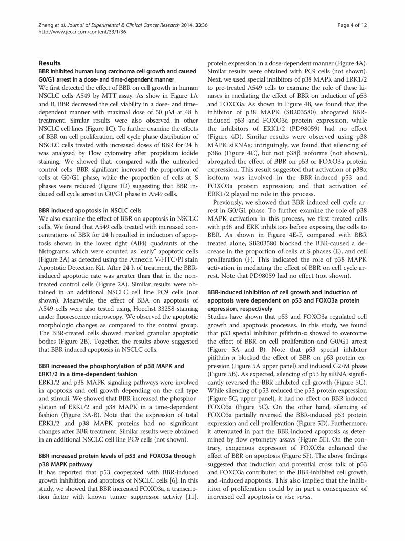
ResultsBBR inhibited human lung carcinoma cell growth and causedG0/G1 arrest in a dose- and time-dependent mannerWe first detected the effect of BBR on cell growth in humanNSCLC cells A549 by MTT assay. As show in Figure 1Aand B, BBR decreased the cell viability in a dose- and time-dependent manner with maximal dose of 50 μM at 48 htreatment. Similar results were also observed in otherNSCLC cell lines (Figure 1C). To further examine the effectsof BBR on cell proliferation, cell cycle phase distribution ofNSCLC cells treated with increased doses of BBR for 24 hwas analyzed by Flow cytometry after propidium iodidestaining. We showed that, compared with the untreatedcontrol cells, BBR significant increased the proportion ofcells at G0/G1 phase, while the proportion of cells at Sphases were reduced (Figure 1D) suggesting that BBR in-duced cell cycle arrest in G0/G1 phase in A549 cells.
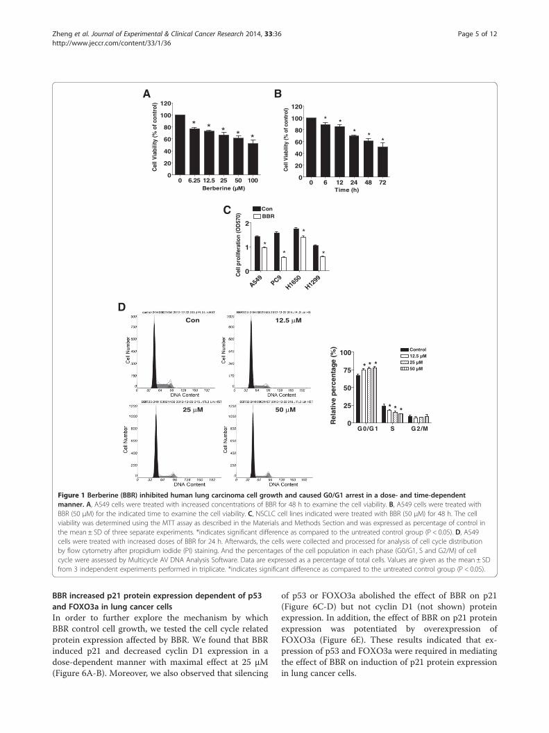
BBR induced apoptosis in NSCLC cellsWe also examine the effect of BBR on apoptosis in NSCLCcells. We found that A549 cells treated with increased con-centrations of BBR for 24 h resulted in induction of apop-tosis shown in the lower right (AB4) quadrants of thehistograms, which were counted as “early” apoptotic cells(Figure 2A) as detected using the Annexin V-FITC/PI stainApoptotic Detection Kit. After 24 h of treatment, the BBR-induced apoptotic rate was greater than that in the non-treated control cells (Figure 2A). Similar results were ob-tained in an additional NSCLC cell line PC9 cells (notshown). Meanwhile, the effect of BBA on apoptosis ofA549 cells were also tested using Hoechst 33258 stainingunder fluorescence microscopy. We observed the apoptoticmorphologic changes as compared to the control group.The BBR-treated cells showed marked granular apoptoticbodies (Figure 2B). Together, the results above suggestedthat BBR induced apoptosis in NSCLC cells.
BBR increased the phosphorylation of p38 MAPK andERK1/2 in a time-dependent fashionERK1/2 and p38 MAPK signaling pathways were involvedin apoptosis and cell growth depending on the cell typeand stimuli. We showed that BBR increased the phosphor-ylation of ERK1/2 and p38 MAPK in a time-dependentfashion (Figure 3A-B). Note that the expression of totalERK1/2 and p38 MAPK proteins had no significantchanges after BBR treatment. Similar results were obtainedin an additional NSCLC cell line PC9 cells (not shown).
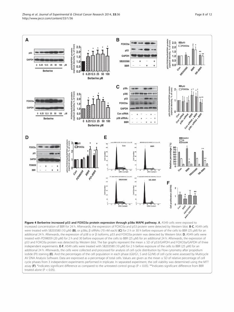
BBR increased protein levels of p53 and FOXO3a throughp38 MAPK pathwayIt has reported that p53 cooperated with BBR-inducedgrowth inhibition and apoptosis of NSCLC cells [6]. In thisstudy, we showed that BBR increased FOXO3a, a transcrip-tion factor with known tumor suppressor activity [11],
protein expression in a dose-dependent manner (Figure 4A).Similar results were obtained with PC9 cells (not shown).Next, we used special inhibitors of p38 MAPK and ERK1/2to pre-treated A549 cells to examine the role of these ki-nases in mediating the effect of BBR on induction of p53and FOXO3a. As shown in Figure 4B, we found that theinhibitor of p38 MAPK (SB203580) abrogated BBR-induced p53 and FOXO3a protein expression, whilethe inhibitors of ERK1/2 (PD98059) had no effect(Figure 4D). Similar results were observed using p38MAPK siRNAs; intriguingly, we found that silencing ofp38α (Figure 4C), but not p38β isoforms (not shown),abrogated the effect of BBR on p53 or FOXO3a proteinexpression. This result suggested that activation of p38αisoform was involved in the BBR-induced p53 andFOXO3a protein expression; and that activation ofERK1/2 played no role in this process.Previously, we showed that BBR induced cell cycle ar-
rest in G0/G1 phase. To further examine the role of p38MAPK activation in this process, we first treated cellswith p38 and ERK inhibitors before exposing the cells toBBR. As shown in Figure 4E-F, compared with BBRtreated alone, SB203580 blocked the BBR-caused a de-crease in the proportion of cells at S phases (E), and cellproliferation (F). This indicated the role of p38 MAPKactivation in mediating the effect of BBR on cell cycle ar-rest. Note that PD98059 had no effect (not shown).
BBR-induced inhibition of cell growth and induction ofapoptosis were dependent on p53 and FOXO3a proteinexpression, respectivelyStudies have shown that p53 and FOXO3a regulated cellgrowth and apoptosis processes. In this study, we foundthat p53 special inhibitor pifithrin-α showed to overcomethe effect of BBR on cell proliferation and G0/G1 arrest(Figure 5A and B). Note that p53 special inhibitorpifithrin-α blocked the effect of BBR on p53 protein ex-pression (Figure 5A upper panel) and induced G2/M phase(Figure 5B). As expected, silencing of p53 by siRNA signifi-cantly reversed the BBR-inhibited cell growth (Figure 5C).While silencing of p53 reduced the p53 protein expression(Figure 5C, upper panel), it had no effect on BBR-inducedFOXO3a (Figure 5C). On the other hand, silencing ofFOXO3a partially reversed the BBR-induced p53 proteinexpression and cell proliferation (Figure 5D). Furthermore,it attenuated in part the BBR-induced apoptosis as deter-mined by flow cytometry assays (Figure 5E). On the con-trary, exogenous expression of FOXO3a enhanced theeffect of BBR on apoptosis (Figure 5F). The above findingssuggested that induction and potential cross talk of p53and FOXO3a contributed to the BBR-inhibited cell growthand -induced apoptosis. This also implied that the inhib-ition of proliferation could by in part a consequence ofincreased cell apoptosis or vise versa.
A549
PC9
H1650
H1299
0
1
2BBR
**
*
*
Con
Cel
l pro
lifer
atio
n (O
D57
0)
0 6.25 12.5 25 50 1000
20
40
60
80
100
120
Berberine (µ )
****
*
Cel
l Via
bilit
y (%
ofc
ontr
ol)
BA
C
0 6 12 24 48 720
20
40
60
80
100
120
* ** *
*
Time (h)
Cel
l Via
bilit
y(%
of c
ontr
ol)
D
G 0/G 1 S G 2/M0
25
50
75
100 Control12.5 µ25 µ50 µ* * *
* * *
Rel
ativ
e p
erce
nta
ge
(%)
Con 12.5 μ
25 μ 50 μ
Figure 1 Berberine (BBR) inhibited human lung carcinoma cell growth and caused G0/G1 arrest in a dose- and time-dependentmanner. A, A549 cells were treated with increased concentrations of BBR for 48 h to examine the cell viability. B, A549 cells were treated withBBR (50 μM) for the indicated time to examine the cell viability. C, NSCLC cell lines indicated were treated with BBR (50 μM) for 48 h. The cellviability was determined using the MTT assay as described in the Materials and Methods Section and was expressed as percentage of control inthe mean ± SD of three separate experiments. *indicates significant difference as compared to the untreated control group (P < 0.05). D, A549cells were treated with increased doses of BBR for 24 h. Afterwards, the cells were collected and processed for analysis of cell cycle distributionby flow cytometry after propidium iodide (PI) staining. And the percentages of the cell population in each phase (G0/G1, S and G2/M) of cellcycle were assessed by Multicycle AV DNA Analysis Software. Data are expressed as a percentage of total cells. Values are given as the mean ± SDfrom 3 independent experiments performed in triplicate. *indicates significant difference as compared to the untreated control group (P < 0.05).
Zheng et al. Journal of Experimental & Clinical Cancer Research 2014, 33:36 Page 5 of 12http://www.jeccr.com/content/33/1/36
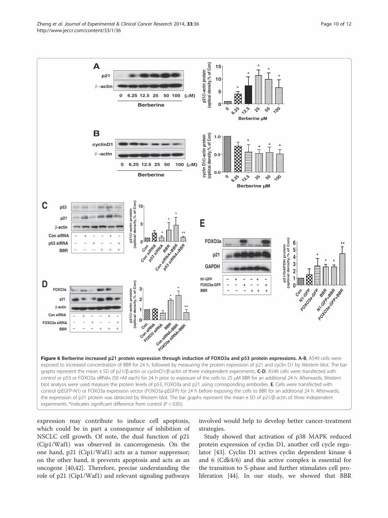
BBR increased p21 protein expression dependent of p53and FOXO3a in lung cancer cellsIn order to further explore the mechanism by whichBBR control cell growth, we tested the cell cycle relatedprotein expression affected by BBR. We found that BBRinduced p21 and decreased cyclin D1 expression in adose-dependent manner with maximal effect at 25 μM(Figure 6A-B). Moreover, we also observed that silencing
of p53 or FOXO3a abolished the effect of BBR on p21(Figure 6C-D) but not cyclin D1 (not shown) proteinexpression. In addition, the effect of BBR on p21 proteinexpression was potentiated by overexpression ofFOXO3a (Figure 6E). These results indicated that ex-pression of p53 and FOXO3a were required in mediatingthe effect of BBR on induction of p21 protein expressionin lung cancer cells.
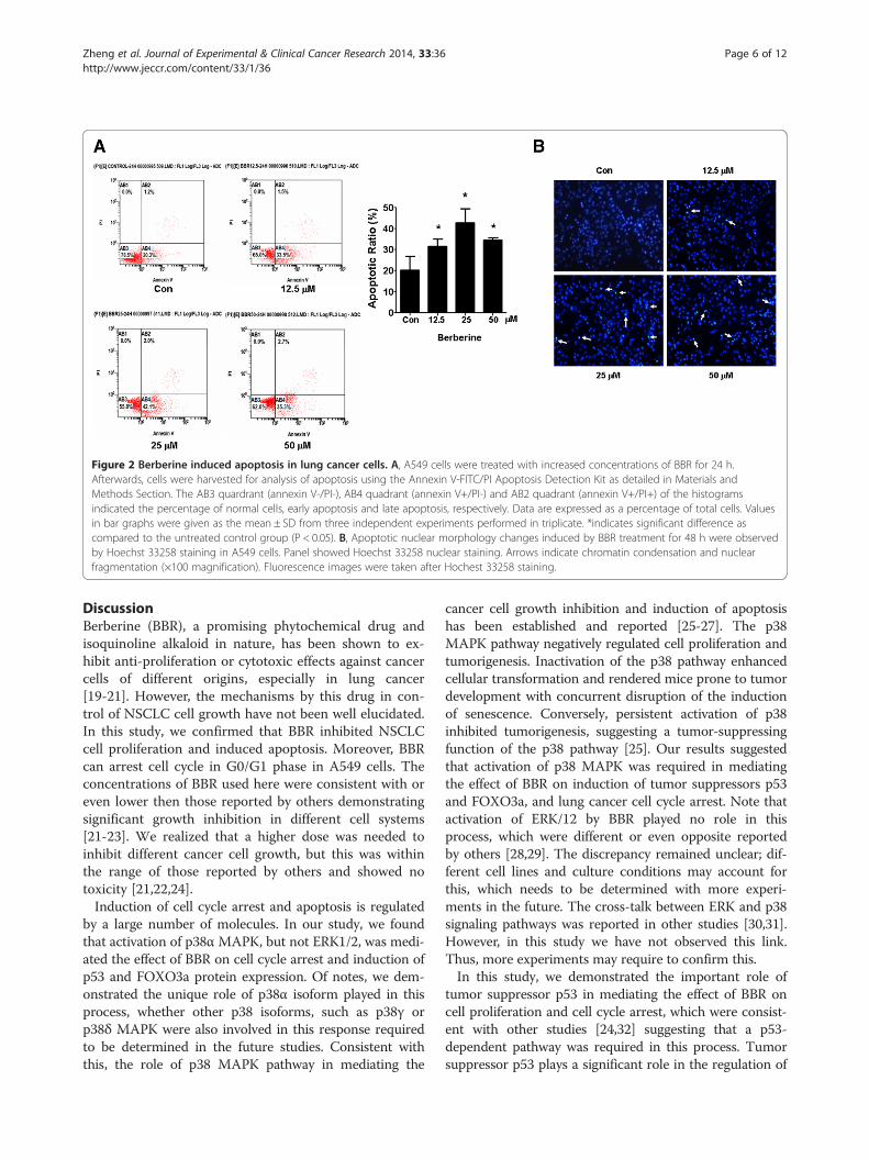
Figure 2 Berberine induced apoptosis in lung cancer cells. A, A549 cells were treated with increased concentrations of BBR for 24 h.Afterwards, cells were harvested for analysis of apoptosis using the Annexin V-FITC/PI Apoptosis Detection Kit as detailed in Materials andMethods Section. The AB3 quardrant (annexin V-/PI-), AB4 quadrant (annexin V+/PI-) and AB2 quadrant (annexin V+/PI+) of the histogramsindicated the percentage of normal cells, early apoptosis and late apoptosis, respectively. Data are expressed as a percentage of total cells. Valuesin bar graphs were given as the mean ± SD from three independent experiments performed in triplicate. *indicates significant difference ascompared to the untreated control group (P < 0.05). B, Apoptotic nuclear morphology changes induced by BBR treatment for 48 h were observedby Hoechst 33258 staining in A549 cells. Panel showed Hoechst 33258 nuclear staining. Arrows indicate chromatin condensation and nuclearfragmentation (×100 magnification). Fluorescence images were taken after Hochest 33258 staining.
Zheng et al. Journal of Experimental & Clinical Cancer Research 2014, 33:36 Page 6 of 12http://www.jeccr.com/content/33/1/36
DiscussionBerberine (BBR), a promising phytochemical drug andisoquinoline alkaloid in nature, has been shown to ex-hibit anti-proliferation or cytotoxic effects against cancercells of different origins, especially in lung cancer[19-21]. However, the mechanisms by this drug in con-trol of NSCLC cell growth have not been well elucidated.In this study, we confirmed that BBR inhibited NSCLCcell proliferation and induced apoptosis. Moreover, BBRcan arrest cell cycle in G0/G1 phase in A549 cells. Theconcentrations of BBR used here were consistent with oreven lower then those reported by others demonstratingsignificant growth inhibition in different cell systems[21-23]. We realized that a higher dose was needed toinhibit different cancer cell growth, but this was withinthe range of those reported by others and showed notoxicity [21,22,24].Induction of cell cycle arrest and apoptosis is regulated
by a large number of molecules. In our study, we foundthat activation of p38α MAPK, but not ERK1/2, was medi-ated the effect of BBR on cell cycle arrest and induction ofp53 and FOXO3a protein expression. Of notes, we dem-onstrated the unique role of p38α isoform played in thisprocess, whether other p38 isoforms, such as p38γ orp38δ MAPK were also involved in this response requiredto be determined in the future studies. Consistent withthis, the role of p38 MAPK pathway in mediating the
cancer cell growth inhibition and induction of apoptosishas been established and reported [25-27]. The p38MAPK pathway negatively regulated cell proliferation andtumorigenesis. Inactivation of the p38 pathway enhancedcellular transformation and rendered mice prone to tumordevelopment with concurrent disruption of the inductionof senescence. Conversely, persistent activation of p38inhibited tumorigenesis, suggesting a tumor-suppressingfunction of the p38 pathway [25]. Our results suggestedthat activation of p38 MAPK was required in mediatingthe effect of BBR on induction of tumor suppressors p53and FOXO3a, and lung cancer cell cycle arrest. Note thatactivation of ERK/12 by BBR played no role in thisprocess, which were different or even opposite reportedby others [28,29]. The discrepancy remained unclear; dif-ferent cell lines and culture conditions may account forthis, which needs to be determined with more experi-ments in the future. The cross-talk between ERK and p38signaling pathways was reported in other studies [30,31].However, in this study we have not observed this link.Thus, more experiments may require to confirm this.In this study, we demonstrated the important role of
tumor suppressor p53 in mediating the effect of BBR oncell proliferation and cell cycle arrest, which were consist-ent with other studies [24,32] suggesting that a p53-dependent pathway was required in this process. Tumorsuppressor p53 plays a significant role in the regulation of
A
24 (h)8420.50
B
24 (h)8420.50 0 0.5 2 4 8 240
1
2
3 **
** *
Time (h)
0 0.5 2 4 8 240
1
2
3
4
* **
*
*
Time (h)
p-ERK1/2 (Thr202/Tyr204)
ERK1/2
p-p38 MPAK (Thr180/Tyr182)
p38 MAPK
GAPDH
GAPDH
Berberine (25 μM)
Berberine (25 μM)
P-E
RK
1/2
/GA
PD
H p
rote
in(o
pti
cal
de
nsit
y,
% o
f C
on
)p
-p-3
8M
AP
K/G
AP
DH
pro
tein
(op
tica
l d
en
sit
y,
% o
f C
on
)
Figure 3 Berberine increased the phosphorylation of p38 MAPK and ERK1/2 in a time-dependent manner. A-B, A549 cells were treatedwith BBR (25 μM) in the indicated times, and cell lysate was harvested and the expression of the phosphorylated or total protein of ERK1/2, p38MAPK were measured by Western blot analysis using corresponding antibodies. GAPDH was used as loading control. The bar graphs representedthe densitometry results of p-ERK (A) or p38 MAPK (B)/GAPDH as mean ± SD of at least three separate experiments. *indicates significantdifference from untreated control cells (P < 0.05).
Zheng et al. Journal of Experimental & Clinical Cancer Research 2014, 33:36 Page 7 of 12http://www.jeccr.com/content/33/1/36
cell growth, cell cycle arrest, and apoptosis in various can-cers [33,34]. p53 controls both the G2/M and the G1 cellcycle checkpoints and mediates reversible growth arrest inhuman fibroblasts [35]. Increased expression of wild-typep53 arrested cells late in the G1 stage of the cell cycle bystimulating the synthesis of inhibitors of cyclin-dependentkinase p21 (CIP1/WAF1) [35]. Consistent with this, wefound that BBR increased p21 protein expression in hu-man lung cancer A549 cells, which was eliminated (notobserved) in cells silencing of p53 gene. This suggestedthat BBR-induced p21 (CIP1/WAF1) was through p53-dependent pathway. P21 (CIP1/WAF1) is a direct targetof p53 [36], thus, p53 mediated induction of p21 (CIP1/WAF1) at least contributed to the inhibitory effect ofBBR on cell proliferation and cell cycle arrest.On the other hand, our results suggested that activation
of p38 MAPK mediated the BBR-induced FOXO3a pro-tein expression and the latter also contributed to the BBR-inhibited cell growth and -induced apoptosis. It is possiblethat the inhibition of proliferation can be in part a conse-quence of increased cell apoptosis or vise versa. TheFOXO3a is an important tumor suppressor and is under-expressed in many cancers. There are a number of paral-lels between FOXO3a and p53, both play a pivotal role inregulating the cellular response to stress and damage sig-nals, inducing cell cycle arrest, apoptosis, and DNA repair
[37]. Several studies showed that FOXO3a interacts withp53, and that FOXO3a is a p53 target gene [15,38]. In thisstudy, we demonstrated that the potential interaction andmutually exclusive events of p53 and FOXO3a may con-tribute to enhance BBR-induced apoptosis and -inhibitedcell proliferation. However, the detailed mechanismunderlining the regulation of these transcriptional net-works in mediating the effect of BBR on the control oflung cancer cell survival needs to be elucidated.Our results also demonstrated a causative role of
FOXO3a in mediated the effect of BBR on p21 (CIP1/WAF1) expression. We showed that the knockdown ofFOXO3a blocked, while overexpression of FOXO3a aug-mented the increase in p21 (CIP1/WAF1) protein ex-pression in BBR-treated cells. These, together with theobservation from silencing of p53 experiments indicatedthat p21 (CIP1/WAF1) is not only the direct target ofp53 but also function as FOXO3a downstream effector,which may be through the p53-independent way [17].p53 and FOXO3a share similar target genes includingp21(CIP1/WAF1), FOXO factors bind to the promoterof p21 to induce cell cycle arrest at the G1/S transition[39]. Given the fact that p21 (CIP1/WAF1) is involved inregulation of fundamental cellular processes, such as cellproliferation, differentiation, regulation of gene tran-scription and apoptosis [40,41]. BBR-induced FOXO3a
A
GAPDH
p53
100 μM502512.56.250
FOXO3a
GAPDH
100 μM502512.56.250
0 6.2512.5 25 50 1000.0
0.5
1.0
1.5
2.0
2.5
* **
*
*
Berberine µ
Berberine
Berberine
0 6.2512.5 25 50 1000.00.51.01.52.02.5
* * * * *
Berberine µ
FO
XO
3a/G
AP
DH
pro
tein
(op
tical d
en
sit
y,%
of
Co
n)
p53/G
AP
DH
pro
tein
(op
tical d
en
sit
y,%
of
Co
n)
FOXO3a
p53
GAPDH
PD98059
BBR
+ − +− + +
D
−−
p53,F
OX
O3a/G
AP
DH
pro
tein
(op
tical d
en
sit
y,
% o
f co
n)
Con
PD98
059
BBR
PD+B
BR
0
1
2
3 p53
FOXO3a
*
*
*
*
ConBBR
SB2035
80
SB+BBR0
1020304050607080
G0/G1SG2/M
***
* **
Rel
ativ
e pe
rcen
tage
( %
)
Con
E
BBR
SB203580 SB+BBR
F
Con
BBR
SB20
3580
BBR+S
B25
3580
0
1
2
***
Cel
l pro
lifer
atio
n (O
D75
0)
SB203580
BBR
−−
+ − +− + +
FOXO3a
p53
GAPDH
B
Con
SB2035
80BBR
SB+BBR
0.00.51.01.52.02.5 p53
FOXO3a * *
****
p53,F
OX
O3a/G
AP
DH
pro
tein
(op
tica
l d
en
sit
y,
% o
f c
on
)
p38 siRNAαBBR
−−
+ +−+ +−
Con siRNA
− − − + + +−−
− −
p53
FOXO3a
GAPDH
p38α
p38β
C
p53,F
OX
O3a/G
AP
DH
pro
tein
(op
tica
l d
en
sit
y,
% o
f c
on
)
Con
Con siR
NA
siR
NA
αp3
8
BBR
Con siR
NA + B
BR
siRNA +
BBR
αp3
8
0
1
2p53FOXO3a * *
****
* *
Figure 4 Berberine increased p53 and FOXO3a protein expression through p38α MAPK pathway. A, A549 cells were exposed toincreased concentration of BBR for 24 h. Afterwards, the expression of FOXO3a and p53 protein were detected by Western blot. B-C, A549 cellswere treated with SB203580 (10 μM) (B), or p38α, β siRNAs (70 nM each) (C) for 2 h or 30 h before exposure of the cells to BBR (25 μM) for anadditional 24 h. Afterwards, the expression of p38 α or β isoforms, p53 and FOXO3a protein was detected by Western blot. D, A549 cells weretreated with PD98059 (20 μM) for 2 h and 30 before exposure of the cells to BBR (25 μM) for an additional 24 h. Afterwards, the expression ofp53 and FOXO3a protein was detected by Western blot. The bar graphs represent the mean ± SD of p53/GAPDH and FOXO3a/GAPDH of threeindependent experiments. E-F, A549 cells were treated with SB203580 (10 μM) for 2 h before exposure of the cells to BBR (25 μM) for anadditional 24 h. Afterwards, the cells were collected and processed for analysis of cell cycle distribution by Flow cytometry after propidiumiodide (PI) staining (E). And the percentages of the cell population in each phase (G0/G1, S and G2/M) of cell cycle were assessed by MulticycleAV DNA Analysis Software. Data are expressed as a percentage of total cells. Values are given as the mean ± SD of relative percentage of cellcycle phases from 3 independent experiments performed in triplicate. In separated experiment, the cell viability was determined using the MTTassay (F). *indicates significant difference as compared to the untreated control group (P < 0.05). **Indicates significant difference from BBRtreated alone (P < 0.05).
Zheng et al. Journal of Experimental & Clinical Cancer Research 2014, 33:36 Page 8 of 12http://www.jeccr.com/content/33/1/36
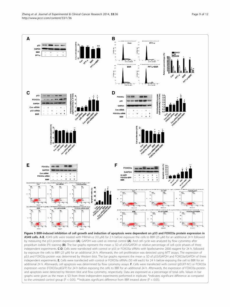
Figure 5 BBR-induced inhibition of cell growth and induction of apoptosis were dependent on p53 and FOXO3a protein expression inA549 cells. A-B, A549 cells were treated with Pifithrin-α (10 μM) for 2 h before exposure the cells to BBR (25 μM) for an additional 24 h followedby measuring the p53 protein expression (A). GAPDH was used as internal control (A). And cell cycle was analyzed by flow cytometry afterpropidium iodide (PI) staining (B). The bar graphs represent the mean ± SD of p53/GAPDH or relative percentage of cell cycle phases of threeindependent experiments. C-D, Cells were transfected with control or p53 or FOXO3a siRNAs with lipofectamine 2000 reagent for 24 h, followedby exposure the cells to BBR (25 μM) for an additional 24 h. Afterwards, the cell proliferation was detected using MTT assays. The expression ofp53 and FOXO3a protein was determined by Western blot. The bar graphs represent the mean ± SD of p53/GAPDH and FOXO3a/GAPDH of threeindependent experiments. E, Cells were transfected with control or FOXO3a siRNAs (50 nM each) for 24 h before exposing the cell to BBR for anadditional 24 h. Afterwards, cell apoptosis was determined by flow cytometry assays. F, Cells were transfected with control (pEGFP-N1) or FOXO3aexpression vector (FOXO3a-pEGFP) for 24 h before exposing the cells to BBR for an additional 24 h. Afterwards, the expression of FOXO3a proteinand apoptosis were detected by Western blot and flow cytometry, respectively. Data are expressed as a percentage of total cells. Values in bargraphs were given as the mean ± SD from three independent experiments performed in triplicate. *indicates significant difference as comparedto the untreated control group (P < 0.05). **Indicates significant difference from BBR treated alone (P < 0.05).
Zheng et al. Journal of Experimental & Clinical Cancer Research 2014, 33:36 Page 9 of 12http://www.jeccr.com/content/33/1/36
A
-actin
cyclinD1
100 (μM)502512.56.250
-actin
p21
100 (μM)502512.56.250
B
06.25
12.5 25 50100
0.0
0.5
1.0
*
* * * *
Berberine µ
06.2
512.5 25 50
1000
5
10
15
*
* * *
*
Berberine µ
Berberine
Berberine
p21/
β-ac
tinpr
otei
n(o
ptic
al d
ensi
ty,%
of C
on)
cycl
inD
1/β-
actin
prot
ein
(opt
ical
den
sity
,% o
f Con
)
FOXO3a siRNA
BBR
Con siRNA
FOXO3a
β-actin
p21
C
D
p53 siRNA
BBR
− + − − + −− − + − − +
Con siRNA
− − − + + +
p53
p21
β-actin
Con
Con si
RNA
FOXO3 s
iRNA
BBR
Con si
RNA+BBR
FOXO3a
siRNA+B
BR
0
1
2
3
*
*
**
− + − − + −− − + − − +− − − + + +
Con
Con
siR
NA
p53 siRNA
BBR
Con siR
NA+BBR
p53 siRNA+B
BR0
5
10
* **
**
p21/β
-acti
np
rote
in(o
pti
ca
l d
en
sit
y,%
of
Co
n)
p21/β
-acti
np
rote
in(o
pti
ca
l d
en
sit
y,%
of
Co
n)
E
FOXO3a
GAPDH
N1-GFP – + – – + –FOXO3a-GFP – – + – – + BBR – – – + + +
p21
p21/G
AP
DH
pro
tein
(op
tica
l d
en
sit
y,%
of
Co
n)
Con
N1-GFP
FOXO
3a-G
FPBBR
N1-GFP
+BBR
FOXO
3a-G
FP+B
BR0123456
* *
**
*
Figure 6 Berberine increased p21 protein expression through induction of FOXO3a and p53 protein expressions. A-B, A549 cells wereexposed to increased concentration of BBR for 24 h, followed by measuring the protein expression of p21 and cyclin D1 by Western blot. The bargraphs represent the mean ± SD of p21/β-actin or cyclinD1/β-actin of three independent experiments. C-D, A549 cells were transfected withcontrol or p53 or FOXO3a siRNAs (50 nM each) for 24 h prior to exposure of the cells to 25 μM BBR for an additional 24 h. Afterwards, Westernblot analysis were used measure the protein levels of p53, FOXO3a and p21 using corresponding antibodies. E, Cells were transfected withcontrol (pEGFP-N1) or FOXO3a expression vector (FOXO3a-pEGFP) for 24 h before exposing the cells to BBR for an additional 24 h. Afterwards,the expression of p21 protein was detected by Western blot. The bar graphs represent the mean ± SD of p21/β-actin of three independentexperiments. *indicates significant difference from control (P < 0.05).
Zheng et al. Journal of Experimental & Clinical Cancer Research 2014, 33:36 Page 10 of 12http://www.jeccr.com/content/33/1/36
expression may contribute to induce cell apoptosis,which could be in part a consequence of inhibition ofNSCLC cell growth. Of note, the dual function of p21(Cip1/Waf1) was observed in cancerogenesis. On theone hand, p21 (Cip1/Waf1) acts as a tumor suppressor;on the other hand, it prevents apoptosis and acts as anoncogene [40,42]. Therefore, precise understanding therole of p21 (Cip1/Waf1) and relevant signaling pathways
involved would help to develop better cancer-treatmentstrategies.Study showed that activation of p38 MAPK reduced
protein expression of cyclin D1, another cell cycle regu-lator [43]. Cyclin D1 actives cyclin dependent kinase 4and 6 (Cdk4/6) and this active complex is essential forthe transition to S-phase and further stimulates cell pro-liferation [44]. In our study, we showed that BBR
Zheng et al. Journal of Experimental & Clinical Cancer Research 2014, 33:36 Page 11 of 12http://www.jeccr.com/content/33/1/36
decreased the cyclin D1 protein expression, but this wasnot through the p53- or FOXO3a-dependent pathway,which consistent with other studies [45] although oppos-ite results were observed [12,46]. Thus, more studies arerequired to elucidate the truly connections and precisemechanism underlining this. In addition, whether theBBR-induced pro-apoptotic signaling by p38 MAPK isalso activated and the functions of FOXO3a are regulatedby p38 MAPK in cells silencing of p53 need to be deter-mined. This may further elucidate pleiotropic anti-cancermechanisms of this medicinal phyto-chemical compound.
ConclusionIn summary, our data demonstrate that BBR inhibits growthand induces cell cycle arrest in G0/G1 phase, and apoptosisin NSCLC cells through p38α MAPK-mediated induction ofp53 and FOXO3a, followed by p21 protein expression.Thus, the parallel induction and mutually exclusive inter-action of p53 and FOXO3a, which act in concert, contributeto mediate the overall responses of NSCLC cell to BBR.
AbbreviationsBBR: Berberine; TCM: Traditional Chinese medicine; MTT: 3-(4, 5)-dimethylthiahiazo(−z-y1)-3,5-di-phenytetrazoliumromide; p38 MAPK: P38mitogen activated protein kinases; ERK1/2: Extracellular signal-regulatedkinase 1/2; siRNA: Small interfering RNA; NSCLC: Non small lung carcinoma;FOXO3a: Forkhead box-O 3a; PI3-K: Phosphatidylinositol 3-kinase;DMSO: Dimethylsulfoxide; PBS: Phosphate-buffered saline; RT: Roomtemperature; Cdk4/6: Cyclin dependent kinase 4 and 6; PI: Propidium iodide.
Competing interestsThe authors declare that they have no competing interests.
Authors’ contributionsSH is fully responsible for the study designing, experiment adjustment anddrafting the manuscript. FZ performed most of the experiments involved. QTcarried out transfection assays and some protein measurement by Westernblot and statistical analysis. SYZ conducted the densitometry, statisticalanalysis and participated in coordination manuscript. JJW executed the MTTassays, FOXO3a overexpression experiments and statistical analysis. ZYLfulfilled MTT and Western Blot analysis. LLL and WYW coordinated andprovided important suggestions including some agents, and critical read themanuscript. All authors read and approved the final manuscript.
AcknowledgmentsWe thank Dr. Frank M. J. Jacobs (Rudolf Magnus Institute of Neuroscience,University Medical Center, Utrecht, the Netherland) for providing theFOXO3a-GFP and N1-GFP plasmids. This work was supported in part by theSpecial Science and Technology Join fund from Guangdong ProvincialDepartment of Science and Technology-Guangdong Academy of TraditionalChinese Medicine (2012A032500011) and grant from the National NatureScientific Foundation of China (81272614).
Author details1Laboratory of Tumor Molecular Biology and Targeted Therapies of ChineseMedicine, 4th Floor, Scientific Research Building, Neihuan West Road No. 55,University City, Panyu District, Guangzhou, Guangdong Province, P. R. China,510006. 2Department of Medical Oncology, University of GuangzhouTraditional Chinese Medicine, Guangdong Provincial Hospital of ChineseMedicine, Guangzhou, Guangdong Province, China, 510120.
Received: 30 December 2013 Accepted: 14 April 2014Published: 26 April 2014
References1. Siegel R, Naishadham D, Jemal A: Cancer statistics, 2013. CA Canc J Clin
2013, 63(1):11–30.2. Yang Y, Xie Y, Xian L: Breast cancer susceptibility gene 1 (BRCA1) predict
clinical outcome in platinum- and toxal-based chemotherapy innon-small-cell lung cancer (NSCLC) patients: a system review andmeta-analysis. J Exp Clin Canc Res 2013, 32:15.
3. Li X, Yang G, Zhang Y, Yang J, Chang J, Sun X, Zhou X, Guo Y, Xu Y, Liu J,Bensoussan A: Traditional Chinese medicine in cancer care: a review ofcontrolled clinical studies published in Chinese. PloS one 2013, 8(4):e60338.
4. Singh IP, Mahajan S: Berberine and its derivatives: a patent review(2009–2012). Expert Opin Ther Pat 2013, 23(2):215–231.
5. Vuddanda PR, Chakraborty S, Singh S: Berberine: a potentialphytochemical with multispectrum therapeutic activities. Expert OpinInvestig Drugs 2010, 19(10):1297–1307.
6. Katiyar SK, Meeran SM, Katiyar N, Akhtar S: p53 cooperates berberine-induced growth inhibition and apoptosis of non-small cell human lungcancer cells in vitro and tumor xenograft growth in vivo. Mol Carcinog2009, 48(1):24–37.
7. Milner J: Forms and functions of p53. Semin Canc Biol 1994, 5(3):211–219.8. Jin S: p53, autophagy and tumor suppression. Autophagy 2005, 1(3):171–173.9. Raj N, Attardi LD: Tumor suppression: p53 alters immune surveillance to
restrain liver cancer. Curr Biol 2013, 23(12):R527–R530.10. Hann SS, Zheng F, Zhao S: Targeting 3-phosphoinositide-dependent
protein kinase 1 by N-acetyl-cysteine through activation of peroxisomeproliferators activated receptor alpha in human lung cancer cells, therole of p53 and p65. J Exp Clin Canc Res 2013, 32:43.
11. Brunet A, Bonni A, Zigmond MJ, Lin MZ, Juo P, Hu LS, Anderson MJ, ArdenKC, Blenis J, Greenberg ME: Akt promotes cell survival by phosphorylatingand inhibiting a forkhead transcription factor. Cell 1999, 96(6):857–868.
12. Schmidt M, Fernandez de Mattos S, van der Horst A, Klompmaker R, KopsGJ, Lam EW, Burgering BM, Medema RH: Cell cycle inhibition by FoxOforkhead transcription factors involves downregulation of cyclin D.Mol Cell Biol 2002, 22(22):7842–7852.
13. Yang JY, Zong CS, Xia W, Yamaguchi H, Ding Q, Xie X, Lang JY, Lai CC, ChangCJ, Huang WC, Huang H, Kuo HP, Lee DF, Li LY, Lien HC, Cheng X, Chang KJ,Hsiao CD, Tsai FJ, Tsai CH, Sahin AA, Muller WJ, Mills GB, Yu D, Hortobagyi GN,Hung MC: ERK promotes tumorigenesis by inhibiting FOXO3a via MDM2-mediated degradation. Nat Cell Biol 2008, 10(2):138–148.
14. Chen W, Yang Q, Roeder RG: Dynamic interactions and cooperativefunctions of PGC-1alpha and MED1 in TRalpha-mediated activation ofthe brown-fat-specific UCP-1 gene. Mol Cell 2009, 35(6):755–768.
15. Renault VM, Thekkat PU, Hoang KL, White JL, Brady CA, Kenzelmann Broz D,Venturelli OS, Johnson TM, Oskoui PR, Xuan Z, Santo EE, Zhang MQ, VogelH, Attardi LD, Brunet A: The pro-longevity gene FoxO3 is a direct targetof the p53 tumor suppressor. Oncogene 2011, 30(29):3207–3221.
16. Macleod KF, Sherry N, Hannon G, Beach D, Tokino T, Kinzler K, VogelsteinB, Jacks T: p53-dependent and independent expression of p21 duringcell growth, differentiation, and DNA damage. Genes Dev 1995,9(8):935–944.
17. Hauck L, Harms C, Grothe D, An J, Gertz K, Kronenberg G, Dietz R, Endres M,von Harsdorf R: Critical role for FoxO3a-dependent regulation ofp21CIP1/WAF1 in response to statin signaling in cardiac myocytes.Circ Res 2007, 100(1):50–60.
18. Wen Q, Duan X, Liao R, Little P, Gao G, Jiang H, Lalit S, Quirion R, Zheng W:Characterization of intracellular translocation of forkhead transcriptionfactor O (FoxO) members induced by NGF in PC12 cells. Neurosci Lett2011, 498(1):31–36.
19. Yip NK, Ho WS: Berberine induces apoptosis via the mitochondrialpathway in liver cancer cells. Oncol Rep 2013, 30(3):1107–1112.
20. Yan L, Yan K, Kun W, Xu L, Ma Q, Tang Y, Jiao W, Gu G, Fan Y, Xu Z:Berberine inhibits the migration and invasion of T24 bladder cancer cellsvia reducing the expression of heparanase. Tumour Biol 2013,34(1):215–221.
21. Fu L, Chen W, Guo W, Wang J, Tian Y, Shi D, Zhang X, Qiu H, Xiao X, KangT, Huang W, Wang S, Deng W: Berberine targets AP-2/hTERT, NF-kappaB/COX-2, HIF-1alpha/VEGF and cytochrome-c/caspase signaling tosuppress human cancer cell growth. PloS one 2013, 8(7):e69240.
22. Li J, Cao B, Liu X, Fu X, Xiong Z, Chen L, Sartor O, Dong Y, Zhang H:Berberine suppresses androgen receptor signaling in prostate cancer.Mol Canc Ther 2011, 10(8):1346–1356.
Zheng et al. Journal of Experimental & Clinical Cancer Research 2014, 33:36 Page 12 of 12http://www.jeccr.com/content/33/1/36
23. Park KS, Kim JB, Bae J, Park SY, Jee HG, Lee KE, Youn YK: Berberineinhibited the growth of thyroid cancer cell lines 8505C and TPC1.Yonsei Med J 2012, 53(2):346–351.
24. Mahata S, Bharti AC, Shukla S, Tyagi A, Husain SA, Das BC: Berberinemodulates AP-1 activity to suppress HPV transcription and downstreamsignaling to induce growth arrest and apoptosis in cervical cancer cells.Mol Cancer 2011, 10:39.
25. Hui L, Bakiri L, Stepniak E, Wagner EF: p38alpha: a suppressor of cellproliferation and tumorigenesis. Cell Cycle 2007, 6(20):2429–2433.
26. Lee HJ, Auh QS, Lee YM, Kang SK, Chang SW, Lee DS, Kim YC, Kim EC:Growth inhibition and apoptosis-inducing effects of Cudraflavone B inhuman oral cancer cells via MAPK, NF-kappaB, and SIRT1 signalingpathway. Planta Med 2013, 79(14):1298–1306.
27. Park HS, Hwang HJ, Kim GY, Cha HJ, Kim WJ, Kim ND, Yoo YH, Choi YH:Induction of apoptosis by fucoidan in human leukemia U937 cellsthrough activation of p38 MAPK and modulation of Bcl-2 family.Mar Drugs 2013, 11(7):2347–2364.
28. Cok A, Plaisier C, Salie MJ, Oram DS, Chenge J, Louters LL: Berberineacutely activates the glucose transport activity of GLUT1. Biochimie 2011,93(7):1187–1192.
29. Burgeiro A, Gajate C, el Dakir H, Villa-Pulgarin JA, Oliveira PJ, Mollinedo F:Involvement of mitochondrial and B-RAF/ERK signaling pathways inberberine-induced apoptosis in human melanoma cells. Anti-cancer drugs2011, 22(6):507–518.
30. Cheng B, Song J, Zou Y, Wang Q, Lei Y, Zhu C, Hu C: Responses ofvascular smooth muscle cells to estrogen are dependent on balancebetween ERK and p38 MAPK pathway activities. Int J Cardiol 2009,134(3):356–365.
31. Finch AR, Caunt CJ, Perrett RM, Tsaneva-Atanasova K, McArdle CA: Dualspecificity phosphatases 10 and 16 are positive regulators ofEGF-stimulated ERK activity: indirect regulation of ERK signals byJNK/p38 selective MAPK phosphatases. Cell Signal 2012,24(5):1002–1011.
32. Li J, Gu L, Zhang H, Liu T, Tian D, Zhou M, Zhou S: Berberine repressesDAXX gene transcription and induces cancer cell apoptosis. Lab Invest2013, 93(3):354–364.
33. Halacli SO, Canpinar H, Cimen E, Sunguroglu A: Effects of gammairradiation on cell cycle, apoptosis and telomerase activity in p53wild-type and deficient HCT116 colon cancer cell lines. Oncol Lett 2013,6(3):807–810.
34. Ma LL, Sun WJ, Wang Z, Zh GY, Li P, Fu SB: Effects of silencing of mutantp53 gene in human lung adenocarcinoma cell line Anip973. J Exp ClinCanc Res 2006, 25(4):585–592.
35. Agarwal ML, Agarwal A, Taylor WR, Stark GR: p53 controls both the G2/Mand the G1 cell cycle checkpoints and mediates reversible growtharrest in human fibroblasts. Proc Natl Acad Sci USA 1995,92(18):8493–8497.
36. Pelletier J, Dayan F, Durivault J, Ilc K, Pecou E, Pouyssegur J, Mazure NM:The asparaginyl hydroxylase factor-inhibiting HIF is essential for tumorgrowth through suppression of the p53-p21 axis. Oncogene 2012,31(24):2989–3001.
37. Lam M, Carmichael AR, Griffiths HR: An aqueous extract of Fagonia creticainduces DNA damage, cell cycle arrest and apoptosis in breast cancercells via FOXO3a and p53 expression. PloS one 2012, 7(6):e40152.
38. Nemoto S, Fergusson MM, Finkel T: Nutrient availability regulatesSIRT1 through a forkhead-dependent pathway. Sci 2004,306(5704):2105–2108.
39. Seoane J, Le HV, Shen L, Anderson SA, Massague J: Integration of Smadand forkhead pathways in the control of neuroepithelial andglioblastoma cell proliferation. Cell 2004, 117(2):211–223.
40. Warfel NA, El-Deiry WS: p21WAF1 and tumourigenesis: 20 years after.Curr Opin Oncol 2013, 25(1):52–58.
41. Nanda K, Miyoshi N, Nakamura Y, Shimoji Y, Tamura Y, Nishikawa Y, UenakaiK, Kohno H, Tanaka T: Extract of vinegar “Kurosu” from unpolished riceinhibits the proliferation of human cancer cells. J Exp Clin Canc Res 2004,23(1):69–75.
42. Liedtke C, Trautwein C: A dual role of p21 in liver regeneration andhepatocarcinogenesis. Hepatol 2008, 48(5):1713–1714.
43. Pillai MS, Sapna S, Shivakumar K: p38 MAPK regulates G1-S transition inhypoxic cardiac fibroblasts. Int J Biochem Cell Biol 2011, 43(6):919–927.
44. Zhong Z, Yeow WS, Zou C, Wassell R, Wang C, Pestell RG, Quong JN, QuongAA: Cyclin D1/cyclin-dependent kinase 4 interacts with filamin A andaffects the migration and invasion potential of breast cancer cells.Canc Res 2010, 70(5):2105–2114.
45. Shen G, Xu C, Chen C, Hebbar V, Kong AN: p53-independent G1 cell cyclearrest of human colon carcinoma cells HT-29 by sulforaphane isassociated with induction of p21CIP1 and inhibition of expression ofcyclin D1. Canc Chemother Pharmacol 2006, 57(3):317–327.
46. Choudhuri T, Pal S, Das T, Sa G: Curcumin selectively induces apoptosis inderegulated cyclin D1-expressed cells at G2 phase of cell cycle in ap53-dependent manner. J Biol Chem 2005, 280(20):20059–20068.
doi:10.1186/1756-9966-33-36Cite this article as: Zheng et al.: p38α MAPK-mediated induction andinteraction of FOXO3a and p53 contribute to the inhibited-growth andinduced-apoptosis of human lung adenocarcinoma cells by berberine.Journal of Experimental & Clinical Cancer Research 2014 33:36.
Submit your next manuscript to BioMed Centraland take full advantage of:
• Convenient online submission
• Thorough peer review
• No space constraints or color figure charges
• Immediate publication on acceptance
• Inclusion in PubMed, CAS, Scopus and Google Scholar
• Research which is freely available for redistribution
Submit your manuscript at www.biomedcentral.com/submit
![Mechanisms and functions of p38 MAPK signalling and functions of p38 MAPK signalling 405 Both MKK3 and MKK6 are highly specific for p38 MAPKs [14,23].Inaddition,p38αcanbealsophophorylatedbyMKK4,an](https://static.fdocument.org/doc/165x107/5ae2800d7f8b9a097a8d0b79/mechanisms-and-functions-of-p38-mapk-signalling-and-functions-of-p38-mapk-signalling.jpg)