2012 11-30 leveraging greece's greatest assets economic impact-v2 1b
molecules
Review
Natural α-Glucosidase and Protein Tyrosine Phosphatase 1BInhibitors: A Source of Scaffold Molecules for Synthesis ofNew Multitarget Antidiabetic Drugs
Massimo Genovese , Ilaria Nesi , Anna Caselli and Paolo Paoli *
�����������������
Citation: Genovese, M.; Nesi, I.;
Caselli, A.; Paoli, P. Natural
α-Glucosidase and Protein Tyrosine
Phosphatase 1B Inhibitors: A Source
of Scaffold Molecules for Synthesis of
New Multitarget Antidiabetic Drugs.
Molecules 2021, 26, 4818. https://
doi.org/10.3390/molecules26164818
Academic Editor: Masahide
Hamaguchi
Received: 27 May 2021
Accepted: 7 August 2021
Published: 9 August 2021
Publisher’s Note: MDPI stays neutral
with regard to jurisdictional claims in
published maps and institutional affil-
iations.
Copyright: © 2021 by the authors.
Licensee MDPI, Basel, Switzerland.
This article is an open access article
distributed under the terms and
conditions of the Creative Commons
Attribution (CC BY) license (https://
creativecommons.org/licenses/by/
4.0/).
Department of Experimental and Clinical Biomedical Sciences “Mario Serio”, University of Florence,50139 Florence, Italy; [email protected] (M.G.); [email protected] (I.N.);[email protected] (A.C.)* Correspondence: [email protected]; Tel.: +39-055-2751248
Abstract: Diabetes mellitus (DM) represents a group of metabolic disorders that leads to acute andlong-term serious complications and is considered a worldwide sanitary emergence. Type 2 diabetes(T2D) represents about 90% of all cases of diabetes, and even if several drugs are actually available forits treatment, in the long term, they show limited effectiveness. Most traditional drugs are designedto act on a specific biological target, but the complexity of the current pathologies has demonstratedthat molecules hitting more than one target may be safer and more effective. The purpose of thisreview is to shed light on the natural compounds known as α-glucosidase and Protein TyrosinePhosphatase 1B (PTP1B) dual-inhibitors that could be used as lead compounds to generate newmultitarget antidiabetic drugs for treatment of T2D.
Keywords: PTP1B; α-glucosidase; insulin signaling; drug discovery; type 2 diabetes
1. Introduction
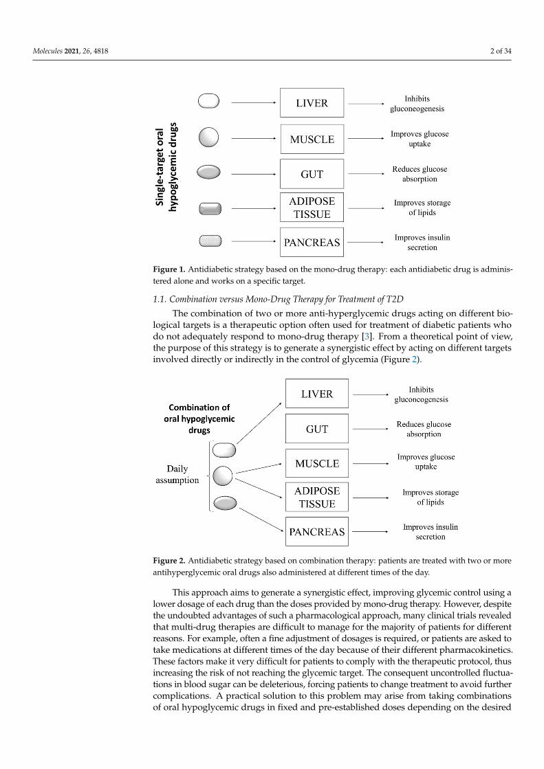
Type 2 diabetes is a complex pathology characterized by hyperglycemia and metabolicabnormalities affecting different organs and tissues, such as liver, muscle, adipose tissue,and pancreas. To date, subjects affected by T2D can rely on several oral antihyperglycemicdrugs showing different mechanisms of action to keep glycaemia under control. Thesedrugs include: inhibitors of intestinal α-glucosidases, which delay intestinal absorption ofglucose; metformin, which blocks hepatic gluconeogenesis; different types of secretagoguesthat stimulate the release of insulin from pancreatic β-cells; thiazolidinediones, whichstimulate the storage of circulating fatty acids into adipocytes, thereby improving insulinsensitivity in several peripheral tissues; and the sodium/glucose cotransporter 2 (SGLT-2)inhibitors, which impair the re-uptake of glucose in the renal tubules [1]. The choiceof the most appropriate hypoglycemic drug for a patient depends on several factors,such as the patient’s general condition, the presence of comorbidities, tolerance, and thepatient’s response to the drug. Generally, most diabetic patients showing hyperglycemiawithout further pathological complications respond positively to single-drug based therapy(Figure 1), experiencing a decrease of blood sugar levels and an improvement of theirgeneral conditions.
However, many clinical studies showed that benefits obtained with this approachare transient, and in the medium to long term, patients experience a gradual rise in bloodsugar and a worsening of general health conditions. In some cases, the up-scaling of drugdosage can allow regaining of the glycemic target, with the hope that, at the same time,no adverse effects related to high doses of the drug occur [2]. The failure of mono-drugtherapy is mainly due to the inability of such drugs to replace physiological functions ofinsulin. Indeed, even if these drugs are able to compensate a specific metabolic defect, theyunexpectedly induce severe unbalance in other metabolic pathways.
Molecules 2021, 26, 4818. https://doi.org/10.3390/molecules26164818 https://www.mdpi.com/journal/molecules
Molecules 2021, 26, 4818 2 of 34
Molecules 2021, 26, x 2 of 35
insulin. Indeed, even if these drugs are able to compensate a specific metabolic defect, they unexpectedly induce severe unbalance in other metabolic pathways.
Figure 1. Antidiabetic strategy based on the mono-drug therapy: each antidiabetic drug is adminis-tered alone and works on a specific target.
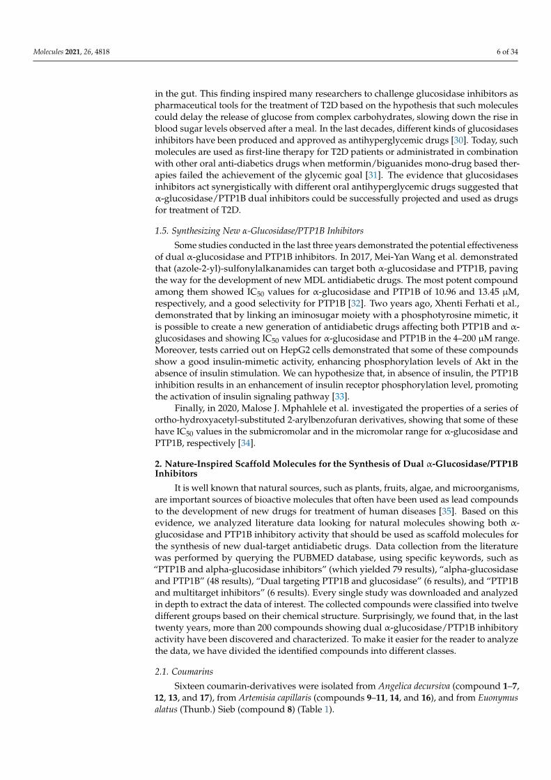
1.1. Combination versus Mono-Drug Therapy for Treatment of T2D The combination of two or more anti-hyperglycemic drugs acting on different bio-
logical targets is a therapeutic option often used for treatment of diabetic patients who do not adequately respond to mono-drug therapy [3]. From a theoretical point of view, the purpose of this strategy is to generate a synergistic effect by acting on different targets involved directly or indirectly in the control of glycemia (Figure 2).
Figure 2. Antidiabetic strategy based on combination therapy: patients are treated with two or more antihyperglycemic oral drugs also administered at different times of the day.
This approach aims to generate a synergistic effect, improving glycemic control using a lower dosage of each drug than the doses provided by mono-drug therapy. However, despite the undoubted advantages of such a pharmacological approach, many clinical tri-als revealed that multi-drug therapies are difficult to manage for the majority of patients for different reasons. For example, often a fine adjustment of dosages is required, or pa-tients are asked to take medications at different times of the day because of their different
Figure 1. Antidiabetic strategy based on the mono-drug therapy: each antidiabetic drug is adminis-tered alone and works on a specific target.
1.1. Combination versus Mono-Drug Therapy for Treatment of T2D
The combination of two or more anti-hyperglycemic drugs acting on different bio-logical targets is a therapeutic option often used for treatment of diabetic patients whodo not adequately respond to mono-drug therapy [3]. From a theoretical point of view,the purpose of this strategy is to generate a synergistic effect by acting on different targetsinvolved directly or indirectly in the control of glycemia (Figure 2).
Molecules 2021, 26, x 2 of 35
insulin. Indeed, even if these drugs are able to compensate a specific metabolic defect, they unexpectedly induce severe unbalance in other metabolic pathways.
Figure 1. Antidiabetic strategy based on the mono-drug therapy: each antidiabetic drug is adminis-tered alone and works on a specific target.
1.1. Combination versus Mono-Drug Therapy for Treatment of T2D The combination of two or more anti-hyperglycemic drugs acting on different bio-
logical targets is a therapeutic option often used for treatment of diabetic patients who do not adequately respond to mono-drug therapy [3]. From a theoretical point of view, the purpose of this strategy is to generate a synergistic effect by acting on different targets involved directly or indirectly in the control of glycemia (Figure 2).
Figure 2. Antidiabetic strategy based on combination therapy: patients are treated with two or more antihyperglycemic oral drugs also administered at different times of the day.
This approach aims to generate a synergistic effect, improving glycemic control using a lower dosage of each drug than the doses provided by mono-drug therapy. However, despite the undoubted advantages of such a pharmacological approach, many clinical tri-als revealed that multi-drug therapies are difficult to manage for the majority of patients for different reasons. For example, often a fine adjustment of dosages is required, or pa-tients are asked to take medications at different times of the day because of their different
Figure 2. Antidiabetic strategy based on combination therapy: patients are treated with two or moreantihyperglycemic oral drugs also administered at different times of the day.
This approach aims to generate a synergistic effect, improving glycemic control using alower dosage of each drug than the doses provided by mono-drug therapy. However, despitethe undoubted advantages of such a pharmacological approach, many clinical trials revealedthat multi-drug therapies are difficult to manage for the majority of patients for differentreasons. For example, often a fine adjustment of dosages is required, or patients are asked totake medications at different times of the day because of their different pharmacokinetics.These factors make it very difficult for patients to comply with the therapeutic protocol, thusincreasing the risk of not reaching the glycemic target. The consequent uncontrolled fluctua-tions in blood sugar can be deleterious, forcing patients to change treatment to avoid furthercomplications. A practical solution to this problem may arise from taking combinationsof oral hypoglycemic drugs in fixed and pre-established doses depending on the desired

Molecules 2021, 26, 4818 3 of 34
effects. This strategy reduces the complexity of therapeutic regimen and improves patientadherence to treatment [4]. Overall, the clinical studies carried out so far confirmed thatcombinatorial therapies imply, in the short to medium term, significant benefits compared tomono-therapy. However, in the long term, the efficacy of this therapeutic approach remainsto be confirmed [5]. In conclusion, all evidence suggests that multitargets therapies seem toguarantee a better quality of life for people affected by T2D.
1.2. Toward the Multiple Designed Ligands (MDLs) for Treatment of T2D
Studies performed in recent decades have unexpectedly shown that, contrary to whatwas originally hypothesized, numerous single-target drugs behave as multiple ligandsin vivo [6]. In clinical practice, it is not easy to predict the in-vivo effects of a moleculecapable of acting on different targets. In fact, depending on the dose and its pharmacoki-netics, it could generate both positive or negative effects especially if used in treatment ofchronic multifactorial diseases. However, the belief that MDLs can offer many advantagesover combinatorial mono-therapies has prompted many researchers in developing newmultiple-ligands drugs for T2D treatment (Figure 3). In principle, a molecule capable ofacting as an MDL should offer the advantages of a combination therapy but with fewerside effects.
Molecules 2021, 26, x 3 of 35
pharmacokinetics. These factors make it very difficult for patients to comply with the ther-apeutic protocol, thus increasing the risk of not reaching the glycemic target. The conse-quent uncontrolled fluctuations in blood sugar can be deleterious, forcing patients to change treatment to avoid further complications. A practical solution to this problem may arise from taking combinations of oral hypoglycemic drugs in fixed and pre-established doses depending on the desired effects. This strategy reduces the complexity of therapeu-tic regimen and improves patient adherence to treatment [4]. Overall, the clinical studies carried out so far confirmed that combinatorial therapies imply, in the short to medium term, significant benefits compared to mono-therapy. However, in the long term, the effi-cacy of this therapeutic approach remains to be confirmed [5]. In conclusion, all evidence suggests that multitargets therapies seem to guarantee a better quality of life for people affected by T2D.
1.2. Toward the Multiple Designed Ligands (MDLs) for Treatment of T2D Studies performed in recent decades have unexpectedly shown that, contrary to what
was originally hypothesized, numerous single-target drugs behave as multiple ligands in vivo [6]. In clinical practice, it is not easy to predict the in-vivo effects of a molecule capable of acting on different targets. In fact, depending on the dose and its pharmacokinetics, it could generate both positive or negative effects especially if used in treatment of chronic multifactorial diseases. However, the belief that MDLs can offer many advantages over combinatorial mono-therapies has prompted many researchers in developing new multi-ple-ligands drugs for T2D treatment (Figure 3). In principle, a molecule capable of acting as an MDL should offer the advantages of a combination therapy but with fewer side effects.
Figure 3. Antidiabetic strategy based on administration of multiple designed ligands: a single drug regulates different specific targets.
Although this idea is considered exciting, the identification of appropriate multifunc-tional scaffolds represents the main challenge for researchers engaged in the generation of new MDLs. The debate among scientists regarding the best strategy to obtain the best multifunctional scaffold remains heated. Large-scale screening and knowledge-based ap-proaches are considered the most effective strategies for designing and developing multi-target molecules. The first approach relies on the fact that a known drug actually behaves like a multiple ligand. Therefore, based on this hypothesis, synthesizing new molecules would no longer be required, but rather it would be sufficient to identify new potential ligands in addition to those already known. Conversely, the knowledge-based approach
Figure 3. Antidiabetic strategy based on administration of multiple designed ligands: a single drugregulates different specific targets.
Although this idea is considered exciting, the identification of appropriate multifunc-tional scaffolds represents the main challenge for researchers engaged in the generationof new MDLs. The debate among scientists regarding the best strategy to obtain the bestmultifunctional scaffold remains heated. Large-scale screening and knowledge-basedapproaches are considered the most effective strategies for designing and developingmulti-target molecules. The first approach relies on the fact that a known drug actuallybehaves like a multiple ligand. Therefore, based on this hypothesis, synthesizing newmolecules would no longer be required, but rather it would be sufficient to identify newpotential ligands in addition to those already known. Conversely, the knowledge-basedapproach exploits previous information obtained through structure-activity analysis (SAR)focusing on the mode of interaction between molecules of interest and their biologicaltargets. Then, once more appropriate molecules are identified, these are chemically linkedor combined together to generate new multi-target drugs that will be subsequently testedto evaluate their real effectiveness.
Besides those aforementioned, several in-silico methods can be used to select po-tential pharmacophores useful for assembling new MDLs. The advantage of using thecomputational approach is its ability to easily perform high-throughput analyses, starting

Molecules 2021, 26, 4818 4 of 34
from databases containing hundreds to thousands of different compounds. The selectedpharmacophores then can be linked together to produce new MDLs. In a forward step, themost promising molecules can be further modified to improve their affinity towards theligands and safety profile while reducing their toxicity [7].
1.3. New MDLs for Treatment of T2D
The emerging interest of scientists and pharmaceutical companies versus multipleligand drugs is evidenced by the growing number of MDLs produced in the last yearsfor treatment of T2D. Coskun and co-workers projected and synthesized dual glucose-dependent insulinotropic polypeptide receptor (GIP-R) and glucagon-like peptide-1 re-ceptor (GLP-1) agonists and demonstrated that treatment with such molecule stimulatedinsulin release, leading to a significant reduction of both fasting and postprandial gly-caemia [8]. In recent years, convincing evidence suggested that GLP-1 acts as anorexigenicpeptide binding to GLP-1R in the hypothalamic region, inducing satiety [9]. Interestingly,it has been demonstrated that, in the brain, GLP-1 acts synergistically with PYY (peptideYY), a peptide that is co-secreted with GLP-1 from enteroendocrine L cells of the intestineand binds NPY2R (Neuropeptide Y receptor Y2 receptor). This finding stimulated manyresearchers to evaluate the activity of new GLP-1R/NPY2R agonists as antidiabetic agents.It has been demonstrated that GLP-1R/NPY2R dual agonists in vivo exert anorectic effectsalong with the ability to reduce blood glucose levels, thereby confirming that they couldact as promising anti-obesity and antihyperglycemic agents [10].
Among all biological targets, peroxisome proliferation-activated receptors (PPARs)are considered some of the most effective ones for treatment of T2D. Various type of PPARsare differently expressed in human tissues; namely, PPAR-α, δ, and γ, have been identifiedand characterized to date. The PPAR-α is highly expressed in liver, kidney, heart muscle,and vascular endothelial cells, where its activation promotes fatty acid oxidation, therebyavoiding accumulation of intracellular lipids depots. Treatment with PPAR-α agonistsincreases cardiac performances in diabetic patients, reducing the risk of stroke [11]. PPAR-δis ubiquitously expressed, and its activation leads to different effects. In muscle cells,PPAR-δ activation stimulates fatty acids oxidation, while it reduces glucose utilization.In adipose cells, PPAR-δ activation increases the expression of genes involved in fattyacids β-oxidation and energy dissipation via uncoupling of fatty acids oxidation and ATPproduction [12].
Interestingly, it has been demonstrated that the balance of fatty acids oxidation andsynthesis can affect inflammatory and immunosuppressive T cells and macrophages. Inmacrophages, PPAR-δ activation impairs polarization toward M2-like phenotype withreduced inflammatory potential. According to this hypothesis, antidiabetic functions ofPPAR-δ have been associated with reduced inflammatory signalling [13]. PPARγ is largelyexpressed in adipose tissue, and its activation promotes proliferation and differentiationof preadipocytes into adipocytes. Moreover, PPARγ agonists stimulate deposition offatty acids into adipocytes, lowering fatty acids blood levels, preventing hyperlipidemia,and increasing peripheral insulin sensitivity [14]. Saroglitazar, a PPARα/γ dual agonist,has been recently approved in India for treatment of diabetic dyslipidemia based on thepromising results obtained in clinical trials. Diabetic patients treated with Saroglitazarshowed no adverse events, an improved lipidemic profile, an increased insulin sensitivityand β-cell function [15]. Recently, a novel dual peroxisome proliferator-activated receptoralpha/delta (PPAR-α/δ) agonist was synthesized and tested on animal models. Besidesprotecting the liver from inflammation, and fibrosis, the administration of dual agonistsdecreased hepatic lipids accumulation, protecting animals from the development of liversteatosis [16,17]. Finally, many efforts have been made to generate pan PPAR agonistscombining the pharmacophore motif of PPAR-α, β, and G agonists. Such molecules reducelipids accumulation in the liver and improve liver damage, inflammation, fibrosis, andinsulin resistance [18,19]. Although many new pan PPAR agonists have demonstratedtheir efficacy as antidiabetic drugs in the preclinical phase, subsequent clinical studies have

Molecules 2021, 26, 4818 5 of 34
shown their limitations and revealed their intrinsic toxicity. For these reasons, studies onthese molecules have not progressed further [20].
Very recently, Qi Pan and coworkers evaluated the antidiabetic activity of GLP-1-Fc-FGF21 on diabetic and obese mice models. This new dual targeting agonist, able totarget both the GLP-1 and FGF21 (Fibroblast growth factor 2) pathway, showed a potentantihyperglycemic activity and caused a marked weight loss, suppressing the appetiteand reducing caloric intake. Together, these results suggested that GLP-1/FGF21 dualagonists possess all characteristics to become promising new drugs to fight diabetes andobesity [21].
1.4. Dual α-Glucosidase/PTP1B Inhibitors: A New Drugs against Type 2 Diabetes?
Clinical studies revealed that many people show evident signs of metabolic abnormal-ities years before the diagnosis of T2D. Insulin resistance (IR) is one of the most commonabnormalities. IR can affect liver, skeletal muscle, adipose tissue, pancreas, and hypothala-mic region, generating several metabolic dysfunctions and promoting the onset of T2D.Besides genetic factors, a diet rich in simple carbohydrates, sedentary behavior, and obesityare thought to be the main risk factors responsible for the development of insulin resis-tance [22]. These evidence suggest that all measures that limit glucose absorption andincrease insulin sensitivity should be the first-line approaches recommended to reduce therisk of developing T2D.
We are convinced that MDLs targeting both PTP1B and α-glucosidases could be usedto reach the goal (Figure 4).
Molecules 2021, 26, x 5 of 35
sensitivity and β-cell function [15]. Recently, a novel dual peroxisome proliferator-acti-vated receptor alpha/delta (PPAR-α/δ) agonist was synthesized and tested on animal models. Besides protecting the liver from inflammation, and fibrosis, the administration of dual agonists decreased hepatic lipids accumulation, protecting animals from the de-velopment of liver steatosis [16,17]. Finally, many efforts have been made to generate pan PPAR agonists combining the pharmacophore motif of PPAR-α, β, and ɣ agonists. Such molecules reduce lipids accumulation in the liver and improve liver damage, inflamma-tion, fibrosis, and insulin resistance [18,19]. Although many new pan PPAR agonists have demonstrated their efficacy as antidiabetic drugs in the preclinical phase, subsequent clin-ical studies have shown their limitations and revealed their intrinsic toxicity. For these reasons, studies on these molecules have not progressed further [20].
Very recently, Qi Pan and coworkers evaluated the antidiabetic activity of GLP-1-Fc-FGF21 on diabetic and obese mice models. This new dual targeting agonist, able to target both the GLP-1 and FGF21 (Fibroblast growth factor 2) pathway, showed a potent antihy-perglycemic activity and caused a marked weight loss, suppressing the appetite and re-ducing caloric intake. Together, these results suggested that GLP-1/FGF21 dual agonists possess all characteristics to become promising new drugs to fight diabetes and obesity [21].
1.4. Dual α-Glucosidase/PTP1B Inhibitors: A New Drugs against Type 2 Diabetes? Clinical studies revealed that many people show evident signs of metabolic abnor-
malities years before the diagnosis of T2D. Insulin resistance (IR) is one of the most com-mon abnormalities. IR can affect liver, skeletal muscle, adipose tissue, pancreas, and hy-pothalamic region, generating several metabolic dysfunctions and promoting the onset of T2D. Besides genetic factors, a diet rich in simple carbohydrates, sedentary behavior, and obesity are thought to be the main risk factors responsible for the development of insulin resistance [22]. These evidence suggest that all measures that limit glucose absorption and increase insulin sensitivity should be the first-line approaches recommended to reduce the risk of developing T2D.
We are convinced that MDLs targeting both PTP1B and α-glucosidases could be used to reach the goal (Figure 4).
Figure 4. Mechanism of action of a dual PTP1B/α-glucosidase inhibitor. Such molecules possess a structure able to interact with both targets, thereby leading to their inhibition.
The tyrosine phosphatase 1B (PTP1B) acts as a key negative regulator of insulin re-ceptor, and a plethora of studies confirmed that uncontrolled activity of this enzyme is one of the main causes that lead to IR [23]. According to this hypothesis, it has been demonstrated that the overexpression of PTP1B promotes IR in liver [24], muscle [25], adipose tissue [26], pancreas [27], and brain [28]. Conversely, many studies confirmed that PTP1B downregulation or inhibition improves insulin sensitivity, normalizes blood glu-cose levels, and protects from obesity and the onset of T2D [29]. Overall, such evidence
Figure 4. Mechanism of action of a dual PTP1B/α-glucosidase inhibitor. Such molecules possess astructure able to interact with both targets, thereby leading to their inhibition.
The tyrosine phosphatase 1B (PTP1B) acts as a key negative regulator of insulinreceptor, and a plethora of studies confirmed that uncontrolled activity of this enzymeis one of the main causes that lead to IR [23]. According to this hypothesis, it has beendemonstrated that the overexpression of PTP1B promotes IR in liver [24], muscle [25],adipose tissue [26], pancreas [27], and brain [28]. Conversely, many studies confirmedthat PTP1B downregulation or inhibition improves insulin sensitivity, normalizes bloodglucose levels, and protects from obesity and the onset of T2D [29]. Overall, such evidencesuggest that PTP1B targeting could generate a pleiotropic effect, improving insulin responsein liver, muscle adipose tissue, pancreas, and brain, thereby correcting most metabolicabnormalities observed in diabetic patients. Since PTP1B does not have a role in regulatingintestinal absorbance of glucose, it is improbable that small molecules designed to targetthis enzyme could be used to regulate intestinal absorption of glucose.
Monosaccharides, such as glucose, fructose, and galactose, are the only sugars ab-sorbed by gut. Oligosaccharides derived from starch digestion are processed by pancreaticα-amylase and intestinal α-glucosidase to produce free glucose that is then uploaded fromintestinal cells. Therefore, the rate of blood glucose raising mainly depends on the gutglucose concentration that, in turn, is influenced by the activity of glucosidases present
Molecules 2021, 26, 4818 6 of 34
in the gut. This finding inspired many researchers to challenge glucosidase inhibitors aspharmaceutical tools for the treatment of T2D based on the hypothesis that such moleculescould delay the release of glucose from complex carbohydrates, slowing down the rise inblood sugar levels observed after a meal. In the last decades, different kinds of glucosidasesinhibitors have been produced and approved as antihyperglycemic drugs [30]. Today, suchmolecules are used as first-line therapy for T2D patients or administrated in combinationwith other oral anti-diabetics drugs when metformin/biguanides mono-drug based ther-apies failed the achievement of the glycemic goal [31]. The evidence that glucosidasesinhibitors act synergistically with different oral antihyperglycemic drugs suggested thatα-glucosidase/PTP1B dual inhibitors could be successfully projected and used as drugsfor treatment of T2D.
1.5. Synthesizing New α-Glucosidase/PTP1B Inhibitors
Some studies conducted in the last three years demonstrated the potential effectivenessof dual α-glucosidase and PTP1B inhibitors. In 2017, Mei-Yan Wang et al. demonstratedthat (azole-2-yl)-sulfonylalkanamides can target both α-glucosidase and PTP1B, pavingthe way for the development of new MDL antidiabetic drugs. The most potent compoundamong them showed IC50 values for α-glucosidase and PTP1B of 10.96 and 13.45 µM,respectively, and a good selectivity for PTP1B [32]. Two years ago, Xhenti Ferhati et al.,demonstrated that by linking an iminosugar moiety with a phosphotyrosine mimetic, itis possible to create a new generation of antidiabetic drugs affecting both PTP1B and α-glucosidases and showing IC50 values for α-glucosidase and PTP1B in the 4–200 µM range.Moreover, tests carried out on HepG2 cells demonstrated that some of these compoundsshow a good insulin-mimetic activity, enhancing phosphorylation levels of Akt in theabsence of insulin stimulation. We can hypothesize that, in absence of insulin, the PTP1Binhibition results in an enhancement of insulin receptor phosphorylation level, promotingthe activation of insulin signaling pathway [33].
Finally, in 2020, Malose J. Mphahlele et al. investigated the properties of a series ofortho-hydroxyacetyl-substituted 2-arylbenzofuran derivatives, showing that some of thesehave IC50 values in the submicromolar and in the micromolar range for α-glucosidase andPTP1B, respectively [34].
2. Nature-Inspired Scaffold Molecules for the Synthesis of Dual α-Glucosidase/PTP1BInhibitors
It is well known that natural sources, such as plants, fruits, algae, and microorganisms,are important sources of bioactive molecules that often have been used as lead compoundsto the development of new drugs for treatment of human diseases [35]. Based on thisevidence, we analyzed literature data looking for natural molecules showing both α-glucosidase and PTP1B inhibitory activity that should be used as scaffold molecules forthe synthesis of new dual-target antidiabetic drugs. Data collection from the literaturewas performed by querying the PUBMED database, using specific keywords, such as“PTP1B and alpha-glucosidase inhibitors” (which yielded 79 results), “alpha-glucosidaseand PTP1B” (48 results), “Dual targeting PTP1B and glucosidase” (6 results), and “PTP1Band multitarget inhibitors” (6 results). Every single study was downloaded and analyzedin depth to extract the data of interest. The collected compounds were classified into twelvedifferent groups based on their chemical structure. Surprisingly, we found that, in the lasttwenty years, more than 200 compounds showing dual α-glucosidase/PTP1B inhibitoryactivity have been discovered and characterized. To make it easier for the reader to analyzethe data, we have divided the identified compounds into different classes.
2.1. Coumarins
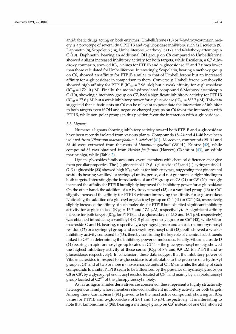
Sixteen coumarin-derivatives were isolated from Angelica decursiva (compound 1–7,12, 13, and 17), from Artemisia capillaris (compounds 9–11, 14, and 16), and from Euonymusalatus (Thunb.) Sieb (compound 8) (Table 1).
Molecules 2021, 26, 4818 7 of 34
Table 1. Coumarins.
Compound IC50 (µM)ReferencePTP1B α-Glucosidase
1 (+)-trans-decursidinol 2.33 ± 0.07 11.32 ± 0.56 [36]
2 Pd-C-I 4.32 ± 0.12 17.40 ± 0.33 [36]3 Pd-C-II 6.17 ± 0.31 24.74 ± 0.89 [36]
4 2′-Isopropylpsoralene 10.78 ± 0.17 85.82 ± 0.99 [37]
5 4′-HydroxyPd-C-III 5.39 ± 0.19 77.30 ± 1.07 [37]
6 Pd-C-III 11.98 ± 0.43 36.77 ± 1.04 [36]
7 4′-MethoxyPd-C-I 6.62 ± 0.77 89.19 ± 0.77 [37]
8 Euonymalatus 13.7 ± 0.2 9.1 ± 0.5 [38]9 Esculetin 10.1 ± 1.9 82.92 ± 4.81 [39]
10 6-Methoxyartemicapin C 27.61 ± 8.30 563.75 ± 6.55 [39]
11 Daphnetin 164.50 ± 7.44 560.20 ± 19.60 [39]12 Decursidin 11.22 ± 0.39 79.09 ± 0.11 [37]13 Decursinol 58.90 ± 1.07 65.29 ± 0.81 [37]14 Scopoletin 227.28 ± 26.71 159.16 ± 11.71 [39]15 Selaginolide A 7.40 ± 0.28 7.52 ± 0.37 [40]16 Umbelliferone 274.86 ± 20.27 633.94 ± 23.78 [39]
17 Umbelliferone6-carboxylic 7.98 ± 0.91 172.10 ± 0.19 [37]
The dihydroxanthyletin-type family includes (+)-trans-decursinol (1), Pd-C-I (2), Pd-C-II (3), Pd-C-III (4), 4′-Hydroxy Pd-C-III (5), and 4′-Methoxy Pd-C-I (7). Among these,(+)-trans-decursinol (1), a molecule bearing two hydroxyl groups on C3 and C4 of pyrenemoiety, resulted the most potent inhibitor of both targets, showing IC50 values of 2.33 and11.32 µM for PTP1B and α-glucosidase, respectively. Conversely, Decursinol (12), whichis lacking in OH on C4, showed a lower affinity for both targets. This result suggestedthe pivotal role of this hydroxyl group in improving the inhibitory activity of naturaldihydroxanthyletin-type coumarins. Interestingly, the replacement of OH on C4 with asenecioyl group produced Pd-C-II (3), which exhibited a slightly decreased inhibitoryactivity in respect to (1), indicating that this group only partially succeeds in substitutingthe hydroxyl group. Pd-C-I (2), a derivative bearing a senecioyl group on C3, showedIC50 values for PTP1B and α-glucosidase similar to those of (+)-trans-decursinol. Onthe other hand, 4′-Hydroxy Pd-C-III (5), bearing an angeloyl group on C3, maintained asignificant inhibitory activity toward PTP1B but showed a weaker inhibitory activity onα-glucosidase in comparison to (+)-trans-decursinol, suggesting that the nature of aliphaticchain is crucial to stabilize the α-glucosidase-inhibitor complex. The substitution of 4 OHwith an acetyl group leads to Pd-C-III (6), which showed a lower affinity for PTP1B buta better inhibitory capacity towards the α-glucosidase compared to (5). Conversely, theinsertion of a methoxy group on C4 of Pd-C-I leads to Methoxy Pd-C-I (7), a compoundshowing an inhibitory activity on PTP1B comparable to that of parental molecule buta slower affinity for the α-glucosidase. Finally, Decursidin (12), bearing two senecioylgroups on C3 and C4, respectively, showed a reduced inhibitory activity for both targetscompared to (+)-trans-decursinol, thereby confirming the OH groups are important toreinforce the binding of dihydroxanthyletin-type coumarins with PTP1B and α-glucosidase.Among phenyl-coumarins, Selaginolide A (15), a 7-hydroxycoumarin derivatives bearing a4-hydroxy-3,6-dimethoxyphenyl ring linked to C3, was proven the most potent comparedto Euonymalatus (8), showing the latter had a more complex structure and several OHgroups. However, both compounds showed a balanced and potent inhibitory activity forboth targets, suggesting that phenolic coumarins are good lead compounds to generate new
Molecules 2021, 26, 4818 8 of 34
antidiabetic drugs acting on both enzymes. Umbelliferone (16) or 7-hydroxycoumarin moi-ety is a prototype of several dual PTP1B and α-glucosidase inhibitors, such as Esculetin (9),Daphnetin (8), Scopoletin (14), Umbelliferone 6-carboxylic (17), and 6-Methoxy artemicapinC (10). Daphnetin, bearing an additional OH group on C8 compared to Umbelliferone,showed a slight increased inhibitory activity for both targets, while Esculetin, a 6,7 dihy-droxy coumarin, showed IC50 values for PTP1B and α-glucosidase 27 and 7 times lowerthan those calculated for Umbelliferone. Interestingly, Scopoletin, bearing a methoxy groupon C6, showed an affinity for PTP1B similar to that of Umbelliferone but an increasedaffinity for α-glucosidase in comparison to them. Conversely, Umbelliferone 6-carboxylicshowed high affinity for PTP1B (IC50 = 7.98 µM) but a weak affinity for α-glucosidase(IC50 = 172.10 µM). Finally, the mono-hydroxylated compound 6-Methoxy artemicapinC (10), showing a methoxy group on C7, had a significant inhibitory activity for PTP1B(IC50 = 27.6 µM) but a weak inhibitory power for α-glucosidase (IC50 = 563.7 µM). This datasuggested that substituents on C6 can be relevant to potentiate the interaction of inhibitorto both targets even if OH and negative-charged groups on C6 favor the interaction withPTP1B, while non-polar groups in this position favor the interaction with α-glucosidase.
2.2. Lignans
Numerous lignans showing inhibitory activity toward both PTP1B and α-glucosidasehave been recently isolated from various plants. Compounds 18–24 and 41–48 have beenisolated from Viburnum macrocephalum f. keteleeri [41]. Moreover, compounds 25–31 and33–40 were extracted from the roots of Limonium gmelinii (Willd.) Kuntze [42], whilecompound 32 was obtained from Hizikia fusiformis (Harvey) Okamura [43], an ediblemarine alga, while (Table 2).
Lignans glycosides family accounts several members with chemical differences that givethem peculiar properties. The (+)-pinoresinol 4-O-β-D-glucoside (22) and (+)-syringaresinol 4-O-β-D-glucoside (23) showed high IC50 values for both enzymes, suggesting that pinoresinolscaffolds bearing vanilloyl or syringoyl units, per se, did not guarantee a tight binding toboth targets. Interestingly, the introduction of an OH group on C8 (21) or C8′ (20) stronglyincreased the affinity for PTP1B but slightly improved the inhibitory power for α-glucosidase.On the other hand, the addition of a p-hydroxybenzoyl (45) or a vanilloyl group (46) to C6′′
slightly increased the affinity for PTP1B without improving the affinity for α-glucosidase.Noticeably, the addition of a glucosyl or galactosyl group on C4′′ (41) or C2′′ (42), respectively,slightly increased the affinity of such molecules for PTP1B but exhibited significant inhibitoryactivity for α-glucosidase (IC50 = 16.7 and 17.1 µM, respectively). A significant affinityincrease for both targets (IC50 for PTP1B and α-glucosidase of 25.8 and 16.1 µM, respectively)was obtained introducing a vanilloyl-4-O-β-glucopyranosyl group on C6′′ (43), while Vibur-macroside G and H, bearing, respectively, a syringoyl group and an α-L-rhamnopyranosylresidue (47) or a syringoyl group and α-D-xylopyranosyl unit (48), both showed a weakerinhibitory activity compared to (43), thereby confirming the key role of chemical substituentslinked to C6′′ in determining the inhibitory power of molecules. Finally, Viburmacroside D(44) bearing an apiofuranosyl group located at C2′′′′ of the glucopyranosyl moiety, showedthe highest inhibitory activity of these series (IC50 of 8.9 and 9.9 µM for PTP1B and α-glucosidase, respectively). In conclusion, these data suggest that the inhibitory power ofViburmacrosides in respect to α-glucosidase is attributable to the presence of a hydroxylgroup at C4′ and of two or more monosaccharide units at C4. Meanwhile, the ability of suchcompounds to inhibit PTP1B seem to be influenced by the presence of hydroxyl groups onC8 or C8′, by a glycosyl-phenolic acyl residue located at C6′′, and mainly by an apiofuranosylgroup located at C2′′′′ of the glucopyranosyl moiety.
As far as lignanamides derivatives are concerned, these represent a highly structurallyheterogenous family whose members showed a different inhibitory activity for both targets.Among these, Cannabisin I (31) proved to be the most active compound, showing an IC50value for PTP1B and α-glucosidase of 2.01 and 1.5 µM, respectively. It is interesting tonote that Limoniumin B (34), bearing a methoxyl group on C3′ instead of one OH, showed
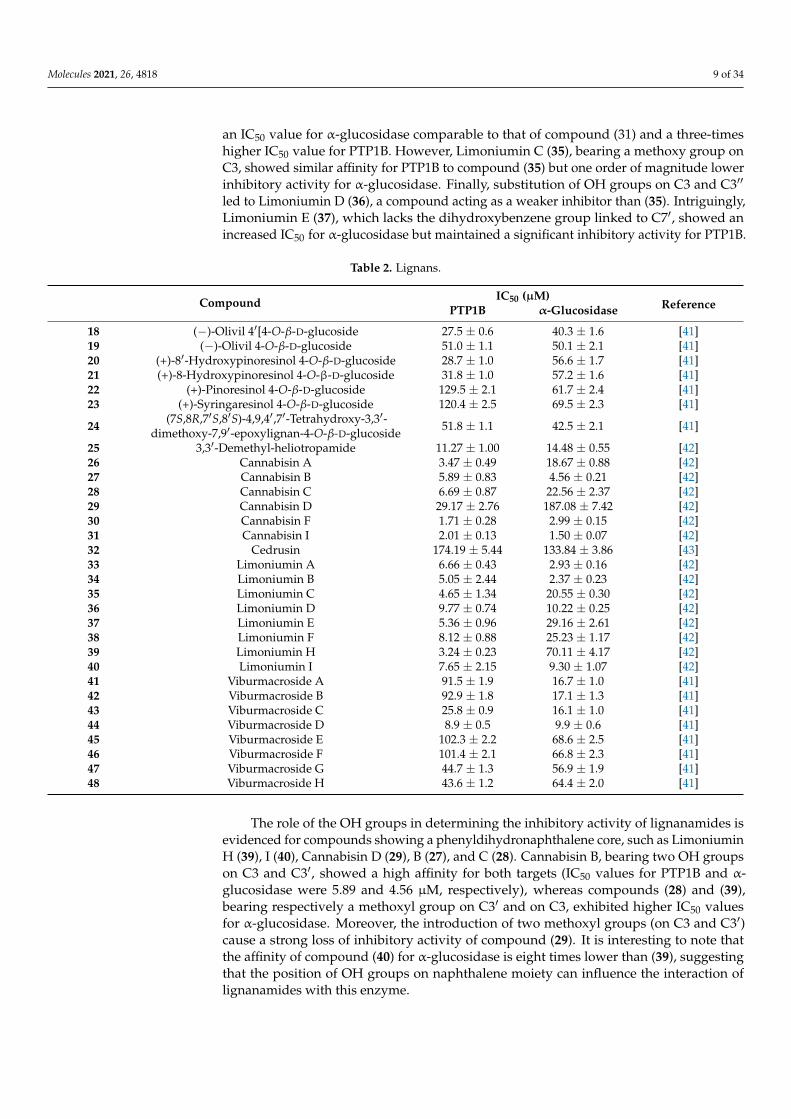
Molecules 2021, 26, 4818 9 of 34
an IC50 value for α-glucosidase comparable to that of compound (31) and a three-timeshigher IC50 value for PTP1B. However, Limoniumin C (35), bearing a methoxy group onC3, showed similar affinity for PTP1B to compound (35) but one order of magnitude lowerinhibitory activity for α-glucosidase. Finally, substitution of OH groups on C3 and C3′′
led to Limoniumin D (36), a compound acting as a weaker inhibitor than (35). Intriguingly,Limoniumin E (37), which lacks the dihydroxybenzene group linked to C7′, showed anincreased IC50 for α-glucosidase but maintained a significant inhibitory activity for PTP1B.
Table 2. Lignans.
Compound IC50 (µM)ReferencePTP1B α-Glucosidase
18 (−)-Olivil 4′[4-O-β-D-glucoside 27.5 ± 0.6 40.3 ± 1.6 [41]19 (−)-Olivil 4-O-β-D-glucoside 51.0 ± 1.1 50.1 ± 2.1 [41]20 (+)-8′-Hydroxypinoresinol 4-O-β-D-glucoside 28.7 ± 1.0 56.6 ± 1.7 [41]21 (+)-8-Hydroxypinoresinol 4-O-β-D-glucoside 31.8 ± 1.0 57.2 ± 1.6 [41]22 (+)-Pinoresinol 4-O-β-D-glucoside 129.5 ± 2.1 61.7 ± 2.4 [41]23 (+)-Syringaresinol 4-O-β-D-glucoside 120.4 ± 2.5 69.5 ± 2.3 [41]
24 (7S,8R,7′S,8′S)-4,9,4′,7′-Tetrahydroxy-3,3′-dimethoxy-7,9′-epoxylignan-4-O-β-D-glucoside 51.8 ± 1.1 42.5 ± 2.1 [41]
25 3,3′-Demethyl-heliotropamide 11.27 ± 1.00 14.48 ± 0.55 [42]26 Cannabisin A 3.47 ± 0.49 18.67 ± 0.88 [42]27 Cannabisin B 5.89 ± 0.83 4.56 ± 0.21 [42]28 Cannabisin C 6.69 ± 0.87 22.56 ± 2.37 [42]29 Cannabisin D 29.17 ± 2.76 187.08 ± 7.42 [42]30 Cannabisin F 1.71 ± 0.28 2.99 ± 0.15 [42]31 Cannabisin I 2.01 ± 0.13 1.50 ± 0.07 [42]32 Cedrusin 174.19 ± 5.44 133.84 ± 3.86 [43]33 Limoniumin A 6.66 ± 0.43 2.93 ± 0.16 [42]34 Limoniumin B 5.05 ± 2.44 2.37 ± 0.23 [42]35 Limoniumin C 4.65 ± 1.34 20.55 ± 0.30 [42]36 Limoniumin D 9.77 ± 0.74 10.22 ± 0.25 [42]37 Limoniumin E 5.36 ± 0.96 29.16 ± 2.61 [42]38 Limoniumin F 8.12 ± 0.88 25.23 ± 1.17 [42]39 Limoniumin H 3.24 ± 0.23 70.11 ± 4.17 [42]40 Limoniumin I 7.65 ± 2.15 9.30 ± 1.07 [42]41 Viburmacroside A 91.5 ± 1.9 16.7 ± 1.0 [41]42 Viburmacroside B 92.9 ± 1.8 17.1 ± 1.3 [41]43 Viburmacroside C 25.8 ± 0.9 16.1 ± 1.0 [41]44 Viburmacroside D 8.9 ± 0.5 9.9 ± 0.6 [41]45 Viburmacroside E 102.3 ± 2.2 68.6 ± 2.5 [41]46 Viburmacroside F 101.4 ± 2.1 66.8 ± 2.3 [41]47 Viburmacroside G 44.7 ± 1.3 56.9 ± 1.9 [41]48 Viburmacroside H 43.6 ± 1.2 64.4 ± 2.0 [41]
The role of the OH groups in determining the inhibitory activity of lignanamides isevidenced for compounds showing a phenyldihydronaphthalene core, such as LimoniuminH (39), I (40), Cannabisin D (29), B (27), and C (28). Cannabisin B, bearing two OH groupson C3 and C3′, showed a high affinity for both targets (IC50 values for PTP1B and α-glucosidase were 5.89 and 4.56 µM, respectively), whereas compounds (28) and (39),bearing respectively a methoxyl group on C3′ and on C3, exhibited higher IC50 valuesfor α-glucosidase. Moreover, the introduction of two methoxyl groups (on C3 and C3′)cause a strong loss of inhibitory activity of compound (29). It is interesting to note thatthe affinity of compound (40) for α-glucosidase is eight times lower than (39), suggestingthat the position of OH groups on naphthalene moiety can influence the interaction oflignanamides with this enzyme.

Molecules 2021, 26, 4818 10 of 34
2.3. Xanthones
Several alkylated xanthones active on both PTP1B and α-glucosidases have beenextracted from roots bark of C. Cochinchinense [44] (Table 3).
Table 3. Xanthones.
Compound IC50 (µM)ReferencePTP1B α-Glucosidase
49 γ-Mangostin 2.8 1.7 [44]50 α-Mangostin 5.5 5.7 [44]51 1,3,7-Trihydroxy-2,4-diisoprenylxanthone 10.5 25.5 [44]52 7-Geranyloxy-1,3-dihydroxyxanthone 35.1 20.6 [44]53 Caratoxanthone A 2.4 4.8 [44]54 Cochinchinone Q 52.5 72.7 [44]55 Cochinechinone A 5.2 17.4 [44]56 Cochinxanthone A 25.2 13.7 [44]57 Cratoxanthone E 7.2 16.1 [44]58 Cratoxanthone F 12.5 10.1 [44]59 Cratoxylone 24.1 30.7 [44]60 Pruniflorone S 7.05 28.4 [44]61 Toralactone gentiobioside 81.15 ± 0.15 37.60 ± 0.79 [45]
All compounds isolated behaved as good inhibitors of both targets, showing IC50values in the 1.7–80 µM range for α-glucosidase and between 2.8 and 52.5 µM for PTP1B.Although selected xanthones possess different substituents, they behaved as mixed-typeinhibitors of α-glucosidases and competitive inhibitors of PTP1B. This finding suggeststhat xanthone moiety has a key role in determining the interaction with the active site ofboth targets even if aliphatic chains and hydroxyl groups linked to xanthone structure caninfluence the affinity of each compound.
The γ-Mangostin (49), bearing two prenyl chains on C2 and C8 and four OH groups,resulted as the most potent inhibitor of this series, showing IC50 values for PTP1B andα-glucosidase of 2.8 and 1.7 µM, respectively. The replacement of OH group on C7 with amethoxyl group (α-Mangostin, 50) slightly affected affinity for both targets, while the affin-ity of Cratoxylone (59), also bearing an hydroxylated prenyl chain on C2, was reduced eighttimes for PTP1B and eighteen times for α-glucosidase. The displacement of prenyl chainfrom C8 to C4 impaired the inhibitory activity of 1,3,7-Trihydroxy-2,4-diisoprenylxanthone(51), while the replacement of prenyl chain on C4 with a geranyl group (Cochinechinone A,55) strongly improved the affinity of the molecule for PTP1B. Interestingly, hydroxylationof the prenyl chain on C2 (Caratoxanthone A, 53) but not the hydroxylation of the geranylchain on C4 (Cratoxanthone F, 58) makes compound (53) an inhibitor almost as efficientas compound (49). The Pruniflorone S (60) showed IC50 values similar to those of com-pound (55), suggesting that the substitution of OH groups with an aliphatic chain did notresult in a further enhancement of inhibitory power. 7-Geranyloxy-1,3-dihydroxyxanthone(52) and Cochinxanthone A (56) showed a weak inhibitory activity toward both targets,indicating that the presence of a single geranyl chain was not functional to improve theinhibitory activity of these compounds. Finally, Cochinchinone Q (54), which does notbear aliphatic chains, resulted as the weaker inhibitor of this series toward both targets,thereby confirming the importance of aliphatic chains in stabilizing the xanthones-enzymecomplexes.
2.4. Terpenes
Many terpenes acting on both α-glucosidase and PTP1B have been isolated in the lastyears from Aralia elata (62, 63), Euonymus alatus (Thunb.) Sieb. (71, 74), Hizikia fusiformis (64,65), Agrimonia pilosa (80, 81), Panax ginseng C.A. Meyer (66–70), Myrtus communis Linn. (72,73, 75, 78, 79), and Pueraria lobata (76, 77) (Table 4).
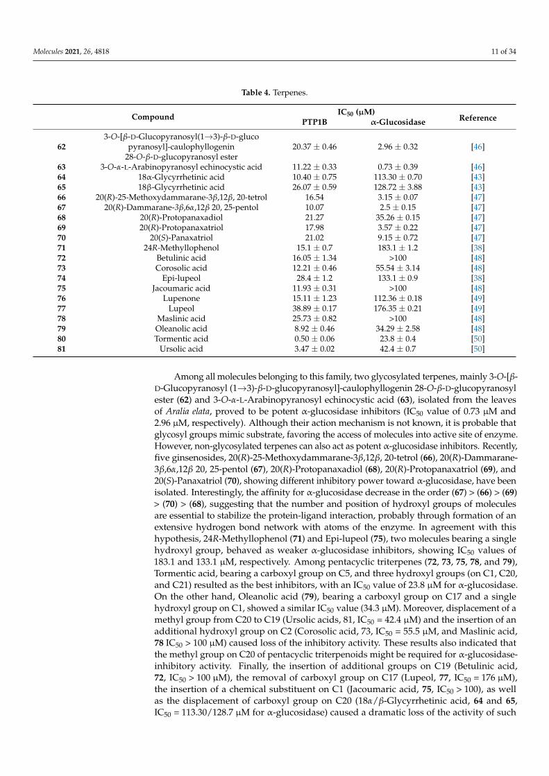
Molecules 2021, 26, 4818 11 of 34
Table 4. Terpenes.
Compound IC50 (µM)ReferencePTP1B α-Glucosidase
623-O-[β-D-Glucopyranosyl(1→3)-β-D-gluco
pyranosyl]-caulophyllogenin28-O-β-D-glucopyranosyl ester
20.37 ± 0.46 2.96 ± 0.32 [46]
63 3-O-α-L-Arabinopyranosyl echinocystic acid 11.22 ± 0.33 0.73 ± 0.39 [46]64 18α-Glycyrrhetinic acid 10.40 ± 0.75 113.30 ± 0.70 [43]65 18β-Glycyrrhetinic acid 26.07 ± 0.59 128.72 ± 3.88 [43]66 20(R)-25-Methoxydammarane-3β,12β, 20-tetrol 16.54 3.15 ± 0.07 [47]67 20(R)-Dammarane-3β,6α,12β 20, 25-pentol 10.07 2.5 ± 0.15 [47]68 20(R)-Protopanaxadiol 21.27 35.26 ± 0.15 [47]69 20(R)-Protopanaxatriol 17.98 3.57 ± 0.22 [47]70 20(S)-Panaxatriol 21.02 9.15 ± 0.72 [47]71 24R-Methyllophenol 15.1 ± 0.7 183.1 ± 1.2 [38]72 Betulinic acid 16.05 ± 1.34 >100 [48]73 Corosolic acid 12.21 ± 0.46 55.54 ± 3.14 [48]74 Epi-lupeol 28.4 ± 1.2 133.1 ± 0.9 [38]75 Jacoumaric acid 11.93 ± 0.31 >100 [48]76 Lupenone 15.11 ± 1.23 112.36 ± 0.18 [49]77 Lupeol 38.89 ± 0.17 176.35 ± 0.21 [49]78 Maslinic acid 25.73 ± 0.82 >100 [48]79 Oleanolic acid 8.92 ± 0.46 34.29 ± 2.58 [48]80 Tormentic acid 0.50 ± 0.06 23.8 ± 0.4 [50]81 Ursolic acid 3.47 ± 0.02 42.4 ± 0.7 [50]
Among all molecules belonging to this family, two glycosylated terpenes, mainly 3-O-[β-D-Glucopyranosyl (1→3)-β-D-glucopyranosyl]-caulophyllogenin 28-O-β-D-glucopyranosylester (62) and 3-O-α-L-Arabinopyranosyl echinocystic acid (63), isolated from the leavesof Aralia elata, proved to be potent α-glucosidase inhibitors (IC50 value of 0.73 µM and2.96 µM, respectively). Although their action mechanism is not known, it is probable thatglycosyl groups mimic substrate, favoring the access of molecules into active site of enzyme.However, non-glycosylated terpenes can also act as potent α-glucosidase inhibitors. Recently,five ginsenosides, 20(R)-25-Methoxydammarane-3β,12β, 20-tetrol (66), 20(R)-Dammarane-3β,6α,12β 20, 25-pentol (67), 20(R)-Protopanaxadiol (68), 20(R)-Protopanaxatriol (69), and20(S)-Panaxatriol (70), showing different inhibitory power toward α-glucosidase, have beenisolated. Interestingly, the affinity for α-glucosidase decrease in the order (67) > (66) > (69)> (70) > (68), suggesting that the number and position of hydroxyl groups of moleculesare essential to stabilize the protein-ligand interaction, probably through formation of anextensive hydrogen bond network with atoms of the enzyme. In agreement with thishypothesis, 24R-Methyllophenol (71) and Epi-lupeol (75), two molecules bearing a singlehydroxyl group, behaved as weaker α-glucosidase inhibitors, showing IC50 values of183.1 and 133.1 µM, respectively. Among pentacyclic triterpenes (72, 73, 75, 78, and 79),Tormentic acid, bearing a carboxyl group on C5, and three hydroxyl groups (on C1, C20,and C21) resulted as the best inhibitors, with an IC50 value of 23.8 µM for α-glucosidase.On the other hand, Oleanolic acid (79), bearing a carboxyl group on C17 and a singlehydroxyl group on C1, showed a similar IC50 value (34.3 µM). Moreover, displacement of amethyl group from C20 to C19 (Ursolic acids, 81, IC50 = 42.4 µM) and the insertion of anadditional hydroxyl group on C2 (Corosolic acid, 73, IC50 = 55.5 µM, and Maslinic acid,78 IC50 > 100 µM) caused loss of the inhibitory activity. These results also indicated thatthe methyl group on C20 of pentacyclic triterpenoids might be required for α-glucosidase-inhibitory activity. Finally, the insertion of additional groups on C19 (Betulinic acid,72, IC50 > 100 µM), the removal of carboxyl group on C17 (Lupeol, 77, IC50 = 176 µM),the insertion of a chemical substituent on C1 (Jacoumaric acid, 75, IC50 > 100), as wellas the displacement of carboxyl group on C20 (18α/β-Glycyrrhetinic acid, 64 and 65,IC50 = 113.30/128.7 µM for α-glucosidase) caused a dramatic loss of the activity of such
Molecules 2021, 26, 4818 12 of 34
molecules for this target. As far as the inhibitory activity on PTP1B is concerned, allcompounds behave as good inhibitors, showing IC50 values in the 0.5–38.9 µM range.Among all, the most potent PTP1B inhibitors were Tormentic acid (IC50 0.5 µM), Ursolicacid (IC50 = 3.4 µM), Oleanolic acid (IC50 = 8.9 µM), and 20(R)-Dammarane-3β,6α,12β 20,25-pentol (IC50 = 10 µM). Of note, compounds 62, 63, and 66–82 showed a good inhibitoryactivity against both targets, suggesting that terpenic moiety represents a versatile scaffoldstructure to generate new bifunctional inhibitors targeting both α-glucosidase and PTP1B.
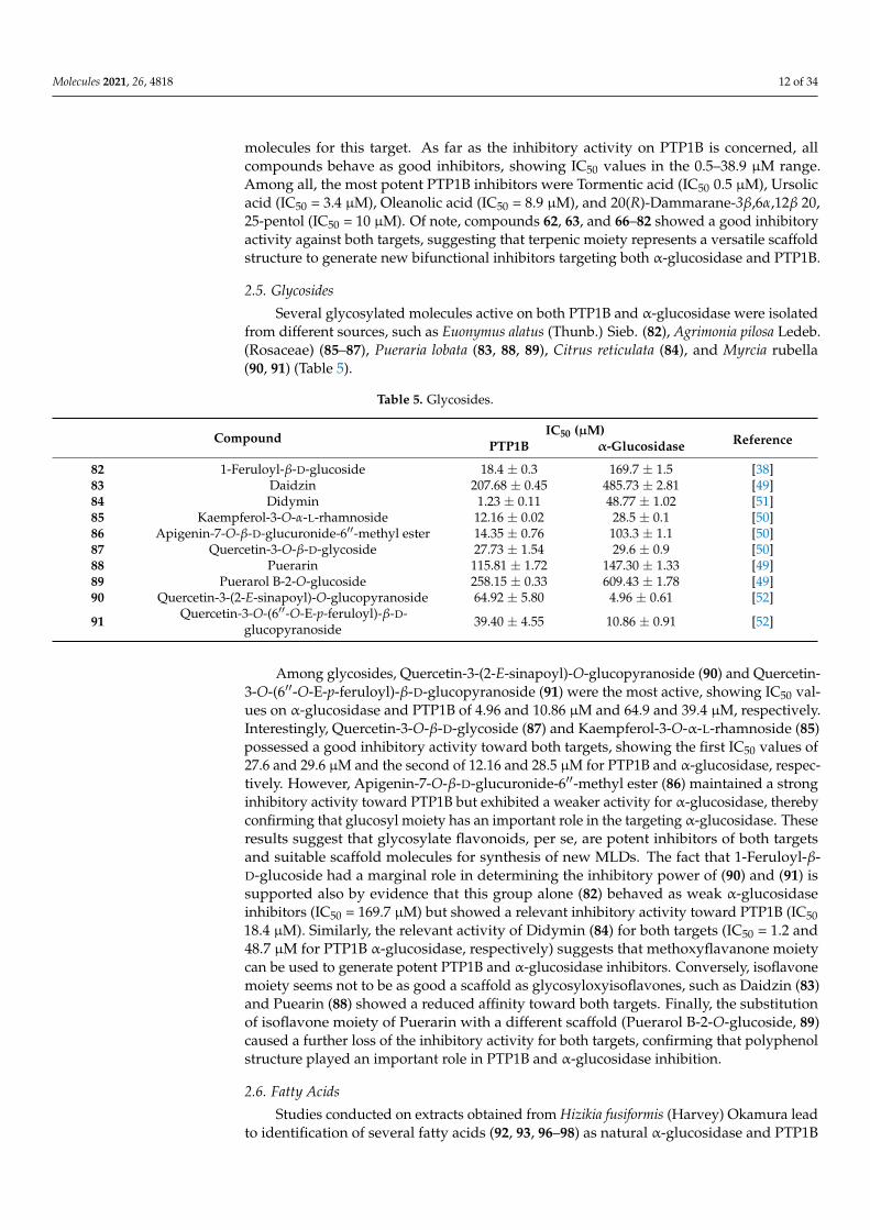
2.5. Glycosides
Several glycosylated molecules active on both PTP1B and α-glucosidase were isolatedfrom different sources, such as Euonymus alatus (Thunb.) Sieb. (82), Agrimonia pilosa Ledeb.(Rosaceae) (85–87), Pueraria lobata (83, 88, 89), Citrus reticulata (84), and Myrcia rubella(90, 91) (Table 5).
Table 5. Glycosides.
Compound IC50 (µM)ReferencePTP1B α-Glucosidase
82 1-Feruloyl-β-D-glucoside 18.4 ± 0.3 169.7 ± 1.5 [38]83 Daidzin 207.68 ± 0.45 485.73 ± 2.81 [49]84 Didymin 1.23 ± 0.11 48.77 ± 1.02 [51]85 Kaempferol-3-O-α-L-rhamnoside 12.16 ± 0.02 28.5 ± 0.1 [50]86 Apigenin-7-O-β-D-glucuronide-6′′-methyl ester 14.35 ± 0.76 103.3 ± 1.1 [50]87 Quercetin-3-O-β-D-glycoside 27.73 ± 1.54 29.6 ± 0.9 [50]88 Puerarin 115.81 ± 1.72 147.30 ± 1.33 [49]89 Puerarol B-2-O-glucoside 258.15 ± 0.33 609.43 ± 1.78 [49]90 Quercetin-3-(2-E-sinapoyl)-O-glucopyranoside 64.92 ± 5.80 4.96 ± 0.61 [52]
91 Quercetin-3-O-(6′′-O-E-p-feruloyl)-β-D-glucopyranoside 39.40 ± 4.55 10.86 ± 0.91 [52]
Among glycosides, Quercetin-3-(2-E-sinapoyl)-O-glucopyranoside (90) and Quercetin-3-O-(6′′-O-E-p-feruloyl)-β-D-glucopyranoside (91) were the most active, showing IC50 val-ues on α-glucosidase and PTP1B of 4.96 and 10.86 µM and 64.9 and 39.4 µM, respectively.Interestingly, Quercetin-3-O-β-D-glycoside (87) and Kaempferol-3-O-α-L-rhamnoside (85)possessed a good inhibitory activity toward both targets, showing the first IC50 values of27.6 and 29.6 µM and the second of 12.16 and 28.5 µM for PTP1B and α-glucosidase, respec-tively. However, Apigenin-7-O-β-D-glucuronide-6′′-methyl ester (86) maintained a stronginhibitory activity toward PTP1B but exhibited a weaker activity for α-glucosidase, therebyconfirming that glucosyl moiety has an important role in the targeting α-glucosidase. Theseresults suggest that glycosylate flavonoids, per se, are potent inhibitors of both targetsand suitable scaffold molecules for synthesis of new MLDs. The fact that 1-Feruloyl-β-D-glucoside had a marginal role in determining the inhibitory power of (90) and (91) issupported also by evidence that this group alone (82) behaved as weak α-glucosidaseinhibitors (IC50 = 169.7 µM) but showed a relevant inhibitory activity toward PTP1B (IC5018.4 µM). Similarly, the relevant activity of Didymin (84) for both targets (IC50 = 1.2 and48.7 µM for PTP1B α-glucosidase, respectively) suggests that methoxyflavanone moietycan be used to generate potent PTP1B and α-glucosidase inhibitors. Conversely, isoflavonemoiety seems not to be as good a scaffold as glycosyloxyisoflavones, such as Daidzin (83)and Puearin (88) showed a reduced affinity toward both targets. Finally, the substitutionof isoflavone moiety of Puerarin with a different scaffold (Puerarol B-2-O-glucoside, 89)caused a further loss of the inhibitory activity for both targets, confirming that polyphenolstructure played an important role in PTP1B and α-glucosidase inhibition.
2.6. Fatty Acids
Studies conducted on extracts obtained from Hizikia fusiformis (Harvey) Okamura leadto identification of several fatty acids (92, 93, 96–98) as natural α-glucosidase and PTP1B

Molecules 2021, 26, 4818 13 of 34
inhibitors. Further fatty acids active on both targets (94, 95) were obtained from Agrimoniapilosa Ledeb. (Rosaceae) (Table 6).
Table 6. Fatty Acids.
Compound IC50 (µM)ReferencePTP1B α-Glucosidase
92 (Z)-Hexadec-12-enoic acid 6.59 ± 0.09 48.05 ± 3.37 [43]93 (Z)-Octaec-9-enoic acid 13.65 ± 0.49 113.44 ± 2.47 [43]94 Methyl 2-hydroxyl tricosanoate 36.39 ± 1.72 112.8 ± 1.7 [50]95 Palmitic acid 0.10 ± 0.03 45.5 ± 1.5 [50]96 (7Z,10Z,13Z)-Octadeca-7,10,13-trienoic acid 13.58 ± 0.10 111.51 ± 1.44 [43]97 (7Z,9Z,11Z,13Z)-Eicosa-7,9,11,13-tetraenoic acid 10.68 ± 0.17 34.85 ± 2.39 [43]98 (8Z,11Z,14Z)-Heptadeca-8,11,14-trienoic acid 11.51 ± 0.52 43.90 ± 0.77 [43]
The isolated compounds (Z)-Hexadec-12-enoic acid (92), Palmitic acid (95), (7Z,9Z,11Z,13Z)-Eicosa-7,9,11,13-tetraenoic acid (97), and (8Z,11Z,14Z)-Heptadeca-8,11,14-trienoic acid (98) exhib-ited the strongest inhibitory activity, with IC50 values comprised between 0.5–11.5 µM for PTP1Band between 34.8–48.05µM range forα-glucosidase. Overall, Palmitic acid, a saturated fatty acid,seemed the most potent inhibitor of PTP1B, showingshowing an affinity for this enzyme similarto that of some unsaturated fatty acids. The only exception is Methyl 2-hydroxyl tricosanoate(94), which showed a reduced affinity for the enzyme. This data suggest that methylation ofcarboxylic acid has a detrimental effect in terms of inhibitory power and that carboxylic acidgroup contributes to stabilize the enzyme-inhibitor complex, probably forming hydrogen bondsor ionic interactions with some amino acids of the target enzyme. Furthermore, as far as theinhibitory activity on α-glucosidase is concerned, is not possible to define a strict correlationbetween fatty acids structure, number of carbon atoms, and inhibitory activity. Palmitic acid,a saturated fatty acid, showed similar activity (IC50 = 45.5 µM) to (Z)-Hexadec-12-enoic acid(92, IC50 = 48.05 µM), a monounsaturated fatty acid or that of (8Z,11Z,14Z)-Heptadeca-8,11,14-trienoic acid (98, IC50 = 43.9 µM), a heptadecenoic acid having three double bonds located atpositions 8, 11, and 14. In addition, (Z)-Octaec-9-enoic acid (93) and (7Z, 10Z, 13Z)-Octadeca-7,10,13-trienoic acid (96), a monounsaturated fatty acid the former and a polyunsaturatedfatty acid the second, showed a similar, never unexpectedly weak inhibitory activity towardsα-glucosidase (IC50 values = 113 and 111 µM, respectively).
2.7. Anthraquinones
Anthraquinones reported in Table 7 were obtained from Cassia obtusifolia L., a legom-nous annual herb growing in tropical countries of Asia [44] (Table 7).
Screening assays carried out using both PTP1B and α-glucosidase revealed that Alater-nin (100) is the most active compound, showing an IC50 value in the low micromolar range(IC50 = 1.22 and 0.99 µM for PTP1B and α-glucosidase, respectively). Furthermore, kineticanalyses revealed that Alaternin acts as a competitive inhibitor of PTP1B and a mixed typeinhibitor of α-glucosidase. In addition, docking analyses performed with PTP1B showedthat hydroxyl groups present on C1, C2, and C6, as well as methyl group present on C3 ofAlaternin, have a key role in stabilizing the Alaternin-PTP1B complex. Moreover, althoughno kinetic and docking data are available on α-glucosidase, it is reasonable to think thathydroxyl groups contribute to stabilize the complex Alaternin/α-glucosidase, too. Accord-ing with this hypothesis, we found that the introduction of a methoxyl group on C1 ofAlaternin generates 2-Hydroxyemodin-1 methylether (99) that showed a reduced activityfor both targets. Moreover, the removal of the OH group on C6 leads to Obstusifolin (113),a molecule showing a good affinity for PTP1B but a reduced affinity for α-glucosidase. Onthe other hand, the removal of the OH group from C2 leads to Emodin (110), a moleculethat possesses an IC50 value unchanged compared to Alaternin but with a lower affinity forPTP1B. In addition, Chrysophanol (107), obtained by removing the OH group from C6 ofEmodin, showed a very weak affinity for α-glucosidase (IC50 = 0.99 µM of Alaternin versus
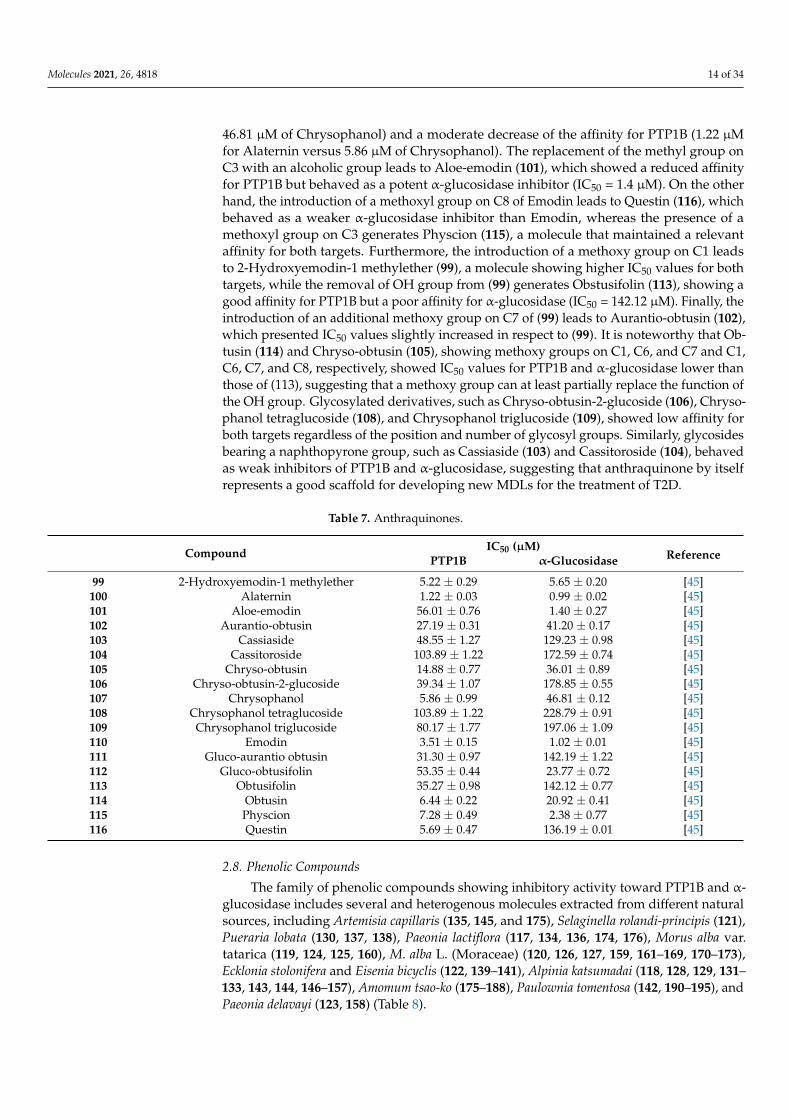
Molecules 2021, 26, 4818 14 of 34
46.81 µM of Chrysophanol) and a moderate decrease of the affinity for PTP1B (1.22 µMfor Alaternin versus 5.86 µM of Chrysophanol). The replacement of the methyl group onC3 with an alcoholic group leads to Aloe-emodin (101), which showed a reduced affinityfor PTP1B but behaved as a potent α-glucosidase inhibitor (IC50 = 1.4 µM). On the otherhand, the introduction of a methoxyl group on C8 of Emodin leads to Questin (116), whichbehaved as a weaker α-glucosidase inhibitor than Emodin, whereas the presence of amethoxyl group on C3 generates Physcion (115), a molecule that maintained a relevantaffinity for both targets. Furthermore, the introduction of a methoxy group on C1 leadsto 2-Hydroxyemodin-1 methylether (99), a molecule showing higher IC50 values for bothtargets, while the removal of OH group from (99) generates Obstusifolin (113), showing agood affinity for PTP1B but a poor affinity for α-glucosidase (IC50 = 142.12 µM). Finally, theintroduction of an additional methoxy group on C7 of (99) leads to Aurantio-obtusin (102),which presented IC50 values slightly increased in respect to (99). It is noteworthy that Ob-tusin (114) and Chryso-obtusin (105), showing methoxy groups on C1, C6, and C7 and C1,C6, C7, and C8, respectively, showed IC50 values for PTP1B and α-glucosidase lower thanthose of (113), suggesting that a methoxy group can at least partially replace the function ofthe OH group. Glycosylated derivatives, such as Chryso-obtusin-2-glucoside (106), Chryso-phanol tetraglucoside (108), and Chrysophanol triglucoside (109), showed low affinity forboth targets regardless of the position and number of glycosyl groups. Similarly, glycosidesbearing a naphthopyrone group, such as Cassiaside (103) and Cassitoroside (104), behavedas weak inhibitors of PTP1B and α-glucosidase, suggesting that anthraquinone by itselfrepresents a good scaffold for developing new MDLs for the treatment of T2D.
Table 7. Anthraquinones.
Compound IC50 (µM)ReferencePTP1B α-Glucosidase
99 2-Hydroxyemodin-1 methylether 5.22 ± 0.29 5.65 ± 0.20 [45]100 Alaternin 1.22 ± 0.03 0.99 ± 0.02 [45]101 Aloe-emodin 56.01 ± 0.76 1.40 ± 0.27 [45]102 Aurantio-obtusin 27.19 ± 0.31 41.20 ± 0.17 [45]103 Cassiaside 48.55 ± 1.27 129.23 ± 0.98 [45]104 Cassitoroside 103.89 ± 1.22 172.59 ± 0.74 [45]105 Chryso-obtusin 14.88 ± 0.77 36.01 ± 0.89 [45]106 Chryso-obtusin-2-glucoside 39.34 ± 1.07 178.85 ± 0.55 [45]107 Chrysophanol 5.86 ± 0.99 46.81 ± 0.12 [45]108 Chrysophanol tetraglucoside 103.89 ± 1.22 228.79 ± 0.91 [45]109 Chrysophanol triglucoside 80.17 ± 1.77 197.06 ± 1.09 [45]110 Emodin 3.51 ± 0.15 1.02 ± 0.01 [45]111 Gluco-aurantio obtusin 31.30 ± 0.97 142.19 ± 1.22 [45]112 Gluco-obtusifolin 53.35 ± 0.44 23.77 ± 0.72 [45]113 Obtusifolin 35.27 ± 0.98 142.12 ± 0.77 [45]114 Obtusin 6.44 ± 0.22 20.92 ± 0.41 [45]115 Physcion 7.28 ± 0.49 2.38 ± 0.77 [45]116 Questin 5.69 ± 0.47 136.19 ± 0.01 [45]
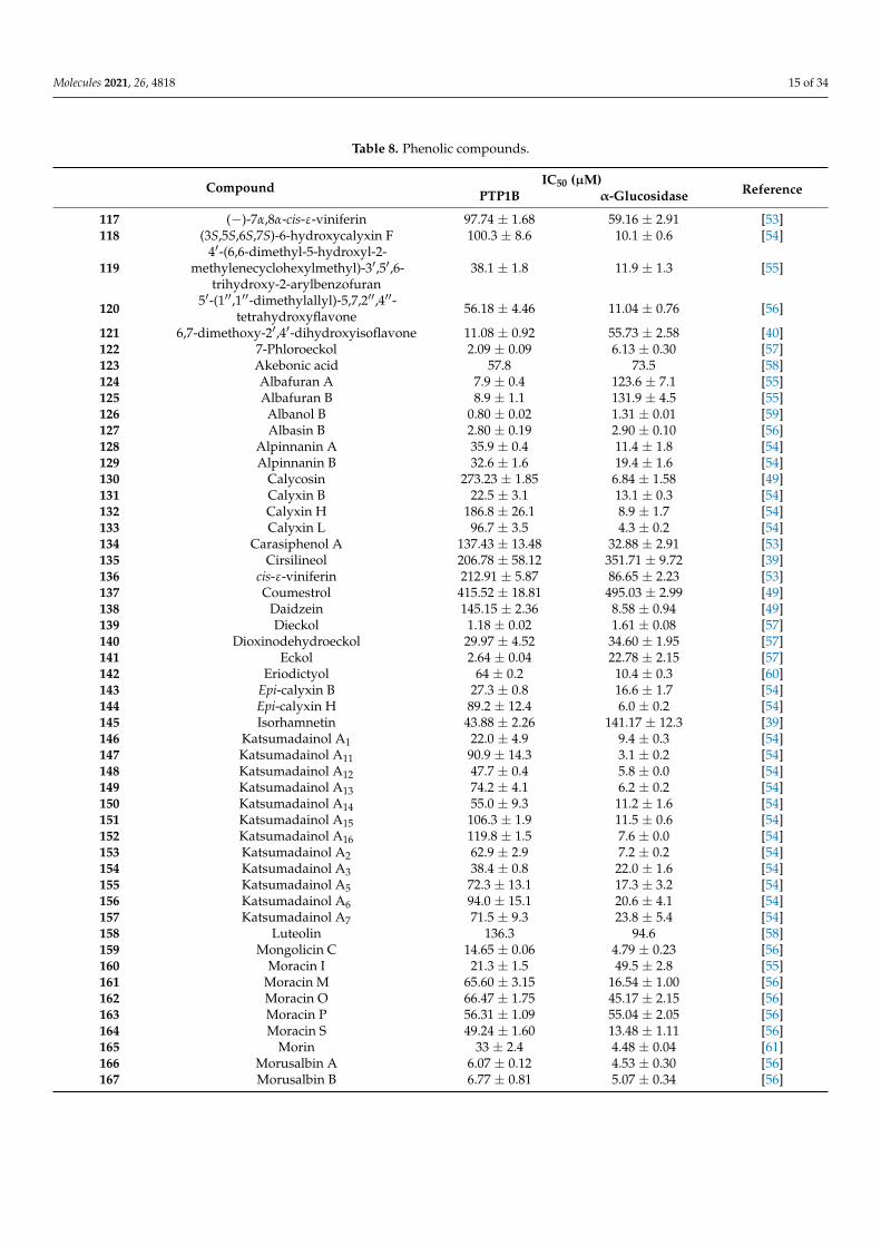
2.8. Phenolic Compounds
The family of phenolic compounds showing inhibitory activity toward PTP1B and α-glucosidase includes several and heterogenous molecules extracted from different naturalsources, including Artemisia capillaris (135, 145, and 175), Selaginella rolandi-principis (121),Pueraria lobata (130, 137, 138), Paeonia lactiflora (117, 134, 136, 174, 176), Morus alba var.tatarica (119, 124, 125, 160), M. alba L. (Moraceae) (120, 126, 127, 159, 161–169, 170–173),Ecklonia stolonifera and Eisenia bicyclis (122, 139–141), Alpinia katsumadai (118, 128, 129, 131–133, 143, 144, 146–157), Amomum tsao-ko (175–188), Paulownia tomentosa (142, 190–195), andPaeonia delavayi (123, 158) (Table 8).
Molecules 2021, 26, 4818 15 of 34
Table 8. Phenolic compounds.
Compound IC50 (µM)ReferencePTP1B α-Glucosidase
117 (−)-7α,8α-cis-ε-viniferin 97.74 ± 1.68 59.16 ± 2.91 [53]118 (3S,5S,6S,7S)-6-hydroxycalyxin F 100.3 ± 8.6 10.1 ± 0.6 [54]
1194′-(6,6-dimethyl-5-hydroxyl-2-
methylenecyclohexylmethyl)-3′,5′,6-trihydroxy-2-arylbenzofuran
38.1 ± 1.8 11.9 ± 1.3 [55]
120 5′-(1′′,1′′-dimethylallyl)-5,7,2′′,4′′-tetrahydroxyflavone 56.18 ± 4.46 11.04 ± 0.76 [56]
121 6,7-dimethoxy-2′,4′-dihydroxyisoflavone 11.08 ± 0.92 55.73 ± 2.58 [40]122 7-Phloroeckol 2.09 ± 0.09 6.13 ± 0.30 [57]123 Akebonic acid 57.8 73.5 [58]124 Albafuran A 7.9 ± 0.4 123.6 ± 7.1 [55]125 Albafuran B 8.9 ± 1.1 131.9 ± 4.5 [55]126 Albanol B 0.80 ± 0.02 1.31 ± 0.01 [59]127 Albasin B 2.80 ± 0.19 2.90 ± 0.10 [56]128 Alpinnanin A 35.9 ± 0.4 11.4 ± 1.8 [54]129 Alpinnanin B 32.6 ± 1.6 19.4 ± 1.6 [54]130 Calycosin 273.23 ± 1.85 6.84 ± 1.58 [49]131 Calyxin B 22.5 ± 3.1 13.1 ± 0.3 [54]132 Calyxin H 186.8 ± 26.1 8.9 ± 1.7 [54]133 Calyxin L 96.7 ± 3.5 4.3 ± 0.2 [54]134 Carasiphenol A 137.43 ± 13.48 32.88 ± 2.91 [53]135 Cirsilineol 206.78 ± 58.12 351.71 ± 9.72 [39]136 cis-ε-viniferin 212.91 ± 5.87 86.65 ± 2.23 [53]137 Coumestrol 415.52 ± 18.81 495.03 ± 2.99 [49]138 Daidzein 145.15 ± 2.36 8.58 ± 0.94 [49]139 Dieckol 1.18 ± 0.02 1.61 ± 0.08 [57]140 Dioxinodehydroeckol 29.97 ± 4.52 34.60 ± 1.95 [57]141 Eckol 2.64 ± 0.04 22.78 ± 2.15 [57]142 Eriodictyol 64 ± 0.2 10.4 ± 0.3 [60]143 Epi-calyxin B 27.3 ± 0.8 16.6 ± 1.7 [54]144 Epi-calyxin H 89.2 ± 12.4 6.0 ± 0.2 [54]145 Isorhamnetin 43.88 ± 2.26 141.17 ± 12.3 [39]146 Katsumadainol A1 22.0 ± 4.9 9.4 ± 0.3 [54]147 Katsumadainol A11 90.9 ± 14.3 3.1 ± 0.2 [54]148 Katsumadainol A12 47.7 ± 0.4 5.8 ± 0.0 [54]149 Katsumadainol A13 74.2 ± 4.1 6.2 ± 0.2 [54]150 Katsumadainol A14 55.0 ± 9.3 11.2 ± 1.6 [54]151 Katsumadainol A15 106.3 ± 1.9 11.5 ± 0.6 [54]152 Katsumadainol A16 119.8 ± 1.5 7.6 ± 0.0 [54]153 Katsumadainol A2 62.9 ± 2.9 7.2 ± 0.2 [54]154 Katsumadainol A3 38.4 ± 0.8 22.0 ± 1.6 [54]155 Katsumadainol A5 72.3 ± 13.1 17.3 ± 3.2 [54]156 Katsumadainol A6 94.0 ± 15.1 20.6 ± 4.1 [54]157 Katsumadainol A7 71.5 ± 9.3 23.8 ± 5.4 [54]158 Luteolin 136.3 94.6 [58]159 Mongolicin C 14.65 ± 0.06 4.79 ± 0.23 [56]160 Moracin I 21.3 ± 1.5 49.5 ± 2.8 [55]161 Moracin M 65.60 ± 3.15 16.54 ± 1.00 [56]162 Moracin O 66.47 ± 1.75 45.17 ± 2.15 [56]163 Moracin P 56.31 ± 1.09 55.04 ± 2.05 [56]164 Moracin S 49.24 ± 1.60 13.48 ± 1.11 [56]165 Morin 33 ± 2.4 4.48 ± 0.04 [61]166 Morusalbin A 6.07 ± 0.12 4.53 ± 0.30 [56]167 Morusalbin B 6.77 ± 0.81 5.07 ± 0.34 [56]
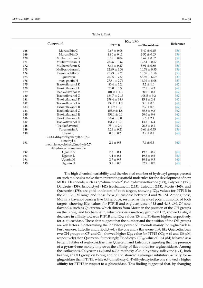
Molecules 2021, 26, 4818 16 of 34
Table 8. Cont.
Compound IC50 (µM)ReferencePTP1B α-Glucosidase
168 Morusalbin C 9.67 ± 0.08 5.40 ± 0.45 [56]169 Morusalbin D 1.90 ± 0.12 3.55 ± 0.03 [56]170 Mulberrofuran G 0.57 ± 0.04 1.67 ± 0.02 [59]171 Mulberrofuran H 78.96 ± 3.62 12.51 ± 0.57 [56]172 Mulberrofuran K 8.49 ± 0.27 5.91 ± 0.80 [56]173 Mulberro-furan L 32.89 ± 1.38 10.76 ± 0.55 [56]174 Paeonilactiflorol 27.23 ± 2.25 13.57 ± 1.56 [53]175 Quercetin 20.35 ± 7.56 58.93 ± 6.69 [39]176 trans-gnetin H 27.81 ± 2.74 14.39 ± 8.08 [53]175 Tsaokoflavanol K 80.4 ± 3.2 5.2 ± 1.0 [62]178 Tsaokoflavanol L 73.0 ± 0.5 37.5 ± 4.3 [62]179 Tsaokoflavanol M 101.0 ± 4.3 58.0 ± 0.3 [62]180 Tsaokoflavanol O 134.7 ± 21.3 108.5 ± 9.2 [62]181 Tsaokoflavanol P 359.4 ± 14.9 15.1 ± 2.4 [62]182 Tsaokoflavanol A 238.2 ± 1.0 9.0 ± 0.6 [62]183 Tsaokoflavanol B 114.9 ± 0.1 7.7 ± 0.8 [62]184 Tsaokoflavanol C 155.9 ± 1.8 35.8 ± 9.3 [62]185 Tsaokoflavanol E 356.1 ± 0.1 20.0 ± 0.6 [62]186 Tsaokoflavanol F 56.4 ± 5.0 5.6 ± 2.1 [62]187 Tsaokoflavanol H 151.7 ± 0.1 13.3 ± 6.4 [62]188 Tsaokoflavanol J 75.1 ± 2.4 26.8 ± 0.1 [62]189 Yunanensin A 5.26 ± 0.21 3.64 ± 0.35 [56]190 Ugonin J 0.6 ± 0.2 3.9 ± 0.2 [60]
191
2-(3,4-dihydroxyphenyl)-6-((2,2-dimethyl-6-
methylenecyclohexyl)methyl)-5,7-dihydroxychroman-4-one
2.1 ± 0.5 7.4 ± 0.3 [60]
192 Ugonin S 7.3 ± 0.4 19.2 ± 0.5 [60]193 Ugonin L 4.4 ± 0.2 19.3 ± 0.6 [60]194 Ugonin M 2.7 ± 0.3 10.4 ± 0.3 [60]195 Ugonin U 3.1 ± 0.7 32.9 ± 0.7 [60]
The high chemical variability and the elevated number of hydroxyl groups presenton such molecules make them interesting scaffold molecules for the development of newMDLs. Flavonoids, such as 6,7-dimethoxy-2′,4′-dihydroxyisoflavone (121), Calycosin (130),Daidzein (138), Eriodictyol (142) Isorhamnetin (145), Luteolin (158), Morin (165), andQuercetin (175), are good inhibitors of both targets, showing IC50 values for PTP1B inthe 20–136 µM range and those for α-glucosidase between 4 and 94 µM. Among these,Morin, a flavanol bearing five OH groups, resulted as the most potent inhibitor of bothtargets, showing IC50 values for PTP1B and α-glucosidase of 33 and 4.48 µM. Of note,flavanols, such as Quercetin, which differs from Morin in the position of the OH groupson the B ring, and Isorhamnetin, which carries a methoxy group on C3′, showed a slightdecrease in affinity towards PTP1B and IC50 values 13- and 31-times higher, respectively,for α-glucosidase. These data suggest that the number and the position of the OH groupsare key factors in determining the inhibitory power of flavonols mainly for α-glucosidase.Furthermore, Luteolin and Eriodictyol, a flavone and a flavanone that, like Quercetin, beartwo OH groups on C3′ and C4′, showed higher IC50 value for PTP1B (IC50 = 64 and 136 µM,respectively) than Quercetin. Surprisingly, Eriodictyol (IC50 value of 10.4 µM) behaved as abetter inhibitor of α-glucosidase than Quercetin and Luteolin, suggesting that the presenceof a pyran-4-one moiety improves the affinity of flavonoids for α-glucosidase. Amongthe isoflavones, Calycosin (130) and 6,7-dimethoxy-2′,4′-dihydroxyisoflavone (121), bothbearing an OH group on B-ring and on C7, showed a stronger inhibitory activity for α-glugosidase than PTP1B, while 6,7-dimethoxy-2′,4′-dihydroxyisoflavone showed a higheraffinity for PTP1B in respect to α-glucosidase. This finding suggested that, by changing
Molecules 2021, 26, 4818 17 of 34
position and number of OH and methoxyl groups, it is possible to generate inhibitors witha different affinity for both targets. Among all compounds, Ugonins appear of particu-lar interest because of their low molecular weight and high inhibitory activity. UgoninJ (190) e, 2-(3,4-dihydroxyphenyl)-6-((2,2-dimethyl-6-methylene cyclohexyl)methyl)-5,7-dihydroxychroman-4-one (191), was found to be the most effective inhibitor of both PTP1B(IC50 = 0.6 and 2.1 µM, respectively) and α-glucosidase (IC50 = 3.9 and 7.4 µM, respec-tively), acting as competitive inhibitors against the first target and as non-competitiveinhibitors against the second. Moreover, Ugonin M (194), bearing a furane moiety anda cyclohexyl group linked in a different position in respect to (190) and (191), showeda similar inhibitory activity versus both targets even if acting as a mixed-type inhibitor(binds both free enzyme and enzyme-substrate complex). Finally, Ugonin S (192), L (193),and U (195), bearing a tetrahydro-7H-pyran [2,3-c] xanthen-1-one group, showed a slightlyweaker affinity for PTP1B but 5–8-times lower inhibitory activity for α-glucosidase com-pared to Ugonin J. Among high-molecular-weight polyphenols, the most active compoundswere 7-Phloroeckol (122), Albanol B (126), Albasin B (127), Dieckol (139), Morusalbin A–D(166–169), Mulberrofuran G (170), Mulberrofuran K (172), and Yunanensin A (189). Inter-estingly, all these compounds are characterized by a balanced inhibitory activity and highaffinity for both targets, as confirmed by the fact that IC50 values for both targets fall in thelow micromolar range. Interestingly, kinetic and insilico docking analyses revealed thatAlbasin B, Morusalbin D, and Yunanensin A, some the most active compounds, behavedas mixed-type inhibitors of PTP1B and as competitive inhibitors of α-glucosidase [54].
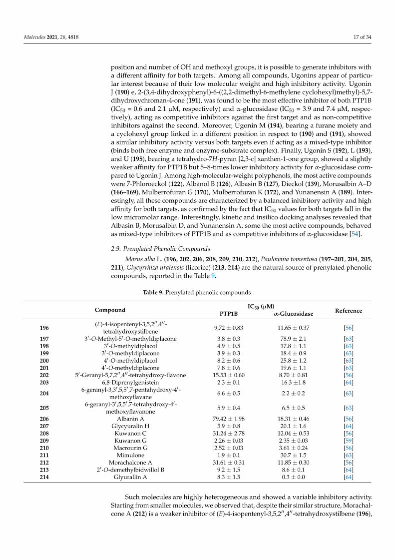
2.9. Prenylated Phenolic Compounds
Morus alba L. (196, 202, 206, 208, 209, 210, 212), Paulownia tomentosa (197–201, 204, 205,211), Glycyrrhiza uralensis (licorice) (213, 214) are the natural source of prenylated phenoliccompounds, reported in the Table 9.
Table 9. Prenylated phenolic compounds.
Compound IC50 (µM)ReferencePTP1B α-Glucosidase
196 (E)-4-isopentenyl-3,5,2′′,4′′-tetrahydroxystilbene 9.72 ± 0.83 11.65 ± 0.37 [56]
197 3′-O-Methyl-5′-O-methyldiplacone 3.8 ± 0.3 78.9 ± 2.1 [63]198 3′-O-methyldiplacol 4.9 ± 0.5 17.8 ± 1.1 [63]199 3′-O-methyldiplacone 3.9 ± 0.3 18.4 ± 0.9 [63]200 4′-O-methyldiplacol 8.2 ± 0.6 25.8 ± 1.2 [63]201 4′-O-methyldiplacone 7.8 ± 0.6 19.6 ± 1.1 [63]202 5′-Geranyl-5,7,2′′,4′′-tetrahydroxy-flavone 15.53 ± 0.60 8.70 ± 0.81 [56]203 6,8-Diprenylgenistein 2.3 ± 0.1 16.3 ±1.8 [64]
204 6-geranyl-3,3′,5,5′,7-pentahydroxy-4′-methoxyflavane 6.6 ± 0.5 2.2 ± 0.2 [63]
205 6-geranyl-3′,5,5′,7-tetrahydroxy-4′-methoxyflavanone 5.9 ± 0.4 6.5 ± 0.5 [63]
206 Albanin A 79.42 ± 1.98 18.31 ± 0.46 [56]207 Glycyuralin H 5.9 ± 0.8 20.1 ± 1.6 [64]208 Kuwanon C 31.24 ± 2.78 12.04 ± 0.53 [56]209 Kuwanon G 2.26 ± 0.03 2.35 ± 0.03 [59]210 Macrourin G 2.52 ± 0.03 3.61 ± 0.24 [56]211 Mimulone 1.9 ± 0.1 30.7 ± 1.5 [63]212 Morachalcone A 31.61 ± 0.31 11.85 ± 0.30 [56]213 2′-O-demethylbidwillol B 9.2 ± 1.5 8.6 ± 0.1 [64]214 Glyurallin A 8.3 ± 1.5 0.3 ± 0.0 [64]
Such molecules are highly heterogeneous and showed a variable inhibitory activity.Starting from smaller molecules, we observed that, despite their similar structure, Morachal-cone A (212) is a weaker inhibitor of (E)-4-isopentenyl-3,5,2′′,4′′-tetrahydroxystilbene (196),
Molecules 2021, 26, 4818 18 of 34
suggesting that the position of dihydroxyphenyl groups influence the activity of suchprenylated molecules. Concerning the flavonoid compounds, we observed that Mimu-lone (211), bearing a geranylated-naringenin based structure, is a potent PTP1B inhibitor(IC50 = 1.9 µM), and a good α-glucosidase inhibitor (IC50 = 30 µM), thereby confirmingthat flavonoids are good lead molecules for synthesis of new drugs targeting both enzymes.Modifications of Mimulone structure have a different impact on the inhibitory power ofchemical derivatives depending on the chemical groups introduced. For instance, theintroduction of a methoxy group on C3′ (199) or C4′ (201) of “B” aromatic ring reducedaffinity for PTP1B but improved that for α-glucosidase, whereas the introduction of asecond methoxy group on C5′ (197) strongly reduced the affinity of the molecule for α-glucosidase. Moreover, the introduction of OH group on C3 of keto-pyrene moiety (198,200) did not improve the inhibitory power, whereas the presence of two OH groups onC3′and C5′ together with a methoxy group on C4′ generated two molecules, (204) and(205), showing IC50 values in the low micromolar range toward both targets. This findingsuggests that the OH group on “B” ring has a key role in enhancing the targets–ligand in-teractions. Prenylated Morin derivatives, such as Albanin A (206), showed a better affinityfor α-glucosidase than for PTP1B, while Kuwanon C (208), showing two prenyl groups,had increased affinity for both enzymes. A stronger inhibitory activity was observed whenprenyl chains were linked to C6 and C8 of Genistein (203), suggesting that position of “B”ring strongly influences the inhibitory activity of prenylated phenol molecules. GlycyuralinH (207), which has the same 3-hydroxyisoflavanone skeleton of Genistein, behaved asweaker inhibitor of Kuwanon C (208), suggesting that modification of “B” ring of Genisteinis not a good strategy to improve the inhibitory activity. The presence of a single geranylchain on C5′ of “B” ring of Morin suitably increased the inhibitory power of 5′-Geranyl-5,7,2′′,4′′-tetrahydroxy-flavone (202) for PTP1B and α-glucosidase. This result indicates thatthe length of the aliphatic chain can be modulated to improve affinity of inhibitor for bothtargets. On the other hand, the introduction of additional 2,4-dihydroxyphenyl groups (209)resulted in a strong enhancement of inhibitory power of Kuwanon G, thereby confirmingthat OH groups contribute to strengthening the stability of the enzyme-inhibitor complex.Isoprenylated coumarones proved to be good inhibitors, showing 2′-O-demethylbidwillolB (213) similar IC50 values for both targets, while Glyurallin A (214) appeared to havea greater affinity for α-glucosidase. Finally, the Diels-Alder adduct Macrourin G (210)behaved as a potent inhibitor of both enzymes. Interestingly, kinetic and docking analysesrevealed that Macrourin G binds into allosteric site identified by Wiesmann in PTP1B [65],while it acts as competitive inhibitor of α-glucosidase, interacting with some residues ofcatalytic site of enzyme [56].
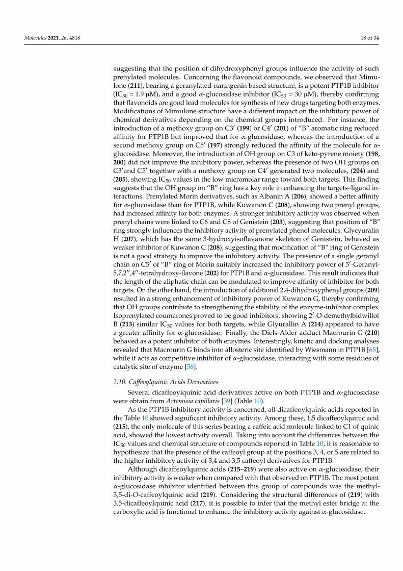
2.10. Caffeoylquinic Acids Derivatives
Several dicaffeoylquinic acid derivatives active on both PTP1B and α-glucosidasewere obtain from Artemisia capillaris [39] (Table 10).
As the PTP1B inhibitory activity is concerned, all dicaffeoylquinic acids reported inthe Table 10 showed significant inhibitory activity. Among these, 1,5 dicaffeoylquinic acid(215), the only molecule of this series bearing a caffeic acid molecule linked to C1 of quinicacid, showed the lowest activity overall. Taking into account the differences between theIC50 values and chemical structure of compounds reported in Table 10, it is reasonable tohypothesize that the presence of the caffeoyl group at the positions 3, 4, or 5 are related tothe higher inhibitory activity of 3,4 and 3,5 caffeoyl derivatives for PTP1B.
Although dicaffeoylquinic acids (215–219) were also active on α-glucosidase, theirinhibitory activity is weaker when compared with that observed on PTP1B. The most potentα-glucosidase inhibitor identified between this group of compounds was the methyl-3,5-di-O-caffeoylquinic acid (219). Considering the structural differences of (219) with3,5-dicaffeoylquinic acid (217), it is possible to infer that the methyl ester bridge at thecarboxylic acid is functional to enhance the inhibitory activity against α-glucosidase.
Molecules 2021, 26, 4818 19 of 34
Table 10. Caffeoylquinic Acid.
Compound IC50 (µM)ReferencePTP1B α-Glucosidase
215 1,5-Dicaffeoylquinic acid 16.05 ± 1.45 146.06 ± 0.07 [39]216 3,4-Dicaffeoylquinic acid 2.60 ± 0.24 128.07 ± 1.67 [39]217 3,5-Dicaffeoylquinic acid 2.02 ± 0.46 217.40 ± 5.45 [39]218 4,5-Dicaffeoylquinic acid 3.21 ± 0.23 229.94 ± 1.32 [39]219 Methyl-3,5-di-O-caffeoylquinic acid 2.99 ± 0.42 86.95 ± 4.10 [39]
2.11. Alkaloids
Eighteen different alkaloids able to inhibit both PTP1B and α-glucosidase were ex-tracted from Clausena anisum-olens [66] (Table 11).
Table 11. Alkaloids.
Compound IC50 (µM)ReferencePTP1B α-Glucosidase
220 3-Formyl-1-hydroxycarbazole 4.36 ± 0.09 87.36 ± 0.69 [66]221 Clauraila B 23.89 ± 0.22 76.64 ± 0.69 [66]222 Clausenaline E 32.86 ± 0.17 20.97 ± 0.63 [66]223 Clausenaline F 28.42 ± 0.26 168.75 ± 1.87 [66]224 Clausenanisine A 0.58 ± 0.05 3.28 ± 0.16 [66]225 Clausenanisine B 0.87 ± 0.06 8.27 ± 0.42 [66]226 Clausenanisine C 28.79 ± 0.16 9.38 ± 0.23 [66]227 Clausenanisine D 38.48 ± 0.32 12.37 ± 0.62 [66]228 Clausenanisine E 27.96 ± 0.15 96.17 ± 1.28 [66]229 Clausenanisine F 2.47 ± 0.09 176.32 ± 1.09 [66]230 Clausine I 3.96 ± 0.07 16.78 ± 0.45 [66]231 Clausine Z 5.39 ± 0.12 17.28 ± 0.36 [66]232 Clausines B 31.45 ± 0.32 16.83 ± 0.78 [66]233 Clauszoline M 26.37 ± 0.16 143.57 ± 1.02 [66]234 Clauszoline N 24.43 ± 0.25 15.79 ± 0.57 [66]235 Dihydromupamine 15.26 ± 0.26 62.89 ± 0.82 [66]236 Euchrestifoline 1.28 ± 0.07 11.96 ± 0.29 [66]237 Kurryame 27.93 ± 0.28 192.23 ± 0.78 [66]
It is interestingly to observe that among all carbazole, Clausenanisine A (224) exhibitedthe lowest IC50 values on both targets, suggesting that the carbazole moiety bearing a five-membered cyclic ether, a methoxy group, and a short aliphatic chain represents a promisinglead structure for developing new MLDs active on both PTP1B and α-glucosidase. Takinginto account that Clausenanisine B (225) showed inhibitory activity similar to Clause-nanisine A (224), we argue that the insertion of a tetrahydro-pyran-4-one group resultedin a slight decrease of the affinity for both targets. The loss of the OH group from C2′
of Clausenanisine B (225), of both OH and carbonyl groups, or the introduction of amethoxyl or hydroxyl group on C8 of carbazole moiety generated Euchrestifoline (236), Di-hydromupamine (235), Clauraila B (221), and Kurryame (237), whose affinity for PTP1B andα-glucosidase steadily decreased. However, the most relevant decrease of affinity occurredafter the loss of the carbonic group present on the pyrene moiety, suggested that ketocar-bonyl group on C1′ of Clausenanisine B is responsible of the significant inhibitory activityof molecule for both enzymes. Clausenanisine F (229), which bears a carboxyl group on C3and an OH group on C1, showed lower but always significant inhibitory activity for PTP1Bwhile it proved to be a very weak inhibitor of α-glucosidase. The addition of a methoxygroup on C2 or C6 of (229) leads to Clausenanisine C (226) and Clausenaline E (222), twomolecules with different inhibitory activity. The first one showed a weak affinity for PTP1Bbut behaved as a potent α-glucosidase inhibitor. The latter showed a reduced affinity forboth PTP1B α-glucosidase when compared to (229). Finally, Clausenaline F (223), bearing

Molecules 2021, 26, 4818 20 of 34
two methoxy groups on C1 and C2, and Clausines B (232), possessing 2 methoxy groupson C6 and C8, showed a decreased affinity for PTP1B in comparison with (229). However,Clausines B resulted a better α-glucosidase inhibitor than Clausenaline F. The replacementof the carboxyl group of (229) with a formyl group leads to 3-Formyl-1-hydroxycarbazole(220), which showed an affinity for PTP1B similar to (229) but an increased affinity forα-glucosidase. The loss of the OH on C1 of (231) leads to Clausenanisine D (227), whichshowed a higher IC50 value for PTP1B but a similar affinity for the α-glucosidase comparedto (220). Changing the position of one or both OH groups present on (220) we obtainClauszoline N (234) and Clauszoline M (233). The affinity of these compounds is similar,but that for α-glucosidase differs, (233) being a very bad inhibitor of this enzyme. Theaddition of one methoxy group on C6 of 3-Formyl-1-hydroxycarbazole (220) leads to Clau-sine I (230), which possessed a similar affinity for PTP1B as (220) but an enhanced affinityfor α-glucosidase compared to 3-Formyl-1-hydroxycarbazole. Finally, the introduction oftwo methoxy groups on C6 and C8 of Clauszoline M (233) leads to Clausines B (232), amolecule that showed an affinity for PTP1B similar to that of (233) but a high affinity forα-glucosidase.
2.12. Others
Several different molecules were obtained from different natural sources, such as Euony-mus alatus (Thunb.) Sieb. [38], Hizikia fusiformis [43], Pueraria lobata [49], Paeonia lactiflora [53],Ecklonia stolonifera and Eisenia bicyclis [57], and Artemisia capillaris [67] (Table 12).
Table 12. Others.
Compound IC50 (µM)ReferencePTP1B α-Glucosidase
238 Capillin 31.90 ± 0.15 332.96 ± 1.44 [67]239 Capillinol 159.99 ± 0.39 464.53 ± 2.69 [67]240 Dioxinodehydroeckol 29. 97 ± 4.52 34.60 ± 1.95 [57]241 Eckol 2.64 ± 0.04 22.78 ± 2.15 [57]242 Phlorofucofuroeckol-A 0.56 ± 0.10 1.37 ± 0.05 [57]243 Phloroglucinol 55.48 ± 1.85 141.18 ± 13.1 [57]244 p-propoxybenzoic acid 14.8 ± 0.9 10.5 ± 0.8 [38]245 Sargachromenol 11.80 ± 3.35 42.41 ± 3.09 [68]246 Sargaquinoic acid 14.15 ± 0.02 96.17 ± 3.48 [68]247 Suffruticosol A 86.26 ± 8.47 15.57 ± 2.14 [53]248 Suffruticosol B 53.93 ± 2.88 10.82 ± 2.96 [53]249 Suffruticosol C 131.24 ± 3.93 45.10 ± 4.81 [53]250 Tuberosin 183.95 ± 2.27 28.07 ± 3.10 [49]251 Tyramine 188.06 ± 3.21 273.23 ± 5.65 [43]
Some of these, such as Phloroglucinol (243), Dioxinodehydroeckol (240), Eckol (241), andPhlorofucofuroeckol-A (242), are naturally occurring phloroglucinol polymers. Phlorofucofuroeckol-A exhibited the higher inhibitory activity, with IC50 values for PTP1B and α-glucosidase of 0.56and 1.37 µM, respectively. Moreover, kinetic analyses revealed that Phlorofucofuroeckol-A actsas a non-competitive inhibitor of both PTP1B and α-glucosidase. Interestingly, the affinity ofEckol, Dioxinodehydroeckol, and Phloroglucinol is progressively decreasing, suggesting that thephloroglucinol polymers ensure a stronger interaction with both targets, probably due to the highnumber of hydrogen bonds that are generated between the hydroxyl groups of molecules and theamino acids of the target.
p-propoxybenzoic acid (244) showed a slight decrease of affinity for both targets(IC50 values for PTP1B and α-glucosidase were 14.8 and 10.5 µM, respectively), whileSargachromenol (245) and Sargaquinoic acid (246), showed a similar affinity for PTP1B buta decreased affinity for α-glucosidase in respect to (244). All other compounds showed aweak affinity for both targets.

Molecules 2021, 26, 4818 21 of 34
3. From Natural World to Bench-Side
What emerges from the data reported in this review is that the natural world canprovide a large number of chemically different scaffold molecules active on both PTP1B andα-glucosidase. The list includes a large number of good inhibitors—molecules showing IC50values in the low micromolar range (0.1–20 µM) for both targets—and other compoundswith IC50 values that gradually increase up to the value of 440 µM for the PTP1B and600 µM for the α-glycosidase (Figure 5 and Table 13).
Molecules 2021, 26, x 22 of 35
Table 13. Distribution frequency of IC50 values for PTP1B and α-glucosidase.
IC50 (µM) n° of Molecules * PTP1B α-Glucosidase
0.1–10 84 (34.2%) 65 (26.5%) 10–20 31 (12.6%) 51 (20.8%) 20–30 29 (11.8%) 21 (8.6%) 30–40 20 (8.1%) 14 (5.7%) 40–50 6 (2.4%) 14 (5.7%) 50–60 13 (5.3%) 11 (4.6%) 60–650 63 (25.6%) 69 (28.1%)
* Total number of analyzed molecules = 250.
Figure 5. Distribution of the IC50 values calculated for both PTP1B (left) and α-glucosidase (right). The IC50 values are categorized as shown in Table 13.
It is interesting to note that the 250 known MDLs show a greater affinity for PTP1B than for α-glucosidase, as evidenced by the fact that the average IC50 values for the en-zymes are 6.0 ± 4.1 µM and 42.9 ± 52.0 µM, respectively. Although this difference could seem an obstacle to the development of balanced dual PTP1B/α-glucosidase inhibitors, it may not represent a true problem, especially considering the different localization and physiological role of the two enzymes. The α-glucosidase localizes in the intestinal lumen while PTP1B in the cytosol of human cells [69]. This first consideration suggests that α-glucosidase targeting does not require very potent inhibitors, and significant inhibition of this enzyme can easily be achieved by taking oral drug during or immediately after a meal. By ensuring protection from the acidic pH of the stomach, MDLs could reach the intestine intact and target the enzyme. Furthermore, the lower potency of MDLs for α-glucosidase could be easily compensated by increasing drug dosage. As a precautionary measure, it could be of great importance to evaluate the ability of these molecules to in-hibit the activity of intestinal glucose transporters in order to avoid the risk of inducing a hypoglycemic condition [70]. Conversely, targeting PTP1B is a more complex matter. In fact, MDLs designed to act on PTP1B should have good bioavailability to ensure their absorption through the intestinal wall and their rapid uptake by cells that compose liver, muscle, adipose tissue, pancreas, and brain, where this enzyme acts as a negative regula-tor of the insulin receptor. Moreover, considering that mammalian cells express different types of tyrosine phosphatases, each of which carries out important regulatory functions, a good/high specificity of MDLs for PTP1B is essential to avoid the induction of severe side effects. Unfortunately, in most cases, information about the specificity of such mole-cules are not yet available. Conversely, relatively more informations are available about the action mechanism of natural MDLs (Table 14). It is interesting to note that several of the most effective MDLs identified act as non-competitive inhibitors of PTP1B, suggesting that they interact with sites different from the active site of enzyme. Non-competitive and allosteric PTP1B inhibitors actually represent the most promising molecules for develop-ing new antidiabetic drugs since they could ensure a more adequate and specific action, thereby reducing the risk of inducing negative side effects [71].
Figure 5. Distribution of the IC50 values calculated for both PTP1B (left) and α-glucosidase (right).The IC50 values are categorized as shown in Table 13.
Table 13. Distribution frequency of IC50 values for PTP1B and α-glucosidase.
IC50 (µM)n◦ of Molecules *
PTP1B α-Glucosidase
0.1–10 84 (34.2%) 65 (26.5%)10–20 31 (12.6%) 51 (20.8%)20–30 29 (11.8%) 21 (8.6%)30–40 20 (8.1%) 14 (5.7%)40–50 6 (2.4%) 14 (5.7%)50–60 13 (5.3%) 11 (4.6%)
60–650 63 (25.6%) 69 (28.1%)* Total number of analyzed molecules = 250.
It is interesting to note that the 250 known MDLs show a greater affinity for PTP1B thanfor α-glucosidase, as evidenced by the fact that the average IC50 values for the enzymesare 6.0 ± 4.1 µM and 42.9 ± 52.0 µM, respectively. Although this difference could seem anobstacle to the development of balanced dual PTP1B/α-glucosidase inhibitors, it may notrepresent a true problem, especially considering the different localization and physiologicalrole of the two enzymes. The α-glucosidase localizes in the intestinal lumen while PTP1Bin the cytosol of human cells [69]. This first consideration suggests that α-glucosidasetargeting does not require very potent inhibitors, and significant inhibition of this enzymecan easily be achieved by taking oral drug during or immediately after a meal. By ensuringprotection from the acidic pH of the stomach, MDLs could reach the intestine intact andtarget the enzyme. Furthermore, the lower potency of MDLs for α-glucosidase could beeasily compensated by increasing drug dosage. As a precautionary measure, it could be ofgreat importance to evaluate the ability of these molecules to inhibit the activity of intestinalglucose transporters in order to avoid the risk of inducing a hypoglycemic condition [70].Conversely, targeting PTP1B is a more complex matter. In fact, MDLs designed to act onPTP1B should have good bioavailability to ensure their absorption through the intestinalwall and their rapid uptake by cells that compose liver, muscle, adipose tissue, pancreas,and brain, where this enzyme acts as a negative regulator of the insulin receptor. Moreover,considering that mammalian cells express different types of tyrosine phosphatases, eachof which carries out important regulatory functions, a good/high specificity of MDLs forPTP1B is essential to avoid the induction of severe side effects. Unfortunately, in mostcases, information about the specificity of such molecules are not yet available. Conversely,
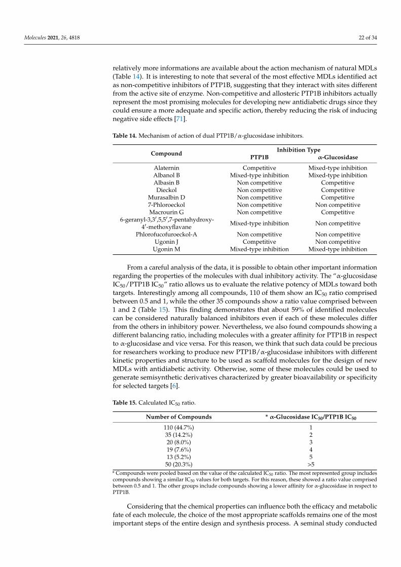
Molecules 2021, 26, 4818 22 of 34
relatively more informations are available about the action mechanism of natural MDLs(Table 14). It is interesting to note that several of the most effective MDLs identified actas non-competitive inhibitors of PTP1B, suggesting that they interact with sites differentfrom the active site of enzyme. Non-competitive and allosteric PTP1B inhibitors actuallyrepresent the most promising molecules for developing new antidiabetic drugs since theycould ensure a more adequate and specific action, thereby reducing the risk of inducingnegative side effects [71].
Table 14. Mechanism of action of dual PTP1B/α-glucosidase inhibitors.
Compound Inhibition TypePTP1B α-Glucosidase
Alaternin Competitive Mixed-type inhibitionAlbanol B Mixed-type inhibition Mixed-type inhibitionAlbasin B Non competitive CompetitiveDieckol Non competitive Competitive
Murasalbin D Non competitive Competitive7-Phloroeckol Non competitive Non competitiveMacrourin G Non competitive Competitive
6-geranyl-3,3′,5,5′,7-pentahydroxy-4′-methoxyflavane Mixed-type inhibition Non competitive
Phlorofucofuroeckol-A Non competitive Non competitiveUgonin J Competitive Non competitive
Ugonin M Mixed-type inhibition Mixed-type inhibition
From a careful analysis of the data, it is possible to obtain other important informationregarding the properties of the molecules with dual inhibitory activity. The “α-glucosidaseIC50/PTP1B IC50” ratio allows us to evaluate the relative potency of MDLs toward bothtargets. Interestingly among all compounds, 110 of them show an IC50 ratio comprisedbetween 0.5 and 1, while the other 35 compounds show a ratio value comprised between1 and 2 (Table 15). This finding demonstrates that about 59% of identified moleculescan be considered naturally balanced inhibitors even if each of these molecules differfrom the others in inhibitory power. Nevertheless, we also found compounds showing adifferent balancing ratio, including molecules with a greater affinity for PTP1B in respectto α-glucosidase and vice versa. For this reason, we think that such data could be preciousfor researchers working to produce new PTP1B/α-glucosidase inhibitors with differentkinetic properties and structure to be used as scaffold molecules for the design of newMDLs with antidiabetic activity. Otherwise, some of these molecules could be used togenerate semisynthetic derivatives characterized by greater bioavailability or specificityfor selected targets [6].
Table 15. Calculated IC50 ratio.
Number of Compounds * α-Glucosidase IC50/PTP1B IC50
110 (44.7%) 135 (14.2%) 220 (8.0%) 319 (7.6%) 413 (5.2%) 5
50 (20.3%) >5* Compounds were pooled based on the value of the calculated IC50 ratio. The most represented group includescompounds showing a similar IC50 values for both targets. For this reason, these showed a ratio value comprisedbetween 0.5 and 1. The other groups include compounds showing a lower affinity for α-glucosidase in respect toPTP1B.
Considering that the chemical properties can influence both the efficacy and metabolicfate of each molecule, the choice of the most appropriate scaffolds remains one of the mostimportant steps of the entire design and synthesis process. A seminal study conducted

Molecules 2021, 26, 4818 23 of 34
by Lipinski and coworkers lead to formulation of general criteria useful to predict in-vivo bioavailability and degree of oral absorption of a molecule. The molecules that aremost likely to be absorbed are characterized by a molecular weight lower than 500 Da, amiLogP value comprised in the 0–5 range, a number of hydrogen bond acceptors <10, anumber of hydrogen bond donors <5, a TPSA (topological polar surface area) value <140 Å,and a number of rotatable bonds <10–20 [72]. Based on this information, we decided toanalyze the properties of the 60 most potent compounds among those reviewed to predicttheir bioavailability. Interestingly, we found that, on average, the compounds meet thecriteria mentioned before (Table 16). This result reinforces the hypothesis that most of thesecompounds possess a good bioavailability and can be considered promising lead moleculesfor the development of new MDLs.
Table 16. Physicochemical properties of the sixty most potent MDLs obtained from PUBCHEM database.
Parameters Average Values of the Top Sixty Compounds Lipinski’s Criteria
miLogP 1 4.9 ± 2 0–5hydrogen bond
acceptors 2 6.4 ± 4 <10
hydrogen bond donors3 3.8 ± 2 <5
TPSA 4 112 ± 59 <140 Årotatable bonds 5 5.2 ± 4 <10–20Molecular weight 432 ± 127 Da <500 Da
1 Octanol/Water Partition Coefficient (partition coefficients are useful in estimating the distribution of drugs withinthe cells: hydrophobic drugs, showing high octanol–water partition coefficients, are mainly distributed to lipidbilayers of cells, whereas hydrophilic compounds possess a low octanol–water partition coefficients and accumulatein cytoplasm of the cells). 2 Number of hydrogen bond acceptors (the number of hydrogen bond acceptors inthe structure). 3 Number of hydrogen bond donors in the structure (the number of hydrogen bond donors in thestructure.). 4 TPSA represents the surface sum over all polar atoms in a molecule. 5 Any single-order, non-ring bondwhere atoms on either side of the bond are in turn bound to nonterminal heavy (i.e., non-hydrogen) atoms.
Besides the influence on bioavailability, we wondered if the aforementioned chemical-physical parameters could influence the activity of MDLs toward designed targets. To givean answer, we analyzed the correlation between the IC50 values for both enzymes and eachparameter described in Table 16 (Figures 6 and 7).
Interestingly, taking into account the PTP1B inhibitory activity, we found that theTPSA, the number of hydrogen donors and acceptors, the complexity, and the molecularweight of molecules are inversely related to IC50 value, while the miLogP shows a directcorrelation with the calculated IC50 value. A weak correlation was observed between thenumber of rotatable bonds and the IC50 value. This evidence suggests that the extensionof the molecules and their ability to form hydrogen bonds with the atoms of the enzymeplay an important role in determining their interaction strength with PTP1B, while thehydrophobicity of the molecules represents an unfavorable factor. However, differentresults were obtained analyzing the IC50 values for α-glucosidase. Parameters such asTPSA, number of hydrogen donors and acceptors, complexity, and molecular weighthave a poor correlation with the IC50 value, while the parameter miLogP seems inverselycorrelated with the inhibitory power of the molecules. Finally, the number of rotatablebonds appears directly related to the IC50 value for α-glucosidase. These data suggest thatthis enzyme preferentially interacts with rigid molecules characterized by a hydrophilicprofile even if the interaction of MDLs with the enzyme is not strongly influenced byformation of hydrogen bond or by molecular weight. This is in accordance with the factthat this enzyme works in an aqueous extracellular medium and binds glucose polymers,molecules showing a relatively high rigidity.

Molecules 2021, 26, 4818 24 of 34Molecules 2021, 26, x 25 of 35
Figure 6. Relationship between the chemical-physical properties and the IC50 values for PTP1B of the best sixty compounds. Chemical-physical data were recovered from PubChem database and analyzed using OriginPro 2021 Software.
Figure 6. Relationship between the chemical-physical properties and the IC50 values for PTP1B ofthe best sixty compounds. Chemical-physical data were recovered from PubChem database andanalyzed using OriginPro 2021 Software.
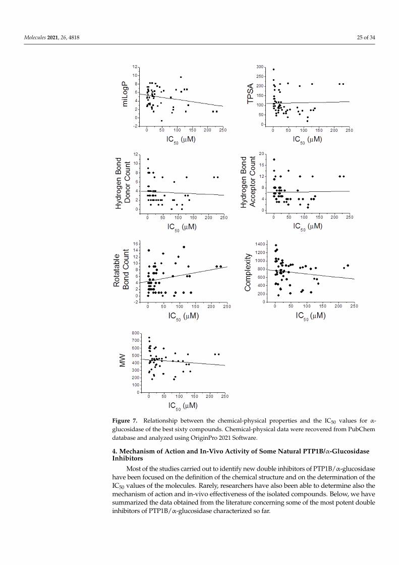
Molecules 2021, 26, 4818 25 of 34Molecules 2021, 26, x 26 of 35
Figure 7. Relationship between the chemical-physical properties and the IC50 values for α-gluco-sidase of the best sixty compounds. Chemical-physical data were recovered from PubChem data-base and analyzed using OriginPro 2021 Software.
4. Mechanism of Action and In-Vivo Activity of Some Natural PTP1B/α-Glucosidase Inhibitors
Most of the studies carried out to identify new double inhibitors of PTP1B/α-gluco-sidase have been focused on the definition of the chemical structure and on the determi-nation of the IC50 values of the molecules. Rarely, researchers have also been able to de-termine also the mechanism of action and in-vivo effectiveness of the isolated compounds. Below, we have summarized the data obtained from the literature concerning some of the most potent double inhibitors of PTP1B/α-glucosidase characterized so far.
Figure 7. Relationship between the chemical-physical properties and the IC50 values for α-glucosidase of the best sixty compounds. Chemical-physical data were recovered from PubChemdatabase and analyzed using OriginPro 2021 Software.
4. Mechanism of Action and In-Vivo Activity of Some Natural PTP1B/α-GlucosidaseInhibitors
Most of the studies carried out to identify new double inhibitors of PTP1B/α-glucosidasehave been focused on the definition of the chemical structure and on the determination of theIC50 values of the molecules. Rarely, researchers have also been able to determine also themechanism of action and in-vivo effectiveness of the isolated compounds. Below, we havesummarized the data obtained from the literature concerning some of the most potent doubleinhibitors of PTP1B/α-glucosidase characterized so far.

Molecules 2021, 26, 4818 26 of 34
Phlorofucofuroeckol-A (242), Dieckol (139), and 7-Phloroeckol (122) are phlorotanninsextracted by the edible brown algae arame (Ecklonia bicyclis) and turuarame (Eckloniastolonifera).
Molecules 2021, 26, x 27 of 35
Phlorofucofuroeckol-A (242), Dieckol (139), and 7-Phloroeckol (122) are phlorotan-nins extracted by the edible brown algae arame (Ecklonia bicyclis) and turuarame (Ecklonia stolonifera).
(139) Dieckol
IC50PTP1B = 1.2 µM IC50α-glucosidase = 1.6 µM
(242) Phlorofucofuroeckol-A IC50PTP1B = 0.56 µM
IC50α-glucosidase = 1.37 µM
(122) 7-Phloroeckol
IC50PTP1B = 2.1 µM IC50α-glucosidase = 6.1 µM
Kinetic analyses revealed that these compounds acted as non-competitive inhibitors of PTP1B, while only Phlorofucofuroeckol-A and 7-Phloroeckol behaved as non-compet-itive inhibitors of α-glucosidase. Conversely, Dieckol acted as competitive inhibitor of the latter. A recent investigation on rat insulinoma cells showed that treatment with Dieckol reduced oxidative stress and apoptosis caused by exposition of cells to high glucose levels, suggesting that this compound could protect β-pancreatic cells against damages induced by hyperglycemia [73].
A recent in-vivo study demonstrated that Phlorofucofuroeckol-A delayed intestinal absorption of dietary carbohydrates in diabetic mice models, suggesting that this mole-cule could be used to generate new drugs able to reduce post-prandial glucose levels in diabetic patients [74]. High blood glucose levels increase the production of advanced gly-cation end products that, in turn, contribute to the onset of numerous diabetes-related complications, such as nephropathy, retinopathy, atherosclerosis, and neurodegenerative diseases. Recently, Su Hui Seong et al. demonstrated that Phlorofucofuroeckol-A inhibits non-enzymatic insulin glycation and Aβ aggregation. This finding suggests that this mol-ecule could be used to preserve insulin function and prevent aggregation of the Aβ pep-tide, thus maintaining the vitality of neurons and avoiding Aβ-mediated brain damage [75]. Besides, it has been demonstrated that Phlorofucofuroeckol-A acts as a potent non-competitive inhibitor of human monoamine oxidase-A, confirming that this compound could be useful to prevent neuronal disorders [76]. A further study showed that Phlorofu-cofuroeckol-A is effective in contrasting negative effects induced by high fatty diet as well as in reducing leptin resistance in hypothalamic neurons and microglia. This evidence suggests that such compound could be used to fight leptin resistance and to control weight in obese subjects [77].
Results of studies conducted on diabetic mice models confirmed the antidiabetic ac-tivity of Dieckol (139) in vivo. Intra peritoneal injections (10–20 mg/Kg for 14 days) of Dieckol in C57BL/KsJ-db/db diabetic mice lead to a significant reduction of both blood glucose and serum insulin levels as well as of body weight. Moreover, it has been reported that treated mice showed either increased phosphorylation levels of AMPK (5′ adenosine monophosphate-activated protein kinase) or Akt (Akt kinase) and a concomitant increase
Kinetic analyses revealed that these compounds acted as non-competitive inhibitors ofPTP1B, while only Phlorofucofuroeckol-A and 7-Phloroeckol behaved as non-competitiveinhibitors of α-glucosidase. Conversely, Dieckol acted as competitive inhibitor of thelatter. A recent investigation on rat insulinoma cells showed that treatment with Dieckolreduced oxidative stress and apoptosis caused by exposition of cells to high glucose levels,suggesting that this compound could protect β-pancreatic cells against damages inducedby hyperglycemia [73].
A recent in-vivo study demonstrated that Phlorofucofuroeckol-A delayed intestinalabsorption of dietary carbohydrates in diabetic mice models, suggesting that this moleculecould be used to generate new drugs able to reduce post-prandial glucose levels in diabeticpatients [74]. High blood glucose levels increase the production of advanced glycationend products that, in turn, contribute to the onset of numerous diabetes-related complica-tions, such as nephropathy, retinopathy, atherosclerosis, and neurodegenerative diseases.Recently, Su Hui Seong et al. demonstrated that Phlorofucofuroeckol-A inhibits non-enzymatic insulin glycation and Aβ aggregation. This finding suggests that this moleculecould be used to preserve insulin function and prevent aggregation of the Aβ peptide, thusmaintaining the vitality of neurons and avoiding Aβ-mediated brain damage [75]. Besides,it has been demonstrated that Phlorofucofuroeckol-A acts as a potent non-competitiveinhibitor of human monoamine oxidase-A, confirming that this compound could be usefulto prevent neuronal disorders [76]. A further study showed that Phlorofucofuroeckol-A iseffective in contrasting negative effects induced by high fatty diet as well as in reducingleptin resistance in hypothalamic neurons and microglia. This evidence suggests thatsuch compound could be used to fight leptin resistance and to control weight in obesesubjects [77].
Results of studies conducted on diabetic mice models confirmed the antidiabeticactivity of Dieckol (139) in vivo. Intra peritoneal injections (10–20 mg/Kg for 14 days) ofDieckol in C57BL/KsJ-db/db diabetic mice lead to a significant reduction of both bloodglucose and serum insulin levels as well as of body weight. Moreover, it has been reportedthat treated mice showed either increased phosphorylation levels of AMPK (5′ adenosinemonophosphate-activated protein kinase) or Akt (Akt kinase) and a concomitant increase

Molecules 2021, 26, 4818 27 of 34
of the activity of several antioxidant enzymes [78]. Seung-Hong Lee and co-workersshowed that the treatment of type 2 diabetic mice with a Dieckol-rich extract from Eckloniacava caused a significant reduction of blood glycosylated hemoglobin and plasma insulinlevels and an improvement of glucose tolerance. Furthermore, diabetic mice treated withDieckol-rich extract showed a reduction of both plasma and hepatic lipids levels comparedto control mice group [79]. Similar results were obtained from tests carried out on eightypre-diabetic male and female adults. In this case, the administration of Dieckol-rich extract(1500 mg per day for 12 weeks) caused a significant reduction of postprandial glucose,insulin, and C-peptide levels compared to control group. Moreover, no significant adverseevents or changes of biochemical and hematological parameters were observed duringtreatment. Taken together, these results confirmed that algae extracts rich in Dieckol andother phlorotannins possess antidiabetic activity, and their consumption may contribute toregularize glycemia in prediabetic and diabetic subjects [80].
Mulberrofuran G (170), Albanol B (122), and Kuwanon G (209) are flavonoids extractedfrom root bark of Morus Alba Linn.
Molecules 2021, 26, x 28 of 35
of the activity of several antioxidant enzymes [78]. Seung-Hong Lee and co-workers showed that the treatment of type 2 diabetic mice with a Dieckol-rich extract from Ecklonia cava caused a significant reduction of blood glycosylated hemoglobin and plasma insulin levels and an improvement of glucose tolerance. Furthermore, diabetic mice treated with Dieckol-rich extract showed a reduction of both plasma and hepatic lipids levels com-pared to control mice group [79]. Similar results were obtained from tests carried out on eighty pre-diabetic male and female adults. In this case, the administration of Dieckol-rich extract (1500 mg per day for 12 weeks) caused a significant reduction of postprandial glu-cose, insulin, and C-peptide levels compared to control group. Moreover, no significant adverse events or changes of biochemical and hematological parameters were observed during treatment. Taken together, these results confirmed that algae extracts rich in Dieckol and other phlorotannins possess antidiabetic activity, and their consumption may contribute to regularize glycemia in prediabetic and diabetic subjects [80].
Mulberrofuran G (170), Albanol B (122), and Kuwanon G (209) are flavonoids ex-tracted from root bark of Morus Alba Linn.
(170) Mulberrofuran G
IC50PTP1B = 0.57 µM IC50α-glucosidase = 1.7 µM
(122) Albanol B IC50PTP1B = 0.80 µM
IC50α-glucosidase = 1.3 µM
(209) Kuwanon G IC50PTP1B = 2.26 µM
IC50α-glucosidase = 2.35 µM
Paudel P. et al. demonstrated that such compounds acted as potent mixed-type en-zyme inhibitors against PTP1B and α-glucosidase [59]. In accordance with the results of kinetic analyses, docking in silico revealed that these compounds interacted with both the catalytic and an allosteric site present on PTP1B surface. Similar results were obtained analyzing the interaction mode of compounds with α-glucosidase enzyme, thereby con-firming the mixed-type inhibition mode. Studying the effects on liver cells, the authors reported that treatment with these compounds decreased the expression of PTP1B and stimulated glucose uptake, suggesting that they act as insulin-sensitizing agents [59]. Sev-eral phytochemicals extracted from leaves and fruits of different members belonging to the Moraceae family showed anti-obesity and anti-diabetic activity, enhancing insulin sig-naling pathway, glucose uptake, and inhibiting hepatic gluconeogenesis [81,82].
Like the above compounds, also Morusalbin D (169), Albasin B (127), and Macrourin G (210) were extracted from Morus Alba Linn.
Paudel P. et al. demonstrated that such compounds acted as potent mixed-type en-zyme inhibitors against PTP1B and α-glucosidase [59]. In accordance with the results ofkinetic analyses, docking in silico revealed that these compounds interacted with both thecatalytic and an allosteric site present on PTP1B surface. Similar results were obtained ana-lyzing the interaction mode of compounds with α-glucosidase enzyme, thereby confirmingthe mixed-type inhibition mode. Studying the effects on liver cells, the authors reportedthat treatment with these compounds decreased the expression of PTP1B and stimulatedglucose uptake, suggesting that they act as insulin-sensitizing agents [59]. Several phyto-chemicals extracted from leaves and fruits of different members belonging to the Moraceaefamily showed anti-obesity and anti-diabetic activity, enhancing insulin signaling pathway,glucose uptake, and inhibiting hepatic gluconeogenesis [81,82].
Like the above compounds, also Morusalbin D (169), Albasin B (127), and MacrourinG (210) were extracted from Morus Alba Linn.
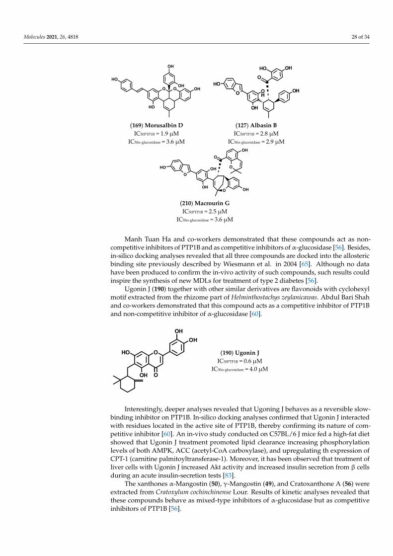
Molecules 2021, 26, 4818 28 of 34Molecules 2021, 26, x 29 of 35
(169) Morusalbin D
IC50PTP1B = 1.9 µM IC50α-glucosidase = 3.6 µM
(127) Albasin B IC50PTP1B = 2.8 µM
IC50α-glucosidase = 2.9 µM
(210) Macrourin G IC50PTP1B = 2.5 µM
IC50α-glucosidase = 3.6 µM
Manh Tuan Ha and co-workers demonstrated that these compounds act as non-com-petitive inhibitors of PTP1B and as competitive inhibitors of α-glucosidase [56]. Besides, in-silico docking analyses revealed that all three compounds are docked into the allosteric binding site previously described by Wiesmann et al. in 2004 [65]. Although no data have been produced to confirm the in-vivo activity of such compounds, such results could in-spire the synthesis of new MDLs for treatment of type 2 diabetes [56].
Ugonin J (190) together with other similar derivatives are flavonoids with cyclohexyl motif extracted from the rhizome part of Helminthostachys zeylanicawas. Abdul Bari Shah and co-workers demonstrated that this compound acts as a competitive inhibitor of PTP1B and non-competitive inhibitor of α-glucosidase [60].
(190) Ugonin J IC50PTP1B = 0.6 µM
IC50α-glucosidase = 4.0 µM
Interestingly, deeper analyses revealed that Ugoning J behaves as a reversible slow-binding inhibitor on PTP1B. In-silico docking analyses confirmed that Ugonin J interacted with residues located in the active site of PTP1B, thereby confirming its nature of compet-itive inhibitor [60]. An in-vivo study conducted on C57BL/6 J mice fed a high-fat diet showed that Ugonin J treatment promoted lipid clearance increasing phosphorylation lev-els of both AMPK, ACC (acetyl-CoA carboxylase), and upregulating th expression of CPT-1 (carnitine palmitoyltransferase-1). Moreover, it has been observed that treatment of liver cells with Ugonin J increased Akt activity and increased insulin secretion from β cells dur-ing an acute insulin-secretion tests [83].
The xanthones α-Mangostin (50), γ-Mangostin (49), and Cratoxanthone A (56) were extracted from Cratoxylum cochinchinense Lour. Results of kinetic analyses revealed that these compounds behave as mixed-type inhibitors of α-glucosidase but as competitive inhibitors of PTP1B [56].
Manh Tuan Ha and co-workers demonstrated that these compounds act as non-competitive inhibitors of PTP1B and as competitive inhibitors ofα-glucosidase [56]. Besides,in-silico docking analyses revealed that all three compounds are docked into the allostericbinding site previously described by Wiesmann et al. in 2004 [65]. Although no datahave been produced to confirm the in-vivo activity of such compounds, such results couldinspire the synthesis of new MDLs for treatment of type 2 diabetes [56].
Ugonin J (190) together with other similar derivatives are flavonoids with cyclohexylmotif extracted from the rhizome part of Helminthostachys zeylanicawas. Abdul Bari Shahand co-workers demonstrated that this compound acts as a competitive inhibitor of PTP1Band non-competitive inhibitor of α-glucosidase [60].
Molecules 2021, 26, x 29 of 35
(169) Morusalbin D
IC50PTP1B = 1.9 µM IC50α-glucosidase = 3.6 µM
(127) Albasin B IC50PTP1B = 2.8 µM
IC50α-glucosidase = 2.9 µM
(210) Macrourin G IC50PTP1B = 2.5 µM
IC50α-glucosidase = 3.6 µM
Manh Tuan Ha and co-workers demonstrated that these compounds act as non-com-petitive inhibitors of PTP1B and as competitive inhibitors of α-glucosidase [56]. Besides, in-silico docking analyses revealed that all three compounds are docked into the allosteric binding site previously described by Wiesmann et al. in 2004 [65]. Although no data have been produced to confirm the in-vivo activity of such compounds, such results could in-spire the synthesis of new MDLs for treatment of type 2 diabetes [56].
Ugonin J (190) together with other similar derivatives are flavonoids with cyclohexyl motif extracted from the rhizome part of Helminthostachys zeylanicawas. Abdul Bari Shah and co-workers demonstrated that this compound acts as a competitive inhibitor of PTP1B and non-competitive inhibitor of α-glucosidase [60].
(190) Ugonin J IC50PTP1B = 0.6 µM
IC50α-glucosidase = 4.0 µM
Interestingly, deeper analyses revealed that Ugoning J behaves as a reversible slow-binding inhibitor on PTP1B. In-silico docking analyses confirmed that Ugonin J interacted with residues located in the active site of PTP1B, thereby confirming its nature of compet-itive inhibitor [60]. An in-vivo study conducted on C57BL/6 J mice fed a high-fat diet showed that Ugonin J treatment promoted lipid clearance increasing phosphorylation lev-els of both AMPK, ACC (acetyl-CoA carboxylase), and upregulating th expression of CPT-1 (carnitine palmitoyltransferase-1). Moreover, it has been observed that treatment of liver cells with Ugonin J increased Akt activity and increased insulin secretion from β cells dur-ing an acute insulin-secretion tests [83].
The xanthones α-Mangostin (50), γ-Mangostin (49), and Cratoxanthone A (56) were extracted from Cratoxylum cochinchinense Lour. Results of kinetic analyses revealed that these compounds behave as mixed-type inhibitors of α-glucosidase but as competitive inhibitors of PTP1B [56].
Interestingly, deeper analyses revealed that Ugoning J behaves as a reversible slow-binding inhibitor on PTP1B. In-silico docking analyses confirmed that Ugonin J interactedwith residues located in the active site of PTP1B, thereby confirming its nature of com-petitive inhibitor [60]. An in-vivo study conducted on C57BL/6 J mice fed a high-fat dietshowed that Ugonin J treatment promoted lipid clearance increasing phosphorylationlevels of both AMPK, ACC (acetyl-CoA carboxylase), and upregulating th expression ofCPT-1 (carnitine palmitoyltransferase-1). Moreover, it has been observed that treatment ofliver cells with Ugonin J increased Akt activity and increased insulin secretion from β cellsduring an acute insulin-secretion tests [83].
The xanthones α-Mangostin (50), γ-Mangostin (49), and Cratoxanthone A (56) wereextracted from Cratoxylum cochinchinense Lour. Results of kinetic analyses revealed thatthese compounds behave as mixed-type inhibitors of α-glucosidase but as competitiveinhibitors of PTP1B [56].
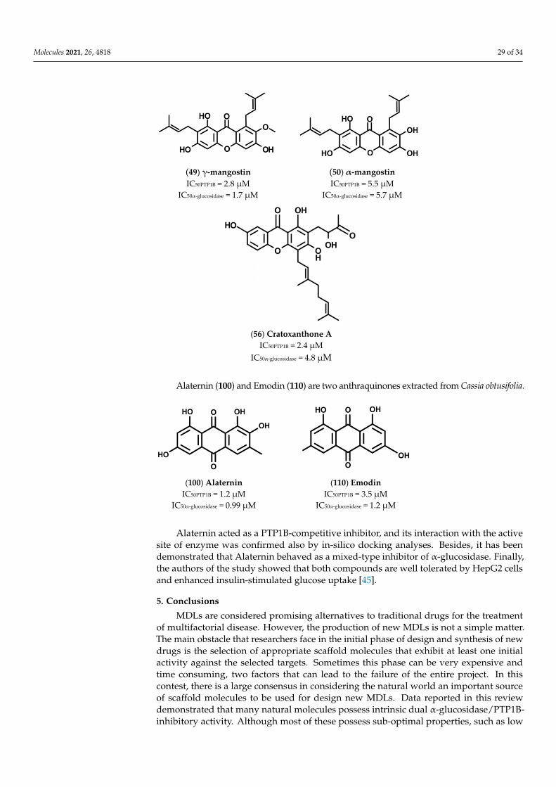
Molecules 2021, 26, 4818 29 of 34Molecules 2021, 26, x 30 of 35
(49) γ-mangostin IC50PTP1B = 2.8 µM
IC50α-glucosidase = 1.7 µM
(50) α-mangostin IC50PTP1B = 5.5 µM
IC50α-glucosidase = 5.7 µM
(56) Cratoxanthone A
IC50PTP1B = 2.4 µM IC50α-glucosidase = 4.8 µM
Alaternin (100) and Emodin (110) are two anthraquinones extracted from Cassia ob-tusifolia.
(100) Alaternin
IC50PTP1B = 1.2 µM IC50α-glucosidase = 0.99 µM
(110) Emodin IC50PTP1B = 3.5 µM
IC50α-glucosidase = 1.2 µM
Alaternin acted as a PTP1B-competitive inhibitor, and its interaction with the active site of enzyme was confirmed also by in-silico docking analyses. Besides, it has been demonstrated that Alaternin behaved as a mixed-type inhibitor of α-glucosidase. Finally, the authors of the study showed that both compounds are well tolerated by HepG2 cells and enhanced insulin-stimulated glucose uptake [45].
5. Conclusions MDLs are considered promising alternatives to traditional drugs for the treatment of
multifactorial disease. However, the production of new MDLs is not a simple matter. The main obstacle that researchers face in the initial phase of design and synthesis of new drugs is the selection of appropriate scaffold molecules that exhibit at least one initial ac-tivity against the selected targets. Sometimes this phase can be very expensive and time consuming, two factors that can lead to the failure of the entire project. In this contest, there is a large consensus in considering the natural world an important source of scaffold molecules to be used for design new MDLs. Data reported in this review demonstrated
Alaternin (100) and Emodin (110) are two anthraquinones extracted from Cassia obtusifolia.
Molecules 2021, 26, x 30 of 35
(49) γ-mangostin IC50PTP1B = 2.8 µM
IC50α-glucosidase = 1.7 µM
(50) α-mangostin IC50PTP1B = 5.5 µM
IC50α-glucosidase = 5.7 µM
(56) Cratoxanthone A
IC50PTP1B = 2.4 µM IC50α-glucosidase = 4.8 µM
Alaternin (100) and Emodin (110) are two anthraquinones extracted from Cassia ob-tusifolia.
(100) Alaternin
IC50PTP1B = 1.2 µM IC50α-glucosidase = 0.99 µM
(110) Emodin IC50PTP1B = 3.5 µM
IC50α-glucosidase = 1.2 µM
Alaternin acted as a PTP1B-competitive inhibitor, and its interaction with the active site of enzyme was confirmed also by in-silico docking analyses. Besides, it has been demonstrated that Alaternin behaved as a mixed-type inhibitor of α-glucosidase. Finally, the authors of the study showed that both compounds are well tolerated by HepG2 cells and enhanced insulin-stimulated glucose uptake [45].
5. Conclusions MDLs are considered promising alternatives to traditional drugs for the treatment of
multifactorial disease. However, the production of new MDLs is not a simple matter. The main obstacle that researchers face in the initial phase of design and synthesis of new drugs is the selection of appropriate scaffold molecules that exhibit at least one initial ac-tivity against the selected targets. Sometimes this phase can be very expensive and time consuming, two factors that can lead to the failure of the entire project. In this contest, there is a large consensus in considering the natural world an important source of scaffold molecules to be used for design new MDLs. Data reported in this review demonstrated
Alaternin acted as a PTP1B-competitive inhibitor, and its interaction with the activesite of enzyme was confirmed also by in-silico docking analyses. Besides, it has beendemonstrated that Alaternin behaved as a mixed-type inhibitor of α-glucosidase. Finally,the authors of the study showed that both compounds are well tolerated by HepG2 cellsand enhanced insulin-stimulated glucose uptake [45].
5. Conclusions
MDLs are considered promising alternatives to traditional drugs for the treatmentof multifactorial disease. However, the production of new MDLs is not a simple matter.The main obstacle that researchers face in the initial phase of design and synthesis of newdrugs is the selection of appropriate scaffold molecules that exhibit at least one initialactivity against the selected targets. Sometimes this phase can be very expensive andtime consuming, two factors that can lead to the failure of the entire project. In thiscontest, there is a large consensus in considering the natural world an important sourceof scaffold molecules to be used for design new MDLs. Data reported in this reviewdemonstrated that many natural molecules possess intrinsic dual α-glucosidase/PTP1B-inhibitory activity. Although most of these possess sub-optimal properties, such as low

Molecules 2021, 26, 4818 30 of 34
bioavailability and unfavorable pharmacokinetic or a lowspecificity, semi- or fully syntheticsuperior derivatives could be easily obtained starting from them. This approach couldallow researchers to quickly overcome the first experimental phase, allowing them to focuson the optimization phase aimed at balancing the activity of the molecules toward thebiological targets, increasing theirspecificityor bioavailability.
The data we reported confirmed that many natural molecules, including some lig-nanamides, xanthones, anthraquinones, and several phenolic compounds, show high/balancedinhibitory activity toward PTP1B and α-glucosidase, making these as interesting lead com-pounds for synthesis of new MDLs. Most of these compounds are characterized by thepresence of several hydroxylated phenolic rings or aliphatic chains. What often emerges fromSAR analyses is that the number and the position of hydroxyl groups or aliphatic chainsare essential to ensure a close interaction between the inhibitors and both enzymes and thatby changing one of these parameters, it is possible to influence both inhibitory power andspecificity for targets. Moreover, we found that complexity and molecular weight are twoparameters strictly related with the IC50 values of molecules, confirming that the higher thenumber of OH groups, the higher the affinity for targets. However, small molecules such asAlaternin, Ugonin J, or α-Mangostin also showed high affinity for both targets, suggestingthat it is possible to reach a high inhibitory activity also starting from small scaffold molecules.This is another interesting aspect concerning the mechanism of action of these molecules. Datareported in Table 14 shows that smaller molecules mainly behave as competitive inhibitorsfor PTP1B and bigger ones as non-competitive inhibitors. This evidence suggests that smallmolecules bearing a hydroxylated phenyl ring could mimic the phenolic structure of thenatural substrate, the phosphotyrosine, and for this reason, they could interact more easilywith the active site of the enzyme. Conversely, larger molecules, due to their steric hindrance,would not be able to access the active site but could bind to allosteric sites on the surface of theenzyme.
To the best of our knowledge, few synthetic PTP1B/α-glucosidase inhibitors havebeen produced still to date, and none of these has been evaluated in clinical trials to date,which is probably due to the fact that development of such kinds of molecules is still in itsinfancy. We hope that the information reported in this review will be useful to researchersdedicated to the design and synthesis of novel dual PTP1B/α-glucosidase inhibitors tobe used routinely for the treatment of patients affected by T2D, obesity, and metabolicsyndrome.
Author Contributions: Conceptualization, P.P. and M.G.; methodology, I.N.; software, M.G.; valida-tion, A.C., P.P., and M.G.; formal analysis, P.P.; investigation, P.P.; resources, P.P.; data curation, P.P.;writing—original draft preparation, P.P., A.C., M.G., and I.N.; writing—review and editing, P.P., A.C.,M.G., and I.N.; supervision, P.P.; project administration, P.P.; funding acquisition, P.P. All authorshave read and agreed to the published version of the manuscript.
Funding: This research was funded by University of Study of Florence, Italy (Fondo Ateneo ex 60%).
Conflicts of Interest: The authors declare no conflict of interest.
References1. Brunetti, L.; Kalabalik, J. Management of type-2 diabetes mellitus in adults: Focus on individualizing non-insulin therapies.
Pharm. Ther. 2012, 37, 687–696.2. Del Prato, S.; Felton, A.-M.; Munro, N.; Nesto, R.; Zimmet, P.; Zinman, B.; Global Partnership for Effective Diabetes Management.
Improving glucose management: Ten steps to get more patients with type 2 diabetes to glycaemic goal. Int. J. Clin. Pract. 2005, 59,1345–1355. [CrossRef] [PubMed]
3. Vos, R.C.; van Avendonk, M.J.; Jansen, H.; Goudswaard, A.N.; van den Donk, M.; Gorter, K.; Kerssen, A.; Rutten, G.E. Insulinmonotherapy compared with the addition of oral glucose-lowering agents to insulin for people with type 2 diabetes already oninsulin therapy and inadequate glycaemic control. Cochrane Database Syst. Rev. 2016, 9, CD006992. [CrossRef] [PubMed]
4. Hutchins, V.; Zhang, B.; Fleurence, R.L.; Krishnarajah, G.; Graham, J. A systematic review of adherence, treatment satisfactionand costs, in fixed-dose combination regimens in type 2 diabetes. Curr. Med. Res. Opin. 2011, 27, 1157–1168. [CrossRef]
5. Milligan, S. Combination therapy for the improvement of long-term macrovascular and microvascular outcomes in type 2diabetes: Rationale and evidence for early initiation. J. Diabetes Complicat. 2016, 30, 1177–1185. [CrossRef]
Molecules 2021, 26, 4818 31 of 34
6. Morphy, R.; Rankovic, Z. Designed multiple ligands. An emerging drug discovery paradigm. J. Med. Chem. 2005, 48, 6523–6543.[CrossRef]
7. Artasensi, A.; Pedretti, A.; Vistoli, G.; Fumagalli, L. Type 2 Diabetes Mellitus: A Review of Multi-Target Drugs. Molecules 2020,25, 1987. [CrossRef]
8. Coskun, T.; Sloop, K.W.; Loghin, C.; Alsina-Fernandez, J.; Urva, S.; Bokvist, K.B.; Cui, X.; Briere, D.A.; Cabrera, O.;Roell, W.C.; et al. LY3298176, a novel dual GIP and GLP-1 receptor agonist for the treatment of type 2 diabetes mellitus: Fromdiscovery to clinical proof of concept. Mol. Metab. 2018, 18, 3–14. [CrossRef] [PubMed]
9. Burmeister, M.A.; Brown, J.D.; Ayala, J.E.; Stoffers, D.A.; Sandoval, D.A.; Seeley, R.J.; Ayala, J.E. The glucagon-like peptide-1receptor in the ventromedial hypothalamus reduces short-term food intake in male mice by regulating nutrient sensor activity.Am. J. Physiol. Endocrinol. Metab. 2017, 313, E651–E662. [CrossRef]
10. Chepurny, O.G.; Bonaccorso, R.L.; Leech, C.A.; Wöllert, T.; Langford, G.M.; Schwede, F.; Roth, C.L.; Doyle, R.P.; Holz, G.G.Chimeric peptide EP45 as a dual agonist at GLP-1 and NPY2R receptors. Sci. Rep. 2018, 8, 1–14. [CrossRef]
11. Cheng, C.-F.; Ku, H.-C.; Lin, H. PGC-1α as a Pivotal Factor in Lipid and Metabolic Regulation. Int. J. Mol. Sci. 2018, 19, 3447.[CrossRef]
12. Wang, Y.-X.; Lee, C.-H.; Tiep, S.; Yu, R.T.; Ham, J.; Kang, H.; Evans, R.M. Peroxisome-Proliferator-Activated Receptor δ ActivatesFat Metabolism to Prevent Obesity. Cell 2003, 113, 159–170. [CrossRef]
13. Matsushita, Y.; Ogawa, D.; Wada, J.; Yamamoto, N.; Shikata, K.; Sato, C.; Tachibana, H.; Toyota, N.; Makino, H. Activation ofperoxisome proliferator-activated receptor delta inhibits streptozotocin-induced diabetic nephropathy through anti-inflammatorymechanisms in mice. Diabetes 2011, 60, 960–968. [CrossRef] [PubMed]
14. Laplante, M.; Sell, H.; MacNaul, K.L.; Richard, D.; Berger, J.P.; Deshaies, Y. PPAR-gamma activation mediates adipose depot-specific effects on gene expression and lipoprotein lipase activity: Mechanisms for modulation of postprandial lipemia anddifferential adipose accretion. Diabetes 2003, 52, 291–299. [CrossRef]
15. Jain, N.; Bhansali, S.; Kurpad, A.V.; Hawkins, M.; Sharma, A.; Kaur, S.; Rastogi, A.; Bhansali, A. Effect of a Dual PPAR α/γ agoniston Insulin Sensitivity in Patients of Type 2 Diabetes with Hypertriglyceridemia- Randomized double-blind placebo-controlledtrial. Sci. Rep. 2019, 9, 1–9. [CrossRef] [PubMed]
16. Zhou, Z.; Deng, L.; Hu, L.; Ren, Q.; Cai, Z.; Wang, B.; Li, Z.; Zhang, L. Hepatoprotective effects of ZLY16, a dual peroxisomeproliferator-activated receptor α/δ agonist, in rodent model of nonalcoholic steatohepatitis. Eur. J. Pharmacol. 2020, 882, 173300.[CrossRef]
17. Staels, B.; Rubenstrunk, A.; Noel, B.; Rigou, G.; Delataille, P.; Millatt, L.J.; Baron, M.; Lucas, A.; Tailleux, A.; Hum, D.W.; et al.Hepatoprotective effects of the dual peroxisome proliferator-activated receptor alpha/delta agonist, GFT505, in rodent models ofnonalcoholic fatty liver disease/nonalcoholic steatohepatitis. Hepatology 2013, 58, 1941–1952. [CrossRef] [PubMed]
18. Chen, W.; Fan, S.; Xie, X.; Xue, N.; Jin, X.; Wang, L. Novel PPAR pan agonist, ZBH ameliorates hyperlipidemia and insulinresistance in high fat diet induced hyperlipidemic hamster. PLoS ONE 2014, 9, e96056. [CrossRef]
19. Li, M.H.; Chen, W.; Wang, L.L.; Sun, J.L.; Zhou, L.; Shi, Y.C.; Wang, C.H.; Zhong, B.H.; Shi, W.G.; Guo, Z.W. RLA8-A New andHighly Effective Quadruple PPAR-α/γ/δ and GPR40 Agonist to Reverse Nonalcoholic Steatohepatitis and Fibrosis. J. Pharmacol.Exp. Ther. 2019, 369, 67–77. [CrossRef]
20. Ramachandran, U.; Kumar, R.; Mittal, A. Fine tuning of PPAR ligands for type 2 diabetes and metabolic syndrome. Mini Rev.Med. Chem. 2006, 6, 563–573. [CrossRef]
21. Pan, Q.; Lin, S.; Li, Y.; Liu, L.; Li, X.; Gao, X.; Yan, J.; Gu, B.; Chen, X.; Li, W.; et al. A novel GLP-1 and FGF21 dual agonist hastherapeutic potential for diabetes and non-alcoholic steatohepatitis. EBioMedicine 2021, 63, 103202. [CrossRef]
22. Yaribeygi, H.; Farrokhi, F.R.; Butler, A.E.; Sahebkar, A. Insulin resistance: Review of the underlying molecular mechanisms. J. Cell.Physiol. 2019, 234, 8152–8161. [CrossRef]
23. Abdelsalam, S.S.; Korashy, H.M.; Zeidan, A.; Agouni, A. The Role of Protein Tyrosine Phosphatase (PTP)-1B in CardiovascularDisease and Its Interplay with Insulin Resistance. Biomolecules 2019, 9, 286. [CrossRef] [PubMed]
24. Haj, F.G.; Zabolotny, J.M.; Kim, Y.-B.; Kahn, B.B.; Neel, B.G. Liver-specific protein-tyrosine phosphatase 1B (PTP1B) re-expressionalters glucose homeostasis of PTP1B-/-mice. J. Biol. Chem. 2005, 280, 15038–15046. [CrossRef]
25. Zabolotny, J.M.; Haj, F.G.; Kim, Y.-B.; Kim, H.-J.; Shulman, G.I.; Kim, J.K.; Neel, B.G.; Kahn, B.B. Transgenic overexpressionof protein-tyrosine phosphatase 1B in muscle causes insulin resistance, but overexpression with leukocyte antigen-relatedphosphatase does not additively impair insulin action. J. Biol. Chem. 2004, 279, 24844–24851. [CrossRef] [PubMed]
26. Rondinone, C.M.; Trevillyan, J.M.; Clampit, J.; Gum, R.J.; Berg, C.; Kroeger, P.; Frost, L.; Zinker, B.A.; Reilly, R.; Ulrich, R.; et al.Protein tyrosine phosphatase 1B reduction regulates adiposity and expression of genes involved in lipogenesis. Diabetes 2002, 51,2405–2411. [CrossRef]
27. Lu, B.; Gu, P.; Xu, Y.; Ye, X.; Wang, Y.; DU, H.; Shao, J. Overexpression of protein tyrosine phosphatase 1B impairs glucose-stimulated insulin secretion in INS-1 cells. Minerva Endocrinol. 2016, 41, 1–9.
28. Zhang, Z.-Y.; Dodd, G.T.; Tiganis, T. Protein Tyrosine Phosphatases in Hypothalamic Insulin and Leptin Signaling. TrendsPharmacol. Sci. 2015, 36, 661–674. [CrossRef]
29. Elchebly, M.; Payette, P.; Michaliszyn, E.; Cromlish, W.; Collins, S.; Loy, A.L.; Normandin, D.; Cheng, A.; Himms-Hagen, J.;Chan, C.C.; et al. Increased insulin sensitivity and obesity resistance in mice lacking the protein tyrosine phosphatase-1B gene.Science 1999, 283, 1544–1548. [CrossRef]
Molecules 2021, 26, 4818 32 of 34
30. Joshi, S.R.; Standl, E.; Tong, N.; Shah, P.; Kalra, S.; Rathod, R. Therapeutic potential of α-glucosidase inhibitors in type 2 diabetesmellitus: An evidence-based review. Expert Opin. Pharmacother. 2015, 16, 1959–1981. [CrossRef] [PubMed]
31. Yang, H.K.; Lee, S.H.; Shin, J.; Choi, Y.H.; Ahn, Y.B.; Lee, B.W.; Rhee, E.J.; Min, K.W.; Yoon, K.H. Acarbose Add-on Therapyin Patients with Type 2 Diabetes Mellitus with Metformin and Sitagliptin Failure: A Multicenter, Randomized, Double-Blind,Placebo-Controlled Study. Diabetes Metab. J. 2019, 43, 287–301. [CrossRef] [PubMed]
32. Wang, M.-Y.; Cheng, X.-C.; Chen, X.-B.; Li, Y.; Zang, L.-L.; Duan, Y.-Q.; Chen, M.-Z.; Yu, P.; Sun, H.; Wang, R.-L. Synthesis andbiological evaluation of novel N-aryl-ω-(benzoazol-2-yl)-sulfanylalkanamides as dual inhibitors of α-glucosidase and proteintyrosine phosphatase 1B. Chem. Biol. Drug Des. 2018, 92, 1647–1656. [CrossRef] [PubMed]
33. Ferhati, X.; Matassini, C.; Fabbrini, M.G.; Goti, A.; Morrone, A.; Cardona, F.; Moreno-Vargas, A.J.; Paoli, P. Dual targeting ofPTP1B and glucosidases with new bifunctional iminosugar inhibitors to address type 2 diabetes. Bioorg. Chem. 2019, 87, 534–549.[CrossRef]
34. Mphahlele, M.J.; Choong, Y.S.; Maluleka, M.M.; Gildenhuys, S. Synthesis, In Vitro Evaluation and Molecular Docking ofthe 5-Acetyl-2-aryl-6-hydroxybenzo[b]furans against Multiple Targets Linked to Type 2 Diabetes. Biomolecules 2020, 10, 418.[CrossRef]
35. Atanasov, A.G.; Waltenberger, B.; Pferschy-Wenzig, E.-M.; Linder, T.; Wawrosch, C.; Uhrin, P.; Temml, V.; Wang, L.; Schwaiger, S.;Heiss, E.H.; et al. Discovery and resupply of pharmacologically active plant-derived natural products: A review. Biotechnol. Adv.2015, 33, 1582–1614. [CrossRef] [PubMed]
36. Ali, M.Y.; Jannat, S.; Jung, H.A.; Choi, J.S. Insulin-Mimetic Dihydroxanthyletin-Type Coumarins from Angelica decursiva withProtein Tyrosine Phosphatase 1B and α-Glucosidase Inhibitory Activities and Docking Studies of Their Molecular Mechanisms.Antioxidants 2021, 10, 292. [CrossRef] [PubMed]
37. Ali, M.Y.; Jannat, S.; Jung, H.A.; Jeong, H.O.; Chung, H.Y.; Choi, J.S. Coumarins from Angelica decursiva inhibit α-glucosidaseactivity and protein tyrosine phosphatase 1B. Chem. Biol. Interact. 2016, 252, 93–101. [CrossRef]
38. Jeong, S.-Y.; Nguyen, P.-H.; Zhao, B.-T.; Ali, M.Y.; Choi, J.-S.; Min, B.-S.; Woo, M.-H. Chemical Constituents of Euonymus alatus(Thunb.) Sieb. and Their PTP1B and α-Glucosidase Inhibitory Activities. Phytother. Res. 2015, 29, 1540–1548. [CrossRef]
39. Nurul Islam, M.; Jung, H.A.; Sohn, H.S.; Kim, H.M.; Choi, J.S. Potent α-glucosidase and protein tyrosine phosphatase 1B inhibitorsfrom Artemisia capillaris. Arch. Pharm. Res. 2013, 36, 542–552. [CrossRef]
40. Nguyen, D.-T.; To, D.-C.; Tran, T.-T.; Tran, M.-H.; Nguyen, P.-H. PTP1B and α-glucosidase inhibitors from Selaginella rolandi-principis and their glucose uptake stimulation. J. Nat. Med. 2021, 75, 186–193. [CrossRef]
41. Zhao, C.-C.; Chen, J.; Shao, J.-H.; Zhang, X.-H.; Gu, W.-Y.; Shen, J.; Liu, Y. Lignan Constituents from the Fruits of Viburnummacrocephalum f. keteleeri and their α-Amylase, α-Glucosidase, and Protein Tyrosine Phosphatase 1B Inhibitory Activities. J.Agric. Food Chem. 2020, 68, 11151–11160. [CrossRef]
42. Tuohongerbieke, A.; Li, J.; Sabir, G.; Xin, X.; Hu, M.; Duan, X.; Liu, L.; Tang, D.; Zhu, J.; Aisa, H.A. Lignanamides from the roots ofLimonium gmelinii (Willd.) Kuntze and their anti-diabetic, cytotoxic and anti-inflammatory activities. Phytochemistry 2021, 184,112648. [CrossRef]
43. Seong, S.H.; Nguyen, D.H.; Wagle, A.; Woo, M.H.; Jung, H.A.; Choi, J.S. Experimental and Computational Study to Revealthe Potential of Non-Polar Constituents from Hizikia fusiformis as Dual Protein Tyrosine Phosphatase 1B and α-GlucosidaseInhibitors. Mar. Drugs 2019, 17, 302. [CrossRef]
44. Li, Z.P.; Song, Y.H.; Uddin, Z.; Wang, Y.; Park, K.H. Inhibition of protein tyrosine phosphatase 1B (PTP1B) and α-glucosidase byxanthones from Cratoxylum cochinchinense, and their kinetic characterization. Bioorg. Med. Chem. 2018, 26, 737–746. [CrossRef]
45. Jung, H.A.; Ali, M.Y.; Choi, J.S. Promising Inhibitory Effects of Anthraquinones, Naphthopyrone, and Naphthalene Glycosides,from Cassia obtusifolia on α-Glucosidase and Human Protein Tyrosine Phosphatases 1B. Molecules 2017, 22, 28. [CrossRef][PubMed]
46. Zhang, Y.; Han, F.-Y.; Wu, J.; Song, S.-J. Triterpene saponins with a-glucosidase and PTP1B inhibitory activities from the leaves ofAralia elata. Phytochem. Lett. 2018, 26, 179–183. [CrossRef]
47. Yang, N.; Zhang, S.; Yang, S.; Guo, Z.; Zhang, X.; Zhao, Y. The inhibition of α-glycosidase and protein tyrosine phosphatase 1B(PTP1B) activities by ginsenosides from Panax ginseng C.A. Meyer and simultaneous determination by HPLC-ELSD. J. Funct.Foods 2016, 23, 188–197. [CrossRef]
48. Liang, C.; Staerk, D.; Kongstad, K.T. Potential of Myrtus communis Linn. as a bifunctional food: Dual high-resolution PTP1B andα-glucosidase inhibition profiling combined with HPLC-HRMS and NMR for identification of antidiabetic triterpenoids andphloroglucinol derivatives. J. Funct. Foods 2020, 64, 103623. [CrossRef]
49. Seong, S.H.; Roy, A.; Jung, H.A.; Jung, H.J.; Choi, J.S. Protein tyrosine phosphatase 1B and α-glucosidase inhibitory activities ofPueraria lobata root and its constituents. J. Ethnopharmacol. 2016, 194, 706–716. [CrossRef]
50. Na, B.; Nguyen, P.-H.; Zhao, B.-T.; Vo, Q.-H.; Min, B.S.; Woo, M.H. Protein tyrosine phosphatase 1B (PTP1B) inhibitory activityand glucosidase inhibitory activity of compounds isolated from Agrimonia pilosa. Pharm. Biol. 2016, 54, 474–480. [CrossRef][PubMed]
51. Ali, M.Y.; Zaib, S.; Rahman, M.M.; Jannat, S.; Iqbal, J.; Park, S.K.; Chang, M.S. Didymin, a dietary citrus flavonoid exhibits anti-diabetic complications and promotes glucose uptake through the activation of PI3K/Akt signaling pathway in insulin-resistantHepG2 cells. Chem. Biol. Interact. 2019, 305, 180–194. [CrossRef]
Molecules 2021, 26, 4818 33 of 34
52. de Cássia Lemos Lima, R.; Kato, L.; Kongstad, K.T.; Staerk, D. Brazilian insulin plant as a bifunctional food: Dual high-resolution PTP1B and α-glucosidase inhibition profiling combined with HPLC-HRMS-SPE-NMR for identification of antidiabeticcompounds in Myrcia rubella Cambess. J. Funct. Foods 2018, 45, 444–451. [CrossRef]
53. Zhang, C.-C.; Geng, C.-A.; Huang, X.-Y.; Zhang, X.-M.; Chen, J.-J. Antidiabetic Stilbenes from Peony Seeds with PTP1B,α-Glucosidase, and DPPIV Inhibitory Activities. J. Agric. Food Chem. 2019, 67, 6765–6772. [CrossRef] [PubMed]
54. He, X.-F.; Chen, J.-J.; Li, T.-Z.; Hu, J.; Zhang, X.-M.; Geng, C.-A. Diarylheptanoid-chalcone hybrids with PTP1B and α-glucosidasedual inhibition from Alpinia katsumadai. Bioorg. Chem. 2021, 108, 104683. [CrossRef]
55. Zhang, Y.-L.; Luo, J.-G.; Wan, C.-X.; Zhou, Z.-B.; Kong, L.-Y. Geranylated 2-arylbenzofurans from Morus alba var. tatarica andtheir α-glucosidase and protein tyrosine phosphatase 1B inhibitory activities. Fitoterapia 2014, 92, 116–126. [CrossRef]
56. Ha, M.T.; Seong, S.H.; Nguyen, T.D.; Cho, W.-K.; Ah, K.J.; Ma, J.Y.; Woo, M.H.; Choi, J.S.; Min, B.S. Chalcone derivatives from theroot bark of Morus alba L. act as inhibitors of PTP1B and α-glucosidase. Phytochemistry 2018, 155, 114–125. [CrossRef]
57. Moon, H.E.; Islam, N.; Ahn, B.R.; Chowdhury, S.S.; Sohn, H.S.; Jung, H.A.; Choi, J.S. Protein tyrosine phosphatase 1B andα-glucosidase inhibitory Phlorotannins from edible brown algae, Ecklonia stolonifera and Eisenia bicyclis. Biosci. Biotechnol.Biochem. 2011, 75, 1472–1480. [CrossRef]
58. Huang, Q.; Chen, J.-J.; Pan, Y.; He, X.-F.; Wang, Y.; Zhang, X.-M.; Geng, C.-A. Chemical profiling and antidiabetic potency ofPaeonia delavayi: Comparison between different parts and constituents. J. Pharm. Biomed. Anal. 2021, 198, 113998. [CrossRef]
59. Paudel, P.; Yu, T.; Seong, S.H.; Kuk, E.B.; Jung, H.A.; Choi, J.S. Protein Tyrosine Phosphatase 1B Inhibition and Glucose UptakePotentials of Mulberrofuran G, Albanol B, and Kuwanon G from Root Bark of Morus alba L. in Insulin-Resistant HepG2 Cells:An In Vitro and In Silico Study. Int. J. Mol. Sci. 2018, 19, 1542. [CrossRef] [PubMed]
60. Shah, A.B.; Yoon, S.; Kim, J.H.; Zhumanova, K.; Ban, Y.J.; Lee, K.W.; Park, K.H. Effectiveness of cyclohexyl functionality inugonins from Helminthostachys zeylanica to PTP1B and α-glucosidase inhibitions. Int. J. Biol. Macromol. 2020, 165, 1822–1831.[CrossRef] [PubMed]
61. Paoli, P.; Cirri, P.; Caselli, A.; Ranaldi, F.; Bruschi, G.; Santi, A.; Camici, G. The insulin-mimetic effect of Morin: A promisingmolecule in diabetes treatment. Biochim. Biophys. Acta 2013, 1830, 3102–3111. [CrossRef] [PubMed]
62. He, X.-F.; Chen, J.-J.; Li, T.-Z.; Zhang, X.-K.; Guo, Y.-Q.; Zhang, X.-M.; Hu, J.; Geng, C.-A. Nineteen New Flavanol-Fatty AlcoholHybrids with α-Glucosidase and PTP1B Dual Inhibition: One Unusual Type of Antidiabetic Constituent from Amomum tsao-ko.J. Agric. Food Chem. 2020, 68, 11434–11448. [CrossRef] [PubMed]
63. Song, Y.H.; Uddin, Z.; Jin, Y.M.; Li, Z.; Curtis-Long, M.J.; Kim, K.D.; Cho, J.K.; Park, K.H. Inhibition of protein tyrosinephosphatase (PTP1B) and α-glucosidase by geranylated flavonoids from Paulownia tomentosa. J. Enzyme Inhib. Med. Chem. 2017,32, 1195–1202. [CrossRef] [PubMed]
64. Fan, J.-R.; Kuang, Y.; Dong, Z.-Y.; Yi, Y.; Zhou, Y.-X.; Li, B.; Qiao, X.; Ye, M. Prenylated Phenolic Compounds from the Aerial Partsof Glycyrrhiza uralensis as PTP1B and α-Glucosidase Inhibitors. J. Nat. Prod. 2020, 83, 814–824. [CrossRef] [PubMed]
65. Wiesmann, C.; Barr, K.J.; Kung, J.; Zhu, J.; Erlanson, D.A.; Shen, W.; Fahr, B.J.; Zhong, M.; Taylor, L.; Randal, M.; et al. Allostericinhibition of protein tyrosine phosphatase 1B. Nat. Struct. Mol. Biol. 2004, 11, 730–737. [CrossRef]
66. Liu, Y.-P.; Li, Y.-J.; Zhao, Y.-Y.; Guo, J.-M.; Liu, Y.-Y.; Wang, X.-P.; Shen, Z.-Y.; Qiang, L.; Fu, Y.-H. Carbazole alkaloids from thefruits of Clausena anisum-olens with potential PTP1B and α-glucosidase inhibitory activities. Bioorg. Chem. 2021, 110, 104775.[CrossRef]
67. Islam, M.N.; Choi, R.J.; Jung, H.A.; Oh, S.H.; Choi, J.S. Promising anti-diabetic potential of capillin and capillinol isolated fromArtemisia capillaris. Arch. Pharm. Res. 2016, 39, 340–349. [CrossRef]
68. Ali, M.Y.; Kim, D.H.; Seong, S.H.; Kim, H.-R.; Jung, H.A.; Choi, J.S. α-Glucosidase and Protein Tyrosine Phosphatase 1B InhibitoryActivity of Plastoquinones from Marine Brown Alga Sargassum serratifolium. Mar. Drugs 2017, 15, 368. [CrossRef]
69. Fueller, J.; Egorov, M.V.; Walther, K.A.; Sabet, O.; Mallah, J.; Grabenbauer, M.; Kinkhabwala, A. Subcellular Partitioning of ProteinTyrosine Phosphatase 1B to the Endoplasmic Reticulum and Mitochondria Depends Sensitively on the Composition of Its TailAnchor. PLoS ONE 2015, 10, e0139429. [CrossRef]
70. Rieg, T.; Vallon, V. Development of SGLT1 and SGLT2 inhibitors. Diabetologia 2018, 61, 2079–2086. [CrossRef]71. Shinde, R.N.; Kumar, G.S.; Eqbal, S.; Sobhia, M.E. Screening and identification of potential PTP1B allosteric inhibitors using in
silico and in vitro approaches. PLoS ONE 2018, 13, e0199020. [CrossRef]72. Lipinski, C.A.; Lombardo, F.; Dominy, B.W.; Feeney, P.J. Experimental and computational approaches to estimate solubility and
permeability in drug discovery and development settings. Adv. Drug Deliv. Rev. 2001, 46, 3–26. [CrossRef]73. Lee, S.-H.; Park, M.-H.; Kang, S.-M.; Ko, S.-C.; Kang, M.-C.; Cho, S.; Park, P.-J.; Jeon, B.-T.; Kim, S.-K.; Han, J.-S.; et al. Dieckol
isolated from Ecklonia cava protects against high-glucose induced damage to rat insulinoma cells by reducing oxidative stressand apoptosis. Biosci. Biotechnol. Biochem. 2012, 76, 1445–1451. [CrossRef]
74. You, H.-N.; Lee, H.-A.; Park, M.-H.; Lee, J.-H.; Han, J.-S. Phlorofucofuroeckol A isolated from Ecklonia cava alleviates postprandialhyperglycemia in diabetic mice. Eur. J. Pharmacol. 2015, 752, 92–96. [CrossRef] [PubMed]
75. Seong, S.H.; Paudel, P.; Jung, H.A.; Choi, J.S. Identifying Phlorofucofuroeckol-A as a Dual Inhibitor of Amyloid-β25-35 Self-Aggregation and Insulin Glycation: Elucidation of the Molecular Mechanism of Action. Mar. Drugs 2019, 17, 600. [CrossRef]
76. Seong, S.H.; Paudel, P.; Choi, J.-W.; Ahn, D.H.; Nam, T.-J.; Jung, H.A.; Choi, J.S. Probing Multi-Target Action of Phlorotanninsas New Monoamine Oxidase Inhibitors and Dopaminergic Receptor Modulators with the Potential for Treatment of NeuronalDisorders. Mar. Drugs 2019, 17, 377. [CrossRef]
Molecules 2021, 26, 4818 34 of 34
77. Oh, S.; Son, M.; Choi, J.; Choi, C.H.; Park, K.Y.; Son, K.H.; Byun, K. Phlorotannins from Ecklonia cava Attenuates Palmitate-Induced Endoplasmic Reticulum Stress and Leptin Resistance in Hypothalamic Neurons. Mar. Drugs 2019, 17, 570. [CrossRef][PubMed]
78. Kang, M.-C.; Wijesinghe, W.A.J.P.; Lee, S.-H.; Kang, S.-M.; Ko, S.-C.; Yang, X.; Kang, N.; Jeon, B.-T.; Kim, J.; Lee, D.-H.; et al.Dieckol isolated from brown seaweed Ecklonia cava attenuates type II diabetes in db/db mouse model. Food Chem. Toxicol. 2013,53, 294–298. [CrossRef] [PubMed]
79. Lee, S.-H.; Min, K.-H.; Han, J.-S.; Lee, D.-H.; Park, D.-B.; Jung, W.-K.; Park, P.-J.; Jeon, B.-T.; Kim, S.-K.; Jeon, Y.-J. Effects of brownalga, Ecklonia cava on glucose and lipid metabolism in C57BL/KsJ-db/db mice, a model of type 2 diabetes mellitus. Food Chem.Toxicol. 2012, 50, 575–582. [CrossRef]
80. Lee, S.-H.; Jeon, Y.-J. Efficacy and safety of a dieckol-rich extract (AG-dieckol) of brown algae, Ecklonia cava, in pre-diabeticindividuals: A double-blind, randomized, placebo-controlled clinical trial. Food Funct. 2015, 6, 853–858. [CrossRef]
81. Tanabe, K.; Nakamura, S.; Omagari, K.; Oku, T. Repeated ingestion of the leaf extract from Morus alba reduces insulin resistancein KK-Ay mice. Nutr. Res. 2011, 31, 848–854. [CrossRef] [PubMed]
82. Singab, A.N.B.; El-Beshbishy, H.A.; Yonekawa, M.; Nomura, T.; Fukai, T. Hypoglycemic effect of Egyptian Morus alba root barkextract: Effect on diabetes and lipid peroxidation of streptozotocin-induced diabetic rats. J. Ethnopharmacol. 2005, 100, 333–338.[CrossRef] [PubMed]
83. Chang, T.-C.; Chiou, W.-C.; Lai, W.-H.; Huang, H.-C.; Huang, Y.-L.; Liu, H.-K.; Liang, Y.-C.; Huang, C. Ugonin J improvesmetabolic disorder and ameliorates nonalcoholic fatty liver disease by regulating the AMPK/AKT signaling pathway. Pharmacol.Res. 2021, 163, 105298. [CrossRef] [PubMed]