MRI-Visible Poly(ε-caprolactone) with Controlled Contrast Agent Ratios for Enhanced Visualization...
9
MRI-Visible Poly(ε-caprolactone) with Controlled Contrast Agent Ratios for Enhanced Visualization in Temporary Imaging Applications Sarah El Habnouni, Benjamin Nottelet,* Vincent Darcos, Barbara Porsio, Laurent Lemaire, † Florence Franconi, ‡ Xavier Garric, and Jean Coudane Institut des Biomolé cules Max Mousseron (IBMM), UMR CNRS 5247, University of Montpellier 1, University of Montpellier 2 − ENSCM, Faculty of Pharmacy, 15 Av. C. Flahault, Montpellier, 34093, France † Micro et Nanome ́ decines Biomimé tiques (MINT), UMR-S 1066, Universite ́ d’Angers, 4 rue Larrey, 49933 Angers Cedex9, France ‡ PIAM, UFR Sciences, 2bd Lavoisier, 49045 Angers, France * S Supporting Information ABSTRACT: Hydrophobic macromolecular contrast agents (MMCAs) are highly desirable to provide safe and efficient magnetic resonance (MR) visibility to implantable medical devices. In this study, we report on the synthesis and evaluation of novel biodegradable poly(ε-caprolactone)-based MMCAs. Poly(α-propargyl-ε-caprolactone-co-ε-caprolactone)s containing 2, 5, and 10 mol % of propargyl groups have been prepared by ring-opening copolymerization of ε-caprolactone and the corresponding propargylated lactone. In parallel, a diazido derivative of the clinically used diethylenetriaminepentaacetic acid (DTPA)/Gd 3+ complex has been synthesized. Finally, MRI-visible poly(ε-caprolactone)s (PCLs) were obtained by the efficient click ligation of these compounds via a Cu I -catalyzed [3 + 2] cycloaddition. ICP-MS analyses confirmed the efficient coupling of the complex on the PCL backbone with the MRI-visible PCLs containing 1.0, 2.6, and 3.6 wt % of Gd 3+ . The influence of the Gd 3+ grafting density on the T 1 relaxation times and on the MRI visibility of the novel biodegradable MMCAs was evaluated. Finally, their stability and cytocompatibility were assessed with regard to their potential as innovative MRI-visible biomaterials for biomedical applications. ■ INTRODUCTION Magnetic resonance imaging (MRI) is today one of the noninvasive technique of choice to provide high spatial and temporal resolutions in clinical diagnosis and staging of human diseases. 1 Unfortunately, in the case of polymeric prostheses there is an inability to provide a postoperative image of implanted prostheses, as polymeric biomaterials are intrinsically transparent to X-rays and are invisible using clinical MRI. 2,3 In some cases, the signal void of the prosthetic material can be useful for diagnoses, however it strongly depends on the size of the prostheses and on the site of implantation. Therefore this inability to visualize implanted material restricts the evaluation of the tissue integration, the postoperative fixation and the material’s fate in the body. 4 This led us to consider in the past the possibility to produce radio-opaque degradable polyesters. A first example was provided by the radio-opaque poly(ε- caprolactone) (PCL) obtained by the anionic chemical modification of PCL with iodine to generate a poly(α-iodo-ε- caprolactone-co-ε-caprolactone) copolymer. 5 More recently, this postmodification strategy was applied to the synthesis of MRI-visible PCL and MRI-visible poly(methyl acrylate) (PMA) that were among the first examples of hydrophobic MRI-visible thermoplastics. 6,7 Indeed, although MRI polymers have attracted much attention in the recent past, most efforts are dedicated to water-soluble polycondensate or amphiphilic structures to be used in drug delivery approaches. 8−17 In particular, a preferred strategy relies on the modification of diblock amphiphilic copolymers with a contrast agent to yield macromolecular contrast agents (MMCAs) able to self- assemble into micellar systems. 13,18−22 These water-soluble MMCAs are rapidly excreted and cannot be used for the MRI visualization of non water-soluble implanted devices. Another strategy relies in the development of nonclassical MRI techniques, as exemplified by the recent use of amide proton transfer MRI technique to visualize hydrophilic and fast resorbing collagen coatings. 23 In this regard, the originality of the postmodification approach used by our group lies in the possibility of providing hydrophobic and multivalent MMCAs. However, one limitation was the lack of fine control over the final substitution degree of the polymeric backbone and the necessity to use protection/deprotection steps to covalently bind the ligand diethylenetriaminepentaacetic acid (DTPA) before final complexation of the Gd 3+ MRI contrast agent. Yet, control of the overall amount of contrast agent, as well as its repartition along the polymer chain and its chemical environ- ment, are known to influence the resulting relaxivity and visualization. 24 The strategy chosen in this work to prepare hydrophobic and multivalent MMCAs addresses these points. It is based on the convergent synthesis of an azide-containing contrast agent, and of a propargyl-functionalized PCL whose late ligation occurs via Cu I -catalyzed [3 + 2] cycloaddition reaction. Practically, the control of the functionalization of Received: July 5, 2013 Revised: September 4, 2013 Article pubs.acs.org/Biomac © XXXX American Chemical Society A dx.doi.org/10.1021/bm400978a | Biomacromolecules XXXX, XXX, XXX−XXX
Transcript of MRI-Visible Poly(ε-caprolactone) with Controlled Contrast Agent Ratios for Enhanced Visualization...
MRI-Visible Poly(ε-caprolactone) with Controlled Contrast AgentRatios for Enhanced Visualization in Temporary ImagingApplicationsSarah El Habnouni, Benjamin Nottelet,* Vincent Darcos, Barbara Porsio, Laurent Lemaire,†
Florence Franconi,‡ Xavier Garric, and Jean Coudane
Institut des Biomolecules Max Mousseron (IBMM), UMR CNRS 5247, University of Montpellier 1, University of Montpellier 2 −ENSCM, Faculty of Pharmacy, 15 Av. C. Flahault, Montpellier, 34093, France†Micro et Nanomedecines Biomimetiques (MINT), UMR-S 1066, Universite d’Angers, 4 rue Larrey, 49933 Angers Cedex9, France‡PIAM, UFR Sciences, 2bd Lavoisier, 49045 Angers, France
*S Supporting Information
ABSTRACT: Hydrophobic macromolecular contrast agents (MMCAs) are highly desirable to provide safe and efficientmagnetic resonance (MR) visibility to implantable medical devices. In this study, we report on the synthesis and evaluation ofnovel biodegradable poly(ε-caprolactone)-based MMCAs. Poly(α-propargyl-ε-caprolactone-co-ε-caprolactone)s containing 2, 5,and 10 mol % of propargyl groups have been prepared by ring-opening copolymerization of ε-caprolactone and thecorresponding propargylated lactone. In parallel, a diazido derivative of the clinically used diethylenetriaminepentaacetic acid(DTPA)/Gd3+ complex has been synthesized. Finally, MRI-visible poly(ε-caprolactone)s (PCLs) were obtained by the efficientclick ligation of these compounds via a CuI-catalyzed [3 + 2] cycloaddition. ICP-MS analyses confirmed the efficient coupling ofthe complex on the PCL backbone with the MRI-visible PCLs containing 1.0, 2.6, and 3.6 wt % of Gd3+. The influence of theGd3+ grafting density on the T1 relaxation times and on the MRI visibility of the novel biodegradable MMCAs was evaluated.Finally, their stability and cytocompatibility were assessed with regard to their potential as innovative MRI-visible biomaterials forbiomedical applications.
■ INTRODUCTION
Magnetic resonance imaging (MRI) is today one of thenoninvasive technique of choice to provide high spatial andtemporal resolutions in clinical diagnosis and staging of humandiseases.1 Unfortunately, in the case of polymeric prosthesesthere is an inability to provide a postoperative image ofimplanted prostheses, as polymeric biomaterials are intrinsicallytransparent to X-rays and are invisible using clinical MRI.2,3 Insome cases, the signal void of the prosthetic material can beuseful for diagnoses, however it strongly depends on the size ofthe prostheses and on the site of implantation. Therefore thisinability to visualize implanted material restricts the evaluationof the tissue integration, the postoperative fixation and thematerial’s fate in the body.4 This led us to consider in the pastthe possibility to produce radio-opaque degradable polyesters.A first example was provided by the radio-opaque poly(ε-caprolactone) (PCL) obtained by the anionic chemicalmodification of PCL with iodine to generate a poly(α-iodo-ε-caprolactone-co-ε-caprolactone) copolymer.5 More recently,this postmodification strategy was applied to the synthesis ofMRI-visible PCL and MRI-visible poly(methyl acrylate) (PMA)that were among the first examples of hydrophobic MRI-visiblethermoplastics.6,7 Indeed, although MRI polymers haveattracted much attention in the recent past, most efforts arededicated to water-soluble polycondensate or amphiphilicstructures to be used in drug delivery approaches.8−17 Inparticular, a preferred strategy relies on the modification ofdiblock amphiphilic copolymers with a contrast agent to yield
macromolecular contrast agents (MMCAs) able to self-assemble into micellar systems.13,18−22 These water-solubleMMCAs are rapidly excreted and cannot be used for the MRIvisualization of non water-soluble implanted devices. Anotherstrategy relies in the development of nonclassical MRItechniques, as exemplified by the recent use of amide protontransfer MRI technique to visualize hydrophilic and fastresorbing collagen coatings.23 In this regard, the originality ofthe postmodification approach used by our group lies in thepossibility of providing hydrophobic and multivalent MMCAs.However, one limitation was the lack of fine control over thefinal substitution degree of the polymeric backbone and thenecessity to use protection/deprotection steps to covalentlybind the ligand diethylenetriaminepentaacetic acid (DTPA)before final complexation of the Gd3+ MRI contrast agent. Yet,control of the overall amount of contrast agent, as well as itsrepartition along the polymer chain and its chemical environ-ment, are known to influence the resulting relaxivity andvisualization.24 The strategy chosen in this work to preparehydrophobic and multivalent MMCAs addresses these points.It is based on the convergent synthesis of an azide-containingcontrast agent, and of a propargyl-functionalized PCL whoselate ligation occurs via CuI-catalyzed [3 + 2] cycloadditionreaction. Practically, the control of the functionalization of
Received: July 5, 2013Revised: September 4, 2013
Article
pubs.acs.org/Biomac
© XXXX American Chemical Society A dx.doi.org/10.1021/bm400978a | Biomacromolecules XXXX, XXX, XXX−XXX
propargylated PCLs is conveniently obtained by ROP of εCLand αPgεCL, as recently described by our group,25 whereas theuse of a click chemistry reaction for ligation ensures aquantitative functionalization of the polymer by the contrastagent. Finally, the direct coupling of the Gd3+ complex on thepolymer avoids the late complexation step between themacromolecular ligand and Gd3+, which is generally of lowerefficiency to control the final amount of immobilized contrastagent.7
We thus propose in this work a so far nonexplored strategyfor the efficient and controlled Gd3+-functionalization ofaliphatic degradable polyesters to provide biocompatible andslow degrading hydrophobic MRI-visible thermoplastics forlong-term MR visualization. Synthesis of copolyesters MMCAswith finely controlled compositions is described. The influenceof the copolymers nature is discussed with respect to their MRvisualization. Finally, stability of these MMCAs and theircytocompatibility are assessed.
■ EXPERIMENTAL SECTIONMaterials. Isopropanol, ε-caprolactone, and toluene were dried
over calcium hydride for 24 h at room temperature and distilled underreduced pressure. Tetrahydrofuran was dried by refluxing over abenzophenone-sodium mixture and distilled. All other materials wereobtained from Aldrich and were used without further purification.NMR Spectroscopy. 1H and 13C NMR spectra were recorded on a
Bruker spectrometer (AMX300) operating at 300 and 75 MHz,respectively. Deuterated chloroform or deuterated dimethyl sulfoxidewere used as solvents. Chemical shifts were expressed in ppm withrespect to tetramethylsilane (TMS).Relaxation times for polymers dissolved in DMSO-d6 were
measured on an AMX400 Bruker spectrometer operating at 400MHz. T1 measurements were obtained using T1 inversion/recoverysequence. Analyses were carried out at 80 °C to facilitate the molecularmobility of the polymer. A fixed concentration of 11 mg/mL was usedfor all samples.FT-IR Spectroscopy. Infrared spectra were recorded on a Perkin-
Elmer Spectrum 100 FT-IR spectrometer using the attenuated totalreflectance (ATR) method.LC-MS Analyses. LC/MS analyses were performed on a Q-TOF
(Waters) spectrometer fitted with an electrospray interface. Solventused for HPLC and LC/MS were HPLC grade. MALDI analyses wereperformed on a Ultra-Flex III (Bruker) spectrometer using a dithranolmatrix.Size Exclusion Chromatography. Size exclusion chromatography
(SEC) was performed at room temperature on a Waters systemequipped with a guard column, a 600 mm PLgel 5 mm Mixed Ccolumn (Polymer Laboratories), and a Waters 410 refractometricdetector. Calibration was established with poly(styrene) standardsfrom Polymer Laboratories. THF was used as eluent at a flow rate of 1mL/min.ICP-MS Analyses. Gd3+ was quantified using an Element XR sector
field ICP-MS (inductively coupled plasma-mass spectrometry) atGeosciences in Montpellier (University Montpellier II). Internalstandardization used an ultrapure solution enriched with indium.Synthesis and Characterization of MRI-Visible PCL. Synthesis
of Diazido Functionalized Diethylenetriaminepentaacetic Acid(diN3-DTPA) 1. In a first step, 1-azido-3-aminopropane was preparedaccording to a previously described procedure.26 A solution of 3-chloropropylamine hydrochloride (4 g; 30.8 mmol) and sodium azide(6 g; 92.3 mmol) in water (1 mL per mmol) was heated at 80 °Covernight. After removing most of the water by distillation undervacuum, the reaction mixture was cooled in an ice bath. Diethyl ether(50 mL) and KOH pellets (4 g) were added, keeping the temperaturebelow 10 °C in an ice bath. After separation of the organic phase, theaqueous layer was further extracted with diethyl ether (2 × 20 mL).The combined organic layers were dried over MgSO4 andconcentrated to give oil which was purified by distillation. Colorless
oil (2.46 g) was obtained (80% yield; Figure S1). RMN 1H (300 MHz,CDCl3) δ (ppm): 3.33 (t, 2H, CH2N3), 2.55 (t, 2H, CH2NH2), 2.34(s, 2H, NH2), 1.55 (q, 2H, CH2CH2CH2). FT-IR (ATR, cm−1): 2100(N3).
In a second step, diN3-DTPA was synthesized according to amodified described procedure.27 Typically, 1-azido-3-aminopropane(0.616 g; 6.2 mmol) was reacted with DTPA dianhydride (1 g; 2.8mmol) in anhydrous DMF (20 mL) at room temperature and undernitrogen for 24 h. DMF was distilled under vacuum until 2−3 mL involume and precipitated into diethyl ether. The obtained solid wasfurther dissolved in water and lyophilized to yield a white solid (1.48 g,95% yield). RMN 1H (300 MHz, DMSO) δ (ppm): 3.4 (6H,CH2CO2H), 3.1 (12H, N(CH2)2N and NCH2C(O)), 2.8 (8H,CH2NHC(O) and CH2N3), 1.7 (4H, CH2CH2N3). FT-IR (ATR,cm−1): 2100 (N3). LC-MS (ES+, m/z): 558.4 Da [M + H+].
Caution: ω-aminoalkyl azides being potentially explosives all reactionsinvolving this compound were carried out with the appropriate protectionunder a well ventilated hood.
Preparation of Clickable Gd3+ Complex 2. Pyridine (1.44 mL; 17.9mmol) was added to an aqueous solution of 1 (1 g; 1.79 mmol). After10 min stirring at room temperature a clear solution was obtained andGdCl3·6H2O (1.33 g; 3.58 mmol) was added. Complexation was let torun under stirring at 40 °C for 24 h. Water and pyridine were removedby distillation under vacuum. The resulting solid was dissolved inwater before purification on a Chelex 100 resin. After lyophylization 2was obtained as a white solid (90% yield). Absence of free Gd3+ wasconfirmed by a methyl thymol blue (MTB) test28 (Figure S2), whereasthe content of complexed Gd3+ in 2 was quantitatively determined byICP-MS. FT-IR (ATR, cm−1): 2100 (N3). MALDI-TOF (dithranolm/z): [M + H]+ m/z 713.16 Da; [2M + H]+ m/z calcd, 1423.33 Da;theoretical, 713.17 and 1425.34. Gd3+ content (ICP-MS): 15.4 wt %.
Synthesis of Poly(α -propargyl-ε -caprolactone-co-ε -caprolactone)s P(Pg-CL) 3. α-Propargyl-ε-caprolactone (αPgεCL)was synthesized from εCL by an anionic modification approachalready described elsewhere.25 Polymerization was carried out in bulkusing standard Schlenk technique under an inert atmosphere of argon.Amounts of εCL and αPgεCL were added in the reaction flaskaccording to the targeted compositions. In a typical experiment, εCL(2.66 g; 29.6 mmol), αPgεCL (0.5 g; 3.29 mmol), Sn(Oct)2 (66.4 mg;0.164 mol), and isopropanol (25 μL; 0.328 mmol) were placed in anoven-dried Schlenk tube. The tube was fitted with a rubber septum.The solution was further degassed by three freeze−pump−thaw cycles.The resulting mixture was stirred at 140 °C for 3.5 h. To stop thereaction, polymerization was quenched by addition of an excess of 1 NHCl. The reaction mixture was poured into cold methanol. Theprecipated polymer 3 was collected by filtration and dried in vacuum.1H NMR (300 MHz, CDCl3) δ (ppm): 4.96 (m, (CH3)2CH), 4.02 (t,CH2O), 3.60 (m, CH2OH), 2.50 (m, COCHCH2), 2.26 (t, COCH2),1.96 (m, CCH), 1.49−1.70 (m, CH2-CH2-CH2-CH2-O-), 1.29−1.42 (m, m, CH2-CH2-CH2-CH2-O-), 1.18 (d, (CH3)2CH). IR (ATR,cm−1): 3280 (CCH), 1720 (CO). MnSEC = 18000 g/mol, Đ =1.7.
Synthesis of MRI-Visible Poly(ε-caprolactone)s (MRI-PCLs) 4.Copolymer 3, complex 2 (3 equiv/αPgεCL units) and CuBr (2 equiv/αPgεCL units) were solubilized in DMF. The solution was degassedby three freeze−pump−thaw cycles. N,N,N′,N″,N″-Pentamethyldie-thylenetriamine (PMDETA) (2 equiv/ αPgεCL units) degassed byargon bubbling was added to the reaction medium. The reaction wascarried out for 48 h at room temperature under stirring. The crudemedium was dissolved in THF for dialysis (MWCO 3500 g/mol)against distilled water. Copolymer 4 was recovered after removal of thesolvents and drying in vacuum. Copolymers with 1.0, 2.6, and 3.6 wt %of Gd3+, as determined by ICP-MS, were noted 41, 42, and 43,respectively.
MRI Visualization. MR Imaging Protocol. MR imaging experi-ments were performed on a Bruker Avance DRX system (BrukerBiospin SA, Wissembourg, France). The system was equipped with a150 mm vertical superwide-bore magnet operating at 7 T, a 84 mminner diameter shielded gradient set capable of 144 mT·m−1 maximumgradient strength and a 30 mm diameter birdcage resonator. Modified
Biomacromolecules Article
dx.doi.org/10.1021/bm400978a | Biomacromolecules XXXX, XXX, XXX−XXXB

or native PCL films (∼1 cm2) were embedded in a degassed 1% (w/w) agarose gel prior to imaging. Gadolinium-free samplescorresponded to a PCL film. To test signal enhancement, T1 weightingwas introduced into the MR images using an inversion pulse29 in rapidthree-dimensional (3-D) acquisition with relaxation enhancement(RARE) sequence (TR = 3000 ms; mean echo time (TEm) = 8 ms;RARE factor = 8; FOV = 3 × 3 × 1.5 cm; matrix 128 × 128 × 64).Inversion time was set at 1100 ms, sufficient to allow canceling of theembedding gel.Stability Studies. MRI-visible films were prepared with high (0.4 wt
%) and low (0.1 wt %) contents of Gd3+. Films were prepared bydissolution of the appropriate amounts of PCL and 42 in dichloro-methane followed by solvent evaporation. Film samples (10 mg) werecut out for stability studies. Released gadolinium was quantified byICP-MS after sample immersion in 10 mL of PBS saline buffer andstirring at 130 rpm and at 37 °C. At scheduled time points (1, 3, 7, 15,30, and 90 days), 1 mL aliquots of solution were taken and replaced byfresh buffer. Gd3+ was quantified by ICP-MS.
In Vitro Cytocompatibility. Murine fibroblasts cells (designatedL929) were used to assess the in vitro cytocompatibility of thematerials as recommended by the International and EuropeanStandards (ISO 10993−5:2009). L929 cells were cultured inDMEMα supplemented with 10% fetal bovine serum (FBS), penicillin(100 U/mL), streptomycin (100 μg/mL), and glutamine (2 mM).Sample disks (ø 15 mm) were cut from copolymer films anddisinfected in ethanol for 30 min before immersion in a solution ofsterile PBS containing penicillin and streptomycin (1 mg/mL) andincubation for 48 h at 37 °C. Films were then rinsed 2 times withsterile PBS before soaking for 12 h in sterile PBS. After disinfection,disks were placed in TCPS 12-well plates and Viton O-rings were usedto maintain the samples on the bottom of the wells and avoid cellsgrowing on TCPS underneath the samples. Disks were finally seededwith 1 × 10⁴ cells and viability was evaluated after 1, 2, and 3 daysusing PrestoBlue assay, which reflects the number of living cellspresent on a surface at a given time point. At scheduled time points,culture medium was removed and replaced by 1 mL of fresh medium
Scheme 1. Reaction Scheme for the Synthesis of MRI-PCLs 4a
aReagents and conditions: (a) NaN3, water, 80 °C, overnight; (b) DTPA dianhydride, DMFan, RT, 24 h; (c) pyridine, RT, 10 min/GdCl3·6H2O, 40°C, 24 h; (d) LDA, THFan, −78 °C, 1 h/propargyl bromide, THFan, −30 °C, 3 h; (e) εCL, Sn(Oct)2, iPrOH, 140 °C, 3.5 h; (f) CuBr, PMDETA,DMF, RT, 48 h.
Biomacromolecules Article
dx.doi.org/10.1021/bm400978a | Biomacromolecules XXXX, XXX, XXX−XXXC
containing 10% of PrestoBlue. After 2 h of incubation at 37 °C, 200 μLof supernatant were taken from each well and analyzed forfluorescence at 530 nm (ex.) and 615 nm (em.) with a Victor X3(Perkin-Elmer).
■ RESULTS AND DISCUSSION
Synthesis of MRI-Visible Poly(ε-caprolactone)s (MRI-PCLs). Preparation of Clickable Gd3+ Complex 2. The presentwork aims at providing MRI-PCLs with controlled content ofgadolinium by coupling of an azide-containing contrast agent toa propargyl-functionalized PCL obtained by ROP of εCL andαPgεCL. In this regard, the first step consisted in thepreparation of diazido functionalized diethylenetriaminepenta-acetic acid (diN3-DTPA; Scheme 1). Our choice to use abifunctional ligand was motivated by scale-up considerationswith regard to the final context of biomaterials where multigramscale MRI-PCLs would be required. Indeed, although describedin the literature, the synthesis of monofunctional DOTA-,DTTA-, or DTPA-based ligands is a multistep process thatrequires careful purifications.30−33 Commercially monofunc-tional ligands are also available but, due to the mentionedsynthetic problems encountered, they still are specialtychemicals whose expensive costs are limiting their use toapplications where very low functionalizations are required, likefor example in the case of polymer monosubstitution.19 Wethus preferred to base our approach on a straightforward andcost-effective preparation of diN3-DTPA. As will be discussedin In Vitro Stability of MRI-PCLs, although difunctional ligandspossess less coordinating groups, the use of such a ligand wasnot detrimental to the overall complex stability. A similarapproach was already used by Perez-Baena et al.27 whoprepared dipropargyl-DTPA for cross-linking reactions ofpolyacrylic derivatives. In the present work, compound 1 wasreadily synthesized from commercially available DTPAdianhydride and an excess of 1-azido-3-aminopropane, by anamidification reaction in almost quantitative yield. Nomonofunctional derivative was produced under the chosenconditions as proved by the single peak at 558.4 Da [M + H+]obtained in LC-MS analysis (data not shown) in accordancewith the theoretical value of 558.3 Da. The formation of thedesired diazide was further corroborated by 1H NMR and FT-IR analyses (Figure S1). The subsequent complexation of 1with GdCl3·6H2O yielded chelate 2 in good yield. Purificationon Chelex 100 resin was used to efficiently remove thenoncomplexed Gd3+. The absence of free Gd3+ was confirmedby a methyl thymol blue (MTB) test (λABS max = 425 nm;Figure S2). MALDI-TOF analysis showed three peakscorresponding to residual free chelator 1 at 558.2 Da and theprotonated complex 2 under a monomeric form (713.2 Da)and a dimeric form (1423.3 Da) resulting from sharedelectrostatic interactions between two complexes 2. ICP-MSanalysis showed a 70% complexation ratio of Gd3+ with 1. It
should be noted that complexation conditions vary in literaturedepending on the chelate. Classically, temperature and reactiontime are used to optimize complexation. In this work, highertemperatures could not be used to guarantee the conservationof the thermo-labile azide groups, and complexation time wasdoubled without benefice. However, it is well-known thatcommercial contrast agents like Magnevist, which chelatingagent is DTPA, contain non complexed ligands (ca. 2%) toensure the chelation of potentially released free Gd3+.34 Onemay wonder the potential biological effect of the 30%remaining free chelate, especially with regard to a potentialcomplexation of calcium in vivo. This point will be addressed inthe last paragraph regarding biocompatibility.
Synthesis of MRI-Visible Poly(ε-caprolactone)s (MRI-PCLs)4. In parallel to the synthesis of 2, propargylated PCLs (P(Pg-CL)) 3 were prepared. This was achieved by the copoly-merization of αPgεCL and εCL, as already described by ourgroup.25 ROP allowed generating a family of copolymers with acontrolled ratio of propargylated units whose properties aresummarized in Table 1. Quantitative incorporation of thepropargylated units was obtained. Polymers were characterizedby NMR, SEC, and FT-IR. Increased intensities of the signalscharacteristic of the propargyl group at 1.96 ppm on the NMRspectra and at 3300 cm−1 on the FT-IR spectra were observed(Figures S3−S5). Our aim being to study the influence of thecontrast agent grafting density on the polymer visualization,three copolymers were prepared with 2, 5, and 10% ofpropargyl units (31, 32, and 33, respectively). We limited ourstudy to low ratios because our previous work on MRI-visiblePCLs proved the efficient visualization of polymers incorporat-ing low amounts of contrast agents.6
MRI-PCLs 4 were finally obtained by a CuI-catalyzed [3 + 2]cycloaddition between the azide groups of 2 and the alkynegroups of 3. Typically, a 3 equiv excess of 2 relative to thealkyne moles present in copolymers 3 was used and reactionwas performed in a dilute regime to avoid nondesiredintermolecular cross-linking and to favor the reaction of 2with a single propargyl group (Scheme 2). Under theseconditions, complex 2 was efficiently coupled to P(Pg-CL)copolymers 3 via 1,2,3-triazole groups without precipitation ofthe resulting polymer that remained soluble in the reactionmedium. Compounds 4 were first characterized by NMR andFT-IR. FT-IR spectra showed bands at 1690 cm−1 correspond-ing to the carboxylate groups engaged in the complexation ofGd3+. Bands at 2100 cm−1 corresponding to residual free azidegroups were also visible (Figure 1), thus, confirming that part ofligands 2 reacted with a single propargyl group (Scheme 2a).1H NMR experiments showed the expected peaks of PCL witha strong perturbation of the proton signal that increased withthe content of immobilized contrast agent on the polymerbackbone (Figure 2). As expected, peak broadening wasobserved as a consequence of the perturbation induced by
Table 1. Properties of P(Pg-CL)s 3 and MRI-PCLs 4
P(Pg-CL)s MRI-PCLs
FαPrεCLMn SEC
a
(g/mol) Đ
Gd wt%single ligation(calculated)
Gd wt%cross-linking(calculated)
Gd wt%b
(experimental)Gd mol%c
(experimental)
P(Pg-CL)2 31 2 34000 2.3 P(DPTA[Gd]-CL)2 41 2.4 1.2 1.0 0.8P(Pg-CL)5 32 5 25000 1.9 P(DPTA[Gd]-CL)5 42 5.4 2.7 2.6 2.1P(Pg-CL)10 33 10 18000 1.7 P(DPTA[Gd]-CL)10 43 8.6 4.3 3.6 3.0
aSEC analyses with THF as solvent and PS standards. bDetermined by ICP-MS. cCalculated from the weight percentages.
Biomacromolecules Article
dx.doi.org/10.1021/bm400978a | Biomacromolecules XXXX, XXX, XXX−XXXD

Gd3+, and no quantification could be done by NMR. A MTBtest was performed after decomplexation of Gd3+ by treatmentof MRI-PCLs in nitric acid to obtain a qualitative visualizationof the amount of Gd3+ present in the macromolecular contrastagents (Figure S6). To quantitatively characterize the MRI-PCLs, ICP-MS analyses were performed. Results are listed inTable 1 and can be used to determine the type of ligation, thatis, single ligation, intra-, or intermolecular cross-linking. Indeed,two ideal cases can be considered with either only the singleligation occurring (Scheme 2a) or only double ligations, that is,intra- or intermolecular cross-linking taking place (Scheme2b,c). The expected weight percentages of Gd3+ should betwice higher in the first case compared to the second one.Calculated values of these two ideal scenarios are provided inTable 1. Results show a good agreement between theexperimental amounts of Gd3+ found and the values calculatedwhen considering only intra- or intermolecular cross-linking.Starting from copolymers 31, 32, and 33, containing 2, 5, and10% of alkyne groups, respectively, the weight percentages ofcomplexed gadolinium obtained were 1.0, 2.6, and 3.6 wt %, tobe compared with theoretical values of 1.2, 2.7, and 4.3 wt %,respectively. These results, combined to the free azido groupsshown by FT-IR analysis and the maintained solubility of thecopolymers 4, tend to demonstrate that the intramolecularcross-linking scenario shown in Scheme 2b is predominant inour system with only a limited amount of 2 reacting via a singleligation. The concordance between the calculated and theexperimental values also proves the soundness of the chosenstrategy based on the use of complex rather than on thepostcomplexation of Gd3+ with a PCL macroligand. Indeed, thisstrategy enabled the efficient coupling of contrast agents on thedegradable PCL backbones without decomplexation ofgadolinium from 2 during the coupling reaction, which yieldsMRI-visible PLCs with predetermined amounts of Gd3+.
MR Characterizations. MRI-PCLS T1 Relaxation TimeMeasurements. The effect of grafted gadolinium was measuredby determining the T1 relaxation time for hydrogen atoms onthe MRI-PCLs backbone. As expected, the longitudinalrelaxation time T1 of hydrogen atoms on MRI-PCLs wassignificantly lower than in genuine PCL (Table 2). As observed
in our previous study,6 T1 decreased with Gd3+ substitutionratio, and all protons of the MRI-PCLs were similarly impacted.At this point, one can note that, although not obtained by thesame chemical strategies, T1 relaxation times were comparableto the ones obtained by Blanquer et al.6 Typically, for thereported PCL containing 0.8 wt % of Gd3+ a relaxation time ofabout 600 ms was found. This matches the 550 ms found for 41containing 1.0 wt % of Gd3+. Advantageously, the chemicalstrategy reported here allows the targeting of defined Gd3+
contents. As a consequence, it is possible to precisely evaluatethe relative decrease of T1 relaxation time for the protons of
Scheme 2. Possible Structures of MRI-PCLs 4 as a Functionof the Type of Ligationa
a(a) Single ligation, (b) intramolecular crosslinking, and (c)intermolecular crosslinking (in brown: PCL backbone, in blue: ligand2, in green: free azide groups).
Figure 1. FT-IR spectra of MRI-PCLs copolymers 41−3. The plain linehighlights the band at 1690 cm−1 corresponding to the carboxylategroups engaged in the complexation of Gd3+.
Figure 2. 1H NMR (DMSO-d6, 300 MHz) spectra of PCL, P(Pg-CL)32, and MRI-PCLs copolymers 41−3 (marks correspond to DMSO-d6at 2.5 ppm and residual water at 3.3 ppm).
Table 2. Longitudinal Relaxation Times T1 of PCL andCompounds 32 and 41−3
a
T1 relaxation time (ms)
proton δ (ppm) PCL 32 41 42 43
A 4.0 1554 1558 536 273 252B 2.3 1587 1694 562 289 346C 1.3 1514 1505 554 291 196D 1.4 1512 1502 552 299 289
a400 MHz, DMSO-d6, 80 °C, 11 mg/mL.
Biomacromolecules Article
dx.doi.org/10.1021/bm400978a | Biomacromolecules XXXX, XXX, XXX−XXXE
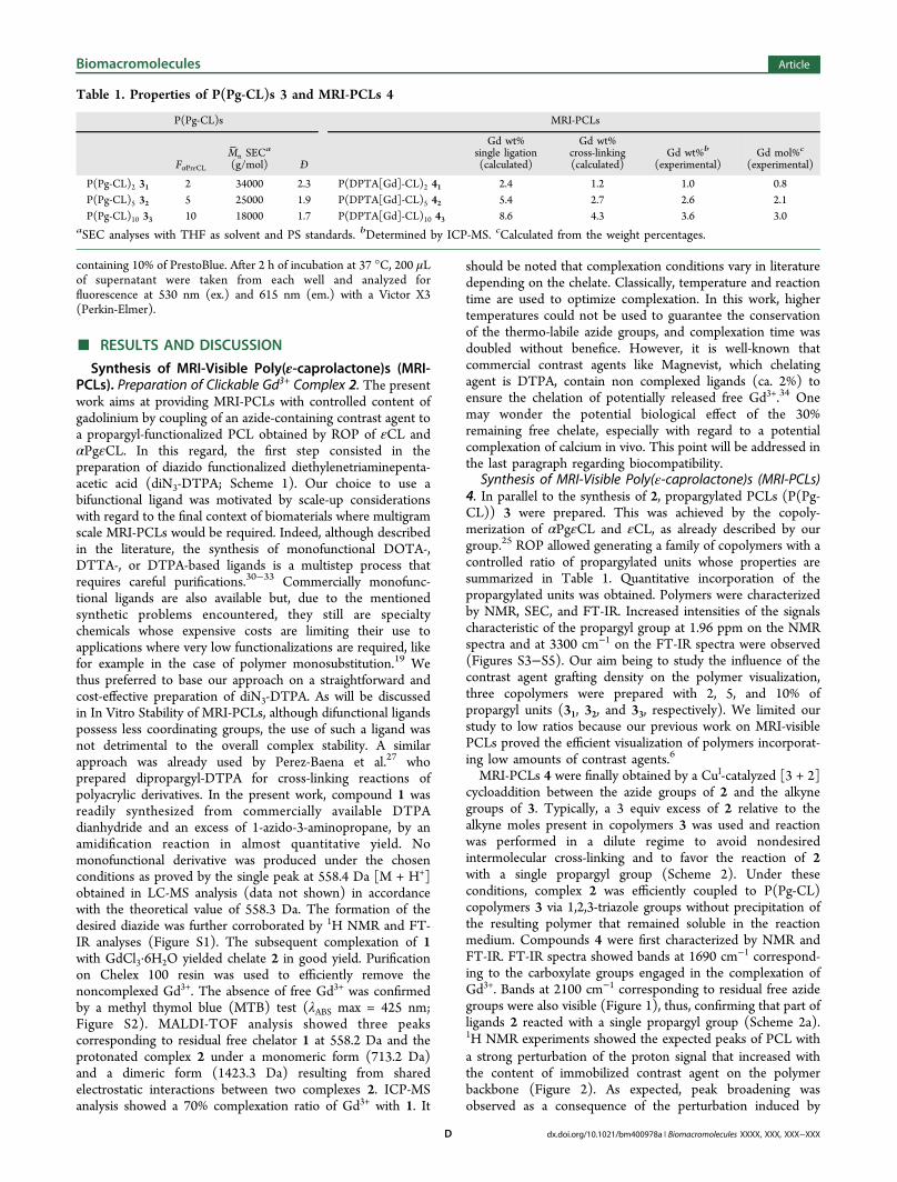
compounds 4 compared to genuine PCL as a function of theirgadolinium content. This is shown in Figure 3. To clarify the
discussion, grafting densities, that is, molar percentages of CLunits substituted by Gd3+, have been calculated based on theexperimental values of Gd3+ weight percentages (Table 1) andused in Figure 3. Two main observations can be drawn fromthis nonlinear evolution. First, it is noticeable that T1 relaxationtimes are strongly influenced even when low grafting densitiesof Gd3+ are found on the polymers backbone, as shown by the70% decrease of T1 in copolymer 41 that contains only 0.8 mol% of Gd3+. Second, it is clearly visible that a limit is rapidlyreached with almost no further decrease of T1 above 2.1 mol %of Gd3+. These results are of importance in the frame of MRIapplications as they show that limited amounts of Gd3+ may beused, which is of benefice when considering the toxicity of freeGd3+.MR-Imaging of MRI-PCLs. Having in hand MRI-PCLs with
defined contents of gadolinium, we were interested inevaluating the influence of (i) the overall amount of gadoliniumpresent in the samples and (ii) the grafting density of Gd3+
complexes immobilized on the PCL backbone on the MRvisualization. The final aim was to evaluate the optimumcompromise between MRI-visibility and low dose of Gd3+. For
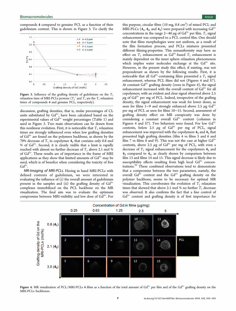
this purpose, circular films (10 mg, 0.8 cm2) of mixed PCL andMRI-PLCs (41, 42, and 43) were prepared with increasing Gd3+
concentrations in the range 2−40 μg of Gd3+ per film. T1 signalenhancement was compared to a PCL control film. One shouldnote that films morphologies were not uniform, as a result ofthe film formation process, and PCLs mixtures presenteddifferent filming-properties. This nonuniformity may have aneffect on T1 enhancement as Gd3+-based T1 enhancement ismainly dependent on the inner sphere relaxation phenomenonwhich implies water molecules exchange at the Gd3+ site.However, in the present study this effect, if existing, was notpreponderant as shown by the following results. First, it isnoticeable that all Gd3+-containing films presented a T1 signalenhancement, whereas PCL films did not (Figures 4 and S7).At constant Gd3+ grafting density (rows in Figure 4), the signalenhancement increased with the overall content of Gd3+ for allcopolymers, with an evident and clear signal observed above 2.5μg of Gd3+ per mg of PCL. Indeed, irrespective of the graftingdensity, the signal enhancement was weak for lower doses, asseen for films 1−9 and strongly enhanced above 2.5 μg Gd3+
per mg of PCL as seen for films 10−15. Second, assessment ofgrafting density effect on MR conspicuity was done byconsidering a constant overall Gd3+ content (columns inFigures 4 and S7). Two behaviors were found. For low Gd3+
contents, below 2.5 μg of Gd3+ per mg of PCL, signalenhancement was improved with the copolymers 42 and 43 thatpresented high grafting densities (film 4 vs films 5 and 6 andfilm 7 vs films 8 and 9). This was not the case at higher Gd3+
contents, above 2.5 μg of Gd3+ per mg of PCL, with even adecrease of T1 signal enhancement for the copolymers 42 and43 compared to 41, as clearly shown by comparison betweenfilm 13 and films 14 and 15. This signal decrease is likely due tosusceptibility effects resulting from high local Gd3+ concen-trations.35 These combined observations tend to demonstratethat a compromise between the two parameters, namely, theoverall Gd3+ content and the Gd3+ grafting density on thepolymer backbone, seems to be necessary for optimal MRvisualization. This corroborates the evolution of T1 relaxationtimes that showed that above 2.1 mol % no further T1 decreasewas observed. It also confirms the fact that a fine control ofGd3+ content and grafting density is of first importance for
Figure 3. Influence of the grafting density of gadolinium on the T1relaxation time of MRI-PCLs protons (T1′ and T1 are the T1 relaxationtimes of compounds 4 and genuine PCL, respectively).
Figure 4. MR visualization of PCL/MRI-PCLs 4 films as a function of the total amount of Gd3+ per film and of the Gd3+ grafting density on theMRI-PCLs backbones.
Biomacromolecules Article
dx.doi.org/10.1021/bm400978a | Biomacromolecules XXXX, XXX, XXX−XXXF
optimal MR visualization. In vivo implantations should now beplanned to confirm our approach and thoroughly evaluate thebenefit of MRI-visibility of MRI-PCLs.Stability and Cytocompatibility of MRI-PCLs. In Vitro
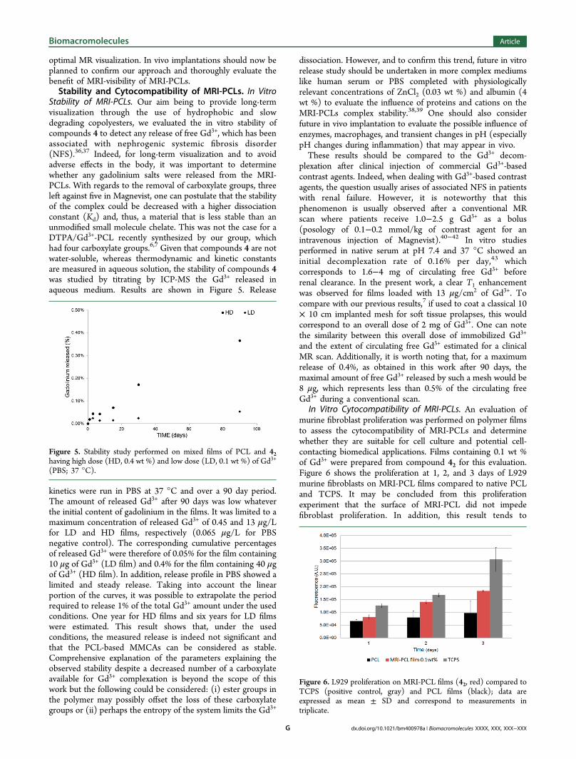
Stability of MRI-PCLs. Our aim being to provide long-termvisualization through the use of hydrophobic and slowdegrading copolyesters, we evaluated the in vitro stability ofcompounds 4 to detect any release of free Gd3+, which has beenassociated with nephrogenic systemic fibrosis disorder(NFS).36,37 Indeed, for long-term visualization and to avoidadverse effects in the body, it was important to determinewhether any gadolinium salts were released from the MRI-PCLs. With regards to the removal of carboxylate groups, threeleft against five in Magnevist, one can postulate that the stabilityof the complex could be decreased with a higher dissociationconstant (Kd) and, thus, a material that is less stable than anunmodified small molecule chelate. This was not the case for aDTPA/Gd3+-PCL recently synthesized by our group, whichhad four carboxylate groups.6,7 Given that compounds 4 are notwater-soluble, whereas thermodynamic and kinetic constantsare measured in aqueous solution, the stability of compounds 4was studied by titrating by ICP-MS the Gd3+ released inaqueous medium. Results are shown in Figure 5. Release
kinetics were run in PBS at 37 °C and over a 90 day period.The amount of released Gd3+ after 90 days was low whateverthe initial content of gadolinium in the films. It was limited to amaximum concentration of released Gd3+ of 0.45 and 13 μg/Lfor LD and HD films, respectively (0.065 μg/L for PBSnegative control). The corresponding cumulative percentagesof released Gd3+ were therefore of 0.05% for the film containing10 μg of Gd3+ (LD film) and 0.4% for the film containing 40 μgof Gd3+ (HD film). In addition, release profile in PBS showed alimited and steady release. Taking into account the linearportion of the curves, it was possible to extrapolate the periodrequired to release 1% of the total Gd3+ amount under the usedconditions. One year for HD films and six years for LD filmswere estimated. This result shows that, under the usedconditions, the measured release is indeed not significant andthat the PCL-based MMCAs can be considered as stable.Comprehensive explanation of the parameters explaining theobserved stability despite a decreased number of a carboxylateavailable for Gd3+ complexation is beyond the scope of thiswork but the following could be considered: (i) ester groups inthe polymer may possibly offset the loss of these carboxylategroups or (ii) perhaps the entropy of the system limits the Gd3+
dissociation. However, and to confirm this trend, future in vitrorelease study should be undertaken in more complex mediumslike human serum or PBS completed with physiologicallyrelevant concentrations of ZnCl2 (0.03 wt %) and albumin (4wt %) to evaluate the influence of proteins and cations on theMRI-PCLs complex stability.38,39 One should also considerfuture in vivo implantation to evaluate the possible influence ofenzymes, macrophages, and transient changes in pH (especiallypH changes during inflammation) that may appear in vivo.These results should be compared to the Gd3+ decom-
plexation after clinical injection of commercial Gd3+-basedcontrast agents. Indeed, when dealing with Gd3+-based contrastagents, the question usually arises of associated NFS in patientswith renal failure. However, it is noteworthy that thisphenomenon is usually observed after a conventional MRscan where patients receive 1.0−2.5 g Gd3+ as a bolus(posology of 0.1−0.2 mmol/kg of contrast agent for anintravenous injection of Magnevist).40−42 In vitro studiesperformed in native serum at pH 7.4 and 37 °C showed aninitial decomplexation rate of 0.16% per day,43 whichcorresponds to 1.6−4 mg of circulating free Gd3+ beforerenal clearance. In the present work, a clear T1 enhancementwas observed for films loaded with 13 μg/cm2 of Gd3+. Tocompare with our previous results,7 if used to coat a classical 10× 10 cm implanted mesh for soft tissue prolapses, this wouldcorrespond to an overall dose of 2 mg of Gd3+. One can notethe similarity between this overall dose of immobilized Gd3+
and the extent of circulating free Gd3+ estimated for a clinicalMR scan. Additionally, it is worth noting that, for a maximumrelease of 0.4%, as obtained in this work after 90 days, themaximal amount of free Gd3+ released by such a mesh would be8 μg, which represents less than 0.5% of the circulating freeGd3+ during a conventional scan.
In Vitro Cytocompatibility of MRI-PCLs. An evaluation ofmurine fibroblast proliferation was performed on polymer filmsto assess the cytocompatibility of MRI-PCLs and determinewhether they are suitable for cell culture and potential cell-contacting biomedical applications. Films containing 0.1 wt %of Gd3+ were prepared from compound 42 for this evaluation.Figure 6 shows the proliferation at 1, 2, and 3 days of L929murine fibroblasts on MRI-PCL films compared to native PCLand TCPS. It may be concluded from this proliferationexperiment that the surface of MRI-PCL did not impedefibroblast proliferation. In addition, this result tends to
Figure 5. Stability study performed on mixed films of PCL and 42having high dose (HD, 0.4 wt %) and low dose (LD, 0.1 wt %) of Gd3+
(PBS; 37 °C).
Figure 6. L929 proliferation on MRI-PCL films (42, red) compared toTCPS (positive control, gray) and PCL films (black); data areexpressed as mean ± SD and correspond to measurements intriplicate.
Biomacromolecules Article
dx.doi.org/10.1021/bm400978a | Biomacromolecules XXXX, XXX, XXX−XXXG
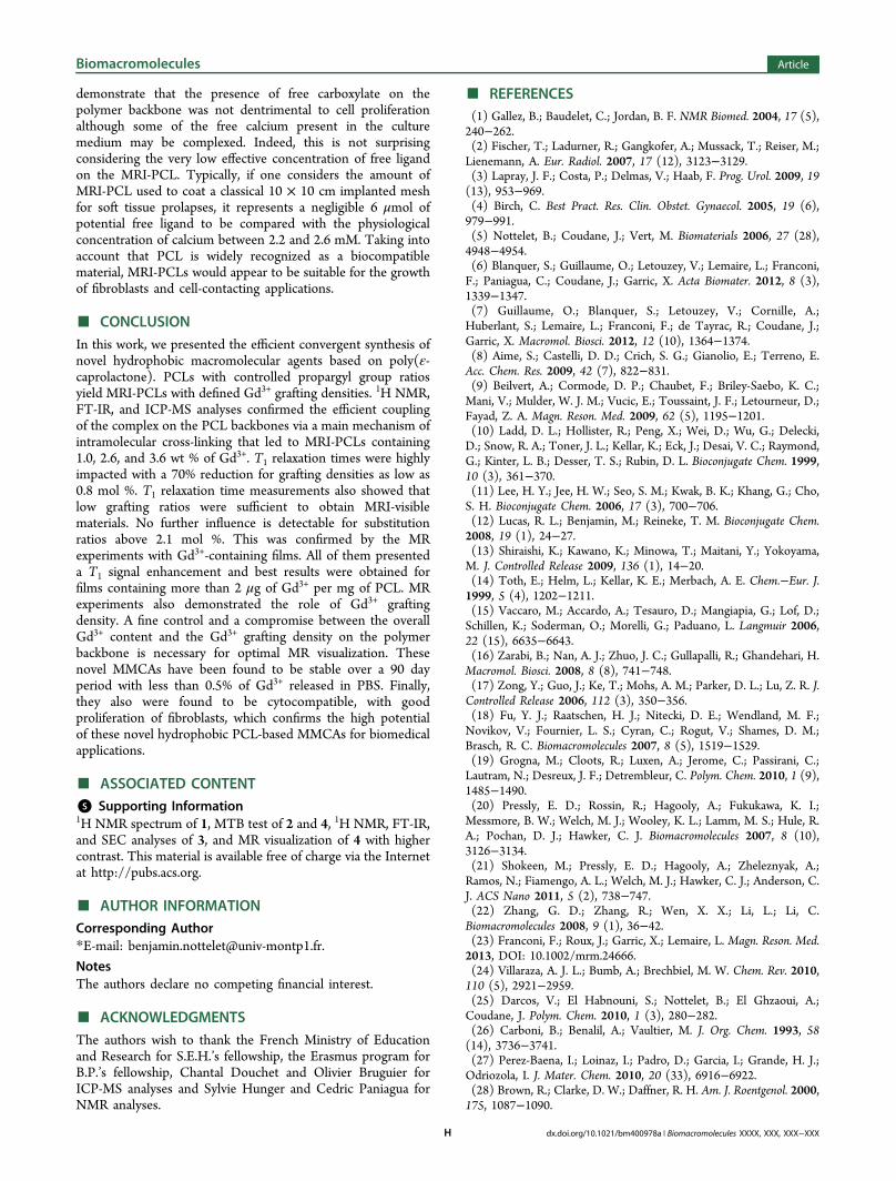
demonstrate that the presence of free carboxylate on thepolymer backbone was not dentrimental to cell proliferationalthough some of the free calcium present in the culturemedium may be complexed. Indeed, this is not surprisingconsidering the very low effective concentration of free ligandon the MRI-PCL. Typically, if one considers the amount ofMRI-PCL used to coat a classical 10 × 10 cm implanted meshfor soft tissue prolapses, it represents a negligible 6 μmol ofpotential free ligand to be compared with the physiologicalconcentration of calcium between 2.2 and 2.6 mM. Taking intoaccount that PCL is widely recognized as a biocompatiblematerial, MRI-PCLs would appear to be suitable for the growthof fibroblasts and cell-contacting applications.
■ CONCLUSIONIn this work, we presented the efficient convergent synthesis ofnovel hydrophobic macromolecular agents based on poly(ε-caprolactone). PCLs with controlled propargyl group ratiosyield MRI-PCLs with defined Gd3+ grafting densities. 1H NMR,FT-IR, and ICP-MS analyses confirmed the efficient couplingof the complex on the PCL backbones via a main mechanism ofintramolecular cross-linking that led to MRI-PCLs containing1.0, 2.6, and 3.6 wt % of Gd3+. T1 relaxation times were highlyimpacted with a 70% reduction for grafting densities as low as0.8 mol %. T1 relaxation time measurements also showed thatlow grafting ratios were sufficient to obtain MRI-visiblematerials. No further influence is detectable for substitutionratios above 2.1 mol %. This was confirmed by the MRexperiments with Gd3+-containing films. All of them presenteda T1 signal enhancement and best results were obtained forfilms containing more than 2 μg of Gd3+ per mg of PCL. MRexperiments also demonstrated the role of Gd3+ graftingdensity. A fine control and a compromise between the overallGd3+ content and the Gd3+ grafting density on the polymerbackbone is necessary for optimal MR visualization. Thesenovel MMCAs have been found to be stable over a 90 dayperiod with less than 0.5% of Gd3+ released in PBS. Finally,they also were found to be cytocompatible, with goodproliferation of fibroblasts, which confirms the high potentialof these novel hydrophobic PCL-based MMCAs for biomedicalapplications.
■ ASSOCIATED CONTENT*S Supporting Information1H NMR spectrum of 1, MTB test of 2 and 4, 1H NMR, FT-IR,and SEC analyses of 3, and MR visualization of 4 with highercontrast. This material is available free of charge via the Internetat http://pubs.acs.org.
■ AUTHOR INFORMATIONCorresponding Author*E-mail: [email protected].
NotesThe authors declare no competing financial interest.
■ ACKNOWLEDGMENTSThe authors wish to thank the French Ministry of Educationand Research for S.E.H.’s fellowship, the Erasmus program forB.P.’s fellowship, Chantal Douchet and Olivier Bruguier forICP-MS analyses and Sylvie Hunger and Cedric Paniagua forNMR analyses.
■ REFERENCES(1) Gallez, B.; Baudelet, C.; Jordan, B. F. NMR Biomed. 2004, 17 (5),240−262.(2) Fischer, T.; Ladurner, R.; Gangkofer, A.; Mussack, T.; Reiser, M.;Lienemann, A. Eur. Radiol. 2007, 17 (12), 3123−3129.(3) Lapray, J. F.; Costa, P.; Delmas, V.; Haab, F. Prog. Urol. 2009, 19(13), 953−969.(4) Birch, C. Best Pract. Res. Clin. Obstet. Gynaecol. 2005, 19 (6),979−991.(5) Nottelet, B.; Coudane, J.; Vert, M. Biomaterials 2006, 27 (28),4948−4954.(6) Blanquer, S.; Guillaume, O.; Letouzey, V.; Lemaire, L.; Franconi,F.; Paniagua, C.; Coudane, J.; Garric, X. Acta Biomater. 2012, 8 (3),1339−1347.(7) Guillaume, O.; Blanquer, S.; Letouzey, V.; Cornille, A.;Huberlant, S.; Lemaire, L.; Franconi, F.; de Tayrac, R.; Coudane, J.;Garric, X. Macromol. Biosci. 2012, 12 (10), 1364−1374.(8) Aime, S.; Castelli, D. D.; Crich, S. G.; Gianolio, E.; Terreno, E.Acc. Chem. Res. 2009, 42 (7), 822−831.(9) Beilvert, A.; Cormode, D. P.; Chaubet, F.; Briley-Saebo, K. C.;Mani, V.; Mulder, W. J. M.; Vucic, E.; Toussaint, J. F.; Letourneur, D.;Fayad, Z. A. Magn. Reson. Med. 2009, 62 (5), 1195−1201.(10) Ladd, D. L.; Hollister, R.; Peng, X.; Wei, D.; Wu, G.; Delecki,D.; Snow, R. A.; Toner, J. L.; Kellar, K.; Eck, J.; Desai, V. C.; Raymond,G.; Kinter, L. B.; Desser, T. S.; Rubin, D. L. Bioconjugate Chem. 1999,10 (3), 361−370.(11) Lee, H. Y.; Jee, H. W.; Seo, S. M.; Kwak, B. K.; Khang, G.; Cho,S. H. Bioconjugate Chem. 2006, 17 (3), 700−706.(12) Lucas, R. L.; Benjamin, M.; Reineke, T. M. Bioconjugate Chem.2008, 19 (1), 24−27.(13) Shiraishi, K.; Kawano, K.; Minowa, T.; Maitani, Y.; Yokoyama,M. J. Controlled Release 2009, 136 (1), 14−20.(14) Toth, E.; Helm, L.; Kellar, K. E.; Merbach, A. E. Chem.−Eur. J.1999, 5 (4), 1202−1211.(15) Vaccaro, M.; Accardo, A.; Tesauro, D.; Mangiapia, G.; Lof, D.;Schillen, K.; Soderman, O.; Morelli, G.; Paduano, L. Langmuir 2006,22 (15), 6635−6643.(16) Zarabi, B.; Nan, A. J.; Zhuo, J. C.; Gullapalli, R.; Ghandehari, H.Macromol. Biosci. 2008, 8 (8), 741−748.(17) Zong, Y.; Guo, J.; Ke, T.; Mohs, A. M.; Parker, D. L.; Lu, Z. R. J.Controlled Release 2006, 112 (3), 350−356.(18) Fu, Y. J.; Raatschen, H. J.; Nitecki, D. E.; Wendland, M. F.;Novikov, V.; Fournier, L. S.; Cyran, C.; Rogut, V.; Shames, D. M.;Brasch, R. C. Biomacromolecules 2007, 8 (5), 1519−1529.(19) Grogna, M.; Cloots, R.; Luxen, A.; Jerome, C.; Passirani, C.;Lautram, N.; Desreux, J. F.; Detrembleur, C. Polym. Chem. 2010, 1 (9),1485−1490.(20) Pressly, E. D.; Rossin, R.; Hagooly, A.; Fukukawa, K. I.;Messmore, B. W.; Welch, M. J.; Wooley, K. L.; Lamm, M. S.; Hule, R.A.; Pochan, D. J.; Hawker, C. J. Biomacromolecules 2007, 8 (10),3126−3134.(21) Shokeen, M.; Pressly, E. D.; Hagooly, A.; Zheleznyak, A.;Ramos, N.; Fiamengo, A. L.; Welch, M. J.; Hawker, C. J.; Anderson, C.J. ACS Nano 2011, 5 (2), 738−747.(22) Zhang, G. D.; Zhang, R.; Wen, X. X.; Li, L.; Li, C.Biomacromolecules 2008, 9 (1), 36−42.(23) Franconi, F.; Roux, J.; Garric, X.; Lemaire, L. Magn. Reson. Med.2013, DOI: 10.1002/mrm.24666.(24) Villaraza, A. J. L.; Bumb, A.; Brechbiel, M. W. Chem. Rev. 2010,110 (5), 2921−2959.(25) Darcos, V.; El Habnouni, S.; Nottelet, B.; El Ghzaoui, A.;Coudane, J. Polym. Chem. 2010, 1 (3), 280−282.(26) Carboni, B.; Benalil, A.; Vaultier, M. J. Org. Chem. 1993, 58(14), 3736−3741.(27) Perez-Baena, I.; Loinaz, I.; Padro, D.; Garcia, I.; Grande, H. J.;Odriozola, I. J. Mater. Chem. 2010, 20 (33), 6916−6922.(28) Brown, R.; Clarke, D. W.; Daffner, R. H. Am. J. Roentgenol. 2000,175, 1087−1090.
Biomacromolecules Article
dx.doi.org/10.1021/bm400978a | Biomacromolecules XXXX, XXX, XXX−XXXH
(29) Vonarbourg, A.; Sapin, A.; Lemaire, L.; Franconi, F.; Menei, P.;Jallet, P.; Le Jeune, J. J. Magn. Reson. Mater. Phys., Biol. Med. 2004, 17(3−6), 133−139.(30) Anelli, P. L.; Fedeli, F.; Gazzotti, O.; Lattuada, L.; Lux, G.;Rebasti, F. Bioconjugate Chem. 1999, 10 (1), 137−140.(31) Bryson, J. M.; Chu, W. J.; Lee, J. H.; Reineke, T. M. BioconjugateChem. 2008, 19 (8), 1505−1509.(32) Fernandez-Trillo, F.; Pacheco-Torres, J.; Correa, J.; Ballesteros,P.; Lopez-Larrubia, P.; Cerdan, S.; Riguera, R.; Fernandez-Megia, E.Biomacromolecules 2011, 12 (8), 2902−2907.(33) Parac-Vogt, T. N.; Kimpe, K.; Laurent, S.; Pierart, C.; Elst, L. V.;Muller, R. N.; Binnemans, K. Eur. J. Inorg. Chem. 2004, 17, 3538−3543.(34) Product Monograph: MAGNEVIST Gadopentetate DimeglumineInjection; Bayer Inc.: Toronto, Ontario, 2012; submission control No.156553, pp 1−38.(35) Villringer, A.; Rosen, B. R.; Belliveau, J. W.; Ackerman, J. L.;Lauffer, R. B.; Buxton, R. B.; Chao, Y. S.; Wedeen, V. J.; Brady, T. J.Magn. Reson. Med. 1988, 6 (2), 164−174.(36) Goulle, J. P.; Saussereau, E.; Mahieu, L.; Bouige, D.; Groenwont,S.; Guerbet, M.; Lacroix, C. J. Anal. Toxicol. 2009, 33 (2), 92−98.(37) Thakral, C.; Abraham, J. L. Radiol. Clin. 2009, 47 (5), 841−853.(38) Baranyai, Z.; Palinkas, Z.; Uggeri, F.; Brucher, E. Eur. J. Inorg.Chem. 2010, 13, 1948−1956.(39) Laurent, S.; Elst, L. V.; Henrotte, V.; Muller, R. N. Chem.Biodiversity 2010, 7 (12), 2846−2855.(40) VIDAL: le Dictionnaire, 88th ed.; Vidal: Paris, 2012.(41) Grobner, T. Nephrol. Dial. Transpl. 2006, 21 (4), 1104−1108.(42) Idee, J. M.; Port, M.; Raynal, I.; Schaefer, M.; Le Greneur, S.;Corot, C. Fund. Clin. Pharmacol. 2006, 20 (6), 563−576.(43) Frenzel, T.; Lengsfeld, P.; Schirmer, H.; Hutter, J.; Weinmann,H. J. Invest. Radiol. 2008, 43 (12), 817−828.
Biomacromolecules Article
dx.doi.org/10.1021/bm400978a | Biomacromolecules XXXX, XXX, XXX−XXXI