Magnetic Resonance Spectroscopy – EPR and NMR · Magnetic Resonance Spectroscopy – EPR and NMR...
21
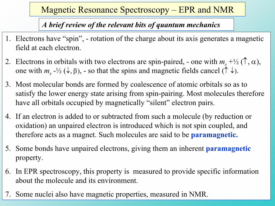
Magnetic Resonance Spectroscopy – EPR and NMR 1. Electrons have “spin”, - rotation of the charge about its axis generates a magnetic field at each electron. 2. Electrons in orbitals with two electrons are spin-paired, - one with m s +½ (↑, α), one with m s -½ (↓, β), - so that the spins and magnetic fields cancel (↑↓). 3. Most molecular bonds are formed by coalescence of atomic orbitals so as to satisfy the lower energy state arising from spin-pairing. Most molecules therefore have all orbitals occupied by magnetically “silent” electron pairs. 4. If an electron is added to or subtracted from such a molecule (by reduction or oxidation) an unpaired electron is introduced which is not spin coupled, and therefore acts as a magnet. Such molecules are said to be paramagnetic. 5. Some bonds have unpaired electrons, giving them an inherent paramagnetic property. 6. In EPR spectroscopy, this property is measured to provide specific information about the molecule and its environment. 7. Some nuclei also have magnetic properties, measured in NMR. A brief review of the relevant bits of quantum mechanics
Transcript of Magnetic Resonance Spectroscopy – EPR and NMR · Magnetic Resonance Spectroscopy – EPR and NMR...
Magnetic Resonance Spectroscopy – EPR and NMR
1. Electrons have “spin”, - rotation of the charge about its axis generates a magnetic field at each electron.
2. Electrons in orbitals with two electrons are spin-paired, - one with ms +½ (↑, α), one with ms -½ (↓, β), - so that the spins and magnetic fields cancel (↑ ↓).
3. Most molecular bonds are formed by coalescence of atomic orbitals so as to satisfy the lower energy state arising from spin-pairing. Most molecules therefore have all orbitals occupied by magnetically “silent” electron pairs.
4. If an electron is added to or subtracted from such a molecule (by reduction or oxidation) an unpaired electron is introduced which is not spin coupled, and therefore acts as a magnet. Such molecules are said to be paramagnetic.
5. Some bonds have unpaired electrons, giving them an inherent paramagneticproperty.
6. In EPR spectroscopy, this property is measured to provide specific information about the molecule and its environment.
7. Some nuclei also have magnetic properties, measured in NMR.
A brief review of the relevant bits of quantum mechanics

Electron Paramagnetic Resonance (EPR) spectroscopy. What can we use it for?
• Assay of concentration and concentration changes: redox state of paramagnetic species such as iron-sulfur clusters, semiquinones, flavin free-radicals, cytochromes, P450, photosynthetic reaction center components, amino acid radicals (Tyr, Trp), etc.
• Paramagnetic metals such as Mn2+, vanadyl ions (Mg2+-analogue) at catalytic centers.
• Measurement of local polarity and local structural mobility using nitroxide spin probes.
• Measurement of distance through spin-coupling with remote paramagnetic centers.
• Measurement of structure through interaction with local nuclear spins. ENDOR and pulsed methods (ESEEM) provide both distances and angles to give details of structure in the reaction environment close to the paramagnetic center (NMR through EPR).
• Measurement of electron distribution on paramagnetic species.
• Measurement of interactions (H-bonding) with local solvent (H2O).

Nuclear Magnetic Resonance (NMR) spectroscopy
1. The atomic nucleus is made up of protons (+-ve charge) and neutrons (neutral).
2. Like electrons, protons and neutrons (or nucleons) are quantum mechanical entities, and their energetic properties can be described by operators, wave functions, and quantum numbers.
3. Both protons and neutrons have a spin ½, with spin quantum number, mI, which can have values of ↑ (up, +½, or α), or ↓ (down, -½, or β).
4. Total nuclear energy levels are lower if the constituent protons and neutrons are spin-paired, so that for most nuclei, the spins (and magnetic fields) cancel. Protons are spin-paired with protons, neutrons with neutrons.
5. Isotopes that have an odd number of either protons or neutrons have a net spin, according to the Table below:
1 or 2 or 3 …OddOdd
1/2 or 3/2 or 5/2 …EvenOdd
1/2 or 3/2 or 5/2 …OddEven0EvenEven
Nuclear spin, (I)Number of neutronsNumber of protons
What can we use NMR spectroscopy for?
Because of the wide range of nuclei that have spins, the applications of NMR are more widespread than those of EPR. Resolution can be enhanced by isotopic substitution for those nuclei that either are rare, or have no significant population, at natural abundance.
• Identification and characterization of reaction products in chemistry through characteristic chemical shifts in energy for particular nuclei.
• Proton exchange rates between -OH, -COOH, etc., and H2O
• Spin-spin coupling to identify bonding interactions.
• By extension of the above, - NMR can be used to obtain structures for macromolecules at atomic resolution in solution.
• Magnetic Resonance Imaging (MRI), - a major advance in medical imaging of internal tissues, - based on proton NMR.

Spin and magnetism
Electrons and nuclei have magnetic properties only because they carry charges and move. The magnetic force generated by a moving charge is proportional to the current. In the case of a spinning entity, this translates into how fast it spins.
Why do different isotopes have different nuclear magnetic properties?Since the nuclear charge is dependent on atomic number (the number of protons), and different isotopes have the same atomic number (otherwise they are different elements), they must all carry the same charge. Since there’s no change in charge on adding neutrons, why is there a change in magnetic properties?
If a nucleus has an even number of protons and an even number of neutrons, all the spins compensate, and the nucleus has no net spin. The nuclear magnetism arises from the spin introduced by an extra neutron, which gives a net angular momentum.
The rate of spinning is dependent on angular momentum and therefore on mass. For any particular mass, a given increment in spin on adding a neutron will start the whole nucleus spinning at a rate dependent on the combined mass.
The magnetic difference between isotopes therefore depends in a complicated way on the mass of the nucleus (the atomic weight), the number of charges it carries (the atomic number), and how many unpaired nucleons are present.

Some nuclear magnets of importance in biological studies
As we shall see later, the gyromagnetic ratio (γ) determines the energy at which electromagnetic radiation will flip the spin of a nuclear magnet. A flip occurs when the energy matches (or is in resonance with) the energy of the transition. The energy needed (expressed in terms of frequency) depends on the applied magnetic field (the strength of the magnet), - 4.7 Tesla in this case.

Energy of transition
ν =E/h
(E = hν)
Energy (ν)
magnetic field strength (B0)0
Energy (ν)
magnetic field strength (B0)0
πγν2
0hBhE ==∆mI = ±½
mI = -½
mI = +½
ms = ±½
ms = -½
ms = +½
0BghE βν ==∆
EPR ~9.5GHz @ 0.34 T
NMR ~10-200 MHz @ 4.7 T
RT=0.593 kcal/mol at 298oK
ν - frequency Hz
ν - wavenumber cm-1
λ- wavelength nm
~
cν
λν ==
1~
RT=0.002 kcal/mol at 10oK

On the nature of electromagnetic waves, and the transitions thatabsorb or generate them
Our knowledge of electromagnetic waves comes from their interactions with matter. We measure them by the transitions they generate in atoms, molecules, and other antennae, and they are generated by excitation of transitions that decay by emitting a photon. They are called electromagnetic, not because they are either electrical or magnetic entities, but because they generate or are generated by electrical transitions, which have a linked magnetic component.
The energy of photons is expressed in terms of frequency, wavenumber or wavelength, all of which are related to energy through the Planck constant. All photons move at the speed of light (~1ft/ns), - this relates frequency to wavelength.
The frequency is related to the timescale of the transition associated with absorption or generation of the photon.
Energy of transition
ν =E/h
(E = hν)
ν - frequency Hz
ν - wavenumber cm-1
λ- wavelength nm
cν
λν ==
1~
The transitions associated with EPR occur with timescales in the ns range; those for NMR with timescales in the µs range. These timescales are of importance in terms of saturation, and the use of pulsed techniques, as we will see later.
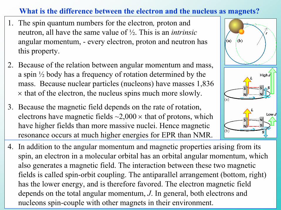
What is the difference between the electron and the nucleus as magnets?1. The spin quantum numbers for the electron, proton and
neutron, all have the same value of ½. This is an intrinsic angular momentum, - every electron, proton and neutron has this property.
2. Because of the relation between angular momentum and mass, a spin ½ body has a frequency of rotation determined by the mass. Because nuclear particles (nucleons) have masses 1,836 × that of the electron, the nucleus spins much more slowly.
3. Because the magnetic field depends on the rate of rotation, electrons have magnetic fields ~2,000 × that of protons, which have higher fields than more massive nuclei. Hence magnetic resonance occurs at much higher energies for EPR than NMR.
4. In addition to the angular momentum and magnetic properties arising from its spin, an electron in a molecular orbital has an orbital angular momentum, which also generates a magnetic field. The interaction between these two magnetic fields is called spin-orbit coupling. The antiparallel arrangement (bottom, right) has the lower energy, and is therefore favored. The electron magnetic field depends on the total angular momentum, J. In general, both electrons and nucleons spin-couple with other magnets in their environment.

Energy (ν)
magnetic field strength (B0)0
πγν2
0hBhE ==∆mI = ±½
mI = -½
mI = +½
Nuclear Magnetic Resonance
In the absence of an external magnetic field, the spins of the nuclei are arranged more or less at random. When a magnetic field is applied, the spins align with the field, just like bar magnets. However, because the energy involved is so small, they can flip direction relatively easily.
The consequence is that the energy level of the nucleus splits in a magnetic field, as shown on the right. The population of spins in the mI +½ (α) state is (very slightly) higher than in the mI -½ (β) state, because the energy is lower.

When photons of the “right” energy are absorbed, the spin of thenucleus flips between the two states. If we look at the diagram, we can see that the energy gap between the two states is dependent on the strength of the applied magnetic field. As a consequence, photons are absorbed which have a particular energy at a particular field. This is what is meant by resonance.
Here, γ is the gyromagnetic ratio for the nucleus, B0 is the magnetic field strength, and h is Planck’s constant.
When we come to discuss pulsed NMR, we will need to refer to circular motion. Here the preferred frame of reference is that of the circular motion of precession. We define the precession or Larmor frequency:
πγν2
0hBhE ==∆
00
above, thein onsubstitutiby or 2
Bγω
πυω
=
=

Some consequences of the energy scale.
The transitions leading to NMR absorption have energies in the radio frequency range, depending on nucleus (γ) and the strength of the magnetic field generated by the magnet.
NMR machines are rated by the frequency at which the proton is in NMR resonance for the magnet they are built around, so we have 200 MHz, 500 MHz, 750 MHz and even 1 GHz NMR spectrometers. To achieve the higher fields, high electrical currents are needed, which can be achieved using superconducting coils, - these are generally called superconducting magnets (costing $M).
Since ω = 2πν = γB0, for a 200 MHz machine we need a magnet generating 4.7 Tesla; for a 500 MHz machine we need 11.74 Tesla, etc.
As we increase the energy gap (increase frequency), the small differences of energy for transitions of nuclear magnets in different environments are also increased, and our NMR spectrum will be better resolved. In addition, we also increase the population difference for the two spin states as we increase the energy (∆E/RT is increased), as discussed in the next slide.
These factors make a big difference in the amount of time needed to generate a data set, - for example in solution of the structure of a protein.
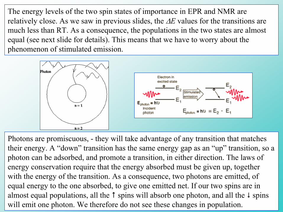
The energy levels of the two spin states of importance in EPR and NMR are relatively close. As we saw in previous slides, the ∆E values for the transitions are much less than RT. As a consequence, the populations in the two states are almost equal (see next slide for details). This means that we have to worry about the phenomenon of stimulated emission.
Photons are promiscuous, - they will take advantage of any transition that matches their energy. A “down” transition has the same energy gap as an “up” transition, so a photon can be adsorbed, and promote a transition, in either direction. The laws of energy conservation require that the energy absorbed must be given up, together with the energy of the transition. As a consequence, two photons are emitted, of equal energy to the one absorbed, to give one emitted net. If our two spins are in almost equal populations, all the spins will absorb one photon, and all the spins will emit one photon. We therefore do not see these changes in population.
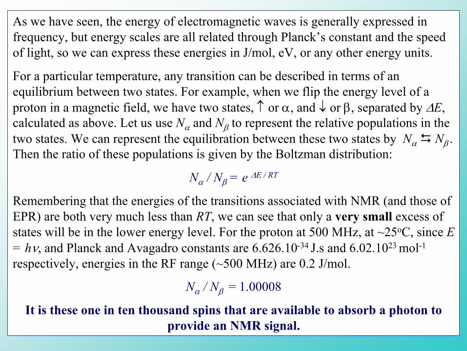
As we have seen, the energy of electromagnetic waves is generally expressed in frequency, but energy scales are all related through Planck’s constant and the speed of light, so we can express these energies in J/mol, eV, or any other energy units.
For a particular temperature, any transition can be described in terms of an equilibrium between two states. For example, when we flip the energy level of a proton in a magnetic field, we have two states, ↑ or α, and ↓ or β, separated by ∆E, calculated as above. Let us use Nα and Nβ to represent the relative populations in the two states. We can represent the equilibration between these two states by Nα Nβ . Then the ratio of these populations is given by the Boltzman distribution:
Nα / Nβ = e ∆E / RT
Remembering that the energies of the transitions associated with NMR (and those of EPR) are both very much less than RT, we can see that only a very small excess of states will be in the lower energy level. For the proton at 500 MHz, at ~25oC, since E = hν, and Planck and Avagadro constants are 6.626.10-34 J.s and 6.02.1023 mol-1
respectively, energies in the RF range (~500 MHz) are 0.2 J/mol.
Nα / Nβ = 1.00008
It is these one in ten thousand spins that are available to absorb a photon to provide an NMR signal.
Electron Paramagnetic Resonance
Energy (ν)
magnetic field strength (B0)0
ms = ±½
ms = -½
ms = +½
0BghE βν ==∆
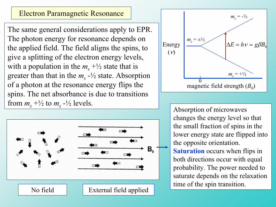
The same general considerations apply to EPR. The photon energy for resonance depends on the applied field. The field aligns the spins, to give a splitting of the electron energy levels, with a population in the ms +½ state that is greater than that in the ms -½ state. Absorption of a photon at the resonance energy flips the spins. The net absorbance is due to transitions from ms +½ to ms -½ levels.
No field External field applied
Absorption of microwaves changes the energy level so that the small fraction of spins in the lower energy state are flipped into the opposite orientation. Saturation occurs when flips in both directions occur with equal probability. The power needed to saturate depends on the relaxation time of the spin transition.
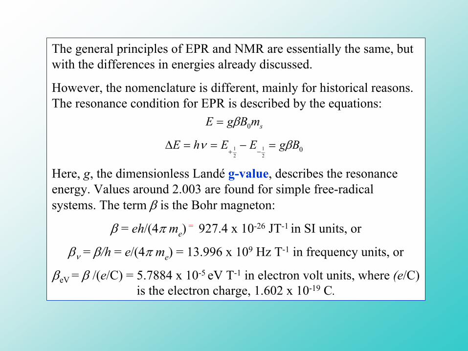
The general principles of EPR and NMR are essentially the same, but with the differences in energies already discussed.
However, the nomenclature is different, mainly for historical reasons. The resonance condition for EPR is described by the equations:
Here, g, the dimensionless Landé g-value, describes the resonance energy. Values around 2.003 are found for simple free-radical systems. The term β is the Bohr magneton:
β = eh/(4π me) = 927.4 x 10⁻²⁶ JT-1 in SI units, or
βν = β/h = e/(4π me) = 13.996 x 10⁹ Hz T-1 in frequency units, or
βeV = β /(e/C) = 5.7884 x 10⁻⁵ eV T-1 in electron volt units, where (e/C) is the electron charge, 1.602 x 10⁻¹⁹ C.
021
21 BgEEhE βν =−==∆
−+
smBgE 0β=
Two different approaches to magnetic spectroscopy, CW and pulsed methods
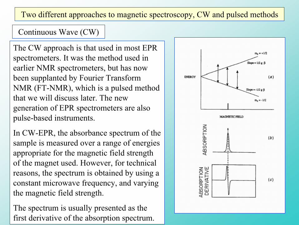
Continuous Wave (CW)
The CW approach is that used in most EPR spectrometers. It was the method used in earlier NMR spectrometers, but has now been supplanted by Fourier Transform NMR (FT-NMR), which is a pulsed method that we will discuss later. The new generation of EPR spectrometers are also pulse-based instruments.
In CW-EPR, the absorbance spectrum of the sample is measured over a range of energies appropriate for the magnetic field strength of the magnet used. However, for technical reasons, the spectrum is obtained by using a constant microwave frequency, and varying the magnetic field strength.
The spectrum is usually presented as the first derivative of the absorption spectrum.

Comparison of an EPR spectrometer, and a UV-VIS spectrophotometer
In this comparison, several points are noteworthy.
1. In a spectrophotometer, the spectrum is generated by varying the wavelength. As we just discussed, in a CW-EPR spectrometer, we use a fixed wavelength (frequency), but vary the magnetic field. This is achieved by the field scan and modulation coils associated with the magnet.
2. In the spectrophotometer shown, the light-beam is chopped by a rotating sector. This gives it a period function, and allows use of phase and frequency sensitive detection electronics, which improve the signal to noise ratio.
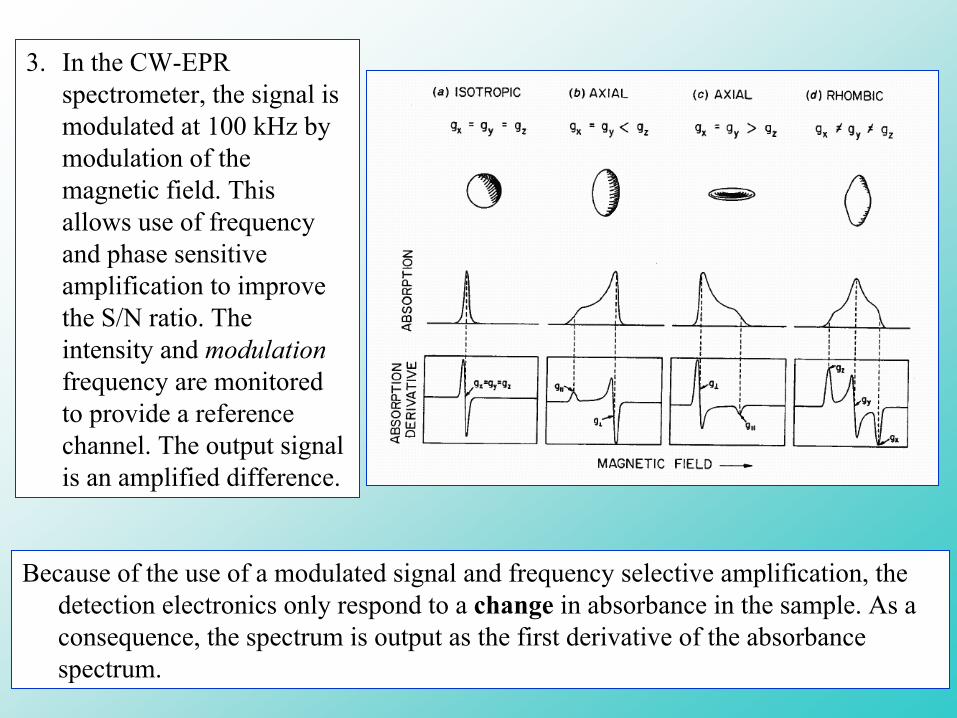
3. In the CW-EPR spectrometer, the signal is modulated at 100 kHz by modulation of the magnetic field. This allows use of frequency and phase sensitive amplification to improve the S/N ratio. The intensity and modulationfrequency are monitored to provide a reference channel. The output signal is an amplified difference.
Because of the use of a modulated signal and frequency selective amplification, the detection electronics only respond to a change in absorbance in the sample. As a consequence, the spectrum is output as the first derivative of the absorbance spectrum.
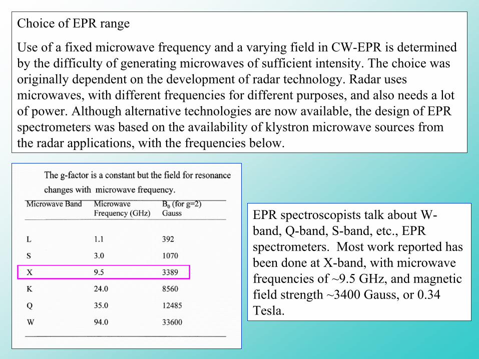
Choice of EPR range
Use of a fixed microwave frequency and a varying field in CW-EPR is determined by the difficulty of generating microwaves of sufficient intensity. The choice was originally dependent on the development of radar technology. Radar uses microwaves, with different frequencies for different purposes, and also needs a lot of power. Although alternative technologies are now available, the design of EPR spectrometers was based on the availability of klystron microwave sources from the radar applications, with the frequencies below.
EPR spectroscopists talk about W-band, Q-band, S-band, etc., EPR spectrometers. Most work reported has been done at X-band, with microwave frequencies of ~9.5 GHz, and magnetic field strength ~3400 Gauss, or 0.34 Tesla.

magnet
sample
microwave bridge
EPR tubeselectronics
computer for data analysis