Inflammasome-Mediated Secretion of IL-1 in Human Monocytes through TLR2 Activation; Modulation by...
12
of June 3, 2014. This information is current as Acids Activation; Modulation by Dietary Fatty in Human Monocytes through TLR2 β Inflammasome-Mediated Secretion of IL-1 Rutledge and Daniel H. Hwang Ryan G. Snodgrass, Shurong Huang, Il-Whan Choi, John C. http://www.jimmunol.org/content/191/8/4337 doi: 10.4049/jimmunol.1300298 September 2013; 2013; 191:4337-4347; Prepublished online 16 J Immunol Material Supplementary 8.DC1.html http://www.jimmunol.org/content/suppl/2013/09/16/jimmunol.130029 References http://www.jimmunol.org/content/191/8/4337.full#ref-list-1 , 46 of which you can access for free at: cites 88 articles This article Subscriptions http://jimmunol.org/subscriptions is online at: The Journal of Immunology Information about subscribing to Permissions http://www.aai.org/ji/copyright.html Submit copyright permission requests at: Email Alerts http://jimmunol.org/cgi/alerts/etoc Receive free email-alerts when new articles cite this article. Sign up at: Print ISSN: 0022-1767 Online ISSN: 1550-6606. All rights reserved. 9650 Rockville Pike, Bethesda, MD 20814-3994. The American Association of Immunologists, Inc., is published twice each month by The Journal of Immunology at Univ of Laval Biblio/Sec Des Acquisitions on June 3, 2014 http://www.jimmunol.org/ Downloaded from at Univ of Laval Biblio/Sec Des Acquisitions on June 3, 2014 http://www.jimmunol.org/ Downloaded from
Transcript of Inflammasome-Mediated Secretion of IL-1 in Human Monocytes through TLR2 Activation; Modulation by...
of June 3, 2014.This information is current as
AcidsActivation; Modulation by Dietary Fattyin Human Monocytes through TLR2
βInflammasome-Mediated Secretion of IL-1
Rutledge and Daniel H. HwangRyan G. Snodgrass, Shurong Huang, Il-Whan Choi, John C.
http://www.jimmunol.org/content/191/8/4337doi: 10.4049/jimmunol.1300298September 2013;
2013; 191:4337-4347; Prepublished online 16J Immunol
MaterialSupplementary
8.DC1.htmlhttp://www.jimmunol.org/content/suppl/2013/09/16/jimmunol.130029
Referenceshttp://www.jimmunol.org/content/191/8/4337.full#ref-list-1
, 46 of which you can access for free at: cites 88 articlesThis article
Subscriptionshttp://jimmunol.org/subscriptions
is online at: The Journal of ImmunologyInformation about subscribing to
Permissionshttp://www.aai.org/ji/copyright.htmlSubmit copyright permission requests at:
Email Alertshttp://jimmunol.org/cgi/alerts/etocReceive free email-alerts when new articles cite this article. Sign up at:
Print ISSN: 0022-1767 Online ISSN: 1550-6606. All rights reserved.9650 Rockville Pike, Bethesda, MD 20814-3994.The American Association of Immunologists, Inc.,
is published twice each month byThe Journal of Immunology
at Univ of L
aval Biblio/Sec D
es Acquisitions on June 3, 2014
http://ww
w.jim
munol.org/
Dow
nloaded from
at Univ of L
aval Biblio/Sec D
es Acquisitions on June 3, 2014
http://ww
w.jim
munol.org/
Dow
nloaded from
The Journal of Immunology
Inflammasome-Mediated Secretion of IL-1b in HumanMonocytes through TLR2 Activation; Modulation by DietaryFatty Acids
Ryan G. Snodgrass,*,† Shurong Huang,* Il-Whan Choi,‡ John C. Rutledge,x and
Daniel H. Hwang*,†
Many studies have shown that TLR4- and TLR2-deficient mice are protected from high-fat diet–induced inflammation and insulin
resistance, suggesting that saturated fatty acids derived from the high-fat diet activate TLR-mediated proinflammatory signaling
pathways and induce insulin resistance. However, evidence that palmitic acid, the major dietary saturated fatty acid, can directly
activate TLR has not been demonstrated. In this article, we present multiple lines of evidence showing that palmitic acid directly
activates TLR2, a major TLR expressed on human monocytes, by inducing heterodimerization with TLR1 in an NADPH oxidase–
dependent manner. Dimerization of TLR2 with TLR1 was inhibited by the n-3 fatty acid docosahexaenoic acid. Activation of
TLR2 by palmitic acid leads to expression of pro–IL-1b that is cleaved by caspase-1, which is constitutively present in monocytes,
to release mature IL-1b. Our results reveal mechanistic insight about how palmitic acid activates TLR2, upregulates NALP3
expression, and induces inflammasome-mediated IL-1b production in human monocytes, which can trigger enhanced inflamma-
tion in peripheral tissues, and suggest that these processes are dynamically modulated by the types of dietary fat we consume.
The Journal of Immunology, 2013, 191: 4337–4347.
Chronic inflammation is one of the key causative conditionsfor the development and progression of many chronicdiseases. Low-grade chronic inflammation characteri-
zed by elevated circulating concentrations of proinflammatory cyto-kines, acute-phase proteins, and adhesion molecules is known to beassociated with obesity and insulin resistance. What causes andmediates low-grade chronic inflammation and how it can be sup-pressed through dietary means that can provide preventive efficacyfor inflammatory chronic disease are challenging questions.Blood monocytes are sentinel immune effector cells that detect
and respond to invading pathogens and dietary components ab-sorbed from the gut. Activated blood monocytes can extravasateinto peripheral tissues and become resident macrophages or den-
dritic cells (DCs) that can trigger inflammatory signals in responseto exogenous and endogenous stimuli (1, 2). Recent animal studies
have shown that a high-fat diet increases infiltration of macro-
phages and DCs, which originate from circulating blood mono-
cytes, in adipose tissue where they play a central role in the
development of chronic inflammation and insulin resistance (3).
Therefore, activation of proinflammatory pathways in circulating
blood monocytes is the gateway toward enhanced inflammation in
various tissues. IL-1b is a major cytokine involved in the activa-
tion of blood monocyte and proinflammatory signaling pathways
in peripheral tissues. Unlike other proinflammatory cytokines, IL-
1b production is tightly regulated by a unique two-signal mech-
anism. The primary signals induce the expression of pro–IL-1b
mediated, in part, through the activation of TLRs. The secondary
signals activate the NALP3 inflammasome, an intracellular sig-
naling complex composed of NALP3, procaspase-1, and the
adaptor protein ASC. Formation of the inflammasome complex
results in proteolytic cleavage of procaspase-1 to yield active
caspase-1, which, in turn, cleaves pro–IL-1b producing mature
and active IL-1b. Mature IL-1b released extracellularly binds its
cognate receptor in a paracrine manner, resulting in amplification
of proinflammatory responses (4–7). However, recent findings
suggest that the two-signal model for NALP3 inflammasome-
mediated IL-1b secretion may not be applicable to all IL-1b–se-
creting cells including blood monocytes and DCs. Tissue macro-
phages require both primary and secondary signals for NALP3
inflammasome-mediated IL-1b secretion. In contrast, blood mono-
cytes have constitutively active NALP3 inflammasome; therefore,
TLR stimulation alone is sufficient to induce inflammasome-
mediated IL-1b production in monocytes (8–10). Similarly, both
bone marrow–derived and splenic DCs secrete substantial amounts
of inflammasome-mediated IL-1b upon TLR activation in the ab-
sence of secondary signals (3, 11). Therefore, regulation of NALP3
inflammasome-mediated IL-1b production in mononuclear phag-
ocytes appears to be cell-type specific.
*U.S. Department of Agriculture, Agricultural Research Service Western HumanNutrition Research Center, Davis, CA 95616; †Department of Nutrition, Universityof California, Davis, Davis, CA 95616; ‡Department of Microbiology, Inje Univer-sity, Busan 614-735, Republic of Korea; and xDepartment of Internal Medicine,University of California, Davis, CA 95616
Received for publication January 31, 2013. Accepted for publication August 8, 2013.
This work was supported by the National Institutes of Health (Grants R01DK064007, R01 DK41868, R01 AG039094) and the Western Human NutritionResearch Center/Agricultural Research Service/U.S. Department of Agriculture(5306-51530-017-00D).
The contents of this publication are solely the responsibility of the authors and do notnecessarily represent the official views of the National Institutes of Health or othergranting agencies. The U.S. Department of Agriculture is an equal opportunity em-ployer and provider.
Address correspondence and reprint requests to Dr. Daniel H. Hwang, Western Hu-man Nutrition Research Center (USDA/ARS), 430 West Health Sciences Drive,Davis, CA 95616. E-mail address: [email protected]
The online version of this article contains supplemental material.
Abbreviations used in this article: BMDC, bone marrow–derived dendritic cell;BMDM, bone marrow–derived macrophage; DC, dendritic cell; DHA, docosahexae-noic acid; FRET, fluorescence resonance energy transfer; LPL, lipoprotein lipase;Nod, nucleotide-binding oligomerization domain–containing protein; NOX2,NADPH oxidase 2; Pam3CSK4, Pam3CysSerLys4; PRR, pattern recognition re-ceptor; PUFA, polyunsaturated fatty acid; ROS, reactive oxygen species; SFA, sat-urated fatty acid; siRNA, small interfering RNA; TGRL, triglyceride-rich lipoprotein;TR-FRET, time resolved FRET.
www.jimmunol.org/cgi/doi/10.4049/jimmunol.1300298
at Univ of L
aval Biblio/Sec D
es Acquisitions on June 3, 2014
http://ww
w.jim
munol.org/
Dow
nloaded from
Pattern recognition receptors (PRRs) induce innate immuneresponses by recognizing invariant pathogen-associated molecularpatterns leading to activation of downstream signaling pathwaysand the expression of a diverse array of proinflammatory gene pro-ducts that are required for host defense against invading pathogens.In addition, PRRs are activated by endogenous molecules derivedfrom tissue injury and elicit sterile inflammation to initiate wound-healing processes (12, 13). Activation of PRRs can also be modu-lated by dietary components and their metabolites bridging immuneresponses to metabolic homeostasis (3, 14–19).Our previous studies first revealed that a medium-chain satu-
rated fatty acid (SFA), lauric acid, can activate TLR4, TLR2, andnucleotide-binding oligomerization domain–containing protein(Nod) 2–mediated signaling pathways, whereas the n-3 fattyacid, docosahexaenoic acid (DHA), inhibits lauric acid–inducedactivation of TLRs or Nod-mediated signaling pathways (15, 20–26). Many studies with animal models also showed that TLR4 orTLR2 deletion or mutant mice were protected from high-fat diet–induced inflammation and insulin resistance (3, 14, 16, 27–31),suggesting that high saturated fat diet activates TLR-mediatedproinflammatory signaling pathways and induces insulin resis-tance. However, evidence that SFAs derived from high-fat dietcan directly activate TLRs in humans has not been demonstrated.In this article, we present multiple lines of evidence that palmiticacid, the major dietary SFA, directly activates TLR2, a predomi-nant TLR expressed on blood monocytes, by inducing hetero-dimerization with TLR1. The dimerization of TLR2 with TLR1 asassessed by time resolved-fluorescence resonance energy transfer(TR-FRET) was inhibited by DHA. Activation of TLR2 leads toexpression of pro–IL-1b that is cleaved by caspase-1, which isactivated by constitutively active inflammasome, to release matureIL-1b in blood monocytes. Our results reveal mechanistic insightabout how the SFA, palmitic acid, induces inflammasome-mediatedIL-1b production and suggest that inflammasome-mediated IL-1bproduction in human blood monocytes, which can trigger enhancedinflammation in peripheral tissues, is dynamically modulated by thetypes of dietary fat we consume.
Materials and MethodsReagents
LPS (catalog no. 421) was purchased from List Biological Laboratories(Campbell, CA). Pam3CysSerLys4 (Pam3CSK4; catalog no. tlrl-pms) andTAK-242 (catalog no. tlrl-cli95) were purchased from Invivogen (SanDiego, CA). BSA (catalog no. 30-AB79, Lot no. A10072001) was pur-chased from Fitzgerald Industries International (Acton, MA). Palmitic acid(catalog no. P5585), sodium palmitate (catalog no. P9767), sodium saltDHA (catalog no. D8768), lipoprotein lipase (LPL) (catalog no. L2254),endotoxin-free water, polymyxin B (catalog no. P4932), and Ab for b-actinwere purchased from Sigma (Saint Louis, MO). Apocynin (catalog no.178385) and Ac-YVAD-AOM (catalog no. 400015) were purchased fromEMD Chemicals (Darmstadt, Germany).
Cell culture
THP-1 cells (human monocytic cell line, ATCC TIB-202) were cultured inRPMI 1640medium (ATCC catalog no. 30-2001) containing 10% (v/v) FBS(Premium Select, catalog no. S11550, Lot no. K0109 and K11050; AtlantaBiologicals, Lawrenceville, GA), 0.05 mM 2-ME, 100 U/ml penicillin, and100 mg/ml streptomycin. Cells were maintained at 37˚C in a 5% CO2/airenvironment. Cells were seeded at 1 3 106/ml and incubated in serum-poor medium (0.25% and 1.0% FBS) before treatment.
Fatty acid preparations
A stock solution of 100 mM sodium palmitate (C16:0) was prepared in 70%ethanol in a glass vial and heated to 60˚C. Sodium palmitate was reheated to60˚C and vortexed before use to give appropriate final concentration indi-cated in the figures. A stock solution of 10 mM DHA sodium salt wasprepared in endotoxin-free water, flushed with nitrogen, then sealed in air-
tight tube and stored at220˚C until use. Palmitic acid solubilized with BSA(C16:0-BSA) was carried out as previously described (32). In brief, palmiticacid was dissolved in 100% ethanol (250 or 500 mM), then mixed with BSAin a 10:1 molar ratio in 0.25% FBS/RPMI 1640 medium. The mixture wassonicated in a water bath for 15 min, then rocked at 55˚C for 15 min. BSA-solubilized palmitic acid was filtered through a 0.22-mm filter before use.
We used sodium palmitate instead of BSA-solubilized palmitic acid inour studies because we initially found that many commercially availableBSA preparations contain contaminants with agonist activity for certainPRRs (32). We also found that sodium palmitate is able to stimulate PRR-mediated signaling pathways in most suspension cells such as THP-1monocytes and primary human blood monocytes. However, palmitic acidneeds to be solubilized with albumin (BSA) to stimulate PRR-mediatedsignaling pathways in adherent cells. After recently discovering a BSApreparation (catalog no. 30-AB79, Lot no. A10072001; Fitzgerald Indus-tries International) that did not significantly induce PRR target geneproducts, we found that BSA-solubilized palmitic acid showed similarresults as sodium palmitate with regard to IL-1b production in THP-1 cells(Supplemental Fig. 1).
Plasmids
Expression vectors pDisplay, pDisplay-TLR1, pDisplay-TLR2, pDisplay-TLR4, and pDisplay-TLR6 were obtained from Adeline Hajjar (Univer-sity of Washington, Seattle, WA). MD2 was provided by Kensuke Miyake(Tokyo University, Japan). (2x)-NF-kB–luciferase reporter construct wasprovided by Frank Mercurio (Signal Pharmaceuticals, San Diego, CA).pRSV–b-galactosidase plasmid was from Jongdae Lee (University ofCalifornia, San Diego, CA). The plasmid DNA from these expressionvectors was prepared in large scale for transfection using the EndoFreePlasmid Maxi Kit (Qiagen, Valencia, CA).
Transfections and luciferase assays
Transient transfections were carried out using SuperFect transfection reagent(Qiagen) according to the manufacturer’s recommendations. HEK293T cellswere seeded at 2 3 105 per well in 24-well plates and cotransfected the fol-lowing day with 10 ng each of pDisplay-TLR4 and MD2, or pDisplay-TLR2and pDisplay-TLR1, or pDisplay-TLR2 and pDisplay-TLR6, in addition to50 ng (2x)-NF-kB–luciferase reporter and 10 ng pRSV–b-galactosidaseexpression vectors. Twenty nanograms of pDisplay empty vector was usedin addition to the earlier amounts of (2x)-NF-kB and pRSV–b-galactosi-dase for transfection as controls. Twenty-four hours after transfection, thecells were serum-starved in 0.25% FBS/DMEM medium for 6 h followedby 12-h treatment with BSA-solubilized palmitic acid in the same low se-rum medium. The cells were lysed. Luciferase and b-galactosidaseenzyme activities were determined from the lysate supernatants usingthe luciferase and b-galactosidase enzyme assay system (Promega,Madison, WI) according to the manufacturer’s instructions. Luciferaseactivity was normalized by b-galactosidase activity to correct differences intransfection efficiency among samples. Each experiment was repeated atleast three times.
Immunoblot and ELISA assays
Immunoblotting was performed as previously described (17). In brief,THP-1 cells were lysed by sonication in cell lysis buffer (Cell SignalingTechnology, Danvers, MA) containing 20 mM Tris-HCl (pH 7.5), 150 mMNaCl, 1 mM Na2EDTA, 1 mM EGTA, 1% Triton, 2.5 mM sodium pyro-phosphate, 1 mM b-glycerophosphate, 1 mM sodium orthovanadate, and 1mg/ml leupeptin plus 1 mM PMSF. Lysate supernatants were collected bycentrifugation and subjected to 10% or 10–20% SDS-PAGE followed byprotein transfer to polyvinylidene difluoride membrane (Bio-Rad, Hercu-les, CA). The membrane was blocked in TBST buffer (20 mM Tris-HCl[pH 7.4], 150 mM NaCl, and 0.05% [v/v] Tween 20) containing 5% nonfatmilk or BSA. The membrane was probed with primary Ab for 1 h at roomtemperature or overnight at 4˚C followed by incubation with HRP-con-jugated secondary Ab (Amersham Biosciences, Piscataway, NJ) for 1 hat room temperature. Proteins were detected by ECL Western blot detec-tion reagents (Amersham Biosciences) followed by exposure to autora-diography film (BioExpress, Kaysville, UT). Anti–IL-1b (3ZD) Ab wasobtained from the National Cancer Institute Preclinical Repository; anti–caspase-1 p10 (C-20) and anti–IkBa (C-21) Abs were obtained from SantaCruz Biotechnology (Santa Cruz, CA). Anti-NALP3 (catalog no. ALX-804-881) Ab was purchased from Enzo Life Science (Plymouth Meeting,PA). Anti-JNK (catalog no. 9252), anti–phospho-JNK (catalog no. 9251),anti–phospho-IkBa (catalog no. 2859), and anti-MyD88 (catalog no. 4283)Abs were purchased from Cell Signaling Technology. Anti–Flotillin-1 (catalogno. 610820) and anti-p47phox (catalog no. 610355) Abs were purchased from
4338 TLR2-MEDIATED INFLAMMASOME ACTIVATION AND FATTY ACIDS
at Univ of L
aval Biblio/Sec D
es Acquisitions on June 3, 2014
http://ww
w.jim
munol.org/
Dow
nloaded from
BD Biosciences (San Jose, CA). Anti-Transferrin Receptor (catalog no.13-6890) Ab was purchased from Life Technologies Corporation(Grand Island, NY). Cell-culture supernatants were analyzed for IL-1busing BD OptEIA Human IL-1b ELISA using a Synergy 2 plate reader(BioTek, Winooski, VT) according to the manufacturer’s instructions.To verify that secreted IL-1b in cell-culture supernatants was the 17-kDabioactive form, we concentrated the supernatants using Amicon UltraCentrifugal Filters (Millipore, Billerica, MA) according to the manu-facturer’s instructions, then performed immunoblot by which pro–IL-1band bioactive IL-1b can be separated by size difference. The immunoblotsshowed only 17-kDa IL-1b, indicating that IL-1b in the supernatant ismostly bioactive IL-1b.
Confocal microscopy
Reactive oxygen species (ROS) was examined with a Zeiss LSM 510confocal microscope. THP-1 cells were serum-starved in 0.25% FBS-RPMI1640 for 12 h, then treated with 10 mM CM-H2DCFDA in prewarmed PBSfor 30 min at 37˚C. Cells were washed three times with warm PBS, thentreated with indicated concentration of sodium palmitate for 20 min in0.25% FBS-RPMI 1640. Cells were then washed with ice-cold PBS, fixedin 10% formalin for 30 min at 4˚C, and washed again with ice-cold PBS.The cells were mounted on glass slides and analyzed with a 40 3 1.3 oilobjective lens using laser excitation at 488 nm.
TLR2 small interfering RNA transfection assays
Silencer Select predesigned and prevalidated negative control and TLR2 smallinterfering RNAs (siRNAs; cat no. 4390843 and 4392420, respectively) werepurchased from Life Technologies Corporation. THP-1 cells were transientlytransfected with siRNAs (25 nM) using Lipofectamine RNAiMAX (catalogno. 13778) reagent following manufacturer’s instructions. Cell-surface TLR2expression was analyzed by flow cytometry 60 h after transfection; then cellswere treated with sodium palmitate (150 mM) and Pam3CSK4 (10 ng/ml) for24 h. Cell-culture supernatants were collected and analyzed for IL-1b byELISA. For cell-surface expression of TLR2 by flow cytometry, transfectedTHP-1 cells were stained for 30 min on ice with anti-TLR2 Ab (cloneTL2.1), then analyzed with a FACSCalibur flow cytometer by collecting50,000 events in the FL1 channel.
TR-FRET assays to determine TLR2/1 dimerization
Anti-TLR2 (clone TL2.1, catalog no. 16-9922-82) and anti-TLR1 (cloneGD2.F4, catalog no. 16-9911-82) Abs extensively characterized in traditionalFRET experiments (33–36) were purchased from eBioscience (San Diego,CA) and custom labeled with europium cryptate (donor) and proprietary d2dye (acceptor), respectively, by CisBio Bioassays (Bedford, MA). THP-1cells (106 cells/ml) were serum-starved in 0.25% FBS-RPMI 1640 with44.6 ng/ml TLR2-europium cryptate and 220 ng/ml TLR1-d2 Abs (donor/acceptor ratio of 1:5) for 12 h. Cells were washed in PBS to remove unboundAb, then resuspended in 0.25% FBS-RPMI 1640. Cells were pretreated for1 h if applicable, then treated for 10 min. Cells were washed in PBS, thenfixed in 1% paraformaldehyde on ice for 20 min. Cells were washed again inPBS, then placed in incubation buffer (calcium- and magnesium-free DPBSwith 400 mM potassium fluoride and 0.1% BSA) at a concentration of 3 3106/ml. Fifty microliters per well was loaded into a half-area white 96-wellplate and analyzed on a Synergy 2 plate reader (BioTek) configured witha 330/80 nm excitation filter and 620/10 and 665/8 emission filters. Fluo-rescence of the europium cryptate donor and d2 acceptor were measured,respectively, at 620 and 665 nm (100-ms time delay, 300-ms integration) upon330 nm excitation. The 620- and 665-nm sample emissions were correctedfor background by subtracting the fluorescence of incubation buffer at therespective wavelengths. The energy transfer ratio for each individual wellwas calculated using the formula: (signal 665 nm/signal 620 nm) 3 104. Ofthe two sequential measurements carried out, the time resolved fluorescenceemission measured at 620 nm is used as an internal reference correlated withcell-surface TLR2 expression levels. At the same time, the d2 emission mea-sured at 665 nm upon 620-nm excitation signifies energy transfer throughproximity of the labeled TLR1 and TLR2 Abs. The principle of TR-FRETis illustrated in Fig. 5C. All experimental samples were run in triplicate.
Isolation of lipid raft fractions using sucrose gradientultracentrifugation
THP-1 cells (5 3 107) stimulated with palmitic acid and Pam3CSK4 wereused to isolate lipid rafts. Cells were incubated in serum-poor RPMI 1640(1.0% FBS) for 12 h before treatment. Lipid rafts were isolated by lysingcells in 750 ml TNE lysis buffer (25 mM Tris-HCl, pH 7.5, 150 mM NaCl,and 5 mM EDTA, with 1% Triton X-100, 1 mM sodium orthovanadate, 5mM sodium fluoride, 1 mM PMSF, and 10 mg/ml each of aprotinin and
leupeptin) for 30 min on ice followed by passage through a 25-gaugeneedle. Lysates were mixed with an equal volume of 80% sucrose in TNEbuffer and placed in an ultracentrifuge tube. Two milliliters of 30% sucrosein TNE buffer was then overlaid followed by 1 ml 5% sucrose in TNEbuffer. Centrifuge tubes were placed in an SW60 rotor in a Beckman L-70Ultracentrifuge and spun for 16 h at 40,000 rpm at 4˚C using maximumacceleration and no brake conditions. After fractionation, eight 500-mlfractions were collected from top down. For immunoblotting, fractionatedsamples were mixed with sample buffer and subjected to electrophoresis.
Preparation of triglyceride-rich lipoproteins and triglyceride-rich lipoprotein lipolysis products
Postprandial blood samples were obtained from healthy male and femalevolunteers 3.5 h after consumption of a standard moderately high-fat meal(40% calories from fat). This time point has been shown to correlate withelevated circulating triglyceride-rich lipoproteins (TGRLs) (37, 38). Allprocedures were conducted under a protocol approved by the HumanSubjects Institutional Review Committee at the University of CaliforniaDavis. Written informed consent was obtained from all study subjectsbefore participating. Blood was collected in 10-ml Vacutainer tubes con-taining K2-EDTA (BD Biosciences). TGRL isolated as previously described(39–41) contained #0.5 endotoxin unit/ml according to a Pyrochrome kitfrom Associates of Cape Cod (Falmouth, MA). Samples were stored overnitrogen gas in sealed tubes at 4˚C. For preparation of TGRL lipolysisproducts, TGRL (50 mg/dl triglycerides) was subjected to enzymatic li-polysis with bovine LPL (5 U/ml) in 0.25% (for THP-1 cell treatments) or2% (for primary monocyte treatments) HI-FBS RPMI 1640 for 60 min at37˚C. The conditioned media were immediately used for incubations withcells in culture for indicated times.
Isolation of primary human monocytes and treatment with fattyacids
Whole blood was collected in 10-ml Vacutainer tubes containing sodiumheparin (BD Biosciences). Blood was centrifuged at 14003 g for 10 min atroom temperature. Plasma was removed and buffy coats were collected,mixed with an equal volume of HBSS, then overlaid on an equal volume ofHistopaque-1077 (Sigma) and centrifuged at 400 3 g for 30 min. PBMCswere isolated, washed twice with HBSS, then resuspended at 1 3 106
monocytes/ml in serum-free RPMI 1640. After 2 h at 37˚C in a 5% CO2/airenvironment, the medium containing nonadherent cells was aspirated, theattached monocytes were washed twice with HBSS to remove residualnonadherent cells, and then cultured in HI-FBS RPMI 1640. Monocyteswere then treated with palmitic acid or TGRL lipolysis products for 24 h.Supernatants were collected and analyzed for IL-1b by ELISA.
Treatment of whole blood with LPL
Blood from healthy male and female volunteers, as approved by the HumanSubjects Institutional Review Committee of the University of CaliforniaDavis, was drawn and prepared at the Western Human Nutrition ResearchCenter-U.S. Department of Agriculture to study whether endogenous SFAsderived from enzymatic hydrolysis of TGRLs can induce inflammasome-mediated IL-1b release. Written, informed consent was obtained fromall study subjects before participating. Fasting and 3.5-h postprandialwhole blood after consumption of a 630-calorie high-fat breakfast con-taining 40% total fat (20% saturated fat), 16% protein, and 44% carbo-hydrate was collected in a 10-ml Vacutainer tube containing sodiumheparin (BD Biosciences) and then diluted 1:1 with serum-free RPMI1640. If necessary, RPMI 1640 diluted whole blood was pretreated withDHA (10 mM) for 1 h, then subjected to enzymatic lipolysis with bovineLPL (4 U/ml) for 24 h at 37˚C in a 5% CO2/air environment. Supernatantswere collected and analyzed for IL-1b by ELISA.
Statistical analyses
One-way ANOVA was used to determine significance of TR-FRET andIL-1b concentration differences in cell-culture supernatants. Tukey’s multi-ple-comparison test was used as a posttest if any differences were noted withANOVA. Differences in IL-1b concentrations in whole blood assays weredetermined using two-way repeated-measures ANOVA. Factors includedmetabolic state (fasting and postprandial) and treatment (control, LPL, andLPL + DHA), and the interaction of metabolic state 3 treatment. Post hoccomparison was accomplished with Bonferroni posttests. Differences withineach metabolic state of the whole blood assays were determined usingrepeated-measures ANOVA followed by Tukey’s multiple-comparison test(GraphPad Software, La Jolla, CA). A p value ,0.05 was considered sta-tistically significant.
The Journal of Immunology 4339
at Univ of L
aval Biblio/Sec D
es Acquisitions on June 3, 2014
http://ww
w.jim
munol.org/
Dow
nloaded from
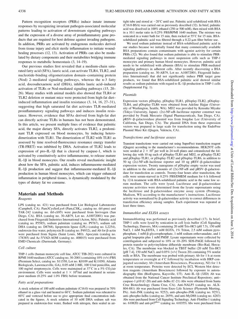
ResultsPalmitic acid induces the expression of pro–IL-1b andsecretion of mature IL-1b in THP-1 monocytes
Sodium palmitate treatment led to the expression of pro–IL-1b ina time- and dose-dependent manner in THP-1 cells, whereas un-treated cells showed undetectable levels of pro–IL-1b (Fig. 1A,1C). Sodium palmitate also induced secretion of mature IL-1b incell-culture supernatant (Fig. 1B, 1C), which requires caspase-1for the cleavage of pro–IL-1b. BSA-solubilized palmitic acid alsoinduced secretion of mature IL-1b in the supernatant in a dose-dependent manner (Supplemental Fig. 1A). Inhibition of caspase-1 activity by Ac-YVAD-AOM reduced sodium palmitate–medi-ated secretion of IL-1b (Fig. 1D). Untreated THP-1 monocytescontain both procaspase-1 and inflammasome-mediated activecaspase-1 (Fig. 1A). These results suggest that unstimulated mono-cytes possess constitutively active inflammasome. Therefore, pri-mary signals inducing the expression of pro–IL-1b mediated bythe activation of TLRs are sufficient for the production of IL-1bin monocytes. In this respect, monocytes differ from macrophages,which do not contain constitutively active inflammasome, and thusrequire the secondary signals for the activation of inflammasomeand the secretion of mature IL-1b (9–11). Untreated THP-1 cellsalso express NALP3 (Fig. 1A), which was enhanced by sodiumpalmitate treatment (Supplemental Fig. 2), suggesting that palmi-tate not only provides the primary signal leading to the expressionof pro–IL-1b, but also potentiates the activation of inflammasomeby upregulating the expression of NALP3.
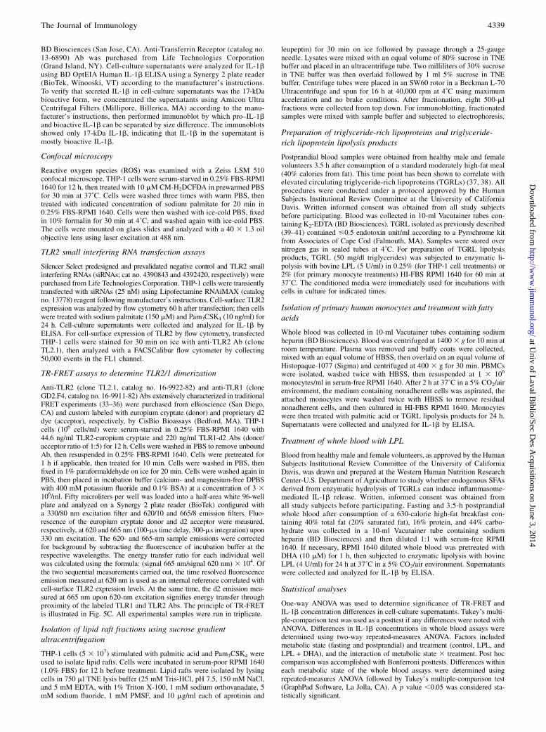
DHA inhibits palmitic acid–induced expression of pro–IL-1band consequent release of mature IL-1b in monocytes
Our previous studies showed that DHA is a pan inhibitor for TLRactivation (20, 21, 23–25). Therefore, we determined whether the
n-3 polyunsaturated fatty acid (PUFA) DHA inhibits palmiticacid–induced expression of pro–IL-1b and secretion of mature IL-1b. Pretreating THP-1 cells with DHA dose-dependently inhibitedthe expression of pro–IL-1b after a 6-h treatment with sodiumpalmitate (150 mM; Fig. 2A) and the secretion of active IL-1b andcaspase-1 after a 24-h treatment (Fig. 2B). Pretreating THP-1 cellswith DHA also dose-dependently inhibited the secretion of matureIL-1b after a 24-h treatment with BSA-solubilized palmitic acid(Supplemental Fig. 1B). These results imply that DHA inhibitspalmitic acid–induced activation of TLRs (primary signal) and thesubsequent expression of pro–IL-1b.
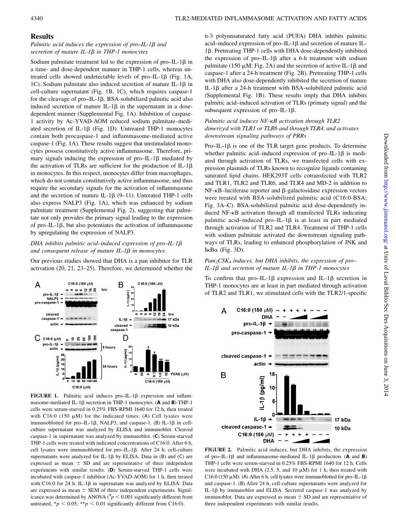
Palmitic acid induces NF-kB activation through TLR2dimerized with TLR1 or TLR6 and through TLR4, and activatesdownstream signaling pathways of PRRs
Pro–IL-1b is one of the TLR target gene products. To determinewhether palmitic acid–induced expression of pro–IL-1b is medi-ated through activation of TLRs, we transfected cells with ex-pression plasmids of TLRs known to recognize ligands containingsaturated lipid chains. HEK293T cells cotransfected with TLR2and TLR1, TLR2 and TLR6, and TLR4 and MD-2 in addition toNF-kB–luciferase reporter and b-galactosidase expression vectorswere treated with BSA-solubilized palmitic acid (C16:0-BSA;Fig. 3A–C). BSA-solubilized palmitic acid dose-dependently in-duced NF-kB activation through all transfected TLRs indicatingpalmitic acid–induced pro–IL-1b is at least in part mediatedthrough activation of TLR2 and TLR4. Treatment of THP-1 cellswith sodium palmitate activated the downstream signaling path-ways of TLRs, leading to enhanced phosphorylation of JNK andIkBa (Fig. 3D).
Pam3CSK4 induces, but DHA inhibits, the expression of pro–IL-1b and secretion of mature IL-1b in THP-1 monocytes
To confirm that pro–IL-1b expression and IL-1b secretion inTHP-1 monocytes are at least in part mediated through activationof TLR2 and TLR1, we stimulated cells with the TLR2/1-specific
FIGURE 1. Palmitic acid induces pro–IL-1b expression and inflam-
masome-mediated IL-1b secretion in THP-1 monocytes. (A and B) THP-1
cells were serum-starved in 0.25% FBS-RPMI 1640 for 12 h, then treated
with C16:0 (150 mM) for the indicated times. (A) Cell lysates were
immunoblotted for pro–IL-1b, NALP3, and caspase-1. (B) IL-1b in cell-
culture supernatant was analyzed by ELISA and immunoblot. Cleaved
caspase-1 in supernatant was analyzed by immunoblot. (C) Serum-starved
THP-1 cells were treated with indicated concentrations of C16:0. After 6 h,
cell lysates were immunoblotted for pro–IL-1b. After 24 h, cell-culture
supernatants were analyzed for IL-1b by ELISA. Data in (B) and (C) are
expressed as mean 6 SD and are representative of three independent
experiments with similar results. (D) Serum-starved THP-1 cells were
incubated with caspase-1 inhibitor (Ac-YVAD-AOM) for 1 h, then treated
with C16:0 for 24 h. IL-1b in supernatant was analyzed by ELISA. Data
are expressed as mean 6 SEM of three independent experiments. Signif-
icance was determined by ANOVA (#p, 0.001 significantly different from
untreated, *p , 0.05, **p , 0.01 significantly different from C16:0).
FIGURE 2. Palmitic acid induces, but DHA inhibits, the expression
of pro–IL-1b and inflammasome-mediated IL-1b production. (A and B)
THP-1 cells were serum-starved in 0.25% FBS-RPMI 1640 for 12 h. Cells
were incubated with DHA (2.5, 5, and 10 mM) for 1 h, then treated with
C16:0 (150 mM). (A) After 6 h, cell lysates were immunoblotted for pro–IL-1b
and caspase-1. (B) After 24 h, cell-culture supernatants were analyzed for
IL-1b by immunoblot and ELISA. Secreted caspase-1 was analyzed by
immunoblot. Data are expressed as mean 6 SD and are representative of
three independent experiments with similar results.
4340 TLR2-MEDIATED INFLAMMASOME ACTIVATION AND FATTY ACIDS
at Univ of L
aval Biblio/Sec D
es Acquisitions on June 3, 2014
http://ww
w.jim
munol.org/
Dow
nloaded from
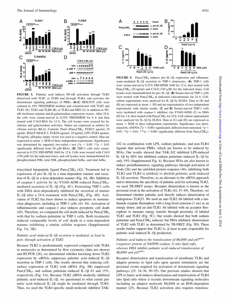
triacylated lipopeptide ligand Pam3CSK4 (42). Treatment led toexpression of pro–IL-1b in a time-dependent manner and secre-tion of IL-1b in a dose-dependent manner (Fig. 4A, 4B). Inhibitionof caspase-1 activity by Ac-YVAD-AOM reduced Pam3CSK4-mediated secretion of IL-1b (Fig. 4C). Pretreating THP-1 cellswith DHA dose-dependently inhibited the secretion of matureIL-1b after a 24-h treatment with Pam3CSK4 (Fig. 4D). Acti-vation of TLR2 has been shown to induce apoptosis in mononu-clear phagocytes, including in THP-1 cells (43, 44). Activation ofinflammasome and caspase-1 also induces pyroptotic cell death(45). Therefore, we compared the cell death induced by Pam3CSK4
with that by sodium palmitate in THP-1 cells. Both treatmentsinduced comparable levels of cell death in a time-dependentmanner exhibiting a similar cellular response (SupplementalFig. 3A, 3B).
Palmitic acid–induced IL-1b secretion is mediated, at least inpart, through activation of TLR2
Because TLR2 is predominantly expressed compared with TLR4in monocytes as determined by flow cytometry (data not shown)and RT-PCR (26), we determined whether knocking down TLR2expression by siRNAs suppresses palmitic acid–induced IL-1bsecretion in THP-1 cells. Our results showed that reducing cell-surface expression of TLR2 with siRNA (Fig. 5B) suppressedPam3CSK4- and sodium palmitate–induced IL-1b 63 and 15%,respectively (Fig. 5A). Because TLR2 siRNA modestly inhibitedpalmitic acid–induced IL-1b, we questioned to what extent pal-mitic acid–induced IL-1b might be mediated through TLR4.Thus, we used the TLR4-specific small-molecule inhibitor TAK-
242 in combination with LPS, sodium palmitate, and non-TLR4ligands that activate PRRs, which are known to be induced bySFAs. Our results showed that TAK-242 inhibited LPS-inducedIL-1b by 80% but inhibited sodium palmitate–induced IL-1b byonly 19% (Supplemental Fig. 4). Because SFAs are also known toinduce proinflammatory signaling pathways through activation ofNods (26) and the unfolded protein response (46), inhibiting bothTLR2 and TLR4 is unlikely to abolish palmitic acid–inducedIL-1b secretion. Therefore, as an alternate to the siRNA approachand to determine the specificity of palmitic acid for activating TLR2,we used TR-FRET assays. Receptor dimerization is known as theproximal event in the activation of TLRs (42, 47–49). Therefore, wedetermined whether palmitic acid directly induces dimerization ofendogenous TLR2/1. We used an anti-TLR2 Ab labeled with a lan-thanide cryptate fluorophore with a long-lived emission (1 ms) as anenergy donor, and an anti-TLR1 Ab labeled with an acceptor fluo-rophore to measure energy transfer through proximity of labeledTLR1 and TLR2 (Fig. 5C). Our results showed that both sodiumpalmitate and Pam3CSK4 induced, but DHA inhibited, dimerizationof TLR2 with TLR1 as determined by TR-FRET (Fig. 5D). Theseresults further support that TLR2 is, at least in part, responsible forpalmitic acid–induced IL-1b production.
Palmitic acid induces the translocation of MyD88 and p47phox
(organizer protein of NADPH oxidase 2) into lipid rafts,whereas DHA inhibits palmitic acid–induced translocation ofMyD88 and p47phox
Receptor dimerization and translocation of membrane TLRs andadaptor proteins to lipid rafts upon agonist stimulation are theproximal events required for activation of downstream signalingpathways (25, 34–36, 50–55). Our previous studies showed thatLPS or lauric acid induces dimerization and translocation of TLR4into lipid rafts where it recruits downstream signaling moleculesincluding an adaptor molecule MyD88 in an ROS-dependentmanner (25). Because TLR2 activation also requires transloca-
FIGURE 3. Palmitic acid induces NF-kB activation through TLR2
dimerized with TLR1 or TLR6 and through TLR4, and activates the
downstream signaling pathways of PRRs. (A–C) HEK293T cells were
cultured in 10% FBS/DMEM medium and cotransfected with TLR2 and
TLR1 (A), TLR2 and TLR6 (B), or TLR4 and MD2 (C) in addition to NF-
kB–luciferase reporter and b-galactosidase expression vectors. After 24 h,
the cells were serum-starved in 0.25% FBS/DMEM for 6 h and then
treated with C16:0-BSA for 12 h. The cell lysates were assayed for lu-
ciferase and galactosidase activities. Values are expressed as relative lu-
ciferase activity (RLA). Controls: Pam3 (Pam3CSK4, TLR2/1 agonist, 10
ng/ml), MALP (MALP-2, TLR2/6 agonist, 10 ng/ml), LPS (TLR4 agonist,
50 ng/ml). pDisplay empty vector was used as a negative control. Data are
expressed as mean 6 SEM of three independent experiments. Significance
was determined by unpaired, two-tailed t test (*p , 0.05, **p , 0.01
significantly different from 50 mM BSA). (D) THP-1 cells were serum-
starved in 0.25% FBS-RPMI 1640 for 12 h. Cells were treated with C16:0
(150 mM) for the indicated times, and cell lysates were immunoblotted for
phosphorylated JNK, total JNK, phosphorylated IkBa, and total IkBa.
FIGURE 4. Pam3CSK4 induces pro–IL-1b expression and inflamma-
some-mediated IL-1b secretion in THP-1 monocytes. (A) THP-1 cells
were serum-starved in 0.25% FBS-RPMI 1640 for 12 h, then treated with
Pam3CSK4 (10 ng/ml) and C16:0 (150 mM) for the indicated times. Cell
lysates were immunoblotted for pro–IL-1b. (B) Serum-starved THP-1 cells
were treated with Pam3CSK4 at indicated concentrations for 24 h. Cell-
culture supernatants were analyzed for IL-1b by ELISA. Data in (A) and
(B) are expressed as mean 6 SD and are representative of two independent
experiments with similar results. (C and D) Serum-starved THP-1 cells
were incubated with caspase-1 inhibitor (Ac-YVAD-AOM) (C) or DHA
(D) for 1 h, then treated with Pam3CSK4 for 24 h. Cell-culture supernatants
were analyzed for IL-1b by ELISA. Data in (C) and (D) are expressed as
mean 6 SEM of three independent experiments. Significance was deter-
mined by ANOVA (#p, 0.001 significantly different from untreated, *p,0.05, **p , 0.01, ***p , 0.001 significantly different from Pam3CSK4).
The Journal of Immunology 4341
at Univ of L
aval Biblio/Sec D
es Acquisitions on June 3, 2014
http://ww
w.jim
munol.org/
Dow
nloaded from
tion and association of the adaptor molecule MyD88 and NADPHoxidase 2 (NOX2) into lipid rafts (51, 56, 57), we determinedwhether palmitic acid induces the translocation of TLR2, MyD88,and NOX2 into lipid rafts of THP-1 cells by isolating lipid raftfractions and examining the translocation of these proteins byimmunoblotting. Our results showed that flotillin-1, which isconstitutively expressed in lipid rafts (58, 59), localized to frac-tions 2 and 3, whereas transferrin receptor, which is constitutivelyexpressed in nonlipid rafts (58), localized to fractions 6–8 (Fig.6A). Although the majority of TLR2 resided in nonlipid raftfractions, a substantial amount was expressed in lipid raft fractionsisolated from unstimulated cells (Fig. 6A). Within 5 min oftreatment, both sodium palmitate and Pam3CSK4 induced trans-location of MyD88 and NOX2 organizer protein p47phox to lipidraft fractions in THP-1 monocytes (Fig. 6B). In contrast, DHAinhibited both sodium palmitate– and Pam3CSK4-induced re-cruitment of both MyD88 and p47phox to lipid raft fractions (Fig.6C).
TLR2-mediated palmitic acid–induced IL-1b is dependent onNADPH oxidase activation and is inhibited by DHA
Based on our results indicating that palmitic acid induces theassembly of NOX2 through translocation of p47phox to lipid rafts,we determined whether palmitic acid–induced assembly of NOX2would induce ROS. Treatment with sodium palmitate inducedROS in a dose-dependent manner as assessed by confocal mi-croscopy but was inhibited by DHA (Fig. 7A). Because NOX2 isknown to be a critical component of the TLR2 signaling complex(56, 57, 60) we determined whether inhibiting NOX2 assembly
would inhibit palmitic acid–induced IL-1b secretion. Apocynin,which inhibits the assembly and activation of NOX2 by interfer-ing with the translocation of p47phox (61, 62) dose-dependentlyinhibited sodium palmitate– and Pam3CSK-induced IL-1b secre-tion (Fig. 7B). In addition, apocynin dose-dependently inhibitedsodium palmitate–induced dimerization of TLR2/1 as assessed byTR-FRET (Fig. 7C). Collectively, these results reveal that palmiticacid and DHA reciprocally modulate activation of TLR2 bymodulating the dimerization of TLR2/1, and that dimerization isdependent on NOX2 activation.
Both exogenous palmitic acid and endogenous fatty acidsderived from the lipolysis products of TGRLs induce, but DHAinhibits, IL-1b production in human peripheral bloodmonocytes and whole blood
Next, we determined whether palmitic acid also induces IL-1bproduction in primary human monocytes. Sodium palmitate in-duced, but DHA inhibited, IL-1b production in primary mono-cytes cultured in 1% HI-FBS RPMI 1640 (Fig. 8A) similar to theresults obtained from the THP-1 monocytic cell line. Next, wedetermined whether a physiological source of dietary SFAs reca-pitulates the effects of palmitic acid. Postprandial lipemia ischaracterized by transient accumulation of TGRLs in the bloodafter ingestion of a fatty meal. In vivo, TGRLs are hydrolyzed byLPL, an enzyme anchored to endothelial cells, releasing free fattyacids into the blood in immediate proximity to blood monocytes.Our previous studies showed that palmitic acid is the major SFAfound in the lipolysis products (free fatty acids) of these TGRLsisolated from subjects consuming the high-fat meal (40). There-
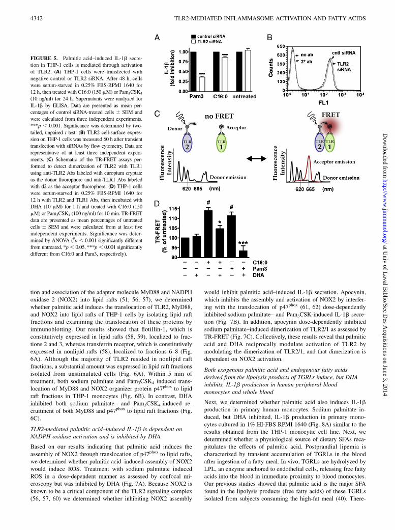
FIGURE 5. Palmitic acid–induced IL-1b secre-
tion in THP-1 cells is mediated through activation
of TLR2. (A) THP-1 cells were transfected with
negative control or TLR2 siRNA. After 48 h, cells
were serum-starved in 0.25% FBS-RPMI 1640 for
12 h, then treated with C16:0 (150 mM) or Pam3CSK4
(10 ng/ml) for 24 h. Supernatants were analyzed for
IL-1b by ELISA. Data are presented as mean per-
centages of control siRNA-treated cells 6 SEM and
were calculated from three independent experiments.
***p , 0.001. Significance was determined by two-
tailed, unpaired t test. (B) TLR2 cell-surface expres-
sion on THP-1 cells was measured 60 h after transient
transfection with siRNAs by flow cytometry. Data are
representative of at least three independent experi-
ments. (C) Schematic of the TR-FRET assays per-
formed to detect dimerization of TLR2 with TLR1
using anti-TLR2 Abs labeled with europium cryptate
as the donor fluorophore and anti-TLR1 Abs labeled
with d2 as the acceptor fluorophore. (D) THP-1 cells
were serum-starved in 0.25% FBS-RPMI 1640 for
12 h with TLR2 and TLR1 Abs, then incubated with
DHA (10 mM) for 1 h and treated with C16:0 (150
mM) or Pam3CSK4 (100 ng/ml) for 10 min. TR-FRET
data are presented as mean percentages of untreated
cells 6 SEM and were calculated from at least five
independent experiments. Significance was deter-
mined by ANOVA (#p , 0.001 significantly different
from untreated, *p, 0.05, ***p, 0.001 significantly
different from C16:0 and Pam3, respectively).
4342 TLR2-MEDIATED INFLAMMASOME ACTIVATION AND FATTY ACIDS
at Univ of L
aval Biblio/Sec D
es Acquisitions on June 3, 2014
http://ww
w.jim
munol.org/
Dow
nloaded from
fore, we determined whether the lipolysis products of TGRLs alsoinduce IL-1b production in primary monocytes or in whole blood.The lipolysis products of TGRLs induced IL-1b production in
primary monocytes (Fig. 8B). Increased IL-1b production by thetreatment of primary monocytes with conditioned media preparedwith LPL without added TGRL is likely due to release of free fattyacids from lipids present in FBS (2% final concentration in me-dia). Lipolysis products of TGRLs also increased heterodimer-ization of TLR2 with TLR1 as assessed by TR-FRET in THP-1monocytes treated in 0.25% FBS (Fig. 8C). Finally, direct treat-ment of fasting and postprandial whole blood with LPL inducedrobust production of IL-1b. Treatment of postprandial wholeblood with LPL induced significantly more IL-1b compared withfasting whole blood. Furthermore, concomitant treatment of bothLPL-treated fasting and postprandial whole-blood samples withDHA inhibited the IL-1b production (Fig. 8D). These resultssuggest that inflammasome-mediated IL-1b production in bloodmonocytes can be dynamically modulated by the types of dietaryfat we consume (Fig. 9).
DiscussionOur results demonstrate that palmitic acid, a predominant dietarySFA, induces, whereas the n-3 fatty acid DHA inhibits, the ex-pression of pro–IL-1b and subsequent release of mature IL-1b inboth THP-1 monocytes and primary human blood monocytes.These results further support the findings that blood monocytescontain active caspase-1 because of the presence of constitu-tively active NALP3 inflammasome (8–10). Therefore, primarysignals inducing the expression of pro–IL-1b are sufficient forinflammasome-mediated IL-1b production in blood monocytes.Pro–IL-1b is one of the target gene products derived from theactivation of TLRs expressed in monocytes. Therefore, any ago-nists that activate TLRs should be able to induce inflammasome-mediated IL-1b production in human blood monocytes withoutsecondary signals required for the activation of inflammasome asdepicted in Fig. 9.Animal studies showed that palmitic acid can activate NALP3
inflammasome in bone marrow–derived macrophages (BMDMs)pretreated with LPS; however, palmitic acid alone did not inducepro–IL-1b expression in BMDMs, suggesting that palmitic acidcan provide the secondary signal to activate inflammasome, butnot the primary signals to induce the expression of pro–IL-1b inBMDMs (19). Thus, for palmitic acid to induce inflammasome-
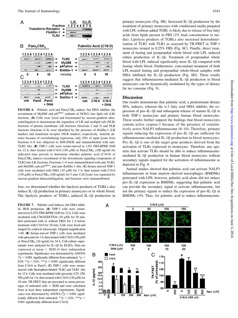
FIGURE 6. Palmitic acid and Pam3CSK4 induce, but DHA inhibits, the
recruitment of MyD88 and p47phox (subunit of NOX2) into lipid raft (LR)
fractions. (A) Cells were lysed and fractionated by sucrose-gradient ultra-
centrifugation to demonstrate the separation of LR and nonlipid raft (NLR)
fractions of plasma membrane. LR fractions (fractions 2 and 3) and NLR
fractions (fractions 6–8) were identified by the presence of flotillin-1 (LR
marker) and transferrin receptor (NLR marker), respectively. Asterisk de-
notes because of overwhelming expression, only 20% of input lysate from
fractions 6–8 was subjected to SDS-PAGE and immunoblotted with anti-
TLR2 Abs. (B) THP-1 cells were serum-starved in 1.0% FBS-RPMI 1640
for 12 h, then treated with C16:0 (150 mM) or Pam3CSK4 (100 ng/ml) for
indicated time periods to determine whether palmitic acid (C16:0) or
Pam3CSK4 induces recruitment of the downstream signaling components of
TLR2 into LR fractions. Fractions 1–4 were immunoblotted with anti-TLR2,
anti-MyD88, anti-p47phox, and anti–flotillin-1 Abs. (C) Serum-starved THP-1
cells were incubated with DHA (10 mM) for 1 h, then treated with C16:0
(150 mM) or Pam3CSK4 (100 ng/ml) for 5 min. Cell lysate was separated by
sucrose-gradient ultracentrifugation, and fractions were immunoblotted.
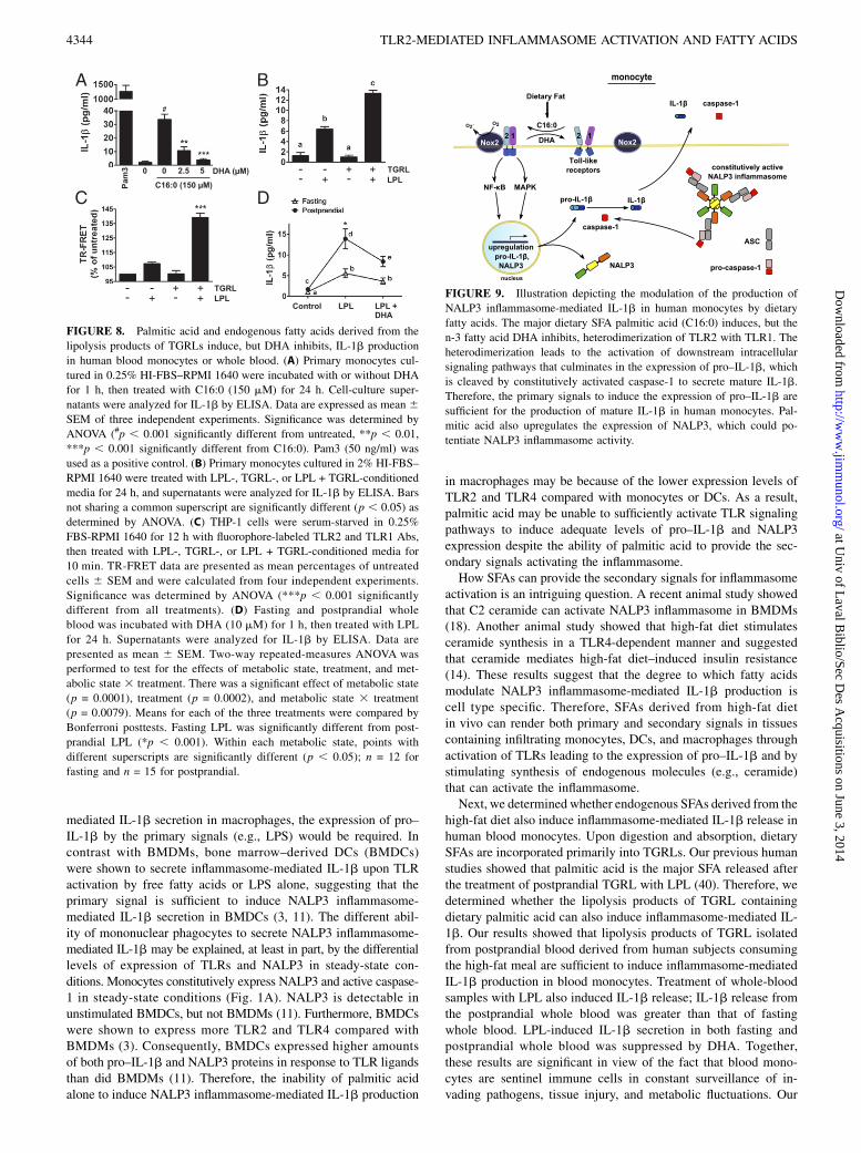
FIGURE 7. Palmitic acid induces, but DHA inhib-
its, ROS production. (A) THP-1 cells were serum-
starved in 0.25% FBS-RPMI 1640 for 12 h. Cells were
incubated with CM-H2DCFDA (10 mM) for 30 min,
then pretreated with or without DHA for 1 h before
treatment with C16:0 for 20 min. Cells were fixed and
imaged by confocal microscopy. Original magnification
340. (B) Serum-starved THP-1 cells were incubated
with apocynin for 1 h, then treated with C16:0 (150mM)
or Pam3CSK4 (10 ng/ml) for 24 h. Cell-culture super-
natants were analyzed for IL-1b by ELISA. Data are
expressed as mean 6 SEM of three independent
experiments. Significance was determined by ANOVA
(#p, 0.001 significantly different from untreated, *p,0.05, **p, 0.01, ***p, 0.001 significantly different
from C16:0 or Pam3). (C) THP-1 cells were serum-
starved with fluorophore-labeled TLR2 and TLR1 Abs
for 12 h. Cells were incubated with apocynin (125, 250,
500mM) for 1 h, then treated with C16:0 (150mM) for
10 min. TR-FRET data are presented as mean percen-
tages of untreated cells 6 SEM and were calculated
from at least three independent experiments. Signifi-
cance was determined by ANOVA (#p, 0.001 signif-
icantly different from untreated, **p , 0.01, ***p ,0.001 significantly different from C16:0).
The Journal of Immunology 4343
at Univ of L
aval Biblio/Sec D
es Acquisitions on June 3, 2014
http://ww
w.jim
munol.org/
Dow
nloaded from
mediated IL-1b secretion in macrophages, the expression of pro–IL-1b by the primary signals (e.g., LPS) would be required. Incontrast with BMDMs, bone marrow–derived DCs (BMDCs)were shown to secrete inflammasome-mediated IL-1b upon TLRactivation by free fatty acids or LPS alone, suggesting that theprimary signal is sufficient to induce NALP3 inflammasome-mediated IL-1b secretion in BMDCs (3, 11). The different abil-ity of mononuclear phagocytes to secrete NALP3 inflammasome-mediated IL-1b may be explained, at least in part, by the differentiallevels of expression of TLRs and NALP3 in steady-state con-ditions. Monocytes constitutively express NALP3 and active caspase-1 in steady-state conditions (Fig. 1A). NALP3 is detectable inunstimulated BMDCs, but not BMDMs (11). Furthermore, BMDCswere shown to express more TLR2 and TLR4 compared withBMDMs (3). Consequently, BMDCs expressed higher amountsof both pro–IL-1b and NALP3 proteins in response to TLR ligandsthan did BMDMs (11). Therefore, the inability of palmitic acidalone to induce NALP3 inflammasome-mediated IL-1b production
in macrophages may be because of the lower expression levels ofTLR2 and TLR4 compared with monocytes or DCs. As a result,palmitic acid may be unable to sufficiently activate TLR signalingpathways to induce adequate levels of pro–IL-1b and NALP3expression despite the ability of palmitic acid to provide the sec-ondary signals activating the inflammasome.How SFAs can provide the secondary signals for inflammasome
activation is an intriguing question. A recent animal study showedthat C2 ceramide can activate NALP3 inflammasome in BMDMs(18). Another animal study showed that high-fat diet stimulatesceramide synthesis in a TLR4-dependent manner and suggestedthat ceramide mediates high-fat diet–induced insulin resistance(14). These results suggest that the degree to which fatty acidsmodulate NALP3 inflammasome-mediated IL-1b production iscell type specific. Therefore, SFAs derived from high-fat dietin vivo can render both primary and secondary signals in tissuescontaining infiltrating monocytes, DCs, and macrophages throughactivation of TLRs leading to the expression of pro–IL-1b and bystimulating synthesis of endogenous molecules (e.g., ceramide)that can activate the inflammasome.Next, we determined whether endogenous SFAs derived from the
high-fat diet also induce inflammasome-mediated IL-1b release inhuman blood monocytes. Upon digestion and absorption, dietarySFAs are incorporated primarily into TGRLs. Our previous humanstudies showed that palmitic acid is the major SFA released afterthe treatment of postprandial TGRL with LPL (40). Therefore, wedetermined whether the lipolysis products of TGRL containingdietary palmitic acid can also induce inflammasome-mediated IL-1b. Our results showed that lipolysis products of TGRL isolatedfrom postprandial blood derived from human subjects consumingthe high-fat meal are sufficient to induce inflammasome-mediatedIL-1b production in blood monocytes. Treatment of whole-bloodsamples with LPL also induced IL-1b release; IL-1b release fromthe postprandial whole blood was greater than that of fastingwhole blood. LPL-induced IL-1b secretion in both fasting andpostprandial whole blood was suppressed by DHA. Together,these results are significant in view of the fact that blood mono-cytes are sentinel immune cells in constant surveillance of in-vading pathogens, tissue injury, and metabolic fluctuations. Our
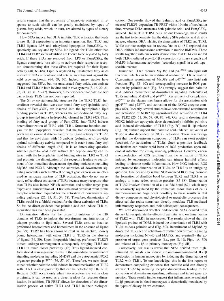
FIGURE 8. Palmitic acid and endogenous fatty acids derived from the
lipolysis products of TGRLs induce, but DHA inhibits, IL-1b production
in human blood monocytes or whole blood. (A) Primary monocytes cul-
tured in 0.25% HI-FBS–RPMI 1640 were incubated with or without DHA
for 1 h, then treated with C16:0 (150 mM) for 24 h. Cell-culture super-
natants were analyzed for IL-1b by ELISA. Data are expressed as mean 6SEM of three independent experiments. Significance was determined by
ANOVA (#p , 0.001 significantly different from untreated, **p , 0.01,
***p , 0.001 significantly different from C16:0). Pam3 (50 ng/ml) was
used as a positive control. (B) Primary monocytes cultured in 2% HI-FBS–
RPMI 1640 were treated with LPL-, TGRL-, or LPL + TGRL-conditioned
media for 24 h, and supernatants were analyzed for IL-1b by ELISA. Bars
not sharing a common superscript are significantly different (p , 0.05) as
determined by ANOVA. (C) THP-1 cells were serum-starved in 0.25%
FBS-RPMI 1640 for 12 h with fluorophore-labeled TLR2 and TLR1 Abs,
then treated with LPL-, TGRL-, or LPL + TGRL-conditioned media for
10 min. TR-FRET data are presented as mean percentages of untreated
cells 6 SEM and were calculated from four independent experiments.
Significance was determined by ANOVA (***p , 0.001 significantly
different from all treatments). (D) Fasting and postprandial whole
blood was incubated with DHA (10 mM) for 1 h, then treated with LPL
for 24 h. Supernatants were analyzed for IL-1b by ELISA. Data are
presented as mean 6 SEM. Two-way repeated-measures ANOVA was
performed to test for the effects of metabolic state, treatment, and met-
abolic state 3 treatment. There was a significant effect of metabolic state
(p = 0.0001), treatment (p = 0.0002), and metabolic state 3 treatment
(p = 0.0079). Means for each of the three treatments were compared by
Bonferroni posttests. Fasting LPL was significantly different from post-
prandial LPL (*p , 0.001). Within each metabolic state, points with
different superscripts are significantly different (p , 0.05); n = 12 for
fasting and n = 15 for postprandial.
FIGURE 9. Illustration depicting the modulation of the production of
NALP3 inflammasome-mediated IL-1b in human monocytes by dietary
fatty acids. The major dietary SFA palmitic acid (C16:0) induces, but the
n-3 fatty acid DHA inhibits, heterodimerization of TLR2 with TLR1. The
heterodimerization leads to the activation of downstream intracellular
signaling pathways that culminates in the expression of pro–IL-1b, which
is cleaved by constitutively activated caspase-1 to secrete mature IL-1b.
Therefore, the primary signals to induce the expression of pro–IL-1b are
sufficient for the production of mature IL-1b in human monocytes. Pal-
mitic acid also upregulates the expression of NALP3, which could po-
tentiate NALP3 inflammasome activity.
4344 TLR2-MEDIATED INFLAMMASOME ACTIVATION AND FATTY ACIDS
at Univ of L
aval Biblio/Sec D
es Acquisitions on June 3, 2014
http://ww
w.jim
munol.org/
Dow
nloaded from
results suggest that the propensity of monocyte activation in re-sponse to such stimuli can be greatly modulated by types ofplasma fatty acids, which, in turn, are altered by types of dietaryfat consumed.How SFAs induce, but DHA inhibits, TLR activation that leads
to pro–IL-1b expression is an important question. Both TLR4 andTLR2 ligands LPS and triacylated lipopeptide Pam3CSK4, re-spectively, are acylated by SFAs. No ligands for TLRs other thanTLR4 and TLR2 so far identified are known to be acylated by fattyacids. If these SFAs are removed from LPS or Pam3CSK4, theligands completely lose ability to activate their respective recep-tor, demonstrating that these SFAs are required for their ligandactivity (48, 63–68). Lipid A acylated by unsaturated fatty acidsinstead of SFAs is nontoxic and acts as an antagonist against thewild type endotoxin (64, 69, 70). Indeed, many studies havesuggested that SFAs, but not unsaturated fatty acids, can activateTLR4 and TLR2 in both in vitro and in vivo systems (3, 16, 20, 21,23, 24, 30, 31, 71–77). However, direct evidence that palmitic acidcan activate TLRs has not been reported.The X-ray crystallographic structure for the TLR2-TLR1 het-
erodimer revealed that two ester-bound fatty acyl (palmitic acid)chains of Pam3CSK4 are inserted into the hydrophobic lipidbinding pocket in TLR2, whereas the amide-bound fatty acylgroup is inserted into a hydrophobic channel in TLR1 (42). Thus,binding of fatty acyl groups of Pam3CSK4 into TLR2 inducesheterodimerization of TLR2 with TLR1. Structure-function anal-ysis for the lipopeptides revealed that the two ester-bound fattyacids are an essential determinant for its ligand activity for TLR2,and that the acyl chain with 16 carbons (palmitic acid) providesoptimal stimulatory activity compared with ester-bound fatty acylchains of different length (63). It is an interesting questionwhether palmitic acid itself without the peptide moiety can in-teract with the hydrophobic lipid binding sites in TLR2 or TLR1and promote the dimerization of the receptors leading to recruit-ment of the immediate downstream signaling molecules includingMyD88 and NOX2. Although the activation of downstream sig-naling molecules such as NF-kB or target gene expression are oftenused as surrogate markers of TLR activation, they do not neces-sarily reflect direct activation of TLRs because many receptors otherthan TLRs also induce NF-kB activation and similar target geneexpression. Dimerization of TLRs is the most proximal event for thereceptor activation required for the activation of downstream sig-naling pathways (25, 35, 51, 54, 78). Therefore, dimerization ofTLRs would be a faithful readout for the direct activation of TLRs.So far, no direct evidence that palmitic acid can induce TLR di-merization has ever been presented.Dimerization allows for the proper orientation of the TIR
domains of TLRs to induce the recruitment and interaction ofadaptor proteins in lipid rafts. Several TLRs exist as inactive,preformed heterodimers and homodimers in the absence of ligand(42, 79). TLR2 has been shown to exist as an inactive, looselybound heterodimer with either TLR1 or TLR6 in the absenceof ligand (34, 80). Upon lipopeptide binding, preformed TLR2/1dimers undergo rearrangement subsequently bringing TLR2 andTLR1 in much closer proximity (42). This ligand-induced con-formational rearrangement results in the immediate recruitment ofsignaling molecules including MyD88 and the cytoplasmic NOX2organizer protein p47phox (56, 57, 60). Therefore, we next deter-mined whether palmitic acid induces heterodimerization of TLR2with TLR1 in close proximity that can be detected by TR-FRET.Because FRET occurs only when two receptors are within closeproximity, it can be used as a direct readout for receptor dimer-ization. In addition, TR-FRET allows for detection of the dimer-ization process of native TLR1 and TLR2 in their biological
context. Our results showed that palmitic acid or Pam3CSK4 in-creased TLR2/1-dependent TR-FRETwithin 10 min of incubationtime, whereas DHA attenuates both palmitic acid– and Pam3CSK4-induced TR-FRET in THP-1 cells. To our knowledge, these resultsare the first to demonstrate that the dietary SFA palmitic acid directlyinduces, whereas DHA inhibits, the dimerization of TLR2 and TLR1.While our manuscript was in review, Yan et al. (81) reported thatDHA inhibits inflammasome activation in murine BMDMs. Theseresults together with our results demonstrate that DHA can inhibitboth TLR-mediated pro–IL-1b expression (primary signal) andNALP3 inflammasome activation (secondary signal) in a cell-type–specific manner.Dimerized TLR2 recruits MyD88 and p47phox into lipid raft
fractions, which can be an additional readout of TLR activation.Concomitant recruitment of MyD88 and p47phox into lipid raftfractions (Fig. 6B, 6C) and corresponding increase in ROS gen-eration by palmitic acid (Fig. 7A) strongly suggest that palmiticacid induces recruitment of downstream signaling molecules ofTLRs including MyD88 and p47phox. Recruitment of cytoplasmicp47phox to the plasma membrane allows for the association withgp91phox and p22phox, and activation of the NOX2 enzyme com-plex (82). Recently, several studies have demonstrated recruitmentand activation of NOX2 by agonist-induced activation of TLR4and TLR2 (25, 51, 56, 57, 60, 83, 84). Our results showing thatNOX2 inhibitor apocynin dose-dependently inhibits palmiticacid–induced dimerization of TLR2 (Fig. 7C) and IL-1b release(Fig. 7B) further support that palmitic acid–induced activation ofTLR2 is also dependent on NOX2 activation. These results sug-gest that the downstream component of TLRs can exert positivefeedback for activation of TLRs. Such a positive feedbackmechanism can render rapid burst of ROS production upon mi-crobial infection for killing invading pathogens. However, exces-sive production of ROS resulting from the activation of TLRsinduced by endogenous molecules can trigger harmful effectsleading to chronic sterile inflammation. How NOX-induced ROScan promote the dimerization of TLR4 or TLR2 is an intriguingquestion. One possibility is that NOX-induced ROS may promotethe formation of disulfide bond between TLR2 and TLR1 as ananalogy to many redox-sensitive proteins (85–88). Dimerizationof TLR2 involves formation of a disulfide bond (89), which maybe sensitively regulated by the immediate redox status of cell’smicroenvironment. Significant corollary to these results is thatdietary components and physiological or metabolic processes thataffect cellular redox status can directly modulate TLR-mediatedinflammatory responses and their subsequent consequences.We next determined whether endogenous SFAs derived from
dietary fat recapitulate the effects of palmitic acid on dimerizationof TLR2 with TLR1 in monocytes. The results showed that thelipolysis product of TGRL induced the dimerization of TLR2 withTLR1 as does palmitic acid (Fig. 8C). Recruitment of MyD88 bydimerized TLR2 led to activation of further downstream signalingmolecules including NF-kB and MAPKs, and eventually the ex-pression of target gene products (i.e., pro–IL-1b; Figs. 1A, 3D)and release of IL-1b in primary monocytes (Fig. 8B).Collectively, our results reveal that SFAs derived from high-
saturated fat meals can induce inflammasome-mediated IL-1bproduction in human monocytes by inducing the dimerization ofTLR2 with TLR1. To our knowledge, this is the first report todemonstrate compelling evidence that palmitic acid can directlyactivate TLR2 by inducing receptor dimerization leading to theactivation of downstream signaling pathways and target gene ex-pression. These results further suggest that inflammasome-mediatedIL-1b production in blood monocytes is dynamically modulated bythe types of dietary fat we consume.
The Journal of Immunology 4345
at Univ of L
aval Biblio/Sec D
es Acquisitions on June 3, 2014
http://ww
w.jim
munol.org/
Dow
nloaded from
DisclosuresThe authors have no financial conflicts of interest.
References1. Geissmann, F., S. Jung, and D. R. Littman. 2003. Blood monocytes consist of
two principal subsets with distinct migratory properties. Immunity 19: 71–82.2. Geissmann, F., M. G. Manz, S. Jung, M. H. Sieweke, M. Merad, and K. Ley.
2010. Development of monocytes, macrophages, and dendritic cells. Science327: 656–661.
3. Nguyen, M. T., S. Favelyukis, A. K. Nguyen, D. Reichart, P. A. Scott, A. Jenn,R. Liu-Bryan, C. K. Glass, J. G. Neels, and J. M. Olefsky. 2007. A subpopulationof macrophages infiltrates hypertrophic adipose tissue and is activated by freefatty acids via Toll-like receptors 2 and 4 and JNK-dependent pathways. J. Biol.Chem. 282: 35279–35292.
4. Bryant, C., and K. A. Fitzgerald. 2009. Molecular mechanisms involved ininflammasome activation. Trends Cell Biol. 19: 455–464.
5. Lamkanfi, M., and V. M. Dixit. 2009. The inflammasomes. PLoS Pathog. 5:e1000510.
6. Martinon, F., K. Burns, and J. Tschopp. 2002. The inflammasome: a molecularplatform triggering activation of inflammatory caspases and processing of proIL-beta. Mol. Cell 10: 417–426.
7. Martinon, F., A. Mayor, and J. Tschopp. 2009. The inflammasomes: guardians ofthe body. Annu. Rev. Immunol. 27: 229–265.
8. Carta, S., S. Tassi, I. Pettinati, L. Delfino, C. A. Dinarello, and A. Rubartelli.2011. The rate of interleukin-1beta secretion in different myeloid cells varieswith the extent of redox response to Toll-like receptor triggering. J. Biol. Chem.286: 27069–27080.
9. Netea, M. G., C. A. Nold-Petry, M. F. Nold, L. A. Joosten, B. Opitz, J. H. van derMeer, F. L. van de Veerdonk, G. Ferwerda, B. Heinhuis, I. Devesa, et al. 2009.Differential requirement for the activation of the inflammasome for processingand release of IL-1beta in monocytes and macrophages. Blood 113: 2324–2335.
10. Netea, M. G., A. Simon, F. van de Veerdonk, B. J. Kullberg, J. W. Van der Meer, andL. A. Joosten. 2010. IL-1beta processing in host defense: beyond the inflammasomes.PLoS Pathog. 6: e1000661.
11. He, Y., L. Franchi, and G. Nunez. 2013. TLR agonists stimulate Nlrp3-dependentIL-1b production independently of the purinergic P2X7 receptor in dendriticcells and in vivo. J. Immunol. 190: 334–339.
12. Krysko, D. V., P. Agostinis, O. Krysko, A. D. Garg, C. Bachert, B. N. Lambrecht,and P. Vandenabeele. 2011. Emerging role of damage-associated molecular pat-terns derived from mitochondria in inflammation. Trends Immunol. 32: 157–164.
13. Piccinini, A. M., and K. S. Midwood. 2010. DAMPening inflammation bymodulating TLR signalling. Mediators Inflamm. 2010: pii:672395.
14. Holland, W. L., B. T. Bikman, L. P. Wang, G. Yuguang, K. M. Sargent,S. Bulchand, T. A. Knotts, G. Shui, D. J. Clegg, M. R. Wenk, et al. 2011. Lipid-induced insulin resistance mediated by the proinflammatory receptor TLR4requires saturated fatty acid-induced ceramide biosynthesis in mice. J. Clin.Invest. 121: 1858–1870.
15. Hwang, D. 2001. Modulation of the expression of cyclooxygenase-2 by fattyacids mediated through toll-like receptor 4-derived signaling pathways. FASEBJ. 15: 2556–2564.
16. Shi, H., M. V. Kokoeva, K. Inouye, I. Tzameli, H. Yin, and J. S. Flier. 2006.TLR4 links innate immunity and fatty acid-induced insulin resistance. J. Clin.Invest. 116: 3015–3025.
17. Stienstra, R., J. A. van Diepen, C. J. Tack, M. H. Zaki, F. L. van de Veerdonk,D. Perera, G. A. Neale, G. J. Hooiveld, A. Hijmans, I. Vroegrijk, et al. 2011.Inflammasome is a central player in the induction of obesity and insulin resis-tance. Proc. Natl. Acad. Sci. USA 108: 15324–15329.
18. Vandanmagsar, B., Y. H. Youm, A. Ravussin, J. E. Galgani, K. Stadler,R. L. Mynatt, E. Ravussin, J. M. Stephens, and V. D. Dixit. 2011. The NLRP3inflammasome instigates obesity-induced inflammation and insulin resistance.Nat. Med. 17: 179–188.
19. Wen, H., D. Gris, Y. Lei, S. Jha, L. Zhang, M. T. Huang, W. J. Brickey, andJ. P. Ting. 2011. Fatty acid-induced NLRP3-ASC inflammasome activationinterferes with insulin signaling. Nat. Immunol. 12: 408–415.
20. Lee, J. Y., A. Plakidas, W. H. Lee, A. Heikkinen, P. Chanmugam, G. Bray, andD. H. Hwang. 2003. Differential modulation of Toll-like receptors by fatty acids:preferential inhibition by n-3 polyunsaturated fatty acids. J. Lipid Res. 44: 479–486.
21. Lee, J. Y., K. H. Sohn, S. H. Rhee, and D. Hwang. 2001. Saturated fatty acids,but not unsaturated fatty acids, induce the expression of cyclooxygenase-2mediated through Toll-like receptor 4. J. Biol. Chem. 276: 16683–16689.
22. Lee, J. Y., J. Ye, Z. Gao, H. S. Youn, W. H. Lee, L. Zhao, N. Sizemore, andD. H. Hwang. 2003. Reciprocal modulation of Toll-like receptor-4 signalingpathways involving MyD88 and phosphatidylinositol 3-kinase/AKT by saturatedand polyunsaturated fatty acids. J. Biol. Chem. 278: 37041–37051.
23. Lee, J. Y., L. Zhao, H. S. Youn, A. R. Weatherill, R. Tapping, L. Feng,W. H. Lee, K. A. Fitzgerald, and D. H. Hwang. 2004. Saturated fatty acidactivates but polyunsaturated fatty acid inhibits Toll-like receptor 2 dimerizedwith Toll-like receptor 6 or 1. J. Biol. Chem. 279: 16971–16979.
24. Weatherill, A. R., J. Y. Lee, L. Zhao, D. G. Lemay, H. S. Youn, andD. H. Hwang. 2005. Saturated and polyunsaturated fatty acids reciprocallymodulate dendritic cell functions mediated through TLR4. J. Immunol. 174:5390–5397.
25. Wong, S. W., M. J. Kwon, A. M. Choi, H. P. Kim, K. Nakahira, andD. H. Hwang. 2009. Fatty acids modulate Toll-like receptor 4 activation through
regulation of receptor dimerization and recruitment into lipid rafts in a reactiveoxygen species-dependent manner. J. Biol. Chem. 284: 27384–27392.
26. Zhao, L., M. J. Kwon, S. Huang, J. Y. Lee, K. Fukase, N. Inohara, andD. H. Hwang. 2007. Differential modulation of Nods signaling pathways by fattyacids in human colonic epithelial HCT116 cells. J. Biol. Chem. 282: 11618–11628.
27. Ehses, J. A., D. T. Meier, S. Wueest, J. Rytka, S. Boller, P. Y. Wielinga,A. Schraenen, K. Lemaire, S. Debray, L. Van Lommel, et al. 2010. Toll-likereceptor 2-deficient mice are protected from insulin resistance and beta celldysfunction induced by a high-fat diet. Diabetologia 53: 1795–1806.
28. Himes, R. W., and C. W. Smith. 2010. Tlr2 is critical for diet-induced metabolicsyndrome in a murine model. FASEB J. 24: 731–739.
29. Suganami, T., T. Mieda, M. Itoh, Y. Shimoda, Y. Kamei, and Y. Ogawa. 2007.Attenuation of obesity-induced adipose tissue inflammation in C3H/HeJ micecarrying a Toll-like receptor 4 mutation. Biochem. Biophys. Res. Commun. 354:45–49.
30. Suganami, T., K. Tanimoto-Koyama, J. Nishida, M. Itoh, X. Yuan, S. Mizuarai,H. Kotani, S. Yamaoka, K. Miyake, S. Aoe, et al. 2007. Role of the Toll-likereceptor 4/NF-kappaB pathway in saturated fatty acid-induced inflammatorychanges in the interaction between adipocytes and macrophages. Arterioscler.Thromb. Vasc. Biol. 27: 84–91.
31. Suganami, T., X. Yuan, Y. Shimoda, K. Uchio-Yamada, N. Nakagawa,I. Shirakawa, T. Usami, T. Tsukahara, K. Nakayama, Y. Miyamoto, et al. 2009.Activating transcription factor 3 constitutes a negative feedback mechanism thatattenuates saturated Fatty acid/toll-like receptor 4 signaling and macrophageactivation in obese adipose tissue. Circ. Res. 105: 25–32.
32. Huang, S., J. M. Rutkowsky, R. G. Snodgrass, K. D. Ono-Moore,D. A. Schneider, J. W. Newman, S. H. Adams, and D. H. Hwang. 2012. Saturatedfatty acids activate TLR-mediated proinflammatory signaling pathways. J. LipidRes. 53: 2002–2013.
33. Liang, S., M. Wang, K. Triantafilou, M. Triantafilou, H. F. Nawar, M. W. Russell,T. D. Connell, and G. Hajishengallis. 2007. The A subunit of type IIb enterotoxin(LT-IIb) suppresses the proinflammatory potential of the B subunit and its abilityto recruit and interact with TLR2. J. Immunol. 178: 4811–4819.
34. Triantafilou, M., F. G. Gamper, R. M. Haston, M. A. Mouratis, S. Morath,T. Hartung, and K. Triantafilou. 2006. Membrane sorting of toll-like receptor(TLR)-2/6 and TLR2/1 heterodimers at the cell surface determines heterotypicassociations with CD36 and intracellular targeting. J. Biol. Chem. 281: 31002–31011.
35. Triantafilou, M., F. G. Gamper, P. M. Lepper, M. A. Mouratis, C. Schumann,E. Harokopakis, R. E. Schifferle, G. Hajishengallis, and K. Triantafilou. 2007.Lipopolysaccharides from atherosclerosis-associated bacteria antagonize TLR4,induce formation of TLR2/1/CD36 complexes in lipid rafts and trigger TLR2-induced inflammatory responses in human vascular endothelial cells. Cell.Microbiol. 9: 2030–2039.
36. Triantafilou, M., M. Manukyan, A. Mackie, S. Morath, T. Hartung, H. Heine, andK. Triantafilou. 2004. Lipoteichoic acid and toll-like receptor 2 internalizationand targeting to the Golgi are lipid raft-dependent. J. Biol. Chem. 279: 40882–40889.
37. Chan, J. W., D. Motton, J. C. Rutledge, N. L. Keim, and T. Huser. 2005. Ramanspectroscopic analysis of biochemical changes in individual triglyceride-richlipoproteins in the pre- and postprandial state. Anal. Chem. 77: 5870–5876.
38. Hyson, D. A., T. G. Paglieroni, T. Wun, and J. C. Rutledge. 2002. Postprandiallipemia is associated with platelet and monocyte activation and increasedmonocyte cytokine expression in normolipemic men. Clin. Appl. Thromb.Hemost. 8: 147–155.
39. Eiselein, L., D. W. Wilson, M. W. Lame, and J. C. Rutledge. 2007. Lipolysisproducts from triglyceride-rich lipoproteins increase endothelial permeability,perturb zonula occludens-1 and F-actin, and induce apoptosis. Am. J. Physiol.Heart Circ. Physiol. 292: H2745–H2753.
40. Wang, L., R. Gill, T. L. Pedersen, L. J. Higgins, J. W. Newman, andJ. C. Rutledge. 2009. Triglyceride-rich lipoprotein lipolysis releases neutral andoxidized FFAs that induce endothelial cell inflammation. J. Lipid Res. 50: 204–213.
41. Wang, Y. I., J. Schulze, N. Raymond, T. Tomita, K. Tam, S. I. Simon, andA. G. Passerini. 2011. Endothelial inflammation correlates with subject trigly-cerides and waist size after a high-fat meal. Am. J. Physiol. Heart Circ. Physiol.300: H784–H791.
42. Jin, M. S., S. E. Kim, J. Y. Heo, M. E. Lee, H. M. Kim, S. G. Paik, H. Lee, andJ. O. Lee. 2007. Crystal structure of the TLR1-TLR2 heterodimer induced bybinding of a tri-acylated lipopeptide. Cell 130: 1071–1082.
43. Aliprantis, A. O., R. B. Yang, M. R. Mark, S. Suggett, B. Devaux, J. D. Radolf,G. R. Klimpel, P. Godowski, and A. Zychlinsky. 1999. Cell activation and ap-optosis by bacterial lipoproteins through toll-like receptor-2. Science 285: 736–739.
44. Aliprantis, A. O., R. B. Yang, D. S. Weiss, P. Godowski, and A. Zychlinsky.2000. The apoptotic signaling pathway activated by Toll-like receptor-2. EMBOJ. 19: 3325–3336.
45. Miao, E. A., J. V. Rajan, and A. Aderem. 2011. Caspase-1-induced pyroptoticcell death. Immunol. Rev. 243: 206–214.
46. Fu, S., S. M. Watkins, and G. S. Hotamisligil. 2012. The role of endoplasmicreticulum in hepatic lipid homeostasis and stress signaling. Cell Metab. 15: 623–634.
47. Jenkins, K. A., and A. Mansell. 2010. TIR-containing adaptors in Toll-like re-ceptor signalling. Cytokine 49: 237–244.
48. Jin, M. S., and J. O. Lee. 2008. Structures of the toll-like receptor family and itsligand complexes. Immunity 29: 182–191.
4346 TLR2-MEDIATED INFLAMMASOME ACTIVATION AND FATTY ACIDS
at Univ of L
aval Biblio/Sec D
es Acquisitions on June 3, 2014
http://ww
w.jim
munol.org/
Dow
nloaded from
49. O’Neill, L. A., and A. G. Bowie. 2007. The family of five: TIR-domain-containing adaptors in Toll-like receptor signalling. Nat. Rev. Immunol. 7:353–364.
50. Fernandez-Lizarbe, S., M. Pascual, M. S. Gascon, A. Blanco, and C. Guerri.2008. Lipid rafts regulate ethanol-induced activation of TLR4 signaling inmurine macrophages. Mol. Immunol. 45: 2007–2016.
51. Nakahira, K., H. P. Kim, X. H. Geng, A. Nakao, X. Wang, N. Murase,P. F. Drain, X. Wang, M. Sasidhar, E. G. Nabel, et al. 2006. Carbon monoxidedifferentially inhibits TLR signaling pathways by regulating ROS-inducedtrafficking of TLRs to lipid rafts. J. Exp. Med. 203: 2377–2389.
52. Olsson, S., and R. Sundler. 2006. The role of lipid rafts in LPS-induced signalingin a macrophage cell line. Mol. Immunol. 43: 607–612.
53. Shin, D. M., C. S. Yang, J. Y. Lee, S. J. Lee, H. H. Choi, H. M. Lee, J. M. Yuk,C. V. Harding, and E. K. Jo. 2008. Mycobacterium tuberculosis lipoprotein-induced association of TLR2 with protein kinase C zeta in lipid rafts contrib-utes to reactive oxygen species-dependent inflammatory signalling in macro-phages. Cell. Microbiol. 10: 1893–1905.
54. Triantafilou, M., K. Miyake, D. T. Golenbock, and K. Triantafilou. 2002. Mediatorsof innate immune recognition of bacteria concentrate in lipid rafts and facilitatelipopolysaccharide-induced cell activation. J. Cell Sci. 115: 2603–2611.
55. Triantafilou, M., S. Morath, A. Mackie, T. Hartung, and K. Triantafilou. 2004.Lateral diffusion of Toll-like receptors reveals that they are transiently confinedwithin lipid rafts on the plasma membrane. J. Cell Sci. 117: 4007–4014.
56. Ogier-Denis, E., S. B. Mkaddem, and A. Vandewalle. 2008. NOX enzymes andToll-like receptor signaling. Semin. Immunopathol. 30: 291–300.
57. Yang, C. S., D. M. Shin, K. H. Kim, Z. W. Lee, C. H. Lee, S. G. Park, Y. S. Bae,and E. K. Jo. 2009. NADPH oxidase 2 interaction with TLR2 is required forefficient innate immune responses to mycobacteria via cathelicidin expression. J.Immunol. 182: 3696–3705.
58. Dhungana, S., B. A. Merrick, K. B. Tomer, and M. B. Fessler. 2009. Quantitativeproteomics analysis of macrophage rafts reveals compartmentalized activation ofthe proteasome and of proteasome-mediated ERK activation in response to li-popolysaccharide. Mol. Cell. Proteomics 8: 201–213.
59. Li, N., A. Mak, D. P. Richards, C. Naber, B. O. Keller, L. Li, and A. R. Shaw.2003. Monocyte lipid rafts contain proteins implicated in vesicular traffickingand phagosome formation. Proteomics 3: 536–548.
60. Yang, C. S., D. M. Shin, H. M. Lee, J. W. Son, S. J. Lee, S. Akira,M. A. Gougerot-Pocidalo, J. El-Benna, H. Ichijo, and E. K. Jo. 2008. ASK1-p38MAPK-p47phox activation is essential for inflammatory responses during tu-berculosis via TLR2-ROS signalling. Cell. Microbiol. 10: 741–754.
61. Stolk, J., T. J. Hiltermann, J. H. Dijkman, and A. J. Verhoeven. 1994. Character-istics of the inhibition of NADPH oxidase activation in neutrophils by apocynin,a methoxy-substituted catechol. Am. J. Respir. Cell Mol. Biol. 11: 95–102.
62. Wedgwood, S., S. Lakshminrusimha, K. N. Farrow, L. Czech, S. F. Gugino,F. Soares, J. A. Russell, and R. H. Steinhorn. 2012. Apocynin improves oxy-genation and increases eNOS in persistent pulmonary hypertension of thenewborn. Am. J. Physiol. Lung Cell. Mol. Physiol. 302: L616–L626.
63. Buwitt-Beckmann, U., H. Heine, K. H. Wiesmuller, G. Jung, R. Brock, andA. J. Ulmer. 2005. Lipopeptide structure determines TLR2 dependent cell ac-tivation level. FEBS J. 272: 6354–6364.
64. Kitchens, R. L., R. J. Ulevitch, and R. S. Munford. 1992. Lipopolysaccharide(LPS) partial structures inhibit responses to LPS in a human macrophage cellline without inhibiting LPS uptake by a CD14-mediated pathway. J. Exp. Med.176: 485–494.
65. Kumazawa, Y., M. Nakatsuka, H. Takimoto, T. Furuya, T. Nagumo, A. Yamamoto,Y. Homma, K. Inada, M. Yoshida, M. Kiso, et al. 1988. Importance of fatty acidsubstituents of chemically synthesized lipid A-subunit analogs in the expression ofimmunopharmacological activity. Infect. Immun. 56: 149–155.
66. Munford, R. S., and C. L. Hall. 1986. Detoxification of bacterial lip-opolysaccharides (endotoxins) by a human neutrophil enzyme. Science 234:203–205.
67. Rietschel, E. T., T. Kirikae, F. U. Schade, U. Mamat, G. Schmidt, H. Loppnow,A. J. Ulmer, U. Zahringer, U. Seydel, F. Di Padova, et al. 1994. Bacterial en-dotoxin: molecular relationships of structure to activity and function. FASEB J.8: 217–225.
68. Schletter, J., H. Heine, A. J. Ulmer, and E. T. Rietschel. 1995. Molecularmechanisms of endotoxin activity. Arch. Microbiol. 164: 383–389.
69. Krauss, J. H., U. Seydel, J. Weckesser, and H. Mayer. 1989. Structural analysisof the nontoxic lipid A of Rhodobacter capsulatus 37b4. Eur. J. Biochem. 180:519–526.
70. Qureshi, N., K. Takayama, and R. Kurtz. 1991. Diphosphoryl lipid A obtainedfrom the nontoxic lipopolysaccharide of Rhodopseudomonas sphaeroides is anendotoxin antagonist in mice. Infect. Immun. 59: 441–444.
71. Eguchi, K., I. Manabe, Y. Oishi-Tanaka, M. Ohsugi, N. Kono, F. Ogata, N. Yagi,U. Ohto, M. Kimoto, K. Miyake, et al. 2012. Saturated fatty acid and TLRsignaling link b cell dysfunction and islet inflammation. Cell Metab. 15: 518–533.
72. Joosten, L. A., M. G. Netea, E. Mylona, M. I. Koenders, R. K. Malireddi,M. Oosting, R. Stienstra, F. L. van de Veerdonk, A. F. Stalenhoef,E. J. Giamarellos-Bourboulis, et al. 2010. Engagement of fatty acids with Toll-like receptor 2 drives interleukin-1b production via the ASC/caspase 1 pathway inmonosodium urate monohydrate crystal-induced gouty arthritis. Arthritis Rheum.62: 3237–3248.
73. Kim, F., M. Pham, I. Luttrell, D. D. Bannerman, J. Tupper, J. Thaler, T. R. Hawn,E. W. Raines, and M. W. Schwartz. 2007. Toll-like receptor-4 mediates vascularinflammation and insulin resistance in diet-induced obesity. Circ. Res. 100:1589–1596.
74. Schaeffler, A., P. Gross, R. Buettner, C. Bollheimer, C. Buechler, M. Neumeier,A. Kopp, J. Schoelmerich, and W. Falk. 2009. Fatty acid-induced induction ofToll-like receptor-4/nuclear factor-kappaB pathway in adipocytes links nutri-tional signalling with innate immunity. Immunology 126: 233–245.
75. Schwartz, E. A., W. Y. Zhang, S. K. Karnik, S. Borwege, V. R. Anand,P. S. Laine, Y. Su, and P. D. Reaven. 2010. Nutrient modification of the innateimmune response: a novel mechanism by which saturated fatty acids greatlyamplify monocyte inflammation. Arterioscler. Thromb. Vasc. Biol. 30: 802–808.
76. Senn, J. J. 2006. Toll-like receptor-2 is essential for the development ofpalmitate-induced insulin resistance in myotubes. J. Biol. Chem. 281: 26865–26875.
77. Bourguignon, L. Y., C. C. Spevak, G. Wong, W. Xia, and E. Gilad. 2009.Hyaluronan-CD44 interaction with protein kinase C(epsilon) promotes onco-genic signaling by the stem cell marker Nanog and the Production of microRNA-21, leading to down-regulation of the tumor suppressor protein PDCD4, anti-apoptosis, and chemotherapy resistance in breast tumor cells. J. Biol. Chem. 284:26533–26546.
78. Triantafilou, M., P. M. Lepper, R. Olden, I. S. Dias, and K. Triantafilou. 2011.Location, location, location: is membrane partitioning everything when it comesto innate immune activation? Mediators Inflamm. 2011: 186093.
79. Latz, E., A. Verma, A. Visintin, M. Gong, C. M. Sirois, D. C. Klein,B. G. Monks, C. J. McKnight, M. S. Lamphier, W. P. Duprex, et al. 2007.Ligand-induced conformational changes allosterically activate Toll-like receptor9. Nat. Immunol. 8: 772–779.
80. Tapping, R. I., and P. S. Tobias. 2003. Mycobacterial lipoarabinomannanmediates physical interactions between TLR1 and TLR2 to induce signaling. J.Endotoxin Res. 9: 264–268.
81. Yan, Y., W. Jiang, T. Spinetti, A. Tardivel, R. Castillo, C. Bourquin, G. Guarda,Z. Tian, J. Tschopp, and R. Zhou. 2013. Omega-3 fatty acids prevent inflam-mation and metabolic disorder through inhibition of NLRP3 inflammasomeactivation. Immunity 38: 1154–1163.
82. Bedard, K., and K. H. Krause. 2007. The NOX family of ROS-generatingNADPH oxidases: physiology and pathophysiology. Physiol. Rev. 87: 245–313.
83. DeLeo, F. R., J. Renee, S. McCormick, M. Nakamura, M. Apicella, J. P. Weiss,and W. M. Nauseef. 1998. Neutrophils exposed to bacterial lipopolysaccharideupregulate NADPH oxidase assembly. J. Clin. Invest. 101: 455–463.
84. Simon, F., and R. Fernandez. 2009. Early lipopolysaccharide-induced reactiveoxygen species production evokes necrotic cell death in human umbilical veinendothelial cells. J. Hypertens. 27: 1202–1216.
85. Ago, T., T. Liu, P. Zhai, W. Chen, H. Li, J. D. Molkentin, S. F. Vatner, andJ. Sadoshima. 2008. A redox-dependent pathway for regulating class II HDACsand cardiac hypertrophy. Cell 133: 978–993.
86. Cumming, R. C., N. L. Andon, P. A. Haynes, M. Park, W. H. Fischer, andD. Schubert. 2004. Protein disulfide bond formation in the cytoplasm duringoxidative stress. J. Biol. Chem. 279: 21749–21758.
87. Miki, H., and Y. Funato. 2012. Regulation of intracellular signalling throughcysteine oxidation by reactive oxygen species. J. Biochem. 151: 255–261.
88. Yang, Y., Y. Song, and J. Loscalzo. 2007. Regulation of the protein disulfideproteome by mitochondria in mammalian cells. Proc. Natl. Acad. Sci. USA 104:10813–10817.
89. Tao, X., Y. Xu, Y. Zheng, A. A. Beg, and L. Tong. 2002. An extensively asso-ciated dimer in the structure of the C713S mutant of the TIR domain of humanTLR2. Biochem. Biophys. Res. Commun. 299: 216–221.
The Journal of Immunology 4347
at Univ of L
aval Biblio/Sec D
es Acquisitions on June 3, 2014
http://ww
w.jim
munol.org/
Dow
nloaded from