In silico Analysis of Pectin Lyase and Pectinase Sequences · In silico Analysis of Pectin Lyase...
11
Pectinases are a group of enzymes involved in degra- dation of pectin, that includes various enzymes classified into various classes and subclasses depending on the sub- strate specificity and mode of action, for example, methyl deesterases, hydrolases, and lyases. According to the cleavage site, pectinases are divided into three groups: (i) hydrolases consisting of polygalacturonase, PG (EC 3.2.1.15); (ii) lyase/trans-eliminases comprising pectin lyase, PNL (EC 4.2.2.10), and pectate lyase, PL (EC 4.2.2.2); (iii) pectin esterase, PE (EC 3.1.1.11) [1, 2]. Pectin esterase catalyzes the deesterification of methyl ester linkages of galacturonan backbone of pectic sub- stances to release acidic pectins and methanol [3]. The resulting pectin is then acted upon by PG and PL [4]. Production, biochemical characterization, and applica- tions of PNL have been reviewed extensively [1]. Among all pectinases, PNLs are of particular interest because these degrade pectin polymers directly by a β-elimination mechanism that results in the formation of 4,5-unsaturat- ed oligogalacturonides, while other pectinases act sequentially to degrade the pectin molecule completely. Pectin esterases are found in plants, plant pathogenic bacteria, and fungi [5], while PGs are widely distributed among fungi, bacteria, and many yeasts [6]. They are also found in higher plants and some plant parasitic nema- todes. Pectate lyases are mostly produced by bacteria. The isolation and characterization of pectolytic enzymes is well documented. Numerous studies on fungal ISSN 0006-2979, Biochemistry (Moscow), 2009, Vol. 74, No. 9, pp. 1049-1055. © Pleiades Publishing, Ltd., 2009. Published in Russian in Biokhimiya, 2009, Vol. 74, No. 9, pp. 1286-1293. 1049 Abbreviations: MEME, Multiple EM for Motif Elicitation; NCBI, National Center for Biotechnology Information; NJ, Neighbor-Joining method; PCR, polymerase chain reaction; PE, pectin esterase; PG, polygalacturonase; PL, pectate lyase; PNL, pectin lyase; UPGMA method, Unweighted Pair Group method with Arithmetic Mean method. * To whom correspondence should be addressed. In silico Analysis of Pectin Lyase and Pectinase Sequences P. K. Yadav 1,2 , V. K. Singh 3 , S. Yadav 2 , K. D. S. Yadav 2 , and D. Yadav 1,3 * 1 Department of Biotechnology, Deen Dayal Upadhyaya Gorakhpur University, Gorakhpur (U. P.), India; E-mail: [email protected]; [email protected] 2 Department of Chemistry, Deen Dayal Upadhyaya Gorakhpur University, Gorakhpur (U. P.), India 3 Department of Molecular Biology and Genetic Engineering, CBSH, G. B. Pant University of Agriculture and Technology, Pantnagar (Uttarakhand), India Received October 13, 2008 Revision received December 22, 2008 Abstract—A total of 48 full-length protein sequences of pectin lyases from different source organisms available in NCBI were subjected to multiple sequence alignment, domain analysis, and phylogenetic tree construction. A phylogenetic tree con- structed on the basis of the protein sequences revealed two distinct clusters representing pectin lyases from bacterial and fun- gal sources. Similarly, the multiple accessions of different source organisms representing bacterial and fungal pectin lyases also formed distinct clusters, showing sequence level homology. The sequence level similarities among different groups of pectinase enzymes, viz. pectin lyase, pectate lyase, polygalacturonase, and pectin esterase, were also analyzed by subjecting a single protein sequence from each group with common source organism to tree construction. Four distinct clusters repre- senting different groups of pectinases with common source organisms were observed, indicating the existing sequence level similarity among them. Multiple sequence alignment of pectin lyase protein sequence of different source organisms along with pectinases with common source organisms revealed a conserved region, indicating homology at sequence level. A con- served domain Pec_Lyase_C was frequently observed in the protein sequences of pectin lyases and pectate lyases, while Glyco_hydro_28 domains and Pectate lyase-like β-helix clan domain are frequently observed in polygalacturonases and pectin esterases, respectively. The signature amino acid sequence of 41 amino acids, i.e. TYDNAGVLPITVN- SNKSLIGEGSKGVIKGKGLRIVSGAKNI, related with the Pec_Lyase_C is frequently observed in pectin lyase protein sequences and might be related with the structure and enzymatic function. DOI: 10.1134/S0006297909090144 Key words: pectin lyase, pectate lyase, polygalacturonase, pectin esterase, domain analysis
Transcript of In silico Analysis of Pectin Lyase and Pectinase Sequences · In silico Analysis of Pectin Lyase...
Pectinases are a group of enzymes involved in degra-
dation of pectin, that includes various enzymes classified
into various classes and subclasses depending on the sub-
strate specificity and mode of action, for example, methyl
deesterases, hydrolases, and lyases. According to the
cleavage site, pectinases are divided into three groups: (i)
hydrolases consisting of polygalacturonase, PG (EC
3.2.1.15); (ii) lyase/trans-eliminases comprising pectin
lyase, PNL (EC 4.2.2.10), and pectate lyase, PL (EC
4.2.2.2); (iii) pectin esterase, PE (EC 3.1.1.11) [1, 2].
Pectin esterase catalyzes the deesterification of methyl
ester linkages of galacturonan backbone of pectic sub-
stances to release acidic pectins and methanol [3]. The
resulting pectin is then acted upon by PG and PL [4].
Production, biochemical characterization, and applica-
tions of PNL have been reviewed extensively [1]. Among
all pectinases, PNLs are of particular interest because
these degrade pectin polymers directly by a β-elimination
mechanism that results in the formation of 4,5-unsaturat-
ed oligogalacturonides, while other pectinases act
sequentially to degrade the pectin molecule completely.
Pectin esterases are found in plants, plant pathogenic
bacteria, and fungi [5], while PGs are widely distributed
among fungi, bacteria, and many yeasts [6]. They are also
found in higher plants and some plant parasitic nema-
todes. Pectate lyases are mostly produced by bacteria.
The isolation and characterization of pectolytic
enzymes is well documented. Numerous studies on fungal
ISSN 0006-2979, Biochemistry (Moscow), 2009, Vol. 74, No. 9, pp. 1049-1055. © Pleiades Publishing, Ltd., 2009.
Published in Russian in Biokhimiya, 2009, Vol. 74, No. 9, pp. 1286-1293.
1049
Abbreviations: MEME, Multiple EM for Motif Elicitation;
NCBI, National Center for Biotechnology Information; NJ,
Neighbor-Joining method; PCR, polymerase chain reaction;
PE, pectin esterase; PG, polygalacturonase; PL, pectate lyase;
PNL, pectin lyase; UPGMA method, Unweighted Pair Group
method with Arithmetic Mean method.
* To whom correspondence should be addressed.
In silico Analysis of Pectin Lyase and Pectinase Sequences
P. K. Yadav1,2, V. K. Singh3, S. Yadav2, K. D. S. Yadav2, and D. Yadav1,3*
1Department of Biotechnology, Deen Dayal Upadhyaya Gorakhpur University, Gorakhpur (U. P.),
India; E-mail: [email protected]; [email protected] of Chemistry, Deen Dayal Upadhyaya Gorakhpur University, Gorakhpur (U. P.), India
3Department of Molecular Biology and Genetic Engineering, CBSH,
G. B. Pant University of Agriculture and Technology, Pantnagar (Uttarakhand), India
Received October 13, 2008
Revision received December 22, 2008
Abstract—A total of 48 full-length protein sequences of pectin lyases from different source organisms available in NCBI were
subjected to multiple sequence alignment, domain analysis, and phylogenetic tree construction. A phylogenetic tree con-
structed on the basis of the protein sequences revealed two distinct clusters representing pectin lyases from bacterial and fun-
gal sources. Similarly, the multiple accessions of different source organisms representing bacterial and fungal pectin lyases
also formed distinct clusters, showing sequence level homology. The sequence level similarities among different groups of
pectinase enzymes, viz. pectin lyase, pectate lyase, polygalacturonase, and pectin esterase, were also analyzed by subjecting
a single protein sequence from each group with common source organism to tree construction. Four distinct clusters repre-
senting different groups of pectinases with common source organisms were observed, indicating the existing sequence level
similarity among them. Multiple sequence alignment of pectin lyase protein sequence of different source organisms along
with pectinases with common source organisms revealed a conserved region, indicating homology at sequence level. A con-
served domain Pec_Lyase_C was frequently observed in the protein sequences of pectin lyases and pectate lyases, while
Glyco_hydro_28 domains and Pectate lyase-like β-helix clan domain are frequently observed in polygalacturonases and
pectin esterases, respectively. The signature amino acid sequence of 41 amino acids, i.e. TYDNAGVLPITVN-
SNKSLIGEGSKGVIKGKGLRIVSGAKNI, related with the Pec_Lyase_C is frequently observed in pectin lyase protein
sequences and might be related with the structure and enzymatic function.
DOI: 10.1134/S0006297909090144
Key words: pectin lyase, pectate lyase, polygalacturonase, pectin esterase, domain analysis
1050 YADAV et al.
BIOCHEMISTRY (Moscow) Vol. 74 No. 9 2009
pectolytic enzymes have been carried out and several fun-
gal PNL genes have been isolated and characterized from
Aspergillus niger [7-9], A. oryzae [10], and Glomerella cin-
gulata [11]. Pectinases have extensive applications in
extraction, clarification, and cloud stabilization of fruit
juices, in degumming and retting of natural fibers (ramie,
hemp, flax, bast), maceration of plant tissues, isolation of
protoplasts, and saccharification of biomass [12-17]. A
number of PEs have been purified and biochemically
characterized [18].
Crystal structures of pectin lyase A (PNLA) from two
strains of A. niger, N400 and 4M-147 [19], reveal that
PNLA folds into a parallel β-sheet and shares many of the
structural features of PL despite not more than 17%
sequence identity after pair-wise structure-based align-
ment. These shared structural features include amino
acid stacks and an asparagine ladder. The substrate-bind-
ing clefts of these two PNLs are dominated by aromatic
residues and are enveloped by negative electrostatic
potential. The major difference between these two PNLA
structures is in the conformation of the loop formed by
residues 182-187. These observed differences are due to
the different pH values of crystallization. The three-
dimensional structure of pectin lyase B (PNLB) from A.
niger has also been determined by crystallographic tech-
niques at resolution of 1.7 Å [20].
In this communication, an attempt has been made
to investigate the protein sequences of PNLs from differ-
ent source organisms along with protein sequences of
PNL, PL, PG, and PE common source organisms using
various bioinformatics tools to reveal the sequence level
similarity. Further in silico domain analysis of these
sequences can provide insight into possible functions
associated with the existing active site of the enzyme,
which might be a target for genetic manipulation for
enhanced activity of the enzyme. The multiple sequence
alignment of different PNL protein sequences from dif-
ferent organisms can provide us an opportunity to design
degenerate primers for PCR amplification of PNL gene
family, which can further be used for cloning and overex-
pression.
MATERIALS AND METHODS
All the sequences of PNL, PL, PG, and PE of dif-
ferent source organisms available in GenBank were
downloaded from NCBI (http://www.ncbi.nlm.nih.gov).
A total of 494, 717, 937, and 172 protein sequences of
PNL, PL, PG, and PE, respectively, representing major
groups of pectinases were downloaded. Only the full-
length protein sequences were considered for in silico
analysis.
The programs Clustalw [21] and Seaview
(http://pbil.univlyon1.fr/software/seaview.html) were
used for multiple sequence alignment. Mega 3.1 [22] was
used for dendrogram construction by Neighbor-Joining
(NJ) [23], Minimum Evolution, and UPGMA methods
[24]. For domain search, the Pfam site (http://
www.sanger.ac.uk/Software/Pfam/search.shtml) was
used. Domain analysis was done using MEME
(http://meme.sdsc.edu/meme/meme.html). The con-
served protein motifs deduced by MEME were subjected
to biological functional analysis using protein BLAST,
and domains were studied with Interproscan providing
the best possible match based on highest similarity score.
The accession numbers of PNL protein sequences along
with the source are listed in Table 1, while the accession
numbers of PNL, PL, PE, and PG with common source
are listed in Table 2.
Accession number
CAA43130, AK37997,XP001402523, CAK47350,AAW03313, CAK48529,XP001389926, AAA32701,ХР001401061, CAA46521,CAD34589
XP001216214, XP001208581,EAU37973, EAU31855
XP753624, EAL91586, XP749007,XP753604, EAL86969, EAL91566
XP001274337, EAW12911
ABO38857
AAM23009, AAM23008,AAD43565
ABH03046
YP049604, CAG74408
BAA06847
NP794039, AAO57734, YP 276124, AAZ35927
BAA12119, AAB84422
AAA21817, AAF22244
ABF50856, ABF50854
YP001285824, ABI36836
YP001224597
XP001259650, EAW17753
CAA01024, CAA01023
Source
A. niger
A. terreus
A. fumigatus
A. clavatus
A. oryzae
Penicillium griseoroseum
P. occitanis
Erwinia carotovora
Pseudomonas marginalis
Ps. syringae
Bacillus subtilis
Colletotrichum gloeosporioides
Emericella nidulans
Geobacillus virus
Synechococcus sp.
Neosartorya fischeri
Synthetic construct
Table 1. List of pectin lyase protein sequences reported
from different sources
In silico ANALYSIS OF PECTIN LYASE AND PECTINASE SEQUENCES 1051
BIOCHEMISTRY (Moscow) Vol. 74 No. 9 2009
Motif
1
2
3
4
5
6
7
8
9
10
Width
41
50
50
41
29
29
21
21
21
15
Best possible match
QN I A I TD I N PKY VWGGDA I T L DDCDMVW I DHV T TAR I GRQH
NCY I DGR TD Y S A T CDGHHYWGMY LDG S NDMV TMKGNY I YHTSGRMPKVQG
S ATP V Y PKT TDE L V S Y LGDDE P RV I V L T K T FDF TGT EGT TTETGCAPWGT
T YDNAGV L P I TV N SNK S L I GE G SKG V I KGK GL R I V S GAKNI (Pec_lyase_C)
T LLH A VNNYWYDNSGH AF E I GE GGY V L A E
F TS P DA S TNE QCMS Y L GRAC Q L NGF GS S G
CQVA I NKHDWCDNY Q P DA PK V
A AAV GV SGA P EGFA KGV TGGG
QADT DF L SK F KGKN I A S AHA Y
GNVFQNV PT V LE S P I
Table 3. Different motifs commonly observed in PNL sequences with best possible match amino acid sequences (see Fig. 2)
Motif
1
2
3
4
5
6
7
8
9
10
Width
50
50
29
41
21
41
41
50
50
50
Best possible match
EWEGP L I R F S GKD I T V EQA SG A V I DGDG S RWWDGEGT NGGKTKP KFMYAH
Y PKT TD ELV S Y LGDS E PRV I I L TK TF DF R G TEG TT TE TGCAPWGTAS KCQ
V WGGDA I R I QNSDMVW IDHC E T AR I R RQH
NQDDC I A I N S GEN I E F SGGV C S GGHGL S I G SV G GRDDNT VE (Glyco_hydro_28)
MMTMHHNYWYH I NGR A PKLMR (Pec_lyase_C)
A YE I Y I LCGDGS C S DWTWEGV D I TGGK T S D KCE NV P SGA SC
I VLGTQ ADNRMT I S NS F I NGE S DY S A T CN GHHY WGMYL DGS
S QNGVR I KT I YDE TGS V SE V T Y S NI KL S G I TK Y G I V I E QDYENG S PTGTP
NTLL HA VNNY WHDNS GHA F E I G EGGY V L A EGNV FQNVP A VAE S P I EGQLF
PKVS V T YDNAGV LG I T VNS NK S L I GE G S AGV I KGKGLR I V SGAKN I I I QN
Table 4. Different motifs commonly observed in different pectinase protein sequences with best possible match amino
acid sequences (see Fig. 3)
Organism
Pectobacterium carotovorum
Bacillus sp
Erwinia sp.
A. niger
E. nidulans
N. fischeri
A. fumigatus
A. clavatus
A. oryzae
Penicillium sp.
PE
P55743
ABV62479
CAA59151
CAK38387
ABF50865
XP001263676
XP747054
XP001270784
BAA75474
Table 2. List of protein sequences of pectinases reported from common source organisms
PG
CAA35998
BAB85762
CAB99319
CAA41693
S24156
XP001266657
XP753090
XP001272239
BAA03244
ABS50231
PL
CAA43402
ABO37788
CAA47821
CAK40523
ABF50894
XP 001265533
XP749213
EAW15258
ABM60783
PNL
AAA24856
AAB 84422
CAG74408
AAW03313
ABF50854
EAW17753
EAL86969
EAW12911
ABO38857
AAS55382
Accession number
1052 YADAV et al.
BIOCHEMISTRY (Moscow) Vol. 74 No. 9 2009
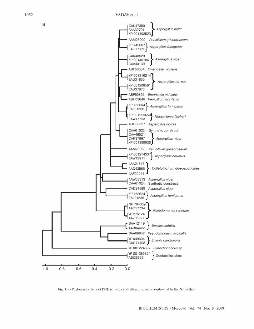
Fig. 1. a) Phylogenetic trees of PNL sequences of different sources constructed by the NJ method.
1.0 0.8 0.6 0.4 0.2 0.0
a CAK47350
AAA32701
XP 001402523
AAM23009
XP 749007
EAL86969
CAK48529
XP 001401061
CAA43130
ABF50854
XP 001216214
EAU31855
XP 001208581
EAU37973
ABF50856
ABH03046
XP 753604
EAL91566
XP 001259650
EAW17753
ABO38857
CAA01023
CAA46521
CAK37997
XP 001389926
AAM23008
XP 001274337
EAW12911
AAA21817
AAD43565
AAF22244
AAW03313
CAA01024
CAD34589
XP 753624
EAL91586
NP 794039
AAO57734
YP 276124
AAZ35927
BAA12119
AAB84422
BAA06847
YP 049604
CAG74408
YP 001224597
YP 001285824
ABI36836
Aspergillus niger
Penicillium griseoroseum
Aspergillus fumigatus
Aspergillus niger
Emericella nidulans
Aspergillus terreus
Emericella nidulans
Penicillium occitanis
Aspergillus fumigatus
Neosartorya fischeri
Aspergillus oryzae
Synthetic construct
Aspergillus niger
Penicillium griseoroseum
Aspergillus clavatus
Colletotrichum gloeosporioides
Synthetic construct
Aspergillus niger
Aspergillus niger
Aspergillus fumigatus
Pseudomonas syringae
Bacillus subtilis
Pseudomonas marginalis
Erwinia carotovora
Synechococcus sp.
Geobacillus virus
In silico ANALYSIS OF PECTIN LYASE AND PECTINASE SEQUENCES 1053
BIOCHEMISTRY (Moscow) Vol. 74 No. 9 2009
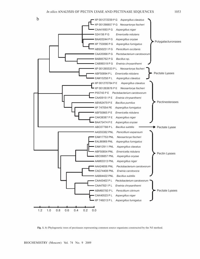
Fig. 1. b) Phylogenetic trees of pectinases representing common source organisms constructed by the NJ method.
1.2 1.0 0.8 0.6 0.4 0.00.2
b XP 001272239 P G Aspergillus clavatus
XP 001266657 P G Neosartorya fischeri
CAA41693 P G Aspergillus niger
S24156 P G Emericella nidulans
BAA03244 P G Aspergillus oryzae
XP 753090 P G Aspergillus fumigatus
ABS50231 P G Penicillium occitanis
CAA35998 P G Pectobacterium carotovorum
BAB85762 P G Bacillus sp.
CAB99319 P G Erwinia chrysanthemi
XP 001265533 P L Neosartorya fischeri
ABF50894 P L Emericella nidulans
EAW15258 P L Aspergillus clavatus
XP 001270784 P E Aspergillus clavatus
P55743 P E Pectobacterium carotovorum
XP 001263676 P E Neosartorya fischeri
CAA59151 P E Erwinia chrysanthemi
ABV62479 P E Bacillus pumilus
XP 747054 PE Aspergillus fumigatus
ABF50865 P E Emericella nidulans
CAK38387 P E Aspergillus niger
BAA75474 P E Aspergillus oryzae
ABO37788 P L Bacillus subtilis
AAS55382 PNL Penicillium expansum
EAW17753 PNL Neosartorya fischeri
EAL86969 PNL Aspergillus fumigatus
EAW12911 PNL Aspergillus clavatus
ABF50854 PNL Emericella nidulans
ABO38857 PNL Aspergillus oryzae
AAW03313 PNL Aspergillus niger
AAA24856 PNL Pectobacterium carotovorum
CAG74408 PNL Erwinia carotovora
AAB84422 PNL Bacillus subtilis
CAA43402 P L Pectobacterium carotovorum
CAA47821 P L Erwinia chrysanthemi
ABM60783 P L Penicillium citrinum
CAK40523 P L Aspergillus niger
XP 749213 P L Aspergillus fumigatus
Pectate Lyases
Pectin Lyases
Pectate Lyase
Pectinesterases
Pectate Lyases
Polygalacturonases
1054 YADAV et al.
BIOCHEMISTRY (Moscow) Vol. 74 No. 9 2009
RESULTS AND DISCUSSION
A total of 48 full-length protein sequences of pectin
lyase enzyme from different source organisms along with
two synthetic sequences were considered for in silico
analysis. The phylogenetic tree constructed revealed two
distinct clusters for bacterial and fungal PNLs, while
multiple accessions of Aspergillus, Pseudomonas,
Penicillium, Erwinia, Bacillus, Emericella, Geobacillus,
Neosartorya accessions formed distinct clusters showing
sequence level similarity (Fig. 1a). Two synthetic
sequences (accession numbers CAA01023 and
CAA01024) were closely related to A. niger PNL (Fig.
1a). The phylogenetic tree constructed by NJ [23],
Minimum Evolution, and UPGMA [24] methods
revealed a more or less similar pattern (see Supplement 1
in the electronic version of the article on the site of this
journal, http://protein.bio.msu.ru/biokhimiya). This
clearly indicates the existence of sequence level similarity
of PNLs produced from common source organisms.
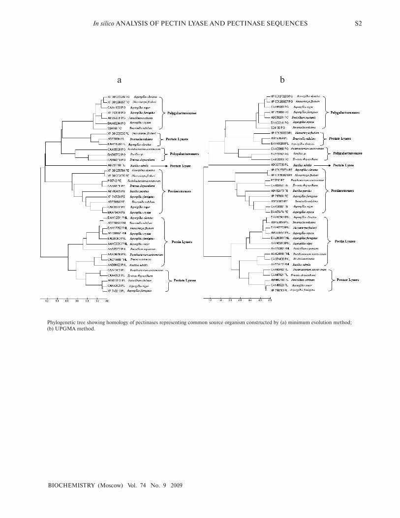
When protein sequence of pectinases produced from
common sources (A. niger, A. fumigatus, A. clavatus, A.
oryzae, Penicillium sp., Erwinia sp., Bacillus sp., E. nidu-
lans, N. fischeri, and P. carotovorum) were subjected to
phylogenetic tree construction using NJ (Fig. 1b),
Minimum Evolution, and UPGMA methods
(Supplement 1), four distinct clusters of PNL, PL, PG,
and PE groups of enzymes were observed.
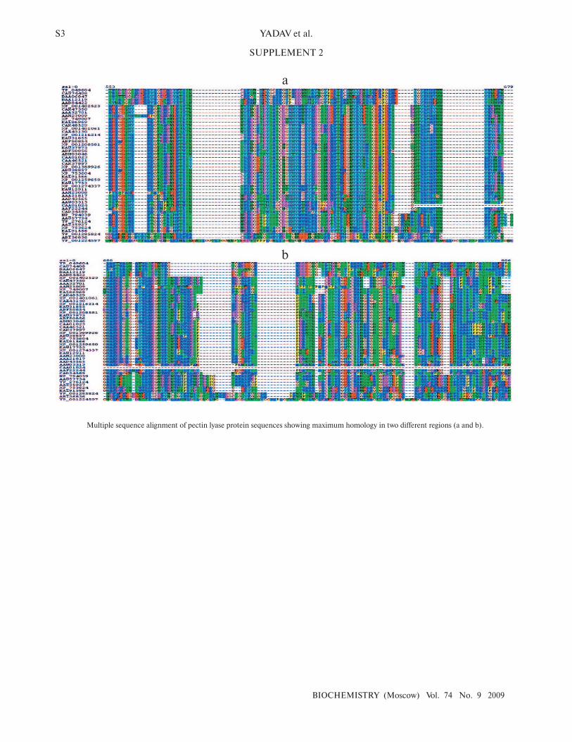
The multiple sequence alignment of 48 protein
sequences of PNL proteins revealed a stretch of con-
served protein sequences from residues 553 to 679 and
from 680 to 806 (see Supplement 2 in the electronic ver-
sion of the article on the site of this journal http://
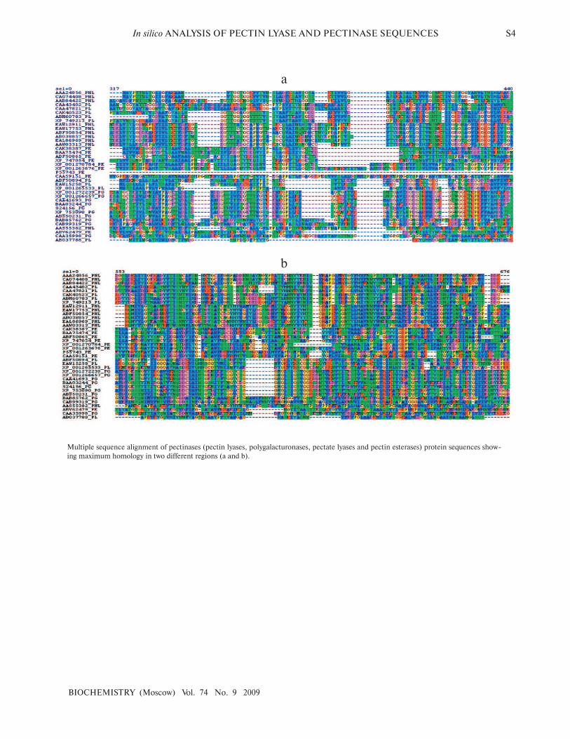
protein.bio.msu.ru/biokhimiya). Similarly, when protein
sequences of pectinases were subjected to multiple
sequence alignment, homology was observed at two dif-
ferent locations — from residue 317 to 440 and from 553
to 676 (Supplement 2).
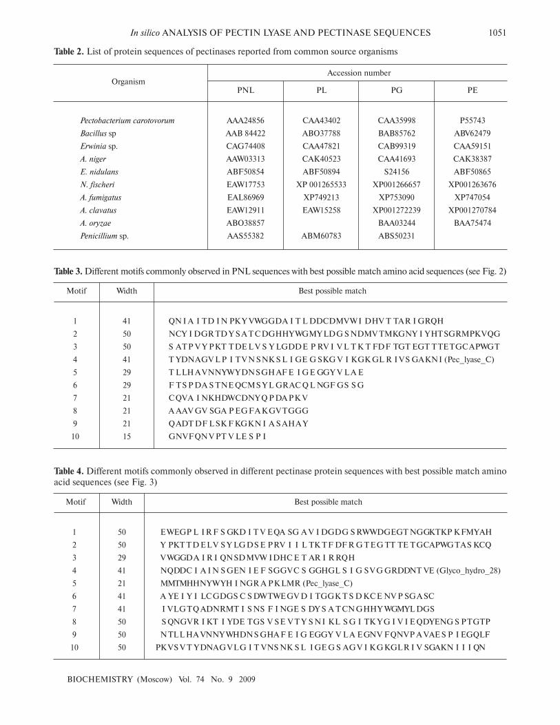
Domain analysis [25, 26] of PNL protein sequences
revealed the presence of Pec_Lyase_C domain consis-
tently, irrespective of sources, except YP_001224597
(Synechococcus sp.), ABI36836 (G. virus), and
YP_001285824 (G. virus). The ten motifs frequently
observed in PNL protein sequences along with the stretch
of amino acids and its width is provided (Fig. 2; see color
insert). A set of 41 amino acid residues, i.e. TYD-
NAGVLPITVNSNKSLIGEGSKGVIKGKGLRIVS-
GAKNI, involved in formation of a right-handed β-helix
structure is associated with the Pec_Lyase_C domain
(Table 3), which is indicative of its important structural
role. The presence and importance of right-handed β-
helix in PNLs and PLs has been proved by many workers
from time to time by solving crystal structures of PNLA
[19], PNLB [20] from A. niger, as well as pectate lyase C
[27], pectate lyase E [28] (both from E. chrysanthemi),
and also PL crystal structure of B. subtilis [29]. This might
explain the stability of the enzyme in hostile extracellular
environment during plant virulence [30]. Although the
mechanism of pectic cleavage differs for the hydrolases
(i.e. PG) and the lyases (i.e. PNL and PL), the substrate
binding sites are found within a cleft formed on the exte-
rior of the parallel β-helix fold in both cases [30].
Similarly, domain analysis of PNL, PL, PE, and PG
protein sequences from common sources revealed the
presence of different conserved sequences. The domain
Pec_Lyase_C was frequently observed with PNL and PL
belonging to the same group of enzymes, i.e. trans-elimi-
nases. In the case of PGs belonging to glycosyl hydrolase
family associated with cell wall metabolism, a domain
Glyco_hydro_28 was frequently observed. The ten most
frequently observed motifs along with signature protein
sequences of maximum matches are shown in Fig. 3 (see
color insert) and Table 4. In case of PEs, frequently
observed domain represents a member of the Pectate
lyase-like β-helix clan. The presence of common and
unique domains among different pectinases might confer
its structural flexibility, which directly influences its cat-
alytic activity.
In silico analysis of PNL protein sequences and its
comparison with other pectinases has revealed the
sequence-based similarity existing among different pecti-
nases and clustering in distinct groups based on its source
organisms and nature of the mechanism of enzymatic
activity. In silico domain analysis confirms the existence
of the different groups of pectinases based on the pres-
ence of unique domains, like PLs and PNLs having a
common domain Pec_Lyase_C belong to a common
group, i.e. trans-eliminases. While PGs having unique
domain Glyco_hydro_28 belong to hydrolases, and PEs
with Pectate lyase-like β-helix clan domain belong to the
esterase group. The presence and absence of specific
domains has direct relation with the structural and func-
tional organization of different groups of pectinases.
The authors wish to acknowledge the support of
DBT funded Sub-DIC Bioinformatics Center,
Department of Molecular Biology and Genetic
Engineering, College of Basic Sciences and Humanities,
G. B. Pant University of Agriculture and Technology,
Pantnagar for providing software and tools utilized in the
present study. P.K.Y. and S.Y. are grateful to the Council
of Scientific and Industrial Research (CSIR) and
Department of Science and Technology (DST),
Government of India, for providing the financial support
in the form of Senior Research Fellowship and Women
Scientists Fellowship, respectively.
REFERENCES
1. Yadav, S., Yadav, P. K., Yadav, D., and Yadav, K. D. S.
(2009) Process Biochem., 44, 1-10.
In silico ANALYSIS OF PECTIN LYASE AND PECTINASE SEQUENCES 1055
BIOCHEMISTRY (Moscow) Vol. 74 No. 9 2009
2. Visser, J., Bussink, H. J., and Witteveen, C. (2004) in Gene
Expression in Recombinant Microorganisms (Smith, A., ed.)
Marcel Dekker, Inc., New York, pp. 241-306.
3. Cosgrove, D. J. (1997) Ann. Rev. Cell. Dev. Biol., 13, 171-201.
4. Prade, R. A., Zohan, D., Ayoubi, P., and Mort, A. J. (1999)
Biotechnol. Gene Eng. Rev., 16, 361-391.
5. Jayani, S. R., Saxena, S., and Gupta, R. (2005) Process
Biochem., 40, 2931-2944.
6. Luh, B. S., and Phaff, H. J. (1951) Arch. Biochem. Biophys.,
33, 212-227.
7. Gysler, C., Harmsen, J. A. M., Kester, H. C. M., Visser, J.,
and Heim, J. (1990) Gene, 89, 101-108.
8. Someren, M. A. K., Harmsen, J. A. M., Kester, H. C. M.,
and Visser, J. (1991) Curr. Gen., 20, 293-299.
9. Someren, M. A. K., Flipphi, M., de Graaff, L., van den
Broeck, H., Kester, H., Hinnen, A., and Visser, J. (1992)
Mol. Gen. Genom., 234, 113-120.
10. Kitamoto, N., Yasuda, Y. S., Ohmiya, K., and Tsukagoshi,
N. (2001) Biosci. Biotechnol. Biochem., 65, 209-212.
11. Templeton, M. D., Sharrock, K. R., Bowen, J. K.,
Crowhurst, R. N., and Rikkerink, E. H. (1994) Gene, 142,
141-146.
12. Kilara, A. (1982) Process Biochem., 23, 35-41.
13. Naidu, G. S. N., and Panda, T. (1998) Bioprocess. Eng., 9,
355-361.
14. Alkorta, I., Garbisu, G., Llama, M. J., and Serra, J. L.
(1998) Process Biochem., 33, 21-28.
15. Blanco, P., Sieiro, C., Reboredo, N. M., and Villa, T. G.
(1997) Arch. Microbiol., 167, 284-288.
16. Takebe, I., Otsuki, Y., and Aoki, S. (1968) Plant Cell
Physiol., 9, 115-124.
17. Beldman, G., Rombouts, F. M., Voragen, A. G. J., and
Pilnik, W. (1984) Enz. Microb. Technol., 6, 503-507.
18. Whitaker, J. R. (1991) in Microbial Enzymes and
Biotechnology (Fogarty, W. M., and Kelly, C. T., eds.)
Elsevier Applied Science, London-New York, pp. 133-
175.
19. Mayans, O., Scott, M., Connerton, I., Gravesen, T.,
Benen, J., Visser, J., Pickersgill, R., and Jenkins, J. (1997)
Structure, 5, 677-689.
20. Vitali, J., Schick, B., Kester, H. C. M., Visser, J., and
Jurnak, F. (1998) Plant Physiol., 116, 69-80.
21. Lassmann, T., and Sonnhammer, E. L. (2006) Nucleic Acids
Res., 34 (Web Server Issue), W596-9.
22. Kumar, S., Tamura, K., and Nei, M. (2004) Brief.
Bioinformatics, 5, 150-163.
23. Saitou, N., and Nei, M. (1987) Mol. Biol. Evol., 4, 406-425.
24. Shi, G. Y., Jie, T. Y., Bing, T. H., Hua, W. K., and Wei, C.
K. (2007) Chin. J. Agric. Biotechnol., 4, 33-38.
25. Timothy, L. B., and Gribskov, M. (1997) J. Comp. Biol., 4,
45-59.
26. Timothy, L. B., and Gribskov, M. (1998) Bioinformatics, 14,
48-54.
27. Yoder, M. D., Keen, N. T., and Jurnak, F. (1993) Science,
260, 1503-1507.
28. Lietzke, S. E., Yoder, M. D., Keen, N. T., and Jurnak, F.
(1994) Plant Physiol., 106, 849-862.
29. Pickersgill, R., Jenkins, J., Harris, G., Nasser, W., and
Robert-Baudouy, J. (1994) Nat. Struct. Biol., 1, 717-723.
30. Herron, S. R., Benen, J. A. E., Scavetta, R. D., Visser, J.,
and Jurnak, F. (2000) Proc. Natl. Acad. Sci. USA, 97, 8762-
8769.
S1 YADAV et al.
BIOCHEMISTRY (Moscow) Vol. 74 No. 9 2009
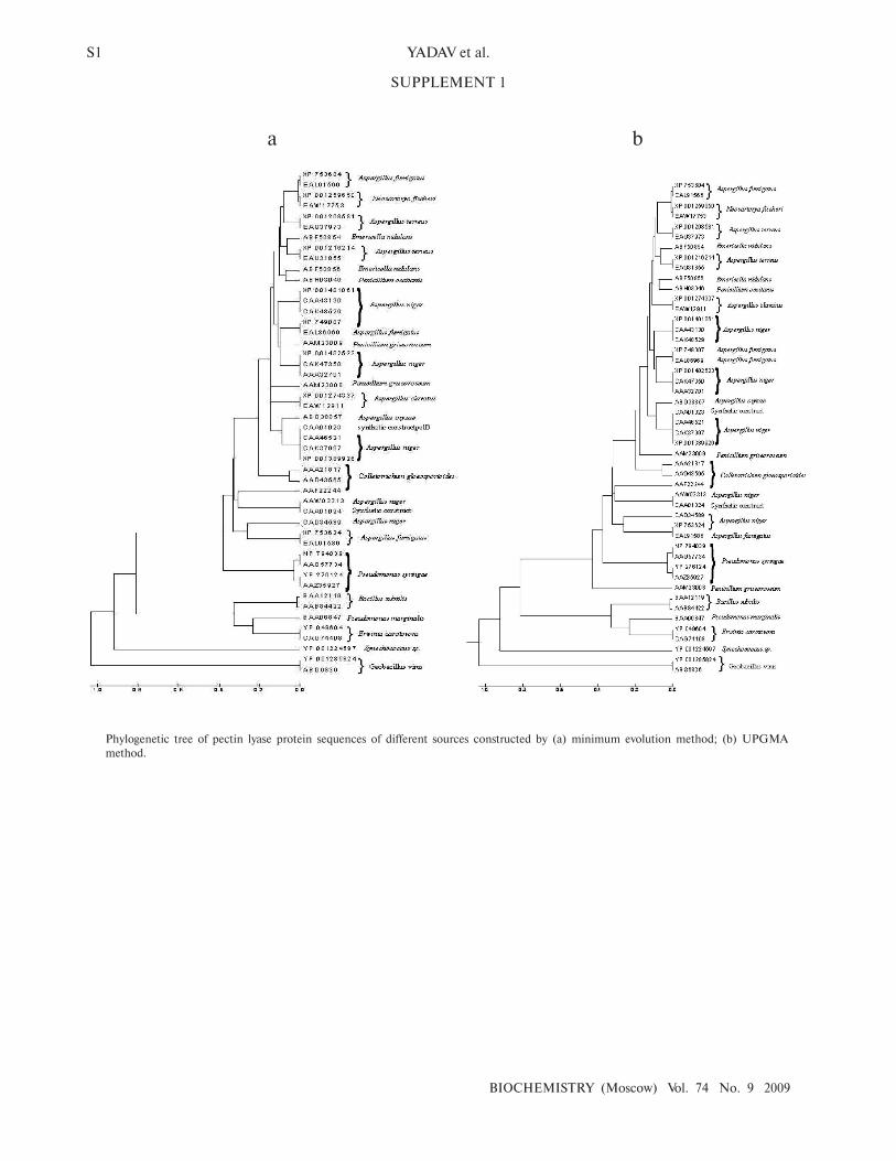
SUPPLEMENT 1
a b
Phylogenetic tree of pectin lyase protein sequences of different sources constructed by (a) minimum evolution method; (b) UPGMA
method.
In silico ANALYSIS OF PECTIN LYASE AND PECTINASE SEQUENCES S2
BIOCHEMISTRY (Moscow) Vol. 74 No. 9 2009
a b
Phylogenetic tree showing homology of pectinases representing common source organism constructed by (a) minimum evolution method;
(b) UPGMA method.
S3 YADAV et al.
BIOCHEMISTRY (Moscow) Vol. 74 No. 9 2009
SUPPLEMENT 2
a
b
Multiple sequence alignment of pectin lyase protein sequences showing maximum homology in two different regions (a and b).
In silico ANALYSIS OF PECTIN LYASE AND PECTINASE SEQUENCES S4
BIOCHEMISTRY (Moscow) Vol. 74 No. 9 2009
a
b
Multiple sequence alignment of pectinases (pectin lyases, polygalacturonases, pectate lyases and pectin esterases) protein sequences show-
ing maximum homology in two different regions (a and b).


![In silico analysis of compounds characterized from ethanolic ...eprints.covenantuniversity.edu.ng/10478/1/20420-74354-2...1 25.637 1.16 Bicyclo[3.1.1]heptane, 2,6,6-trimethyl-138.24992](https://static.fdocument.org/doc/165x107/60f88dd94a7e5669bd2167ee/in-silico-analysis-of-compounds-characterized-from-ethanolic-1-25637-116.jpg)