I. INTRODUCTION - Shodhgangashodhganga.inflibnet.ac.in/bitstream/10603/26651/7/07_chapter 1.pdf ·...
57
I. INTRODUCTION
Transcript of I. INTRODUCTION - Shodhgangashodhganga.inflibnet.ac.in/bitstream/10603/26651/7/07_chapter 1.pdf ·...

I. INTRODUCTION
Introduction
1
Chitin is the most widespread amino polysaccharide in nature and is estimated
annually to be produced almost as much as cellulose. It is a cationic
aminopolysaccharide, composed of β (1-4) linked N-acetyl-D-glucosamine
(NAG) residues. It is present in the exoskeleton of invertebrates e.g.
crustaceans, molluscs, marine diatoms and insects and in algae and fungi
among microorganisms. Derivatives of chitin oligomers have also been
implicated as morphogenic factors in the communication between leguminous
plants and Rhizobium and even in vertebrates, where they may be important
during early stages of embryogenesis (Bakkers et al., 1999). Annual synthesis
of this polysaccharide in fresh water and marine ecosystems is estimated to be
600 and 1600 million tons, respectively (Cauchie, 1997). The best characterized
sources of chitin are shellfish (including shrimp, crab, lobster and krill), oyster
and squid, harvested in quantities of about 29.9, 1.4 and 0.7 million tons per
annum (Synowiecki and Al-Khateeb, 2000). In India alone 60,000 to 80,000
tonnes of chitinous wastes are produced annually, from which a lot of chitin
can be recovered (Suresh and Chandrasekaran, 1998). Chitin is the most
underexploited biomass resource available on Earth. At present only a small
quantity of shell waste is utilized for animal feed or chitin isolation
(Synowiecki and Al-Khateeb, 2003).
Conventionally these wastes are disposed off either by burning or land filling but
these methods are harmful to the environment since burning releases carbon
dioxide and carbon monoxide to the environment, which adds to global warming
while land filling is harmful due to slow rate of degradation and concomitant
release of a potent pollutant of ground water, namely, ammonia (Muzzarelli,
1997; Das et al., 2012). The cost of transporting such waste, environmental
pollution concern and ethical questions as to the morality of ignoring 70-80% of
the dry weight of the catch have highlighted the necessity of finding alternative
method (Simpson and Haard, 1985; Nicol, 1991; Vyas and Deshpande, 1991).
Utilization of such chitinous wastes for the production of some useful products is
being considered lately, and two different approaches are being investigated:
(a) The formation of a useful product such as chitin and chitosan through
biological (Gagne and Simpson, 1993) and chemical treatment (Brine et al.,
Introduction
2
1981), for use in sewage treatment, animal feed, food preservation, and
formulations of biofungicides (Muzzarelli, 1997; Gohel et al., 2005)
(b) Using the waste as a carbon source in fermentation processes for the
production of useful products such as chitinolytic enzymes by microorganisms
(Wang et al. 2001; Gohel et al., 2007).
Recently the commercial value of chitin has increased because of the beneficial
properties of its soluble derivatives, which are suitable in chemistry,
biotechnology, agriculture, food processing, cosmetics, veterinary, medicine,
dentistry, environment protection and paper or textile production (Synowiecki
and Al-Khateeb, 2003; Tharanathan and Kittur, 2003; Das et al., 2012).
Generation of this enormous amount of waste and more importantly the
increasing commercial value of the soluble derivatives of chitin necessitates the
development of a suitable process for solubilization of chitinous waste and its
conversion into useful oligomers. The chemical methods by which these
polymers and their oligomers are produced commercially involve treatment with
harsh chemicals like hydrochloric acid and sodium hydroxide. Besides being
environmentally unsafe, the use of these chemicals leads to products that lack
uniformity. The enzymatic methods, which employ enzymes such as chitinases,
are mild and eco-friendly, and thus preferred over chemical methods.
CHITIN: OCCURRENCE, STRUCTURAL ORGANIZATION, BIOSYNTHESIS, ASSOCIATIONS AND HYDROLYSIS Chitin, a Greek word for ‘envelop’, was discovered in 1811 as a substance
occurring in mushrooms. Chitin is very widely distributed especially in animals,
and it also exists in less evolved taxonomic groups such as protozoa (Ruiz-
Herrera, 1978). In plants, chitinous cell walls are only found in those forms, such
as fungi and moulds that like animals find considerable nitrogen in their food.
Chitin is also believed to constitute the cell wall of some lower green plants such
as chlorophyceae (Tharanathan and Kittur, 2003). Chitinous structures are
mainly of ectodermal origin in multicellular animals and form the characteristic
exoskeleton of most of the invertebrates (Jeuniaux, 1971). Arthropods are
particularly able to synthesize chitin, and concentrations of up to 85% are found
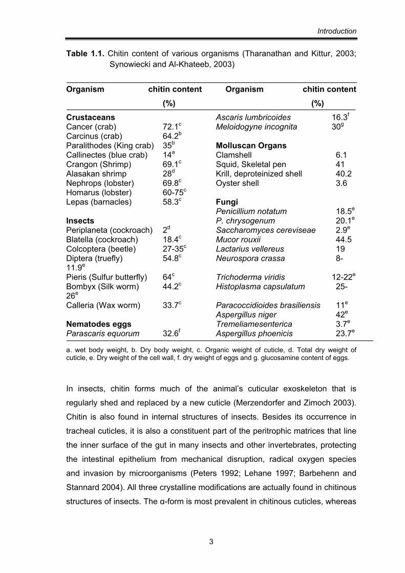
in their shells. The chitin content in various organisms is given in Table 1.1.
Introduction
3
Table 1.1. Chitin content of various organisms (Tharanathan and Kittur, 2003; Synowiecki and Al-Khateeb, 2003)
Organism chitin content Organism chitin content (%) (%)
Crustaceans Ascaris lumbricoides 16.3f Cancer (crab) 72.1c Meloidogyne incognita 30g Carcinus (crab) 64.2b Paralithodes (King crab) 35b Molluscan Organs Callinectes (blue crab) 14a Clamshell 6.1 Crangon (Shrimp) 69.1c Squid, Skeletal pen 41 Alasakan shrimp 28d Krill, deproteinized shell 40.2 Nephrops (lobster) 69.8c Oyster shell 3.6 Homarus (lobster) 60-75c Lepas (barnacles) 58.3c Fungi Penicillium notatum 18.5e Insects P. chrysogenum 20.1e Periplaneta (cockroach) 2d Saccharomyces cereviseae 2.9e Blatella (cockroach) 18.4c Mucor rouxii 44.5 Colcoptera (beetle) 27-35c Lactarius vellereus 19 Diptera (truefly) 54.8c Neurospora crassa 8-11.9e Pieris (Sulfur butterfly) 64c Trichoderma viridis 12-22e Bombyx (Silk worm) 44.2c Histoplasma capsulatum 25-26e Calleria (Wax worm) 33.7c Paracoccidioides brasiliensis 11e
Aspergillus niger 42e
Nematodes eggs Tremeliamesenterica 3.7e Parascaris equorum 32.6f Aspergillus phoenicis 23.7e a. wet body weight, b. Dry body weight, c. Organic weight of cuticle, d. Total dry weight of cuticle, e. Dry weight of the cell wall, f. dry weight of eggs and g. glucosamine content of eggs.
In insects, chitin forms much of the animal’s cuticular exoskeleton that is
regularly shed and replaced by a new cuticle (Merzendorfer and Zimoch 2003).
Chitin is also found in internal structures of insects. Besides its occurrence in
tracheal cuticles, it is also a constituent part of the peritrophic matrices that line
the inner surface of the gut in many insects and other invertebrates, protecting
the intestinal epithelium from mechanical disruption, radical oxygen species
and invasion by microorganisms (Peters 1992; Lehane 1997; Barbehenn and
Stannard 2004). All three crystalline modifications are actually found in chitinous
structures of insects. The α-form is most prevalent in chitinous cuticles, whereas
Introduction
4
the β and γ forms are frequently found in cocoons (Kenchington, 1976; Peters,
1992). Peritrophic matrices usually comprise α- and β -chitins.
Among microorganisms, chitin is widely distributed in fungi, occurring in
Basidiomycetes, Ascomycetes, and Phycomycetes, where it is a component of
the cell walls and structural membranes of mycelia, stalks, and spores. The
amounts vary between traces and up to 45% of the organic fraction, the rest
being mostly proteins, glucans and mannans (Roberts, 1992). Variations in the
amounts of chitin may depend on physiological parameters in natural
environments as well as on the fermentation conditions in biotechnological
processing or in cultures of fungi. Hyphal walls of the Oomycete Pythium
ultimum contain cellulose and chitin, whereas the Ascomycete Fusarium
oxysporum and the Basidiomycete Rhizoctonia solani contain only chitin (Cherif
et al., 1993). The zoopathogenic fungi Cryptococcus neoformans, Pityrosporum
canis and Rhizopus oryzae contain chitin, but not β-(1,3)-glucan (Nicholas et al.,
1994). The mycelia, and the caps and stalks of fruiting bodies of four edible
mushrooms Lentinus edodes, Lycophyllum shimeji, Pleurotussajor-caju, and
Volvariella volvacea contain chitin as a minor component (Cheung, 1996). Slime
moulds (Myxomycetes) and bacteria (Schizomycetes) are devoid of chitin.
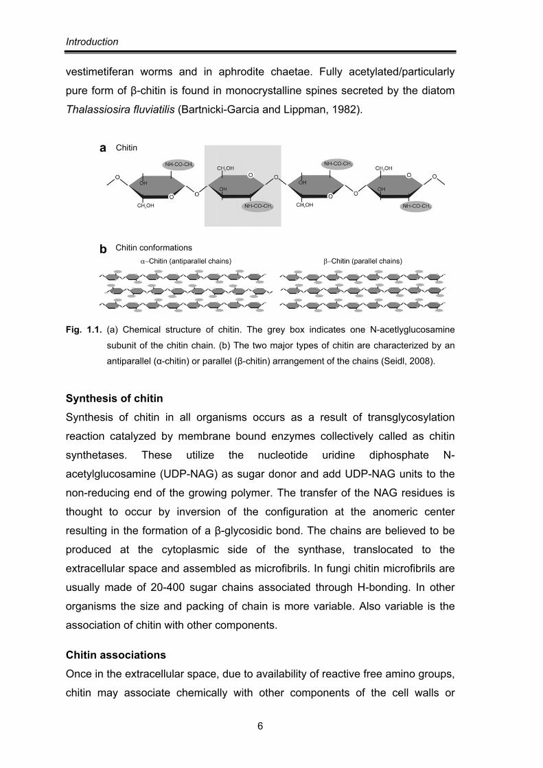
Structural organization of chitin Chitin is a homopolymer consisting of N-acetylglucosamine residues linked by
β-(1-4)-glycosidic bonds. In the linear chitin chain, every single sugar is rotated
by 180° with respect to its neighbouring sugars. Thus, the repeating unit in the
chitin chain is chitobiose. Chitin polymers tend to form microfibrils (also referred
to as rods or crystallites) that are stabilized by hydrogen bonds formed between
the amine and carbonyl groups (Fig. 1.1). X-ray diffraction analysis suggested
that chitin is a polymorphic substance that depends upon the source, and
occurs in two crystalline allomorphs, a more abundant α- and less common
β- form. In both forms chitin chains are arranged in sheets, the chains in any
one sheet have the same direction. They mainly differ in the degree of
hydration, in the size of the unit cell and in the number of chitin chains per unit
cell (Rudall and Kenchington, 1973; Kramer and Koga, 1986). While α-chitin has
no water molecule within its unit cell, the unit cell of β-chitin expands along its
Introduction
5
β parameter when the samples are immersed in water, yielding the β-chitin
dihydrate. β-chitin can also accept other small molecules such as a series of
linear alcohols that intercalates into its lattice to yield crystallosolvates. These
crystallosolvates keep the fibrillar morphology of the parent chitin, and on
drying, they revert normally to the anhydrous structure without any apparent
modification of morphology and crystallinity (Saito et al., 2000).
In α-chitin adjacent sheets have the chains oriented in opposite direction i.e.
they are antiparallel, while in β-chitin, the adjacent sheets are parallel or have
the same direction (Fig. 1.1) [Aranaz et. al., 2009]. In both structures, the sheets
are tightly held by a number of inter sheet H-bonds dominated by CO-NH
hydrogen bonds. In α-chitin there are also some intra-sheet H-bonds involving
the association of hydroxymethyl groups of adjacent chains. A third form, called
gamma-chitin, is considered to be a distorted version of either α- or β- forms,
where every third sheet has the opposite direction (Tharanathan and
Kittur, 2003). In addition, non-crystalline, transient states have also been
reported in fungi (Vermeulen and Wessels, 1986).
The anti-parallel arrangement of chitin molecules in the α form allows tight
packaging into chitin microfibrils, consisting of many single chitin chains that are
stabilized by a high number of hydrogen bonds formed within and
between the molecules. This arrangement may contribute significantly to the
physicochemical properties of the chitinous structure such as mechanical
strength and stability (Giraud-Guille and Bouligand, 1986). By contrast, in the
β- and γ-chains, packing tightness and number of inter-chain hydrogen bonds
are reduced, resulting in an increased number of hydrogen bonds with water.
The high degree of hydration and reduced packaging tightness result in more
flexible and soft chitinous structures as found in insects’ peritrophic matrices or
cocoons (Merzendorfer and Zimoch, 2003).
α-chitin occurs in fungal and yeast cell walls, in krills, in lobsters and crabs
tendons and shells, in shrimp shells and in insect cuticles. The chitin formed
from recrystallization (Saito et al 2000), in vitro synthesis or enzymatic
polymerization is also α-chitin. The commercial source of β-chitin is squid pens.
It also occurs in the spines of some diatoms, in tubes of pogonophoran and
Introduction
6
vestimetiferan worms and in aphrodite chaetae. Fully acetylated/particularly
pure form of β-chitin is found in monocrystalline spines secreted by the diatom
Thalassiosira fluviatilis (Bartnicki-Garcia and Lippman, 1982).
Fig. 1.1. (a) Chemical structure of chitin. The grey box indicates one N-acetlyglucosamine
subunit of the chitin chain. (b) The two major types of chitin are characterized by an
antiparallel (α-chitin) or parallel (β-chitin) arrangement of the chains (Seidl, 2008).
Synthesis of chitin Synthesis of chitin in all organisms occurs as a result of transglycosylation
reaction catalyzed by membrane bound enzymes collectively called as chitin
synthetases. These utilize the nucleotide uridine diphosphate N-
acetylglucosamine (UDP-NAG) as sugar donor and add UDP-NAG units to the
non-reducing end of the growing polymer. The transfer of the NAG residues is
thought to occur by inversion of the configuration at the anomeric center
resulting in the formation of a β-glycosidic bond. The chains are believed to be
produced at the cytoplasmic side of the synthase, translocated to the
extracellular space and assembled as microfibrils. In fungi chitin microfibrils are
usually made of 20-400 sugar chains associated through H-bonding. In other
organisms the size and packing of chain is more variable. Also variable is the
association of chitin with other components.
Chitin associations Once in the extracellular space, due to availability of reactive free amino groups,
chitin may associate chemically with other components of the cell walls or
Introduction
7
exoskeletons, thus acquiring different properties. In invertebrates, protein forms
covalent links with chitin, giving rise to pliable and flexible structures with higher
strength. Whether in its α-or β-form, chitin is covalently linked to arthropodins,
resilins, and sclerotins to form more or less stable glycoproteins through
aspartyl and histidyl residues (Tharanathan and Kittur, 2003). The resulting
chitin-protein complexes gain stability, providing hardness and rigidity and can
associate with additional substances such as lipoproteins and waxes which
provide impermeability properties to exoskeletons. Chitin protein aggregates
provides a substrate for calcium and silica deposition. This mineralization
process seems to be common in crustaceans and molluscs, where it provides
rigidity and ensures stability to the exoskeletons. Chitin also forms conjugates
with carotenoids, giving color to the tissues in insects and crustaceans
(Fox, 1973).
In fungi, chitin associates with glycoproteins and polysaccharides such as
galactomannan and glucans (Muzzarelli et al., 1980). In the macromolecular
network of the cell wall, glucan chains are linked to chitin through their reducing
ends via amino acids, particularly lysine (Sietsma and Wessels, 1981).
Apart from the linkages of the two polymers to each other, both chitin
and β-glucan chains are also hydrogen bonded among themselves
(Tharanathan and Kittur, 2003).
Chitin hydrolysis As an important source of carbon and nitrogen, chitin is recycled by many
saprophytic microorganisms including bacteria and fungi. Many saprophytic
chitinolytic microbes produce a whole chitinolytic system comprising an
endochitinase, chitobiase and an exochitinase whose synergistic or consecutive
actions degrade chitin to free sugar NAG (N-acetylglucosamine).
The possibility of exploiting these organisms to economically generate
commercially useful products has been explored. Wang et al. (2001) reported
microbial reclamation of shellfish wastes for the production of chitinases where
they prepared shrimp and crab shell powder by treating shellfish processing
waste with boiling and crushing and it was used as a substrate for chitinolytic
microorganisms. Rattanakit et al. (2002) reported a chitinase formulation by
Introduction
8
using shrimp shellfish waste as a substrate for solid state cultivation of
Aspergillus sp. SI-13. Besides enzymes, examples of SCP, ethanol and
biodiesel production from chitinous waste have been cited in literature (Ferrer et
al., 1996; Cody et al., 1990; Zang et al., 2011). An economically viable
biotechnological process for such direct utilization of chitinous residues is,
however, still awaited.
Alternatively, chitin can also be extracted from shellfish waste and digested to
yield oligomers and monomers that find growing market. Chitin is inert to most
commercially available solvents but is fairly stable under mild acidic and basic
conditions and thus obtained as the residue remaining after decomposition of
the other components with acid and alkali (Bade, 1997; No and Meyers, 1997).
Presently, shells are first treated with dilute hydrochloric acid at room
temperature to remove metal salts, primarily calcium carbonate. The decalcified
shells are ground and heated at about 100°C in 1-2 M sodium hydroxide to
decompose proteins and pigments. Sometimes repetition of the treatments may
be required. α-chitin is obtained on drying as almost colorless to off-white flakes
or powdery materials in 30–35% yield based on dried shrimp shells (Sannan et
al., 1976). Starting from crab shells, the yield is usually lower than that from
shrimp shells because of the higher content of calcium carbonate. β-chitin from
squid pens are associated with proteins and only a small amount of calcium
carbonate and thus can be isolated under similar, but milder conditions
(Kurita et al., 2006).
Thus extracted chitin is used almost solely as a raw material for the production
of glucosamine along with oligosaccharides and chitosan (Kurita et al., 2006).
The generation of these soluble and industrially important products require more
rigorous acid and base treatment (Einbu and Varun et al., 2008). These
chemical methods are associated with the problems of low yield, high cost of
purification and environmental pollution (Sakai, 1995; Sukwattanasinitt et al.,
2002; Wang et al., 2010). Alternatively, with its advantages in environmental
compatibility, low cost and reproducibility, enzymatic hydrolysis has attracted
attention of scientists in recent years (Yang et al., 2000; Kadokura et al., 2007).
Especially for the production of chitooligosaccharides, enzymatic hydrolysis has
Introduction
9
distinct advantage over acid hydrolysis because the composition of
oligosaccharides can be adjusted readily by changing either the amount or type
of chitinolytic enzymes or controlling reaction conditions (Lee et al., 1996)
CHITIN HYDROLYZING ENZYMES
Classification Chitinases catalyze the hydrolysis of chitin to its oligosaccharides and can be
classified into two major categories (Graham and Sticklen, 1994).
Endochitinases (EC 3.2.1.14) cleave chitin randomly at internal sites, generating
soluble, low molecular mass multimers of NAG such as chitotetraose, chitotriose
and diacetylchitobiose. While exochitinases are further divided into
two subcategories: chitobiosidases and β-(1,4) N-acetyl glucosaminidases.
Chitobiosidases (EC 3.2.1.29) catalyze the progressive release of
diacetylchitobiose starting at the non-reducing end of chitin microfibril. They
form diacetylchitobioses and no monocaccharide or oligosaccharides are
formed. β-(1,4) N-acetyl glucosaminidases (GlcNAcase, EC 3.2.1.30) or
chitobiases catalyze the release of terminal, non-reducing N-acetylglucosamine
residues in an exo-type fashion from chitin and oligomers of chitin, but in
general they have the highest affinity for the dimer N,N’-diacetylchitobiose
(GlcNAc)2 and convert it into two monomers (Horsch et al. 1997). As the
enzyme has broad substrate specificity, it can also be called β-(1,4) N-acetyl
hexosaminidase (HexNAcase, EC 3.2.1.52) [Cannon et al 1994].
However, the enzymatic properties of chitinases are more complex and versatile
than reflected in the exo-/endo classification. Detailed studies of the chitinolytic
system of the bacterium Serratia marcescens demonstrated another way to
classify the enzymatic properties of chitinases by grouping them into processive
and non-processive enzymes (Horn et al. 2006; Sorbotten et al. 2005;
Uchiyama et al. 2001). Processive chitinases do not release the substrate after
hydrolytic cleavage but slide it through the active site-tunnel for the next
cleavage step to occur. The presence of a carbohydrate binding domain can
enhance processivity, but is not essential for it.
Introduction
10
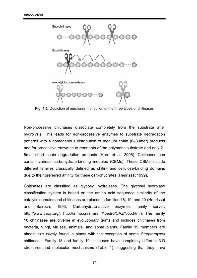
Fig. 1.2. Depiction of mechanism of action of the three types of chitinases
Non-processive chitinases dissociate completely from the substrate after
hydrolysis. This leads for non-processive enzymes to substrate degradation
patterns with a homogenous distribution of medium chain (6–30mer) products
and for processive enzymes to remnants of the polymeric substrate and only 2–
8mer short chain degradation products (Horn et al. 2006). Chitinases can
contain various carbohydrate-binding modules (CBMs). These CBMs include
different families classically defined as chitin- and cellulose-binding domains
due to their preferred affinity for these carbohydrates (Henrissat 1999).
Chitinases are classified as glycosyl hydrolases. The glycosyl hydrolase
classification system is based on the amino acid sequence similarity of the
catalytic domains and chitinases are placed in families 18, 19, and 20 (Henrissat
and Bairoch, 1993; Carbohydrate-active enzymes family server,
http://www.cazy.org/; http://afmb.cnrs.mrs.fr/*pedro/CAZY/db.html). The family
18 chitinases are diverse in evolutionary terms and includes chitinases from
bacteria, fungi, viruses, animals, and some plants. Family 19 members are
almost exclusively found in plants with the exception of some Streptomyces
chitinases. Family 18 and family 19 chitinases have completely different 3-D
structures and molecular mechanisms (Table 1), suggesting that they have
Introduction
11
arisen from different ancestors (Hamel et al 1997; Suzuki et al 1999). The family
20 includes the β-N-acetylhexosaminidases from bacteria, fungi, and mammals.
As chitin in most of the organisms occur as a heteropolymer of acetylated and
deacetylated glucosamine residues, thus chitin deacetylases and
chitosanases too form a part of the repertoire of chitin hydrolyzing enzymes.
Chitin/chitooligosaccharide deacetylases cleaves off the acetyl group from NAG
residues in chitin/ chitooligosaccharide and chitosanase hydrolyze the β-
glycosidic bond between deacetylated chitin residues. Chitin deacetylases are
included in the carbohydrate esterase family 4 of glycoside hydrolases (Caufrier
et al., 2003), while chitosanases have been classified into family 5, 8, 46, 75
and 80 of glycoside hydrolases (Cheng et al., 2006).
Table 1.2. Differences between family 18 and 19 of chitinases.
Family 18 Family 19
Occurrence Bacteria, fungi, viruses and animals
Plants and Streptomyces griseus chitinase C
Mode of action Retention of the anomeric configuration Inversion
Catalytic mechanism Substrate assisted catalysis Acid-base catalysis
Sensitivity to allosamidin Sensitive Insensitive
3D Structure (β/α)8 barrel fold Bilobal structure with high α-helical content
Enzyme assays Techniques and methods have been developed for the estimation of randomly
hydrolyzing endo-chitinases and exo-hydrolytic N-acetylglucosaminidase
activities employing various soluble and insoluble substrates. Chitinase activity
is often determined by measuring the amount of reducing sugars liberated from
colloidal chitin by enzyme activity (Reissig et al., 1955). Endochitinase activity is
also measured by the reduction of turbidity of a suspension of colloidal chitin
(Tronsmo and Harman, 1993).
Chromogenic substrates such as dye-labelled chitin and p-nitrophenyl (pNP)
labelled substrates are also used to measure chitinase activity by estimating
released dye/pNP spectrophotometrically. A carboxymethyl-substituted soluble
chitin covalently linked with Remazol Brilliant Violet 5R is suitable for the
Introduction
12
screening of chitinolytic microorganisms and for detection of chitinase activity by
plate-clearing assay (Wirth and Wolf, 1990). The method is based on the
precipitability of the non-hydrolyzed chitin by HCl. The absorbance in the
supernatant (containing lower oligomers) is used to measure the enzyme
activity. The use of p-nitrophenyl- labelled substrates allow for detection of three
different chitinase types by acting as dimeric, trimeric, and tetrameric
substrates, respectively. Glucosaminidase, chitobiosidase and endochitinase
activities are determined by measuring the release of p-nitrophenyl from pNP-
GlcNAc, pNP-(GlcNAc)2 and pNP-(Glc- NAc)3, respectively (Harman et al.,
1993; Robert and Selitrennikoff, 1988; Patil et al., 2000).
Trudel and Asselin (1989) developed a technique for the detection of chitinase
activity after native or denaturing polyacrylamide gel electrophoresis (PAGE) by
incorporating glycol chitin into the gel. As glycol chitin exhibits high affinity
towards Calcofluor white M2R, the lysis zones can be visualized by UV
illumination as non-fluorescent dark bands in contrast to the fluorescent intact
glycol chitin.
In addition, enzyme activity can also be detected on gels by using fluorescent
substrates (Tronsmo and Harman, 1993). The chitinases appear as fluorescent
bands under UV light because of enzymatic hydrolysis of fluorescent
4-methylumbelliferone from the GlcNAc mono- and oligosaccharides, such as
4-MU-GlcNAc, 4-MU-(GlcNAc)2 and 4-MU- (GlcNAc)3. The dimer is the
preferred substrate for glucosaminidases. Chitobiosidases release fluorescent
product from only the trimeric substrate and endochitinases are identified by
digestion of the tetrameric substrate (Haran et al., 1995).
Sources of chitinases/Chitinolytic organisms Chitinases have broad spectrum of distribution in nature including bacteria,
fungi, nematodes, plants, insects, fish and human (Jeauniaux, 1966). The
physiological functions of chitinases depend on their source.
Introduction
13
Fungi Chitinases and glucanases are important class of hydrolytic enzymes that are
essential for the fungal kingdom and are required for the maintenance of wall
plasticity, during cell expansion and division in yeasts, and during spore
germination, hyphal branching and septum formation in filamentous fungi. They
may contribute to breakage and re-forming of bonds within and between
polymers, leading to re-modelling of the cell wall during growth and
morphogenesis. For example, during budding in S. cerevisiae a division septum
is laid down between the cells and within a ring of chitin deposited at the bud
site. Degradation of this material leads to cell separation and involves an
extensively glycosylated endochitinase with an apparent molecular mass of
approximately 130 kDa. Disruption of the gene encoding this enzyme or a
potent chitinase inhibitor led to cell separation defect and pseudohyphal growth
(Kuranda & Robbins, 1991; Sakuda et al., 1990; King & Butler, 1998).
When the dimorphic pathogen of humans, Candida albicans was grown in a
yeast phase, the transcrips of chitinase genes, CHT2 and CHT3 were greater as
compared to a mycelial phase and gene disruption experiments suggested a
role for the gene product in cell separation (Kuranda & Robbins, 1991;
McCreath et al., 1995).
Plasmids of some strains of yeast encode toxins that kill other strains. Killer
toxins of some strains of Kluyveromyces lactis and Pichia acaciae have
chitinase activity. In K. lactis the toxin is a trimeric protein, with the
intracellular gamma subunit responsible for killing a susceptible cell of
Saccharomyces cerevisae, while the alpha subunit has exochitinase activity
essential for the action of toxin, shown by the inhibition of activity by allosamidin
(Magliani et al., 1997).
Fungal cell wall chitinases also have roles during sporulation in filamentous
fungi, as the specific chitinase inhibitors allosamidin or demethylallosamidin
inhibited fragmentation of hyphae into arthroconidia (Yamanaka et al., 1994;
Sandor et al., 1998). Disruption of the gene encoding the A. nidulans chitinase,
ChiA, led to a decrease in the frequency of spore germination and a lower
hyphal growth rate (Takaya et al., 1998). However in A. fumigatus, disruption of
Introduction
14
gene encoding chitinase, ChiB1p had no effect on growth and morphogenesis
and may contribute to the digestion and utilization of exogenous chitin (Reichard
et al., 2000/Jaques et al., 2003). A single organism may produce several
chitinases for various purposes. There are 15 potential chitinase open reading
frames (ORFs) in the genome of A. nidulans. Among these, the class
V endochitinase ChiB was shown to play an important role in autolysis.
Levels of ChiB significantly increased when the fungal cells were starved for
carbon sources, an induced condition for hyphal autolysis of A. nidulans
(Yamazaki et al., 2007).
Chitinases play an important role in the mycoparasitic progress, especially in the
cell wall penetration and nutrient utilization by degrading the cell walls in fungi
(Inbar and Chet, 1995; Elad et al., 1983; Chet et al., 1993). It has been reported
that disruption of ech42 gene affects mycoparasitism in T. harzianum (Woo et
al., 1998). Chitinases from other parasitic fungi have also been reported, such
as Aphanocladium album (Kunz et al., 1992), Gliocladium virens (Di Pietro et
al., 1993), Fusarium chlamydosporum (Mathivanan et al., 1998), Trichothecium
roseum (Li et al., 2004), Stachybotry elegans (Taylor et al., 2002), Talaromyces
flavus (Li et al., 2005), Beauveria bassiana (Bidochka et al., 1993; Peng et al.,
1996; Fang et al., 2005), Metarhizium anisopliae (Pinto et al., 1997; St. Leger et
al., 1996; Kang et al., 1999) Verticillium chlamydosporium and V. suchlasporium
(Tikhonov et al., 2002). Similarly, chitinases are involved in the association of
plant roots with mycorrhizal or endophytic fungi. Examples include
Hebeloma syrjense (Tibbett and Sanders, 2002), Neotyphodium and Epichloe
(Li et al., 2004).
Bacterial Sources Many prokaryotes efficiently digest the crystalline and complexed chitin and
utilize it as a carbon source, thereby playing an important role in recycling this
compound. The major producers of chitin degrading enzymes are in the
genera Aeromonas (Chen et al., 1991; Lin et al., 1997; Kojima et al., 2005; Lan
et al., 2006), Serratia (Xia et al., 2011), Enterobacter (Dahiya et al., 2005;
Chernin et al., 1995; Velusamy and Kim, 2011), Vibrio (Svitil et al., 1997;
Li and Roseman, 2003; Sugintal et al., 2010), Streptomyces (Ohno et al., 1996;
Introduction
15
Kim et al., 2003; Tsujibo et al., 1993), and Bacillus (Battacharya et al.,
2007;Wang et al., 2001; Wang et al., 2002; Cho et al., 2011).
Prokaryotes are generally the only chitin degraders/responsible for chitin
recycling in extreme environments. One such environment is the rumen of
herbivores where Clostridium sp. is involved in the anaerobic degradation of
chitin (Simunek et al., 2004; Tishchenko et al., 2010). Clostridium thermocellum
is a thermophilic bacterium that degrades/hydrolyze chitin in hot springs and
self-heated, rotting biomass (Zverlov et al., 2002). Other aerobic thermophilic
bacteria that produce chitinase are Thermococcus kodakaraensis KOD1
(Imanaka et al., 2001), Pyrococcus furiosus (Oku and Ishikawa, 2006) and
Thermococcus chitonophagus (Andronopoulou et al., 2004).
The chitinolytic systems of bacteria involve multiple isomeric forms of chitinases
that include endochitinases, exochitinases and N-acetylglucosaminidases.
Chitinolytic system of Bacillus circulans WL-12 is one of the most extensively
studied systems so far. Six chitinases with differing enzyme activities have been
detected in the culture supernatant of this bacterium grown in the presence of
chitin (Watanabe et al., 1990; Watanabe et al., 1992). The chitinolytic machinery
of Serratia marcescens comprises three chitinases ChiA, ChiB and ChiC which
preferentially produce dimers, a chitin binding protein CBP21 (Kolstad et al.,
2005) and a hexosaminidase which further degrades the major end product of
chitinases i.e. chitobiose. ChiA and ChiB are processive exochitinases which
degrade chitin chains in opposite directions, while ChiC is a nonprocessive
endochitinase. Three separate chitinase genes have been identified in
Streptomyces lividans (Miyashita et al., 1991). Chitinases with diverse
enzymatic properties may be synthesized from separate genes as in
Streptomyces lividans, or result from proteolytic processingas in an
Alteromonas sp. (Tsujibo et al., 1993) and Streptomyces olivaceoviridis
(Romaguera et al., 1992).
Animals Chitinases or chitinase-like proteins have been found in all insect species
studied belonging to different orders including dipterans, lepidopterans,
coleopterans, hemipterans and hymenopterans (Koga et al., 1997; Souza-Neto
Introduction
16
et al., 2003). Insect chitinases are almost exclusively endochitinases and have
little or no exochitinase activity as shown by their inability to hydrolyze
methylumbelliferyl-N-acetylglucosamine or p-nitrophenyl-N-acetylglucosamine
or chitobiose. They often act in concert with N-acetylglucosaminidase. The
combined action of endochitinase and N-acetylglucosaminidase is synergistic
and leads to rapid depolymerization of chitin in insects (Fukamizo and Kramer,
1985). Only baculoviral chitinases, which may have a bacterial origin, have been
reported to have both endo- and exo-chitinolytic activity (Hawtin et al., 1997). In
some cases, additional proteins with chitin-binding domains (CBD) but devoid of
catalytic activity help in the degradation of chitin (Vaaje-Kolstad et al., 2005).
The main function of insect chitinases is in the turnover of chitin-containing
extracellular matrices such as the insect cuticle and the peritrophic matrix (PM)
during molting. Insects periodically shed their old exoskeletons and either
continuously or periodically shed their peritrophic membranes and resynthesize
new ones (Lehane et al., 1997). This process is mediated by the elaboration of
chitinases in the moulting fluid that accumulates in the space between the old
cuticle and the epidermis and in gut tissue. The NAG-containing products of
hydrolysis are ultimately recycled for the synthesis of a new cuticle (Kramer and
Muthukrishanan, 1997). In addition, chitinases may have a digestive function in
insects, if their diet contains chitin. Chitinase-like proteins that lack enzymatic
activity may have roles in immunity or as growth factors (Arakrane and
Muthukrishanan, 2010).
Chitinases have been generally considered to lack in mammalian bodies due to
the absence of chitin. However, recent studies have identified chitinases and
chitinase-like proteins (CLPs) belonging to the glycohydrolase family 18 in mice
and human. Chitotriosidase from human macrophages (Renkema et al ., 1995)
and acidic mammalian chitinase AMCase the mouse stomach (Boot et al., 2001)
possess chitinase enzymatic activity, whereas other mammalian chitinases,
including CLPs, do not possess this activity as a result of mutations in their
highly conserved putative active sites (Chang et al., 2001). Mammalian
chitinases with enzymatic activity have a chitin-binding domain that contains six
cysteine residues responsible for their binding to chitin (Tjoelker et al, 2000).
Introduction
17
These mammalian chitinases possess a conserved sequence motif
(DXXDXDXE) on strand β4, and catalytic activity in these chitinases is mediated
by the glutamic acid (E), which protonates the glycosidic bond with chitin (Aalten
et al., 2001). CLPs (also termed chitolectins) do not contain typical chitin-binding
domains, but still can bind to chitin with high affinity.
Mammalian chitinases and chitinase like proteins are induced at sites of
inflammation (such as parasitic infections) (Chang et al., 2001) and remodelling
(Ostergaard et al., 2002). This raises the possibility that these molecules play
active roles in human anti-parasite and anti-infective defense and repair
responses.
Plants Chitinases have been reported from many monocotyledonous and
dicotyledonous plant species and occur in widely different tissues, including
embryos, seeds, cotyledons, stems, leaves, roots and flowers. Many plant
chitinases are expressed constitutively, generally at a low level (Punja and
Zhang, 1993). No substrate for this enzyme is identified in plants, whereas chitin
is commonly a component of fungal cell walls and insect exoskeletons,
organisms which include many important pathogens and pests. Besides, the
dramatic increase in chitinase levels by various biotic (fungi, bacteria, viruses
and viroids) and abiotic factors (ethylene, salicylic acid, salt solutions, ozone
and UV light) suggest their role in plant defence response (Collinge et al.,
1993). Furthermore, chitinase has been shown to accumulate around fungal
hyphal material in Planta (Benhamou et al., 1990; Wubben etal., 1992) and it
has now been demonstrated that enhanced chitinase levels in transgenic plants
can indeed reduce the damage caused by pathogens (Broglie etal., 1991).
Chitinase expression is also under developmental control in certain organs and
tissues and seems to have an important function in early embryo development,
indicating additional non-defensive roles (de Jong et al., 1992; Lotan et al.,
1989; Neale et al., 1990). Chitinase can inactivate the lipo-oligosaccharide
signal molecules produced by certain Rhizobium strains (Roche et al., 1991)
which are responsible for the induction of root hair deformations, cortical cell
divisions and nodule development in the roots of legume hosts (Truchet et al.,
Introduction
18
1991). Similar relations have been observed during symbiosis formation
between plants and mycorrhizal fungi (Xie et al., 1999). When the fungus which
infects the roots is compatible, its chitooligosaccharides are hydrolysed by plant
chitinases, and the plant defence reaction is reduced. However, if the fungus is
not symbiotic, then the chitinases present in the roots do not cleave the fungal
elicitors, which subsequently bind to plasmalemma receptors and trigger a
hypersensitive reaction (Salzer et al., 1997). Many purified plant endochitinases
also show some degree of lysozyme (EC 3.2.1.17) activity, i.e. they can
hydrolyse β-1,4-linkages between N-acetylmuramic acid and GlcNAc residues
in bacterial cell wall peptidoglucan (Boller, 1988; Majeau et al., 1990; Roberts
and Selitrennikoff, 1988).
Some apoplastic chitinases from monocotyledonous plants also have antifreeze
activity (Kasprzewska, 2003). Recently a chitinase with antifreeze activity is also
purified from the corolla of a dicot, wintersweet (Zhang et al., 2010). It is also
suggested that chitinase take part in programmed cell death (PCD) (Hangel et
al., 1998, Passarinho et al., 2001). This conclusion is supported by the
observation that the chitinase (EP3) gene is activated earlier in Daucus carota
cells which are in an apoptosis preceding stage (Hangel et al., 1998). It seems
that in Arabidopsis thaliana, class IV chitinase, similarly to Daucus carota EP3,
is involved in regulation of processes leading to PCD (Passarinho et al., 2001).
Thermophilic microbes Thermophilic organisms are particularly important in biotechnology as a source
of thermostable enzymes. Though thermostable enzymes have been reported
from mesophiles, the productivity is high in thermophiles and the expectancy of
finding more thermostable and more chemoresistant enzyme is higher in
thermophiles than in mesophiles (Maheshwari et al., 2000; Vieille and Zeikus,
2001). Exploration of chitinases from thermophilic bacteria and fungi is also
interesting from the point of view of understanding the mechanism of
thermotolerance of such enzymes.
The thermophilic organisms Bacillus licheniformis X-7u (Takayanagi et al.,
1991), Bacillus sp. BG-11 (Bharat and Hoondal, 1998) and Streptomyces
thermoviolaceus OPC-520 were reported to be the major sources of chitinases
Introduction
19
(Tsujibo et al., 1995; Tsujibo et al., 2000). A thermophilic Bacillus strain isolated
from chitin-containing compost produces three different endochitinases in its
culture fluid showing temperature optima of 75, 65 and 75 °C (Kenji et al.,
1994). Thermostable exochitinases were also isolated from Bacillus
stearothermophilus CH-4, isolated from a compost of organic solid wastes
Kenji et al., 1998).
Clostridium thermocellum, an anaerobic, saccharolytic, thermophilic bacterium
that occurs in hot springs and self-heated, rotting biomass, produces an
endochitinase as part of its cellulosome (Zverlov et al., 2002). Thermostable
chitinase are also reported from the extreme thermophilic anaerobic archaeon
Thermococcus chitinophagus (Huber et al., 1995; Andronopoulou and Vorgias,
2003), Thermococcus kodakaraensis (Tanaka et al., 2003) and Pyrococcus
furiosus (Gao et al., 2003).
Thermophily in fungi is not as extreme as in eubacteria or archaea, however,
0.06% of recorded fungal species are able to breach the upper temperature limit
of eukaryotes and can thrive at temperatures between 45 and 55°C (Cooney
and Emerson, 1964; Maheshwari et al., 2000). A thermophilic fungus is one that
grows optimally at or above 40°C (Crisan, 1973). Thermophilic fungi have been
reported from decomposing organic materials like wood chip, animal dung, plant
straw, municipal refuge etc. in which the activity of mesophiles results in
thermogenic conditions or man-made habitats such as cooling towers,
effluent of nuclear power reactors and ducts employed for thermal insulation
(Johri et al., 1999).
Chitinolytic enzymes have been described from a few thermophilic fungi; Mucor
miehei, Talaromyces emersonii, T. leycettanus and Thermomyces lanuginosus
(Jensen and Olsen, 1999). Talaromyces emersonii produces an inducible
chitinolytic system consisting of a chitinase and an N-acetylglusamidase
with optimal activities in the range of 65 to 75°C (Hendy et al., 1990;
McCormack et al., 1991). A 48 kD chitinase is reported from Thermomyces
lanuginosus and is stable for 20 min at 70°C and for 25 min at 65°C (Guo et al.,
2005). Recently, a chitinase encoding 1326 bp cDNA was sequenced and its
alignment with other family 18 chitinases showed low overall homology except
Introduction
20
for highly conserved regions among microbial chitinases (Guo et al., 2006;
Guo et al., 2008). Likewise the catalytic domains of two chitinases from
Thermoascus aurantiacus var. levisporus and Chaetomium thermophilum are
similar with other family 18 glycosyl hydrolases. Both these enzymes have high
thermostability (Li et al., 2010). Chitin deacetylase has also been reported in
the cell extracts of the thermophile Mucor miehei (Kauss et al., 1983).
PRODUCTION Microbial chitinase has been produced by liquid batch fermentation, continuous
fermentation, and fed-batch fermentation. In addition to these, solid-state
fermentation and biphasic cell systems have also been used for the production
of chitinase. Generally, chitinase produced from microorganisms is inducible in
nature.
Submerged fermentation Extracellular chitinase production is reported to be influenced by media
components such as carbon sources, nitrogen sources, and agricultural
residues such as rice bran, wheat bran, etc. (Bhushan 1998; Dahiya et al.
2005b). Several other physical factors such as aeration, pH, and incubation
temperature also affect chitinase production. Chitinase production levels from
different organisms and process variables are listed in table 1.3.
Table 1.3. Chitinase production in submerged fermentation
Organism Production levels
Medium pH Temp. Ferment-ation time
Reference
Trichoderma harzianum TUBF 966
14.7 U/ml 1.5% colloidal chitin, 0.42% peptone
5.5 30ºC 96h Sandhya et al. 2004
Verticillium lecanii F091
19.9 mU/ml 4.52% (w/v) maltose, 1.79% marine peptone extract, 0.41% shrimp powder and 0.3% soy protein
4 24ºC 144h Liu et al. 2003

Introduction
21
Bacillus sp. BG-11 76 U/ml 1% swollen chitin and 0.5% glucose
8.5 50ºC 72h Bhushan 2000
Alcaligens xylosoxydans
29 U/ml 1.5% chitin, 0.03% yeast extract and 0.012% tween-20
8.5 30ºC 72h Vaidya et al. 2003
Pantoea dispersa 452.56 U/ml
0.5% chitin, 0.05% peptone, 0.005% yeast extract and 0.05% urea
7.2 30 144h Gohel et al., 2006
Colletotrichum gloeosporioides
2.4 U 0.5% colloidal chitin
7 28 5d Souza et al., 2005
Talaromyces emersonii
0.45 µM/h/ml
1-2% Chitin, mineral salt medium
5 45 240h McComark et al., 1991
Physical factors affecting chitinase production
Temperature Temperature is an important factor that influences growth and metabolism of all
microorganisms. Mesophilic as well as thermophilic microorganisms have
been reported to produce chitinases. The optimum temperature for chitinase
production from most mesophiles lies in the range between 24-37°C. For
example, Vibrio alginolyticus produces chitinase optimally at 37°C (Ohishi et al.,
1996), Bacillus pabuli K1 (Frandberg and Schnurer, 1994) and Serratia
marcescens at 30°C (Khoury et al., 1997), Streptomyces lividans at 25-30°C,
Stachybotrys elegans at 24°C (Tweddell et al., 1994), Myrothecium verucaria
(Vyas and Deshpande, 1989), Tricoderma harzianum (Kapat et al., 1996) and
Fusarium chlamydosporum (Mathivanan et al., 1997) at 28°C.
Thermophilic microorganisms produce chitinases at relatively higher
temperatures. Bacillus licheniformis secretes four chitinases (I-IV) when grown
at 50°C (Takayanagi et al., 1991). Thermococcus chitinophagus produces an
extracellular, a membrane associated and an periplasmic chitinase at 85°C
(Andronopoulou et al., 2004). A thermophilic fungi, Talaromyces emersonii
Introduction
22
CBS8 1470 produces a thermostable chitinase at 45°C (McComack et al., 1991)
whereas Thermomyces lanuginosus SY-2 has been reported to produce
chitinase at 50°C (Guo et al., 2008).
pH Microorganisms generally produce chitinase in the pH range of 5-8. For
example, Bacillus licheniformis (Takayanagi et al., 1991), Vibrio alginolyticus
(Ohishi et al., 1996), Colletotrichum gloeosporioides (Souza et al., 2005),
Trichoderma viridae F-19 (Rogalski et al., 1997) and Streptomyces cinereoruber
(Tagawa and Okazaki et al., 1991) produce chitinase in batch culture at pH 7.0.
While Trichoderma harzianum (Kapat et al., 1996), Talaromyces emersonii
(McComack et al., 1991) and Nocardia orientalis (Usui et al., 1984) produce
chitinase optimally when the medium pH is 5. Organisms that produce
chitinase in an alkaline (pH 8) production medium are Alcaligenes xylosoxydens
(Macmil et al., 2005) and Serratia marcescens (Khoury et al., 1997).
Agitation In submerged fermentation, agitation influencing dissolved oxygen tension is an
important parameter that affects the productivity of a process. Although agitation
improves the mixing and mass and heat transfer in a fermentor, it may also
have many negative effects on morphological state of the organism such as
rupture of cells, vacuolation and autolysis that can cause a decrease in
productivity (Cui et al., 1997). Agitation rate is also one of the most critical
parameter used for scale up (Felse and Panda, 2000). Felse and Panda (2000)
optimized the agitator speed for maximal chitinase production in a bioreactor.
They observed a consistent increase in cell growth, chitinase production, and
chitin conversion up to agitator speed of 224 rpm and thereafter it declined. At
lower agitator speed, the uptake of substrate was very low due to incomplete
mixing and/or mass transfer resistance. At higher agitation speeds, chitinase
production rapidly declined. This may be due to shear inactivation of the
enzyme. Paenibacillus sp. CHE-N1 yielded optimal chitinase activity at an
aeration rate of 3 vvm and an agitation rate of 200 rpm. At higher and lower
agitation speeds, shear stress and mass transfer limitation come into play. Liu et
al. (2003) studied the effects of submerged cultivation parameters on chitinase
Introduction
23
production by Verticillium lecanii and found that the highest enzyme production
was attained at 150 rpm and pH 4.
Nutritional parameters
Carbon sources are important effectors for biomass and enzyme production.
The chitinase production is generally inducible by chitin and catabolically
repressed by glucose. Chitinase synthesis is regulated by products of chitin
degradation through an inducer-repressor mechanism. High chitinase activity
was found only in cultures supplied with chitin, but not with other polymers such
as pectin, xylan and cellulose, which is indicative of induction (St Leger, 1986);
a repressible constitutive enzyme should have appeared in high levels on
polysaccharides which, especially in insoluble forms, may be insufficient
to produce catabolite repression (Cooper, 1977). Vaidya et al. (2001),
while studying the effect of different carbon sources on endochitinase
production by Alcaligens xylosoxydans and Souza et al. (2005) in Colletotrichum
gloesporioides, found that glucose, lactose, glucosamine, xylose, and sucrose
induced lower titres of the enzyme when compared to chitin. The low production
levels on carbon sources other than chitin may be due to constitutive
production, since this enzyme is involved in some stages of fungal development.
Vaidya et al. (2001) observed a synergistic effect of arabinose and chitin on
chitinase production in Alcaligenes xylooxydans. Gupta et al. (1995) had also
reported a similar effect in Streptomyces viridificans and suggested a possible
relationship with the arabinose operon or its product in the induction of the
chitinolytic system.
Chitinase synthesis was repressed by glucose and N-acetylglucosamine (NAG)
in Trichoderma harzianum and by glucose in Streptomyces thermoviolaceus.
A suppressing effect of glucose was also reported on chitinase production by
Streptomyces lividans by Miyashita et al. (1991). Chitinase production by
Streptomyces lydicus WYEC108 was induced by colloidal chitin,
N-acetylglucosamine and diacetyl-chitooligosaccharides, and repressed by
various pentoses, hexoses and high levels of glucose. Generally organisms
possess a chitinolytic assembly with different chitinases responding to different
Introduction
24
regulators, thus the effect of glucose and hydrolysis products such as NAG on
chitinase synthesis is complex and varies with the organism.
Among all carbon sources studied for all organisms, colloidal chitin was found to
be the best carbon source. A cell wall fragment was the best carbon source next
to chitin for fungal chitinase production, while no reports are available on the
use of cell wall fragments for bacterial chitinase production (Felse and Panda,
2000). Chitinase production by B. pabuli K1 was induced by chitin as well as by
chito-oligosaccharides. The induction seemed to be most efficient with chitin as
inducer. The explanation given for the lower activities obtained with (GIcNAc)2,
(GlcNAc)3, and (GICNAC)4 was catabolite repression because of the initially
high concentrations of soluble substrate. When the insoluble chitin is
hydrolysed, chitooligosaccharides may be released so slowly that
concentrations remain below catabolite repression threshold (Frandberg and
Schnurer, 1994).
Monreal and Reese (1969) found that the production of chitinases from Serratia
marcescens was repressed by GlcNAc. Chitinase production in Streptomyces
lividans was induced by chitobiose and chitin but not by GlcNAc (Neugebauer
et al. 1991). However, slow-feeding of M. anisopliae cultures with sugars or alanine (about 20 pg ml-l h-1) in a carbon deficient medium to prevent catabolite
repression demonstrated that the most effective inducer of chitinase was
N-acetylglucosamine. Increasing the rate of release of N-acetylglucosamine
decreased chitinase synthesis by about 87 % while causing a sevenfold
increase in growth. Reducing group determination showed that GlcNAc, when
supplied at 20 pg ml-1 h-1, never exceeded 30 pg ml-1 in culture media. At the
higher supply rate, however, GlcNAc accumulated in cultures to as much as
1 mg ml-1. These values suggest that GlcNAc may cause catabolite repression
of the chitinase when in excess of the immediate growth requirements of the
organism (St Leger et al., 1986). Induction of chitinase synthesis is apparently
not specific to GlcNAc as glucosamine also allowed production of the enzyme.
This may be an adaptation by the fungus to the fact that chitin from
natural sources appears to be partially deacetylated (Hackman &
Goldberg, 1965, 1974).
Introduction
25
Polymers like starch, laminarin and β-glucan repress chitinase production in
bacterial strains. It is suggested that other polymer hydrolysing enzymes, such
as amylases and β -glucanases, were synthesized, which in turn produced
glucose at concentrations high enough to cause catabolite repression
(Frandberg and Schnurer, 1994). In fungi addition of pectin, lamanarin, starch
and glucan increase chitinase production (Felse and Panda, 2000). High
concentrations of CMcellulose and pectin appeared to stimulate chitinase
production in B. pabuli K1 and it appears that it could grow to a limited extent on
these polymers, thereby producing more chitinase synthesizing cells without any
repression of activity (Frandberg and Schnurer, 1994).
Nitrogen sources As far as nitrogen sources are concerned organic and complex sources support
more chitinase production than the inorganic and simple ones (Souza et. al.,
2005). Vaidya et al., (2001) reported a significant increase in chitinase
production by Alcaligenes xylosoxydans only when organic nitrogen sources
such as yeast extract and peptone were used, the inorganic ones had either no
or inhibitory effect on enzyme production. Organic nitrogen sources such as
peptone and tryptone increased chitinase production, while inorganic N-sources
such as sodium nitrate and ammonium nitrate reduced the enzyme yield in
T. harzianum TUBF 966 (Sandhya et al. 2004). In wheat bran and colloidal
chitin medium, yeast extract (1%) enhanced chitinase yield by 127.5%,
while ammonium chloride adversely affected its yield (Suresh and
Chandrasekaran, 1999).
Organic nitrogen sources do not necessarily increase chitinase production and
yeast extract (Suresh and Chandrasekaran, 1998), peptone (Kapat et al., 1996),
beef extract and malt extract (Huang et al., 1996) have also been reported to
reduce the enzyme yield. Addition of urea into the medium was found to reduce
chitinase yield by 39.7% for T. harzianum (Sandhya et al., 2004). Kapat et al.
(1996) also observed that exclusion of urea form the medium increased
chitinase production.
All microorganisms require certain mineral elements for growth and metabolism
and some also require certain growth factors such as vitamins. In many media,
Introduction
26
magnesium, phosphorus, potassium, sulphur, calcium and chlorine are essential
and must be added as distinct components (Stanbury et al., 1997). Dipotassium
hydrogen phosphate and magnesium sulphate are most commonly added
mineral elements/salts in chitinase production media (Vaidya et al., 2003; Souza
et al., 2005; Gohel et al., 2006; Sandhya et al., 2004). Other minor mineral
elements such as cobalt, copper, iron, manganese, molybdenum and zinc are
also essential and are either present in other major medium components such
as yeast extract or can be added as trace elements (Felse and Panda, 2000;
Kao et al., 2007).
Solid state fermentation Solid state fermentation (SSF) is defined as the cultivation of microorganisms on
moist solid supports, either on inert carriers or on insoluble substrates that can,
in addition, be used as carbon and energy source. The fermentation takes place
in the absence or near absence of free water, thus being close to the natural
environment to which microorganisms are adapted (Pandey et al. 2000;
Holker et al., 2004). Direct comparison of various parameters such as growth
rate, productivity or volume activity favoured solid state fermentation over
submerged fermentation (SmF) in the majority of cases. The cost-factor for the
production of “bulk-ware” enzymes in most cases also favours SSF over SmF
(Tengerdy et al., 1996). Biological parameters, such as the stability of the
produced enzymes at high temperature or extreme pH, have also been reported
to be better in SSF (Deschamps and Huet 1985; Acuna-Arguelles et al. 1995).
Catabolite repression or protein degradation by proteases, the severe problems
in SmF, were often reduced or absent in SSF (Solis-Pereira et al. 1993; Aguilar
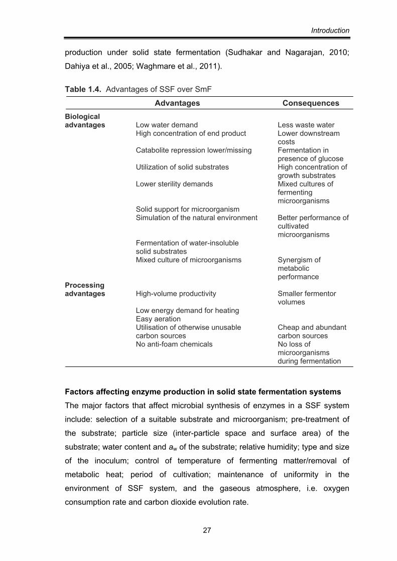
et al. 2001). Table 1.4 lists some advantages of SSF over SmF.
A large number of microorganisms, including bacteria, yeast and fungi produce
different groups of enzymes. Trichoderma spp. and Aspergillus spp. have most
widely been used for hydrolytic enzymes, e.g. chitinases, cellulases, xylanases.
Besides Beauveria spp., Penicillium spp., Verticillium lecanii and
Fusarium oxysporum are also reported to produce chitinase on solid substrates
(Table 1.5). Among prokaryotes Serratia marcescens, Enterobacter sp. NRG4
and Oerskovia xanthineolytica NCIM 2839 have been used for chitinase
Introduction
27
production under solid state fermentation (Sudhakar and Nagarajan, 2010;
Dahiya et al., 2005; Waghmare et al., 2011).
Table 1.4. Advantages of SSF over SmF
Advantages Consequences Biological advantages Low water demand Less waste water High concentration of end product Lower downstream
costs Catabolite repression lower/missing Fermentation in presence of glucose Utilization of solid substrates High concentration of
growth substrates Lower sterility demands Mixed cultures of
fermenting microorganisms
Solid support for microorganism Simulation of the natural environment Better performance of cultivated
microorganisms Fermentation of water-insoluble solid substrates Mixed culture of microorganisms Synergism of
metabolic performance
Processing advantages High-volume productivity Smaller fermentor
volumes Low energy demand for heating Easy aeration Utilisation of otherwise unusable Cheap and abundant carbon sources carbon sources No anti-foam chemicals No loss of
microorganisms during fermentation
Factors affecting enzyme production in solid state fermentation systems The major factors that affect microbial synthesis of enzymes in a SSF system
include: selection of a suitable substrate and microorganism; pre-treatment of
the substrate; particle size (inter-particle space and surface area) of the
substrate; water content and aw of the substrate; relative humidity; type and size
of the inoculum; control of temperature of fermenting matter/removal of
metabolic heat; period of cultivation; maintenance of uniformity in the
environment of SSF system, and the gaseous atmosphere, i.e. oxygen
consumption rate and carbon dioxide evolution rate.
Introduction
28
Table 1.5. Chitinase production in Solid state fermentation
Microorganism Chitinase production
Substrate Temp. Initial pH
Reference
Trichoderma harzianum
3.18 U/gds Wheat bran moistened with 65.7% salt solution, 1% (w/w) colloidal chitin, 2% (w/w) yeast extract
30ºC 4.5 Nampoothiri et al. 2004
Enterobacter sp. 1431 U/gss wheatbran:flake chitin 1, moisture 80%, inoculum size 2.6 ml, incubation time 168 h.
30ºC - Dahiya et al. 2005
Fusarium oxysporum F3(NAGdase)
23.6% U/gss
wheat bran and chitin (10:1)
30ºC 6 Gkargkas et al., 2004
Penicillium chrysogenum PPCS1&2
3809 & 2516 U/gss
wheat bran & 0.1% chitin
24ºC 5.0 (PPCS1) 4.0 (PPCS2)
Patidar et al. 2005a
Beauveria bassiana (marine isolate)
246.6 U/gIDS
Wheat bran, 1% (w/w) colloidal chitin, 75% aged sea water (5:5 w/v)
27ºC 9.2 Suresh and Chandra-sekaran, 1999
Beauveria felina RD101
6.34 U/IDS per hr
Wheat bran and 100% MS-HCl
28 5 Patidar et al., 2005b
Agro-industrial residues are generally considered the best substrates for the
SSF processes. A number of such substrates have been employed for the
cultivation of microorganisms to produce host of enzymes e.g. sugar cane
bagasse, wheat bran, rice bran, maize bran, wheat straw, rice straw, rice husk,
corncobs, coconut coir pith, banana waste, tea waste, cassava waste, palm oil
mill waste, sugar beet pulp, sweet sorghum pulp, apple pomace, etc.
The selection of a substrate for enzyme production in a SSF process depends

Introduction
29
upon cost and availability of the substrate, and thus may involve screening of
several agro-industrial residues. In chitinase production processes including
different organisms wheat bran has most commonly been used (Table 1.5).
Conversely, Sudhakar and Nagarajan (2010) have used rice bran and
Barranco-Florido et al. (2002) used sugarcane pith bagasse for support in SSF.
It is crucial to provide optimized water content, and control the water activity (aw)
of the fermenting substrate, for the availability of water in lower or higher
concentrations affects microbial activity adversely. Moreover, water has
profound impact on the physico-chemical properties of the solids and this, in
turn, affects the overall process productivity. Water activities below 0.9 do not
support most bacterial growth, but yeast and fungi can grow at water activities of
0.7 and greater. Thus, the low moisture environment of many solid state
fermentations favours yeast and fungi Chisti et al., 1999). In some cases, the
optimal water activities for growth and product formation differ (Prior et al.,
1992). The optimal water activity depends also on factors such as agitation rate
and cultivation temperature (Prior et al., 1992). Because the water activity
depends on the concentration of dissolved solutes, sometimes salts, sugar, or
other solutes are added to alter the water activity. Furthermore the fermentation
process itself leads to changes in water activity as products are formed
and the substrate is hydrolyzed. Oxidation of carbohydrates produces water
(Chisti et al., 1999). During fermentation the water activity is controlled by
aeration with humified air and sometimes with intermittent water spray.
Aeration with water saturate air has commonly been found to increase the
moisture content of the substrate. Relative humidity of the aeration gas is
typically 60-80%.
The microbial biomass concentration in SSF is lower than seen in submerged
culture (Tengerdy, 1985), but because there is little water, the heat generation
per unit fermenting mass tends to be much greater in SSF. Temperature can
rise rapidly because there is little water to absorb the heat (i.e. the mean
specific capacity of the fermenting mass is much lower than that of water). Solid
state fermentations are practiced without pH control (Lonsane, 1985) other than
any adjustments made during substrate preparation. Other than that, the
Introduction
30
buffering capacity of substrates is relied on to check large changes in pH during
fermentation (Lonsane, 1985). Many substrates are effective buffers particularly
true of protein-rich substrates.
Different types of fermenters (bioreactors) have been employed for various
purposes in SSF systems. Laboratory studies are generally carried out in
Erlenmeyer flasks, beakers, petri dishes, trays, jars and glass tubes (as column
fermenter). The development of a simple and practical fermentor with
automation is yet to be achieved for the SSF processes (Pandey, 1991).
In spite of all the above advantages, SSF is currently used only to a limited
extent for enzyme and secondary metabolite production due to severe process
engineering problems (Holker et al., 2004). The major problems are (a) the low
amenability of the process to regulation (b) strongly heterogeneous fermentation
conditions due to build up of gradients in temperature, pH, moisture, oxygen,
substrate and inoculum (c) difficult to scale up mainly because of unavailability
of suitable reactors (d) difficulty in determining biomass which is essential for
kinetic studies and (e) complicated downstream processes for product
purification resulting from the use of heterogeneous organic growth substrates
(Holker and Lenz, 2005; Rahardjo et al., 2006; Singhania et al., 2009).
Process optimisation When many variables control a process, the classical approach to optimize the
outcome of the process is the ‘one-variable-at-a-time’ approach, that way
any change could be linked to one different variable. It involves the study of
behaviour of a system at several levels of one variable being studied, while
maintaining rest variables at fixed levels. For each variable, the best value is
found and then, the process is repeated for next variable, until all variables have
been optimised. This approach is based on the assumption that effect of every
variable is mutually independent and may be effective in some situations, but it
is inefficient and takes too many experiments to optimize the process. Besides
the influence of noise increases as the number of experiments are high.
So to investigate systems involving several factors in presence of variability and
noise, a statistically designed set of experiments is required, in which all

Introduction
31
pertinent factors are varied simultaneously and systematically in a single set of
10-20 experiments. These statistical experimental designs are more effective
and widely used in many for process optimisation (Minocha et al., 2007; Kumar
and Satyanarayana, 2007; Singh and Satyanarayana, 2008). An experimental
design is a collection of predetermined settings of the process variables.
Each process variable is called an experimental factor and each combination of
setting for process variables represents a run. A response variable is a measure
of process performance, and each value of the response variable is called an
observation. Each experiment carefully explores the experimental space while
studying many variables using a small number of observations. Statistical
optimisation not only allows quick screening of a large experimental domain, but
also reflects the role of each component. Using a mathematical model, the
levels as well as the interactive effects of variables giving maximum response
can be determined.
Plackett-Burman design Plackett-Burman designs are experimental designs presented in 1946 by Robin
L. Plackett and J.P. Burman. It is the most commonly used among screening
designs that provides a simple model with information about dominating
variables, and their ranges. This design is very useful for economically detecting
large main effects, assuming all interactions are negligible when compared with
the few important main effects.
According to the design, the number of variables to be studied is N-1, where
N is the total number of experiments (multiple of four). When the number of
variables is not N-1 where N is multiple of four, dummy or unassigned variables
are included in the design, which help to calculate the statistical variation in the
data. Each variable is studied at two concentrations, high (H) and low (L)
values. The high and low values are chosen large enough to ensure that any
effective concentration is included in the range. The high and low values for
dummy variables are kept same, thus they must not have any effect of
response/output. The effect of each variable or factor is the difference between
the average of the measurements made at the high level of that factor and the
average of the measurements made at the low level of that factor. If the
Introduction
32
statistical variation were to be negligible the effect of dummy variables should
be zero. Therefore the average effect of dummy variables is taken as a measure
of the statistical error. An ‘F’ test is performed to compare the square of the
effect of an individual variable with that of the dummy variable and found out the
importance of the variable. The critical variables are selected on the basis of
their F-values; higher F-values indicate greater influence of that variable on the
product formation.
Response surface methodology Once critical factors and the region of interest, where the factor’s level influence
the response, is known, the next step is to determine the optimum combination
of factors. This is achieved by response surface methodology. The method was
introduced by G.E.P. Box and K.B. Wilson in 1951. The most popular response
surface design is the central composite design. The design consists of 3
different sets of experimental runs:
1. Factorial points (2k): The number of factors to be included in the design, each
having two levels coded as +1 and -1.
2. The axial/star points (2k): The centre points are augmented with a group of
‘star points’ that allow estimation of curvature. A central composite design
always contains twice as many star points as there are factors in the design.
The star points represent new extreme values (low and high) for each factor
in the design. These are coded as +α and –α.
3. The centre points: Experimental runs whose values of each factor are the
medians of the values used in the factorial portion. This point is often
replicated in order to improve the precision of the experiment and coded as 0.
Here k is the number of factors being studied. The response of individual runs is
used to fit a model by least squares technique. Adequacy of the proposed
model is then revealed using the diagnostic checking tests provided by analysis
of variance (ANOVA). The response surface plots are employed to study the
surfaces and locate the optimum. In several industrial processes, RSM is almost
routinely used to evaluate the results and efficiency of the operations (Beg et
al., 2003; Weska et al., 2007).
Introduction
33
Many authors have reported increase in chitinase titres using Plackett-Burman
screening design and response surface methodology. Vaidya et al. (2003)
optimized medium components for chitinase production by Alcaligenes
xylosoxydans. They found that chitin, yeast extract and tween 80 significantly
affect chitinase production through PB screening and obtained 2.4 fold increase
in chitinase production by optimizing their concentration in the medium by RSM.
Nawani and Kapadnis (2004) used statistical experimental designs to optimize
nutritional and process parameters for chitinase production by three strains of
Streptomyces. The most significant factors identified by 2-level fractional
factorial design were chitin, yeast extract, ammonium sulphate, trace elements,
pH and temperature. Optimizing the values of these variables by RSM led to
9.3-29% increase in chitinase titres by the strains.
Effect of 19 different medium components on chitinase production by marine
isolate Pantoea dispersa was studied by Plackett-Burman design and the
variables chitin, peptone, yeast extract, urea, NH4NO3, NaCl, CaCl2, KBr,
MgSO4.7H2O, KNO3 and KH2PO4 were found significant with confidence level at
or above 95%. It was also observed that the 22nd medium gave highest
chitinase production among the 24 experimental runs. Comparison of chitinase
activity in this and the basal medium showed 3.95-fold and 2.31-fold higher
endochitinase and chitobiase production respectively (Gohel et al., 2006).
PURIFICATION A necessary prerequisite to initiate purification is to have protein in sufficiently
high concentration. Different methods such as, precipitation using salts such as
ammonium sulphate (Teotia et al., 2004; Guo et al., 2004), or organic solvents
such as acetone (Kragh et al., 1991; Muskhazli et al., 2006), lyophilisation
(Watanabe et al., 1990), ultrafiltration etc. have been used for concentrating
crude chitinase preparations. The most commonly used methods for purifying
chitinase involves ion exchange chromatography and gel filtration (Kragh et al.,
1991). A list of some chitinases purified from different microbes is presented in
table 1.6.
Intro
duct
ion
34
Tabl
e 1.
6. C
hara
cter
izat
ion
of c
hitin
ases
from
diff
eren
t mic
ro-o
rgan
ism
s O
rgan
ism
Pr
oces
sivi
ty
Opt
. pH
/ pI
M
W k
D
Opt
. A
ctiv
atio
n In
hibi
tion
Re
fere
nce
ran
ge
tem
p.
Bact
eria
St
rept
omyc
es
- 5
3.7
30
- -
- H
ara
et a
l., 1
989
eryt
hrae
us
Pseu
dom
onas
ae
rogi
nosa
K-1
87
- 8
& 7
5.
2 60
& 3
0 50
&
Cu2+
M
n2+,
Mg2
+,
Wan
g an
d Ch
ang,
199
7
& 4
.8
40
ºC
Zn
2+, g
luta
thio
ne,
dith
ioth
reito
l, an
d
2-
mer
capt
o-et
hano
l. A
erom
onas
sch
bert
ii -
4.8
- 75
-
- -
Guo
et a
l., 2
004
Aer
omon
as h
ydro
phila
su
bsp.
ana
erog
ens
A52
-
7 4.
6 11
0 45
-
Mon
oido
acet
ate,
Ya
buki
et a
l., 1
986
N
-eth
ylm
alei
mid
e,
Hg2
+ an
d A
s3+
Cl
ostr
idiu
m E
-16
- 5-
7
77 &
98
- -
Cu2+
, Fe2
+, H
g2+
Ko
naga
ya e
t al.,
200
6
Zn2+
, & F
e2+
Bu
rkho
lder
ia c
epac
ia
stra
in K
H2
Endo
4.
5 5.
9 34
50
ºC
- -
Oga
wa
et a
l., 2
002
Alc
alig
enes
xyl
ooxy
dans
-
5 -
45
50ºC
-
Cu2,
Na2
Va
diya
et a
l., 2
003
St
rept
omyc
es
hels
tedi
i AJ-
7 -
7 -
55
50ºC
Co
2+
Hg2
+, N
i2, P
b2+
Joo,
200
5 Ps
eudo
mon
as s
p.
YHS
A2
Exo
- -
67
- -
- Le
e et
al.,
200
0
Intro
duct
ion
35
Stre
ptom
yces
ther
mo-
vi
olac
eus
OPC
-520
En
do
8-10
3.
8 40
70
-80°
C -
Cu2+
, Hg2
+, B
a2+,
Cd2+
, Sn2
+, N
-Bro
mo-
succ
inim
ide,
2-h
ydro
xy-
5-
nitr
oben
zyl b
rom
ide
woo
dwar
ds r
eage
nt K
Ts
ujib
o et
al.,
199
3 St
rept
omyc
es g
riseu
s -
5-7
- 31
45
-50°
C -
- H
oste
r et
al.
2005
St
rept
omyc
es s
p. N
K105
7 En
do
4 -
48
60
Hg2
+ N
AG
N
awan
i and
Kap
adni
s, 2
004
Ex
o 6
- 35
40
N
AG
H
g2+
Ba
cillu
s sp
. DA
U10
1 Bi
func
tiona
l
Chito
bios
idas
e
&
-N-a
cety
l-
gluc
oam
inid
ase
7.5
- 66
60
ºC
Co2+
Zn
2+, C
u2+
and
Hg2
+ Le
e et
al.,
200
7 Ba
cillu
s sp
. 13.
26
- 7-
8 -
60
60ºC
M
g2+,
Ni2
+ M
n2+,
Co2
+, C
a2+
Yuli
et a
l. 20
04
Rhod
othe
rmus
mar
inus
En
do, e
xo
on
<pe
ntam
er
4.5-
5 -
42
70ºC
-
- H
obel
et a
l., 2
005
Cellu
lom
onas
uda
e
ndo
-
8.5
70
- -
- Re
guer
a an
d Le
schi
ne, 2
003
Vibr
io a
lgin
olyt
icus
283
-
6.5
- 65
-
- -
Sugi
nta,
200
7 Ba
cillu
s sp
. BG
11
- 7.
5-9
- -
45-5
5ºC
Ca2+
, Ni2
+
Ag2
+, H
g2+,
dith
io-
Bhus
han
and
Hoo
ndal
, 199
8
tr
itonX
100
thre
itol,
b-m
erca
pto-
et
hano
l, gl
utat
hion
e,
io
doac
etic
aci
d an
d
io
doac
et-a
mid
e Ba
cillu
s pu
milu
s SG
2 E
xo
4.5
- 6
3 -
- -
Ahm
adia
n et
al.,
200
7 (H
alot
oler
ant)
5.1
74
Ba
cillu
s am
ylo-
7 5.
8 14
40
ºC
Zn2+
Cu
2+, H
g2+,
ace
tone
W
ang
et a
l., 2
002
liqui
feci
ens
V656
6 5.
3 17
40
ºC
Fe2+
H
g2+
and
acet
one
Baci
llus
subt
ilis
W-1
18
endo
6
6 20
.6
37ºC
-
- W
ang
et a
l., 2
006
Intro
duct
ion
36
Serr
atia
mar
cesc
ens
NK1
-
6.2
- -
47ºC
-
- N
awan
i and
Kap
adni
s,
20
01
Mic
robi
ospo
ra s
p. V
2 -
3 --
35
60
ºC
- H
g2+
Naw
ani e
t al.,
200
2 G
liocl
adiu
m v
irens
En
do
4-6
7.8
41
- -
- Pi
etro
et a
l., 1
993
Piro
myc
es c
omm
unis
O
TS1
(ana
erob
ic)
Endo
6
(at 3
9ºC)
4.4
44
.8
- -
Hg2
+, a
llosa
mid
in
Saku
rada
et a
l. 19
98
Ther
mop
hilic
bac
teri
a Ba
cillu
s sp
. HSA
, 3-1
a -
7 -
22-7
9 60
M
g2+,
Ca2
+,
Co2+
, Fe2
+, Z
n2+
Nat
sir
et a
l., 2
010
Mn2
+ Ba
cillu
s sp
. Hu1
-
6.5
- -
60
Mg2
+, C
a2+,
Cu
2+, D
TT, E
DTA
D
ai e
t al.,
201
1
Zn
2+
O
ersk
ovia
xan
thin
eoly
tica
- C1
7.5
- -
50
Mn2
+, C
u2+
Hg2
+ W
aghm
are
and
Gho
sh, 2
010
NCI
M 2
839
C2
8 -
- 55
Baci
llus
MH
-1
endo
6.
5 5.
3 71
75
M
n2+,
Ca2
+ A
g, H
g, (N
AG
)2
Saka
i et a
l., 1
998
en
do
5.5
4.8
62
65
endo
5.
5 4.
7 53
75
Ra
lsto
nia
sp. A
471
ChiA
End
o 5
4.7
70
70ºC
Cu
2+, M
n2+
- Su
tris
no e
t al.,
200
4
Ca
2+, M
g2+
ChiB
5
- 45
60
-
- U
dea
et a
l., 2
005
Ch
iC
- -
- 55
Co
H
g U
dea
et a
l., 2
009
Hyp
erth
erm
ophi
lic b
acte
ria
and
arch
ea
Pyro
cocc
us fu
rios
us
Endo
(Chi
A)
6 -
40
90-9
5 -
- G
ao e
t al.,
200
3
Exo
(Chi
B)
6 -
55
90-9
5 -
-
Ther
moc
occu
s ch
itino
- En
o (C
hi70
) 7
- 70
70
-
- A
ndro
nopo
ulou
and
ph
agus
Ex
o (C
hi50
) 6
- 50
80
-
- Vo
rgia
s, 2
004
T. k
odak
arae
nsis
Ex
o 5
- 13
4 85
-
- Ta
naka
et a
l., 1
999
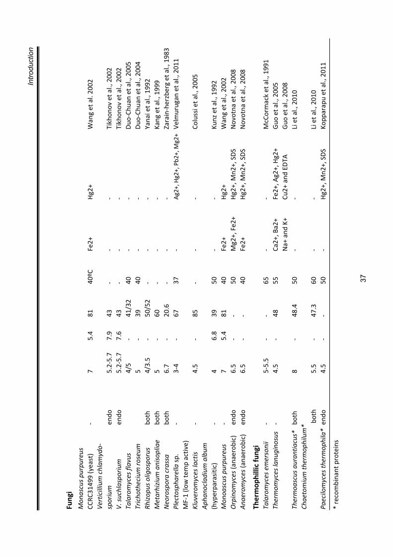
Intro
duct
ion
37
Fung
i M
onas
cus
purp
ureu
s
CCRC
3149
9 (y
east
) -
7 5.
4 81
40
ºC
Fe2+
H
g2+
Wan
g et
al.
2002
Ve
rtic
illiu
m c
hlam
ydo-
sp
oriu
m
endo
5.
2-5.
7 7.
9 43
-
- -
Tikh
onov
et a
l., 2
002
V. s
uchl
aspo
rium
en
do
5.2-
5.7
7.6
43
- -
- Ti
khon
ov e
t al.,
200
2 Ta
laro
myc
es fl
avus
4/5
- 41
/32
40
- -
Duo
-Chu
an e
t al.,
200
5 Tr
icho
thec
ium
rose
um
5
- 39
40
-
- D
uo-C
huan
et a
l., 2
004
Rhiz
opus
olig
ospo
rus
both
4/
3.5
- 50
/52
- -
- Ya
nai e
t al.,
199
2 M
etar
hizi
um a
niso
plia
e bo
th
5 -
60
- -
- Ka
ng e
t al.,
199
9 N
eoro
spor
a cr
assa
bo
th
6.7
- 20
.6
- -
- Za
rain
-her
zber
g et
al.,
198
3 Pl
ecto
spha
rella
sp.
-
3-4
- 67
37
-
Ag2
+, H
g2+,
Pb2
+, M
g2+
Velm
urug
an e
t al.,
201
1
MF-
1 (lo
w te
mp
activ
e)
Kluv
erom
yces
lact
is
- 4.
5 -
85
- -
- Co
luss
i et a
l., 2
005
Aph
anoc
ladi
um a
lbum
(h
yper
para
sitic
) -
4 6.
8 39
50
-
- Ku
nz e
t al.,
199
2 M
onoa
scus
pur
pure
us
- 7
5.4
81
40
Fe2+
H
g2+
Wan
g et
al.,
200
2 O
rpin
omyc
es (a
naer
obic
) en
do
6.5
- -
50
Mg2
+, F
e2+
Hg2
+, M
n2+,
SD
S N
ovot
na e
t al.,
200
8 A
naer
omyc
es (a
naer
obic
) en
do
6.5
- -
40
Fe2+
H
g2+,
Mn2
+, S
DS
Nov
otna
et a
l., 2
008
Ther
mop
hilli
c fu
ngi
Tala
rom
yces
em
erso
nii
- 5-
5.5
- -
65
- -
McC
orm
ack
et a
l., 1
991
Ther
mom
yces
lanu
gino
sus
- 4.
5 -
48
55
Ca2+
, Ba2
+ Fe
2+, A
g2+,
Hg2
+ G
uo e
t al.,
200
5
N
a+ a
nd K
+ Cu
2+ a
nd E
DTA
G
uo e
t al.,
200
8 Th
erm
oasc
us a
uran
tiacu
s*
both
8
- 48
.4
50
- -
Li e
t al.,
201
0 Ch
aeto
miu
m th
erm
ophi
lum
*
b
oth
5.5
- 47
.3
60
- -
Li e
t al.,
201
0 Pa
ecilo
myc
es th
erm
ophi
la*
endo
4.
5 -
- 50
-
Hg2
+, M
n2+,
SD
S Ko
ppar
apu
et a
l., 2
011
* re
com
bina
nt p
rote
ins
Introduction
38
Frequently affinity chromatography using various chitinous substrates has also
been employed. Macroporous beaded chitin cross linked with 2,3-epoxypropyl
chloride was used as an affinity matrix for column chromatography for chitinase
purification from Streptomyces kurssanovii (Tikonov et al., 1998). Macroporous
cross-linked chitin column was used to purify chitinase from the nematophagous
fungi Verticillium chlamydosporium and V. suchlasporium. The column could
separate different chitinases on the basis of their affinity to chitin
(Tikonov et al., 2002). Teotia et al. (2004) purified chitinases to homogeneity
from Neurospora crassa, cabbage and puffballs by affinity precipitation with
chitosan and obtained a fold purification of 27, 15 and 30 respectively.
Acid swollen colloidal chitin was used for affinity purification of chitinase from
Vibrio carchariae (Sunginta et al., 2000). An affinity matrix consisting of
N,N’,N”-triacetylchitotriose immobilized on agarose beads has been used for
chitinase purification from Medicago sativa (Minic et al., 1998).
In addition, a rapid process for purification of chitinase by an aqueous two-
phase extraction (ATPS) and salt fractionation from the latex of Carica
papaya has been reported (Nitsawang and Khanasawud, 2006). The whole
latex, including the insoluble material, was extracted by 5% PEG-12%
ammonium sulphate. Chitinase was purified from the salt-rich bottom phase by
precipitation with 11% ammonium sulfate and 20% sodium chloride,
respectively. 2.4 mg enzyme/g of wet latex and 4.2 folds of purification were
obtained. In a modified ATPS procedure, incorporation of macroaffinity ligand
like chitosan resulted in drastic increase in partitioning of chitinase
(from three different sources, N. crassa, cabbage and puffball) in PEG phase.
A 34-fold purification and 86% recovery could be obtained in case of N. crassa.
Similiarly, cabbage enzyme was purified 20-fold with 80% activity recovery and
puffball enzyme could be purified 38-folds with 88% recovery of the chitinase
activity (Teotia et al., 2004).
MECHANISM OF ACTION There are two general mechanistic pathways for acid catalyzed glycosyl
hydrolysis that results in the following:
Introduction
39
1. Retention of the stereochemistry of the anomeric oxygen at C-1 relative to the
initial configuration
The catalytic mechanism of glycosyl hydrolases is discussed with reference to
the retaining mechanism of hen egg white lysozyme. It involves two catalytic
residues and proceeds through a geometrically distorted oxocarbonium
intermediate (Fig. 1.3). The β-1,4 glycosidic bond between sugars is broken
involving the protonation of the leaving group alcohol leading to an
oxocarbenium ion intermediate, which is then stabilized by a second carboxylate
(either through covalent or electrostatic interactions). A nucleophilic attack by
water which replaces the leaving sugar group yields the hydrolysis products,
which necessarily retains the initial anomeric configuration. This is commonly
referred to as the double displacement mechanism of hydrolysis. It has been
suggested, based on site directed mutagenesis, that Glu 204 and Asp 200
may be the catalytic residues for the chitinase from Bacillus circulans
(Watanabe et al., 1993).
Family 18 chitinases are reported to yield hydrolysis products that retain the
anomeric configuration at C-1. However, chitinases, chitobiases, and related
enzymes from glycosyl hydrolase families 18 and 20, which act also with
retention of configuration, have only a single glutamic acid in their active sites
but no aspartate (Tews et al., 1997). Their x-ray crystal structure reveals the
absence of a second acidic residue in the active site, which is capable of
stabilizing the oxocarbenium ion. Stabilization of the oxocarbonium ion is a
fundamental prerequisite of the mechanism since oxocarbonium ions have
extremely short lifetime in solution. It has been reported that the retaining
mechanism of these chitinases involve substrate assistance (Tews et al. 1997).
That is, the N-acetyl group at position 2 of the scissile sugar may itself facilitate
the reaction via formation of a transient oxazolium intermediate (Terwisscha et
al., 1995, Drouillard et al., 1997).
2. Inversion of the stereochemistry
Although the x-ray crystal structure of family 19 chitinase is similar to that of hen
egg white lysozyme, especially in the substrate binding and catalytic core
suggesting a similar double-displacement mechanism, the hydrolysis products
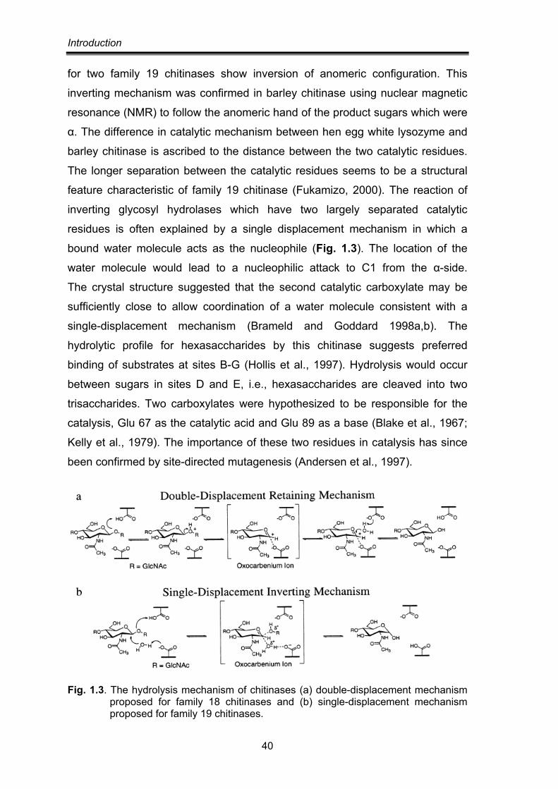
Introduction
40
for two family 19 chitinases show inversion of anomeric configuration. This
inverting mechanism was confirmed in barley chitinase using nuclear magnetic
resonance (NMR) to follow the anomeric hand of the product sugars which were
α. The difference in catalytic mechanism between hen egg white lysozyme and
barley chitinase is ascribed to the distance between the two catalytic residues.
The longer separation between the catalytic residues seems to be a structural
feature characteristic of family 19 chitinase (Fukamizo, 2000). The reaction of
inverting glycosyl hydrolases which have two largely separated catalytic
residues is often explained by a single displacement mechanism in which a
bound water molecule acts as the nucleophile (Fig. 1.3). The location of the
water molecule would lead to a nucleophilic attack to C1 from the α-side.
The crystal structure suggested that the second catalytic carboxylate may be
sufficiently close to allow coordination of a water molecule consistent with a
single-displacement mechanism (Brameld and Goddard 1998a,b). The
hydrolytic profile for hexasaccharides by this chitinase suggests preferred
binding of substrates at sites B-G (Hollis et al., 1997). Hydrolysis would occur
between sugars in sites D and E, i.e., hexasaccharides are cleaved into two
trisaccharides. Two carboxylates were hypothesized to be responsible for the
catalysis, Glu 67 as the catalytic acid and Glu 89 as a base (Blake et al., 1967;
Kelly et al., 1979). The importance of these two residues in catalysis has since
been confirmed by site-directed mutagenesis (Andersen et al., 1997).
Fig. 1.3. The hydrolysis mechanism of chitinases (a) double-displacement mechanism proposed for family 18 chitinases and (b) single-displacement mechanism proposed for family 19 chitinases.
Introduction
41
MULTIPLE FORMS OF CHITINASES Most chitinolytic microorganisms have been found to produce more than one
kind of chitinase. Trichoderma harzianum produce seven individual chitinases:
two N-acetyl-glucosaminidases (102 and 73 kDa), four endochitinases (52, 42,
33 and 31 kDa) and one chitobiosidase (40 kDa) [Haran et al., 1995; Harman et
al., 1993; De La Cruz et al., 1992]. Talaromyces flavus produces at least two
kinds of chitinases (Li et al., 2005). The mycoparasite Stachybotrys elegans
produces two exochitinases and one endochitinase (Taylor et al., 2002). The
entomopathogenic fungus Metarhizium anisopliae produces at least six different
chitinases (Pinto et al., 1997; St. Leger et al., 1996; Kang et al., 1999; St. Leger
et al., 1991). Chitinolytic system of Bacillus circulans WL-12 is one of the most
extensively studied systems so far in prokaryotes. Six chitinases have been
detected in the culture supernatant of this bacterium grown in the presence of
chitin (Watanabe et al., 1990; Watanabe et al., 1992)
Research finds that these multiple chitinases have a mutually synergistic and
complementary effect between them. For example, chitinases CHIT33, CHIT37,
and CHIT42 of T. harzianum show the synergistic action on cell wall
degradation (De La Cruz et al., 1992). Mixing the activities of the he
endochitinase and chitobiosidase from T. harzianum results in the synergistic
increase in antifungal activity (Lorito et al., 1993). A similar phenomenon has
been reported in plant chitinases (Jach et al., 1995) and bacterial chitinases
(Suzuki et al., 2002).
The chitinolytic machinery of Serratia marcescens comprises three chitinases
ChiA, ChiB and ChiC which preferentially produce dimers, a chitin binding
protein CBP21 (Kolstad et al., 2005) and a hexosaminidase which further
degrades the major end product of chitinases i.e. NAG dimer. ChiA and ChiB
are processive exochitinases which degrade chitin chains in opposite directions,
while ChiC is a nonprocessive endochitinase. Chitinases often contain one or
more non-catalytic domains called as carbohydrate binding modules (CBMs)
that help in binding and degrading the insoluble substrate. CBP21 is a CBM that
binds to β-chitin.
Introduction
42
SUBFAMILIES OF MICROBIAL CHITINASES Based on chitinase structure and amino acid sequence, chitinases are
separated into different subfamilies, classes, or groups. Bacterial chitinases are
clearly separated into three major subfamilies, A, B, and C (Watanabe et al.,
1993). Plant chitinases are divided in five different classes. Class I and Class II
chitinases are similar in their catalytic domains. Class I chitinases have a
chitin-binding domain. This domain is separated from the catalytic domain by a
hinge region. Class II chitinases lack both the chitin-binding domain and the
hinge region. Class III chitinases have higher homology to fungal chitinases
than to other plant chitinase classes. Class IV chitinases are similar to
Class I chitinases but they are smaller in size due to certain deletions. Class V
chitinases show some homology with bacterial exochitinases
(Neuhaus et al., 1996).
Compared with bacterial chitinases and plant chitinases, fungal chitinases are
not well classified. Within family 18 two distinct classes of fungal chitinase may
be identified based on the similarity of enzymes to family 18 chitinases from
plants or bacteria (Pishko et al., 1995; Takaya et al., 1998). Therefore,
fungal chitinases are divided into fungal/bacterial (corresponding to
Class V) chitinases, similar to chitinases found in bacteria, and fungal/plant
(corresponding to Class III) chitinases, which are similar to chitinases from
plants (Adams, 2004; Takaya et al., 1998; Jaques et al., 2003). Recently,
according to phylogenetic analysis, it has been suggested that fungal chitinases
of family 18 can be divided into three groups: groups A and group B
(corresponding to class V and III of plant chitinases, respectively), whereas a
novel group C comprises high molecular weight chitinases that have a domain
structure similar to Kluyveromyces lactis killer toxins (Seidl et al., 2005).
Fig. 1.4. Comparison of domains of the ChiB1 fumgal/bacterial chitinase (433 aa) and ChiA1 fingal/plant chitinase (825 aa) of A. fumigatus. 1,catalytic domain; 2, Serine/threonine rich domain.
Introduction
43
STRUCTURE OF FUNGAL CHITINASES
Most fungal chitinases belong to family 18 of the glycosylhydrolase superfamily
(Henrissat, 1999). A characteristic of the family 18 chitinases is their multi-
domain structure. Typically, the basic structures of fungal chitinase of family 18
are composed of five domains or regions: N-terminal signal peptide region,
catalytic domain, serine/theronine-rich region, chitin-binding domain, and C-
terminal extension region. Two chitinases Rhizopus oligosporus (chitinase I and
chitinase II) have all the five distinct domains listed above (Yanai et al., 1992).
However, most fungal chitinases lack the last three domains, which do not seem
to be necessary for chitinase activity because naturally occurring chitinases that
lack these regions are still enzymatically active. The derived amino-acid
sequence of a Saccharomyces cerevisiae endochitinase, CTS1, shows four
distinct domains: a signal sequence, a catalytic domain, a serine/theronine rich
region and chitin-binding domain (Kuranda and Robbins, 1991). T. harzianum
33 kDa chitinase includes a putative signal peptide region and a catalytic
domain, but lacks the other three domains (Limon et al., 1995) [Figure 1.5].
Fig. 1.5. The structure of fungal chitinase of family 18. (a) S. cerevisiae endochitinase (CTS1); (b) R. oligosporus chitinase (CHI1); (c) T. harzianum chitinase (CHIT33); (1) signal peptide; (2) catalytic domain; (3) serine/theronine-rich region; (4) chitin-binding domain; (5) C-terminal extension region (Duo-Chuan, 2006).
The biochemical functions of these domains are as suggested by their names.
A signal peptide is predicted to precede the N-terminal region of many mature
chitinases. The signal peptide presumably mediates secretion of the enzyme
and it is cleaved off by signal peptidases after the protein has been transported
across the membrane. The chitinases lacking secretory signal sequence are
shown to be intracellular chitinase, and they may function during morphogenesis
(Takaya et al., 1998; Seidl et al., 2005). The catalytic domain of fungal
chitinases, responsible for the hydrolysis of the substrate, comprises the
N-terminal half of the enzyme. Sequence alignments reveal two highly
Introduction
44
conserved regions within the catalytic domain. The two consensus regions or
motifs, SxGG and DxxDxDxE, correspond to a substrate-binding site and a
catalytic domain, respectively, in family 18 chitinases (Henrissat, 1991).
It is proposed that two conserved carboxylic acids are important in the retaining,
substrate assisted catalytic mechanism of family 18 hydrolases (Perrakis
et al., 2004). One is supposed to act as a general acid to protonate the leaving
group and glutamate serves this role in a wide range of glycohydrolases
(Monzingo et al., 1996). A second carboxylic acid, generally aspartate in
chitinases either assists the catalytic glutamate or acts as base to stabilize the
transition state (Papanikolau et al., 2001; Bortone et al., 2002).
Apart from N-terminal signal peptide region and catalytic domain, the structure
of some fungal chitinases reported has serine/theronine-rich region and chitin-
binding domain (ChBD), such as Saccharomyces cerevisiae endochitinase,
CTS1 (Kuranda and Robbins, 1991), Kluyveromyces lactis endochitinase,
KlCts1p (Colussi et al., 2005), Trichoderma reesei Chi18-13 and Chi18-16 and
Rhizopus ologosporus chitinase I and Chitinase II (Yanai et al., 1992).
The serine/theronine-rich region of fungal chitinases is ususlly glycosylated
with sugar chains post-translationally to yield the mature protein.
The glycosylation sites may be necessary for the secretion of the protein and
maintenance of its stability.
Fungal chitinases are thought to be anchored to the cell wall or their substrate
through the chitin-binding domain (Kuranda and Robbins, 1991). A conserved
six-cysteine motif, which likely mediates the tertiary structure or protein–protein
interaction through the formation of disulfide bridges, is found in the chitin-
binding domain of CTS1 and KlCts1p (Kuranda and Robbins, 1991; Colussi
et al., 2005). The chitin-binding domain of Trichoderma reesei Chi18-13 and
Chi18-16 has high similarity with a fungal cellulose-binding domain (CBD),
consisting of four strictly conserved aromatic amino acid residues that are
implicated in the interaction with cellulose (Seidl et al., 2005; Kraulis et al.,
1989). The chitin-binding domain differs from the substrate-binding site in the
catalytic domain (Henrissat, 1999). The chitin-binding domain of Kluyveromyces
lactis KlCts1p functions independent of the catalytic domain (Colussi et al.,
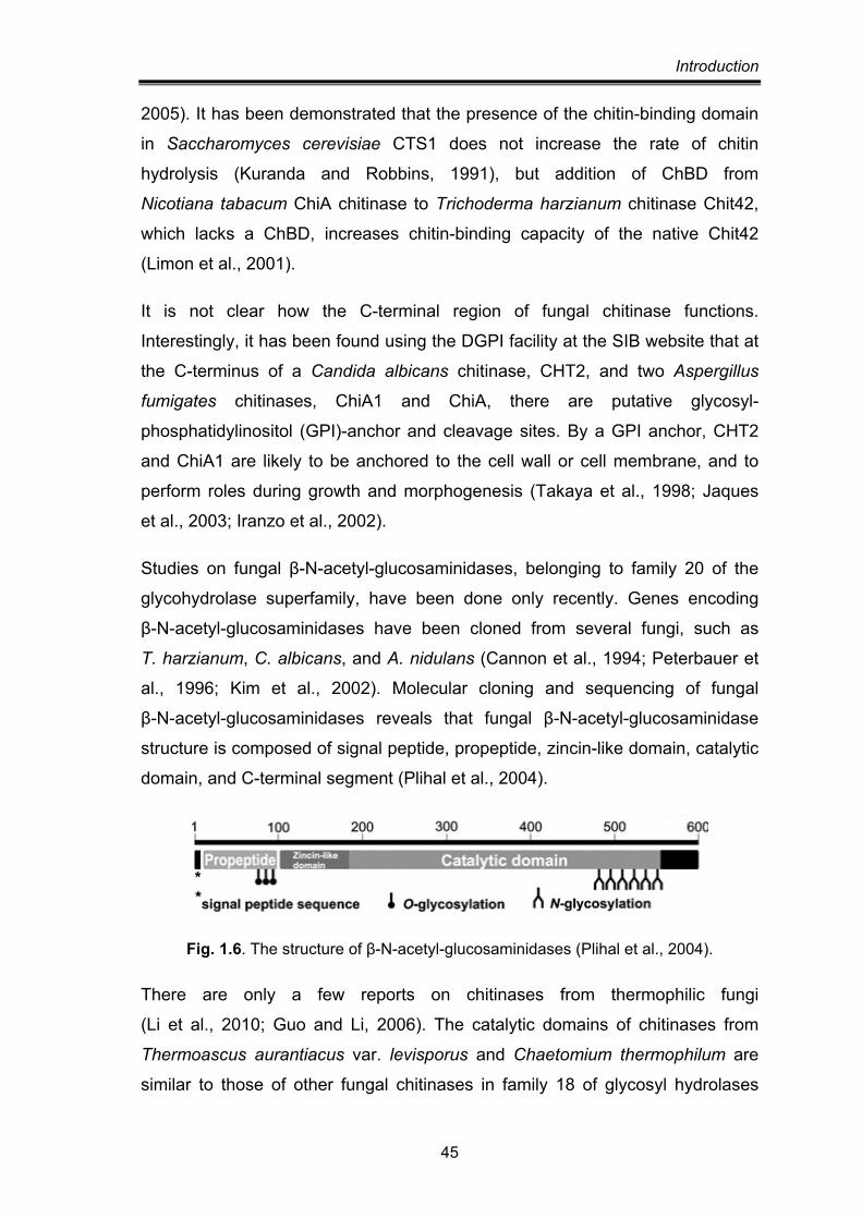
Introduction
45
2005). It has been demonstrated that the presence of the chitin-binding domain
in Saccharomyces cerevisiae CTS1 does not increase the rate of chitin
hydrolysis (Kuranda and Robbins, 1991), but addition of ChBD from
Nicotiana tabacum ChiA chitinase to Trichoderma harzianum chitinase Chit42,
which lacks a ChBD, increases chitin-binding capacity of the native Chit42
(Limon et al., 2001).
It is not clear how the C-terminal region of fungal chitinase functions.
Interestingly, it has been found using the DGPI facility at the SIB website that at
the C-terminus of a Candida albicans chitinase, CHT2, and two Aspergillus
fumigates chitinases, ChiA1 and ChiA, there are putative glycosyl-
phosphatidylinositol (GPI)-anchor and cleavage sites. By a GPI anchor, CHT2
and ChiA1 are likely to be anchored to the cell wall or cell membrane, and to
perform roles during growth and morphogenesis (Takaya et al., 1998; Jaques
et al., 2003; Iranzo et al., 2002).
Studies on fungal β-N-acetyl-glucosaminidases, belonging to family 20 of the
glycohydrolase superfamily, have been done only recently. Genes encoding
β-N-acetyl-glucosaminidases have been cloned from several fungi, such as
T. harzianum, C. albicans, and A. nidulans (Cannon et al., 1994; Peterbauer et
al., 1996; Kim et al., 2002). Molecular cloning and sequencing of fungal
β-N-acetyl-glucosaminidases reveals that fungal β-N-acetyl-glucosaminidase
structure is composed of signal peptide, propeptide, zincin-like domain, catalytic
domain, and C-terminal segment (Plihal et al., 2004).
Fig. 1.6. The structure of β-N-acetyl-glucosaminidases (Plihal et al., 2004).
There are only a few reports on chitinases from thermophilic fungi
(Li et al., 2010; Guo and Li, 2006). The catalytic domains of chitinases from
Thermoascus aurantiacus var. levisporus and Chaetomium thermophilum are
similar to those of other fungal chitinases in family 18 of glycosyl hydrolases
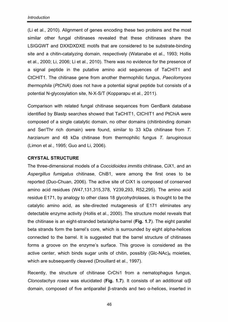
Introduction
46
(Li et al., 2010). Alignment of genes encoding these two proteins and the most
similar other fungal chitinases revealed that these chitinases share the
LSIGGWT and DXXDXDXE motifs that are considered to be substrate-binding
site and a chitin-catalyzing domain, respectively (Watanabe et al., 1993; Hollis
et al., 2000; Li, 2006; Li et al., 2010). There was no evidence for the presence of
a signal peptide in the putative amino acid sequences of TaCHIT1 and
CtCHIT1. The chitinase gene from another thermophilic fungus, Paecilomyces
thermophila (PtChiA) does not have a potential signal peptide but consists of a
potential N-glycosylation site, N-X-S/T (Kopparapu et al., 2011).
Comparison with related fungal chitinase sequences from GenBank database
identified by Blastp searches showed that TaCHIT1, CtCHIT1 and PtChiA were
composed of a single catalytic domain, no other domains (chitinbinding domain
and Ser/Thr rich domain) were found, similar to 33 kDa chitinase from T.
harzianum and 48 kDa chitinase from thermophilic fungus T. lanuginosus
(Limon et al., 1995; Guo and Li, 2006).
CRYSTAL STRUCTURE The three-dimensional models of a Coccidioides immitis chitinase, CiX1, and an
Aspergillus fumigatus chitinase, ChiB1, were among the first ones to be
reported (Duo-Chuan, 2006). The active site of CiX1 is composed of conserved
amino acid residues (W47,131,315,378, Y239,293, R52,295). The amino acid
residue E171, by analogy to other class 18 glycohydrolases, is thought to be the
catalytic amino acid, as site-directed mutagenesis of E171 eliminates any
detectable enzyme activity (Hollis et al., 2000). The structure model reveals that
the chitinase is an eight-stranded beta/alpha-barrel (Fig. 1.7). The eight parallel
beta strands form the barrel’s core, which is surrounded by eight alpha-helices
connected to the barrel. It is suggested that the barrel structure of chitinases
forms a groove on the enzyme’s surface. This groove is considered as the
active center, which binds sugar units of chitin, possibly (Glc-NAc)6 moieties,
which are subsequently cleaved (Drouillard et al., 1997).
Recently, the structure of chitinase CrChi1 from a nematophagus fungus,
Clonostachys rosea was elucidated (Fig. 1.7). It consists of an additional α/β
domain, composed of five antiparallel β-strands and two α-helices, inserted in
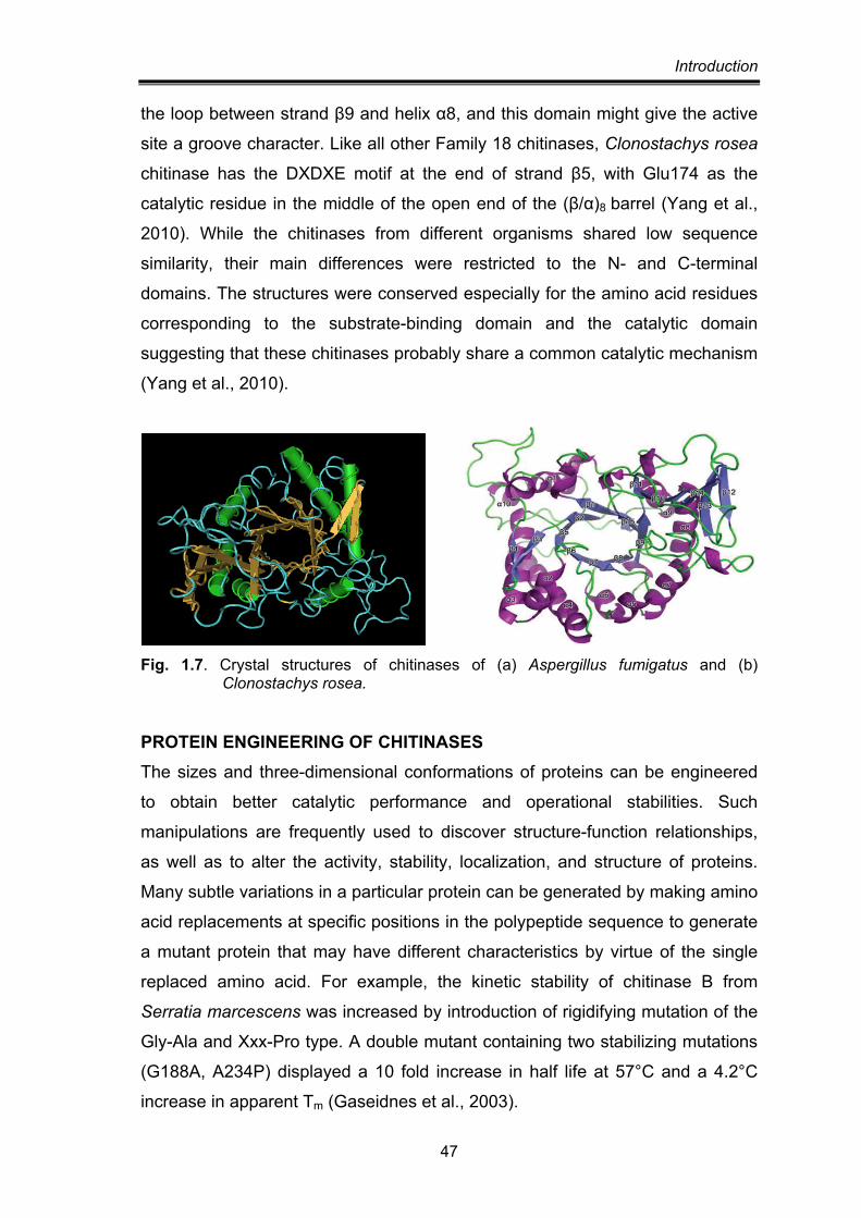
Introduction
47
the loop between strand β9 and helix α8, and this domain might give the active
site a groove character. Like all other Family 18 chitinases, Clonostachys rosea
chitinase has the DXDXE motif at the end of strand β5, with Glu174 as the
catalytic residue in the middle of the open end of the (β/α)8 barrel (Yang et al.,
2010). While the chitinases from different organisms shared low sequence
similarity, their main differences were restricted to the N- and C-terminal
domains. The structures were conserved especially for the amino acid residues
corresponding to the substrate-binding domain and the catalytic domain
suggesting that these chitinases probably share a common catalytic mechanism
(Yang et al., 2010).
Fig. 1.7. Crystal structures of chitinases of (a) Aspergillus fumigatus and (b)
Clonostachys rosea.
PROTEIN ENGINEERING OF CHITINASES The sizes and three-dimensional conformations of proteins can be engineered
to obtain better catalytic performance and operational stabilities. Such
manipulations are frequently used to discover structure-function relationships,
as well as to alter the activity, stability, localization, and structure of proteins.
Many subtle variations in a particular protein can be generated by making amino
acid replacements at specific positions in the polypeptide sequence to generate
a mutant protein that may have different characteristics by virtue of the single
replaced amino acid. For example, the kinetic stability of chitinase B from
Serratia marcescens was increased by introduction of rigidifying mutation of the
Gly-Ala and Xxx-Pro type. A double mutant containing two stabilizing mutations
(G188A, A234P) displayed a 10 fold increase in half life at 57°C and a 4.2°C
increase in apparent Tm (Gaseidnes et al., 2003).
Introduction
48
Catalytic potential of proteins can be also altered through substitution of amino
acids. Martinez et al. (2012) have reported that the mutagenesis of two
GH-18 glycoside hydrolase, B. circulans WL-12 chitinase A1 (Bc ChiA1) and
Trichoderma harzanium chitinase 42 (Th Chit42), and usage of E. coli produced
building blocks to generate long chitooligosaccharides. The conserved catalytic
residues of Bc ChiA1 and Th Chit42 were mutated individually, and each mutant
was screened for its transglycosylation behaviour. The mutants D200A and
D202A of Bc ChiA1, together with D170N and to a lesser extent D170A of Th
Chit42 proved to be active for chitiobiose oxazoline polymerization. These
mutants have additionally retained the ability to catalyze transglycosylation
reaction on natural COs, whereas their hydrolytic activity is abolished. Thus the
generation of complex mixtures of various reaction products obtained due to
hydrolytic activity of the enzymes and uncontrollable random transglycosylation
reactions by wild type chitinases could be avoided (Martinez et al., 2012).
In addition to the substitution of amino acids at specific positions, amino acids
can be deleted from the sequence, either individually or in groups. Such
deletion mutants have been a useful way to create smaller proteins that contain
only one or a few of these domains so that the individual properties can be
studied. A chitinase from Pyrococcus kodakarensis, ChiA has two catalytic
domains and deletion mutants having either of the two showed that these two
domains are functional independently as thermostable chitinases. It was also
observed that the C-terminal half of ChiA was much more thermostable than the
N-terminal half. The removal of the N-terminal region might induce a more
thermostable conformation (Tanaka et al., 1999).
Deletion of 304 C-terminal amino acid residues from Aeromonas caviae
chitinase ChiA, yielded a mutant with lower optimum temperature and higher
relative activity on chitin, colloidal chitin and 4-methylumbelliferyl-N-N′-N′′-
triacetylchitotriose (Lin et al., 2001). Removal of C-terminal regions in chitinases
from Bacillus licheniformis (145 amino acid residues containing putative chitin
binding and fibronectin domain) and Vibrio parahaemolyticus (30 aa residues)
led to higher thermostabilities and decreased catalytic activitiy towards insoluble
chitin substrates in both mutated enzymes. The activities towards insoluble
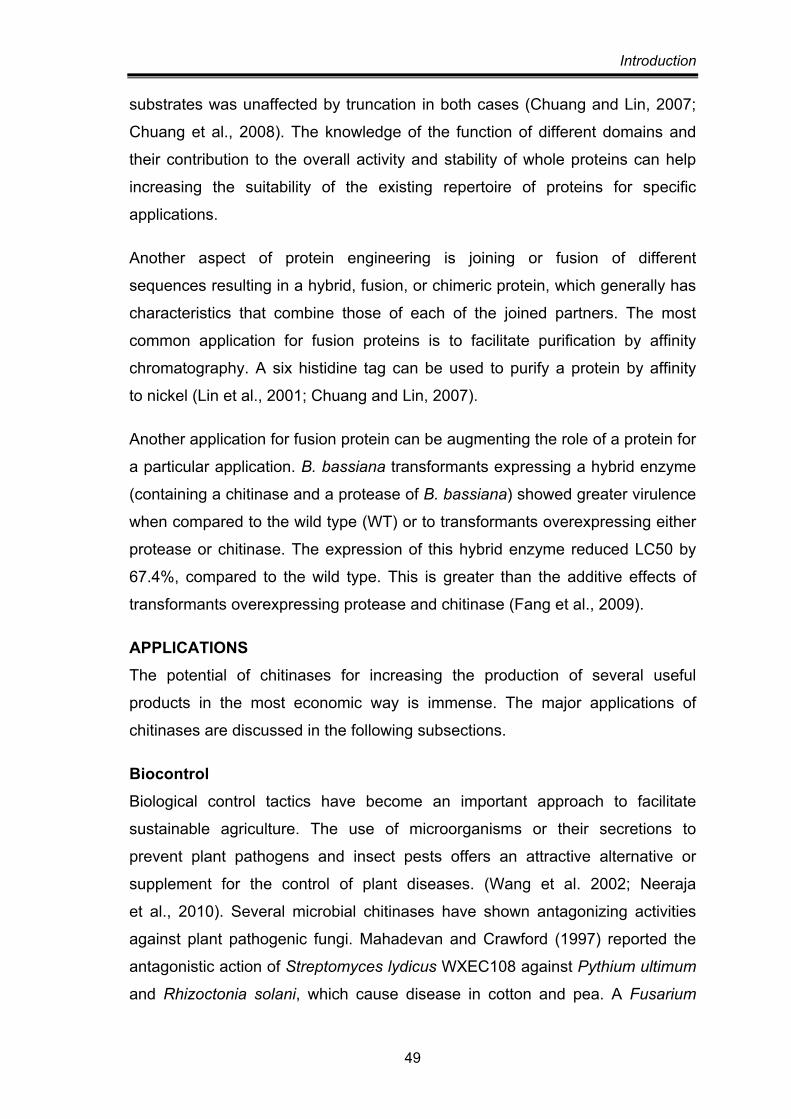
Introduction
49
substrates was unaffected by truncation in both cases (Chuang and Lin, 2007;
Chuang et al., 2008). The knowledge of the function of different domains and
their contribution to the overall activity and stability of whole proteins can help
increasing the suitability of the existing repertoire of proteins for specific
applications.
Another aspect of protein engineering is joining or fusion of different
sequences resulting in a hybrid, fusion, or chimeric protein, which generally has
characteristics that combine those of each of the joined partners. The most
common application for fusion proteins is to facilitate purification by affinity
chromatography. A six histidine tag can be used to purify a protein by affinity
to nickel (Lin et al., 2001; Chuang and Lin, 2007).
Another application for fusion protein can be augmenting the role of a protein for
a particular application. B. bassiana transformants expressing a hybrid enzyme
(containing a chitinase and a protease of B. bassiana) showed greater virulence
when compared to the wild type (WT) or to transformants overexpressing either
protease or chitinase. The expression of this hybrid enzyme reduced LC50 by
67.4%, compared to the wild type. This is greater than the additive effects of
transformants overexpressing protease and chitinase (Fang et al., 2009).
APPLICATIONS The potential of chitinases for increasing the production of several useful
products in the most economic way is immense. The major applications of
chitinases are discussed in the following subsections.
Biocontrol Biological control tactics have become an important approach to facilitate
sustainable agriculture. The use of microorganisms or their secretions to
prevent plant pathogens and insect pests offers an attractive alternative or
supplement for the control of plant diseases. (Wang et al. 2002; Neeraja
et al., 2010). Several microbial chitinases have shown antagonizing activities
against plant pathogenic fungi. Mahadevan and Crawford (1997) reported the
antagonistic action of Streptomyces lydicus WXEC108 against Pythium ultimum
and Rhizoctonia solani, which cause disease in cotton and pea. A Fusarium
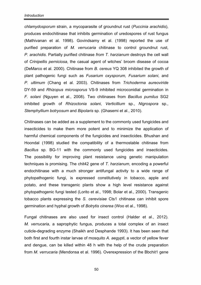
Introduction
50
chlamydosporum strain, a mycoparasite of groundnut rust (Puccinia arachidis),
produces endochitinase that inhibits germination of uredospores of rust fungus
(Mathivanan et al. 1998). Govindsamy et al. (1998) reported the use of
purified preparation of M. verrucaria chitinase to control groundnut rust,
P. arachidis. Partially purified chitinase from T. harzianum destroys the cell wall
of Crinipellis perniciosa, the casual agent of witches’ broom disease of cocoa
(DeMarco et al. 2000). Chitinase from B. cereus YQ 308 inhibited the growth of
plant pathogenic fungi such as Fusarium oxysporum, Fusarium solani, and
P. ultimum (Chang et al. 2003). Chitinases from Trichoderma aureoviride
DY-59 and Rhizopus microsporus VS-9 inhibited microconidial germination in
F. solani (Nguyen et al., 2008). Two chitinases from Bacillus pumilus SG2
inhibited growth of Rhizoctonia solani, Verticillium sp., Nigrospora sp.,
Stemphyllium botryosum and Bipolaris sp. (Ghasemi et al., 2010).
Chitinases can be added as a supplement to the commonly used fungicides and
insecticides to make them more potent and to minimize the application of
harmful chemical components of the fungicides and insecticides. Bhushan and
Hoondal (1998) studied the compatibility of a thermostable chitinase from
Bacillus sp. BG-11 with the commonly used fungicides and insecticides.
The possibility for improving plant resistance using genetic manipulation
techniques is promising. The chit42 gene of T. harzianum, encoding a powerful
endochitinase with a much stronger antifungal activity to a wide range of
phytopathogenic fungi, is expressed constitutively in tobacco, apple and
potato, and these transgenic plants show a high level resistance against
phytopathogenic fungi tested (Lorito et al., 1998; Bolar et al., 2000). Transgenic
tobacco plants expressing the S. cerevisiae Cts1 chitinase can inhibit spore
germination and hyphal growth of Botrytis cinerea (Woo et al., 1998).
Fungal chitinases are also used for insect control (Halder et al., 2012).
M. verrucaria, a saprophytic fungus, produces a total complex of an insect
cuticle-degrading enzyme (Shaikh and Desphande 1993). It has been seen that
both first and fourth instar larvae of mosquito A. aegypti, a vector of yellow fever
and dengue, can be killed within 48 h with the help of the crude preparation
from M. verrucaria (Mendonsa et al. 1996). Overexpression of the Bbchit1 gene
Introduction
51
from the entomopathogenic fungus Beauveria bassiana in transgenic B.
bassiana can significantly enhance the virulence of B. bassiana for aphids
(Treger et al., 1998).
Production of single-cell protein/Bioconversion of chitin waste The solid waste from shellfish processing, mainly composed of chitin, can be
hydrolyzed by chitinases and the resulting sugars can be utilized by other
microorganisms for production of SCP and biofuels. Revah-Moiseev and
Carrod (1981) used the S. marcescens chitinase system to hydrolyze the chitin
and Pichia kudriavazevii to yield SCP (with 45% protein and 8–11% nucleic
acids). The commonly used fungi as the source of SCP are Hansenula
polymorpha, Candida tropicalis, Saccharomyces cerevisiae, and Myrothecium
verrucaria (Dahiya et al., 2006). Vyas and Deshpande (1991) utilized the
chitinolytic enzymes of M. verrucaria and S. cerevisiae for the production of
SCP from chitinous waste reporting the total protein content to be 61%, with
very low contents of nucleic acids (3.1%). Ferrer et al. (1996) used chitin
acid/enzyme hydrolysate as a carbon and energy source for the growth of
Saccharomyces cerevisiae K1V-1116 and reported that the acid and enzymatic
hydrolysis route are comparable when effective production of SCP is concerned.
Cody et al. (1990) reported the applicability of yeast P. tannophilus and
Z. mobilis in production of ethanol on amino sugars, glucosamine and
N-acetylglucosamine. Oleaginous microorganisms like Rhodotorula glutinis,
Rhodococcus opacus and Cryptococcus curvatus possess the ability to
convert NAG into triacylglycerides and store these materials intracellularly (Zang
et al., 2011).
Isolation of protoplasts Fungal protoplasts have been used as an effective experimental tool in studying
cell wall synthesis, enzyme synthesis, and secretion, as well as in strain
improvement for biotechnological applications. Since fungi have chitin in their
cell walls, the chitinolytic enzyme seems to be essential along with other wall-
degrading enzymes for protoplast formation from fungi. Mizuno et al. (1997)
isolated protoplast from Schizophyllum commune using the culture filtrate of
B. circulans KA-304. Dahiya et al. (2005) reported the effectiveness of
Introduction
52
Enterobacter sp. NRG4 chitinase in the generation of protoplasts from
Trichoderma reesei, Pleurotus florida, Agaricus bisporus, and A. niger.
An enzyme complex from B. circulans WL-12 with high chitinase activity was
effective in generating protoplasts from Phaffia rhodozyme (Johnson et al.
1979). A chitinase and a laminarinase from the thermophilic fungus
Paecilomyces uarioti were used to isolate protoplasts of a thermophilic fungus,
Malbranchea sulfurea (Gautam et al., 1996). In biological research, chitinases
from T. harzianum are used for the generation of fungal protoplasts
(Duo-Chuan, 2006).
Medical applications Chitinases can be employed in human health care, such as making ophthalmic
preparations with chitinases and microbiocides. A direct medical use has been
suggested for chitinases in the therapy for fungal diseases in potentiating the
activity of antifungal drugs (Pope and Davis 1979; Orunsi and Trinci 1985).
They can also be used as potential additives in antifungal creams and lotions
due to their topical applications. Chitinases of insect and fungal pathogens can
also be useful as vaccine antigens. Recombinant chitinase of Haemaphysalis
longicornis (hard tick) expressed in E. coli stimulated a specific protective
anti-tick immune response in mice, suggesting its potential as a tick vaccine
(You and Fujisaki, 2009).
In addition, chitinases are also useful in generating chitooligosaccharides,
glucosamine, and GlcNAc that have immense pharmaceutical potential owing to
their strong physiological activities (Dhaiya et al., 2006). For example,
Chitooligomers show enhancing protective effects against infection with some
pathogens like Listeria monocytogenes in mice. The beneficial lowering effect of
oligosaccharides on plasma cholesterol may play an important role in the
prevention and treatment of cardiovascular diseases (Tharanathan and
Kittur, 2003). Chitin oligomers also show the antitumorigenic properties and
inhibit growth of tumor cells via an immuno-enhancing effect.
The potential of glucosamine and N-acetylglucosamine in the treatment
of patients with osteoarthritis has been recognized for several years
(Horstman et al., 1999; Rubin et al., 2001; Matheson and Perry, 2003).
Introduction
53
Glucosamine and NAG are not only the primary building blocks of the structural
matrix of connective tissue in joints, but it also stimulates their synthesis and
inhibits their degradation. In addition, they are capable of scavenging reactive
oxygen species, whose levels are elevated in the synovium of arthritis patients
(Chapman et al., 1986; Sato et al., 1988; Gracy et al., 1999). Glucosamine/NAG
has been shown to accelerate wound healing, improve skin hydration, and
decrease wrinkles. In addition, as an inhibitor of tyrosinase activation, it inhibits
melanin production and is useful in treatment of disorders of hyperpigmentation.
GlcNAc also shows promise as an inexpensive and nontoxic treatment in
chronic inflammatory bowel disease. The oral administration of GlcNAc to
children with severe ulcerative colitis and Crohn’s disease led to histological
improvement, and a significant increase in epithelial and lamina propria
glycosaminoglycans and intracellular GlcNAc.
THERMOPHILIC FUNGI/MOULDS A key physical factor determining the sustenance of living beings on earth is
temperature. The origin of life on earth occurred in extreme temperatures and
microbes recovered from such extremes are until now only prokaryotes.
Eukaryotes can grow only up to 60°C or so, herein too the extremes are shown
by a small number of fungal species (Sharma and Johri, 1992). These
thermophilic moulds when grown at high temperatures exhibit greater saturation
of lipids and a different mode of lipid storage (Satyanarayana and Johri, 1999).
Several enzymes of these moulds are more thermostable than the mesophilic
fungi. Their proteins have been found to be stable upto 95°C or higher
(Satyanarayana et al., 1992).
Many definitions of this phenomenon of thermophily in fungi occur in literature.
Crisan (1959) defined thermophilic fungi as those whose optimum temperature
for normal growth lies at or above 40°C. Cooney and Emerson (1964) defined
‘A thermophilic fungus is one that has a maximum temperature of growth at or
above 50°C and a minimum temperature for growth at or above 20°C’.
Thermophilic fungi were discovered accidently due to chance contamination of
organic materials incubated at elevated temperatures. The earliest discovered
species were Mucor pusillus from bread by Lindt (Lindt, 1886) and Humicola
Introduction
54
lanuginosa from potato disk by Tsiklinsky (Tsiklinsky, 1899). The first extensive
report on thermophily in fungi was produced by Miehe in 1907, who made a
thorough study of self heating hay and described Thermoascus aurantiacus and
Malbranchea pulchella var. sulfurea (Miehe, 1907). Thermophilic fungi grow in
simple media containing carbon and nitrogen sources and mineral salts,
suggesting that they do not have any special requirement for growth and they
are autotrophic for all vitamins.
Thermophilic fungi are worldwide in distribution and have been reported from
various types of soils and in habitats where decomposition of plant material
results in high temperature. These include: composts, piles of hays, stored
grains, wood chip piles, nesting material of birds and animals, snuff, and
municipal refuse, and other accumulations of organic matter wherein the warm,
humid, and aerobic environment provides the basic physiological conditions for
their development. Man made habitats such as cooling towers, effluents from
nuclear power reactors and ducts employed for thermal insulation (Johri and
Satyanarayana, 1986). In these habitats thermophiles may occur either as
resting propagules or as active mycelia depending on the availability of nutrients
and favourable environmental conditions. Their widespread occurrence could
well be due to the dissemination of propagules from self-heating masses of
organic material (Maheswari et al., 1987).
Thermophilic fungi have high potential for industrial applications as a source of
thermostable enzymes. Biomass-degrading enzymes from thermophilic fungi
consistently demonstrate higher hydrolytic capacity (Wojtczak et al., 1987).
Thermophilic fungal strains are also capable of rapid growth with minimized
viscosity at relatively elevated growth temperatures thereby enhancing
productivity in fermentors (Jensen and Boominathan, 1997).
Though chitinolytic enzymes are produced from a wide range of organisms,
most of them cannot be applied wildly due to the high cost of isolation of
enzymes from natural resources, their instability and activity within narrow
temperature and pH ranges (Dai et. al., 2011). The use of thermostable
enzymes, which are intrinsically stable and active at high temperatures
minimizes stability problems and thus cost. The thermostable biocatalysts are
Introduction
55
also more resistant to denaturing agents, more stable to change in pH and
tolerant to higher solute (reactants) concentrations. Other expected advantages
of thermstable enzymes are a longer shelf life, less contamination problems
and a different substrate spectrum than comparable mesophilic enzymes
(Sonnleitner and Fiechter 1983; Vieille and Zeikus, 2001). Higher processing
temperatures gives the benefit of achieving higher reaction rates and allows
higher substrate concentrations, lower viscosity and minimization of diffusion
limitations of the reactants and the products (Sonnleitner and Fiechter, 1983;
Unsworth et al., 2007). Once expressed in mesophilic hosts, thermophilic
enzymes are also easier to purify by heat treatment (Vieille and Zeikus, 2001).
Among various microbes, filamentous fungi are the most frequently used
organisms in industrial processes. In general, filamentous fungi have
18-20 different GH family 18 proteins (Seidl, 2008). Some mycoparasitic
and entomopathogenic fungi have even 30 or more GH family 18 proteins
(Gao et al., 2011; Kubicek et al., 2011). The variability of fungal chitinases
makes them ideal candidates for the development of enzymes acting on
chitinous carbohydrates used in biotechnology (Hartl et al., 2012). As sources of
thermostable enzymes, thermophilic fungi have a high potential for industrial
applications. Another advantage of thermophilic fungi over their mesophilic
counterparts is better growth characteristics in a fermenter. Filamentous growth
in fermenters is normally accompanied by a high culture viscosity, low mixing
rates, and, consequently, suboptimal transfer of, e.g., oxygen and nutrients,
which may severely hamper protein productivity in large-scale fermentations.
Thermophilic fungal strains are capable of rapid growth with minimized viscosity
at relatively elevated growth temperatures, thereby enhancing productivity in
fermenters (Jensen and Boominathan, 1997).
As compared to mesophilic fungi, data on thermophilic fungal chitinases is
limited. As listed in table 1.6, these chitinases are less diverse and all show an
optimum temperature in the range of 50-65°C, molecular weight of 47-48 kD
and pH optimum of 4.5-5.5 (McComark et al., 1991; Guo et al., 2005, 2008; Li et
al., 2010; Kopparapu et al., 2011). The genes encoding these proteins belong to
family 18 of glycosyl hydrolases and consist of a single catalytic domain
with conserved substrate-binding and catalyzing domain (Li et al., 2010;
Introduction
56
Kopparapu et al., 2011). Active chitinases from Thermoascus aurantiacus var.
levisporus and Chaetomium thermophilum have been expressed in Pichia
pastoris (Li et al., 2010). A chitinase from Paecilomyces thermophila has also
been expressed in Escherichia coli as an intracellular soluble protein
(Kopparapu et al., 2011).
Myceliophthora thermophilia is a thermophilic fungus that grows optimally at
45°C and is able to produce some industrially important hydrolytic enzymes
such as phytase, lipase, cellulose, xylanase and others (Satyanarayana et al.
1985; Singh and Satyanarayana et al. 2006). Recently the 38,744,216 bp
genome of M. thermophila containing 9110 protein coding genes on 7
chromosomes has been sequenced, one of the first among filamentous fungi
(Berka et al., 2011). Like other fungi, it harbours a large number (>210) of
glycosyl hydrolase and polysaccharide lyase and can be considered as all
purpose decomposers (Berka et al., 2011). Based on the peptide sequence of
extracellular proteins produced by Myceliophthora thermophila, previously
classified as Chrysosporium lucknowense C1, two chitinases (a 45 kDa
endochitinase and a 70 kDa exochitinase) were identified in the culture filtrates
(Visser et al., 2011). Homologous production of the 45 kDa endochitinase
reached the level of 7.5 g/L using an expression vector containing chi1
(chitinase gene) promoter region and cbh1 (cellobiohydrolase gene) terminator
region flanking the cloning site for chi1 gene (Visser et al., 2011). However,
detailed studies relating to biochemical characterization and application
potential of the chitinases has not been investigated (Visser et al., 2011).
In view of the biotechnological potential of a thermostable chitinases, the
present investigation was planned and executed with the following objectives:
1. Isolation, screening and selection of a potent chitinase producing thermophilic mould
2. Optimization of enzyme production in submerged and solid state fermentations
3. Purification and characterization of chitinase
4. Testing the applicability of chitinase in the production of chitooligosaccharides and in biocontrol

![University of Western Australia · Web view, as it helps to completely degrade chitin degradation products, generated by secreted chitinases [43,44] and transported through outer](https://static.fdocument.org/doc/165x107/60d97f7be5724d3db967093f/university-of-western-australia-web-view-as-it-helps-to-completely-degrade-chitin.jpg)