High density MicroArrays on Blu-ray discs for massive screening
6
High density MicroArrays on Blu-ray discs for massive screening Tania Arnandis-Chover, Sergi Morais, Miguel Ángel González-Martínez, Rosa Puchades, Ángel Maquieira n Centro de Reconocimiento Molecular y Desarrollo Tecnoló gico, Departamento de Química, Universitat Politècnica de València, Camino de Vera s/n, 46071 Valencia, Spain article info Article history: Received 14 June 2013 Received in revised form 19 July 2013 Accepted 22 July 2013 Available online 29 July 2013 Keywords: Blu-ray High density arrays Multiplex analysis Massive screening Point-of-care abstract The potential and the capabilities of Blu-ray technology (discs and drives) for massive screening applications are presented. High density microarrays are fabricated onto a Blu-ray disc and applied for the determination of microcystin residues and pathogenic microorganisms. Specific probes were physisorbed onto the BD surface and the biorecognition event was displayed using labeled secondary antibody solution and subsequent signal amplification. The attenuation of the reflected light caused by the reaction product is detected by the Blu-ray drive and inversely correlated with analyte concentration. BDs preserve the optical properties according to Blu-ray specifications, ensuring maximum accuracy and sensitivity of the drive during disc scanning. Detection limits of 0.4 μg/L for microcystin LR and 10 0 and 10 1 cfu/mL for Salmonella typhimurium and Cronobacter sakazakii respectively, were achieved, improving considerably the DVD performances and reaching similar sensitivity as real-time quantitative PCR. Blu-ray technology adapted to the analysis of high density arrays highlights the enormous capabilities (high sensitivity, speed-scanning, optical resolution, portability) for point-of-care settings, diagnostics, and high-throughput screening applications. & 2013 Elsevier B.V. All rights reserved. 1. Introduction The need for new advances into the development of cost-effective analytical screening tools has stimulated miniaturization as a route to improve performance, speed-to-answer, and portability. Reducing size scales has practical advantages for life science applications. In line with this, microarray technology is a widely accepted system for functional genomic analysis (Schena et al., 1996), drug discovery (Debouck and Goodfellow, 1999), environmental monitoring (Dugat- Bony et al., 2012) and clinical diagnostics (Schaaf et al., 2011). Microarrays are orderly arrangements of probes spotted in a variety of substrates with a range of functionality, morphology, and physical properties. Microscope glass slides, silicon, metals, carbon (Tang et al., 2012) as well as synthetic polymer-based materials have been used to immobilize passive and covalently nucleic acids, proteins or carbohydrates capture probes in array format to determine binding targets. One of the main reasons for using microarrays in biosensing is due to reduced capture feature sizes employed that overcome mass transfer limitations (Dandy et al., 2007). This limitation is particularly important to produce prompt results. Consequently, the reduction of the feature size is therefore a goal to improve assay sensitivity, capture yield and rapidity. Spot sizes in high density arrays are normally within 80–120 mm, decreasing partly mass transfer limita- tions and supported by the early hypothesis that assay signal increases as spotted area decreases (Ekins and Chu, 1991). Associated with the detection of high density features, microarray scanners are still the most employed optical detectors, achieving significant performances in terms of sensitivity and accuracy (Ramsay, 1998; Basarsky et al., 2000). Also, multiplexed flow cyto- metry is widely used for high-throughput analysis (Krutzik and Nolan, 2006). However, the lack of simple and portable sensing systems combining high resolution detection and subsequent ima- ging at competitive costs prevents the spread of DNA and protein array and other technologies for point of need in low-level labs or outside the lab setting. The optoelectronic mass-produced devices are resources with high potential for inexpensive molecular screening developments, offering good sensitivity and accuracy with minimal technical expertise and maintenance. Optical drives and compact discs (CDs) have been used for set-up chemical and biochemical assays by adapting standard drives as quantitative detectors (Morais et al., 2008; Yu et al., 2013). An audio–video optical storage system consists of an optical drive and corresponding optical media. The main elements of an optical drive are a semiconductor laser, a set of optical elements to shape and focus the laser beam, a disc driving part, and a signal detection system. Compact disc (CD) and Digital Versatile Disc (DVD) technologies have been till now used for sensing Contents lists available at ScienceDirect journal homepage: www.elsevier.com/locate/bios Biosensors and Bioelectronics 0956-5663/$ - see front matter & 2013 Elsevier B.V. All rights reserved. http://dx.doi.org/10.1016/j.bios.2013.07.045 n Corresponding author. Tel.: +34 96 3877007. E-mail address: [email protected] (Á. Maquieira). Biosensors and Bioelectronics 51 (2014) 109–114
Transcript of High density MicroArrays on Blu-ray discs for massive screening
Biosensors and Bioelectronics 51 (2014) 109–114
Contents lists available at ScienceDirect
Biosensors and Bioelectronics
0956-56http://d
n CorrE-m
journal homepage: www.elsevier.com/locate/bios
High density MicroArrays on Blu-ray discs for massive screening
Tania Arnandis-Chover, Sergi Morais, Miguel Ángel González-Martínez,Rosa Puchades, Ángel Maquieira n
Centro de Reconocimiento Molecular y Desarrollo Tecnologico, Departamento de Química, Universitat Politècnica de València, Camino de Vera s/n,46071 Valencia, Spain
a r t i c l e i n f o
Article history:Received 14 June 2013Received in revised form19 July 2013Accepted 22 July 2013Available online 29 July 2013
Keywords:Blu-rayHigh density arraysMultiplex analysisMassive screeningPoint-of-care
63/$ - see front matter & 2013 Elsevier B.V. Ax.doi.org/10.1016/j.bios.2013.07.045
esponding author. Tel.: +34 96 3877007.ail address: [email protected] (Á. Maqui
a b s t r a c t
The potential and the capabilities of Blu-ray technology (discs and drives) for massive screeningapplications are presented. High density microarrays are fabricated onto a Blu-ray disc and applied forthe determination of microcystin residues and pathogenic microorganisms. Specific probes werephysisorbed onto the BD surface and the biorecognition event was displayed using labeled secondaryantibody solution and subsequent signal amplification. The attenuation of the reflected light caused bythe reaction product is detected by the Blu-ray drive and inversely correlated with analyte concentration.
BDs preserve the optical properties according to Blu-ray specifications, ensuring maximum accuracyand sensitivity of the drive during disc scanning. Detection limits of 0.4 μg/L for microcystin LR and 100
and 101 cfu/mL for Salmonella typhimurium and Cronobacter sakazakii respectively, were achieved,improving considerably the DVD performances and reaching similar sensitivity as real-time quantitativePCR. Blu-ray technology adapted to the analysis of high density arrays highlights the enormouscapabilities (high sensitivity, speed-scanning, optical resolution, portability) for point-of-care settings,diagnostics, and high-throughput screening applications.
& 2013 Elsevier B.V. All rights reserved.
1. Introduction
The need for new advances into the development of cost-effectiveanalytical screening tools has stimulated miniaturization as a route toimprove performance, speed-to-answer, and portability. Reducingsize scales has practical advantages for life science applications. Inline with this, microarray technology is a widely accepted system forfunctional genomic analysis (Schena et al., 1996), drug discovery(Debouck and Goodfellow, 1999), environmental monitoring (Dugat-Bony et al., 2012) and clinical diagnostics (Schaaf et al., 2011).
Microarrays are orderly arrangements of probes spotted in avariety of substrates with a range of functionality, morphology,and physical properties. Microscope glass slides, silicon, metals,carbon (Tang et al., 2012) as well as synthetic polymer-basedmaterials have been used to immobilize passive and covalentlynucleic acids, proteins or carbohydrates capture probes in arrayformat to determine binding targets.
One of the main reasons for using microarrays in biosensing isdue to reduced capture feature sizes employed that overcome masstransfer limitations (Dandy et al., 2007). This limitation is particularlyimportant to produce prompt results. Consequently, the reduction ofthe feature size is therefore a goal to improve assay sensitivity,
ll rights reserved.
eira).
capture yield and rapidity. Spot sizes in high density arrays arenormally within 80–120 mm, decreasing partly mass transfer limita-tions and supported by the early hypothesis that assay signalincreases as spotted area decreases (Ekins and Chu, 1991).
Associated with the detection of high density features, microarrayscanners are still the most employed optical detectors, achievingsignificant performances in terms of sensitivity and accuracy(Ramsay, 1998; Basarsky et al., 2000). Also, multiplexed flow cyto-metry is widely used for high-throughput analysis (Krutzik andNolan, 2006). However, the lack of simple and portable sensingsystems combining high resolution detection and subsequent ima-ging at competitive costs prevents the spread of DNA and proteinarray and other technologies for point of need in low-level labs oroutside the lab setting.
The optoelectronic mass-produced devices are resources withhigh potential for inexpensive molecular screening developments,offering good sensitivity and accuracy with minimal technicalexpertise and maintenance. Optical drives and compact discs (CDs)have been used for set-up chemical and biochemical assays byadapting standard drives as quantitative detectors (Morais et al.,2008; Yu et al., 2013). An audio–video optical storage system consistsof an optical drive and corresponding optical media. The mainelements of an optical drive are a semiconductor laser, a set ofoptical elements to shape and focus the laser beam, a disc drivingpart, and a signal detection system. Compact disc (CD) and DigitalVersatile Disc (DVD) technologies have been till now used for sensing
T. Arnandis-Chover et al. / Biosensors and Bioelectronics 51 (2014) 109–114110
applications. In both cases, polycarbonate is mainly the surfacematerial used. But, in response to the demand for greater storagecapacity, in 2002 third-generation optical storage systems werelaunched. One of them, commercially called Blu-ray disc (BD) wasproposed by the Blu-ray Disc Founders (Meinders et al., 2006).
There are several striking characteristics to use BD technologyfor chemical and biochemical sensing. As far as the detection isconcern, these features include a blue laser emitting at 405 nm,expanding the range of optical detection for biomolecular sensing(laser diode emission is 780 nm and 650 nm for CD and DVD,respectively). Also, the higher numerical aperture of the objectivelens (NA¼0.85) allows for focusing with greater precision tosmaller spots, allowing for retrieving more digital information.The laser diode employed in BD drives has a focal spot of 130 mmand the beam is focused down to 0.32 mm onto the groovedsurface. At present, the achievable maximum linear velocity ofan optical disc is 56 m/s. These characteristics make BD technologyvery attractive for scanning high spatial density arrays, becomingan accurate, versatile and affordable optical detector.
In relation to the analytical platform, BDs are high qualitymass-produced surfaces. The structure of a BD is similar to that ofCDs and DVDs, and consists of a 1.1 mm grooved polycarbonatesubstrate with a track pitch of 0.32 mm covered with dielectric,recording and reflecting films, sandwiched by a light transmittinglayer of 10–170 mm. This layer includes also an additional ownproprietary highly hydrophobic scratch-protective film of 2 mm(Enomoto et al., 2005). This film is made of accurately definedcharacteristics to guarantee the optical sensitivity and resolutionof the detector. Also, the unique hydrophobic properties of the filmshould enable effective immobilization of biomolecule probesthrough hydrophobic interactions.
In this manuscript, for the first time the potential of Blu-raytechnology for analytical purposes is demonstrated by developingassays in high density microarray format. As a proof of concept, thedetermination of microcystins based on a competitive immuno-assay, and a multiplex DNA hybridization test for the detection ofSalmonella typhimurium and Cronobacter sakazakii in powderinfant formulas (PIF), was accomplished. For comparison purposes,the study was also carried out in parallel using DVD technology.
2. Experimental
2.1. Chemicals
Buffers (printing buffer, 50 mM sodium carbonate buffer, pH9.6; PBS-T, 10 mM sodium phosphate buffer, 150 mM NaCl, 0.05%Tween 20, pH 7.4, hybridization buffer, 0.30 M sodium chlorideand 0.030 M sodium citrate, containing 50% formamide (v/v), pH7.0) and washing solutions were filtered through a 0.22 mm poresize disc before use. The coating conjugate (BSA-MC), the mono-clonal antibody produced in mouse (mAb-MC) and microcystin-LRstandard were kindly provided by Abraxis (Warminster, PA). Goldlabeled goat anti-mouse immunoglobulins, streptavidin, gold labeledstreptavidin and silver enhancer solutions were from Sigma-Aldrich(Madrid, Spain). Horse-radish peroxidase labeled anti-digoxigeninantibody was purchased to Abcam plc (Cambridge, UK) and TMB(3,3′,5,5′-tetra-methylbenzidine) from Pierce (Thermo Fisher Scien-tific, Barcelona, Spain).
2.2. DNA and protein spotting on disc
Before probes spotting, the discs (CD Rohling-up GmbH,Saarbrücken, Germany) were first rinsed with ethanol and driedby spinning for 1 min at 800 rpm. For microcystin LR microimmu-noassay, the coating protein conjugate was directly spotted onto
the reading surface of the discs using a contact liquid dispenserrobot (BioRobotics MicroGrid II, Digilab, Inc. Marlborough, MA)with 50 mm tips that deposit 50 pL of solution.
For hybridization assays, the strategy to attach DNA probesonto the discs was based on the use of streptavidin to link 5′ biotinmodified oligos. For that, the mixture containing streptavidin andthe specific oligo probe was spotted on BDs as above. DigitalVersatile Discs (DVDs) used for comparison purposes were spottedusing the contact liquid dispenser and a non-contact arrayer(Biodot AD1500, Irvine, CA) that dispenses 25 nL of probe solution.
For building the adsorption isotherms, 25 nL of serial dilutionsof BSA-Cy5 (0.03–125 mg/L) in 0.1 M carbonate/bicarbonate bufferwere arrayed on both BDs and DVDs previously cut in chip shape(7.5�2.5 mm2). After 16 h at 25 1C, the chips were scanned using amicroarray scanner (Molecular Devices, Sunnyvale, CA). The fluor-escence intensity (635 nm) of the spots was quantified using theGenepix software to obtain a calibration curve for the protein.
2.3. Immunoarray for microcystin LR
The working principle of the high density array on Blu-ray discfor the determination of microcystin LR was based on an indirectcompetitive immunoassay format (Morais et al., 2010). Competi-tion occurs between free microcystin-LR and immobilized coatingconjugate for specific monoclonal antibody against MC-LR. As faras the microcystin system optimization is concerned, differentconcentrations of coating conjugate BSA-MC, ranging from 0.4 to400 mg/L were tested against serial dilutions of mAb-MC. After16 h at 4 1C, discs were thoroughly washed with PBS-T, rinsed withdeionized water and dried by hand shaking. Then, 5 μL of mono-clonal antibody solution (1/125 to 1/10,000 v/v) in PBS-T, with andwithout analyte was pipetted onto the 10�10 array (2500 spots/cm2). After 30 min incubation, the disc was washed with deio-nized water. Next, 1.0 mL of gold-labeled secondary antibodysolution (1/50 in PBS-T) was added onto the disc, and the solutionwas evenly distributed using a dummy plastic surface. After15 min at room temperature, the cover surface was removed,and the disc washed and dried as before. Finally, 1.0 mL of silverenhancer solution was applied as developer. The reaction wasstopped after 8 min by washing the disc with water. The generatedinsoluble product modifies the optical properties of the disc,attenuating the reflection intensity detected by the pickup photo-diode of the Blu-ray drive. The signal attenuation was directlyrelated with the analyte concentration in the sample.
2.4. DNA hybridization assays
5′biotinlyated DNA probes were immobilized onto the HD filmof the BD disc through streptavidin linkage. For S. typhimurium andC. sakazakii duplex assay, 62 and 26 mer probes were used,respectively. After spotting the probes as above, the discs wereincubated for 16 h at 4 1C. Next day, the discs were washed withPBS-T, rinsed with MilliQ water, and dried.
For the analysis of powder infant formulas, the samples wereseparately spiked with Salmonella serovar typhimurium group B(CECT 443) and Cronobacter sakazakii (ATCC BBA-894). After DNAextraction, PCR products were amplified as previously described(Arnandis-Chover et al., 2012). Genomic DNA was extracted frombacterial cultures and samples using the DNeasy Blood & Tissue Kit(Qiagen, Inc., Valencia, CA). For hybridization, 1 mL of each PCRproduct was diluted in 100 mL of hybridization buffer, dispensing5 mL onto the arrays and incuabated for 30 min at 37 1C. Next, thehybridization event detected using 1/1000 dilution of HRP-labeledanti-digoxigenin antibody in PBS-T. For that, 1 mL was applied on tothe disc and a 12 cm diameter dummy plastic surface was used forsolution dispersion. After 15 min incubation at room temperature,
T. Arnandis-Chover et al. / Biosensors and Bioelectronics 51 (2014) 109–114 111
the cover surface was removed, and the disc washed with deionizedwater. The hybridization event was developed by adding 1.0 mL TMB,distributing the solution evenly along the whole disc surface asbefore. The reaction was stopped after 8 min by removing thedummy plastic surface and washing the disc with water. Theattenuation of the reflection intensity caused by the generatedinsoluble product was related to the presence of the food-bornepathogen.
The real-time quantitative PCR (qPCR) assays were performedas previously described (Verdoy et al., 2012).
2.5. BD scanning and data analysis
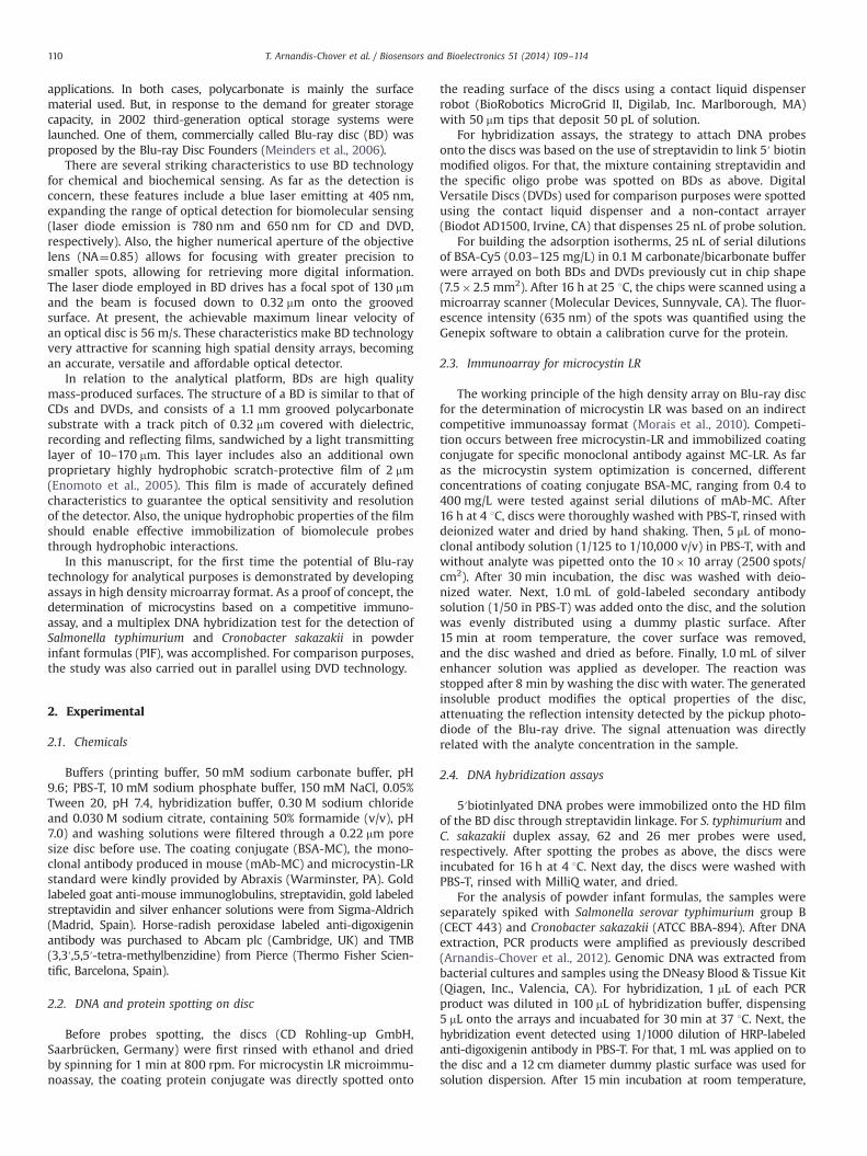
The BD drive used in this study was from LG Electronics Inc.(Englewood Cliffs, NJ), and connected to the PC through a USB2.0universal serial bus interface. The scanning of a recorded BD discwas performed using Nero Disc Speed 4.0 (Nero Inc., Glendale, CA)software. The analog signals were digitized with custom software(Biodisk) that controls the data acquisition board. Biodisk waswritten in Visual C++ and performs the data analysis. Briefly,during BD scanning, the laser diode hits the insoluble productwhich modifies the reflection properties of the BD surface,attenuating the laser beam intensity that reaches the photodiodeof the pickup (Morais et al., 2008). The attenuated analog signalsare directly acquired from the photodiode of the BD drive andrelated to the presence of the target analyte. During disc scanning,signals coming from detection areas are processed for digitizationand deconvoluted into an image. Data analysis was processed bycustom Biodisk software. A schematic representation of the sen-sing system is shown in Fig. 1.
Fig. 1. Schematic diagram of the Blu-ray sensing system. The picture
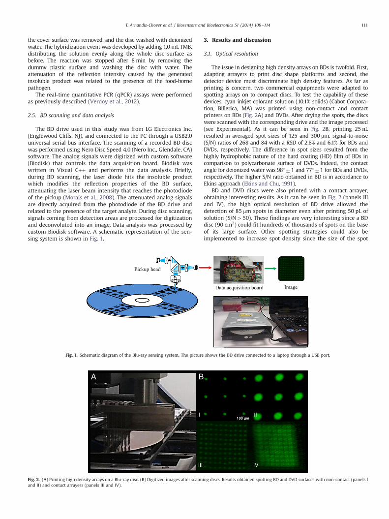
Fig. 2. (A) Printing high density arrays on a Blu-ray disc. (B) Digitized images after scannand II) and contact arrayers (panels III and IV).
3. Results and discussion
3.1. Optical resolution
The issue in designing high density arrays on BDs is twofold. First,adapting arrayers to print disc shape platforms and second, thedetector device must discriminate high density features. As far asprinting is concern, two commercial equipments were adapted tospotting arrays on to compact discs. To test the capability of thesedevices, cyan inkjet colorant solution (10.1% solids) (Cabot Corpora-tion, Billerica, MA) was printed using non-contact and contactprinters on BDs (Fig. 2A) and DVDs. After drying the spots, the discswere scanned with the corresponding drive and the image processed(see Experimental). As it can be seen in Fig. 2B, printing 25 nLresulted in averaged spot sizes of 125 and 300 mm, signal-to-noise(S/N) ratios of 268 and 84 with a RSD of 2.8% and 6.1% for BDs andDVDs, respectively. The difference in spot sizes resulted from thehighly hydrophobic nature of the hard coating (HD) film of BDs incomparison to polycarbonate surface of DVDs. Indeed, the contactangle for deionized water was 98171 and 77171 for BDs and DVDs,respectively. The higher S/N ratio obtained in BD is in accordance toEkins approach (Ekins and Chu, 1991).
BD and DVD discs were also printed with a contact arrayer,obtaining interesting results. As it can be seen in Fig. 2 (panels IIIand IV), the high optical resolution of BD drive allowed thedetection of 85 mm spots in diameter even after printing 50 pL ofsolution (S/N450). These findings are very interesting since a BDdisc (90 cm2) could fit hundreds of thousands of spots on the baseof its large surface. Other spotting strategies could also beimplemented to increase spot density since the size of the spot
shows the BD drive connected to a laptop through a USB port.
ing discs. Results obtained spotting BD and DVD surfaces with non-contact (panels I
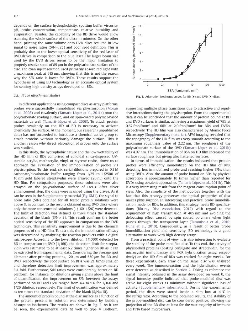
Fig. 3. Adsorption isotherms curves for BD (�) and DVD (■) discs.
T. Arnandis-Chover et al. / Biosensors and Bioelectronics 51 (2014) 109–114112
depends on the surface hydrophobicity, spotting buffer viscosity,pH, probe concentration, temperature, relative humidity andevaporation. Besides, the capability of the BD drive would allowscanning the whole surface of the discs in minutes. On the otherhand, printing the same volume onto DVD discs resulted in lowsignal to noise ratios (S/No25) and poor spot definition. This isprobably due to the lower optical sensitivity of the red laser ofDVD drives in comparison to the blue laser. The larger beam sizeused by the DVD drives seems to be the major limitation toproperly resolve spots of 85 mm in the polycarbonate surface of thedisc. The cyan inject solution used primarily absorb red light witha maximum peak at 615 nm, showing that this is not the reasonwhy the S/N ratio is lower for DVDs. These results support thehypothesis of using BD technology as an accurate optical systemfor sensing high density arrays developed on BDs.
3.2. Probe attachment studies
In different applications using compact discs as array platforms,probes were successfully immobilized via physisorption (Moraiset al., 2008) and covalently (Tamarit-López et al., 2011a) onto thepolycarbonate reading surface, and on spin-coated polymer-basedmaterials as well (Tamarit-López et al., 2008). To attach proteinprobes covalently on the HD of BD is necessary to activatechemically the surface. At the moment, our research (unpublisheddata) has not succeeded to introduce a chemical active group toattach proteins without seriously damage the surface. This isanother reason why direct adsorption of probes onto the surfacewas studied.
In this study, the hydrophobic nature and the low wettability ofthe HD film of BDs comprised of colloidal silica-dispersed UV-curable acrylic, methacrylic, vinyl, or styrene resins, drove us toapproach the evaluation of the immobilization of probes viaphysisorption. To this end, six serial dilutions prepared in 0.1 Mcarbonate/bicarbonate buffer ranging from 1/25 to 1/2500 of10 nm gold labeled streptavidin were arrayed (20 nL) onto theHD film. For comparison purposes, these solutions were alsoarrayed on the polycarbonate surface of DVDs. After silverenhancement step, the discs were scanned using the drives. As itcan be seen in the Supplementary material (Fig. SM1), the signal tonoise ratio (S/N) obtained for all tested protein solutions wereabove 3, in contrast to the results obtained using DVD discs whereonly the more concentrated solutions (1/160–1/26) were detected.The limit of detection was defined as three times the standarddeviation of the blank (S/N43). This result confirms the betteroptical sensitivity of the BD approach in comparison to the DVDtechnology. This sensitivity improvement is due to the chemicalproperties of the HD film. To test this, the immobilization efficacywas determined by analyzing the reaction products with a digitalmicroscope. According to the lower dilution (1/1000) detected forBD in comparison to DVD (1/160), the detection limit for strepta-vidin was estimated to be at least 6.2 times higher on BD as it canbe extracted from experimental data. Considering the average spotdiameter after printing proteins, 120 mm and 550 mm for BD andDVD, respectively, the spot surface on BDs was 21 times smaller,and therefore detection limit improvement was approximately3.4 fold. Furthermore, S/N ratios were considerably better on BDplatform; for instance, for dilutions giving signals above the limitof quantification, the improvement on S/N between the assaysperformed on BD and DVD ranged from 4.4 to 9.6 for 1/160 and1/26 dilution, respectively. The limit of quantification was definedas ten times the standard deviation of the blank (S/N¼10).
The amount of protein bound at the disc surface as a function ofthe protein present in solution was determined by buildingadsorption isotherms. The results are shown in Fig. 3. As it canbe seen, the experimental data fit well to type V isotherm,
suggesting multiple phase transitions due to attractive and repul-sive interactions during the physisorption. From the experimentaldata it can be concluded that the amount of protein bound at BDand DVD surfaces is similar, achieving a maximum yield of 79% at0.67 fmol/mm2 and 68% at 2.0 fmol/mm2 for BDs and DVDs,respectively. The HD film was also characterized by Atomic ForceMicroscopy (Supplementary material). AFM imaging revealed thatthe topography of the HD film was very smooth according to themaximum roughness value of 2.22 nm. The roughness of thepolycarbonate surface of the DVD (Tamarit-López et al., 2011b)was 4.07 nm. The immobilization of BSA on HD film increased thesurface roughness but giving also flattened surfaces.
In terms of immobilization, the results indicated that proteinprobes were efficiently immobilized on the HD film of BDs,detecting less immobilized probe and reaching higher S/Ns thanusing DVDs. Also, the amount of probe bound on BDs by physicaladsorption is approximately 10 times higher than reported forDVDs using covalent approaches (Tamarit-López et al., 2011b). Thisis a very interesting result from the reagent consumption point ofview. Also, the simplicity of the methodology together with thefact that this strategy preserves the optical properties of discmakes physisorption an interesting and practical probe immobili-zation mode for BDs. In addition, this strategy meets BD specifica-tions (Blu-ray Disc Association, 2012) with regard to therequirement of high transmission at 405 nm and avoiding thedefocusing effect caused by spin coated polymers when lightpasses through the transparent layer (Tammisola et al., 2010;Hung et al., 2010). Consequently, as a result of better probeimmobilization yield and sensitivity, BD technology is a goodalternative to work with high density arrays.
From a practical point of view, it is also interesting to evaluatethe stability of the probe-modified disc. To this end, the activity ofphysisorbed proteins (coating conjugate and streptavidin, for themicrocystin immunoassay and PCR hybridization assay, respec-tively) on the HD film of BDs was tracked for eight weeks. Forthese experiments, each array on the same disc was analyzedevery week. The immunoreaction and the hybridization eventswere detected as described in Section 2. Taking as reference thesignal intensity obtained in the assay developed on week 0, thesignal intensity profile indicated that probe-modified disc wasactive for eight weeks as minimum without significant loss ofactivity (Supplementary information). During the experimentalperiod, the discs were stored inside a slim box at 4 1C inthe refrigerator. According to the obtained results, the stability ofthe probe-modified disc can be considered positive; allowing theprojection of the BD disc at least for the vast majority of immuneand DNA based microarrays.
T. Arnandis-Chover et al. / Biosensors and Bioelectronics 51 (2014) 109–114 113
3.3. Proof of concepts
3.3.1. Immunoarray for microcystin LRThe promising results obtained with regard to the capability of
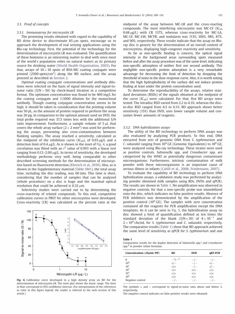
BD drive device to discriminate 85 mm spots, encourage us toapproach the development of real sensing applications using theBlu-ray technology. First, the potential of the technology for thedetermination of microcystin LR was evaluated. The quantificationof these biotoxins is an interesting matter to deal with since mostof the world′s population relies on natural waters as its primarysource for drinking water (World Health Organization, 2003). Forthat, arrays of 10�10 spots of BSA-MC coating conjugate wereprinted (2500 spots/cm2) along the BD surface, and the assayproceed as described in Section 2.
Optimal coating conjugate concentrations and antibody dilu-tions were selected on the basis of signal intensity and signal-to-noise ratio (S/N450) by check-board titration in a competitiveformat. The optimum concentration was found to be 400 mg/L forthe coating conjugate and 1/1000 dilution for the monoclonalantibody. Though coating conjugate concentration seems to behigh, it should be taken in consideration that the printing volumewas 50 pL, so the amount of protein required to perform the assaywas 20 pg. In comparison to the optimal amount used on DVD, thetotal probe required was 37.5 times less with the additional S/Nratio improvement. Furthermore, a sample volume of 5 μL thatcovers the whole array surface (2�2 mm2) was used for perform-ing the assays, preventing also cross-contamination betweenflanking samples. The assay reached a sensitivity, calculated asthe midpoint of the inhibition curve (IC50), of 0.93 μg/L and adetection limit of 0.4 mg/L. As is shown in the inset of Fig. 4, a goodcorrelation was fitted with an r2 value of 0.993 with a linear testranging from 0.12–2.00 mg/L. In terms of sensitivity, the developedmethodology performs very well, being comparable to otherdescribed screening methods for the determination of microcys-tins based on fluorescent detection (Khreich et al., 2010). Also, as isshown in the Supplementary material (Table SM1), the total assaytime, including the disc reading, was 60 min. This time is short,considering that the number of samples that can be analyzed(whole procedure) on a disc is huge and the maximal opticalresolution that could be achieved is 0.32 mm.
Selectivity studies were carried out to by determining thecross-reactivity of related compounds. To this end, competitivecalibration curves in PBST for other microcystins were developed.Cross-reactivity (CR) was calculated as the percent ratio at the
Fig. 4. Calibration curve developed in a high density array on BD for thedetermination of microcystin-LR. The inset plot shows the linear range. The linesin blue correspond to 95% confidence interval. (For interpretation of the referencesto color in this figure legend, the reader is referred to the web version of thisarticle.)
midpoint of the assay between MC-LR and the cross-reactingcompounds. The most interfering microcystin was MC-LY (IC500.68 μg/L) with CR 137%, whereas cross-reactivity for MC-LA,MC-LF, MC-LW, MCYR, and nodularin was 113%, 106%, 98%, 87%,and 90%, respectively. These results indicate that the assay on Blu-ray disc is generic for the determination of an overall content ofmicrocystins, displaying high-congener reactivity and sensitivity.
As far as non-specific binding is concern, the optical signalintensity in the background areas surrounding spots measuredbefore and after the assay procedure was of the same level, indicatingnon-specific adsorption of neither first nor second antibody. Thisnegligible non-specific protein adsorption is a very remarkableadvantage for decreasing the limit of detection by dropping thethreshold of noise in the dose-response curve. Also, it is worth notingthat the high hydrophobicity of the surface did not induce proteinfouling at least under the protein concentration used.
To determine the reproducibility of the assays, relative stan-dard deviations (RSDs) of the signals obtained at the midpoint ofthe curve (IC50) were calculated. For that, five BD arrays weretested. The intradisc RSD varied from 2.2 to 4.3%, whereas the disc-to-disc RSD ranged from 4.5 to 6.1%. BD approach shows bettersensitivity (15%) than DVD, uses lower sample volume and con-sumes fewer amounts of reagents.
3.3.2. DNA hybridization assaysThe utility of the BD technology to perform DNA assays was
also evaluated by analyzing PCR products. To this end, DNAextracted from sets of genomic DNA from S. typhimurium andC. sakazakii ranging from 104 GE (Genomic Equivalents) to 100 GE,were analyzed using Blu-ray technology. These strains were usedas positive controls. Salmonella spp. and Cronobacter spp. arecategorized by the WHO as potentially dangerous contaminantmicroorganisms. Furthermore, intrinsic contamination of milkpowder with these microorganisms is an important cause ofserious illness in infants (Cahill et al., 2008; Friedemann, 2007).
To evaluate the capability of BD technology to perform DNAhybridization assays, a validation study was performed by analyz-ing powder skimmed milk samples using BDs, DVDs and qPCR).The results are shown in Table 1. No amplification was observed innegative controls, for that a non-specific probe was immobilizedonto the disc, which indicates no false positive results. Absence ofPCR inhibitors was demonstrated by the amplification of thepositive control (104 GE). The samples with zero concentrationcontained all the reagents for PCR amplification except the DNAtemplates. As it can be seen in Fig. 5, the hybridization assay ondisc showed a limit of quantification defined as ten times thestandard deviation of the blank (S/N¼10) of 4�10�1 and2�100 cfu/mL for S. typhimurium and C. sakazakii, respectively.The comparative results (Table 1) show that BD approach achievedthe same level of sensitivity as qPCR for S. typhimurium and one
Table 1Comparative results for the duplex detection of Salmonella spp.a and Cronobacterspp.b in powder infant formulas.
Concentration (cfu/mL PIF) BD DVD qRT-PCR
0 �a/�b �/� �/�100 +/� �/� +/�101 +/+ �/� +/�102 +/+ +/+ +/+103 +/+ +/+ +/+104 +/+ +/+ +/+Negative control �/� �/� �/�
The symbols + and – correspond to signal-to-noise ratio above and below 3,respectively.The negative control indicates no false positive results were obtained.
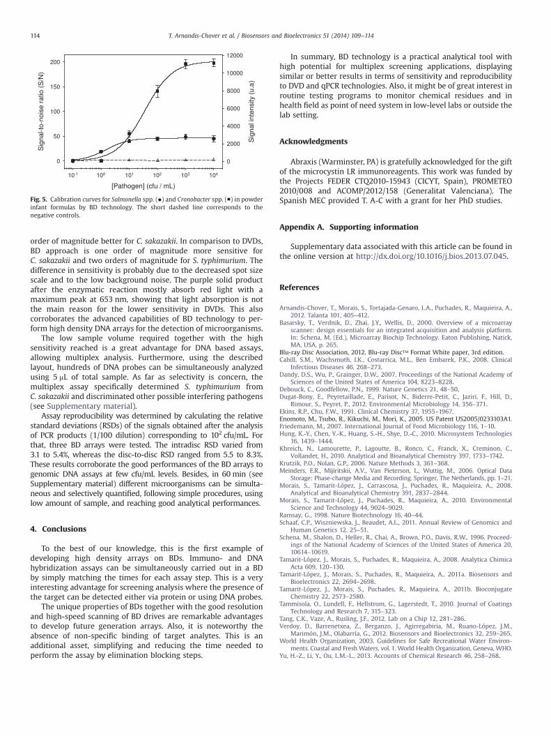
Fig. 5. Calibration curves for Salmonella spp. (�) and Cronobacter spp. (■) in powderinfant formulas by BD technology. The short dashed line corresponds to thenegative controls.
T. Arnandis-Chover et al. / Biosensors and Bioelectronics 51 (2014) 109–114114
order of magnitude better for C. sakazakii. In comparison to DVDs,BD approach is one order of magnitude more sensitive forC. sakazakii and two orders of magnitude for S. typhimurium. Thedifference in sensitivity is probably due to the decreased spot sizescale and to the low background noise. The purple solid productafter the enzymatic reaction mostly absorb red light with amaximum peak at 653 nm, showing that light absorption is notthe main reason for the lower sensitivity in DVDs. This alsocorroborates the advanced capabilities of BD technology to per-form high density DNA arrays for the detection of microorganisms.
The low sample volume required together with the highsensitivity reached is a great advantage for DNA based assays,allowing multiplex analysis. Furthermore, using the describedlayout, hundreds of DNA probes can be simultaneously analyzedusing 5 mL of total sample. As far as selectivity is concern, themultiplex assay specifically determined S. typhimurium fromC. sakazakii and discriminated other possible interfering pathogens(see Supplementary material).
Assay reproducibility was determined by calculating the relativestandard deviations (RSDs) of the signals obtained after the analysisof PCR products (1/100 dilution) corresponding to 102 cfu/mL. Forthat, three BD arrays were tested. The intradisc RSD varied from3.1 to 5.4%, whereas the disc-to-disc RSD ranged from 5.5 to 8.3%.These results corroborate the good performances of the BD arrays togenomic DNA assays at few cfu/mL levels. Besides, in 60 min (seeSupplementary material) different microorganisms can be simulta-neous and selectively quantified, following simple procedures, usinglow amount of sample, and reaching good analytical performances.
4. Conclusions
To the best of our knowledge, this is the first example ofdeveloping high density arrays on BDs. Immuno- and DNAhybridization assays can be simultaneously carried out in a BDby simply matching the times for each assay step. This is a veryinteresting advantage for screening analysis where the presence ofthe target can be detected either via protein or using DNA probes.
The unique properties of BDs together with the good resolutionand high-speed scanning of BD drives are remarkable advantagesto develop future generation arrays. Also, it is noteworthy theabsence of non-specific binding of target analytes. This is anadditional asset, simplifying and reducing the time needed toperform the assay by elimination blocking steps.
In summary, BD technology is a practical analytical tool withhigh potential for multiplex screening applications, displayingsimilar or better results in terms of sensitivity and reproducibilityto DVD and qPCR technologies. Also, it might be of great interest inroutine testing programs to monitor chemical residues and inhealth field as point of need system in low-level labs or outside thelab setting.
Acknowledgments
Abraxis (Warminster, PA) is gratefully acknowledged for the giftof the microcystin LR immunoreagents. This work was funded bythe Projects FEDER CTQ2010-15943 (CICYT, Spain), PROMETEO2010/008 and ACOMP/2012/158 (Generalitat Valenciana). TheSpanish MEC provided T. A-C with a grant for her PhD studies.
Appendix A. Supporting information
Supplementary data associated with this article can be found inthe online version at http://dx.doi.org/10.1016/j.bios.2013.07.045.
References
Arnandis-Chover, T., Morais, S., Tortajada-Genaro, L.A., Puchades, R., Maquieira, A.,2012. Talanta 101, 405–412.
Basarsky, T., Verdnik, D., Zhai, J.Y., Wellis, D., 2000. Overview of a microarrayscanner: design essentials for an integrated acquisition and analysis platform.In: Schena, M. (Ed.), Microarray Biochip Technology. Eaton Publishing, Natick,MA, USA, p. 265.
Blu-ray Disc Association, 2012. Blu-ray Disc™ Format White paper, 3rd edition.Cahill, S.M., Wachsmuth, I.K., Costarrica, M.L., Ben Embarek, P.K., 2008. Clinical
Infectious Diseases 46, 268–273.Dandy, D.S., Wu, P., Grainger, D.W., 2007. Proceedings of the National Academy of
Sciences of the United States of America 104, 8223–8228.Debouck, C., Goodfellow, P.N., 1999. Nature Genetics 21, 48–50.Dugat-Bony, E., Peyretaillade, E., Parisot, N., Biderre-Petit, C., Jaziri, F., Hill, D.,
Rimour, S., Peyret, P., 2012. Environmental Microbiology 14, 356–371.Ekins, R.P., Chu, F.W., 1991. Clinical Chemistry 37, 1955–1967.Enomoto, M., Tsubo, R., Kikuchi, M., Mori, K., 2005. US Patent US2005/0233103A1.Friedemann, M., 2007. International Journal of Food Microbiology 116, 1–10.Hung, K.-Y., Chen, Y.-K., Huang, S.-H., Shye, D.-C., 2010. Microsystem Technologies
16, 1439–1444.Khreich, N., Lamourette, P., Lagoutte, B., Ronco, C., Franck, X., Creminon, C.,
Vollandet, H., 2010. Analytical and Bioanalytical Chemistry 397, 1733–1742.Krutzik, P.O., Nolan, G.P., 2006. Nature Methods 3, 361–368.Meinders, E.R., Mijiritskii, A.V., Van Pieterson, L., Wuttig, M., 2006. Optical Data
Storage: Phase-change Media and Recording. Springer, The Netherlands, pp. 1–21.Morais, S., Tamarit-López, J., Carrascosa, J., Puchades, R., Maquieira, A., 2008.
Analytical and Bioanalytical Chemistry 391, 2837–2844.Morais, S., Tamarit-López, J., Puchades, R., Maquieira, A., 2010. Environmental
Science and Technology 44, 9024–9029.Ramsay, G., 1998. Nature Biotechnology 16, 40–44.Schaaf, C.P., Wiszniewska, J., Beaudet, A.L., 2011. Annual Review of Genomics and
Human Genetics 12, 25–51.Schena, M., Shalon, D., Heller, R., Chai, A., Brown, P.O., Davis, R.W., 1996. Proceed-
ings of the National Academy of Sciences of the United States of America 20,10614–10619.
Tamarit-López, J., Morais, S., Puchades, R., Maquieira, A., 2008. Analytica ChimicaActa 609, 120–130.
Tamarit-López, J., Morais, S., Puchades, R., Maquieira, A., 2011a. Biosensors andBioelectronics 22, 2694–2698.
Tamarit-López, J., Morais, S., Puchades, R., Maquieira, A., 2011b. BioconjugateChemistry 22, 2573–2580.
Tammisola, O., Lundell, F., Hellstrom, G., Lagerstedt, T., 2010. Journal of CoatingsTechnology and Research 7, 315–323.
Tang, C.K., Vaze, A., Rusling, J.F., 2012. Lab on a Chip 12, 281–286.Verdoy, D., Barrenetxea, Z., Berganzo, J., Agirregabiria, M., Ruano-López, J.M.,
Marimón, J.M., Olabarría, G., 2012. Biosensors and Bioelectronics 32, 259–265.World Health Organization, 2003. Guidelines for Safe Recreational Water Environ-
ments. Coastal and FreshWaters, vol. 1. World Health Organization, Geneva, WHO.Yu, H.-Z., Li, Y., Ou, L.M.-L., 2013. Accounts of Chemical Research 46, 258–268.