High beta-lactamase levels change the pharmacodynamics of beta
9
High -Lactamase Levels Change the Pharmacodynamics of -Lactam Antibiotics in Pseudomonas aeruginosa Biofilms Wang Hengzhuang, a Oana Ciofu, b Liang Yang, c,e Hong Wu, a,b,e Zhijun Song, a Antonio Oliver, d Niels Høiby a,b,f Department of Clinical Microbiology, Rigshospitalet, University Hospital of Copenhagen, Copenhagen, Denmark a ; Institute for International Health, Immunology and Microbiology, University of Copenhagen, Copenhagen, Denmark b ; Department of Systems Biology, Center for Systems Microbiology, Technical University of Denmark, Lyngby, Denmark c ; Servicio de Microbiología, Hospital Universitario Son Espases, Palma de Mallorca, Spain d ; The Singapore Centre on Environmental Life Sciences Engineering, Nanyang Technological University, Singapore e ; European Study Group for Biofilms, European Society of Clinical Microbiology and Infectious Diseases, Basel, Switzerland f Resistance to -lactam antibiotics is a frequent problem in Pseudomonas aeruginosa lung infection of cystic fibrosis (CF) pa- tients. This resistance is mainly due to the hyperproduction of chromosomally encoded -lactamase and biofilm formation. The purpose of this study was to investigate the role of -lactamase in the pharmacokinetics (PK) and pharmacodynamics (PD) of ceftazidime and imipenem on P. aeruginosa biofilms. P. aeruginosa PAO1 and its corresponding -lactamase-overproducing mutant, PADDh2Dh3, were used in this study. Biofilms of these two strains in flow chambers, microtiter plates, and on alg- inate beads were treated with different concentrations of ceftazidime and imipenem. The kinetics of antibiotics on the biofilms was investigated in vitro by time-kill methods. Time-dependent killing of ceftazidime was observed in PAO1 biofilms, but con- centration-dependent killing activity of ceftazidime was observed for -lactamase-overproducing biofilms of P. aeruginosa in all three models. Ceftazidime showed time-dependent killing on planktonic PAO1 and PADDh2Dh3. This difference is probably due to the special distribution and accumulation in the biofilm matrix of -lactamase, which can hydrolyze the -lactam antibi- otics. The PK/PD indices of the AUC/MBIC and C max /MBIC (AUC is the area under concentration-time curve, MBIC is the mini- mal biofilm-inhibitory concentration, and C max is the maximum concentration of drug in serum) are probably the best parame- ters to describe the effect of ceftazidime in -lactamase-overproducing P. aeruginosa biofilms. Meanwhile, imipenem showed time-dependent killing on both PAO1 and PADDh2Dh3 biofilms. An inoculum effect of -lactams was found for both plank- tonic and biofilm P. aeruginosa cells. The inoculum effect of ceftazidime for the -lactamase-overproducing mutant PADDh2Dh3 biofilms was more obvious than for PAO1 biofilms, with a requirement of higher antibiotic concentration and a longer period of treatment. D evelopment of resistance to -lactam antibiotics in clinical isolates of Pseudomonas aeruginosa is a common problem in the treatment of chronic lung infection in patients with cystic fibrosis (CF) (1, 2). The chronic lung infection in CF patients is difficult to eradicate because of biofilm formation in the respira- tory tract (3, 4). Typical and classical theories of pharmacokinetics and pharmacodynamics (PK/PD) have been mainly discussed for planktonic microorganisms (5, 6). However, cells in biofilms are different than planktonic cells with regard to growth rate (7), genes expressed (8, 9), and metabolism (10, 11). The role of -lac- tamase in treatment of biofilm infections is still unclear. The dose regimens for -lactam antibiotics on P. aeruginosa biofilms have not been clarified. Ceftazidime is an important antipseudomonal agent. The de- velopment of resistance to ceftazidime on P. aeruginosa growing in biofilms was evaluated in vitro and in vivo in a previous study (12). Resistance to ceftazidime in biofilm-growing P. aeruginosa is re- lated to the low growth rate of bacterial cells in the deep layers of the biofilm and to the production of chromosomal -lactamase (13–15). The kinetics of ceftazidime on -lactamase-overproduc- ing biofilms has not been studied. It was previously shown that -lactamase entrapped in the bio- film matrix might inactivate -lactam antibiotics (16). In previous studies (17, 18), we determined in vivo and in vitro the PK/PD parameters of -lactam antibiotics for biofilm treatments, and we showed that the time-dependent killing of -lactam antibiotics on planktonically grown cells was changed to a dose-dependent kill- ing on biofilms. We hypothesized that the entrapped -lactamase induced by the -lactam treatment might be the cause of changes in the kinetics of killing of antibiotics in biofilms. In the present work, we investigated the influence of hyperproduction of -lac- tamase on the PK/PD parameters by using three different models of in vitro biofilms. Time-kill curve approaches provide more meaningful infor- mation about the interactions between bacteria and antimicrobial agents (19–21). We evaluated the time-kill curves of ceftazidime and imipenem on P. aeruginosa growing in biofilms with basal or stable derepressed production of -lactamase, due to inactivation of the AmpC regulatory genes AmpDh1, Dh2, and Dh3. The well- described inoculum effect, representing the requirement of higher -lactam antibiotic concentrations to inhibit planktonic bacterial growth as the bacterial concentration increases, is related to the Received 10 July 2012 Returned for modification 20 August 2012 Accepted 14 October 2012 Published ahead of print 22 October 2012 Address correspondence to Wang Hengzhuang, [email protected]. Supplemental material for this article may be found at http://dx.doi.org/10.1128 /AAC.01393-12. Copyright © 2013, American Society for Microbiology. All Rights Reserved. doi:10.1128/AAC.01393-12 196 aac.asm.org Antimicrobial Agents and Chemotherapy p. 196 –204 January 2013 Volume 57 Number 1 Downloaded from https://journals.asm.org/journal/aac on 03 January 2022 by 85.163.110.210.
Transcript of High beta-lactamase levels change the pharmacodynamics of beta
High �-Lactamase Levels Change the Pharmacodynamics of �-LactamAntibiotics in Pseudomonas aeruginosa Biofilms
Wang Hengzhuang,a Oana Ciofu,b Liang Yang,c,e Hong Wu,a,b,e Zhijun Song,a Antonio Oliver,d Niels Høibya,b,f
Department of Clinical Microbiology, Rigshospitalet, University Hospital of Copenhagen, Copenhagen, Denmarka; Institute for International Health, Immunology andMicrobiology, University of Copenhagen, Copenhagen, Denmarkb; Department of Systems Biology, Center for Systems Microbiology, Technical University of Denmark,Lyngby, Denmarkc; Servicio de Microbiología, Hospital Universitario Son Espases, Palma de Mallorca, Spaind; The Singapore Centre on Environmental Life SciencesEngineering, Nanyang Technological University, Singaporee; European Study Group for Biofilms, European Society of Clinical Microbiology and Infectious Diseases, Basel,Switzerlandf
Resistance to �-lactam antibiotics is a frequent problem in Pseudomonas aeruginosa lung infection of cystic fibrosis (CF) pa-tients. This resistance is mainly due to the hyperproduction of chromosomally encoded �-lactamase and biofilm formation. Thepurpose of this study was to investigate the role of �-lactamase in the pharmacokinetics (PK) and pharmacodynamics (PD) ofceftazidime and imipenem on P. aeruginosa biofilms. P. aeruginosa PAO1 and its corresponding �-lactamase-overproducingmutant, PA�DDh2Dh3, were used in this study. Biofilms of these two strains in flow chambers, microtiter plates, and on alg-inate beads were treated with different concentrations of ceftazidime and imipenem. The kinetics of antibiotics on the biofilmswas investigated in vitro by time-kill methods. Time-dependent killing of ceftazidime was observed in PAO1 biofilms, but con-centration-dependent killing activity of ceftazidime was observed for �-lactamase-overproducing biofilms of P. aeruginosa in allthree models. Ceftazidime showed time-dependent killing on planktonic PAO1 and PA�DDh2Dh3. This difference is probablydue to the special distribution and accumulation in the biofilm matrix of �-lactamase, which can hydrolyze the �-lactam antibi-otics. The PK/PD indices of the AUC/MBIC and Cmax/MBIC (AUC is the area under concentration-time curve, MBIC is the mini-mal biofilm-inhibitory concentration, and Cmax is the maximum concentration of drug in serum) are probably the best parame-ters to describe the effect of ceftazidime in �-lactamase-overproducing P. aeruginosa biofilms. Meanwhile, imipenem showedtime-dependent killing on both PAO1 and PA�DDh2Dh3 biofilms. An inoculum effect of �-lactams was found for both plank-tonic and biofilm P. aeruginosa cells. The inoculum effect of ceftazidime for the �-lactamase-overproducing mutantPA�DDh2Dh3 biofilms was more obvious than for PAO1 biofilms, with a requirement of higher antibiotic concentration and alonger period of treatment.
Development of resistance to �-lactam antibiotics in clinicalisolates of Pseudomonas aeruginosa is a common problem in
the treatment of chronic lung infection in patients with cysticfibrosis (CF) (1, 2). The chronic lung infection in CF patients isdifficult to eradicate because of biofilm formation in the respira-tory tract (3, 4). Typical and classical theories of pharmacokineticsand pharmacodynamics (PK/PD) have been mainly discussed forplanktonic microorganisms (5, 6). However, cells in biofilms aredifferent than planktonic cells with regard to growth rate (7),genes expressed (8, 9), and metabolism (10, 11). The role of �-lac-tamase in treatment of biofilm infections is still unclear. The doseregimens for �-lactam antibiotics on P. aeruginosa biofilms havenot been clarified.
Ceftazidime is an important antipseudomonal agent. The de-velopment of resistance to ceftazidime on P. aeruginosa growing inbiofilms was evaluated in vitro and in vivo in a previous study (12).Resistance to ceftazidime in biofilm-growing P. aeruginosa is re-lated to the low growth rate of bacterial cells in the deep layers ofthe biofilm and to the production of chromosomal �-lactamase(13–15). The kinetics of ceftazidime on �-lactamase-overproduc-ing biofilms has not been studied.
It was previously shown that �-lactamase entrapped in the bio-film matrix might inactivate �-lactam antibiotics (16). In previousstudies (17, 18), we determined in vivo and in vitro the PK/PDparameters of �-lactam antibiotics for biofilm treatments, and weshowed that the time-dependent killing of �-lactam antibiotics onplanktonically grown cells was changed to a dose-dependent kill-
ing on biofilms. We hypothesized that the entrapped �-lactamaseinduced by the �-lactam treatment might be the cause of changesin the kinetics of killing of antibiotics in biofilms. In the presentwork, we investigated the influence of hyperproduction of �-lac-tamase on the PK/PD parameters by using three different modelsof in vitro biofilms.
Time-kill curve approaches provide more meaningful infor-mation about the interactions between bacteria and antimicrobialagents (19–21). We evaluated the time-kill curves of ceftazidimeand imipenem on P. aeruginosa growing in biofilms with basal orstable derepressed production of �-lactamase, due to inactivationof the AmpC regulatory genes AmpDh1, Dh2, and Dh3. The well-described inoculum effect, representing the requirement of higher�-lactam antibiotic concentrations to inhibit planktonic bacterialgrowth as the bacterial concentration increases, is related to the
Received 10 July 2012 Returned for modification 20 August 2012Accepted 14 October 2012
Published ahead of print 22 October 2012
Address correspondence to Wang Hengzhuang, [email protected].
Supplemental material for this article may be found at http://dx.doi.org/10.1128/AAC.01393-12.
Copyright © 2013, American Society for Microbiology. All Rights Reserved.
doi:10.1128/AAC.01393-12
196 aac.asm.org Antimicrobial Agents and Chemotherapy p. 196–204 January 2013 Volume 57 Number 1
Dow
nloa
ded
from
http
s://j
ourn
als.
asm
.org
/jour
nal/a
ac o
n 03
Jan
uary
202
2 by
85.
163.
110.
210.
production of �-lactamase (22). We have also studied the inocu-lum effect on planktonic and biofilm P. aeruginosa cells treatedwith ceftazidime and imipenem by using the time-kill method.Colistin was used as a control for a completely different mode ofaction (on bacterial membranes) (23) in comparison to �-lactams(on cell wall synthesis), targeting a different subpopulation (thecores of the biofilm mushrooms) compared to �-lactams (thesurfaces of the biofilm mushrooms), another PK/PD parameter(concentration dependent) for comparison with �-lactams (timedependent).
MATERIALS AND METHODSStrains, growth conditions, and chemicals. Laboratory strains, includingwild-type P. aeruginosa PAO1 (basal/ceftazidime-induced �-lactamaseactivity, 7.8 U/1,468 U of nitrocefin hydrolyzed/min/mg of protein) andits overproducing �-lactamase mutant, PA�DDh2Dh3 (10,934 U/9,413
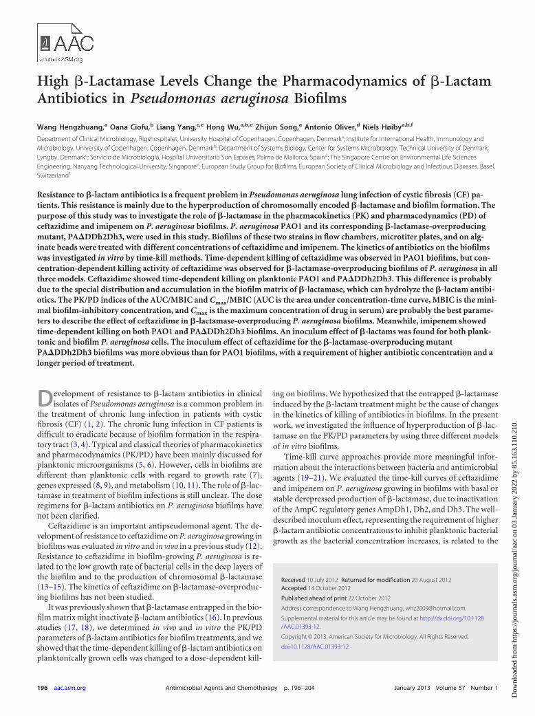
FIG 1 Time-kill curves of planktonic paired strains of PAO1 and �-lactamase-overproducing PA�DDh2Dh3 exposed to ceftazidime and imipenem. (A) PAO1exposed to ceftazidime, inoculum of 5�105 CFU/ml; (B) PA�DDh2Dh3 exposedto ceftazidime, inoculum of 5 � 105 CFU/ml; (C) PAO1 exposed to imipenem,inoculum of 5 � 107 CFU/ml; (D) PA�DDh2Dh3 exposed to imipenem, inocu-lum of 5 � 107 CFU/ml. Portions of similar time-kill curves are not shown.
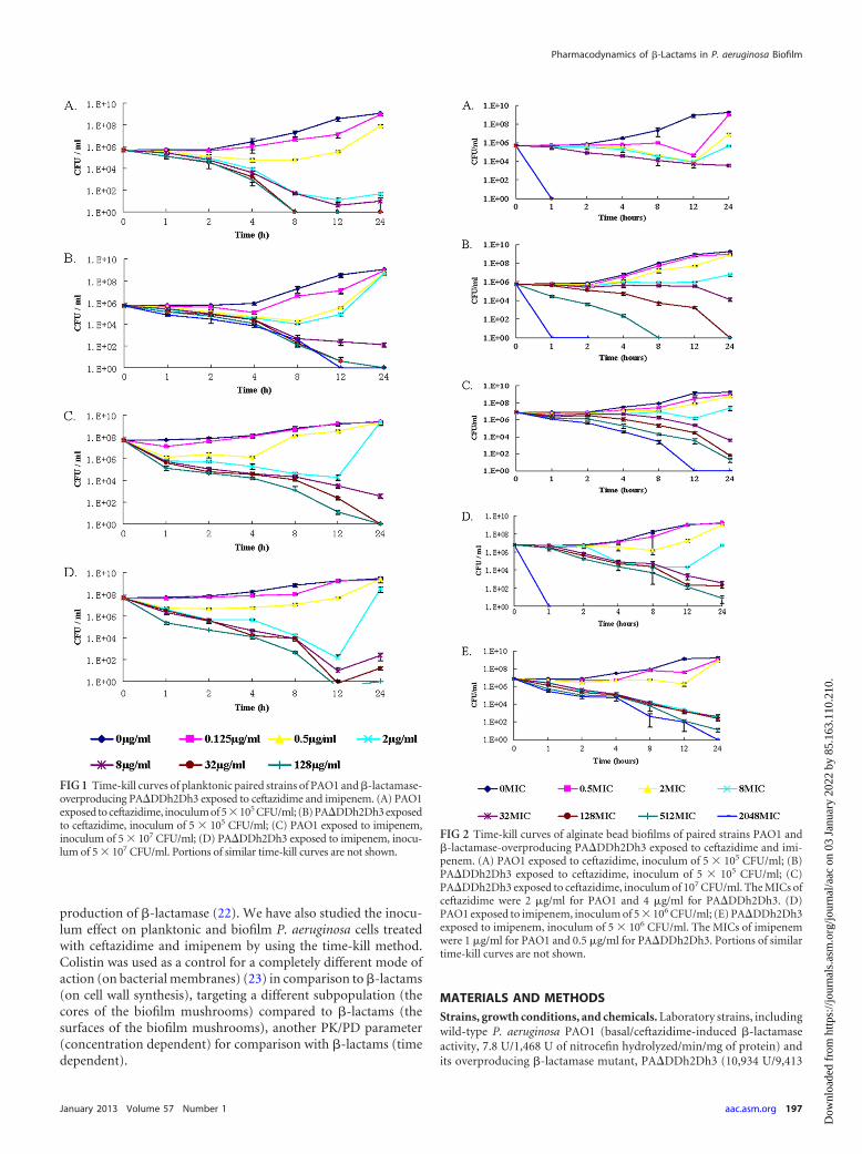
FIG 2 Time-kill curves of alginate bead biofilms of paired strains PAO1 and�-lactamase-overproducing PA�DDh2Dh3 exposed to ceftazidime and imi-penem. (A) PAO1 exposed to ceftazidime, inoculum of 5 � 105 CFU/ml; (B)PA�DDh2Dh3 exposed to ceftazidime, inoculum of 5 � 105 CFU/ml; (C)PA�DDh2Dh3 exposed to ceftazidime, inoculum of 107 CFU/ml. The MICs ofceftazidime were 2 �g/ml for PAO1 and 4 �g/ml for PA�DDh2Dh3. (D)PAO1 exposed to imipenem, inoculum of 5 � 106 CFU/ml; (E) PA�DDh2Dh3exposed to imipenem, inoculum of 5 � 106 CFU/ml. The MICs of imipenemwere 1 �g/ml for PAO1 and 0.5 �g/ml for PA�DDh2Dh3. Portions of similartime-kill curves are not shown.
Pharmacodynamics of �-Lactams in P. aeruginosa Biofilm
January 2013 Volume 57 Number 1 aac.asm.org 197
Dow
nloa
ded
from
http
s://j
ourn
als.
asm
.org
/jour
nal/a
ac o
n 03
Jan
uary
202
2 by
85.
163.
110.
210.
U) (24) were used in this study. The levels of �-lactamase in PAO1 andPA�DDh2Dh3 were detected by using a chromogenic cephalosporin aspreviously described (25). The gfp-tagged strains of PAO1-gfp andPA�DDh2Dh3-gfp for biofilm cultivation in a flow chamber were con-structed by insertion of a mini-Tn7-eGFP-Gmr cassette (26). The growthminimal medium (ABTG medium) for biofilm cultivation in microtiterplates and on alginate beads consisted of 1 mM MgCl2, 0.1 mM CaCl2,15.1 mM (NH4)2SO4, 33.7 mM Na2HPO4 · 2H2O, 22 mM KH2PO4, 51mM NaCl, 0.01 mM FeCl3, 2.5 �g/ml thiamine, and 0.5% glucose (27).ABTG medium was refreshed every 24 h in wells for biofilm cultivation.Ceftazidime (Sandoz GmbH, Kundl, Austria), colistin (Colimycin; Lun-beck A/S, Denmark), and imipenem (Tienam, MSD) were pharmaceuti-cal grade and stored at 4°C. Antibiotics were dissolved in saline and thendiluted with ABTG medium before application.
Standard susceptibility assay. MICs and minimal bactericidal con-centrations (MBCs) for planktonic bacteria were determined by the mi-crotiter method (28). The MICs were also detected by Etest (28).
Killing curves for antibiotics on planktonic cells. Planktonic P.aeruginosa cells in a 0.1-ml volume containing approximately 105,106, or107 CFU/ml were added into microtiter wells with ABTG medium (18).Then, 0.1 ml of different concentrations of ceftazidime, imipenem, andcolistin were mixed into different wells, and cultures were shaken andcultivated at 37°C for 0, 1, 2, 4, 8, 12, and 24 hours. Then, 0.1-ml samples(3 samples/time point/concentration) from wells were serially diluted andcultured on plates for overnight incubation, and CFU counts were deter-mined for killing curves of antibiotics on planktonic cells of PAO1 andPA�DDh2Dh3. Colistin was used as a control antibiotic. This experimentwas performed two times.
Kinetics of ceftazidime, imipenem, and colistin on alginate beadbiofilms. To prepare the biofilm bacteria, planktonic P. aeruginosa cellswere was immobilized on spherical alginate beads, as previously described(29–31). Different inocula of alginate bead biofilms were stained withSYTO 9 (32) (see Fig. S2 in the supplemental material). Alginate beads(105,106, or 107 CFU/ml) were transferred into flat-bottom microtiterplates containing different concentrations (0 to 8,192 �g/ml) of antibiot-ics in 0.2 ml of ABTG medium per well (antibiotic challenge plates) andincubated for 0 h, 1 h, 2 h, 4 h, 8 h, 12 h, or 24 h at 37°C. After antibioticincubation, the samples (3 samples/time point/concentration) were har-vested from wells, serially diluted, and then cultured on plates overnight,and counting of CFU was performed to determine killing curves of anti-biotics for alginate beads. This experiment was performed two times.
Kinetics of ceftazidime, imipenem, and colistin on microtiter bio-films. Isolates were passed twice on agar plates after retrieval from �80°Cstorage and then grown overnight in Luria-Bertani (LB) medium. Afterdilution of the cultures to �107 CFU/ml with ABTG medium, 0.15 ml wastransferred to all except the negative-control wells of a round-bottom96-well microtiter plate (catalog number 163320; Nunc A/S, Roskilde,Denmark). Bacterial biofilms were formed on the surfaces of modifiedpolystyrene microtiter plates (the biofilm growth plates), followed by aer-obic incubation at 37°C for 5 days without shaking. Pilot studies wereperformed to optimize the conditions for biofilm cultivation. The devel-opment from young biofilm to mature biofilm in microtiter wells isshown in Fig. S1 of the supplemental material. Fresh ABTG medium waschanged very 24 h. Relative humidity (RH) for biofilm cultivation wasabove 95%. The crystal violet staining method (33) was employed to checkthe biofilm formation on wells. To determine the microtiter, wells wererinsed three times with sterile water. Then, 0.15 ml ABTG medium wasadded into each well. Biofilms on the surface were scratched by a 10-�lblue inoculation loop (Aktiengesellschaft & Co., Numbrecht Germany)and suspended in the ABTG medium. Biofilms cells were collected fromthe wells into a 50-ml plastic tube, mixed by gentle shaking, and diluted to105, 106, or 107 CFU/ml. Resuspended biofilm cells were stained withSYTO 9 to check biofilm structure (32) (see Fig. S1). Then, 0.1-ml aliquotsof biofilms cells were placed into flat-bottom microtiter plates (catalognumber 269787; Nunc A/S, Roskilde, Denmark) containing antibiotics
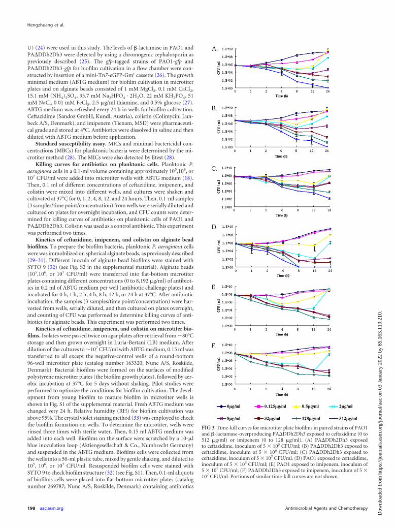
FIG 3 Time-kill curves for microtiter plate biofilms in paired strains of PAO1and �-lactamase-overproducing PA�DDh2Dh3 exposed to ceftazidime (0 to512 �g/ml) or imipenem (0 to 128 �g/ml). (A) PA�DDh2Dh3 exposedto ceftazidime, inoculum of 5 � 105 CFU/ml; (B) PA�DDh2Dh3 exposed toceftazidime, inoculum of 5 � 106 CFU/ml; (C) PA�DDh2Dh3 exposed toceftazidime, inoculum of 5 � 107 CFU/ml. (D) PAO1 exposed to ceftazidime,inoculum of 5 � 105 CFU/ml; (E) PAO1 exposed to imipenem, inoculum of5 � 107 CFU/ml; (F) PA�DDh2Dh3 exposed to imipenem, inoculum of 5 �107 CFU/ml. Portions of similar time-kill curves are not shown.
Hengzhuang et al.
198 aac.asm.org Antimicrobial Agents and Chemotherapy
Dow
nloa
ded
from
http
s://j
ourn
als.
asm
.org
/jour
nal/a
ac o
n 03
Jan
uary
202
2 by
85.
163.
110.
210.
with 0.1-ml aliquots of 2-fold dilutions in ABTG medium per well (anti-biotic challenge plates) and cultured for 1 h to 24 h at 37°C, working withthree wells per concentration. Samples (3 samples/time point/concentra-tion) were collected at different time points. After serial dilution, a 0.1-mlsample was put into each plate and the cells were cultivated for CFUcounting. Then, the killing curves were determined. This experiment wasperformed two times. The surface-detached biofilms of P. aeruginosa cellsare relevant for non-surface-associated biofilms, such as P. aeruginosabiofilms during chronic infection in CF (34).
Flow chamber biofilm formation and kinetics study of ceftazidime.P. aeruginosa biofilms were cultivated at 30°C in flow chambers irrigatedwith a minimal medium (35) supplemented with 0.3 mM glucose as pre-viously reported (36). Biofilms were grown in flow chambers with indi-vidual channel dimensions of 1 by 4 by 40 mm. The flow system wasassembled and prepared as described previously (36). The flow chamberswere inoculated by injecting 350-�l aliquots of overnight cultures dilutedto an optical density at 600 nm of 0.001 into each flow channel by using asmall syringe. After inoculation, the flow channels were left without flowfor 1 h, after which medium flow was started with a Watson Marlow 205Speristaltic pump. The mean flow velocity in the flow chambers was 0.2mm s�1, corresponding to laminar flow with a Reynolds number of 0.02.Day 4 biofilms were treated with different concentrations of ceftazidimefor 2 h, 4 h, 8 h, and 24 h. Then, microscopic observations were performedon a Zeiss LSM510 confocal laser scanning microscope (CLSM; Carl Zeiss,Jena, Germany) equipped with an argon laser detector and filter sets formonitoring of green fluorescent protein (GFP) fluorescence and 0.3 �Mpropidium iodide (PI). Images were obtained using a 40�/1.3 numericalaperture Plan-Neofluar oil objective lens. Vertical cross-sectional imageswere generated using the IMARIS software package (Bitplane AG, Zurich,Switzerland). CLSM images were analyzed by use of the computer pro-gram COMSTAT (35). For CLSM, images acquired at six randomly cho-sen locations were used to analyze biofilm formation of each strain. Afixed threshold value and connected volume filtration were used for allimage stacks. Rates of living/dead biomass (green channel/red channel)were used to make the killing curve for ceftazidime on biofilms. Thisexperiment was performed two times.
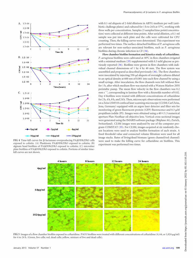
FIG 4 Time-kill curves for �-lactamase-overproducing PA�DDh2Dh3 cellsexposed to colistin. (A) Planktonic PA�DDh2Dh3 exposed to colistin; (B)alginate bead biofilms of PA�DDh2Dh3 exposed to colistin; (C) microtiterplate biofilms of PA�DDh2Dh3 exposed to colistin. Portions of similar time-kill curves are not shown.
FIG 5 Images of a flow chamber biofilm exposed to ceftazidime. PAO1 biofilms were treated with different concentrations of ceftazidime (4, 64, or 1,024 �g/ml)for 4 to 24 h. (Green, live cells; red, dead cells; yellow, mixture of live and dead cells).
Pharmacodynamics of �-Lactams in P. aeruginosa Biofilm
January 2013 Volume 57 Number 1 aac.asm.org 199
Dow
nloa
ded
from
http
s://j
ourn
als.
asm
.org
/jour
nal/a
ac o
n 03
Jan
uary
202
2 by
85.
163.
110.
210.
RESULTSKilling curves of antibiotics on planktonic cells. The killing pro-files of ceftazidime and imipenem against planktonic P. aeruginosaPAO1 and �-lactamase-overproducing PA�DDh2Dh3 are shownin Fig. 1. The bactericidal activities of ceftazidime and imipenemappeared to be time dependent for both planktonic PAO1 andPA�DDh2Dh3 cells. The MBC of ceftazidime for PAO1 was 32�g/ml at the 8-h time point, but for PA�DDh2Dh3 it was 32�g/ml at the 12-h time point approximately. The killing effect ofceftazidime correlated with the time of treatment on both plank-tonic PAO1 and PA�DDh2Dh3 cells. A correlation was shownbetween the killing rate and the treatment time for ceftazidimeand imipenem on both planktonic PAO1 and PA�DDh2Dh3cells. Colistin showed concentration-dependent killing on bothplanktonic PAO1 (18) and PA�DDh2Dh3 cells (see Fig. 4A, be-low).
Killing kinetics of ceftazidime, imipenem, and colistinagainst alginate bead biofilms. Killing profiles of ceftazidime andimipenem for biofilms on alginate beads are shown in Fig. 2. Thebactericidal activity of ceftazidime appeared to be time dependentfor PAO1 on alginate beads but to be concentration dependent forPA�DDh2Dh3 on alginate beads. In the inoculum of 5 � 105
CFU/ml group, a rapid reduction in bacterial burden was seenwithin 1 h when PAO1 on alginate beads was exposed to 128�MIC (256 �g/ml) and when PA�DDh2Dh3 on beads was exposedto 2,048� MIC (8,192 �g/ml) of ceftazidime. The minimal bio-film eradication concentration (MBEC) of ceftazidime wasachieved after 1 h for PAO1 (128� MIC; 256 �g/ml) and after 24h for PA�DDh2Dh3 (128� MIC; 512 �g/ml). The killing effect ofceftazidime was found to correlate with the time of treatment onPAO1 alginate beads (Fig. 2A); however, for PA�DDh2Dh3 onalginate beads it was correlated with the concentration of ceftazi-dime (Fig. 2B and C). Imipenem showed time-dependent killingfor both PAO1 and PA�DDh2Dh3 on alginate beads (Fig. 2D andE). The killing rate on alginate beads was related to the time oftreatment with ceftazidime for PAO1, but it was related to theconcentration of antibiotic for PA�DDh2Dh3. Colistin showedconcentration-dependent killing on alginate bead biofilms ofPA�DDh2Dh3 (see Fig. 4B).
Killing kinetics of ceftazidime, imipenem, and colistinagainst microtiter well biofilms. To confirm the concentration-dependent killing of ceftazidime, as shown with PA�DDh2Dh3 alg-inate beads previously, the killing profiles of ceftazidime against themicrotiter plate biofilms of P. aeruginosa PA�DDh2Dh3 were deter-mined (Fig. 3). Overall, the bactericidal activity of ceftazidime ap-peared to be concentration dependent for microtiter plate biofilmcells of PA�DDh2Dh3 (Fig. 3). The MBEC of ceftazidime onPA�DDh2Dh3 was 512 �g/ml at the 24-h time point for the twoinoculum groups of 5 � 105 CFU/ml and 5 � 106 CFU/ml (Fig. 3Aand B), but a higher concentration of ceftazidime was required forthe 5 � 107 CFU/ml group to eradicate the biofilm cells (Fig. 3C).The killing effect of ceftazidime was found to correlate with theconcentration of treatment on biofilm PA�DDh2Dh3 cells andtime-dependent killing on biofilm PAO1 cells, but the killing ef-fect of imipenem was time dependent on both the PAO1 andPA�DDh2Dh3 microtiter plate biofilms (Fig. 3). Colistin showedconcentration-dependent killing on both PAO1 (18) andPA�DDh2Dh3 microtiter plate biofilms (Fig. 4C). A portion ofthe results for imipenem and colistin in microtiter plate biofilmswas reported in a previous study (18).
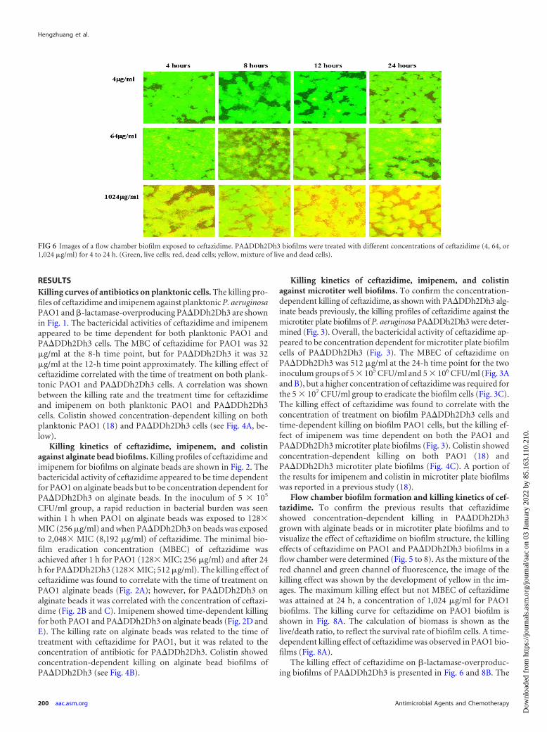
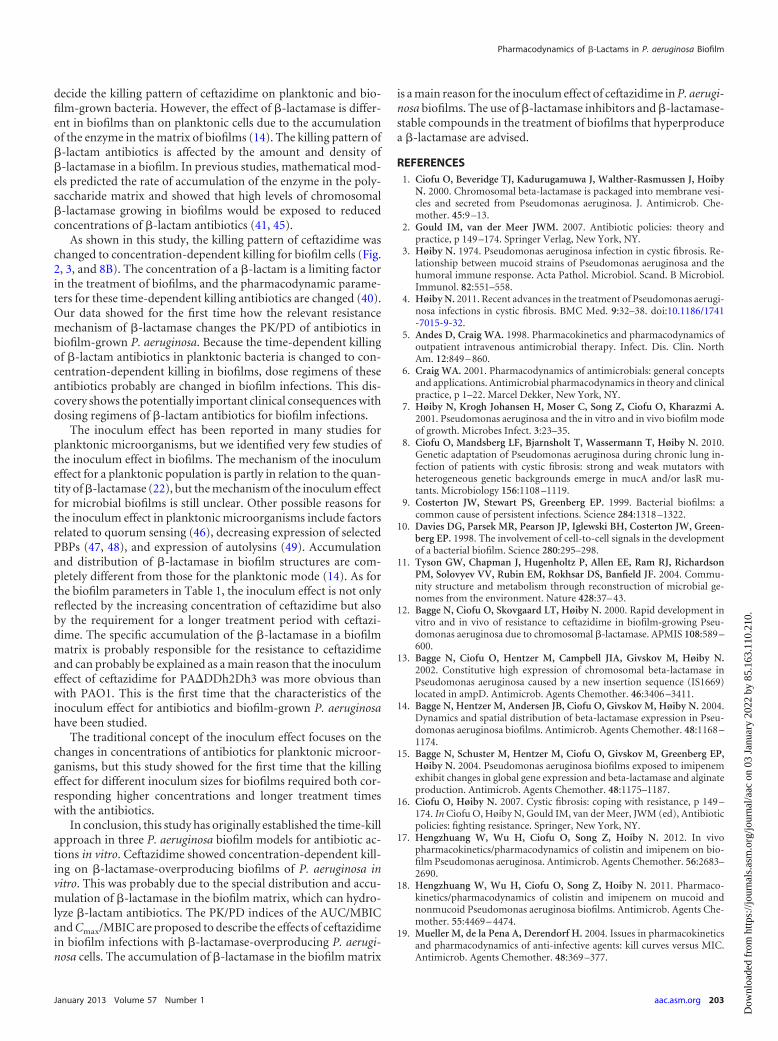
Flow chamber biofilm formation and killing kinetics of cef-tazidime. To confirm the previous results that ceftazidimeshowed concentration-dependent killing in PA�DDh2Dh3grown with alginate beads or in microtiter plate biofilms and tovisualize the effect of ceftazidime on biofilm structure, the killingeffects of ceftazidime on PAO1 and PA�DDh2Dh3 biofilms in aflow chamber were determined (Fig. 5 to 8). As the mixture of thered channel and green channel of fluorescence, the image of thekilling effect was shown by the development of yellow in the im-ages. The maximum killing effect but not MBEC of ceftazidimewas attained at 24 h, a concentration of 1,024 �g/ml for PAO1biofilms. The killing curve for ceftazidime on PAO1 biofilm isshown in Fig. 8A. The calculation of biomass is shown as thelive/death ratio, to reflect the survival rate of biofilm cells. A time-dependent killing effect of ceftazidime was observed in PAO1 bio-films (Fig. 8A).
The killing effect of ceftazidime on �-lactamase-overproduc-ing biofilms of PA�DDh2Dh3 is presented in Fig. 6 and 8B. The
FIG 6 Images of a flow chamber biofilm exposed to ceftazidime. PA�DDh2Dh3 biofilms were treated with different concentrations of ceftazidime (4, 64, or1,024 �g/ml) for 4 to 24 h. (Green, live cells; red, dead cells; yellow, mixture of live and dead cells).
Hengzhuang et al.
200 aac.asm.org Antimicrobial Agents and Chemotherapy
Dow
nloa
ded
from
http
s://j
ourn
als.
asm
.org
/jour
nal/a
ac o
n 03
Jan
uary
202
2 by
85.
163.
110.
210.
maximum killing effect of ceftazidime was reached at 24 h at aconcentration of 1,024 �g/ml, but the killing effect was shown tobe concentration dependent on biofilms of PA�DDh2Dh3.
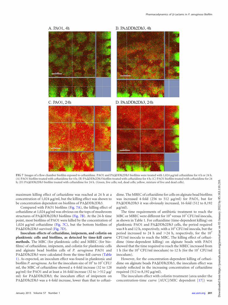
Compared with PAO1 biofilms (Fig. 7A), the killing effect ofceftazidime at 1,024 �g/ml was obvious on the tops of mushroomstructures of PA�DDh2Dh3 biofilms (Fig. 7B). At the 24-h timepoint, most biofilms of PAO1 were killed by the concentration of1,024 �g/ml ceftazidime (Fig. 7C), but the bottom biofilms ofPA�DDh2Dh3 survived (Fig. 7D).
Inoculum effects of ceftazidime, imipenem, and colistin onplanktonic cells and biofilms, as detected by time-kill curvemethods. The MBC (for planktonic cells) and MBEC (for bio-films) of ceftazidime, imipenem, and colistin for planktonic cellsand alginate bead biofilm cells of P. aeruginosa PAO1 andPA�DDh2Dh3 were calculated from the time-kill curves (Table1). As expected, an inoculum effect was found in planktonic andbiofilm P. aeruginosa. As for the inoculum size of 105 to 107 CFU/ml, the MBC of ceftazidime showed a 4-fold increase (32 to 128�g/ml) for PAO1 and at least a 16-fold increase (32 to �512 �g/ml) for PA�DDh2Dh3; the inoculum effect of imipenem onPA�DDh2Dh3 was a 4-fold increase, lower than that to ceftazi-
dime. The MBEC of ceftazidime for cells on alginate bead biofilmswas increased 4-fold (256 to 512 �g/ml) for PAO1, but forPA�DDh2Dh3 it was obviously increased, 16-fold (512 to 8,192�g/ml).
The time requirements of antibiotic treatment to reach theMBC or MBEC were different for 105 versus 107 CFU/ml inocula,as shown in Table 1. For ceftazidime (time-dependent killing) onplanktonic PAO1 and PA�DDh2Dh3 cells, the period requiredwas 8 h and 12 h, respectively, with a 105 CFU/ml inocula, but theperiod increased to 24 h and �24 h, respectively, for the 107
CFU/ml inocula to reach the MBC. The killing effect of ceftazi-dime (time-dependent killing) on alginate beads with PAO1showed that the time required to reach the MBEC increased from1 h (for the 105 CFU/ml inoculum) to 12 h (for the 107 CFU/mlinoculum).
However, for the concentration-dependent killing of ceftazi-dime on alginate beads PA�DDh2Dh3, the inoculum effect wasmainly reflected in the increasing concentration of ceftazidimerequired (512 to 8,192 �g/ml).
The inoculum effect with colistin treatment (area under theconcentration-time curve [AUC]/MIC dependent [17]) was
FIG 7 Images of a flow chamber biofilm exposed to ceftazidime. PAO1 and PA�DDh2Dh3 biofilms were treated with 1,024 �g/ml ceftazidime for 4 h or 24 h.(A) PAO1 biofilm treated with ceftazidime for 4 h; (B) PA�DDh2Dh3 biofilm treated with ceftazidime for 4 h; (C) PAO1 biofilm treated with ceftazidime for 24h; (D) PA�DDh2Dh3 biofilm treated with ceftazidime for 24 h. (Green, live cells; red, dead cells; yellow, mixture of live and dead cells).
Pharmacodynamics of �-Lactams in P. aeruginosa Biofilm
January 2013 Volume 57 Number 1 aac.asm.org 201
Dow
nloa
ded
from
http
s://j
ourn
als.
asm
.org
/jour
nal/a
ac o
n 03
Jan
uary
202
2 by
85.
163.
110.
210.
observed as a slight (2-fold) increase in concentration or in thetreatment time for both planktonic and biofilm PAO1 andPA�DDh2Dh3 cells.
DISCUSSION
Many studies have shown the time-dependent killing effect of cef-tazidime on planktonic bacteria (37–39), but there have been noreports for biofilm-grown organisms. In this study, the killing
curves of ceftazidime and imipenem on biofilm-growing P.aeruginosa were established in order to evaluate their bactericidalactivities in vitro (Fig. 2, 3, and 8). Compared with planktonicbacteria, the time-dependent kinetics of ceftazidime killing onbiofilm-grown bacteria of wild-type P. aeruginosa PAO1 were re-tained. However, for antibiotic treatment of biofilm-growing P.aeruginosa PAO1, higher doses and longer treatment times withceftazidime were required than for planktonic cells (Fig. 1 and 2).These results showed that both treatment time and concentrationshould be considered when treating biofilms. We previously re-ported that the time exceeding the minimal biofilm inhibitoryconcentration (T�MBIC) is a better PK/PD parameter than thetime exceeding the MIC for planktonic cells (T�MIC) for treat-ment of biofilm infections with �-lactam antibiotics (17).
Few studies have looked into the interaction of �-lactamaseand antibiotics in biofilm structures (14). In the present study, thekilling effects of �-lactam antibiotics were investigated on bio-films of a �-lactamase-overproducing strain. The �-lactamase inthe biofilm matrix can lead to the hydrolysis of the �-lactam an-tibiotics before they reach the bacterial cells (40, 41). The source of�-lactamase in biofilms has been considered to be from a sacrifi-cial layer of bacteria exposed to an antibiotic, with release of de-fensive enzymes into the extracellular space (14, 15).
Interestingly, ceftazidime showed time-dependent killing onplanktonic cells but concentration-dependent killing on biofilmsof the �-lactamase-overproducing strain (Fig. 1, 2, and 8). Forbiofilm infections with the �-lactamase-overproducing strain, theoptimized PK/PD parameters for ceftazidime are probably theAUC/MBIC or the maximum concentration of drug in serum[Cmax]/MBIC ratio. In contrast, imipenem, which is more stableto AmpC �-lactamase (42), maintained the time-dependent kill-ing on biofilms of PA�DDh2Dh3 (Fig. 2 and 3).
Although ceftazidime showed concentration-dependent kill-ing on �-lactamase-overproducing biofilms (Fig. 2, 3, and 8B), thekilling was slower than the fast killing observed for typical concen-tration-dependent antibiotics like colistin (Fig. 4). �-Lactam an-tibiotics work mainly by inhibiting cell wall biosynthesis by themicroorganism (43). The �-lactam nucleus of the molecule irre-versibly binds to the penicillin-binding protein (PBP) active sites(44). This irreversible inhibition of the PBPs prevents the finalcross-linking (transpeptidation) of the nascent peptidoglycanlayer, disrupting cell wall synthesis (44). The basic mechanisms
FIG 8 Time-kill curves of flow chamber biofilms of PAO1 and �-lactamase-overproducing PA�DDh2Dh3 exposed to different doses ceftazidime. (A)PAO1 flow chamber biofilm results; (B) PA�DDh2Dh3 flow chamber biofilmresults.
TABLE 1 MBC, MBEC, and inoculum effect of ceftazidime, imipenem, and colistin for planktonic and alginate bead biofilms of P. aeruginosaPAO1 and PA�DDh2Dh3 by the time-kill methoda
Inoculum group
Planktonic cells Biofilm cells on alginate beads
MBC (�g/ml) Timeb (h) MBEC (�g/ml) Timeb (h)
TZ IM CO TZ IM CO TZ IM CO TZ IM CO
PAO1(CFU/ml)105 32 8 8 8 8 2 256 512 64 1 12 2106 32 16 8 12 12 2 512 512 64 8 24 4107 128 32 16 24 24 4 512 �2,048 128 12 �24 8
PA�DDh2Dh3 (CFU/ml)105 32 8 16 12 8 2 512 256 64 24 12 2106 128 16 16 24 8 2 2,048 1,024 64 24 24 4107 �512 32 16 �24 12 4 8192 1,024 64 12 24 4
a TZ, ceftazidime; IM, imipenem; CO, colistin.b Required time for antibiotic treatment to reach the MBC or MBEC.
Hengzhuang et al.
202 aac.asm.org Antimicrobial Agents and Chemotherapy
Dow
nloa
ded
from
http
s://j
ourn
als.
asm
.org
/jour
nal/a
ac o
n 03
Jan
uary
202
2 by
85.
163.
110.
210.
decide the killing pattern of ceftazidime on planktonic and bio-film-grown bacteria. However, the effect of �-lactamase is differ-ent in biofilms than on planktonic cells due to the accumulationof the enzyme in the matrix of biofilms (14). The killing pattern of�-lactam antibiotics is affected by the amount and density of�-lactamase in a biofilm. In previous studies, mathematical mod-els predicted the rate of accumulation of the enzyme in the poly-saccharide matrix and showed that high levels of chromosomal�-lactamase growing in biofilms would be exposed to reducedconcentrations of �-lactam antibiotics (41, 45).
As shown in this study, the killing pattern of ceftazidime waschanged to concentration-dependent killing for biofilm cells (Fig.2, 3, and 8B). The concentration of a �-lactam is a limiting factorin the treatment of biofilms, and the pharmacodynamic parame-ters for these time-dependent killing antibiotics are changed (40).Our data showed for the first time how the relevant resistancemechanism of �-lactamase changes the PK/PD of antibiotics inbiofilm-grown P. aeruginosa. Because the time-dependent killingof �-lactam antibiotics in planktonic bacteria is changed to con-centration-dependent killing in biofilms, dose regimens of theseantibiotics probably are changed in biofilm infections. This dis-covery shows the potentially important clinical consequences withdosing regimens of �-lactam antibiotics for biofilm infections.
The inoculum effect has been reported in many studies forplanktonic microorganisms, but we identified very few studies ofthe inoculum effect in biofilms. The mechanism of the inoculumeffect for a planktonic population is partly in relation to the quan-tity of �-lactamase (22), but the mechanism of the inoculum effectfor microbial biofilms is still unclear. Other possible reasons forthe inoculum effect in planktonic microorganisms include factorsrelated to quorum sensing (46), decreasing expression of selectedPBPs (47, 48), and expression of autolysins (49). Accumulationand distribution of �-lactamase in biofilm structures are com-pletely different from those for the planktonic mode (14). As forthe biofilm parameters in Table 1, the inoculum effect is not onlyreflected by the increasing concentration of ceftazidime but alsoby the requirement for a longer treatment period with ceftazi-dime. The specific accumulation of the �-lactamase in a biofilmmatrix is probably responsible for the resistance to ceftazidimeand can probably be explained as a main reason that the inoculumeffect of ceftazidime for PA�DDh2Dh3 was more obvious thanwith PAO1. This is the first time that the characteristics of theinoculum effect for antibiotics and biofilm-grown P. aeruginosahave been studied.
The traditional concept of the inoculum effect focuses on thechanges in concentrations of antibiotics for planktonic microor-ganisms, but this study showed for the first time that the killingeffect for different inoculum sizes for biofilms required both cor-responding higher concentrations and longer treatment timeswith the antibiotics.
In conclusion, this study has originally established the time-killapproach in three P. aeruginosa biofilm models for antibiotic ac-tions in vitro. Ceftazidime showed concentration-dependent kill-ing on �-lactamase-overproducing biofilms of P. aeruginosa invitro. This was probably due to the special distribution and accu-mulation of �-lactamase in the biofilm matrix, which can hydro-lyze �-lactam antibiotics. The PK/PD indices of the AUC/MBICand Cmax/MBIC are proposed to describe the effects of ceftazidimein biofilm infections with �-lactamase-overproducing P. aerugi-nosa cells. The accumulation of �-lactamase in the biofilm matrix
is a main reason for the inoculum effect of ceftazidime in P. aerugi-nosa biofilms. The use of �-lactamase inhibitors and �-lactamase-stable compounds in the treatment of biofilms that hyperproducea �-lactamase are advised.
REFERENCES1. Ciofu O, Beveridge TJ, Kadurugamuwa J, Walther-Rasmussen J, Hoiby
N. 2000. Chromosomal beta-lactamase is packaged into membrane vesi-cles and secreted from Pseudomonas aeruginosa. J. Antimicrob. Che-mother. 45:9 –13.
2. Gould IM, van der Meer JWM. 2007. Antibiotic policies: theory andpractice, p 149 –174. Springer Verlag, New York, NY.
3. Høiby N. 1974. Pseudomonas aeruginosa infection in cystic fibrosis. Re-lationship between mucoid strains of Pseudomonas aeruginosa and thehumoral immune response. Acta Pathol. Microbiol. Scand. B Microbiol.Immunol. 82:551–558.
4. Høiby N. 2011. Recent advances in the treatment of Pseudomonas aerugi-nosa infections in cystic fibrosis. BMC Med. 9:32–38. doi:10.1186/1741-7015-9-32.
5. Andes D, Craig WA. 1998. Pharmacokinetics and pharmacodynamics ofoutpatient intravenous antimicrobial therapy. Infect. Dis. Clin. NorthAm. 12:849 – 860.
6. Craig WA. 2001. Pharmacodynamics of antimicrobials: general conceptsand applications. Antimicrobial pharmacodynamics in theory and clinicalpractice, p 1–22. Marcel Dekker, New York, NY.
7. Høiby N, Krogh Johansen H, Moser C, Song Z, Ciofu O, Kharazmi A.2001. Pseudomonas aeruginosa and the in vitro and in vivo biofilm modeof growth. Microbes Infect. 3:23–35.
8. Ciofu O, Mandsberg LF, Bjarnsholt T, Wassermann T, Høiby N. 2010.Genetic adaptation of Pseudomonas aeruginosa during chronic lung in-fection of patients with cystic fibrosis: strong and weak mutators withheterogeneous genetic backgrounds emerge in mucA and/or lasR mu-tants. Microbiology 156:1108 –1119.
9. Costerton JW, Stewart PS, Greenberg EP. 1999. Bacterial biofilms: acommon cause of persistent infections. Science 284:1318 –1322.
10. Davies DG, Parsek MR, Pearson JP, Iglewski BH, Costerton JW, Green-berg EP. 1998. The involvement of cell-to-cell signals in the developmentof a bacterial biofilm. Science 280:295–298.
11. Tyson GW, Chapman J, Hugenholtz P, Allen EE, Ram RJ, RichardsonPM, Solovyev VV, Rubin EM, Rokhsar DS, Banfield JF. 2004. Commu-nity structure and metabolism through reconstruction of microbial ge-nomes from the environment. Nature 428:37– 43.
12. Bagge N, Ciofu O, Skovgaard LT, Høiby N. 2000. Rapid development invitro and in vivo of resistance to ceftazidime in biofilm-growing Pseu-domonas aeruginosa due to chromosomal �-lactamase. APMIS 108:589 –600.
13. Bagge N, Ciofu O, Hentzer M, Campbell JIA, Givskov M, Høiby N.2002. Constitutive high expression of chromosomal beta-lactamase inPseudomonas aeruginosa caused by a new insertion sequence (IS1669)located in ampD. Antimicrob. Agents Chemother. 46:3406 –3411.
14. Bagge N, Hentzer M, Andersen JB, Ciofu O, Givskov M, Høiby N. 2004.Dynamics and spatial distribution of beta-lactamase expression in Pseu-domonas aeruginosa biofilms. Antimicrob. Agents Chemother. 48:1168 –1174.
15. Bagge N, Schuster M, Hentzer M, Ciofu O, Givskov M, Greenberg EP,Høiby N. 2004. Pseudomonas aeruginosa biofilms exposed to imipenemexhibit changes in global gene expression and beta-lactamase and alginateproduction. Antimicrob. Agents Chemother. 48:1175–1187.
16. Ciofu O, Høiby N. 2007. Cystic fibrosis: coping with resistance, p 149 –174. In Ciofu O, Høiby N, Gould IM, van der Meer, JWM (ed), Antibioticpolicies: fighting resistance. Springer, New York, NY.
17. Hengzhuang W, Wu H, Ciofu O, Song Z, Hoiby N. 2012. In vivopharmacokinetics/pharmacodynamics of colistin and imipenem on bio-film Pseudomonas aeruginosa. Antimicrob. Agents Chemother. 56:2683–2690.
18. Hengzhuang W, Wu H, Ciofu O, Song Z, Hoiby N. 2011. Pharmaco-kinetics/pharmacodynamics of colistin and imipenem on mucoid andnonmucoid Pseudomonas aeruginosa biofilms. Antimicrob. Agents Che-mother. 55:4469 – 4474.
19. Mueller M, de la Pena A, Derendorf H. 2004. Issues in pharmacokineticsand pharmacodynamics of anti-infective agents: kill curves versus MIC.Antimicrob. Agents Chemother. 48:369 –377.
Pharmacodynamics of �-Lactams in P. aeruginosa Biofilm
January 2013 Volume 57 Number 1 aac.asm.org 203
Dow
nloa
ded
from
http
s://j
ourn
als.
asm
.org
/jour
nal/a
ac o
n 03
Jan
uary
202
2 by
85.
163.
110.
210.
20. Nielsen EI, Viberg A, Lowdin E, Cars O, Karlsson MO, Sandstrom M.2007. Semimechanistic pharmacokinetic/pharmacodynamic model forassessment of activity of antibacterial agents from time-kill curve experi-ments. Antimicrob. Agents Chemother. 51:128 –136.
21. Pankuch GA, Jacobs MR, Appelbaum PC. 1994. Study of comparativeantipneumococcal activities of penicillin G, Rp-59500, erythromycin,sparfloxacin, ciprofloxacin, and vancomycin by using time-kill method-ology. Antimicrob. Agents Chemother. 38:2065–2072.
22. Brook I. 1989. Inoculum effect. Rev. Infect. Dis. 11:361–368.23. Herrmann G, Yang LA, Wu H, Song ZJ, Wang HZ, Høiby N, Ulrich M,
Molin S, Riethmuller J, Doring G. 2010. Colistin-tobramycin combina-tions are superior to monotherapy concerning the killing of biofilm Pseu-domonas aeruginosa. J. Infect. Dis. 202:1585–1592.
24. Juan C, MoyÁ B, Pérez JL, Oliver A. 2006. Stepwise upregulation of thePseudomonas aeruginosa chromosomal cephalosporinase conferringhigh-level �-lactam resistance involves three AmpD homologues. Antimi-crob. Agents Chemother. 50:1780 –1787.
25. O’Callaghan CH, Morris A, Kirby SM, Shingler A. 1972. Novel methodfor detection of �-lactamases by using a chromogenic cephalosporin sub-strate. Antimicrob. Agents Chemother. 1:283–288.
26. Klausen M, Heydorn A, Ragas P, Lambertsen L, Aaes-Jorgensen A,Molin S, Tolker-Nielsen T. 2003. Biofilm formation by Pseudomonasaeruginosa wild type, flagella and type IV pili mutants. Mol. Microbiol.48:1511–1524.
27. Frolund B, Palmgren R, Keiding K, Nielsen PH. 1996. Extraction ofextracellular polymers from activated sludge using a cation exchangeresin. Water Res. 30:1749 –1758.
28. Amsterdam D. 2005. Susceptibility testing of antimicrobials in liquidmedia, p 61–143. In Lorian V (ed), Antibiotics in laboratory medicine, 5thed. Lippincott Williams & Wilkins, Philadelphia, PA.
29. Hoffmann N, Lee B, Hentzer M, Rasmussen TB, Song ZJ, Johansen HK,Givskov M, Høiby N. 2007. Azithromycin blocks quorum sensing andalginate polymer formation and increases the sensitivity to serum andstationary-growth-phase killing of Pseudomonas aeruginosa and attenu-ates chronic P. aeruginosa lung infection in Cftr�/� mice. Antimicrob.Agents Chemother. 51:3677–3687.
30. Pedersen SS, Shand GH, Hansen BL, Hansen GN. 1990. Induction ofexperimental chronic Pseudomonas aeruginosa lung infection with Pseu-domonas aeruginosa entrapped in alginate microspheres. APMIS 98:203–211.
31. Wu H, Song ZJ, Hentzer M, Andersen JB, Heydorn A, Mathee K, MoserC, Eberl L, Molin S, Høiby N, Givskov M. 2000. Detection of N-acylhomoserine lactones in lung tissues of mice infected with Pseudomo-nas aeruginosa. Microbiology 146:2481–2493.
32. Boulos L, Prévost M, Barbeau B, Coallier J, Desjardins R. 1999. LIVE/DEAD(R) BacLight(TM): application of a new rapid staining method fordirect enumeration of viable and total bacteria in drinking water. J. Mi-crobiol. Methods 37:77– 86.
33. O’Toole GA, Kolter R. 1998. Flagellar and twitching motility are neces-sary for Pseudomonas aeruginosa biofilm development. Mol. Microbiol.30:295–304.
34. Alhede M, Kragh KN, Qvortrup K, Allesen-Holm M, van Gennip M,Christensen LD, Jensen PØ, Nielsen AK, Parsek M, Wozniak D. 2011.
Phenotypes of non-attached Pseudomonas aeruginosa aggregates resem-ble surface attached biofilm. PLoS One 6:e27943. doi:10.1371/journal.pone.0027943.
35. Heydorn A, Nielsen AT, Hentzer M, Sternberg C, Givskov M, ErsbøllBK, Molin S. 2000. Quantification of biofilm structures by the novelcomputer program COMSTAT. Microbiology 146:2395–2407.
36. Sternberg C, Tolker-Nielsen T. 2005. Growing and analyzing biofilms inflow cells. Curr. Protoc. Microbiol. Chapter 1:Unit 1B.2.
37. Craig WA. 1995. Interrelationship between pharmacokinetics and phar-macodynamics in determining dosage regimens for broad-spectrumcephalosporins. Diagn. Microbiol. Infect. Dis. 22:89 –96.
38. Mouton JW, Den Hollander J. 1994. Killing of Pseudomonas aeruginosaduring continuous and intermittent infusion of ceftazidime in an in vitropharmacokinetic model. Antimicrob. Agents Chemother. 38:931–936.
39. Mouton JW, Vinks AA, Punt NC. 1997. Pharmacokinetic-pharmacodynamic modeling of activity of ceftazidime during continuousand intermittent infusion. Antimicrob. Agents Chemother. 41:733–738.
40. Ciofu O, Tolker-Nielsen T. 2010. Antibiotic tolerance and resistance inbiofilms, p 215–230. In Bjarnsholt T, Moser C, Jensen PO, Hoiby N (ed),Biofilm infections. Springer, New York, NY.
41. Nichols WW, Evans MJ, Slack MPE, Walmsley HL. 1989. The penetra-tion of antibiotics into aggregates of mucoid and non-mucoid Pseudomo-nas aeruginosa. J. Gen. Microbiol. 135:1291–1303.
42. Moellering RC Jr, Eliopoulos GM, Sentochnik DE. 1989. The carbap-enems: new broad spectrum beta-lactam antibiotics. J. Antimicrob. Che-mother. 24(Suppl A):1–7.
43. Herzberg O, Moult J. 1987. Bacterial resistance to beta-lactam antibiot-ics: crystal structure of beta-lactamase from Staphylococcus aureus PC1 at2.5 Å resolution. Science 236:694 –701.
44. Fisher JF, Meroueh SO, Mobashery S. 2005. Bacterial resistance to�-lactam antibiotics: compelling opportunism, compelling opportunity.ChemInform 36. doi:10.1002/chin.200524267.
45. Dibdin GH, Assinder SJ, Nichols WW, Lambert PA. 1996. Mathematicalmodel of �-lactam penetration into a biofilm of Pseudomonas aeruginosawhile undergoing simultaneous inactivation by released �-lactamases. J.Antimicrob. Chemother. 38:757–769.
46. Hornby JM, Jensen EC, Lisec AD, Tasto JJ, Jahnke B, Shoemaker R,Dussault P, Nickerson KW. 2001. Quorum sensing in the dimorphicfungus Candida albicans is mediated by farnesol. Appl. Environ. Micro-biol. 67:2982–2992.
47. Davies TA, Page MGP, Shang W, Andrew T, Kania M, Bush K. 2007.Binding of ceftobiprole and comparators to the penicillin-binding pro-teins of Escherichia coli, Pseudomonas aeruginosa, Staphylococcus au-reus, and Streptococcus pneumoniae. Antimicrob. Agents Chemother.51:2621–2624.
48. Stevens DL, Van S, Bryant AE. 1993. Penicillin-binding protein expres-sion at different growth stages determines penicillin efficacy in vitro and invivo: an explanation for the inoculum effect. J. Infect. Dis. 167:1401–1405.
49. Li Z, Clarke AJ, Beveridge TJ. 1996. A major autolysin of Pseudomonasaeruginosa: subcellular distribution, potential role in cell growth and di-vision and secretion in surface membrane vesicles. J. Bacteriol. 178:2479 –2488.
Hengzhuang et al.
204 aac.asm.org Antimicrobial Agents and Chemotherapy
Dow
nloa
ded
from
http
s://j
ourn
als.
asm
.org
/jour
nal/a
ac o
n 03
Jan
uary
202
2 by
85.
163.
110.
210.