
Reverse Transcriptase Activity of Hepatitis B Virus Polymerase in ...

Accepted Manuscript
Hepatitis C virus modulates lipid regulatory factor Angiopoietin-like 3 gene
expression by repressing HNF-1α activity
Pelagia Foka, Eirini Karamichali, Georgia Dalagiorgou, Elisavet Serti, Polyxeni
P. Doumba, George Pissas, Athanassios Kakkanas, Dorothea Kazazi,
Emmanouil Kochlios, Maria Gaitanou, John Koskinas, Urania Georgopoulou,
Penelope Mavromara
PII: S0168-8278(13)00610-7
DOI: http://dx.doi.org/10.1016/j.jhep.2013.08.016
Reference: JHEPAT 4844
To appear in: Journal of Hepatology
Received Date: 16 May 2013
Revised Date: 30 July 2013
Accepted Date: 10 August 2013
Please cite this article as: Foka, P., Karamichali, E., Dalagiorgou, G., Serti, E., Doumba, P.P., Pissas, G., Kakkanas,
A., Kazazi, D., Kochlios, E., Gaitanou, M., Koskinas, J., Georgopoulou, U., Mavromara, P., Hepatitis C virus
modulates lipid regulatory factor Angiopoietin-like 3 gene expression by repressing HNF-1α activity, Journal of
Hepatology (2013), doi: http://dx.doi.org/10.1016/j.jhep.2013.08.016
This is a PDF file of an unedited manuscript that has been accepted for publication. As a service to our customers
we are providing this early version of the manuscript. The manuscript will undergo copyediting, typesetting, and
review of the resulting proof before it is published in its final form. Please note that during the production process
errors may be discovered which could affect the content, and all legal disclaimers that apply to the journal pertain.

1
Hepatitis C virus modulates lipid regulatory factor Angiopoietin-like 3 gene
expression by repressing HNF-1α activity
Pelagia Foka1§, Eirini Karamichali1, Georgia Dalagiorgou1, Elisavet Serti1, Polyxeni
P. Doumba2, George Pissas1, Athanassios Kakkanas1, Dorothea Kazazi1, Emmanouil
Kochlios1, Maria Gaitanou3, John Koskinas2, Urania Georgopoulou1 and Penelope
Mavromara1§
1 Molecular Virology Laboratory, Hellenic Pasteur Institute, Athens, Greece
2 2nd Department of Internal Medicine, Medical School of Athens, Hippokration
Hospital , Athens, Greece
3 Laboratory of Cellular and Molecular Neurobiology, Hellenic Pasteur Institute,
Athens, Greece
Keywords: core, lipid metabolism, HCV infection, phosphorylation, transcriptional
regulation.
§ Correspondence should be addressed to Pelagia Foka and Penelope Mavromara:
Molecular Virology Laboratory, Hellenic Pasteur Institute, V. Sofias 127 ave., 115-
21, Athens, Greece, : +302106478877, e-mail: [email protected],
Total word count: 5112, Abstract: 247
Number of Figures: 4, Tables: 1
Potential Conflict of Interest: Nothing to declare.

2
List of Abbreviations
HCV: hepatitis C virus, ANGPTL-3: angiopoietin-like 3, LXR: liver-X receptor,
HNF-1α: hepatic nuclear factor-1α, qRT-PCR: quantitative reverse-transcription
polymerase chain reaction, ELISA: enzyme-linked immunosorbent assay,
Mirk/Dyrk1B: minibrain related kinase/dual-specificity Yak related kinase 1B, RXR:
retinoic-X receptor, VLDL: very low density lipoprotein, SREBP1: sterol regulatory
element binding protein 1, LPL: lipoprotein lipase, HL: hepatic lipase, ApoE:
apolipoprotein E, HDL: high density lipoprotein, LDL: low density lipoprotein,
NASH: non-alcoholic steatohepatitis, RLA: relative luciferase activity, EMSA:
electrophoretic mobility shift assay, ChIP: chromatin immunoprecipitation, ORF:
open reading frame, PPARβ: peroxisome prolifarator-activated receptor β, PI3K:
phosphoinositide-3-kinase.

3
Abstract
Background: HCV relies on host lipid metabolism to complete its life cycle and
HCV core is crucial to this interaction. Liver secreted ANGPTL-3 is an LXR- and
HNF-1α-regulated protein, which plays a key role in lipid metabolism by increasing
plasma lipids via inhibition of lipase enzymes.
Aim: To investigate modulation of ANGPTL-3 by HCV core and identify the
molecular mechanisms involved.
Methods: qRT-PCR and ELISA were used to assess ANGPTL-3 mRNA and protein
levels in HCV patients, the JFH-1 infectious system and liver cell lines. Transfections,
chromatin immunoprecipitation and immunofluorescence delineated parts of the
molecular mechanisms implicated in the core-mediated regulation of ANGPTL-3 gene
expression.
Results: ANGPTL-3 gene expression was decreased in HCV-infected patients and the
JFH-1 infectious system. mRNA and promoter activity levels were down-regulated by
core. The response was lost when an HNF-1α element in ANGPTL-3 promoter was
mutated, while loss of HNF-1α DNA binding to this site was recorded in the presence
of HCV core. HNF-1α mRNA and protein levels were not altered by core. However,
trafficking between nucleus and cytoplasm was observed and then blocked by an
inhibitor of the HNF-1α-specific kinase Mirk/Dyrk1B. Transactivation of LXR/RXR
signalling could not restore core-mediate down-regulation of ANGPTL-3 promoter
activity.
Conclusion: ANGPTL-3 is negatively regulated by HCV in vivo and in vitro. HCV
core represses ANGPTL-3 expression through loss of HNF-1α binding activity and

4
blockage of LXR/RXR transactivation. The putative ensuing increase in serum lipid
clearance and uptake by the liver may sustain HCV virus replication and persistence.
Introduction
HCV patients often develop chronic infection, which is associated with liver
inflammation, fibrosis, steatosis, cirrhosis and hepatocellular carcinoma [1]. HCV
uses cellular lipids for infection, replication, virion assembly, secretion, trafficking,
and escape from host immune surveillance. Viral particles are attached to or
incorporated into VLDL during lipoprotein assembly, then secreted and circulate in
patient serum as Lipo-Viro-Particles [2]. The nucleocapsid core protein accumulates
on the surface of lipid droplets. Disruption of this association in the JFH1 cell culture
model was correlated with viral production loss in culture [3]. Furthermore, core
transgenic mice develop severe hepatic steatosis that eventually leads to
hepatocellular carcinoma [4]. Finally, core has been shown to regulate the activity of
lipid homeostasis- and metabolism-specific transcription factors, such as RXRα [5],
LXRα [6] and SREBP1 [7].
Liver-specific ANGPTL-3 was found responsible for the hypolipidaemia observed in
KK/San mice, which exhibit a hyperglycaemic and hyperinsulinaemic phenotype
reminiscent of human hereditary type 2 diabetes [8]. Overexpression of ANGPTL-3 in
ANGPTL-3-defficient mice resulted in increased triglyceride and VLDL levels
through direct inhibition of the enzymatic activity of LPL and HL [9]. Both enzymes
have lipolytic activity and hydrolyse plasma circulating lipoproteins, while assisting
with their intracellular absorption [9,10]. Conversely, decreased expression of
ANGPTL-3 led to enhanced LPL and HL activity and therefore increased lipolysis of
plasma triglyceride-rich lipoproteins and their clearance by the liver, with an

5
atheroprotective net effect in atherosclerosis-prone ApoE-defficient mice [11]. More
importantly, patients carrying nonsense ANGPTL-3 mutations exhibit reduced plasma
levels of triglycerides, HDL and LDL cholesterol, in what has been described as
“combined hypolipidaemia” [12].
Given that HCV relies heavily on host lipid metabolism for its life cycle, we
investigated the potential modulation of lipid metabolism regulator ANGPTL-3 by
HCV core in vivo and in vitro and looked into the mechanisms that may govern such
an interaction.
Materials and Methods
Materials
Human liver specimens from fine needle biopsies and matching sera were obtained
from HCV-infected patients with mild and advanced fibrosis, patients with NASH and
no liver disease, following their consent and approval by the Ethical and Scientific
Committee of Hippokration Hospital of Athens. Information on the antibodies used
can be found in the Supplement.
Transfection Assays
Information on the cell lines, viruses and plasmids used can be found in the
Supplement. Transient transfections were carried out using JetPEI (Polyplus).
Luciferase and β-galactosidase activities were determined using commercially
available kits (Promega). Luciferase was normalised to β-galactosidase to represent
RLA and every control was arbitrarily set as 100% or 1-fold (black, grey bars), with
all other values taken as a percentage or fold change of this (hatched or open bars).

6
Transfections were performed three times in triplicate. Information on RNA, EMSA,
ChIP, protein and immunofluorescence analyses can be found in the Supplement.
Statistical Analysis
Statistical analysis was performed using Student’s t-test and Spearman’s rank-order
correlation test with p-value≤0.05 considered as statistically significant (*, p-
value≤0.05; **, p-value≤0.005).
Results
HCV core down-regulates ANGPTL-3 gene transcription
Full length core p23 (1-191aa - c191) features unique structure with distinct
functional domains (Fig.1A). A lipid-binding domain responsible for core association
with lipid droplets lies between 118-173aa, while the p16 (1-120aa - c120) peptide
localises in the nucleus [13]. Expression plasmids coding for p23, p16 and the mature
core peptide p21 (1-173aa - c173) were co-transfected with the (-929/+109) human
ANGPTL-3 promoter reporter construct in HepG2 cells. Luciferase assays showed a
75% decrease in promoter activity only by p23 and p21 core proteins (Fig.1B).
Therefore, HCV core regulates ANGPTL-3 promoter activity and sequences situated
within the lipid-binding domain region are required. Co-transfection of the ANGPTL-
3 promoter with expression plasmids coding for core+1/S protein encoded by an ORF
that overlaps the core ORF [14] and the non-structural region NS3-NS5B did not
affect promoter activity (Fig.1B), suggesting that the effect is due to core. qRT-PCR
with mRNA from the tetracycline-inducible core-expressing cell line C2-3 and control
pTRE cells showed that ANGPTL-3 RNA levels dropped by 50%, mimicking
promoter activity (Fig.1C). Finally, we tested HCV core genetic variability on the
core-mediated regulation of ANGPTL-3 gene transcription. We co-transfected

7
expression plasmids for HCV-3a, which has been shown to cause severe steatosis, and
the highly virulent HCV-1b (Met91) with ANGPTL-3 promoter in HepG2 cells. Figure
1D demonstrates a pattern similar to HCV-1a-mediated ANGPTL-3 down-regulation,
implying that common core regions are responsible for ANGPTL-3 modulation.
HNF-1α loss of DNA binding activity is essential for the HCV core-mediated down-
regulation of ANGPTL-3 gene promoter
Transcription factor analysis of ANGPTL-3 promoter revealed adjacent DNA binding
sites for LXR and HNF-1α (Fig.1E), previously shown to be important for inducible
ANGPTL-3 promoter activity [15,16]. Therefore, ANGPTL-3 promoter constructs
with mutated HNF-1α (pmHNF) and LXR (pmLXR) sites were co-transfected with
c191 in HepG2 cells. Surprisingly, HCV core was unable to modulate pmHNF
promoter activity, in contrast to the wild type and pmLXR promoters, indicating that
the HNF-1α cis-acting element is important to core response (Fig.1F). Notably,
mutating the HNF-1α site also reduced the constitutive activity of ANGPTL-3
promoter, as compared to the wild type and pmLXR constructs (data not shown).
EMSA analysis using nuclear extracts from C2-3 and control pTRE cells and a 32P-
radiolabelled oligonucleotide containing both sites, showed complete loss of binding
to this part of the promoter (Suppl. Fig.1A). ChIP revealed that loss of DNA binding
in the presence of core was attributed to reduced affinity of HNF-1α to its
corresponding site on the ANGPTL-3 promoter. Figure 1G demonstrates qRT-PCR
data of DNA immunoprecipitated from C2-3, pTRE and naive HepG2 cells with
HNF-1α and negative IgG antibodies. As expected, liver-specific HNF-1α occupied
the corresponding site in both naïve and pTRE cells, but DNA binding of HNF-1α
was lost in the presence of core.

8
HNF-1α and LXR cis-acting elements may interact in controlling ANGPTL-3 gene
expression in the presence of HCV core
LXR/RXRs were shown to regulate ANGPTL-3 in mice fed a high cholesterol diet
[16]. We investigated the possibility that HCV core regulates ANGPTL-3 negatively
only in the absence of activated LXR/RXR. C2-3/pTRE cells were transfected with
wild type ANGPTL-3 or the mutant pmHNF promoters and incubated with 5μM of
the LXR ligand 22(R)-hydroxycholesterol (22(R)-HC) and the RXR ligand 9-cis-
retinoic acid (9-cis-RA), individually or in combination, for 24h. As published, [16,6],
an up-regulation of ANGPTL-3 promoter activity by 4.5-fold with 9-cis-RA, 2-fold
with 22(R)-HC and a synergistic activation of at least 8-fold by their combination,
was evident in pTRE cells (Fig.1H1). Conversely, Figure 1H2 shows that core
reduced ANGPTL-3 promoter activity even in the presence of activated LXR/RXR
signalling. Surprisingly, the absence of a functional HNF-1α site blocks enhancement
of ANGPTL-3 transcription via LXR/RXR (Fig.1H3). Thus, HNF-1α may be
important to LXR/RXR signalling by helping the LXR/RXR complex to dock on the
LXR site, possibly due to sequence proximity (see diagram in Fig.1E). As expected,
the core effect was abolished with the pmHNF promoter, (Fig.1H4). Additionally,
Western blot analysis in C2-3/pTRE cells revealed a core-induced increase in LXRα/β
protein expression (Fig.1I). Taken together, these data suggest differential LXR/RXR
regulation by core in the milieu of the ANGPTL-3 promoter due to core-induced
absence of HNF-1α from its binding site.
HCV core does not alter HNF-1α gene transcription and protein levels

9
Next, we investigated the core-mediated mechanisms involved in the loss of HNF-1α
DNA binding from ANGPTL-3 promoter. qRT-PCR in C2-3/pTRE cells showed that
this was not due to regulation of HNF-1α gene transcription, since HNF-1α mRNA
levels remained unchanged in the presence of core (Fig.2A). HCV core has been
reported to elicit changes in transcription factor activity through direct binding to
them [5]. However, immunoprecipitation analysis with C2-3/pTRE cell lysates and an
HNF-1α antibody that recognised an internal HNF-1α epitope (80-284aa) failed to
produce evidence of direct interaction between core and HNF-1α (Fig.2B).
Additionally, HNF-1α steady state protein levels did not change in the presence of
core, suggesting that core may alter HNF-1α activity via a post-translational
mechanism.
Finally, overexpression of HNF-1α in co-transfections with c191 and ANGPTL-3
promoter in HepG2 or C2-3/pTRE cells (Suppl. Fig.1B1-1B2) did not alleviate the
effect of core, thereby ruling out the possibility that some core-driven unknown factor
may “mop up” limited amounts of HNF-1α or compete it out of binding to the
promoter.
Trafficking and post-translational modifications of HNF-1α may be responsible for
HCV core-mediated modulation of ANGPTL-3 gene transcription
The absence of changes in HNF-1α RNA and protein levels, led us to examine core-
induced alterations of its localisation status, by subjecting pTRE/C2-3 cell lysates to
subcellular fractionation and Western blotting with an anti-HNF-1α antibody. Figure
2C shows that HNF-1α protein levels were diminished in the C2-3 nuclear fraction
with a concurrent increase in the cytoplasmic fraction, while they remained roughly

10
unchanged in pTRE fractions. Evidently, loss of DNA binding to the ANGPTL-3
promoter is accompanied by, at least partial, accumulation of HNF-1α to the
cytoplasm. Since the net cellular protein amounts remain steady in the presence of
core protein, we hypothesised that the change in HNF-1α cellular localisation may be
due to a core-mediated post-translational modification.
Phosphorylation of HNF-1α has been reported in very few studies [17,18], with
Mirk/Dyrk1B being the only kinase implicated in its phosphorylation [18]. We
utilised the Mirk/Dyrk1B inhibitor harmine, to block a putative HNF-1α
phosphorylation via Mirk/Dyrk1B inhibition. C2-3/pTRE cells were transfected with
the ANGPTL-3 promoter for 24h and 1μM harmine or DMSO was added to the
medium for a further 24h. The experiment was repeated with co-transfection of
HepG2 cells with ANGPTL-3 and c191. In both cases, harmine was able to restore
promoter activity in the presence of core (Fig.2D1-2D2). Lysates from these
experiments were subjected to Western blotting with anti-HNF-1α and anti-
Mirk/Dyrk1B antibodies. Both HNF-1α and Mirk/Dyrk1B protein levels remained
unchanged in the presence of core and harmine (Fig.2E1-2E2). Densitometric analysis
of these blots can be seen in Supplementary Figures 1C1-1C2. Furthermore,
fractionation analysis of C2-3/pTRE cells incubated with harmine demonstrated that
harmine was able to block trafficking of HNF-1α from the nuclear to the cytoplasmic
HNF-1α fraction (Fig.2F1-2F2). Taken together, the above data suggest that core may
affect Mirk/Dyrk1B-mediated HNF-1α phosphorylation status resulting in diminished
HNF-1α binding activity and nuclear levels, while the kinase itself is activated
through some unknown signalling pathway that does not require changes in the
Mirk/Dyrk1B gene expression.

11
Additionally, C2-3/pTRE cells were subjected to immunofluorescence with an anti-
core mAb and the anti-HNF-1α. Cells were fixed on slides and examined by confocal
microscopy. Evidently, core remained cytoplasmic in C2-3 cells together with HNF-
1α. However, in the presence of harmine, HNF-1α was retained in the nucleus in core-
expressing cells (Fig.3A). No core staining was visible in control pTRE cells, where
HNF-1α was localised in the cell nucleus. Co-transfection of HepG2 cells with c191
and an expression plasmid coding for Mirk/Dyrk1B revealed abundant Mirk/Dyrk1B
expression in hepatoma cells, as well as primarily nuclear localisation that remained
unchanged in the presence of core (Fig.3B). Additionally, HNF-1α was found
localised into the cytoplasm, with harmine blocking that trafficking, as seen earlier
(Fig.3C). Overall, these data supported the involvement of Mirk/Dyrk1b kinase to the
HCV core-mediated modulation of ANGPTL-3 gene transcription and a putative
phosphorylation of HNF-1α that causes changes to its trafficking profile, albeit the net
effect of core on Mirk/Dyrk1B remains elusive.
HCV down-regulates ANGPTL-3 gene transcription both in vivo and in vitro
ANGPTL-3 gene expression was investigated during viral infection in vivo. Clinical
and biochemical characteristics of the enrolled patients are given in Table 1. All
patients were HCV RNA positive by qualitative PCR assay, while HCV RNA levels
were recorded for some of them (ranging from 4.5x10⁴ – 1x10⁶IU/ml). Total RNA
isolated from human liver biopsies taken from HCV-infected patients with mild
fibrosis (9 biopsies with stage 1-2) and advanced fibrosis (5 biopsies with stage≥3),
was subjected to semi-quantitative RT-PCR. RNA from patients with no liver disease
(3 biopsies) and NASH (3 biopsies irrespective of fibrosis stage) was used as control.

12
We observed a 50% decrease in ANGPTL-3 mRNA levels from HCV-infected
patients with advanced fibrosis compared to normal control (p≤0.05), while there was
a 2-fold increase in patients with NASH (p≤0.05) (Fig.4A1).
Matched sera from these patients were used in ELISA, in order to examine serum
levels of secreted ANGPTL-3 protein. Figure 4A2 demonstrates that low intrahepatic
ANGPTL-3 mRNA levels corresponded to low serum concentration of the protein in
patients with advanced liver fibrosis (p≤0.005). Reduced serum ANGPTL-3 levels
were also found in patients with mild fibrosis (p≤0.05), in the absence of mRNA
alterations. Possibly, other non-identified factors take part in ANGPTL-3 protein
regulation or secretion during the earlier disease stages. Additionally, sera from
NASH patients were characterised by elevated ANGPTL-3 concentrations, which
agrees with already published data [19]. Taken together, the above results suggest
that ANGPTL-3 is regulated by HCV in vivo and may be involved in HCV
pathogenesis.
The JFH-1 infectious system was used to further examine ANGPTL-3 regulation by
the virus. RNA was isolated from Huh7.5 cells infected with JFH-1 particles for 12-
96h and subjected to qRT-PCR using ANGPTL-3 and HNF-1α primers. Additionally,
whole cell extracts were prepared for HNF-1α Western blot analysis. Endogenous
ANGPTL-3 mRNA levels dropped progressively from 12h onwards, compared to
mock-infected control, agreeing with our in vivo observations (Fig.4B1). Similar
results were obtained when 10μg of in vitro transcribed full length JFH-1 RNA was
electroporated into Huh7-Lunet cells (Suppl. Fig.1D1). Conversely, both HNF-1α
mRNA and protein levels remained unchanged throughout the 96h of infection
(Fig.4B2-4B3). NS3 and core expression were used to evaluate infectivity (Fig.4B4

13
and Suppl. Fig.1D2). These data imply that although there is no clear effect on HNF-
1α gene regulation during HCV infection, the virus may confer changes to liver and
lipid metabolism gene regulation by core-mediated alterations in HNF-1α activity.
Discussion
The strategic role of HCV core in manipulating host lipid metabolism is well
established [2]. Still, the mechanisms in place remain obscure. We demonstrated that
the lipid regulatory factor ANGPTL-3 is negatively modulated both in the HCV JFH-
1 infectious system in vitro and in HCV-infected patients in vivo. HCV core was
responsible for the effect, which was mediated by core’s lipid binding domain, a
region known to be crucial for lipid droplet and endoplasmic reticulum membrane
binding. Both organelles are a source of complex intracellular signalling, orchestrated
by local host or viral proteins [20,21], including core [7].
Given the variations in HCV pathology due to viral genotype [1], we performed a
comparative study on the effect of core proteins from HCV-1a, HCV-1b and HCV-3a
on ANGPTL-3 regulation. These genotypes exhibit good homology in their core
amino-acid sequences, with some differences towards the C-terminus of HCV-3a (see
Suppl. Fig.2A). Nevertheless, these differences are capable of inducing altered
profiles in microarray and miRNA array analysis of Huh7 cells expressing HCV-3a
and HCV-1b core and ultimately justify the variation in HCV genotype pathology
[22,23]. However, the similar levels of ANGPTL-3 promoter activity observed with
the genotypes used, hint towards common ANGPTL-3 regulation that is likely to be
due to signalling events originating from core’s positioning in the cytoplasm.

14
An ANGPTL-3 promoter with a mutated HNF-1α site demonstrated the involvement
of the liver-specific transcription factor HNF-1α in the core-mediated effect. The non-
structural protein NS5A expressed from an HCV subgenomic replicon, has been
shown to bind and transcriptionally regulate HNF-1α with concomitant increase of its
mRNA levels and DNA binding [24]. To our knowledge, this is the first study to
report core-mediated regulation of HNF-1α, albeit by a mechanism that involves loss
of HNF-1α DNA binding. HNF-1α has been implicated in ANGPTL-3 down-
regulation by thyroid hormone, where it was trans-repressed by a mechanism that did
not involve alterations in DNA binding activity [15]. In our study, HNF-1α mRNA
and protein expression did not change notably in the presence of core or during the
first 96h of Huh7.5 infection with JFH-1. When infection was maintained for longer,
we observed a drop in HNF-1α mRNA, in accordance with a previous report (Suppl.
Fig2B) [25]. However, ANGPTL-3 mRNA levels remained low, indicating that core
and not NS5A, is the key, albeit not exclusive, HNF-1α regulator during the early
stages of viral infection. Moreover, use of MG-132 proteasomal inhibitor confirmed
that alterations in HNF-1α protein stability were not responsible for core-mediated
ANGPTL-3 regulation (Suppl. Fig.3A,B). Competition between factors and limited
amounts of regulators is often sufficient to alter gene expression. For example,
ANGPTL-3 is repressed by ligand-activated PPARβ, which competes RXR out of the
LXR/RXR heterodimer that is positioned on the ANGPTL-3 promoter.
Overexpression of RXR was enough to restore ANGPTL-3 expression [26]. HNF-1α
is known to heterodimerise with other transcription factors in order to regulate gene
expression [27]. Therefore, we overexpressed HNF-1α in the presence of core,
however, this could not restore ANGPTL-3 levels. Cellular fractionation and
immunofluorescence analysis revealed a change in localisation from nucleus to the

15
cytoplasm of core-expressing cells. Co-immunoprecipitation experiments in the
presence of core did not discern any physical binding between core and HNF-1α that
could sequester the latter to the cytoplasm. We hypothesised that the altered
trafficking profile of HNF-1α was, at least in part, a result of an HNF-1α
phosphorylation event and was responsible for the observed loss of DNA binding
from the ANGPTL-3 promoter. Phosphorylation-dependent deactivation of HNF-1α
and reduction of its DNA binding ability and nuclear presence was shown by Carriere
et al. These authors showed phosphorylation of HNF-1α but did not offer any
information on the phosphorylating agent [17]. In another study, the nuclear
Serine/Threonine kinase Mirk/Dyrk1B [28] was reported to bind to HNF-1α and
phosphorylate it at Ser249 in vitro. Furthermore, increasing Mirk/Dyrk1B amounts
transactivated HNF-1α in vitro under the instruction of p38 activator MKK3 and the
presence of the accessory protein DCoH, in pancreatic cells [18]. We hypothesised
that Mirk/Dyrk1B may also act as HNF-1α repressor depending on the stimulus, since
such promiscuous behaviour has already been shown for other Dyrk family members
[29]. The lack of commercially available anti-phospho-HNF-1α antibodies, led us to
utilise harmine, an inhibitor of Mirk/Dyrk1B [29], to block putative phosphorylation
of HNF-1α. Our experiments showed that harmine abrogated the core-mediate
regulation of ANGPTL-3 promoter activity, while keeping HNF-1α localisation
nuclear in core-expressing cells. Additionally, neither core nor infection with the JFH-
1 clone (Suppl. Fig.3C) affected Mirk/Dyrk1B mRNA/protein levels or nuclear
localisation,, which could inadvertently lead to HNF-1α transactivation. Taken
together, our data hint towards regulation of endogenous Mirk/Dyrk1B kinase activity
by a core-mediated unknown signalling event that may ultimately lead to HNF-1α
phosphorylation and loss of DNA binding activity.

16
Ligand-activated LXR and RXR are transcription factors of the nuclear receptor
superfamily and crucial for lipid metabolism regulation. Activation by oxysterol,
cholesterol and retinoic acid analogues leads to heterodimer formation and binding to
the LXR element of target promoters [16]. HCV core has been shown to physically
interact with and increase RXR DNA binding activity [5], while elevated LXR protein
levels coupled with LXR/RXR transactivation, have been linked with core-mediated
hepatic lipogenesis via PI3K signalling [30]. This increase in LXR protein levels was
also observed in core-expressing C2-3 cells. Nevertheless, core-mediated LXR/RXR
transactivation was not evident in our system. In fact, even conditions of ligand-
activated LXR signalling failed to alter core-reduced ANGPTL-3 promoter activity.
The core-mediated HNF-1α loss of DNA binding from the promoter may have played
a role in this, due to sequence proximity of the two sites that may obstruct activated
LXR docking on the ANGPTL-3 promoter. Given that LXR/RXR-dependent
regulation of ANGPTL-3 in the steatotic liver may be important for restoration of
normal lipid homeostasis, more experiments are needed to clarify the interplay
between these factors and HCV core.
The ANGPTL-3 target LPL was shown to decrease HCV infectivity by hydrolysing
the lipid content of Lipo-Viro-Particles [10]. We showed that ANGPTL-3 gene
expression was diminished mainly in HCV patients with advanced disease.
Correlation analysis using the Spearman’s rank-order correlation test did not reveal
any significant correlation between the ANGPTL-3 serum levels and the patients’
clinical characteristics, possibly due to the small patient number used in this study. It
has been reported that cirrhotic patients had significant lower levels of HCV viremia

17
as compared to those with earlier stages of liver disease regardless of age and
genotype [31]. Additionally, the metabolic functions of the cirrhotic liver, including
lipid absorption and uptake by the liver are impaired due to portal hypertension and
portal -systemic anastomoses [32]. Consequently, a shortage of lipid compounds
needed for the completion of the viral life cycle may occur. In this instance, HCV
may modulate lipid metabolism in host cells that have the potential for lipid
regulation, such as hepatocytes and macrophages, in order to create a more hospitable
environment for replication. Given that lower levels of ANGPTL-3 result in enhanced
LPL and HL activity in vivo [9], thereby increasing lipid clearance and uptake of
lipoprotein remnants and free fatty acids by the liver, we speculate that core-mediated
down-regulation of ANGPTL-3 may be a way for the virus to support replication and
achieve persistence and by all means an alternative mechanism of steatosis.
In conclusion, this study provides evidence for HCV core-mediated transcriptional
regulation of ANGPTL-3, via a mechanism that involves, at least in part,
phosphorylation-dependent localisation change and deactivation of HNF-1α and
blockage of LXR/RXR signalling (Fig.4C). Trans-repression of HNF-1α has been
linked to deregulated glycolysis, enhanced hepatic lipogenesis and the steatotic
phenotype in hepatocellular adenomas [33] and hepatocarcinoma cells [34]. On the
other hand, ANGPTL-3 modulation by the virus may contribute to persistent
infection. Therefore, it would be challenging to consider both molecules as part of a
core-induced hepatocarcinogenetic mechanism and future targets for therapeutic
intervention against HCV.
Acknowledgements

18
We thank Dr C. Brechot (Institut Pasteur Paris, France) for the HCV-1b cDNA and
Elina Aslanoglou for technical support. This work was partially supported by the
European Union, FP-6, project THOVLEN-01864 and the Hellenic Society for the
study of the Liver.
References
[1] Adinolfi LE, Durante-Manoni E, Zampino R, Ruggiero G. Hepatitis C virus-
associated steatosis – pathogenic mechanisms and clinical implications. Aliment
Pharmacol Ther 2005;22(Suppl.2):52–55.
[2] Moriishi K, Matsuura Y. Exploitation of lipid components by viral and host
proteins for hepatitis C virus infection. Front Microbiol 2012;3:54.
[3] Roingeard P, Hourioux C. (2008). Hepatitis C virus core protein, lipid droplets
and steatosis. J Viral Hep 2008;15:157–164.
[4]. Koike K, Moriya K, Matsuura Y. Animal models for hepatitis C and related liver
disease. Hepatol Res 2010;40:69-82.
[5] Tsutsumi T, Suzuki T, Shimoike T, Suzuki R, Moriya K, Shintani Y, et al.
Interaction of hepatitis C virus core protein with retinoid X receptor α modulates its
transcriptional activity. Hepatology 2002;35:937-946.

19
[6] Moriishi K, Mochizuki R, Moriya K, Miyamoto H, Mori Y, Abe T, et al. Critical
role of PA28γ in hepatitis C virus-associated steatogenesis and hepatocarcinogenesis.
PNAS 2007;104:1661–1666.
[7] Kim KH, Hong SP, Kim K, Park MJ, Kim KJ, Cheong J. HCV core protein
induces hepatic lipid accumulation by activating SREBP1 and PPARγ. Biochem
Biophys Res Comm 2007 ;355:883–888.
[8]. Koishi R, Ando Y, Ono M, Shimamura M, Yasumo H, Fujiwara T, et al. Angptl3
regulates lipid metabolism in mice. Nat Genet 2002;30:151-157.
[9] Shimizugawa T, Ono M, Shimamura M, Yoshida K, Ando Y, Koishi R, et al.
ANGPTL3 decreases very low density lipoprotein triglyceride clearance by inhibition
of lipoprotein lipase. J Biol Chem 2002;277:33742-33748.
[10] Maillard P, Walic M, Meuleman P, Roohvand F, Huby T, Le Goff W, et al.
Lipoprotein lipase inhibits hepatitis C virus (HCV) infection by blocking virus cell
entry. PLoS ONE 2011;6:e26637.
[11] Ando Y, Shimizugawa T, Takeshita S, Ono M, Shimamura M, Koishi R, et al. A
decreased expression of angiopoietin-like 3 is protective against atherosclerosis in
apoE-deficient mice. J Lipid Res 2003;44:1216–1223.

20
[12] Musunuru K, Pirruccello JP, Do R, Peloso GM, Guiducci C, Sougnez C, et al.
Exome sequencing, ANGPTL3 mutations, and familial combined hypolipidemia.
Engl J Med. 2010;363:2220–2222.
[13] McLauchlan J. Properties of the hepatitis C virus core protein: a structural
protein that modulates cellular processes. J Viral Hep 2000;7:2-14.
[14] Vassilaki N, Boleti H, Mavromara P. Expression studies of the core+1 protein of
the hepatitis C virus 1a in mammalian cells. The influence of the core protein and
proteasomes on the intracellular levels of core+1. FEBS J 2007;274:4057-4074.
[15] Fugier C, Tousaint JJ, Prieur X, Plateroti M, Samarut J, Delerive P. The
lipoprotein lipase inhibitor ANGPTL3 is negatively regulated by thyroid hormone. J
Biol Chem 2006;281:11553–11559.
[16] Kaplan R, Zhang T, Hernandez M, Gan FX, Wright SD, Waters MG, et al.
Regulation of the angiopoietin-like protein 3 gene by LXR. J Lipid Res 2003;44:136-
144.
[17] Carriere V, Lacasa M, Rousset M. Activity of hepatocyte nuclear factor 1α and
hepatocyte nuclear factor 1β isoforms is differently affected by the inhibition of
protein phosphatases 1/2A. Biochem J 2001;354:301-308.

21
[18] Lim S, Jin K, Friedman E. Mirk protein kinase is activated by MKK3 and
functions as a transcriptional activator of HNF1α. J Biol Chem 2002;277:25040–
25046.
[19] Yilmaz Y, Ulukaya E, Atug O, Dolar E. Serum concentrations of human
angiopoietin-like protein 3 in patients with nonalcoholic fatty liver disease:
association with insulin resistance. Eur J Gastroenterol Hepatol. 2009;21:1247-1251.
[20] Marciniak SJ, Ron D. Endoplasmic reticulum stress signaling in disease. Physiol
Rev 2006;86:1133–1149.
[21] Bozza PT, Viola JPB. Lipid droplets in inflammation and cancer. Prostaglandins
Leukot Essent Fatty Acids 2010;82:243–250.
[22] Pazienza V, Vinciguerra M, Andriulli A, Mangia A. Hepatitis C virus core
protein genotype 3a increases SOCS-7 expression through PPAR-γ in Huh-7 cells. J
Gen Virol 2010;91:1678-1686.
[23] Gu Y, Xu Y, Jiang L, Cao X, Liu F, Li H, et al. Differentially expressed
microRNAs in Huh-7 cells expressing HCV core genotypes 3a or 1b: potential
functions and downstream pathways. Int J Mol Med 2012;30:374-82.
[24] Qadri I, Iwahashi M, Kullak-Ublick GA, Simon FR. Hepatocyte nuclear factor
HNF1 and HNF4 mediate hepatic multidrug resistance protein 2 up-regulation during
hepatitis C virus gene expression. Mol Pharmacol 2006;70:627–636.

22
[25] Matsui C, Shoji I, Kaneda S, Sianipar IR, Deng L, Hotta H. Hepatitis C virus
infection suppresses GLUT2 gene expression via downregulation of hepatocyte
nuclear factor 1α. J Virol 2012;86:12903-12911.
[26] Matsusue K, Miyoshib A, Yamanoc S, Gonzaleza FJ. Ligand-activated PPARβ
efficiently represses the induction of LXR-dependent promoter activity through
competition with RXR. Mol Cell Endocrinol 2006;256: 23–33.
[27] Mitchelmore C, Troelsen JT, Spodsberg N, Sjostrom H, Noren O. Interaction
between the homeodomain proteins Cdx2 and HNF1α mediates expression of the
lactase-phlorizin hydrolase gene. Biochem J 2000;346:529-535.
[28] Mercer SE, Friedman E. Mirk/Dyrk1B: A multifunctional dual-specificity kinase
involved in growth arrest, differentiation and cell survival. Cell Biochem Biophys
2006;45:303-315.
[29] Aranda S, Laguna A, de la Luna S. DYRK family of protein kinases:
evolutionary relationships, biochemical properties, and functional roles. FASEB J
2011;25:449-462.
[30] Garcia-Mediavilla MV, Pisonero-Vaquero S, Lima-Cabello E, Benedicto I,
Majano PL, Jorquera F, et al. Liver X receptor α-mediated regulation of lipogenesis
by core and NS5A proteins contributes to HCV-induced liver steatosis and HCV
replication. Lab Invest 2012;92:1191–1202.

23
[31] Adinolfi L, Utili R, Adreana A, Tripodi MF, Marracino M, Gambardella M, et al.
Serum HCV RNA levels correlate with histological liver damage and concur with
steatosis in progression of chronic Hepatitis C. Dig Dis Sci 2001;46:1677–1683.
[32] Cabré E, Hernandez-Pérez JM, Fluvia L, Pastor C, Corominas A, Gassull MA.
Absorption and transport of dietary long-chain fatty acids in cirrhosis: a stable-
isotope-tracing study. Am J Clin Nutr 2005;81:692–701.
[33] Rebouissou S, Imbeaud S, Balabaud C, Boulanger V, Bertrand-Michel J, Tercé
F, et al. HNF1α inactivation promotes lipogenesis in human hepatocellular adenoma
independently of SREBP-1 and carbohydrate-response element-binding protein
(ChREBP) activation. J Biol Chem 2007;282:14437–14446.
[34] Pelletier L, Rebouissou S, Paris A, Rathahao-Paris E, Perdu E, Bioulac-Sage P,
et al. Loss of hepatocyte nuclear factor 1α function in human hepatocellular adenomas
leads to aberrant activation of signaling pathways involved in tumorigenesis.
Hepatology 2010;51:557-566.
Figure Legends
Fig. 1. HCV core down-regulates ANGPTL-3 gene expression through
modulation of HNF-1α and LXR/RXR. (A) HCV core diagram (B) HepG2 cells
transfected with ANGPTL-3 promoter and expression plasmids for pCI, truncated
core , core+1/S and non-structural NS3-NS5B (NS) proteins. (C) ANGPTL-3 mRNA
levels in C2-3/pTRE cells. (Rel: Relative) (D) HepG2 cells transfected with
ANGPTL-3 promoter and expression plasmids for pCI, HCV-3a and HCV-1b core
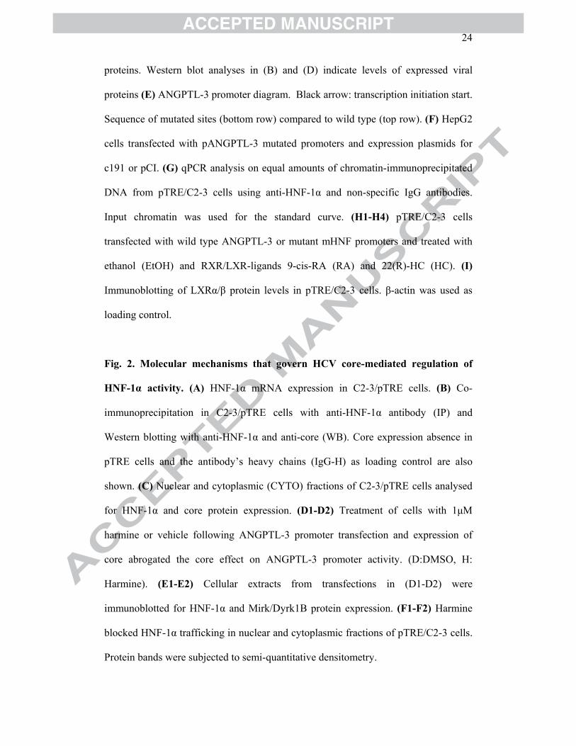
24
proteins. Western blot analyses in (B) and (D) indicate levels of expressed viral
proteins (E) ANGPTL-3 promoter diagram. Black arrow: transcription initiation start.
Sequence of mutated sites (bottom row) compared to wild type (top row). (F) HepG2
cells transfected with pANGPTL-3 mutated promoters and expression plasmids for
c191 or pCI. (G) qPCR analysis on equal amounts of chromatin-immunoprecipitated
DNA from pTRE/C2-3 cells using anti-HNF-1α and non-specific IgG antibodies.
Input chromatin was used for the standard curve. (H1-H4) pTRE/C2-3 cells
transfected with wild type ANGPTL-3 or mutant mHNF promoters and treated with
ethanol (EtOH) and RXR/LXR-ligands 9-cis-RA (RA) and 22(R)-HC (HC). (I)
Immunoblotting of LXRα/β protein levels in pTRE/C2-3 cells. β-actin was used as
loading control.
Fig. 2. Molecular mechanisms that govern HCV core-mediated regulation of
HNF-1α activity. (A) HNF-1α mRNA expression in C2-3/pTRE cells. (B) Co-
immunoprecipitation in C2-3/pTRE cells with anti-HNF-1α antibody (IP) and
Western blotting with anti-HNF-1α and anti-core (WB). Core expression absence in
pTRE cells and the antibody’s heavy chains (IgG-H) as loading control are also
shown. (C) Nuclear and cytoplasmic (CYTO) fractions of C2-3/pTRE cells analysed
for HNF-1α and core protein expression. (D1-D2) Treatment of cells with 1μM
harmine or vehicle following ANGPTL-3 promoter transfection and expression of
core abrogated the core effect on ANGPTL-3 promoter activity. (D:DMSO, H:
Harmine). (E1-E2) Cellular extracts from transfections in (D1-D2) were
immunoblotted for HNF-1α and Mirk/Dyrk1B protein expression. (F1-F2) Harmine
blocked HNF-1α trafficking in nuclear and cytoplasmic fractions of pTRE/C2-3 cells.
Protein bands were subjected to semi-quantitative densitometry.

25
Fig. 3. Subcellular localisation of HNF-1α in the presence of HCV core. (A)
Confocal microscopy images of C2-3/pTRE cells in the absence and presence of
harmine. Cells were immunostained with anti-core (green) and anti-HNF-1α (red)
antibodies. Bar: 8μm. (B) Subcellular localisation of Mirk/Dyrk1B in HepG2 cells
transfected with c191 and Mirk/Dyrk1B or pCI and immunostained with anti-
Mirk/Dyrk1B (red) and anti-core (green) antibodies. (C) White arrows indicate
HepG2 cells transfected with c191 and Mirk/Dyrk1B expression plasmids and
incubated with harmine or DMSO, then stained as in A.
Fig. 4. ANGPTL-3 gene expression is regulated by HCV in vivo and in vitro. (A1)
Intrahepatic ANGPTL-3 mRNA levels in HCV-infected patients. Key: liver biopsies
with mild (MF), advanced (AF) fibrosis, no liver disease (NL). Densitometry analysis
accompanies the agarose gel electrophoretogram. (A2) ANGPTL-3 levels in matching
sera of HCV-infected patients. ANGPTL-3 (B1) and HNF-1α (B2) mRNA levels, as
well as HNF-1α protein levels (B3) in Huh7.5 cells infected with JFH-1 for the
indicated time points. (B4) NS3-2a mRNA and core protein levels were used to
monitor infectivity. (C) Schematic diagram of our hypothesis on core-mediated
regulation of ANGPTL-3 gene expression through repression of HNF-1α activity and
blockage of LXR/RXR activation. (P): phosphorylation event.

0.000.050.100.150.200.250.30
NegIgG HNF-1α NegIgG HNF-1α NegIgG HNF-1α
% In
put
pTRE HepG2C2-3
0%20%40%60%80%
100%120%140%
+pCI +c191 +pCI +c191
RLA *
pmHNF pmLXR
1 191
aa 1 191
53 93 96 116
NLSNLS NLSNLS NLSNLS1 25 38 43 58 64 66 71
hydrophobichydrophobicRNA bindingRNA binding1 78 120 191
Ribosome bindingRibosome binding
NS5A bindingNS5A binding
1
1 191
120
DDX3/CAP-RF bindingDDX3/CAP-RF binding1 59
NLS
36 90TNFR/LTβR binding
PO4PO4 PO4PO4
D2 : lipid droplets interaction & stability118 175
NF-κB activationNF-κB activation1 71 NF-κB activationNF-κB activation
174 191
Smad3/4 interactionSmad3/4 interaction59 126
1 191
aa 1 191
53 93 96 116
NLSNLS NLSNLS NLSNLS1 25 38 43 58 64 66 71
hydrophobichydrophobicRNA bindingRNA binding1 78 120 191
Ribosome bindingRibosome binding
NS5A bindingNS5A binding
1
1 191
120
DDX3/CAP-RF bindingDDX3/CAP-RF binding1 59
NLS
36 90TNFR/LTβR binding
PO4PO4 PO4PO4
D2 : lipid droplets interaction & stability118 175
NF-κB activationNF-κB activation1 71 NF-κB activationNF-κB activation
174 191
Smad3/4 interactionSmad3/4 interaction59 126Smad3/4 interactionSmad3/4 interaction59 126
23kDa21kDa16kDa 16kDa
56-58kDa
0%50%
100%150%200%250%
+pC
I
+c19
1
+c17
3
+c12
0
+c+1
/S
+NS
RLA
** **
23kDa21kDa16kDa 16kDa
56-58kDa23kDa21kDa16kDa 16kDa
56-58kDa
0%50%
100%150%200%250%
+pC
I
+c19
1
+c17
3
+c12
0
+c+1
/S
+NS
RLA
** **
0%
40%
80%
120%
pTRE C2-3
Rel
mR
NA
Exp
ress
ion
*
23kDa
0%
40%
80%
120%
+pCI +c3a +c1b
RLA
**
23kDa
0%
40%
80%
120%
+pCI +c3a +c1b
RLA
**
-929
NFAT SRE Sp1 STAT C/EBP LXR HNF-1αTATA
-863 -581 -472 -422 -370 -114 -89 -36
Luciferase
AGGTTACATTCGTGCAAGTTAACACGGCTTAATGA-TTAACTA
GGGCCCCATTCGTGCATCAATTGTGCGCT--ATGGATTA-CTA
-929
NFAT SRE Sp1 STAT C/EBP LXR HNF-1αTATA
-863 -581 -472 -422 -370 -114 -89 -36
Luciferase-929
NFAT SRE Sp1 STAT C/EBP LXR HNF-1αTATA
-863 -581 -472 -422 -370 -114 -89 -36
LuciferaseLuciferaseLuciferase
AGGTTACATTCGTGCAAGTTAACACGGCTTAATGA-TTAACTA
GGGCCCCATTCGTGCATCAATTGTGCGCT--ATGGATTA-CTA
-
2.0
4.0
6.0
8.0
10.0
pANG+EtOH +RA +HC +RA+HC
RLA
*
*
*pTRE
0%
40%
80%
120%
160%
pTRE C2-3 pTRE C2-3 pTRE C2-3 pTRE C2-3
RLA
+EtOH
* * *
pANG
+RA+HC+HC+RA
*
0%
40%
80%
120%
160%
pTRE C2-3 pTRE C2-3 pTRE C2-3 pTRE C2-3
RLA
+EtOH
pmHNF
+RA+HC+HC+RA
A
B
C
D
E
F
G
H1
H2
H3
H4
LXRα/β
β-actin
pTRE C2-3
50-56kDa LXRα/β
β-actin
pTRE C2-3
50-56kDa50-56kDa
I
0%
40%
80%
120%
160%
pmHNF+EtOH +RA +HC +RA+HC
RLA
pTRE

0%
40%
80%
120%
160%
pTRE C2-3
Rel
mR
NA
Exp
ress
ion pTRE C2-3
core191
IP:HNF-1αWB:HNF-1α
IP:HNF-1αWB:core
IgG-H
pTRE C2-3
core191
IP:HNF-1αWB:HNF-1α
IP:HNF-1αWB:core
IgG-H
corePARP1/2β-actin
HNF-1α
CYTO NUCLEARpTRE C2-3 pTRE C2-3
116kDa
corePARP1/2β-actin
HNF-1α
CYTO NUCLEARpTRE C2-3 pTRE C2-3
corePARP1/2β-actin
HNF-1αcorePARP1/2β-actin
HNF-1α
CYTO NUCLEARCYTO NUCLEARpTRE C2-3 pTRE C2-3
116kDa
HepG2
0%
40%
80%
120%
160%
+V+D +c191+D +c191+HR
LA **
0%
40%
80%
120%
160%
pTRE C2-3 pTRE C2-3
RLA
+D
**
+H
β-actin PARP1/2HNF-1α
CYTO NUCLEARpTRE C2-3 pTRE C2-3 +1μM Harmine
β-actin PARP1/2β-actin PARP1/2HNF-1α
CYTO NUCLEARCYTO NUCLEARpTRE C2-3 pTRE C2-3 +1μM Harmine
pCI c191 c191HepG2 +D +H
HNF-1α
β-actin
Mirk/Dyrk1B 70kDa
pCI c191 c191HepG2 +D +H
HNF-1α
β-actin
Mirk/Dyrk1B 70kDa
HNF-1α
β-actin
Mirk/Dyrk1B
D H D H
pTRE C2-3
HNF-1α
β-actin
Mirk/Dyrk1B
HNF-1αHNF-1α
β-actinβ-actin
Mirk/Dyrk1BMirk/Dyrk1B
D H D H
pTRE C2-3
0%
100%
200%
300%
400%
pTRE C2-3 pTRE C2-3
%ch
ange
(HN
F-1α
/β-a
ctin
or P
AR
P1/2
)
CYTO NUCLEAR
+1μM 1μM Harmine
0%
100%
200%
300%
400%
pTRE C2-3 pTRE C2-3
%ch
ange
(HN
F-1α
/β-a
ctin
or P
AR
P1/2
)
CYTO NUCLEAR
+1μM 1μM Harmine
A B
C
D1 D2
E1 E2
F1
F2

26

0%
40%
80%
120%
160%
Moc
k
12h
24h
48h
72h
96hR
el m
RN
A E
xpre
ssio
n
HNF-1α
0%
40%
80%
120%
Moc
k
12h
24h
48h
72h
96hR
el m
RN
A E
xpre
ssio
n
**
** *
ANGPTL-3
Mock 12h 24h 48h 72h 96hHNF-1αβ-actin
Mock 12h 24h 48h 72h 96hHNF-1αβ-actin
0.0
1.5
3.0
4.5
Moc
k
12h
24h
48h
72h
96hR
el m
RN
A E
xpre
ssio
n
NS3
corecore
0%
100%
200%
300%
NL
HC
V+(M
F)
HC
V+(A
F)
NA
SH
Rel
mR
NA
Exp
ress
ion
*
*
ANGPTL-3
28S rRNA
0%
100%
200%
300%
NL
HC
V+(M
F)
HC
V+(A
F)
NA
SH
Rel
mR
NA
Exp
ress
ion
*
*
ANGPTL-3
28S rRNA
ANGPTL-3
28S rRNA
0%
40%
80%
120%
160%
NL
HC
V+(M
F)
HC
V+(A
F)
NA
SH
% N
L * **
A1
A2
B1
B2
B3
B4
C