Extraction of β-galactosidase and β-glucosidase from the seeds of Tamarindus indica
10
Purushothaman and Murthy 8 Int. J. Biomol. & Biomed. RESEARCH PAPER OPEN ACCESS Extraction of β-galactosidase and β-glucosidase from the seeds of Tamarindus indica Shlini Purushothaman * , Siddalinga Murthy K R Central College Campus, Bangalore University, Bangalore, Karnataka. India Received: 27 August 2011 Revised: 12 September 2011 Accepted: 14 September 2011 Key words: Tamarind, p-nitrophenyl-β-D-glucoside, β-glucosidase, p-nitrophenyl-β-D- galactoside, β-galactosidase, K m and V max . Abstract The enzymes β–galactosidase and β–glucosidase were extracted from the tamarind seeds using different buffers at different pH. Highest activity was obtained with 10 mM sodium acetate buffer, pH 5.6 and 10 mM tris buffer, pH 7.4. The effect of NaCl and Triton X–100 at different concentrations on the extraction of the enzymes indicated 10 mM sodium acetate buffer, pH 5.6 containing 1 M NaCl as a better extractant of the enzyme. The enzyme assay was carried out using p–nitrophenyl–β–D–galactoside and p–nitrophenyl–β–D–glucoside as substrates. Highest enzyme activities were observed on 6 th and 24 th day of germination. The protein content gradually decreased upto 5 th day of germination and suddenly increased on 6 th day. However, on subsequent days of germination, the protein content greatly decreased upto 11 th day. During the latter period of germination (18 th day onwards) the content remained almost constant. The kinetic parameters varied for both β–galactosidase and β–glucosidase. The activity of β– galactosidase was show to have an optimal operating condition at pH 5.5 and a temperature of 50 0 C. The thermostability of the enzyme was in the range of 40 0 C – 70 0 C with the pH stability in the range of 5.0 – 7.0. The Km and Vmax values for pNPGal were determined as 66μM and 2.27nmolesmin -1 . In contrast the activity of β– glucosidase was shown to have an optimal operating condition at pH 5.0 and a temperature of 30 0 C. The thermostability of the enzyme was in the range of 27 0 C – 35 0 C with the pH stability in the range of 4.0 – 7.0. The Km and Vmax values for pNPGlu were determined as 121μM and 5.26nmolesmin -1 . The presented study is a preliminary work carried out for the standardization of protocols. The purification and characterization of β–galactosidase and β– glucosidase is under progress. Corresponding Author: Shlini Purushothaman [email protected] International Journal of Biomolecules and Biomedicine (IJBB) ISSN: 2221-1063 (Print) 2222-503X (Online) Vol. 1, No.3, p. 8-17, 2011 http://www.innspub.net
-
Upload
innspub-net -
Category
Education
-
view
353 -
download
0
Transcript of Extraction of β-galactosidase and β-glucosidase from the seeds of Tamarindus indica
Purushothaman and Murthy
8 Int. J. Biomol. & Biomed.
RESEARCH PAPER OPEN ACCESS
Extraction of β-galactosidase and β-glucosidase from the seeds of
Tamarindus indica
Shlini Purushothaman*, Siddalinga Murthy K R
Central College Campus, Bangalore University, Bangalore, Karnataka. India
Received: 27 August 2011
Revised: 12 September 2011
Accepted: 14 September 2011
Key words: Tamarind, p-nitrophenyl-β-D-glucoside, β-glucosidase, p-nitrophenyl-β-D-
galactoside, β-galactosidase, Km and Vmax.
Abstract
The enzymes β–galactosidase and β–glucosidase were extracted from the tamarind seeds using different buffers at
different pH. Highest activity was obtained with 10 mM sodium acetate buffer, pH 5.6 and 10 mM tris buffer, pH 7.4.
The effect of NaCl and Triton X–100 at different concentrations on the extraction of the enzymes indicated 10 mM
sodium acetate buffer, pH 5.6 containing 1 M NaCl as a better extractant of the enzyme. The enzyme assay was carried
out using p–nitrophenyl–β–D–galactoside and p–nitrophenyl–β–D–glucoside as substrates. Highest enzyme
activities were observed on 6th and 24th day of germination. The protein content gradually decreased upto 5th day of
germination and suddenly increased on 6th day. However, on subsequent days of germination, the protein content
greatly decreased upto 11th day. During the latter period of germination (18th day onwards) the content remained
almost constant. The kinetic parameters varied for both β–galactosidase and β–glucosidase. The activity of β–
galactosidase was show to have an optimal operating condition at pH 5.5 and a temperature of 500C. The
thermostability of the enzyme was in the range of 400C – 700C with the pH stability in the range of 5.0 – 7.0. The Km
and Vmax values for pNPGal were determined as 66μM and 2.27nmolesmin-1. In contrast the activity of β–
glucosidase was shown to have an optimal operating condition at pH 5.0 and a temperature of 300C. The
thermostability of the enzyme was in the range of 270C – 350C with the pH stability in the range of 4.0 – 7.0. The Km
and Vmax values for pNPGlu were determined as 121μM and 5.26nmolesmin-1. The presented study is a preliminary
work carried out for the standardization of protocols. The purification and characterization of β–galactosidase and β–
glucosidase is under progress.
Corresponding Author: Shlini Purushothaman [email protected]
International Journal of Biomolecules and Biomedicine (IJBB) ISSN: 2221-1063 (Print) 2222-503X (Online)
Vol. 1, No.3, p. 8-17, 2011
http://www.innspub.net
Purushothaman and Murthy
9 Int. J. Biomol. & Biomed.
Introduction
The tamarind tree (Tamarindus indica L.) belonging
to family Caesalpinaceae is found in both tropical and
subtropical regions of the world. It is grown extensively
in the dry tracts of Central and South Indian States for
its sour fruit pulp, which is used extensively in the local
confectionary industry and is a common article of trade
in India. The seed comprises 20 – 30 % seed coat or
testa and 70 – 75 % kernel or endosperm (Coronel,
R.E. (1991); (Shankaracharya, N.B. (1998)). The kernel
contains 55 – 70 % polysaccharide, 16 – 20 % proteins,
5 – 7 % oils, and minerals (Shlini and Siddalinga
Murthy, 2011)).
The post-germinative mobilization of seed xyloglucans
has been studied both ultrastructurally and
biochemically (Buckeridge et al., 1992; Edwards et al.,
1985; Reid et al., 1987). Nasturtium xyloglucan
mobilization involves the simultaneous action of four
enzymes, a xyloglucan-specific endo-(1→4)-β-D-
glucanase or xyloglucan endo-transglycosylase (XET)
(Edwards et al., 1986; Fanutti et al., 1993, 1996; Farkas
et al., 1992; de Silva et al., 1993), a β-galactosidase
with action both on polymeric xyloglucan and its
subunit oligosaccharides (Edwards et al., 1988; Reid et
al., 1988), a xyloglucan oligosaccharide-specific α-
xylosidase or oligoxyloglucan exo-xylohydrolase
(Chengappa et al., 1993; Fanutti et al., 1991), and a β-
glucosidase (Edwards et al., 1985).
β-Galactosidases (EC 3.2.1.23), a widespread family of
glycosyl hydrolases, are characterized by their ability to
hydrolyze terminal, non-reducing β-D-galactosyl
residues from β-D-galactosides. Exoglycosidases are
widely distributed in many species, including
microorganisms, animals and plants. These enzymes
are useful in determining the anomeric configuration
and sequence of glycoconjugates. β-D-Galactose is
commonly found in glycoproteins and glycolipids.
Most β-Galactosidases isolated so far, are from
microbial sources (Erick and Steers, 1970; Steers et.
al,. 1971; Glasgow et al., 1977; Hirata et al., 1984;
Tanaka et al., 1988; Nagano et al., 1992; Shigeta et al.,
1983). Plant galactosidases have been characterized
from jack bean (Li et al., 1975), and from wheat grain
(Carratu et al., 1985; Papet et al., 1992). α and β-
mannosidase, and β-galactosidase were identified from
green onion (Kim et al., 1991b).
In plants, β-glucosidases ( EC 3.2.1.21) and related
glycosidases play roles in many biological processes,
including defence, lignifications, phytohormone
activation and cell-wall modification (Esen, 1993).
Their physiological function depends upon their
location and substrate-specificity. Most plant β-
glucosidases belong to glycosyl hydrolase family 1
(GH1), which also includes myrosinases (thio-β-
glucosidases), β-mannosidases. β-galactosidases,
phospho-β-glucosidases and phospho- β-
galactosidases (David et al., 2000).
Glucosidases and galactosidases are distributed widely
in soils and are important enzymes in the soil carbon
cycle. Although some research has been done on β-
glucosidase in some soil types or ecosystems, few have
been conducted in paddy soils.
Numerous studies have shown that β-galactosidases
catalyse the hydrolysis of terminal galactosyl residues
from carbohydrates, glycoproteins and galactolipids
(David et al., 2000). β-Galactosidases action has been
proposed to release stored energy for rapid growth
(lactose hydrolysis in mammals and bacteria,
xyloglucan mobilization in cotyledons), release free Gal
during normal metabolic recycling of galactolipids,
glycoproteins, and cell wall components, and degrade
cell wall components during senescence (Lo et al.,
1979; Bhalla and Dalling, 1984; Maley et al., 1989;
Raghothama et al., 1991; De Veau et al., 1993; Ross et
al., 1993; Buckeridge and Reid, 1994; Hall, 1998).
Many β-galactosidases have specific biosynthetic
activities such as transglycosylation and reverse
hydrolysis under favorable thermodynamic in vitro
Purushothaman and Murthy
10 Int. J. Biomol. & Biomed.
conditions (Bonnin et al., 1995; Yoon and Ajisaka,
1996).
Much attention has been focused on the enzyme β-
galactosidase, which is involved in the bacterial
metabolism of lactose. In addition to normal hydrolysis
of the β-D-galactoside linkage in lactose, some β-D-
galactosidase enzyme may catalyze the formation of
galactooligosaccharides through transfer of one or
more D-galactosyl units onto the D-galactose moiety of
lactose. This transgalactosylation reaction (Huber et
al., 1976) has been shown to be a characteristic of β-
galactosidase enzyme from a great variety of bacterial
and fungal species (Dumortier et al., 1994; Nakao et
al., 1994; Onishi et al., 1995; Yoon et al., 1996.).
The present investigation is aimed at extracting β-
galactosidases and β-glucosidases from Tamarind
seeds during germination. These enzymes from other
sources are unsuitable because of high cost. The
enzyme β-galactosidases and β-glucosidases obtained
from agricultural product such as the seeds of
Tamarind is a heat stable enzyme and can be
commercially exploited for the production of the
enzyme. In the current study, β-galactosidases and β-
glucosidases of Tamarindus indica seeds has been
examined as a new source for producing these
enzymes.
Material and methods
Plant material
The seeds of Tamarindus indica were collected using
random sampling technique (RST) from local areas of
Bangalore district, Karnataka State, India. After
dehulling the fruits, equal samples of seeds were
combined to give one bulk population sample from
which sub samples were taken for test. Collected seed
samples were dried in the sunlight for 24 hrs. After
removing immature and damaged seeds, the dried
matured seeds were washed under tap water, dried and
stored in plastic containers or refrigerator until further
use.
Enzyme extraction
All procedure were carried out at 4o C. Unless
otherwise specified, endosperm tissue was
homogenized (1g/10ml) with 0.05 M sodium acetate
buffer, pH 5.6 and 0.01 M Tris-HCl buffer, pH 7.4 and
also with the same buffers containing different
concentrations of NaCl (0.2, 0.5, 1.0, 2.0, and 4.0 M
NaCl) and 0.5 % Triton X-100, respectively. The
homogenate was centrifuged at 6000g for 20 mins and
the supernatant was used for further assay.
Protein assay
Protein content was determined according to the
method of Lowry et al. (1951) with bovine serum
albumin as standard.
Enzyme assay
β-glucosidase assay is based on the measurement of
the amount of p-nitrophenol formed. The enzyme
reaction was initiated by adding 0.25 ml of the extract
to 0.75 ml of 1.2 mM p-nitrophenyl-β-D-glucoside
(PNPG) in 10Mm acetate buffer, pH 5.6 and incubated
at 37o C for 30 mins. The reaction was stopped by
adding 4.0 ml of 0.1 M sodium hydroxide. The amount
of p-nitrophenol liberated is measued at 440 nm. One
enzyme unit corresponds to 0.5μmoles of ρ-
nitrophenol/min.
β-galactosidase assay is based on the measurement of
the amount of p-nitrophenol formed. The enzyme
reaction was initiated by adding 0.25 ml of the extract
to 0.75 ml of 1.2 mM p-nitrophenyl-β-D-galactoside
(PNPG) in 10Mm acetate buffer, pH 5.6 and incubated
at 37o C for 30 mins. The reaction was stopped by
adding 4.0 ml of 0.1 M sodium hydroxide. The amount
of p-nitrophenol liberated is measued at 440 nm. One
enzyme unit corresponds to 0.5μmoles of ρ-
nitrophenol/min.
Kinetic parameters
The optimum incubation period was determined by
measuring enzyme activity between 00-30mins.The
Purushothaman and Murthy
11 Int. J. Biomol. & Biomed.
optimum pH was determined in reactions carried out
at pH values ranging from 2.0 - 9.0 using 0.2 M buffers
– Acetate (pH 2.0, 2.5, 3.0, 3.5, 4.0 and 5.0); sodium
phosphate (pH 5.2, 6.0 and 7.0); Tris-HCl (pH 7.0, 8.0
and 9.0). The pH stability was determined by
preincubating the enzyme in the above buffers for
30mins and assaying the enzyme at pH 5.6. Optimum
temperature was determined by assaying the enzyme at
different temperatures (70C – 950C). Temperature
stability was determined by preincubating the enzyme
for 30 mins at different temperatures and assaying the
enzyme at 370C. Michaelis-Menten constants were
determined using different substrate concentration
(0.03-0.15µmoles/ml).
Germination studies
Fifty healthy Tamarind seeds were soaked in cooled
50% H2SO4 for 60mins and then dispensed into the
soil (1:1 ratio of acid washed sand and cocco peat).
Germination of seed was monitored. Cotyledons were
collected everyday starting from 1st day till 25th day.
Proteins were extracted as mentioned above and
analysed on PAGE. The samples were checked for
enzyme activity.
Polyacrylamide Gel Electrophoresis (PAGE)
Native-PAGE of 7.5% resolving was performed as
described by Flurkey. Duplicate samples were run for
determination of protein bands. The gel was stained
with 0.1% Coomassie Brilliant BlueR-250, destained
and visualized the bands.
Results and discussion
β-galactosidase are known to increase during
germination. The activity of some glycosidases in
germinating mung bean seeds increased to a high level
between days 4-6. The enzymes which increased
during germination in mung bean are β-N-
acetylglucosidase, β-galactosidase and α-mannosidase.
The increase of β-galactosidase activity during seed
germination was also reported in plants such as pinto
beans (Agrawal and Bahl, 1968), castor beans (Harley
and Beevers, 1985), nasturtium (Edwards et al., 1988),
barley (Giannakouros et al., 1991) and lupin
(Buckeridge and Reid, 1994). The increase of β-
galactosidase activity during the germination of barley
seeds seem to be very moderate when compared with
the many fold increase in the activity of the same
enzyme observed in the seeds of dicotyledon plants
(Giannakouros et al., 1991). Thus, the developmental
regulation of plant β-galactosidase during germination
and growth has become evident. However, much less is
known about the mechanism underlying this
regulation.
(a)
(b)
(c)
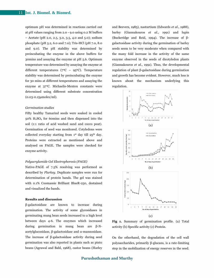
Fig 1. Summary of germination profile. (a) Total
activity (b) Specific activity (c) Protein.
On the otherhand, the degradation of the cell wall
polysaccharides, primarily β-glucans, is a rate-limiting
step in the mobilization of energy reserves in the seed.
Purushothaman and Murthy
12 Int. J. Biomol. & Biomed.
These cell wall degradation products may also provide
a significant source of carbohydrate for the
germinating seedling. The complete hydrolysis of β-
glucans is facilitated by several enzyme activities.
These include endo-(1-4)-β-glucanase, endo-(1-3)-β-
glucanase, and endo-(1-3, 1-4)-β-glucanase capable of
hydrolyzing most of the endosperm β-glucans to a
mixture of β-linked oligosaccharides. In addition, β-
glucosidase activities may be required to further
degrade β-linked oligosaccharides to glucose (Stone
and Clarke, 1992).
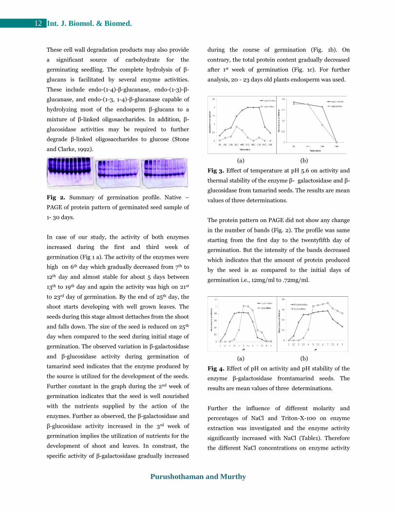
Fig 2. Summary of germination profile. Native –
PAGE of protein pattern of germinated seed sample of
1- 30 days.
In case of our study, the activity of both enzymes
increased during the first and third week of
germination (Fig 1 a). The activity of the enzymes were
high on 6th day which gradually decreased from 7th to
12th day and almost stable for about 5 days between
13th to 19th day and again the activity was high on 21st
to 23rd day of germination. By the end of 25th day, the
shoot starts developing with well grown leaves. The
seeds during this stage almost dettaches from the shoot
and falls down. The size of the seed is reduced on 25th
day when compared to the seed during initial stage of
germination. The observed variation in β-galactosidase
and β-glucosidase activity during germination of
tamarind seed indicates that the enzyme produced by
the source is utilized for the development of the seeds.
Further constant in the graph during the 2nd week of
germination indicates that the seed is well nourished
with the nutrients supplied by the action of the
enzymes. Further as observed, the β-galactosidase and
β-glucosidase activity increased in the 3rd week of
germination implies the utilization of nutrients for the
development of shoot and leaves. In constrast, the
specific activity of β-galactosidase gradually increased
during the course of germination (Fig. 1b). On
contrary, the total protein content gradually decreased
after 1st week of germination (Fig. 1c). For further
analysis, 20 - 23 days old plants endosperm was used.
(a) (b)
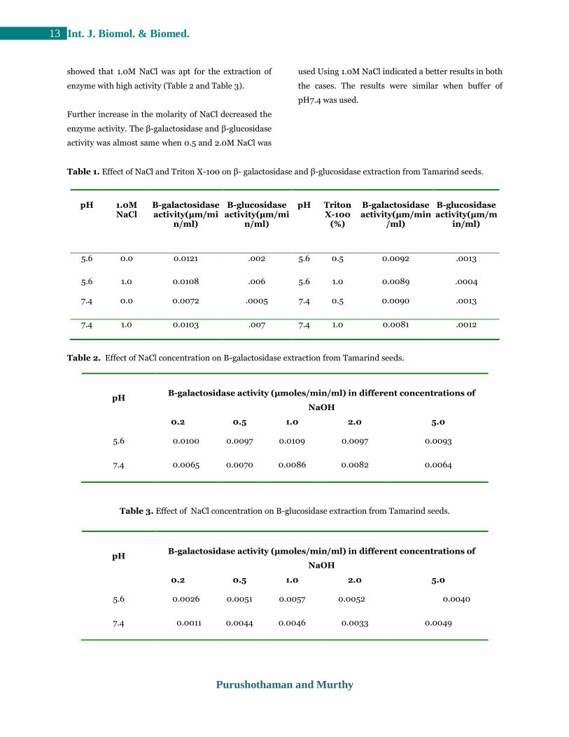
Fig 3. Effect of temperature at pH 5.6 on activity and
thermal stability of the enzyme β- galactosidase and β-
glucosidase from tamarind seeds. The results are mean
values of three determinations.
The protein pattern on PAGE did not show any change
in the number of bands (Fig. 2). The profile was same
starting from the first day to the twentyfifth day of
germination. But the intensity of the bands decreased
which indicates that the amount of protein produced
by the seed is as compared to the initial days of
germination i.e., 12mg/ml to .72mg/ml.
(a) (b)
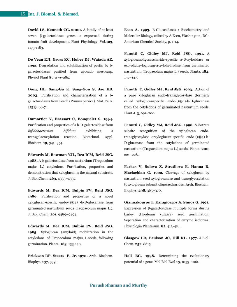
Fig 4. Effect of pH on activity and pH stability of the
enzyme β-galactosidase fromtamarind seeds. The
results are mean values of three determinations.
Further the influence of different molarity and
percentages of NaCl and Triton-X-100 on enzyme
extraction was investigated and the enzyme activity
significantly increased with NaCl (Table1). Therefore
the different NaCl concentrations on enzyme activity
Purushothaman and Murthy
13 Int. J. Biomol. & Biomed.
showed that 1.0M NaCl was apt for the extraction of
enzyme with high activity (Table 2 and Table 3).
Further increase in the molarity of NaCl decreased the
enzyme activity. The β-galactosidase and β-glucosidase
activity was almost same when 0.5 and 2.0M NaCl was
used Using 1.0M NaCl indicated a better results in both
the cases. The results were similar when buffer of
pH7.4 was used.
Table 1. Effect of NaCl and Triton X-100 on β- galactosidase and β-glucosidase extraction from Tamarind seeds.
pH
1.0M NaCl
Β-galactosidase activity(μm/mi
n/ml)
Β-glucosidase
activity(μm/min/ml)
pH
Triton X-100
(%)
Β-galactosidase activity(μm/min
/ml)
Β-glucosidase activity(μm/m
in/ml)
5.6 0.0 0.0121 .002 5.6 0.5 0.0092 .0013
5.6 1.0 0.0108 .006 5.6 1.0 0.0089 .0004
7.4 0.0 0.0072 .0005 7.4 0.5 0.0090 .0013
7.4 1.0 0.0103 .007 7.4 1.0 0.0081 .0012
Table 2. Effect of NaCl concentration on Β-galactosidase extraction from Tamarind seeds.
Table 3. Effect of NaCl concentration on Β-glucosidase extraction from Tamarind seeds.
pH
Β-galactosidase activity (μmoles/min/ml) in different concentrations of
NaOH
0.2 0.5 1.0 2.0 5.0
5.6 0.0100 0.0097 0.0109 0.0097 0.0093
7.4 0.0065 0.0070 0.0086 0.0082 0.0064
pH
Β-galactosidase activity (μmoles/min/ml) in different concentrations of
NaOH
0.2 0.5 1.0 2.0 5.0
5.6 0.0026 0.0051 0.0057 0.0052 0.0040
7.4 0.0011 0.0044 0.0046 0.0033 0.0049

Purushothaman and Murthy
14 Int. J. Biomol. & Biomed.
The kinetic parameters of the unpurified β-
galactosidase and β-glucosidase enzyme are as shown
(Fig. 2 and 3). β-galactosidase and β-glucosidase
enzyme showed highest activity at 700C and 300C. To
examine the heat resistance of the enzyme from
Tamarind seeds, it was pre-incubated at different
temperatures before determination of the activity. The
β-galactosidase enzyme was stable at temperatures of
upto 400C and β-glucosidase enzyme was stable at
temperatures of upto 350C (Fig. 2). The examined β-
galactosidase enzyme displayed a maximum activity at
pH 5.0 and showed stability from pH4.0 to pH 7.0.
Whereas β-glucosidase enzyme displayed a maximum
activity between pH 4.0 and pH 5.0 and showed
stability from pH 4.0 to pH 7.0 (Fig. 3). The kinetic
parameters of Tamarind seed β-galactosidase and β-
glucosidase for the hydrolysis of p–nitrophenyl–β–D–
galactoside and p–nitrophenyl–β–D–glucoside are in
similar order of magnitude to those of other plant
enzymes (Masayuki Sekimata et al., 1989, Yeong Shik
Kim et al., 1993, Sing-Chung et al., 2001, Dong Hoon
et al., 2003, Masayuki et al., 2000, Hazel et al., 1998).
The course of conversion of substrate into product
determined at an increased substrate concentration
indicated that Km value of 66 µM and121 µM and
Vmax value as 2.27 nmoles / min and 5.26nmoles/min
for β-galactosidase and β-glucosidase enzyme
respectively.
Conclusion
Presented results seem to be the first to determine the
production, activity and partial purification of the
enzyme β-galactosidase and β-glucosidase from
Tamarind seeds. Our results suggest that this enzyme
is a β-D-galactosidase as it hydrolysis ρ-nitrophenyl-β-
D-galactoside and β-D-glucosidase as it hydrolysis ρ-
nitrophenyl-β-D-glucoside . Observed thermal stability
of the enzyme β-D-galactosidase allows us to achieve a
high level of substrate conversion. The further
purification and more detailed characterization of the
enzyme from Tamarind seed is currently in progress.
References
Agrawal KM, Bahl OP. 1968. Glycosidases of
Phaseolus vulgaris.II. isolation and general properties.
Journal of Biological Chemistry. 243, 103-111.
Bhalla PL, Dalling MJ. 1984. Characteristics of a b-
galactosidase associated with the stroma of
chloroplasts prepared from mesophyll protoplasts of
the primary leaf of wheat. Plant Physiol 76, 92–95.
Bonnin E, Lahaye M, Vigouroux J, Thibault JF.
1995. Preliminary characterization of a new exo-b-
(1,4)-galactanase with transferase activity. Int J Biol
Macromol 17, 345–351.
Buckeridge MS, Reid JS. 1994. Purification and
properties of a novel b-galactosidase or exo-(134)-b-d-
galactanase from the cotyledons of germinated
Lupinus angustifolius L. seeds. Planta 192, 502–511.
Buckeridge MS, Rocha DC, Reid JSG, Dietrich
SMC. 1992. Xyloglucan structure and post-
germinative metabolism in seeds of Copaifera
langsdorfii from savanna and forest populations.
Physiol. Plant. 86, 1693-1702.
Carratu G, Colacino C, Conti S, Giannattasio M.
1985. Phytochem. 24, 1465.
Chengappa S, Jarman C, Fanutti C, Reid JSG.
1993. Xyloglucan oligosaccharide-specific a-D-
xylosidase: molecular mode of action and cloning of a
cDNA from germinated nasturtium (Tropaeolum
majus L.) seeds. J. Cell. Biochem. (Suppl. 17A), 27.
Coronel RE. 1991. Tamarindus indica L. In Plant
Resources of South East Asia, Wageningen, Pudoc.
No.2. Edible fruits and nuts. (Eds.) Verheij, E.W.M.
and Coronel, R.E., PROSEA Foundation, Bogor,
Indonesia: 298-301.
Purushothaman and Murthy
15 Int. J. Biomol. & Biomed.
David LS, Kenneth CG. 2000. A family of at least
seven β-galactosidase genes is expressed during
tomato fruit development. Plant Physiology, Vol.123,
1173-1183.
De Veau EJI, Gross KC, Huber DJ, Watada AE.
1993. Degradation and solubilization of pectin by b-
galactosidases purified from avocado mesocarp.
Physiol Plant 87, 279–285.
Dong HL, Sang-Gu K, Sang-Gon S, Jae KB.
2003. Purification and characterization of a b-
galactosidases from Peach (Prunus persica). Mol. Cells.
15(1). 68-74.
Dumortier V, Brassart C, Bouquelet S. 1994.
Purification and properties of a b-D-galactosidase from
Bifidobacterium bifidum exhibiting a
transgalactosylation reaction. Biotechnol. Appl.
Biochem. 19, 341–354.
Edwards M, Bowman YJL, Dea ICM, Reid JSG.
1988. A b-galactosidase from nasturtium (Tropaeolum
majus L.) cotyledons. Purification, properties and
demonstration that xyloglucan is the natural substrate.
J. Biol.Chem. 263, 4333–4337.
Edwards M, Dea ICM, Bulpin PV, Reid JSG.
1986. Purification and properties of a novel
xyloglucan-specific endo-(1®4) -b-D-glucanase from
germinated nasturtium seeds (Tropaeolum majus L.).
J. Biol. Chem. 261, 9489–9494.
Edwards M, Dea ICM, Bulpin PV, Reid JSG.
1985. Xyloglucan (amyloid) mobilization in the
cotyledons of Tropaeolum majus L.seeds following
germination. Planta. 163, 133-140.
Erickson RP, Steers E. Jr. 1970. Arch. Biochem.
Biophys. 137, 339.
Esen A. 1993. Β-Glucosidases : Biochemistry and
Molecular Biology, edited by A Esen, Washington, DC :
American Chemical Society, p. 1-14.
Fanutti C, Gidley MJ, Reid JSG. 1991. A
xyloglucanoligosaccharide-specific a-D-xylosidase or
exo-oligoxyloglucan-a-xylohydrolase from germinated
nasturtium (Tropaeolum majus L.) seeds. Planta, 184,
137–147.
Fanutti C, Gidley MJ, Reid JSG. 1993. Action of
a pure xyloglucan endo-transglycosylase (formerly
called xyloglucanspecific endo-(1®4)-b-D-glucanase
from the cotyledons of germinated nasturtium seeds.
Plant J. 3, 691–700.
Fanutti C, Gidley MJ, Reid JSG. 1996. Substrate
subsite recognition of the xyloglucan endo-
transglycosylase orxyloglucan-specific endo-(1®4)-b-
D-glucanase from the cotyledons of germinated
nasturtium (Tropaeolum majus L.) seeds. Planta, 200,
221–228.
Farkas V, Sulova Z, Stratilova E, Hanna R,
Maclachlan G. 1992. Cleavage of xyloglucan by
nasturtium seed xyloglucanase and transglycosylation
to xyloglucan subunit oligosaccharides. Arch. Biochem.
Biophys. 298, 365–370.
Giannakouros T, Karagiorgos A, Simos G. 1991.
Expression of β-galactosidase multiple forms during
barley (Hordeum vulgare) seed germination.
Seperation and characterization of enzyme isoforms.
Physiologia Plantarum. 82, 413-418.
Glasgow LR, Paulson JC, Hill RL. 1977. J.Biol.
Chem. 252, 8615.
Hall BG. 1998. Determining the evolutionary
potential of a gene. Mol Biol Evol 15, 1055–1061.
Purushothaman and Murthy
16 Int. J. Biomol. & Biomed.
Harley S M, Beevers H. 1985. Characterisation and
partial purification of three galactosidases from castor
bean endosperm. Phytochemistry. 24, 1459-1464.
Hazel J Crombie, Sumant Chengappa, Amanda
Hellyer and J S Grant Reid. 1998. A xyloglucan
oligosaccharide-active, transglycosylating β-D-
glucosidase from the cotyledons of nasturtium
(Tropaeolum majus L) seedlings-purification,
properties and characterization of a cDNA clone. The
Plant Journal. 15(1) : 27-38.
Hirata H, Negoro S, Okada H. 1984. J. Bacteriol.
106, 9.
Huber RE, Kurz G, Wallenfels K. 1976. A
quantitation of the factors which affect the hydrolase
and transgalactosylase activities of b-galactosidase (E.
coli) on lactose. Biochemistry 15, 1994–2001.
Kim YS, Lee EB Joo SH. 1991b. Arch. Pharm.
Res. 14, 255.
Kim YS, Lee MY, Park YM. 1992. Kor. Biochem. J.
25, 171.
Li SC, Mazzotta MY, Chien SF, Li YT. 1975. J.
Biol. Chem. 250. 6786.
Lo JT, Mukerji K, Awasthi YC, Hanada E,
Suzuki K, Srivastava SK. 1979. Purification and
properties of sphingolipid b-galactosidase from human
placenta. J Biol Chem 254, 6710–6715.
Maley F, Trimble RB, Tarentino AL, Plummer
TH. 1989. Characterization of glycoproteins and their
associated oligosaccharides through the use of
endoglycosidases. Anal Biochem 180, 195–204.
Masayuki S, Kiyoshi O, Yoichi T, Yohichi H,
Shigeru Y. 1989. A β-Galactosidase fromm Radish
(Raphanus sativus L.) Seeds. Plant Physiol. 90, 567-
574.
Masayuki S, Atsushi I, Hajime I. 2000.
Purification and characterization of a β-glucosidase
from rye (Secale cereal L.) seedlings. Plant
Science.155, 67-74.
Nagano H, Osmori M, Shoji Z, Kawaguchi T,
Arai M. 1992. Biosci. Biotech. Biochem. 56, 674.
Nakao M, Harada M, Kodama Y, Nakayama T,
Shibano Y, Amachi T. 1994. Purification and
characterization of a thermostable b-galactosidase with
high transgalactosylation activity from
Saccharopolyspora rectivirgula. Appl. Microbiol.
Biotechnol. 40, 657–663.
Onishi N, Yamashiro A, Yokozeki K. 1995.
Production of galactooligosaccharide from lactose by
Sterigmatomyces elviae CBS8119. Appl. Environ.
Microbiol. 61, 4022–4025.
Papet MP, Delay D, Monsigny M, Delmotte F.
1992. Biochimie. 74, 53.
Raghothama KG, Lawton KA, Goldsbrough PB,
Woodson WR. 1991. Characterization of an
ethylene-regulated flower senescence-related gene
from carnation. Plant Mol Biol 17, 61–71.
Reid JSG, Edwards M, Dea ICM. 1988. Enzymatic
modification of natural seed gums. In Gums and
Stabilisers for the Food Industry (Phillips,
G.O.,Wedlock, D.J. and Williams,P.A., eds). Oxford:
IRL Press, p. 391–398.
Reis D, Vian B, Darzens D, Roland JC. 1987.
Sequential patterns of intramural digestion of
galactoxyloglucan in tamarind seedlings. Planta. 170,
60-73.
Purushothaman and Murthy
17 Int. J. Biomol. & Biomed.
Ross GS, Redgwell RJ, MacRae EA. 1993.
Kiwifruit b-galactosidase: isolation and activity against
specific fruit cell-wall polysaccharides. Planta 189,
499–506.
Shankaracharya NB. 1998. Tamarind - Chemistry,
Technology and Uses - a critical appraisal. Journal of
Food Technology, 35(3), 193-208.
Shigeta S, Kubota H, Tamura H, Oka S. 1983. J.
Biochem 94, 1827.
Shlini P, Siddalinga MKR. 2011. Extraction of
phenolics, proteins and antioxidant activity from
defatted tamarind kernel powder. Asian J. Research
Chem. 4(6), 936-941.
de Silva J, Jarman CD, Arrowsmith DA,
Stronach MS, Chengappa S, Sidebottom C, Reid
JSG. 1993. Molecular characterization of a
xyloglucan-specific endo-(1®4)-b-Dglucanase
(xyloglucan endo-transglycosylase) from nasturtium
seeds. Plant J. 3, 701–711.
Sing-Chung Li, Jiahn-Wern Han, Kuan-Chung
Chen, Ching-San Chen. 2001. Purification and
characterization of isoforms of β-galactosidases in
mung bean seedlings. Phytochemistry. 57, 349-359.
Steers E, Cuatrecasas P, Pollard H. 1971. J.Biol.
Chem. 246, 196.
Stone BA, Clarke AE. 1992. Chemistry and Biology
of (1-3)-β-D-Glucan. La Trobe University Press,
Melbourne.
Tanaka Y, Kagamishi A, Kiuchi A, Shoji Z.
1988. Agric. Biol. Chem. 52, 1301.
Yeong SK, Kyung SP, Jong GK. 1993. Purification
and characterization of β-galactosidase from green
onion. Korean Biochem J. 26(7), 602-608.
Yoon JH, Ajisaka K. 1996. The synthesis of
galactopyranosly derivatives with b-galactosidases of
different origins. Carbohydr Res 292, 153–163.