Extensive aggregation of α-synuclein and tau in juvenile-onset neuroaxonal dystrophy: an autopsied...
11
CASE REPORT Open Access Extensive aggregation of α-synuclein and tau in juvenile-onset neuroaxonal dystrophy: an autopsied individual with a novel mutation in the PLA2G6 gene-splicing site Yuichi Riku 1,2 , Takeshi Ikeuchi 3 , Hiroyo Yoshino 4 , Maya Mimuro 5 , Kazuo Mano 1 , Yoji Goto 1 , Nobutaka Hattori 6 , Gen Sobue 1 and Mari Yoshida 5* Abstract Background: Infantile neuroaxonal dystrophy (INAD) is a rare autosomal-recessive neurodegenerative disorder. Patients with INAD usually show neurological symptoms with infant onset and die in childhood. Recently, it was reported that mutations in the PLA2G6 gene cause INAD, but neuropathological analysis of genetically confirmed individuals with neuroaxonal dystrophy has been limited. Results: Here, we report a Japanese individual with neuroaxonal dystrophy associated with compound heterozygous mutations in the PLA2G6 gene. A novel splice-site mutation resulting in skipping and missense mutations (p.R538C) in exon 9 was identified in the patient. This patient initially presented with cerebellar ataxia at the age of 3 years, which was followed by symptoms of mental retardation, extrapyramidal signs, and epileptic seizure. The patient survived until 20 years of age. Neuropathological findings were characterized by numerous axonal spheroids, brain iron deposition, cerebellar neuronal loss, phosphorylated alpha-synuclein-positive Lewy bodies (LBs), and phosphorylated-tau-positive neurofibrillary tangles. In particular, LB pathology exhibited a unique distribution with extremely severe cortical involvement. Conclusions: Our results support a genetic clinical view that compound heterozygous mutations with potential residual protein function are associated with a relatively mild phenotype. Moreover, the severe LB pathology suggests that dysfunction of the PLA2G6 gene primarily contributes to LB formation. Keywords: α-synuclein, Infantile neuroaxonal dystrophy, Atypical neuroaxonal dystrophy, PLA2G6 gene, Tau Background Neurodegeneration with brain iron accumulation (NBIA) describes a group of progressive neurodegenerative disor- ders that are pathologically characterized by the presence of axonal spheroids and iron deposition in the brain [1-3]. These neurodegenerative diseases consist of a clinically and genetically heterogeneous group of disorders, includ- ing pantothenate kinase-associated neurodegeneration (PKAN, formerly known as Hallervorden-Spatz disease), infantile neuroaxonal dystrophy (INAD), and an unknown gene mutation-linked idiopathic neuroaxonal dystrophy [1,2,4]. PKAN is caused by mutations in the pantothenate kinase 2 (PANK2) gene, which accounts for the majority of NBIA patients [2]. Recently, it was reported that muta- tions in the phospholipase A2 group VI (PLA2G6) gene cause INAD [5], which is a rare autosomal-recessive neu- rodegenerative disorder. Patients with INAD usually present with psychomotor regression, truncal hypotonia, progressive ataxia, extrapyramidal symptoms, fast waves on an electroencephalogram, and neuro-ophthalmological abnormalities (e.g., optic atrophy, nystagmus, and strabis- mus) with infant onset and die in childhood [1,4,6]. How- ever, in rare cases, patients with NAD caused by PLA2G6 mutations present with heterogeneous neurological mani- festations with onset past infanthood and survive until * Correspondence: [email protected] 5 Institute for Medical Science of Aging, Aichi Medical University, Aichi, Japan Full list of author information is available at the end of the article © 2013 Riku et al.; licensee BioMed Central Ltd. This is an Open Access article distributed under the terms of the Creative Commons Attribution License (http://creativecommons.org/licenses/by/2.0), which permits unrestricted use, distribution, and reproduction in any medium, provided the original work is properly cited. Riku et al. Acta Neuropathologica Communications 2013, 1:12 http://www.actaneurocomms.org/content/1/1/12
Transcript of Extensive aggregation of α-synuclein and tau in juvenile-onset neuroaxonal dystrophy: an autopsied...
CASE REPORT Open Access
Extensive aggregation of α-synuclein and tau injuvenile-onset neuroaxonal dystrophy: anautopsied individual with a novel mutation in thePLA2G6 gene-splicing siteYuichi Riku1,2, Takeshi Ikeuchi3, Hiroyo Yoshino4, Maya Mimuro5, Kazuo Mano1, Yoji Goto1, Nobutaka Hattori6,Gen Sobue1 and Mari Yoshida5*
Abstract
Background: Infantile neuroaxonal dystrophy (INAD) is a rare autosomal-recessive neurodegenerative disorder.Patients with INAD usually show neurological symptoms with infant onset and die in childhood. Recently, it wasreported that mutations in the PLA2G6 gene cause INAD, but neuropathological analysis of genetically confirmedindividuals with neuroaxonal dystrophy has been limited.
Results: Here, we report a Japanese individual with neuroaxonal dystrophy associated with compoundheterozygous mutations in the PLA2G6 gene. A novel splice-site mutation resulting in skipping and missensemutations (p.R538C) in exon 9 was identified in the patient. This patient initially presented with cerebellar ataxia atthe age of 3 years, which was followed by symptoms of mental retardation, extrapyramidal signs, and epilepticseizure. The patient survived until 20 years of age. Neuropathological findings were characterized by numerousaxonal spheroids, brain iron deposition, cerebellar neuronal loss, phosphorylated alpha-synuclein-positive Lewybodies (LBs), and phosphorylated-tau-positive neurofibrillary tangles. In particular, LB pathology exhibited a uniquedistribution with extremely severe cortical involvement.
Conclusions: Our results support a genetic clinical view that compound heterozygous mutations with potentialresidual protein function are associated with a relatively mild phenotype. Moreover, the severe LB pathologysuggests that dysfunction of the PLA2G6 gene primarily contributes to LB formation.
Keywords: α-synuclein, Infantile neuroaxonal dystrophy, Atypical neuroaxonal dystrophy, PLA2G6 gene, Tau
BackgroundNeurodegeneration with brain iron accumulation (NBIA)describes a group of progressive neurodegenerative disor-ders that are pathologically characterized by the presenceof axonal spheroids and iron deposition in the brain [1-3].These neurodegenerative diseases consist of a clinicallyand genetically heterogeneous group of disorders, includ-ing pantothenate kinase-associated neurodegeneration(PKAN, formerly known as Hallervorden-Spatz disease),infantile neuroaxonal dystrophy (INAD), and an unknowngene mutation-linked idiopathic neuroaxonal dystrophy
[1,2,4]. PKAN is caused by mutations in the pantothenatekinase 2 (PANK2) gene, which accounts for the majorityof NBIA patients [2]. Recently, it was reported that muta-tions in the phospholipase A2 group VI (PLA2G6) genecause INAD [5], which is a rare autosomal-recessive neu-rodegenerative disorder. Patients with INAD usuallypresent with psychomotor regression, truncal hypotonia,progressive ataxia, extrapyramidal symptoms, fast waveson an electroencephalogram, and neuro-ophthalmologicalabnormalities (e.g., optic atrophy, nystagmus, and strabis-mus) with infant onset and die in childhood [1,4,6]. How-ever, in rare cases, patients with NAD caused by PLA2G6mutations present with heterogeneous neurological mani-festations with onset past infanthood and survive until
* Correspondence: [email protected] for Medical Science of Aging, Aichi Medical University, Aichi, JapanFull list of author information is available at the end of the article
© 2013 Riku et al.; licensee BioMed Central Ltd. This is an Open Access article distributed under the terms of the CreativeCommons Attribution License (http://creativecommons.org/licenses/by/2.0), which permits unrestricted use, distribution, andreproduction in any medium, provided the original work is properly cited.
Riku et al. Acta Neuropathologica Communications 2013, 1:12http://www.actaneurocomms.org/content/1/1/12
adulthood with a slower disease progression [1,7,8]. Inaddition, mutations of the PLA2G6 gene cause early onsetdystonia-parkinsonism (PARK-14), which is clinically dis-tinguished from NAD by good L-dopa responsiveness,L-dopa–induced dyskinesia, and dementia. These charac-teristics have been typically observed in patients with anolder age of onset and with a longer disease duration com-pared to NAD, with no evidence of cerebellar symptoms[9]. Thus, these clinical phenotypes are collectively termedas PLA2G6-associated neurodegeneration [9].We report a Japanese individual with neuroaxonal dys-
trophy that was associated with a novel compound het-erozygous mutation in a splicing site of the PLA2G6gene. The clinical phenotype of this patient was atypicalfor INAD, occurred during late disease onset, andprolonged the disease course. Histopathological data re-vealed the presence of neuroaxonal spheroids, brain irondepositions, and cerebellar degeneration. Moreover, nu-merous Lewy bodies (LBs) and neurofibrillary tangles(NFTs), which are pathological hallmarks of Parkinson’sdisease (PD) and Alzheimer’s disease (AD), respectively,were observed. Until recently, neuropathological analysisof genetically confirmed neuroaxonal dystrophy has beenstrongly limited due to a small number of patients [1,8].In this study, we describe the clinicopathological charac-teristics of the patient and discuss the neuropathologicalimplication of LBs and NFTs compared with PDand AD.
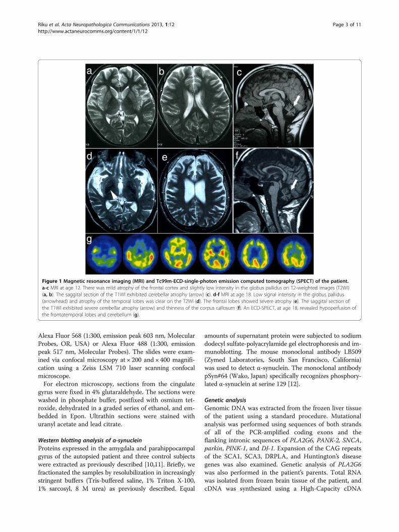
Case presentationClinical historyThe patient was a Japanese man who died at 20 years ofage. He exhibited normal development until the age of3 years, at which time his parents noted his slurredspeech and unstable gait. There was no evidence of aconsanguineous marriage in any of his relatives. Hisgrand-aunt had been diagnosed with “parkinsonism”,and she died at the age of 60; however, her clinical diag-nosis was uncertain. At the age of six, the patient was re-ferred to our hospital due to a progressive gaitdisturbance and dysarthria. A neurological examinationrevealed cerebellar ataxia, bradykinesia, mental retard-ation, and hyperreflexia in the lower limbs withoutpathological reflexes. Truncal hypotonia and abnormal-ities in eye movement were not observed. Cerebral com-puted tomography (CT) showed severe cerebellaratrophy. The patient was clinically diagnosed with juven-ile spinocerebellar degeneration, and taltirelin was ad-ministered for his ataxia; however, it did not have aneffect. At the age of 12, cerebral magnetic resonance im-aging (MRI) revealed severe atrophy of the cerebellumand mild atrophy of the frontal lobes (Figure 1a-c). Thepatient gradually became bedridden until the age of 15and started experiencing repetitive generalized seizures.
He was mainly treated with sodium valproate andphenobarbital. At the age of 18, he was re-admitted toour hospital, although he was nearly bedridden andcould barely sit in a wheelchair at that time. Neuro-logical examination revealed severe dystonia and rigidityin his limbs and neck, a masked face, and severe cerebel-lar ataxia. His tendon reflexes showed hyperreflexia inthe upper limbs and were abolished in his lower limbs.Moreover, his plantar responses were flexor. CT andMRI (Figure 1d-f ) revealed severe cerebellar and fronto-temporal lobe atrophy. The cerebral atrophy was moreprogressive compared to the atrophy observed when hewas 12 years old. By T2-weighted imaging (T2WI), thebilateral globus pallidus (GP) and putamen exhibitedlow signal intensity. Tc99m-ECD-single-photon emissioncomputed tomography revealed hypoperfusion in thefronto-temporal lobes and cerebellum (Figure 1g). Anelectroencephalogram showed multifocal spikes andtheta waves in the right hemisphere in the absence offast waves. The results of the nerve conduction study onthe four limbs were normal. After discharge, a higherdose of valproate reduced the frequency of the patient’sseizures; however, his rigidity and dystonia showed noresponse to L-DOPA treatment. The patient died of as-piration pneumonia.
Materials and methodsNeuropathological analysisThe postmortem interval was 5 hours. The brain andspinal cord were fixed in 20% neutral formalin. Samplesobtained from the main representative regions of thebrain and spinal cord were embedded in paraffin, sec-tioned into 4.5-μm-thick slides, and stained withhematoxylin and eosin (H&E), Klüver-Barrera staining,Prussian blue methods, and Gallyas-Braak (GB) staining.Immunohistochemical studies were performed on 4.5-μm-thick sections using an ENVISION kit (Dako) withdiaminobenzidine (DAB; Wako, Osaka, Japan) as achromogen. The primary antibodies used were anti-phosphorylated alpha-synuclein (p-α-synuclein) (pSyn#64,monoclonal mouse, 1:1000; Wako Pure Chemical Indus-tries, Osaka, Japan), anti-ubiquitin (polyclonal rabbit,1:2000; Dako), anti-amyloid-beta peptide (6F/3D,monoclonal mouse, 1:200; Dako), phosphorylated tau(p-tau) (AT8, monoclonal mouse, 1:2000; Innogenetics,Zwijndrecht, Belgium), anti- TDP-43 (TARDBP, polyclonalrabbit, 1:2500; ProteinTech, IL, USA), and anti-phosphorylated neurofilament (p-NF) (2F11, monoclonalmouse, 1:600; Dako). For double-immunofluorescence la-beling, brain tissues obtained from the amygdala, oculo-motor nucleus, and substantia nigra were sectioned into4.5-μm-thick slides. The primary antibodies were anti-p-α-synuclein antibody and AT8 antibody. The secondaryantibodies were goat anti-mouse IgG coupled with either
Riku et al. Acta Neuropathologica Communications 2013, 1:12 Page 2 of 11http://www.actaneurocomms.org/content/1/1/12
Alexa Fluor 568 (1:300, emission peak 603 nm, MolecularProbes, OR, USA) or Alexa Fluor 488 (1:300, emissionpeak 517 nm, Molecular Probes). The slides were exam-ined via confocal microscopy at × 200 and × 400 magnifi-cation using a Zeiss LSM 710 laser scanning confocalmicroscope.For electron microscopy, sections from the cingulate
gyrus were fixed in 4% glutaraldehyde. The sections werewashed in phosphate buffer, postfixed with osmium tet-roxide, dehydrated in a graded series of ethanol, and em-bedded in Epon. Ultrathin sections were stained withuranyl acetate and lead citrate.
Western blotting analysis of α-synucleinProteins expressed in the amygdala and parahippocampalgyrus of the autopsied patient and three control subjectswere extracted as previously described [10,11]. Briefly, wefractionated the samples by resolubilization in increasinglystringent buffers (Tris-buffered saline, 1% Triton X-100,1% sarcosyl, 8 M urea) as previously described. Equal
amounts of supernatant protein were subjected to sodiumdodecyl sulfate-polyacrylamide gel electrophoresis and im-munoblotting. The mouse monoclonal antibody LB509(Zymed Laboratories, South San Francisco, California)was used to detect α-synuclein. The monoclonal antibodypSyn#64 (Wako, Japan) specifically recognizes phosphory-lated α-synuclein at serine 129 [12].
Genetic analysisGenomic DNA was extracted from the frozen liver tissueof the patient using a standard procedure. Mutationalanalysis was performed using sequences of both strandsof all of the PCR-amplified coding exons and theflanking intronic sequences of PLA2G6, PANK-2, SNCA,parkin, PINK-1, and DJ-1. Expansion of the CAG repeatsof the SCA1, SCA3, DRPLA, and Huntington’s diseasegenes was also examined. Genetic analysis of PLA2G6was also performed in the patient’s parents. Total RNAwas isolated from frozen brain tissue of the patient, andcDNA was synthesized using a High-Capacity cDNA
Figure 1 Magnetic resonance imaging (MRI) and Tc99m-ECD-single-photon emission computed tomography (SPECT) of the patient.a-c MRI at age 12. There was mild atrophy of the frontal cortex and slightly low intensity in the globus pallidus on T2-weighted images (T2WI)(a, b). The saggital section of the T1WI exhibited cerebellar atrophy (arrow) (c). d-f MRI at age 18. Low signal intensity in the globus pallidus(arrowhead) and atrophy of the temporal lobes was clear on the T2WI (d). The frontal lobes showed severe atrophy (e). The saggital section ofthe T1WI exhibited severe cerebellar atrophy (arrow) and thinness of the corpus callosum (f). An ECD-SPECT, at age 18, revealed hypoperfusion ofthe frontotemporal lobes and cerebellum (g).
Riku et al. Acta Neuropathologica Communications 2013, 1:12 Page 3 of 11http://www.actaneurocomms.org/content/1/1/12
Reverse Transcription kit (Applied Biosystems). RT-PCRwas performed using primer pairs to amplify the codingregions of the PLA2G6 gene spanning exons 8–13 (5′-caacgtggagatgatcaagg-3′ and 5-gtcagcatcaccttgggttt-3′)and exons 9–13 (5′-ggaaggcgatcttgactctg-3′ and 5′-gtcagcatcaccttgggttt-3′). The institutional review boardapproved this study.
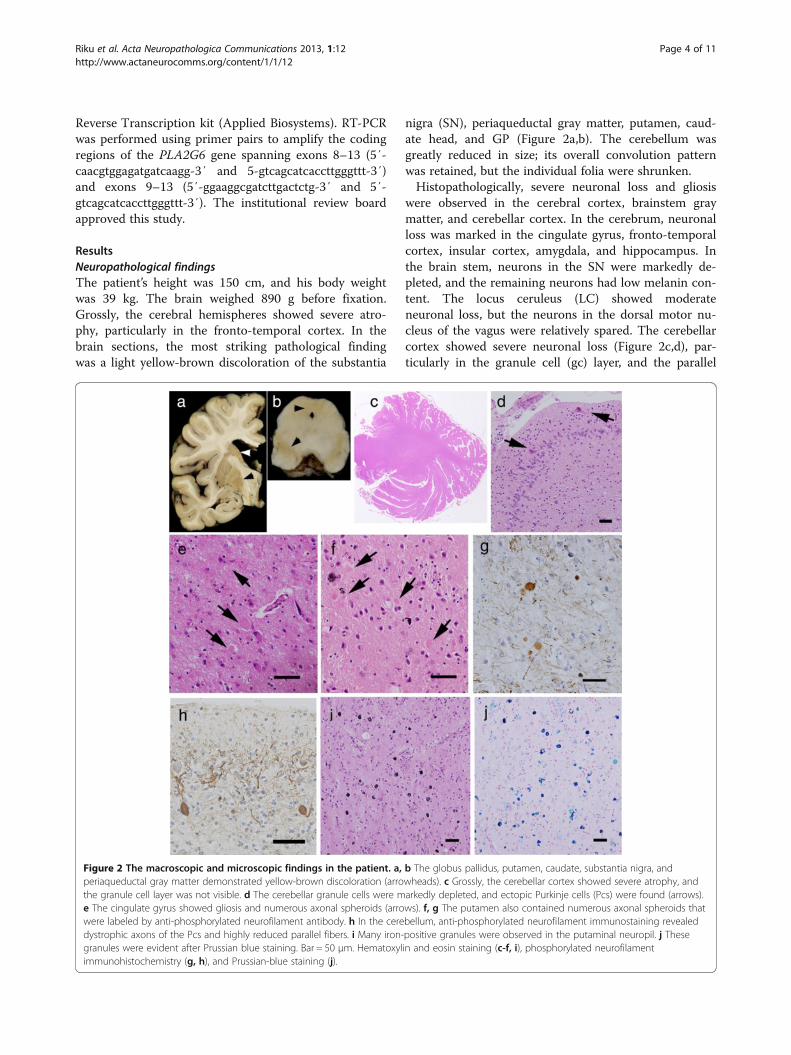
ResultsNeuropathological findingsThe patient’s height was 150 cm, and his body weightwas 39 kg. The brain weighed 890 g before fixation.Grossly, the cerebral hemispheres showed severe atro-phy, particularly in the fronto-temporal cortex. In thebrain sections, the most striking pathological findingwas a light yellow-brown discoloration of the substantia
nigra (SN), periaqueductal gray matter, putamen, caud-ate head, and GP (Figure 2a,b). The cerebellum wasgreatly reduced in size; its overall convolution patternwas retained, but the individual folia were shrunken.Histopathologically, severe neuronal loss and gliosis
were observed in the cerebral cortex, brainstem graymatter, and cerebellar cortex. In the cerebrum, neuronalloss was marked in the cingulate gyrus, fronto-temporalcortex, insular cortex, amygdala, and hippocampus. Inthe brain stem, neurons in the SN were markedly de-pleted, and the remaining neurons had low melanin con-tent. The locus ceruleus (LC) showed moderateneuronal loss, but the neurons in the dorsal motor nu-cleus of the vagus were relatively spared. The cerebellarcortex showed severe neuronal loss (Figure 2c,d), par-ticularly in the granule cell (gc) layer, and the parallel
Figure 2 The macroscopic and microscopic findings in the patient. a, b The globus pallidus, putamen, caudate, substantia nigra, andperiaqueductal gray matter demonstrated yellow-brown discoloration (arrowheads). c Grossly, the cerebellar cortex showed severe atrophy, andthe granule cell layer was not visible. d The cerebellar granule cells were markedly depleted, and ectopic Purkinje cells (Pcs) were found (arrows).e The cingulate gyrus showed gliosis and numerous axonal spheroids (arrows). f, g The putamen also contained numerous axonal spheroids thatwere labeled by anti-phosphorylated neurofilament antibody. h In the cerebellum, anti-phosphorylated neurofilament immunostaining revealeddystrophic axons of the Pcs and highly reduced parallel fibers. i Many iron-positive granules were observed in the putaminal neuropil. j Thesegranules were evident after Prussian blue staining. Bar = 50 μm. Hematoxylin and eosin staining (c-f, i), phosphorylated neurofilamentimmunohistochemistry (g, h), and Prussian-blue staining (j).
Riku et al. Acta Neuropathologica Communications 2013, 1:12 Page 4 of 11http://www.actaneurocomms.org/content/1/1/12
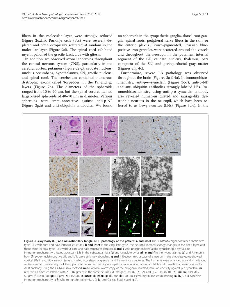
fibers in the molecular layer were strongly reduced(Figure 2c,d,h). Purkinje cells (Pcs) were severely de-pleted and often ectopically scattered at random in themolecular layer (Figure 2d). The spinal cord exhibitedmyelin pallor of the gracile fasciculus with gliosis.In addition, we observed axonal spheroids throughout
the central nervous system (CNS), particularly in thecerebral cortex, putamen (Figure 2e-g), caudate nucleus,nucleus accumbens, hypothalamus, SN, gracile nucleus,and spinal cord. The cerebellum contained numerousdystrophic axons called ‘torpedoes’ in the Pc and gclayers (Figure 2h). The diameters of the spheroidsranged from 10 to 20 μm, but the spinal cord containedlarger-sized spheroids of 40–70 μm in diameter. Variousspheroids were immunoreactive against anti-p-NF(Figure 2g,h) and anti-ubiquitin antibodies. We found
no spheroids in the sympathetic ganglia, dorsal root gan-glia, spinal roots, peripheral nerve fibers in the skin, orthe enteric plexus. Brown-pigmented, Prussian blue-positive iron granules were scattered around the vesselsand throughout the neuropil in the putamen, internalsegment of the GP, caudate nucleus, thalamus, parscompacta of the SN, and periaqueductal gray matter(Figures 2i,j, 4c).Furthermore, severe LB pathology was observed
throughout the brain (Figures 3a-f, 4a). In immunohisto-chemistry, anti-p-α-synuclein (Figure 3c-f), anti-p-NF,and anti-ubiquitin antibodies strongly labeled LBs. Im-munohistochemistry using anti-p-α-synuclein antibodyalso revealed numerous dilated and sausage-like dys-trophic neurites in the neuropil, which have been re-ferred to as Lewy neurites (LNs) (Figure 3d,e). In the
Figure 3 Lewy body (LB) and neurofibrillary tangle (NFT) pathology of the patient. a and inset The substantia nigra contained “brainstem-type” LBs with core and halo (arrows) structures. b and inset In the cingulate gyrus, the neuropil showed spongy changes in the deep layer, andthere were “cortical-type” LBs without core and halo structures (arrows). c and d Anti-phosphorylated alpha-synuclein (p-α-synuclein)immunohistochemistry showed abundant LBs in the substantia nigra (c) and cingulate gyrus (d). e and f In the hypothalamus (e) and Ammon’ shorn (f), p-α-synuclein-positive LBs and LNs were strikingly abundant. g and h Electron microscopy of a neuron in the cingulate gyrus showedcortical LBs in a cortical neuron (asterisk), which consisted of granular and filamentous structures. The filaments were arranged at random withouta clear central zone density. i - l The pyramidal neuron in the hippocampal cortex contained abundant NFTs and threads that were positive forAT-8 antibody using the Gallyas-Braak method. m-o Confocal microscopy of the amygdala revealed immunoreactivity against p-α-synuclein (m,red), which often co-labeled with AT8 (n, green) in the same neurons (o, merged). Bar (a), (b), (c), and (i) = 100 μm; (d), (e), (m), (n), and (o) =50 μm; (f) = 250 μm; (g) = 2 μm; (h) = 0.2 μm; (a-inset), (b-inset), (j), (k), and (l) = 20 μm. Hematoxylin and eosin staining (a, b, j), p-α-synucleinimmunohistochemistry (c-f), AT8 immunohistochemistry (i, k), and Gallyas-Braak staining (l).
Riku et al. Acta Neuropathologica Communications 2013, 1:12 Page 5 of 11http://www.actaneurocomms.org/content/1/1/12
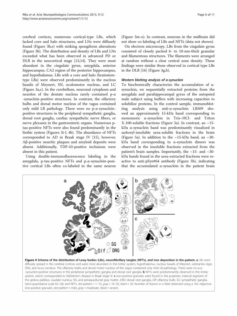
cerebral cortices, numerous cortical-type LBs, whichlacked core and halo structures, and LNs were diffuselyfound (Figure 3b,e) with striking spongiform alterations(Figure 3b). The distribution and density of LBs and LNsexceeded what has been observed in advanced PD orDLB in the neocortical stage [13,14]. They were mostabundant in the cingulate gyrus, amygdala, anteriorhippocampus, CA2 region of the posterior hippocampus,and hypothalamus. LBs with a core and halo (brainstem-type LBs) were observed predominantly in the nucleusbasalis of Meynert, SN, oculomotor nucleus, and LC(Figure 3a,c). In the cerebellum, neuronal cytoplasm andneurites of the dentate nucleus rarely contained p-α-synuclein-positive structures. In contrast, the olfactorybulbs and dorsal motor nucleus of the vagus containedonly mild LB pathology. There were no p-α-synuclein-positive structures in the peripheral sympathetic ganglia,dorsal root ganglia, cardiac sympathetic nerve fibers, ornerve plexuses in the gastroenteric organs. Numerous p-tau-positive NFTs were also found predominantly in thelimbic system (Figures 3i-l, 4b). The abundance of NFTscorresponded to AD in Braak stage IV [15], however,Aβ-positive neuritic plaques and amyloid deposits wereabsent. Additionally, TDP-43-positive inclusions wereabsent in this patient.Using double-immunofluorescence labeling in the
amygdala, p-tau-positive NFTs and p-α-synuclein-posi-tive cortical LBs often co-labeled in the same neuron
(Figure 3m-o). In contrast, neurons in the midbrain didnot show co-labeling of LBs and NFTs (data not shown).On electron microscopy, LBs from the cingulate gyrus
consisted of closely packed 6- to 10-nm-thick granularand filamentous structures. The filaments were arrangedat random without a clear central zone density. Thesefindings were similar those observed in cortical-type LBsin the DLB [16] (Figure 3g,h).
Western blotting analysis of α-synucleinTo biochemically characterize the accumulation of α-synuclein, we sequentially extracted proteins from theamygdala and parahippocampal gyrus of the autopsiedmale subject using buffers with increasing capacities tosolubilize proteins. In the control sample, immunoblot-ting analysis using anti-α-synuclein LB509 sho-wed an approximately 15-kDa band corresponding tomonomeric α-synuclein in Tris–HCl- and TritonX-100-soluble fractions (Figure 5a). In contrast, an ~15-kDa α-synuclein band was predominantly visualized insarkosyl-insoluble urea-soluble fractions in the brain(Figure 5a). In addition to the ~15-kDa band, an ~30-kDa band corresponding to α-synuclein dimers wasobserved in the insoluble fractions extracted from thepatient’s brain samples. Importantly, the ~15- and ~30-kDa bands found in the urea-extracted fractions were re-active to anti-pSyn#64 antibody (Figure 5b), indicatingthat the accumulated α-synuclein in the patient brain
Figure 4 Schema of the distribution of Lewy bodies (LBs), neurofibrillary tangles (NFTs), and iron deposition in the patient. a LBs werediffusely spread in the cerebral cortices and were most abundant in the limbic system, hypothalamus, nucleus basalis of Meynert, substantia nigra(SN), and locus ceruleus. The olfactory bulbs and dorsal motor nucleus of the vagus contained only mild LB pathology. There were no p-α-synuclein-positive structures in the peripheral sympathetic ganglia and dorsal root ganglia. b NFTs were predominantly observed in the limbicsystem, which corresponded to Alzheimer’s disease in Braak stage IV. c Iron-positive granules were found in the putamen, internal segment ofthe globus pallidus, caudate nucleus, SN, and periaqueductal gray matter. DRG: dorsal root ganglia, Olf: olfactory bulb, SG: sympathetic ganglia.Semi-quantitative scale for LBs and NFTs: dot-pattern = 1–10, gray = 10–20, black > 20. Number of lesions in a field observed using a 10× objective.Iron-positive granules: dot-pattern =mild, gray =moderate, black = severe.
Riku et al. Acta Neuropathologica Communications 2013, 1:12 Page 6 of 11http://www.actaneurocomms.org/content/1/1/12
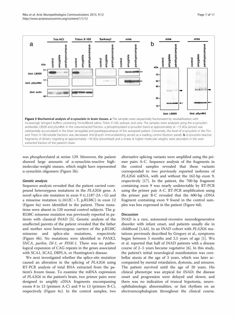
was phosphorylated at serine 129. Moreover, the patientshowed large amounts of α-synuclein-reactive high-molecular-weight smears, which might have representedα-synuclein oligomers (Figure 5b).
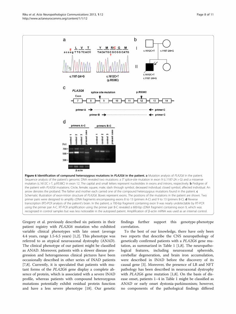
Genetic analysisSequence analysis revealed that the patient carried com-pound heterozygous mutations in the PLA2G6 gene. Anovel splice-site mutation in exon 9 (c.1187-2A > G) anda missense mutation (c.1612C > T, p.R538C) in exon 12(Figure 6a) were identified in the patient. These muta-tions were absent in 120 normal control subjects. The p.R538C missense mutation was previously reported in pa-tients with classical INAD [5]. Genetic analysis of theunaffected parents of the patient revealed that the fatherand mother were heterozygous carriers of the p.R538Cmissense and splice-site mutations, respectively(Figure 6b). No mutations were identified in PANK2,SNCA, parkin, DJ-1, or PINK-1. There was no patho-logical expansion of CAG repeats in the genes associatedwith SCA1, SCA2, DRPLA, or Huntington’s disease.We next investigated whether the splice-site mutation
caused an alteration in the splicing of PLA2G6 usingRT-PCR analysis of total RNA extracted from the pa-tient’s frozen tissue. To examine the mRNA expressionof PLA2G6 in the patient’s brain, two primer pairs weredesigned to amplify cDNA fragments encompassingexons 8 to 13 (primers A-C) and 9 to 13 (primers B-C),respectively (Figure 6c). In the control samples, two
alternative splicing variants were amplified using the pri-mer pairs A-C. Sequence analysis of the fragments inthe control samples revealed that these variantscorresponded to two previously reported isoforms ofPLA2G6 mRNA, with and without the 162-bp exon 9,respectively [17]. In the patient, the 700-bp fragmentcontaining exon 9 was nearly undetectable by RT-PCRusing the primer pair A-C. RT-PCR amplification usingthe primer pair B-C revealed that the 600-bp cDNAfragment containing exon 9 found in the control sam-ples was less expressed in the patient (Figure 6d).
DiscussionINAD is a rare, autosomal-recessive neurodegenerativedisorder with infant onset, and patients usually die inchildhood [1,4,6]. In an INAD cohort with PLA2G6 mu-tations previously described by Gregory et al., symptomsbegan between 5 months and 2.5 years of age [1]. Wuet al. reported that half of INAD patients with a diseasecourse of 2–5 years became vegetative [6]. In this study,the patient’s initial neurological manifestation was cere-bellar ataxia at the age of 3 years, which was later ac-companied by mental retardation, dystonia, and seizures.The patient survived until the age of 20 years. Hisclinical phenotype was atypical for INAD; the diseaseonset and progression were delayed and slower, andthere was no indication of truncal hypotonia, neuro-ophthalmologic abnormalities, or fast rhythms on anelectroencephalogram throughout the clinical course.
Figure 5 Biochemical analysis of α-synuclein in brain tissues. a The samples were sequentially fractionated by resolubilization withincreasingly stringent buffers containing Tris-buffered saline, Triton X-100, sarkosyl, and urea. The samples were analyzed using the α-synucleinantibodies LB509 and pSyn#64. In the urea-extracted fraction, a phosphorylated α-synuclein band at approximately at ~15 kDa (arrow) wassubstantially accumulated in the brain (amygdala and parahippocampus) of the autopsied patient. Conversely, the level of α-synuclein in the Tris-and Triton X-100-soluble fractions was decreased. Anti-β-actin immunoblotting served as a loading control (bottom panel). b α-Synuclein-reactivefragments of dimers migrating at approximately ~30 kDa (arrowhead) and a smear at higher molecular weights were abundant in the urea-extracted fraction of the patient’s brain.
Riku et al. Acta Neuropathologica Communications 2013, 1:12 Page 7 of 11http://www.actaneurocomms.org/content/1/1/12
Gregory et al. previously described six patients in theirpatient registry with PLA2G6 mutation who exhibitedvariable clinical phenotypes with late onset (average4.4 years, range 1.5-6.5 years) [1,2]. This phenotype wasreferred to as atypical neuroaxonal dystrophy (ANAD).The clinical phenotype of our patient might be classifiedas ANAD. Moreover, patients with a slower disease pro-gression and heterogeneous clinical pictures have beenoccasionally described in other series of INAD patients[7,8]. Currently, it is speculated that patients with mu-tant forms of the PLA2G6 gene display a complete ab-sence of protein, which is associated with a severe INADprofile, whereas patients with compound heterozygousmutations potentially exhibit residual protein functionand have a less severe phenotype [18]. Our genetic
findings further support this genotype-phenotypecorrelation.To the best of our knowledge, there have only been
two reports that describe the CNS neuropathology ofgenetically confirmed patients with a PLA2G6 gene mu-tation, as summarized in Table 1 [1,8]. The neuropatho-logical features, including neuroaxonal spheroids,cerebellar degeneration, and brain iron accumulation,were described in INAD before the discovery of itscausal gene [3]. Moreover, the presence of LB and NFTpathology has been described in neuroaxonal dystrophywith PLA2G6 gene mutation [1,8]. On the basis of dis-ease onset, patients 1–4 in Table 1 might be classified asANAD or early onset dystonia-parkinsonism; however,no components of the pathological findings differed
Figure 6 Identification of compound heterozygous mutations in PLA2G6 in the patient. a Mutation analysis of PLA2G6 in the patient.Sequence analysis of the patient’s genomic DNA revealed two mutations: a 5′ splice-site mutation in exon 9 (c.1187-2A > G) and a missensemutation (c.1612C > T, p.R538C) in exon 12. The capital and small letters represent nucleotides in exons and introns, respectively. b Pedigree ofthe patient with PLA2G6 mutations. Circle, female; square, male; slash through symbol, deceased individual; closed symbol, affected individual. Anarrow denotes the proband. The father and mother each carried one of the compound heterozygous mutations found in the patient. cSchematic illustration of exon-intron structure of PLA2G6. Boxes represent exons. The positions of the mutations in the patient are shown. Twoprimer pairs were designed to amplify cDNA fragments encompassing exons 8 to 13 (primers A-C) and 9 to 13 (primers B-C). d Reversetranscription (RT)-PCR analysis of the patient’s brain. In the patient, a 700-bp fragment containing exon 9 was nearly undetectable by RT-PCRusing the primer pair A-C. RT-PCR amplification using the primer pair B-C revealed a 600-bp cDNA fragment containing exon 9, which wasrecognized in control samples but was less noticeable in the autopsied patient. Amplification of β-actin mRNA was used as an internal control.
Riku et al. Acta Neuropathologica Communications 2013, 1:12 Page 8 of 11http://www.actaneurocomms.org/content/1/1/12
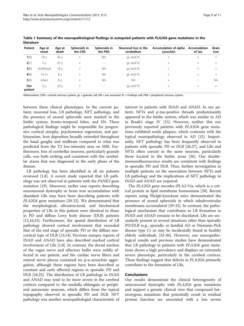
between these clinical phenotypes. In the current pa-tient, neuronal loss, LB pathology, NFT pathology, andthe presence of axonal spheroids were marked in thelimbic system, fronto-temporal lobes, and SN. Thesepathological findings might be responsible for progres-sive cortical atrophy, psychomotor regression, and par-kinsonism. Iron deposition broadly extended throughoutthe basal ganglia and midbrain compared to what waspredicted from the T2 low-intensity area on MRI. Fur-thermore, loss of cerebellar neurons, particularly granulecells, was both striking and consistent with the cerebel-lar ataxia that was diagnosed in the early phase of thedisease.LB pathology has been identified in all six patients
reviewed [1,8]. A recent study reported that LB path-ology was not observed in patients with the PANK2 genemutation [19]. Moreover, earlier case reports describingneuroaxonal dystrophy or brain iron accumulation withabundant LBs may have been describing patients withPLA2G6 gene mutations [20-22]. We demonstrated thatthe morphological, ultrastructural, and biochemicalproperties of LBs in this patient were identical to thosein PD and diffuse Lewy body disease (DLB) patients[12,16,23]. Furthermore, the spatial distribution of LBpathology showed cortical involvement that exceededthat of the end stage of sporadic PD or the diffuse neo-cortical type of DLB [13,14]. Previous autopsy reports ofINAD and ANAD have also described marked corticalinvolvement of LBs [1,8]. In contrast, the dorsal nucleusof the vagus nerve and olfactory bulbs were mildly af-fected in our patient, and the cardiac nerve fibers andenteral nerve plexus contained no p-α-synuclein aggre-gation, although these regions have been described asconstant and early affected regions in sporadic PD andDLB [24,25]. The distribution of LB pathology in INADand ANAD may tend to be more severe in the cerebralcortices compared to the medulla oblongata or periph-eral autonomic neurons, which differs from the typicaltopography observed in sporadic PD and DLB. NFTpathology was another neuropathologial characteristic of
interest in patients with INAD and ANAD. In our pa-tient, NFTs and p-tau-positive threads predominantlyappeared in the limbic system, which was similar to ADin Braak’s stage IV [15]. However, neither this norpreviously reported patients with PLA2G6 gene muta-tions exhibited senile plaques, which contrasts with thetypical neuropathology observed in AD [15]. Import-antly, NFT pathology has been frequently observed inpatients with sporadic PD or DLB [26,27], and LBs andNFTs often coexist in the same neurons, particularlythose located in the limbic areas [26]. Our double-immunofluorescence results are consistent with findingsin sporadic PD and DLB. Thus, further investigation inmultiple patients on the association between NFTs andLB pathology and the implications of NFT pathology inINAD and ANAD are required.The PLA2G6 gene encodes iPLA2-Via, which is a crit-
ical protein in lipid membrane homeostasis [28]. Recentreports using Pla2g6-knockout mice demonstrated thepresence of axonal spheroids in which tubulovesicularmembranes accumulated [29-32]. In contrast, the patho-logical mechanism that contributes to LB formation inINAD and ANAD remains to be elucidated. LBs are sec-ondarily present in several situations other than sporadicPD/DLB (e.g., sporadic or familial AD or Niemann-Pickdisease type C) or may be incidentally found in healthyelderly individuals [33-36]. However, our neuropatho-logical results and previous studies have demonstratedthat LB pathology in patients with PLA2G6 gene muta-tions shows a high prevalence and displays an extremelysevere phenotype, particularly in the cerebral cortices.These findings suggest that defects in PLA2G6 primarilycontribute to the formation of LBs.
ConclusionsOur results demonstrate the clinical heterogeneity ofneuroaxonal dystrophy with PLA2G6 gene mutationsand support a genetic clinical view that compound het-erozygous mutations that potentially result in residualprotein function are associated with a less severe
Table 1 Summary of the neuropathological findings in autopsied patients with PLA2G6 gene mutations in theliterature
Patient Age atonset
Age atdeath
Spheroids inthe CNS
Spheroids inthe PNS
Neuronal loss in thecerebellum
Accumulation of alphasynuclein
Accumulationof tau
Brainiron
1[8] 18 y 36 y + NA gc and Pc + + +
2[1] 3 y 23 y + - gc and Pc + + +
3[8] childhood 18 y + NA gc and Pc + + +
4[8] 14 m 8 y + NA gc and Pc + + +
5[8] infant 8 y + NA NA + - NA
Ourpatient
3 y 20 y + - gc and Pc + + +
Abbreviation: CNS = central nervous system; gc = granule cell; NA = not assessed; Pc = Purkinje cell; PNS = peripheral nervous system.
Riku et al. Acta Neuropathologica Communications 2013, 1:12 Page 9 of 11http://www.actaneurocomms.org/content/1/1/12
phenotype. Neuropathologically, CNS involvement withLBs was striking and exhibited a unique topographycompared with PD. Thus, further investigations on theprocess of LB formation caused by loss of PLA2G6 genefunction may provide new insights into the pathologicalmechanism of neuroaxonal dystrophy and LB formation.
ConsentWritten informed consent was obtained from the pa-tient’s parents for publication of this Case report andany accompanying images. A copy of the written consentis available for review by the Editor-in chief of thisjournal.
Competing interestsThere are no competing interests in the report.
Authors’ contributionsYR, MM, and MY performed clinical and pathological analysis. TI, HY, and HHcarried out the biochemical and genetic studies and drafted the manuscript.GS, KM, and YG helped to draft the manuscript. All authors read andapproved the final manuscript.
AcknowledgementThis work was supported by Grants-in-Aid from the Research Committee ofCNS Degenerative Diseases, the Ministry of Health, Labour and Welfare ofJapan. The study was approved by the ethics committee of JuntendoUniversity and Niigata University, and all subjects gave informed consent.
Author details1Department of Neurology, Nagoya Daiichi Red Cross Hospital, Aichi, Japan.2Department of Neurology, Nagoya University Graduate School of Medicine,Aichi, Japan. 3Department of Neurology, Brain Research Institute, NiigataUniversity, Niigata, Japan. 4Research Institute for Diseases of Old Age,Graduate School of Medicine, Juntendo University, Tokyo, Japan. 5Institutefor Medical Science of Aging, Aichi Medical University, Aichi, Japan.6Department of Neurology, Graduate School of Medicine, JuntendoUniversity, Tokyo, Japan.
Received: 9 February 2013 Accepted: 5 April 2013Published: 9 May 2013
References1. Gregory A, Westaway SK, Holm IE, Kotzbauer PT, Hogarth P, Sonek S, Coryell
JC, Nguyen TM, Nardocci N, Zorzi G, Rodriguez D, Desguerre I, Bertini E,Simonati A, Levinson B, Dias C, Barbot C, Carrilho I, Santos M, Malik I,Gitschier J, Hayflick SJ: Neurodegeneration associated with geneticdefects in phospholipase A2. Neurology 2008, 71:1402–1409.
2. Gregory A, Polster BJ, Hayflick SJ: Clinical and genetic delineation ofneurodegeneration with brain iron accumulation. J Med Genet 2009,46:73–80.
3. Seitelberger F: Neuroaxonal dystrophy: its relation to aging andneurological diseases. In Handbook of Clinical Neurology. Volume 5. Editedby Vinken PJ, Bruyn GW, Klawans HL. Amsterdam: Elsevier; 1986:391–415.
4. Kurian MA, Morgan NV, MacPherson L, Foster K, Peake D, Gupta R, Philip SG,Hendriksz C, Morton JEV, Kingston HM, Rosser EM, Wassmer E, Gissen P,Maher ER: Phenotypic spectrum of neurodegeneration associated withmutations in the PLA2G6 gene (PLAN). Neurology 2009, 70:1623–1629.
5. Morgan NV, Westaway SK, Morton JE, Gregory A, Gissen P, Sonek S, CangulH, Coryell J, Canham N, Nardocci N, Zorzi G, Pasha S, Rodriguez D,Desguerre I, Mubaidin A, Bertini E, Trembath RC, Simonati A, Schanen C,Johnson CA, Levinson B, Woods CG, Wilmot B, Kramer P, Gitschier J, MaherER, Hayflick SJ: PLA2G6, encoding a phospholipase A2, is mutated inneurodegenerative disorders with high brain iron. Nat Genet 2006,38:752–754.
6. Wu Y, Jiang Y, Gao Z, Wang J, Yuan Y, Xiong H, Chang X, Bao X, Zhang Y,Xiao J, Wu X: Clinical study and PLA2G6 mutation screening analysis in
Chinese patients with infantile neuroaxonal dystrophy. Eur J Neurol 2009,16:240–245.
7. Nardocci N, Zorzi G, Farina L, Binelli S, Scaioli W, Ciano C, Verga L, AngeliniL, Savoiardo M, Bugiani O: Infantile neuroaxonal dystrophy: Clinicalspectrum and diagnostic criteria. Neurology 1999, 52:1472–1478.
8. Paisan-Ruitz C, Li A, Schneider SA, Holton JL, Johnson R, Kidd D, Chataway J,Bhatia KP, Lees AJ, Hardy J, Revesz T, Houlden H: Widespread Lewy bodyand tau accumulation in childhood and adult onsetdystonia-parkinsonism cases with PLA2G6 mutations. Neurobiol Aging2012, 33:814–823.
9. Yoshino H, Tomiyama H, Tachibana N, Yoshino H, Tomiyama H, TachibanaN, Ogaki K, Li Y, Funayama M, Hashimoto T, Takashima S, Hattori N:Phenotypic spectrum of patients with PLA2G6 mutation andPARK14-linked parkinsonism. Neurology 2010, 75:1356–1361.
10. Ikeuchi T, Kakita A, Shiga A, Kasuga K, Kaneko H, Tan CF, Idezuka J,Wakabayashi K, Onodera O, Iwatsubo T, Nishizawa M, Takahashi H, IshikawaA: Homozygous and heterozygous patients for SNCA duplication infamily with parkinsonism and dementia. Arch Neurol 2008, 65:514–519.
11. Kaneko H, Kakita A, Kasuga K, Nozaki H, Ishikawa A, Miyashita A, Kuwano R,Ito G, Iwatsubo T, Takahashi H, Nishizawa M, Onodera O, Sisodia SS, IkeuchiT: Enhanced accumulation of phosphorylated α-synuclein and elevatedAβ42/40 ratio caused by expression of the presenilin-1 delta T440mutant associated with familial Lewy body disease and variantAlzheimer disease. J Neurosci 2007, 28:13092–13097.
12. Fujiwara H, Hasegawa M, Dohmae N, Kawashima A, Masliah E, Goldberg MS,Shen J, Takio K, Iwatsubo T: α-synuclein is phosphorylated insynucleinopathy lesions. Nat Cell Biol 2002, 4:160–164.
13. Braak H, Del Tredici K, Rüb U, de Vos RA, Jansen Steur EN, Braak E: Stagingof brain pathology related to sporadic Parkinson’s disease.Neurobiol Aging 2003, 24:197–211.
14. McKeith IG, Dickson DW, Lowe J, Emre M, O’Brien JT, Feldman H, CummingsJ, Duda JE, Lippa C, Perry EK, Aarsland D, Arai H, Ballard CG, Boeve B, BurnDJ, Costa D, Del Ser T, Dubois B, Galasko D, Gauthier S, Goetz CG, Gomez-Tortosa E, Halliday G, Hansen LA, Hardy J, Iwatsubo T, Kalaria RN, Kaufer D,Kenny RA, Korczyn A, Kosaka K, Lee VM, Lees A, Litvan I, Londos E, LopezOL, Minoshima S, Mizuno Y, Molina JA, Mukaetova-Ladinska EB, Pasquier F,Perry RH, Schulz JB, Trojanowski JQ, Yamada M: Diagnosis andmanagement of dementia with Lewy bodies: third report of the DLBConsortium. Neurology 2005, 65:1863–1872.
15. Braak H, Alafuzoff I, Arzberger T, Kretzschmar H, Tredici KD: Staging ofAlzheimer’s disease-associated neurofibrillary pathology using paraffinsections and immunocytochemistry. Acta Neuropathol 2006, 112:389–404.
16. Kosaka K: Lewy bodies in cerebral cortex, report of three cases.Acta Neuropathol (Berl) 1978, 42:127–134.
17. Ma Z, Wang X, Nowatzke W, Ramanadham S, Turk J: Human pancreaticislets express mRNA species encoding two distinct catalytically activeisoforms of group VI phopholipase A2 (iPLA2) that arise from an exon-skipping mechanism of alternative splicing of the transcript from theiPLA2 gene on chromosome 22q13.1. J Biol Chem 1999, 274:9607–9616.
18. Tonelli A, Romaniello R, Grasso R, Cavallini A, Righini A, Bresolin N, BorgattiR, Bassi MT: Novel splice-site mutations and a large intragenic deletion inPLA2G6 associated with a severe and rapidly progressive form ofinfantile neuroaxonal dystrophy. Clin Genet 2010, 78:432–440.
19. Li A, Paudel R, Johnson R, Courtney R, Lees AJ, Holton JL, Hardy J, Revesz T,Houlden H: Pantothenate kinase-associated neurodegeneration is not asynucleinopathy. Neuropathol Appl Neurobiol 2013, 39:121. 131.
20. Wakabayashi K, Fukushima T, Koide R, Horikawa Y, Hasegawa M, WatanabeY, Noda T, Eguchi I, Morita T, Yoshimoto M, Iwatsubo T, Takahashi H:Juvenile-onset generalized neuroaxonal dystrophy (Hallervorden-Spatzdisease) with diffuse neurofibrillary and Lewy body pathology.Acta Neuropathol (Berl) 2000, 99:331–336.
21. Wakabayashi K, Yoshimoto M, Fukushima T, Koide R, Horikawa Y, Morita T,Takahashi H: Widespread occurrence of α-synuclein/NACPimmunoreactive neuronal inclusions in juvenile and adult-onsetHallervorden–Spatz disease with Lewy bodies. Neuropathol Appl Neurobiol1999, 25:363–368.
22. Neumann M, Adler S, Schlüter O, Kremmer E, Benecke R, Kretzschmar HA: α-Synuclein accumulation in a case of neurodegeneration with brain ironaccumulation type 1 (NBIA-1, formerly Hallervorden-Spatz syndrome)with wide spread cortical and brainstem-type Lewy bodies.Acta Neuropathol (Berl) 2000, 100:568–574.
Riku et al. Acta Neuropathologica Communications 2013, 1:12 Page 10 of 11http://www.actaneurocomms.org/content/1/1/12
23. Forno LS: Neuropathology of Parkinson’s disease. J Neuropathol Exp Neurol1996, 55:259–272.
24. Hawkes CH, Tredici KD, Braak H: Review: Parkinson’s disease: a dual-hithypothesis. Neuropathol Appl Neurobiol 2007, 33:599–614.
25. Orimo S, Takahashi A, Uchihara T, Mori F, Kakita A, Wakabayashi K, TakahashiH: Degeneration of cardiac sympathetic nerve begins in the earlydisease process of Parkinson’s disease. Brain Pathol 2007, 17:24–30.
26. Iseki E, Marui W, Kosaka K: Frequent coexistence of Lewy bodies andneurofibrillary tangles in the same neurons of patients with diffuse Lewybody disease. Neurosci Lett 1999, 265:9–12.
27. Ishizawa T, Mattila P, Davies P, Wang D, Dickson DW: Colocalization of tauand alpha-synuclein epitopes in Lewy bodies. J Neuropathol Exp Neurol2003, 62:389–397.
28. Baburina I, Jakowski S: Cellular responses to excess phospholipid. J BiolChem 1999, 14:9400–9408.
29. Beck G, Sugiura Y, Shinzawa K, Kato S, Setou M, Tsujimoto Y, Sakoda S,Sumi-Akamaru H: Neuroaxonal dystrophy in calcium-independentphospholipase A2β deficiency results from insufficient remodeling anddegeneration of mitochondrial and presynaptic membranes. J Neurosci2011, 31:11411–11420.
30. Malik I, Turk J, Mancuso DJ, Montier L, Wohltmann M, Wozniak DF, SchmidtRE, Gross RW, Kotzbauer PT: Disrupted membrane homeostasis andaccumulation of ubiquitinated proteins in a mouse model of infantileneuroaxonal dystrophy caused by PLA2G6 mutations. Am J Pathol 2008,172:406–416.
31. Shinzawa K, Sumi H, Ikawa M, Matsuoka Y, Okabe M, Sakoda S, Tsujimoto Y:Neuroaxonal dystrophy caused by group VIA phospholipase A2
deficiency in mice: a model of human neurodegenerative disease.J Neurosci 2008, 28:2212–2220.
32. Wada H, Yasuda T, Miura I, Watabe K, Sawa C, Kamijuku H, Kojo S, TaniguchiM, Nishino I, Wakana S, Yoshida H, Seino K: Establishment of an improvedmouse model for infantile neuroaxonal dystrophy that shows earlydisease onset and bears a point mutation in Pla2g6. Am J Pathol 2009,175:2257–2263.
33. Hamilton RL: Lewy bodies in Alzheimer’s disease: A neuropathologicalreview of 145 cases using α-synuclein immunohistochemistry. BrainPathol 2000, 10:378–384.
34. Rosenberg CK, Pericak-Vance MA, Saunders AM, Gilbert JR, Gaskell PC,Hulette CM: Lewy body and Alzheimer pathology in a family with theamyloid-β precursor protein APP717 gene mutation. Acta Neuropathol(Berl) 2000, 100:145–152.
35. Saito Y, Ruberu NN, Sawabe M, Arai T, Kazama H, Hosoi T, Yamanouchi H,Murayama S: Lewy body-related α-synucleinopathy in aging.J Neuropathol Exp Neurol 2004, 63:742–749.
36. Saito Y, Suzuki K, Hulette CM, Murayama S: Aberrant phosphorylation of α-synuclein in human Niemann-Pick type C1 disease. J Neuropathol ExpNeurol 2004, 63:323–328.
doi:10.1186/2051-5960-1-12Cite this article as: Riku et al.: Extensive aggregation of α-synuclein andtau in juvenile-onset neuroaxonal dystrophy: an autopsied individualwith a novel mutation in the PLA2G6 gene-splicing site. ActaNeuropathologica Communications 2013 1:12.
Submit your next manuscript to BioMed Centraland take full advantage of:
• Convenient online submission
• Thorough peer review
• No space constraints or color figure charges
• Immediate publication on acceptance
• Inclusion in PubMed, CAS, Scopus and Google Scholar
• Research which is freely available for redistribution
Submit your manuscript at www.biomedcentral.com/submit
Riku et al. Acta Neuropathologica Communications 2013, 1:12 Page 11 of 11http://www.actaneurocomms.org/content/1/1/12