
Cellular Interactions of Synovial Fluid gd T Cells in Juvenile
12
of December 23, 2018. This information is current as Cells in Juvenile Idiopathic Arthritis T δ γ Cellular Interactions of Synovial Fluid and Ilan Bank Maya Gerstein, Meital Nagar, Itamar Goldstein, Shai Padeh Anna Bendersky, Victoria Marcu-Malina, Yackov Berkun, ol.1102403 http://www.jimmunol.org/content/early/2012/04/04/jimmun published online 26 March 2012 J Immunol average * 4 weeks from acceptance to publication Fast Publication! • Every submission reviewed by practicing scientists No Triage! • from submission to initial decision Rapid Reviews! 30 days* • Submit online. ? The JI Why Subscription http://jimmunol.org/subscription is online at: The Journal of Immunology Information about subscribing to Permissions http://www.aai.org/About/Publications/JI/copyright.html Submit copyright permission requests at: Email Alerts http://jimmunol.org/alerts Receive free email-alerts when new articles cite this article. Sign up at: Print ISSN: 0022-1767 Online ISSN: 1550-6606. Immunologists, Inc. All rights reserved. Copyright © 2012 by The American Association of 1451 Rockville Pike, Suite 650, Rockville, MD 20852 The American Association of Immunologists, Inc., is published twice each month by The Journal of Immunology by guest on December 23, 2018 http://www.jimmunol.org/ Downloaded from by guest on December 23, 2018 http://www.jimmunol.org/ Downloaded from
Transcript of Cellular Interactions of Synovial Fluid gd T Cells in Juvenile
of December 23, 2018.This information is current as
Cells in Juvenile Idiopathic Arthritis TδγCellular Interactions of Synovial Fluid
and Ilan BankMaya Gerstein, Meital Nagar, Itamar Goldstein, Shai Padeh Anna Bendersky, Victoria Marcu-Malina, Yackov Berkun,
ol.1102403http://www.jimmunol.org/content/early/2012/04/04/jimmun
published online 26 March 2012J Immunol
average*
4 weeks from acceptance to publicationFast Publication! •
Every submission reviewed by practicing scientistsNo Triage! •
from submission to initial decisionRapid Reviews! 30 days* •
Submit online. ?The JIWhy
Subscriptionhttp://jimmunol.org/subscription
is online at: The Journal of ImmunologyInformation about subscribing to
Permissionshttp://www.aai.org/About/Publications/JI/copyright.htmlSubmit copyright permission requests at:
Email Alertshttp://jimmunol.org/alertsReceive free email-alerts when new articles cite this article. Sign up at:
Print ISSN: 0022-1767 Online ISSN: 1550-6606. Immunologists, Inc. All rights reserved.Copyright © 2012 by The American Association of1451 Rockville Pike, Suite 650, Rockville, MD 20852The American Association of Immunologists, Inc.,
is published twice each month byThe Journal of Immunology
by guest on Decem
ber 23, 2018http://w
ww
.jimm
unol.org/D
ownloaded from
by guest on D
ecember 23, 2018
http://ww
w.jim
munol.org/
Dow
nloaded from
The Journal of Immunology
Cellular Interactions of Synovial Fluid gd T Cells in JuvenileIdiopathic Arthritis
Anna Bendersky,*,1 Victoria Marcu-Malina,*,1 Yackov Berkun,† Maya Gerstein,†
Meital Nagar,‡ Itamar Goldstein,‡ Shai Padeh,† and Ilan Bank*,x
The pathogenesis of juvenile idiopathic arthritis (JIA) is thought to involve multiple components of the cellular immune system,
including subsets of gd T cells. In this study, we conducted experiments to define the functional roles of one of the major synovial
fluid (SF) T cell subsets, Vg9+Vd2+ (Vg9+) T cells, in JIA. We found that as opposed to CD4+ T cells, equally high percentages
(∼35%) of Vg9+ T cells in SF and peripheral blood (PB) produced TNF-a and IFN-g. Furthermore, stimulation with isopentenyl
pyrophosphate (IPP), a metabolite in the mevalonate pathway, which is a specific potent Ag for Vg9Jg1.2+ T cells, similarly
amplified cytokine secretion by SF and PB Vg9+ T cells. Significantly, the SF subset expressed higher levels of CD69 in situ,
suggesting their recent activation. Furthermore, 24-h coculturing with SF-derived fibroblasts enhanced CD69 on the SF > PB
Vg9+ T cells, a phenomenon strongly augmented by zoledronate, a farnesyl pyrophosphate synthase inhibitor that increases
endogenous intracellular IPP. Importantly, although Vg9+ T cell proliferation in response to IPP was significantly lower in SF
than PBMC cultures, it could be enhanced by depleting SF CD4+CD25+FOXP3+ cells (regulatory T cells). Furthermore, coculture
with the Vg9+ T cells in medium containing zoledronate or IPP strongly increased SF-derived fibroblasts’ apoptosis. The findings
that IPP-responsive proinflammatory synovial Vg9+ T cells for which proliferation is partly controlled by regulatory T cells can
recognize and become activated by SF fibroblasts and then induce their apoptosis suggest their crucial role in the pathogenesis and
control of synovial inflammation. The Journal of Immunology, 2012, 188: 000–000.
Asubset of T lymphocytes discovered 25 y ago, gd T cellscombine properties of the adaptive and innate immunesystems (1, 2). In humans, a major subset of gd T cells
express TCR polypeptides in which the V regions are encoded byrearranged Vg9 and Vd2 genes (3). Vg9+Vd2+ (Vg9+) T cells caninteract with dendritic cells, B cells, and macrophages; kill, ina non–MHC-restricted manner, virally infected and malignantlytransformed cells; secrete cytokines and growth factors; and pres-ent Ags to CD4+ and CD8+TCRab+ T cells (4). Most Vg9+
T cells, which form 50–70% of the circulating gd T cell subset inhealthy humans, use the joining g 1.2 gene segment rearranged toVg9 and uniquely can recognize, in a TCR-dependent manner,small nonpeptidic phosphorylated Ags (including isopentenylpyrophosphate [IPP]) produced in the mevalonate biosyntheticpathway when their concentration increases due to its dysregu-
lation (5). For example, these phosphorylated moieties mayaccumulate intracellularly during infection, inflammatory stress,
and malignant transformation of epithelial or mesenchymal cells.
Furthermore, the same cells can recognize with markedly higher
affinity phosphorylated products of the alternative mevalone
pathway in the nonmevalonate (or Rohmer) pathway of isoprenoid
synthesis by various microorganisms (6). Recent evidence sug-
gests these substances are recognized by Vg9Jg1.2Vd2 T cells in
the context of cell-surface membrane-expressed molecules, which
may include ectopic F1-ATPase (7).The mevalonate pathway is critical for the growth of rheuma-
toid synovial fibroblasts (Sfib) because blocking this pathway by
inhibiting hydroxy-3-methylglutaryl CoA reductase induces their
apoptosis (8). The demonstrated activity of this pathway suggests
that Sfib produce IPP, the Ag recognized by Vg9Jg1.2Vd2 T cells,
which could potentially activate a subset of synovial Vg9+ T cells
in the joints of patients with autoimmune synovitis (9).Juvenile idiopathic arthritis (JIA), a clinically heterogeneous
group of different disorders characterized by chronic inflammatory
arthritis in children, is an important cause of short- and long-term
disability (10). Current International League of Associations for
Rheumatology classification differentiates subtypes of JIA on
the basis of clinical manifestations (oligoarthritis, persistent and
extended; rheumatoid factor-positive and -negative polyarthritis;
systemic, psoriatic, enthesitis-related, and other undifferentiatedarthritis) (11). Although the precise etiology is still unknown, JIA
is an autoimmune disease, and T cells are thought to be key
players in this process (12). Interestingly, although the Vg9Jg1.2
gene rearrangement is less commonly expressed among synovial
fluid (SF) than peripheral blood (PB) gd T cells, IPP was recently
found to induce expansion of SF Vg9+ T in virtually all forms
of JIA (13, 14). However, despite the known proinflammatory
potential of Vg9+ T cells in healthy individuals, higher levels
of expansion were associated with clinical remission in these
*Laboratory of Immunoregulation, Sheba Medical Center, Tel Hashomer and SacklerSchool of Medicine, Tel Aviv University, Ramat Gan 52621, Israel; †Department ofPediatrics, Sheba Medical Center, Tel Hashomer and Sackler School of Medicine, TelAviv University, Ramat Gan 52621, Israel; ‡Cancer Research Center, Sheba MedicalCenter, Tel Hashomer and Sackler School of Medicine, Tel Aviv University, RamatGan 52621, Israel; and xDepartment of Medicine, Sheba Medical Center, TelHashomer and Sackler School of Medicine, Tel Aviv University, Ramat Gan52621, Israel
1A.B. and V.M.-M. contributed equally to this work.
Received for publication August 23, 2011. Accepted for publication February 22,2012.
This work was supported in part by the Kandinoff Pessa Polanski Fund.
Address correspondence and reprint requests to Prof. Ilan Bank, Department ofMedicine, Sheba Medical Center, Tel Hashomer and Sackler School of Medicine,Ramat Gan 52621, Israel. E-mail address: [email protected]
Abbreviations used in this article: DI, division index; IPP, isopentenyl pyrophosphate;JIA, juvenile idiopathic arthritis; MC, mononuclear cell; MFI, mean fluorescenceintensity; PB, peripheral blood; rhIL-2, recombinant human IL-2; SF, synovial fluid;SFMC, synovial fluid mononuclear cell; Sfib, synovial fluid-derived fibroblast; Treg,regulatory T cell; TSST1, toxic shock syndrome toxin-1; Vg9+, Vg9+Vd2+.
Copyright� 2012 by The American Association of Immunologists, Inc. 0022-1767/12/$16.00
www.jimmunol.org/cgi/doi/10.4049/jimmunol.1102403
Published April 4, 2012, doi:10.4049/jimmunol.1102403 by guest on D
ecember 23, 2018
http://ww
w.jim
munol.org/
Dow
nloaded from
patients, suggesting that IPP-induced activation of the cells insome unknown manner contributes to control synovial inflam-mation (13). The experiments described in this paper were un-dertaken to further delineate functions and cellular interactions ofIPP-responsive SF Vg9+ T cells that could be relevant to diseaseoutcomes in these patients.
Materials and MethodsPatients
The study was approved by the Institutional Review Board (HelsinkiCommittee) of the Chaim Sheba Medical Center, Ramat Gan, Israel. Allparticipating patients were seen in the Pediatric Rheumatology clinic.Patients fulfilled the criteria of the American College of Rheumatology forJIA (11). Informed consent was signed by parents of all participants. Allsamples of SF and PB were drawn during an acute flare of synovitis, re-quiring intra-articular corticosteroid injection. The patients participating inthe study included individuals with pauciarticular persistent arthritis, withextended JIA, systemic disease, psoriatic arthritis, and polyarthritis. Con-trol samples were drawn from PB of healthy consenting adults.
Isolation of mononuclear cells
PBMC and synovial fluid mononuclear cells (SFMC) were isolated byFicoll Hypaque (Sigma-Aldrich, St. Louis, MO) density centrifugation aspreviously described (15).
Fibroblast cultures
Primary fibroblastic lines were obtained from SF removed from knees ofpatients for therapeutic indications and maintained as previously described(15). In brief, fibroblasts were derived by culturing adherent synovial fluidcells in plastic wells in DMEM-supplemented medium for 48 h followedby removal of all of the nonadherent cells. The adherent cells displayeda fibroblastic morphology and were determined by flow cytometry to ex-press CD90, low levels of the CD49a integrin, to be negative for macro-phage (CD14), T cell (CD3), and B cell (CD20) surface markers, andstained positively for collagen type I determined by Sirius red staining (notshown).
Characterization of T cell subsets by flow cytometry
FITC, allophycocyanin, PerCP, or PE-conjugated nonbinding Ig isotypecontrol mAb (BD Pharmingen) or fluorochrome-conjugated mAb to CD4,CD3, CD25 (BD Pharmingen), or the human Vg9, Vd1, and Vd2 geneproducts (Immunotech) were added to a resuspended pellet of 2 3 105
mononuclear cells (MC) for 45 min on ice (15). Cells were then washed inPBS (pH 7.4; Life Technologies, Invitrogen). The anti-human FOXP3mAb (PCH101) staining kit was purchased from eBioscience and used asper the manufacturer’s instructions. Lymphocytes were gated according totypical forward and side scatter plots using an FACS flow cytometer (BDBiosciences, Mountain View, CA), and analysis of the gated cells wasperformed.
Purification of T cell populations
CD25+ cells were isolated using the MACS system (Miltenyi Biotec)according to the manufacturer’s instructions. SFMC were also stained withFITC-conjugated anti-CD4 and/or PE-conjugated anti-CD25 mAbs (BDPharmingen) for 45 min on ice. PBMC activated with IPP were immu-nodepleted using mAb to CD4 and CD8 to obtain .90% purified Vg9+
T cells. Cells were then washed twice with MACS buffer (0.2 mM EDTAand 0.5% BSA in PBS). Subsequently, cells were incubated with anti-mouse IgG microbeads (Miltenyi Biotec) for 15 min at 4–8˚C andpassed through MACS columns. Cells retained in the columns were elutedand used in experiments after confirmation of purity by FACS.
In vitro stimulation
PBMC or SFMC (1 3 106 cells/ml) were cultured in 96-well, round-bottom tissue-culture plates (Costar) in 200 ml RPMI 1640 medium sup-plemented with 10% FBS, 2 mM L-glutamine, 100 mg/ml penicillin-streptomycin solution, and 100 IU/ml recombinant human IL-2 (rhIL-2;Boehringer Mannheim) and stimulated by addition of IPP (Sigma-Aldrich)as indicated in the respective experiments. An ab TCR selective super-antigen, toxic shock syndrome toxin-1 (TSST1; Sigma-Aldrich) at 10 ng/ml, or rhIL-2 alone was added as indicated. Medium was replaced on days3 and 7 of culture. All cultures were maintained for 10 d except whereindicated, at which time they were used for experiments.
Proliferation assay
T cell proliferation was assessed, as previously described, using FACS-based analysis of serial halving of the vital dye CFSE (16). Briefly, upto 13 107 cells were suspended in 1 ml RPMI 1640 medium and thenpulsed with 2.5 mM CFSE (Molecular Probes) for 10 min (37˚C) withconstant shaking. Subsequently, the CFSE was quenched with supple-mented RPMI 1640 medium, and then the cells were washed twice withlarge volumes of medium. The labeled cells were stimulated and cultured,as indicated, and at the end of culture were analyzed for CFSE dilution.
Cytokine detection
For intracellular cytokine detection, the T cells were activated with 20 ng/mlPMA and 0.8 mM ionomycin (Sigma-Aldrich) in the presence of 2 mg/mlbrefeldin A (GolgiStop; eBioscience) for 4.5 h. Subsequently, the cellswere harvested and stained with anti–Vg9-FITC and CD4-PE and washedtwice in PBS. Cells were fixed and permeabilized using fixation andpermeabilization buffers (eBioscience), then stained in Flow CytometryStaining buffer (eBioscience) with anti–TNF-a, anti–IL-17, and anti–IFN-g–allophycocyanin. IFN-g detection in supernatants was done using theELISA max Deluxe Sets (BioLegend, San Diego, CA) according to themanufacturer’s instructions.
Fibroblast/T cell cocultures
A total of 100 ml (2 3 105 cell/ml) Sfib, resuspended in DMEM-supplemented medium, were placed in 48-well flat-bottom plates (Co-star). After 1 h, during which time fibroblasts formed a homogeneousmonolayer, effector T cells resuspended in RPMI 1640-supplementedmedium were added at the indicated concentration (usually 2 3 106 cell/ml). Cocultures were placed in the humidified 5% CO2 incubator at 37˚Cfor 48 h. To evaluate fibroblast growth, nonadherent cells were decantedand adherent cells fixed in 70% ethanol for 10 min. Cells were then stainedwith 1% (w/v) crystal violet solution for 20 min, followed by extensivewashing with water, and were evaluated by inverted microscopic inspec-tion. To determine apoptosis, fibroblasts were stained with CFSE prior toplating. After coincubation with the designated population of T cells for 48h, adherent cells in each well were treated with trypsin-EDTA solution for3 min (Ta’assiot Biologiot, Beit Haemek, Israel) and washed in PBS. Thecells were spun down at 350 3 g, stained with Annexin V (BD Phar-mingen), and analyzed on an FACScan flow cytometer (BD Biosciences).Analysis of CFSE+ cells was performed and the percentage of Annexin V+
cells recorded.
Suppression assay
Regulatory T cells (Treg), isolated from SFMC or PBMC, were tested fortheir ability to suppress the proliferation of autologous CD252Vg9+ T cellsusing methods similar to those previously described (16). Briefly, CFSE-labeled CD252 SFMC or PBMC at 1 3 105 cells/well were plated into 96-well plates (Costar) and activated by either anti-CD3 mAbs (clone OKT3;2.5 mg/ml), IPP (2 mg/ml), or both reagents in the presence of gradedamounts of CD25+ T cells. Five days later, the T cells were harvested andanalyzed by FACS. Vg9+ T cells proliferation was determined using theproliferation platform of FlowJo 7.2.5 (Tree Star) to calculate the divisionindex (DI) and draw a fit model of generations onto the CFSE dilutionhistograms. The DI reflects the average number of cell divisions that thecells underwent and is considered a good objective value to compare therate of proliferation from sample to sample. Percent suppression wascalculated for each well using the following formula: 100 3 (DI in theabsence of CD25+ T cells 2 DI with added CD25+ T cells)/DI in theabsence of CD25+ T cells.
Statistical analysis
Student t tests (Prism software; GraphPad) were used to analyze the sig-nificance of differences between groups.
ResultsCytokine production by SF Vg9+ T cells and effect of IPPstimulation
To determine the cytokine profile of SF Vg9+ T cells, relevant tosynovial inflammation, freshly isolated SF and PBMC from 12patients with JIA were activated with PMA and ionomycin in thepresence of a Golgi-transport inhibitor and then analyzed for in-tracellular levels of IFN-g, TNF-a, and IL-17. We found that SFCD4+ T cells were significantly enriched for TNF-a– (21.7 6
2 gd T CELLS IN JUVENILE IDIOPATHIC ARTHRITIS
by guest on Decem
ber 23, 2018http://w
ww
.jimm
unol.org/D
ownloaded from
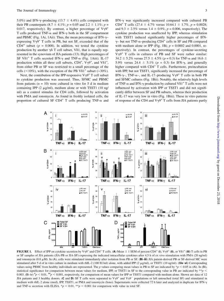
5.0%) and IFN-g–producing (13.7 6 4.4%) cells compared withtheir PB counterparts (8.7 6 4.1%; p = 0.05 and 2.2 6 1.1%; p =0.017, respectively). By contrast, a higher percentage of Vg9+
T cells produced TNF-a and IFN-g both in the SF compartmentand PBMC (Fig. 1Ai, 1Aii). Thus, the mean percentage of IFN-g–expressing Vg9+ T cells in PB, but not SF, exceeded that of theCD4+ subset (p = 0.008). In addition, we tested the cytokineproduction by another gd T cell subset, Vd1, that is equally rep-resented in the synovium of JIA patients (13). High percentages ofSF Vd1+ T cells secreted IFN-g and TNF-a (Fig. 1Aiii). IL-17production within all three cell subsets, CD4+, Vg9+, and Vd1+,from either PB or SF was restricted to a small percentage of thecells (,10%), with the exception of the PB Vd1+ subset (∼30%).Next, the contribution of the IPP-responsive Vg9+ T cell subset
to cytokine production was assessed. Thus, SFMC and PBMCfrom patients (n = 10) were cultured in vitro for 5 d in mediumcontaining IPP (2 mg/ml), medium alone or with TSST1 (10 ng/ml) as a control stimulus for CD4 cells, followed by activationwith PMA and ionomycin. As found in freshly isolated cells, theproportion of cultured SF CD4+ T cells producing TNF-a and
IFN-g was significantly increased compared with cultured PBCD4+ T cells (27.4 6 4.7% versus 10.64.1 6 1.7%, p = 0.0028;and 9.3 6 2.5% versus 1.4 6 0.9%, p = 0.006, respectively). Thecytokine production was unaffected by IPP, whereas stimulationwith TSST1 induced significantly higher percentages of IFN-g– but not TNF-a–producing CD4+ cells in SF and PB comparedwith medium alone or IPP (Fig. 1Bi; p = 0.0002 and 0.0001, re-spectively). In contrast, the percentages of cytokine-secretingVg9+ T cells in cultures of PB and SF were rather similar:34.2 6 5.2% versus 27.5 6 4.5% (p = 0.3) for TNF-a and 16.6 65.9% versus 24.4 6 5.1% (p = 0.3) for IFN-g, and generallyhigher compared with CD4+ T cells. Furthermore, preincubationwith IPP, but not TSST1, significantly increased the percentage ofIFN-g–, TNF-a–, and IL-17–producing Vg9+ T cells in both PBand SFMC cultures (Fig. 1Bii). Notably, the relatively high levelsof TNF-a and IFN-g production by cultured Vd1+ T cells were notinfluenced by activation with IPP or TSST1 and did not signifi-cantly differ between SF and PB subsets, whereas their productionof IL-17 was very low in vitro (Fig. 1Biii). These in vitro patternsof response of the CD4 and Vg9+ T cells from JIA patients partly
FIGURE 1. Effect of IPP on cytokine secretion by Vg9+ and CD4+ T cells. (A) Mean6 1 SEM of percent CD4+ (i), Vg9+ (ii), or Vd1+ (iii) T cells in PB
or SF samples of JIA patients (JIA-PB or JIA-SF) expressing the indicated intracellular cytokines after 4.5 h of ex vivo stimulation with PMA (20 ng/ml)
and ionomycin (0.8 mM). In (A), cells were stimulated immediately after isolation from PB or SF. (Bi–iii) JIA patient-derived PB or SF-derived MC were
stimulated after 5 d of in vitro culture in medium with rhIL-2 (100 IU/ml) alone, with added IPP (2 mg/ml), or TSST1 (10 ng/ml). (Biv–vi) Corresponding
values using PBMC from healthy individuals are represented. The p values comparing mean values in PB to SF are indicated by *p , 0.05 in (Ai). In (Bi),
statistical significance for comparison between mean values for medium, IPP, or TSST1 in SF to the corresponding value in PB are indicated by **p ,0.003. (Bi–iv) #p , 0.01, ##p , 0.001, respectively, for comparison of mean values for IPP or TSST1 compared with medium alone. Shown are data of 12
JIA patients and 3 healthy donors. (C and D) SF T cells were separated to Vg9+ and Vg92 populations or left untouched (total SF) and stimulated in
medium with rhIL-2 alone (med), IPP, TSST1, or PMA and ionomycin (Iono). Supernatants were collected 72 h later and analyzed in duplicate for IFN-g
and TNF-a secretion with ELISA. *p , 0.01, **p , 0.001 for comparison with value in total SF.
The Journal of Immunology 3
by guest on Decem
ber 23, 2018http://w
ww
.jimm
unol.org/D
ownloaded from
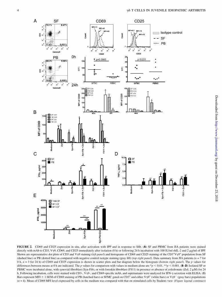
FIGURE 2. CD69 and CD25 expression in situ, after activation with IPP and in response to Sfib. (A) SF and PBMC from JIA patients were stained
directly with mAb to CD3, Vg9, CD69, and CD25 immediately after isolation (0 h) or following 24 h incubation with 100 IU/ml rhIL-2 and 2 mg/ml of IPP.
Shown are representative dot plots of CD3 and Vg9 staining (left panel) and histograms of CD69 and CD25 staining of the CD3+Vg9+ population from SF
(dashed line) or PB (dotted line) as compared with negative control isotype staining (gray fill) (top right panel). Data summary from JIA patients (n = 7 for
0 h, n = 3 for 24 h) of CD69 and CD25 expression is shown in scatter plots and bar diagram below the histogram (bottom right panel). The p values for
differences between means at 0 h are indicated. The p values for comparison with values in medium alone are *p, 0.01, **p, 0.001. (B–D) Isolated SF or
PBMC were incubated alone, with synovial fibroblast (Syn-Fib), or with foreskin fibroblast (FS11) in presence or absence of zoledronate (Zol; 2 mM) for 24
h. Following incubation, cells were stained with CD3-, Vg9-, and CD69-specific mAb, and supernatants were analyzed for IFN-g secretion with ELISA. (B)
Bars represent MFI6 1 SEM of CD69 staining of PB (hatched bars) or SFMC gated on CD3+ and either Vg9+ (white bars) or Vg92 (gray bars) populations
(n = 4). Mean of CD69 MFI level expressed by cells in the medium was compared with that on stimulated cells by Student t test (Figure legend continues)
4 gd T CELLS IN JUVENILE IDIOPATHIC ARTHRITIS
by guest on Decem
ber 23, 2018http://w
ww
.jimm
unol.org/D
ownloaded from
resembled those of these subsets in PBMC isolated from healthyadults (Fig. 1Biv–1Bvi). Thus, IPP specifically and significantlyincreased TNF-a production by cultured Vg9+ T cells fromhealthy donors but not by CD4+ or Vd1+ T cells. In contrast,TSST1 increased this cytokine production only by cultured CD4+
T cells. The effect of IPP on IFN-g production in the healthydonor-derived Vg9+ T cells was not significant, possibly due tothe high percent of spontaneously producing IFN-g+ cells in thesedonors.Further, to analyze the contribution of Vg9+ T cells to the cy-
tokine pool produced by SFMC, we compared cytokine produc-tion by Vg9-depleted SFMC and Vg9+ (.90%) T cells froma donor whose SFMC contained a high percentage (18.7%) ofVg9+ T cells after triggering with IPP, TSST1, or PMA and ion-omycin for 72 h (Fig. 1C, 1D). Very low or absent levels ofcytokines were produced in medium alone, whereas a high con-centration of IFN-g and a significant yet lower concentration ofTNF-a was secreted by unseparated SFMC, Vg92, and Vg9+
SFMC stimulated with PMA and ionomycin. Depletion of theVg9+ T cells almost abolished production of IFN-g and TNF-a,whereas the isolated Vg9+ T cells produced notably higheramounts of TNF-a than unseparated SFMC and Vg92 SFMC inresponse to IPP. By contrast, TSST1-induced cytokine productionwas mainly contributed by Vg92 cells.These results suggest that Vg9+ T cells, specifically, of JIA
patients can strongly contribute to the repertoire of TNF-a, IFN-g,and, to a lesser degree, IL-17 produced systemically and in thesynovium upon activation with IPP.
Activation of SF Vg9+ T cells in vivo and in vitro
To test the hypothesis that SF Vg9+ T cells may be respondingto subset-specific Ags at the site of inflammation, we determinedwhether they express an activated phenotype consistent with arecent encounter with IPP. First, we examined CD69 and CD25,cell-surface markers of recent activation, on the Vg9+ T cells inPB and SF of JIA patients. We found that SF Vg9+ T cellsexpressed significantly higher levels of CD69 (mean fluores-cence intensity [MFI] 89.5 6 15.3) than PB Vg9+ T cells (MFI37.5 6 3.9; p = 0.0065), whereas both subsets express only lowlevels of CD25 in situ (Fig. 2A, 0 h). In vitro stimulation with IPPfor 24 h induced significantly higher levels of CD69 and CD25 onthe two populations of Vg9+ T cells, as compared with cellscultured in IL-2 medium alone (Fig. 2A, 24 h). These resultssuggest that in the inflamed tissue, SF Vg9+ T cells express atleast one of the hallmarks consistent with that of cells recentlyactivated by IPP (i.e., CD69high).Monocytes and dendritic cells are known to activate PB Vg9+
T cells when they are induced to express high levels of IPP, forexample, by zoledronate, an inhibitor of farnesyl pyrophosphatesynthase (17, 18). To determine whether other cellular compo-nents of the synovium also play a role in activating SF Vg9+
T cells, we focused on the role of Sfib, which are critically de-pendent upon the mevalonate pathway, suggesting that they maycontinuously produce the Vg9+ Ag IPP (8).SFMC were isolated from four JIA patients and cocultured for
24 h either alone or with zoledronate in the presence or absence of
Sfib or foreskin fibroblasts and analyzed for CD69 expression. Wefound that brief ex vivo treatment of PBMC and SFMC withzoledronate enhanced CD69 expression specifically on Vg9+ T cellsbut not on Vg92 populations of T cells (Fig. 2C). However,coculturing of the PBMC and SFMC with Sfib, even in the absenceof zoledronate, enhanced CD69 expression on SF Vg9+ . Vg92
T cells, but not on PB-derived cells. Coculturing with foreskinfibroblasts alone, by contrast, did not induce significant changes inCD69 levels within these T cell populations. Importantly, treatingthe cocultures containing either Sfib or foreskin fibroblasts withzoledronate further enhanced CD69 expression on both SF and PBVg9+ T cells specifically. Polyclonal stimulation with anti-CD3, aspredicted, induced nonspecific CD69 upregulation on the majorityof PB- and SF-derived T cells (data not shown).Next, we tested whether contact with Sfib will induce cytokine
expression by SF- and PB-derived T cells. We found that IFN-gsecretion was not increased in the presence of Sfib or foreskinfibroblasts alone. Of note, SFMC spontaneously secreted sig-nificantly higher levels of IFN-g compared with PBMC, andzoledronate alone enhanced IFN-g secretion only in the PBMC.Strikingly, IFN-g secretion could be induced most significantly inboth SFMC and PBMC by coculturing with Sfib or foreskinfibroblasts coupled with zoledronate treatment (Fig. 2D). In gen-eral, a similar pattern of induction of TNF-a secretion was ob-served, although the concentration of this cytokine was muchlower (Fig. 2D). Furthermore, depletion of the Vg9+ T cells, butnot sham depletion, prior to the coculture with Sfib, completelyabrogated the significant increase in IFN-g secretion by Vg9+
T cells following their culture with zoledronate-stimulated fibro-blasts (Fig. 2E).These results show that contact with Sfib alone can specifically
activate SF Vg9+ T cells to express CD69 in vitro, whereas in-duction of significantly enhanced IFN-g (and to a lesser degreeof TNF-a) secretion requires an additional stimulus that can beprovided by zoledronate.
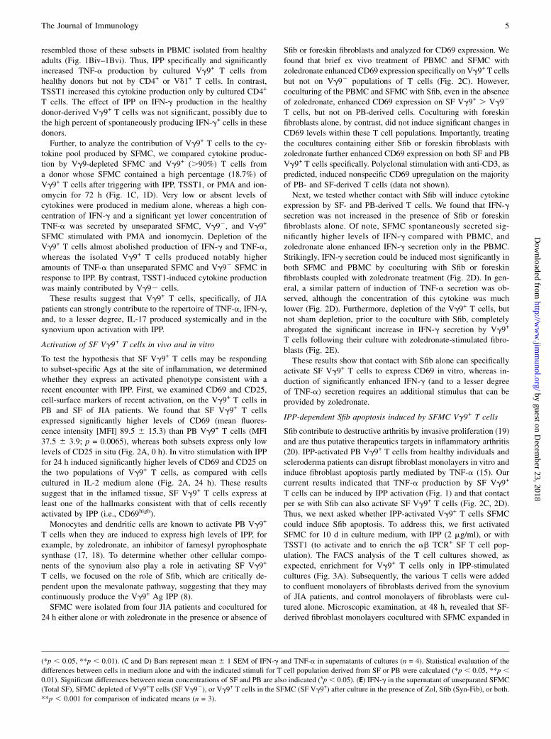
IPP-dependent Sfib apoptosis induced by SFMC Vg9+ T cells
Sfib contribute to destructive arthritis by invasive proliferation (19)and are thus putative therapeutics targets in inflammatory arthritis(20). IPP-activated PB Vg9+ T cells from healthy individuals andscleroderma patients can disrupt fibroblast monolayers in vitro andinduce fibroblast apoptosis partly mediated by TNF-a (15). Ourcurrent results indicated that TNF-a production by SF Vg9+
T cells can be induced by IPP activation (Fig. 1) and that contactper se with Sfib can also activate SF Vg9+ T cells (Fig. 2C, 2D).Thus, we next asked whether IPP-activated Vg9+ T cells SFMCcould induce Sfib apoptosis. To address this, we first activatedSFMC for 10 d in culture medium, with IPP (2 mg/ml), or withTSST1 (to activate and to enrich the ab TCR+ SF T cell pop-ulation). The FACS analysis of the T cell cultures showed, asexpected, enrichment for Vg9+ T cells only in IPP-stimulatedcultures (Fig. 3A). Subsequently, the various T cells were addedto confluent monolayers of fibroblasts derived from the synoviumof JIA patients, and control monolayers of fibroblasts were cul-tured alone. Microscopic examination, at 48 h, revealed that SF-derived fibroblast monolayers cocultured with SFMC expanded in
(*p , 0.05, **p , 0.01). (C and D) Bars represent mean 6 1 SEM of IFN-g and TNF-a in supernatants of cultures (n = 4). Statistical evaluation of the
differences between cells in medium alone and with the indicated stimuli for T cell population derived from SF or PB were calculated (*p , 0.05, **p ,0.01). Significant differences between mean concentrations of SF and PB are also indicated (xp , 0.05). (E) IFN-g in the supernatant of unseparated SFMC
(Total SF), SFMC depleted of Vg9+T cells (SF Vg92), or Vg9+ T cells in the SFMC (SF Vg9+) after culture in the presence of Zol, Sfib (Syn-Fib), or both.
**p , 0.001 for comparison of indicated means (n = 3).
The Journal of Immunology 5
by guest on Decem
ber 23, 2018http://w
ww
.jimm
unol.org/D
ownloaded from
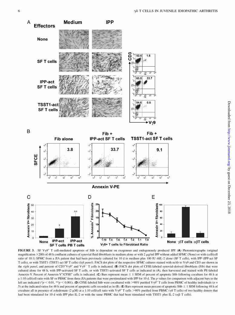
FIGURE 3. SF Vg9+ T cell-mediated apoptosis of Sfib is dependent on exogenous and endogenously produced IPP. (A) Photomicrographs (original
magnification3200) of 48 h confluent cultures of synovial fluid fibroblasts in medium alone or with 2 mg/ml IPP without added SFMC (None) or with (cell/cell
ratio of 10:1) SFMC from a JIA patient that had been previously cultured for 10 d in medium plus 100 IU rhIL-2 alone (SF T cells), with IPP (IPP-act SF
T cells), or with TSST1 (TSST1-act SF T cells) (left panel). FACS dot plots of the respective SFMC cultures stained with mAb to Vg9 and CD3 are shown in
the right panel, and percent of CD3+Vg9+ and Vg92 T cells is indicated. (B) FACS dot plots of CFSE-labeled synovial-derived fibroblasts (Fib) that were
cultured alone for 48 h, with IPP-activated SF T cells, or with TSST1-activated SF T cells as indicated in (A), then harvested and stained with PE-labeled
Annexin-V. Percent of Annexin-V+/CFSE+ cells is indicated. (C) Bars represent mean 6 1 SEM of percent of apoptotic Sfib following coculture for 48 h at
a 1:10 cell/cell ratio with SF or PBMC from three JIA patients that were prestimulated with IPP for 10 d. The p values for comparison with adjacent bars to the
left are indicated (*p , 0.01, **p , 0.001). (D) CFSE-labeled Sfib were cocultured with .90% purified Vg9+ T cells from PBMC of healthy individuals (n =
5) at the indicated ratios for 48 h and percent of apoptotic cells recorded as in (B). (E) Bars represent mean percent of apoptotic Sfib6 1 SEM following 48 h of
coculture all in presence of zoledronate (2 mM) at a 1:10 cell/cell ratio with Vg9+ T cells .90% purified from PBMC (gd T cells) of two healthy donors that
had been stimulated for 10 d with IPP plus IL-2 or with the same PBMC that had been stimulated with TSST1 plus IL-2 (ab T cells).
6 gd T CELLS IN JUVENILE IDIOPATHIC ARTHRITIS
by guest on Decem
ber 23, 2018http://w
ww
.jimm
unol.org/D
ownloaded from
medium with or without TSST1 were grossly indistinguishablefrom control fibroblasts (Fig. 3A, left panel). By contrast, fibro-blast monolayers cocultured with IPP-activated SF T cells in thepresence of IPP showed reduction in numbers and confluence. Ofnote, the apoptosis of CFSE-labeled fibroblasts, as analyzed byAnnexin V staining, was significantly increased by coculturingwith IPP-activated SF T cells and exogenous IPP (Fig. 3B).However, IPP-stimulated SF T cells induced less apoptosis of Sfibas compared with IPP-stimulated PB T cells isolated from JIApatients (Fig. 3C; n = 3). To determine whether apoptosis is de-pendent upon the number of Vg9+ T cells added to the fibroblastcultures, Vg9+ T cells were isolated from IPP-activated PBMCof healthy individuals by negative selection and added at an in-creasing ratio to the Sfib in the presence of IPP. The induction offibroblast apoptosis in the cocultures was highly dependent on theeffector (activated Vg9+ T cells) to target (fibroblasts) cell ratio(Fig. 3D). We also determined whether stimulation with zoledr-onate, which has been shown to increase endogenously producedIPP, can induce effector T cell-dependent fibroblast apoptosis. Weindeed found that apoptosis was specifically induced in zoledro-nate-treated fibroblasts cocultured with purified IPP-activatedVg9+ T cells, but not TSST1-reactive ab T cells (Fig. 3E).Taken together, these results indicated that IPP-reactive Vg9+
T cells from PB and SF of JIA patients can induce Sfib apoptosis,particularly in the presence of zoledronate.
IPP-induced proliferation of SF-derived Vg9+ T cells is lowerthan that of PB-derived Vg9+ T cells
We next assessed the proliferative potential of IPP-responsive SFVg9+ T cells relative to their PB counterparts. The absolutenumber and percentage of Vg9+ T cells in SF or PB in freshlyisolated samples was rather similar, and moreover, their expansionin control medium was minimal upon culturing with IL-2 alone(Fig. 4A, 4B; p = 0.9). However, the IPP dose-dependent expan-sion of Vg9+ T cells was significantly lower in SF versus PBMCcultures, both numerically and as a percentage of the culturedT cells (Fig. 4A, 4B, respectively). Using the CFSE-dilution assaywe further demonstrated that the cell division rate of Vg9+ T cellsfrom SF was also significantly lower compared with those fromPB (31.0 6 19.7% versus 63.5 6 12.5%; p , 0.003; Fig. 4C).Notably, as a control for the specificity of IPP, the proliferation ofSF CD4+ T was induced by TCRab-specific superantigen TSST1but not IPP (Fig. 4C). Interestingly, the rate of IPP-induced pro-liferation of the SF Vg9+ T cells was highly variable amongdifferent patients, and some exhibited quite strong proliferativeresponses (Fig. 4D; 7.8–42.3%; n = 4).These results indicated that proliferation of SF Vg9+ T cells in
response to IPP in vitro is significantly lower than that of theirPBMC counterparts in this cohort of patients, which may explain
FIGURE 4. Proliferative response to IPP. (A) Shown is number of cells
per milliliter6 1 SEM of CD3+Vg9+ T cells of paired PB and SFMC of 14
JIA patients in freshly isolated samples (time 0) and after 10-d culture
in vitro with increasing doses of IPP plus 100 IU/ml rhIL-2. The p values
for comparison between the mean in PB and SF at each concentration of
IPP are indicated (ppp , 0.01, pppp , 0.001). (B) Representative dot plot
FACS analysis of cultured SF and PBMC from a JIA patient stained with
mAb to Vg9 and CD3 after stimulation with the indicated concentration of
IPP. Percent of CD3+Vg9+ T cells in each culture is indicated in the top
right quadrant. (C) CFSE-labeled PB or SFMC cultures were triggered with
rhIL-2 (100 U/ml) alone (Med), or with 2 mg/ml IPP, or 10 ng/ml TSST1.
Five days later, mean percent 6 1 SD of dividing cells within indicated
subsets from flow cytometric analysis was calculated. Number of experi-
ments for each evaluation is indicated in parentheses. Statistical signifi-
cance is shown as *p , 0.05, **p, 0.009, ***p, 0.001, respectively, for
comparison with medium alone. (D) FACS analysis of CFSE-loaded
SFMC gated on Vg9+ T cells derived from four different JIA patients (Pt)
after 5 d in culture with 2 mg/ml IPP and 100 IU rhIL-2. Each peak to the
left of the first on the right indicates a round of cell division corresponding
to the dilution of CFSE. Percent of dividing cells within the Vg9+ subset is
indicated in the top left corner.
The Journal of Immunology 7
by guest on Decem
ber 23, 2018http://w
ww
.jimm
unol.org/D
ownloaded from
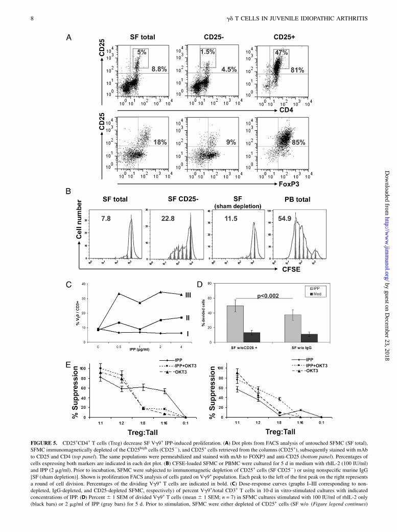
FIGURE 5. CD25+CD4+ T cells (Treg) decrease SF Vg9+ IPP-induced proliferation. (A) Dot plots from FACS analysis of untouched SFMC (SF total),
SFMC immunomagnetically depleted of the CD25high cells (CD252), and CD25+ cells retrieved from the columns (CD25+), subsequently stained with mAb
to CD25 and CD4 (top panel). The same populations were permeabilized and stained with mAb to FOXP3 and anti-CD25 (bottom panel). Percentages of
cells expressing both markers are indicated in each dot plot. (B) CFSE-loaded SFMC or PBMC were cultured for 5 d in medium with rhIL-2 (100 IU/ml)
and IPP (2 mg/ml). Prior to incubation, SFMC were subjected to immunomagnetic depletion of CD25+ cells (SF CD252) or using nonspecific murine IgG
[SF (sham depletion)]. Shown is proliferation FACS analysis of cells gated on Vg9+ population. Each peak to the left of the first peak on the right represents
a round of cell division. Percentages of the dividing Vg9+ T cells are indicated in bold. (C) Dose-response curves (graphs I–III corresponding to non-
depleted, IgG-depleted, and CD25-depleted SFMC, respectively) of percent Vg9+/total CD3+ T cells in 10-d in vitro-stimulated cultures with indicated
concentrations of IPP. (D) Percent 6 1 SEM of divided Vg9+ T cells (mean 6 1 SEM; n = 7) in SFMC cultures stimulated with 100 IU/ml of rhIL-2 only
(black bars) or 2 mg/ml of IPP (gray bars) for 5 d. Prior to stimulation, SFMC were either depleted of CD25+ cells (SF w/o (Figure legend continues)
8 gd T CELLS IN JUVENILE IDIOPATHIC ARTHRITIS
by guest on Decem
ber 23, 2018http://w
ww
.jimm
unol.org/D
ownloaded from
their lower capacity to induce fibroblast apoptosis as observed inthe previous set of experiments (Fig. 3C).
Influence of CD4+CD25+FOXP3+ Treg on IPP-responsiveVg9+ T cells
Treg have been demonstrated in SF of JIA patients, suggestingthey may regulate SF Vg9+ T cell proliferation (21, 22). To probewhether Treg from SF indeed suppress IPP-induced activationof SF Vg9+ T cells in JIA patients, we depleted, using CD25+
microbeads (MACS cell separation system), the majority of CD4+
CD25hiFOXP3+ T cells from the SFMC population. As seen inthe representative example shown in Fig. 5A, CD25+ populationsisolated in these experiments were highly enriched for FOXP3+
cells (85%), contrasting with 18 and 9% FOXP3+ in the unsepa-rated and CD25-depleted SFMC, respectively. We found thatfollowing IPP stimulation, depletion of CD25+ Treg augmentedthe proliferation of Vg9+ T cells in SFMC cultures compared withsham depletion and untouched SFMC, as assessed by FACSanalysis of CFSE dilution within the Vg9+CD3+ events gate (Fig.5B; 22.8, 11.5, and 7.8%, respectively). Nevertheless, the prolif-eration of CD252 SFMC did not reach the rate observed in PBVg9+ T cells. Likewise, in another JIA patient, we found thatCD25+ T cell depletion also augmented proliferation in an IPPdose-dependent manner (Fig. 5C) as compared with sham deple-tion or untouched SFMC cultures. In addition, the cumulative datafrom analogous experiments repeated in seven different JIApatients show significant increase of the proliferation of Vg9+
T cells in SFMC cultures in which CD25+ cells were depletedcompared with sham depletion (49.4 6 8.1% versus 37.2 6 6.8%;p, 0.002; Fig. 5D). We further found that CD25+ T cell depletionsignificantly increased IFN-g+ (CD25-depleted, 68.8 6 8.5%versus nondepleted, 59.6 6 10.8%; p , 0.02), but not TNF-a+
(46.9 6 18 versus 44.9 6 9.7% respectively; p = 0.77) Vg9+
T cells in the 5-d IPP cultures (data not shown). To confirm that SFTreg actively suppress IPP-induced SF Vg9+ T cell proliferation,CD25+ SFMC (.70% FOXP3+) were immunomagnetically sep-arated from SFMC. As a positive control for these experiments,we used PBMC from a healthy adult. These Treg were then addedback to CFSE-loaded CD25-depleted SFMC (Tall) at differentTreg to CD25-SFMC ratios and cultured in medium containinga low concentration of IL-2 alone or with added IPP, anti-CD3mAbs, or IPP plus anti-CD3 mAbs. FACS analysis of CFSE di-lution in Vg9-stained cells after 5 d of culture revealed a strongand dose-dependent suppressive effect of the Treg on proliferationof SF Vg9+ T cells. We found that Treg could suppress the pro-liferation of Vg9+ T cells regardless of the stimuli employed.Interestingly, lower ratios of SF-Treg/Tall were required to ach-ieve ∼60% suppression of Ag-specific (IPP) versus polyclonal-induced (anti-CD3) Vg9+ T cell activation (1:8 versus 1:2, re-spectively; Fig. 5E). Treg from PB of healthy controls also sup-pressed IPP-induced proliferation of Vg9+ T cells, but requireda higher Treg/Tall ratio of 1:1 to achieve ∼60% suppression. Theresults of these experiments are consistent with the hypothesis thatin JIA patients with a reduced in vitro response of Vg9+ T cell toIPP, SF-Treg may be playing a critical suppressive role.
DiscussionThis study shows for the first time, to our knowledge, that Vg9+
T cells in SF of JIA patients consist primarily of a functionallyimportant population of proinflammatory cytokine-secreting cells,including IPP-responsive cells, that are largely in an activatedstate in vivo. Our data further suggest that Sfib can potently drivetheir activation, at least in vitro, in a manner that producesa functional outcome (e.g., IFN-g secretion), especially in thepresence of a reagent that induces cellular accumulation of IPP,a potent Ag for a subset of human Vg9+ T cells (e.g., zoledronate).Furthermore, SF Vg9+ T cells can induce apoptosis of Sfib that areexposed to a reagent, zoledronate (7). These latter findings suggestan immunological mechanism that may underlie the recently de-scribed correlation between recovery from JIA and higher levelsof IPP-responsive synovial Vg9+ T cells (13).Current paradigms of immunopathogenesis of JIA envision
autoreactive ab T cells responding to a limited repertoire ofautoantigens in the joint, initiating the inflammatory response(10). In full-blown clinical disease, the immune response mayexpand to include multiple autoantigens presented by dendriticcells to autoreactive CD4+ T cells that subsequently activate Sfiband innate immune cells, all producing multiple proinflammatorycytokines (12). It has been shown that autoreactive effector CD4+
T cells can be suppressed by CD4+CD25highFOXP3+ T cells,theoretically providing a mechanism to terminate such pathogenicactivation.In this study, we focus on the SF Vg9+ gd T cell subset and show
that a high proportion of these cells secrete TNF-a and IFN-g anda lower percentage IL-17, an effector function that increased uponactivation by IPP presented either by conventional APCs withinthe SFMC population or by adherent fibroblasts. In this respect,TNF-a is a central mediator of multiple aspects of synovial in-flammation that can activate fibroblasts, osteoclasts, B, and T cellsand is an established therapeutic target in JIA (23). IFN-g mayalso play a pathogenic role by activating macrophages, whereasIL-17 increases vascularization and induces influx of neutrophilsto drive inflammation (24, 25). Thus, each of the cytokines shownin this study to be abundantly produced by SF Vg9+ T cells, and inparticular by the IPP-responsive subset, could potentially con-tribute to the inflammatory process in JIA.By contrast, other well-described functions of the same cyto-
kines could paradoxically dampen the inflammatory response. Forexample, contact-dependent induction of Sfib apoptosis by IPP-activated Vg9+ T cells is partly TNF-a dependent, as it can bereduced by blocking mAbs to TNF-a (15). This TNF-mediatedeffect could potentially reduce chronic inflammation and cartilagedestruction, thus producing a protective effect in arthritis. Like-wise, IFN-g can enhance Treg function, inhibit differentiation ofTh17 cells, and attenuate tissue damage (24). These effects ofTNF-a and IFN-g suggest that under certain conditions, a proin-flammatory cytokine could play a beneficial, rather than a patho-genic, role in JIA. The recent finding of a clinical correlationbetween recovery from JIA and the number SF Vg9+ T cells andtheir ability to expand in response to IPP suggests that the cyto-kines produced by activated Vg9+ T cells in the synovium of JIA
CD25+) or of control nonspecific IgG-binding cells (SF w/o IgG). The p value comparing means is shown. (E and F) CD25+ cells were isolated from SFMC
of a JIA patient (E) or PBMC of a healthy adult control (F) and added to the respective CFSE-labeled unseparated SFMC or PBMC in the presence of IPP,
mAb to CD3, or both reagents (see Materials and Methods). After 5 d, cells were removed from the cultures and stained with mAb to Vg9. Percent
inhibition of division of the Vg9+ CFSE-stained cells relative to the divisions in the absence of added CD25+ cells was calculated as described in Materials
and Methods (y-axis). The x-axis indicates ratio of Treg to total MC (Tall) in each culture.
The Journal of Immunology 9
by guest on Decem
ber 23, 2018http://w
ww
.jimm
unol.org/D
ownloaded from
patients may actually tilt the balance toward a beneficial outcome(13).Our data show that both Vg9+ T cells in synovium and in PB are
enriched for cells with the immediate effector capacity to producevarious cytokines, whereas SF CD4+ T cells were selectivelyenriched for immediate effectors relative to their PB counterparts(Fig. 1). This finding is consistent with the concept that as opposedto SF CD4+ T cells that are selected by site-specific autoantigens,Vg9+ T cells recognize ubiquitously expressed phosphorylatedautoantigens, such as IPP (26). However, SF Vg9+ T cells in thesynovium were unique in expressing a phenotype consistent withrecent activation (e.g., expressing a higher level of CD69 thantheir PB counterparts). This suggests that SF Vg9+ T cells haveencountered autoantigens more recently in situ. Moreover, SF-resident Vg9+ T cells, as opposed to their PB counterparts, im-mediately upregulated CD69 when they contacted Sfib, whereasboth populations could be further activated in the presence ofzoledronate. Thus, we postulate that SF Vg9+ T cells may havebeen primed in vivo by exposure to stressed cells expressing highlevels of IPP or by exogenous sources of phosphorylated Ags (e.g.,those produced by bacterial nonmevalonate pathways) and arethus able to respond to the lower levels of IPP that may beexpressed by untreated Sfib. The mechanisms involved in thisheightened level of responsiveness could include elevated ex-pression of costimulatory molecules as well as the selection ofTCR with higher affinity for IPP in the synovium-residing popula-tion. It is also possible that Ags other than IPP expressed by Sfibare being recognized by oligoclonal TCRs that are more abundantin the SF Vg9+ T cell population, but infrequent in PB. Obviously,further experiments are required to address this hypothesis.In some patients, which may include those with relatively weak
proliferative responses, the Vg9+ subset localizing to the SF isunder tight control of SF CD4+ Treg (Fig. 5). However, in themajority of patients with a substantial proliferative response toIPP, Treg control was significant but marginal. We recently re-ported that higher IPP responses, suggesting that Treg are lessefficient in this regard, are associated with better outcomes of JIA(13). The current analysis suggests that strong IPP-induced pro-liferation driven by endogenous upregulation of this metabolitecould lead to local increases of Vg9+ T cells in the synovial mi-croenvironment. This activation might suffice to induce apoptosisof certain Sfib. This IPP-responsive Vg9+ T cell-mediated effectcould play a critical negative-feedback role in amelioratingdisease in JIA patients. This could be more vital in patients withless efficient Treg because fibroblasts contribute to inflammatorycytokines and cartilage destruction (19). To test this postulation,multivariate correlations between the number and function of Tregrelevant to autoreactive SF CD4+ T cells and the IPP-dependentresponses of SF Vg9+ T cells and disease outcomes will be re-quired to dissect the relative importance of each mechanism in thedifferent subgroups of JIA patients. Interestingly in this regard,Macaubas et al. (27) recently reported a downregulation of PB gd
T cells, but not of NK cells, during acute exacerbations of sys-temic JIA specifically, suggesting a unique and specific role of gdT cells during flares of disease in this particular subset of JIApatients. Although we have not dissected the functional role ofVg9+ T cells in this subset in particular, our previous results (13)indicate that Vg9+ T cells in the SF of systemic JIA patients canrespond to IPP. Taken together with the data of Macaubas et al.(27), it could be postulated that during acute flares of systemicJIA, PB Vg9+ T cells migrate to synovium, where they may se-crete TNF-a and IFN-g, thus contributing to local inflammation.Notably, highly efficient induction of apoptosis of fibroblasts by
Vg9+ T cells in vitro required exogenous IPP, suggesting that
in vivo Sfib that present low levels of IPP will likely escape Vg9+
T cell-mediated apoptosis. In contrast, treatment of fibroblastswith zoledronate strongly enhanced SF Vg9+ T cell activation andantifibroblast function. Thus, we envision that when IPP is over-produced by proliferating Sfib (e.g., in the inflamed synovium),the IPP-reactive Vg9+ T cells are activated to express CD69 andinduce their apoptosis. Interestingly, the mevalonate pathway isrequired for matrix metalloproteinase-9–mediated invasive be-havior of murine fibroblasts, suggesting that high levels of IPPmay be a marker of the most invasive Sfib (28). Further studies ofthe in vivo conditions that enhance IPP production by Sfib andtheir interaction with PB Vg9+ T cells may further our under-standing of mechanisms of tissue inflammation in autoimmunearthritis and may lead to the development of novel therapeuticmodalities.
DisclosuresThe authors have no financial conflicts of interest.
References1. Bank, I., R. A. DePinho, M. B. Brenner, J. Cassimeris, F. W. Alt, and L. Chess.
1986. A functional T3 molecule associated with a novel heterodimer on thesurface of immature human thymocytes. Nature 322: 179–181.
2. Brenner, M. B., J. McLean, D. P. Dialynas, J. L. Strominger, J. A. Smith,F. L. Owen, J. G. Seidman, S. Ip, F. Rosen, and M. S. Krangel. 1986. Identifi-cation of a putative second T-cell receptor. Nature 322: 145–149.
3. Morita, C. T., E. M. Beckman, J. F. Bukowski, Y. Tanaka, H. Band, B. R. Bloom,D. E. Golan, and M. B. Brenner. 1995. Direct presentation of nonpeptide prenylpyrophosphate antigens to human gamma delta T cells. Immunity 3: 495–507.
4. Hayday, A. C. 2000. [gamma][delta] cells: a right time and a right place fora conserved third way of protection. Annu. Rev. Immunol. 18: 975–1026.
5. Kabelitz, D., A. Glatzel, and D. Wesch. 2000. Antigen recognition by humangammadelta T lymphocytes. Int. Arch. Allergy Immunol. 122: 1–7.
6. Hintz, M., A. Reichenberg, B. Altincicek, U. Bahr, R. M. Gschwind,A. K. Kollas, E. Beck, J. Wiesner, M. Eberl, and H. Jomaa. 2001. Identificationof (E)-4-hydroxy-3-methyl-but-2-enyl pyrophosphate as a major activator forhuman gammadelta T cells in Escherichia coli. FEBS Lett. 509: 317–322.
7. Mookerjee-Basu, J., P. Vantourout, L. O. Martinez, B. Perret, X. Collet,C. Perigaud, S. Peyrottes, and E. Champagne. 2010. F1-adenosine triphosphatasedisplays properties characteristic of an antigen presentation molecule forVgamma9Vdelta2 T cells. J. Immunol. 184: 6920–6928.
8. Nagashima, T., H. Okazaki, K. Yudoh, H. Matsuno, and S. Minota. 2006. Ap-optosis of rheumatoid synovial cells by statins through the blocking of proteingeranylgeranylation: a potential therapeutic approach to rheumatoid arthritis.Arthritis Rheum. 54: 579–586.
9. Houten, S. M., J. Frenkel, and H. R. Waterham. 2003. Isoprenoid biosynthesis inhereditary periodic fever syndromes and inflammation. Cell. Mol. Life Sci. 60:1118–1134.
10. Ravelli, A., and A. Martini. 2007. Juvenile idiopathic arthritis. Lancet 369: 767–778.
11. Brewer, E. J., Jr., J. Bass, J. Baum, J. T. Cassidy, C. Fink, J. Jacobs, V. Hanson,J. E. Levinson, J. Schaller, and J. S. Stillman; JRA Criteria Subcommittee of theDiagnostic and Therapeutic Criteria Committee of the American RheumatismSection of The Arthritis Foundation. 1977. Current proposed revision of JRACriteria. Arthritis Rheum. 20(Suppl 2): 195–199.
12. Prakken, B., S. Albani, and A. Martini. 2011. Juvenile idiopathic arthritis. Lancet377: 2138–2149.
13. Berkun, Y., A. Bendersky, M. Gerstein, I. Goldstein, S. Padeh, and I. Bank. 2011.GammadeltaT cells in juvenile idiopathic arthritis: higher percentages of syno-vial Vdelta1+ and Vgamma9+ T cell subsets are associated with milder disease.J. Rheumatol. 38: 1123–1129.
14. Olive, C., P. A. Gatenby, and S. W. Serjeantson. 1992. Molecular characteriza-tion of the V gamma 9 T cell receptor repertoire expressed in patients withrheumatoid arthritis. Eur. J. Immunol. 22: 2901–2906.
15. Bendersky, A., N. Markovits, and I. Bank. 2010. Vgamma9+ gammadelta T cellsin systemic sclerosis patients are numerically and functionally preserved andinduce fibroblast apoptosis. Immunobiology 215: 380–394.
16. Goldstein, I., S. Ben-Horin, J. Li, I. Bank, H. Jiang, and L. Chess. 2003. Ex-pression of the alpha1beta1 integrin, VLA-1, marks a distinct subset of humanCD4+ memory T cells. J. Clin. Invest. 112: 1444–1454.
17. Gruenbacher, G., H. Gander, A. Rahm, W. Nussbaumer, N. Romani, andM. Thurnher. 2009. CD56+ human blood dendritic cells effectively promoteTH1-type gammadelta T-cell responses. Blood 114: 4422–4431.
18. Roelofs, A. J., M. Jauhiainen, H. Monkkonen, M. J. Rogers, J. Monkkonen, andK. Thompson. 2009. Peripheral blood monocytes are responsible for gamma-delta T cell activation induced by zoledronic acid through accumulation of IPP/DMAPP. Br. J. Haematol. 144: 245–250.
19. Bartok, B., and G. S. Firestein. 2010. Fibroblast-like synoviocytes: key effectorcells in rheumatoid arthritis. Immunol. Rev. 233: 233–255.
10 gd T CELLS IN JUVENILE IDIOPATHIC ARTHRITIS
by guest on Decem
ber 23, 2018http://w
ww
.jimm
unol.org/D
ownloaded from
20. Kiener, H. P., B. Niederreiter, D. M. Lee, E. Jimenez-Boj, J. S. Smolen, andM. B. Brenner. 2009. Cadherin 11 promotes invasive behavior of fibroblast-likesynoviocytes. Arthritis Rheum. 60: 1305–1310.
21. de Kleer, I. M., L. R. Wedderburn, L. S. Taams, A. Patel, H. Varsani, M. Klein,W. de Jager, G. Pugayung, F. Giannoni, G. Rijkers, et al. 2004. CD4+CD25brightregulatory T cells actively regulate inflammation in the joints of patients with theremitting form of juvenile idiopathic arthritis. J. Immunol. 172: 6435–6443.
22. Nagar, M., J. Jacob-Hirsch, H. Vernitsky, Y. Berkun, S. Ben-Horin,N. Amariglio, I. Bank, Y. Kloog, G. Rechavi, and I. Goldstein. 2010. TNFactivates a NF-kappaB-regulated cellular program in human CD45RA- regula-tory T cells that modulates their suppressive function. J. Immunol. 184: 3570–3581.
23. Feldmann, M., and R. N. Maini. 2010. Anti-TNF therapy, from rationale tostandard of care: what lessons has it taught us? J. Immunol. 185: 791–794.
24. Hu, X., and L. B. Ivashkiv. 2009. Cross-regulation of signaling pathways byinterferon-gamma: implications for immune responses and autoimmune dis-eases. Immunity 31: 539–550.
25. Cua, D. J., and C. M. Tato. 2010. Innate IL-17-producing cells: the sentinels ofthe immune system. Nat. Rev. Immunol. 10: 479–489.
26. Morita, C. T., H. K. Lee, D. S. Leslie, Y. Tanaka, J. F. Bukowski, and E. Marker-Hermann. 1999. Recognition of nonpeptide prenyl pyrophosphate antigens byhuman gammadelta T cells. Microbes Infect. 1: 175–186.
27. Macaubas, C., K. Nguyen, C. Deshpande, C. Phillips, A. Peck, T. Lee, J. L. Park,C. Sandborg, and E. D. Mellins. 2010. Distribution of circulating cells in systemicjuvenile idiopathic arthritis across disease activity states.Clin. Immunol. 134: 206–216.
28. Wang, I. K., S. Y. Lin-Shiau, and J. K. Lin. 2000. Suppression of invasion andMMP-9 expression in NIH 3T3 and v-H-Ras 3T3 fibroblasts by lovastatinthrough inhibition of ras isoprenylation. Oncology 59: 245–254.
The Journal of Immunology 11
by guest on Decem
ber 23, 2018http://w
ww
.jimm
unol.org/D
ownloaded from


![,NDUXV 0= .DPD] 5REXU :ROJD =DSRUR]HF 0D]GD 0; /DQG 5RYHU ... · +dxswvfkhlqzhuihu ,m 3(6 ,m,nduxv 0= .dpd] 5rexu :rojd =dsrur]hf 0d]gd 0; /dqg 5ryhu 'hihqghu /dgd 1lyd + 9 rghu 9](https://static.fdocument.org/doc/165x107/5e0951457bf2e2579f20f0ae/nduxv-0-dpd-5rexu-rojd-dsrurhf-0dgd-0-dqg-5ryhu-dxswvfkhlqzhuihu.jpg)


![Gd(III) complexes: basic relaxometric characterization · Eliana Gianolio EMMI Intensive Programme "Design, Synthesis and Validation of Imaging Probes" R iobs = [CA]r ip + R iw i](https://static.fdocument.org/doc/165x107/5f3cbbb1f7efd235f739b895/gdiii-complexes-basic-relaxometric-characterization-eliana-gianolio-emmi-intensive.jpg)