Hippocampal interleukin-1β in the juvenile and …² in the juvenile and middle-aged rat 365 It has...
15
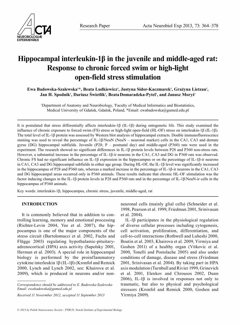
Research Paper Acta Neurobiol Exp 2013, 73: 364–378 © 2013 by Polish Neuroscience Society - PTBUN, Nencki Institute of Experimental Biology INTRODUCTION It is commonly believed that in addition to con- trolling learning, memory and emotional processing (Richter-Levin 2004, Yee et al. 2007), the hip- pocampus is one of the major components of the stress circuit (Bartolomucci et al. 2002, Fuchs and Flgge 2003) regulating hypothalamic-pituitary- adrenocortical (HPA) axis activity (Sapolsky 2001, Herman et al. 2003). A special role in hippocampal biology is performed by the proinflammatory cytokine interleukin 1β (IL-1β) (Kronfol and Remick 2000, Lynch and Lynch 2002, see: Khairova et al. 2009), which is produced in neurons and/or non- neuronal cells (mainly glial cells) (Schneider et al. 1998, Pearson et al. 1999, Friedman 2001, Srinivasan et al. 2004). IL-1β participates in the physiological regulation of diverse cellular processes including cytogenesis, cell activation, proliferation, differentiation, and cell-to-cell interactions (Rothwell and Luheshi 2000, Boutin et al. 2003, Khairova et al. 2009, Yirmiya and Goshen 2011) of a healthy organ (Vitkovic et al. 2000, Tonelli and Postolache 2005) and also under conditions of damage, disease and stress (Friedman 2001, Srinivasan et al. 2004). By taking part in HPA axis modulation (Turnbull and Rivier 1999, Grinevich et al. 2001, Elenkov and Chrousos 2002, Dunn 2006), IL-1β is involved in responses not only to traumatic, but also to physical and psychological stressors (Kronfol and Remick 2000, Goshen and Yirmiya 2009). Hippocampal interleukin-1β in the juvenile and middle-aged rat: Response to chronic forced swim or high-light open-field stress stimulation Ewa Badowska-Szalewska 1 *, Beata Ludkiewicz 1 , Justyna Sidor-Kaczmarek 1 , Grażyna Lietzau 1 , Jan H. Spodnik 1 , Dariusz Świetlik 2 , Beata Domaradzka-Pytel 1 , and Janusz Moryś 1 1 Department of Anatomy and Neurobiology, 2 Faculty of Medical Informatics and Biostatistics, Medical University of Gdańsk, Gdańsk, Poland, *Email: [email protected] It is postulated that stress differentially affects interleukin-1β (IL-1β) during ontogenetic life. This study examined the influence of chronic exposure to forced swim (FS) stress or high-light open-field (HL-OF) stress on interleukin-1β (IL-1β). The total level of IL-1β protein was assessed by Western blot analysis of hippocampal extracts. Double immunofluorescence staining was used to reveal the percentage of IL-1β/NeuN (NeuN – neuronal marker) cells in the CA1, CA3 and dentate gyrus (DG) hippocampal subfields. Juvenile (P28; P – postnatal day) and middle-aged (P360) rats were used in the experiment. The research showed no significant differences in IL-1β protein levels between P28 and P360 non-stress rats. However, a substantial increase in the percentage of IL-1β-ir neurons in the CA1, CA3 and DG in P360 rats was observed. Chronic FS had no significant influence on IL-1β expression in the hippocampus or on the percentage of IL-1β-ir neurons in CA1, CA3 and DG hippocampal subfields in either age group. During HL-OF, the IL-1β level was significantly increased in the hippocampus of P28 and P360 rats, whereas a marked increase in the percentage of IL-1β-ir neurons in the CA1, CA3 and DG hippocampal areas occurred only in P360 animals. These results indicate that chronic HL-OF stimulation was the factor inducing changes in the IL-1β protein levels in P28 and P360 rats and in the percentage of IL-1β/NeuN-ir cells in the hippocampus of P360 animals. Key words: interleukin-1β, hippocampus, chronic stress, juvenile, middle-aged, rat Correspondence should be addressed to E. Badowska-Szalewska Email: [email protected] Received 11 Novermber 2012, accepted 11 September 2013
Transcript of Hippocampal interleukin-1β in the juvenile and …² in the juvenile and middle-aged rat 365 It has...
Research Paper Acta Neurobiol Exp 2013, 73: 364–378
© 2013 by Polish Neuroscience Society - PTBUN, Nencki Institute of Experimental Biology
INTRODUCTION
It is commonly believed that in addition to con-trolling learning, memory and emotional processing (Richter-Levin 2004, Yee et al. 2007), the hip-pocampus is one of the major components of the stress circuit (Bartolomucci et al. 2002, Fuchs and Flugge 2003) regulating hypothalamic-pituitary-adrenocortical (HPA) axis activity (Sapolsky 2001, Herman et al. 2003). A special role in hippocampal biology is performed by the proinflammatory cytokine interleukin 1β (IL-1β) (Kronfol and Remick 2000, Lynch and Lynch 2002, see: Khairova et al. 2009), which is produced in neurons and/or non-
neuronal cells (mainly glial cells) (Schneider et al. 1998, Pearson et al. 1999, Friedman 2001, Srinivasan et al. 2004).
IL-1β participates in the physiological regulation of diverse cellular processes including cytogenesis, cell activation, proliferation, differentiation, and cell-to-cell interactions (Rothwell and Luheshi 2000, Boutin et al. 2003, Khairova et al. 2009, Yirmiya and Goshen 2011) of a healthy organ (Vitkovic et al. 2000, Tonelli and Postolache 2005) and also under conditions of damage, disease and stress (Friedman 2001, Srinivasan et al. 2004). By taking part in HPA axis modulation (Turnbull and Rivier 1999, Grinevich et al. 2001, Elenkov and Chrousos 2002, Dunn 2006), IL-1β is involved in responses not only to traumatic, but also to physical and psychological stressors (Kronfol and Remick 2000, Goshen and Yirmiya 2009).
Hippocampal interleukin-1β in the juvenile and middle-aged rat: Response to chronic forced swim or high-light
open-field stress stimulationEwa Badowska-Szalewska1*, Beata Ludkiewicz1, Justyna Sidor-Kaczmarek1, Grażyna Lietzau1,
Jan H. Spodnik1, Dariusz Świetlik2, Beata Domaradzka-Pytel1, and Janusz Moryś1
1Department of Anatomy and Neurobiology, 2Faculty of Medical Informatics and Biostatistics, Medical University of Gdańsk, Gdańsk, Poland, *Email: [email protected]
It is postulated that stress differentially affects interleukin-1β (IL-1β) during ontogenetic life. This study examined the influence of chronic exposure to forced swim (FS) stress or high-light open-field (HL-OF) stress on interleukin-1β (IL-1β). The total level of IL-1β protein was assessed by Western blot analysis of hippocampal extracts. Double immunofluorescence staining was used to reveal the percentage of IL-1β/NeuN (NeuN – neuronal marker) cells in the CA1, CA3 and dentate gyrus (DG) hippocampal subfields. Juvenile (P28; P – postnatal day) and middle-aged (P360) rats were used in the experiment. The research showed no significant differences in IL-1β protein levels between P28 and P360 non-stress rats. However, a substantial increase in the percentage of IL-1β-ir neurons in the CA1, CA3 and DG in P360 rats was observed. Chronic FS had no significant influence on IL-1β expression in the hippocampus or on the percentage of IL-1β-ir neurons in CA1, CA3 and DG hippocampal subfields in either age group. During HL-OF, the IL-1β level was significantly increased in the hippocampus of P28 and P360 rats, whereas a marked increase in the percentage of IL-1β-ir neurons in the CA1, CA3 and DG hippocampal areas occurred only in P360 animals. These results indicate that chronic HL-OF stimulation was the factor inducing changes in the IL-1β protein levels in P28 and P360 rats and in the percentage of IL-1β/NeuN-ir cells in the hippocampus of P360 animals.
Key words: interleukin-1β, hippocampus, chronic stress, juvenile, middle-aged, rat
Correspondence should be addressed to E. Badowska-Szalewska Email: [email protected]
Received 11 Novermber 2012, accepted 11 September 2013
IL-1β in the juvenile and middle-aged rat 365
It has been observed that certain types of stressful stimuli can affect IL-1β expression in the hippocampus while others cannot (Pugh et al. 1999, Plata-Salamán et al. 2000, Hennessy et al. 2004, Kwon et al. 2008, You et al. 2011). This disparity of findings has led to con-siderable controversy regarding the nature and dura-tion of stressors to induce IL-1β expression (Kronfol and Remick 2000, see: Deak et al. 2003).
It is well known that stress affects the hippocampus throughout an individual’s life span (Fuchs and Flugge 2003, Moynihan 2003, Lupien 2009). Most of the research analysing the effects of stressful situations on hippocampal IL-1β was based on examination of adult and old rats (Schneider et al. 1998, Plata-Salamán et al. 2000, Deak et al. 2003, Kwon et al. 2008, Campuzano et al. 2009), but the relation between a stress response and important stages of life, namely juvenile and middle-aged stages, has not been fully explained. These onto-genic periods are crucial with respect to morphological and functional transformations, which then occur within the limbic system (Pardon 2007, Lupien 2009).
Our previous preliminary semiquantitative analysis, conducted on 28-day-old and 1-year-old rats, showed that acute and chronic high-light open-field (HL-OF) or forced swim (FS), resulted in an increase in IL-1β-immunoreactivity (-ir) in the CA1, CA3 and dentate gyrus (DG) hippocampal subfields of P28 rats, as opposed to P360 animals which showed no clear changes (Badowska-Szalewska et al. 2009). In the experiment, IL-1β-ir was observed mainly in glial cells. Moreover, IL-1β-ir neurons were also observed, especially in middle-aged rats. As a follow-up to our previous study, the aim of our current research was to investigate in detail whether the IL-1β-ir neuron popu-lation in the CA1, CA3 and DG hippocampal subfields of juvenile (P28) and middle-aged (P360) rats is altered by exposure to chronic forced swim (FS) or high-light open-field (HL-OF) stressors. The research was also designed to explore the correlation between age, type of stimulation applied and the IL-1β protein level in the hippocampus.
METHODS
Animals
The research was performed using Wistar Han rats. Rat pups were housed with their mothers (one mother with five male or female pups per cage) from birth
until the end of the experiment when they reached 28 days of age (P28; P-postnatal day). Male animals that were 360-days-old (P360) were also used. All animals were kept under constant temperature (21 ± 1°C) and lighting regimens (light on from 07:00 am to 07:00 pm) in polycarbonate cages (T. IV, 56 cm × 36 cm × 20 cm + 7 cm lid) with sawdust bedding and free access to water and food pellets. The care and treatment of the rats conformed to the guidelines for laboratory ani-mals established by the National Institute of Health and the Local Ethical Committee of the Medical University of Gdańsk. The animals were divided into controls and two experimental groups exposed to chronic forced swim stress or to chronic high-light open-field stress. At the outset of chronic stress stimu-lation, P28 rats were seven days old (P-7). The control non-stress groups, comprising animals from the P28 and P360 age categories, were handled for a few min-utes daily by the same operator and separated from their mothers for 15 min in the same way as experi-mental animals.
Test model
All tests were conducted once a day in 15-minute sessions for 21 consecutive days at the same time, such as between 09:00 am and 02:00 pm. After the testing procedure, the rats were returned to their respective home cages.
Forced swim (FS) test
The FS test procedure applied in this experiment was used in our previous studies (Badowska-Szalewska et al. 2011) and in several other experiments (Grace et al. 2008, Mikhailenko et al. 2010). The rats were placed in a glass cylinder (45-cm high, 20 cm in diam-eter) filled with clear, fresh water (at 22°C) up to a height of 30 cm. If some of the youngest individuals were unable to maintain their noses above the water level during the 15-minute FS exposures to the stres-sor, the time of the test was reduced (this applied to one rat only on the first day after 10 minutes of stimu-lation).
High-light open-field (HL-OF) test
The chronic HL-OF adopted in this study was employed in our former experiments (Badowska-
366 E. Badowska-Szalewska et al.
Szalewska et al. 2011) and in a series of other research projects (Lin et al. 2008, Van Wijk et al. 2008). The apparatus to perform the test consisted of a 100 × 100 × 40 cm wooden box illuminated with a 500-watt halogen lamp. In order to provoke a stress reaction, every animal was gently placed in the centre of the open-field arena, which after each test, was cleaned with water and 70% ethanol.
Methods of IL-1β detection
On postnatal day 28 (P28) or 360 (P360), following the final experimental procedures, all the rats were sacrificed by deep anaesthesia induced with a lethal dose of Nembutal (80 mg/kg of body weight).
Western blot
A total of 36 microdissected hippocampal tissue samples (six rats per group) were obtained using a sur-gical microscope. All the samples were manually homogenized on ice with a Potter-Elvehjem tissue grinder in 4 volumes of homogenization buffer (20 mM Tris-HCl pH 7.5; 0.25 M sucrose; 10 mM EGTA; 2 mM EDTA) containing protease inhibitors (2 mM PMSF; 50 µg/ml leupeptin; 25 µg/ml aprotinin, 10 µg/ml pepstatin A and 2 mM DTT). Denaturated 40-µg protein samples were electrophoretically separated on 12% Tris-SDS gels with Tris-glycine elecrophoresis buffer and subsequently transferred onto a nitrocellu-lose membrane by semi-dry electroblotting. The mem-branes were then blocked with 3% nonfat dry milk in TBS buffer (10 mM Tris pH 8.0; 150 mM NaCl) for 2 hours and incubated overnight at 4°C with primary antibody: rabbit polyclonal anti-IL-1β (1:200; Endogen, USA) diluted in 1.5% milk in TBST (TBS buffer sup-plemented with 0.05% Tween20). Having been washed 3 × 10 min with TBS, the blots were incubated for 2 hours with secondary goat anti-rabbit antibody, horse-radish peroxidase conjugate (1:50 000; Pierce, USA) diluted in TBST. The internal standard of β-actin was used under the abovementioned conditions with mouse monoclonal anti-β-actin primary antibody (1:30 000; Sigma, USA) and rabbit anti-mouse IgG, horseradish peroxidase-conjugated antibody (1:50 000; Sigma, USA). A chemiluminescent signal was developed using the SuperSignal West Pico chemiluminescent system (Pierce, USA) and visualized on an X-ray film.
Immunohistochemical tissue collection and preparation
A total of 39 rats were perfused transcardially with a 0.9% saline solution with heparin, followed by 4% para-formaldehyde solution in 0.1 M phosphate buffer (pH 7.4). Afterwards, the brains were postfixed in 4% paraformalde-hyde for 3–4 hours and transferred to 0.1 M phosphate buffer containing initially 15% sucrose and later 30% sucrose at 4°C until they sank. 40-µm-thick serial coronal sections of brain tissue were cut with a JUNG 1800 cryostat (Leica, Germany). Adjacent sections were stained for IL-1β and NeuN (neuronal marker) using double immunohis-tochemical methods. Free-floating sections were blocked in 10% Normal Goat Serum (NGS) for 2 hours and then incu-bated for 3 days at 4°C in a mixture of primary polyclonal rabbit anti-IL-1β antibody (Endogen; 1:100 dilution) togeth-er with monoclonal mouse anti-NeuN antibody (Chemicon; 1:500 dilution) and 0.3% Triton X-100. After multiple rinses in phosphate buffered saline (PBS), the sections were incubated (room temperature for 2–3 hours) with a mixture of appropriate secondary antibodies: Cy3-conjugated goat anti-rabbit (Jackson ImmunoResearch; 1:600 dilution) and Alexa Fluor 488-conjugated goat-anti-mouse (Molecular Probes; 1:150 dilution). Controls for the immunohistochem-istry, negative for any reactivity, were obtained by repeat-ing the same procedure with the omission of the primary or secondary antibodies. The specificity of the IL-1β antibody was tested by Western blot.
Quantitative analysis
Western blot assessment of IL-1β protein content in the hippocampal formation
In order to assess the level of IL-1β protein expression in the hippocampus, X-ray films were scanned and the optical density (OD) of bands was evaluated using ImageJ 1.38 software (National Institutes of Health, USA). Relative OD ratios were calculated by comparing the OD of IL-1β of each sample with the OD of its internal standard (β-actin). The differences in the relative OD of IL-1β between groups were evaluated.
Assessment of neuronal IL-1β immunoreactivity in the CA1, CA3 and DG hippocampal subfields
The investigated hippocampal areas were selected based on the rat brain atlas by Paxinos and Watson
IL-1β in the juvenile and middle-aged rat 367
(2007); Bregma points from −2.40 to −3.60. The imag-es for analysis were obtained using a Bio-Rad Radiance 2100 (UK) laser scanning confocal microscope equipped with a Krypton/Argon laser and mounted on an Eclipse 600 (Nikon, Japan) fluorescent microscope, using LaserSharp 2000 v.4.0 software (Bio-Rad, UK) for quantitative evaluation. Each of the P28 rat groups contained 5 animals whereas the P360 groups con-tained 7–8. From 4–6 sections of the same hippocam-pal region were evaluated per rat and the data were averaged. The CA2 sector was incorporated in the CA3 area, since in the rat’s hippocampus the CA2 resembles in many respects the terminal portion of the CA3 field (Amaral and Witter 1995).
IL-1β and NeuN-immunoreactive, double-stained sections of hippocampal CA1, CA3 and DG were pho-tographed with a 20× objective, and the pyramidal layers of CA1 and CA3 and the granular layer of DG were additionally photographed with a 60× objective (zoom 2). The photographs were examined by means of LaserPix v. 4.0 software (Bio-Rad; UK). The num-ber of IL-1β/NeuN-ir cells and NeuN-ir cells in the pyramidal layers (CA1 and CA3) and the granular layer (DG) was counted on the photographs at 60× (zoom 2) magnification, whereas the number of IL-1β/NeuN-ir cells and NeuN-ir cells was calculated in the remaining layers of CA1, CA3 and DG on the photo-graphs at 20× magnification. The results were totalled up. At least 60% of the volume of every hippocampal subfield was assessed.
Statistical analysis
Due to the considerable variability among the stud-ied cases, the difference between the tested groups (i.e., the intact-control groups, chronically FS stressed groups and chronically HL-OF stressed groups) and the age groups (juvenile-P28 rats and middle-aged-P360 rats) was assessed using a Kruskal-Wallis non-parametric ANOVA test and post hoc multiple com-parisons of mean ranks. All statistical analyses were carried out using STATISTICA Data Analysis Software (StatSoft, Inc. 2011, www.statsoft.com), version 10. The whole process of statistical inference was per-formed at a significance level of P<0.05. The results of Western blot analyses were expressed as mean relative optical density (OD) ± standard deviation (SD). The findings of the immunohistochemical investigation were expressed as a mean percentage (the number of
IL-1β/NeuN-ir double-labelled cells in relation to all NeuN-ir labelled cells ×100) ± standard deviation (SD).
RESULTS
Western blot analysis
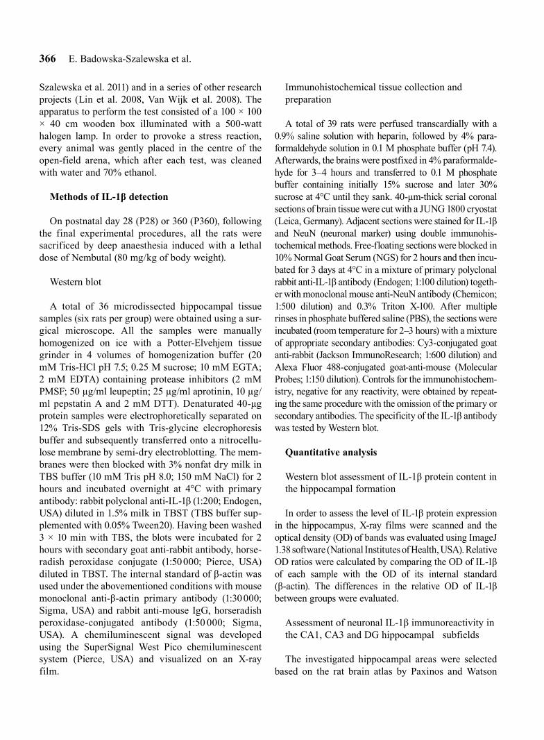
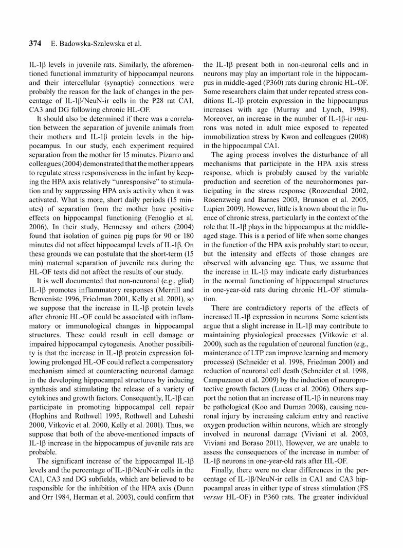
Under non-stress conditions, the hippocampal for-mation revealed the presence of IL-1β protein, and there was no significant difference in its optical den-sity in juvenile (P28) (0.30 ± 0.02) and middle-aged (P360) (0.40 ± 0.10) animals (F1,11=3.33, P=0.0679) (Fig. 1A–B).
The juvenile (P28) rats that underwent the chronic FS test showed no statistically significant disparities in
Fig. 1. Densitometric analysis of changes in the IL-1β pro-tein level after chronic FS or HL-OF stimulation in the hip-pocampus of P28 and P360 rats. The protein level is pre-sented as a ratio of the optical density of IL-1β to the internal standard of β-actin (relative OD). Experimental groups are compared with control groups and P28 with P360 rats. Statistical analyses are made by ANOVA with Kruskall-Wallis post hoc test. Significant increase in the IL-1β protein level after HL-OF versus control is indicated by * (P<0.05) (A). A representative blot is shown (B).
368 E. Badowska-Szalewska et al.
the optical density of hippocampal IL-1β protein (0.43 ± 0.08) in relation to control animals, whereas long-lasting exposure of this group to HL-OF led to an observable increase in IL-1β density (0.55 ± 0.05; F2,15=10.82, P=0.0034, P<0.05).
In P360 subjects, as compared to the control group, no significant differences were found in the optical density of hippocampal IL-1β protein after prolonged exposure to FS (0.45 ± 0.10), but there was an increase of IL-1β after HL-OF (0.63 ± 0.14; F2,18=6.68, P=0.0331, P<0.05) tests (Fig. 1A).
Comparing both age groups (P28 versus P360), optical density variations in hippocampal IL-1β were not detected after the FS (F1,11=1.20, P=0.2733) and HL-OF (F1,11=2.13, P=0.1441) chronic stimulations (Fig. 1A).
Immunohistochemical study
The findings recorded in the subsection were obtained by examining IL-1β/NeuN-ir colocalised cells and NeuN-ir cells. The mean percentage of IL-1β/NeuN-ir cells in relation to all NeuN-ir cells ± standard deviation was determined. There was no statistically significant change in the total number of NeuN-ir cells between any of the comparable groups.
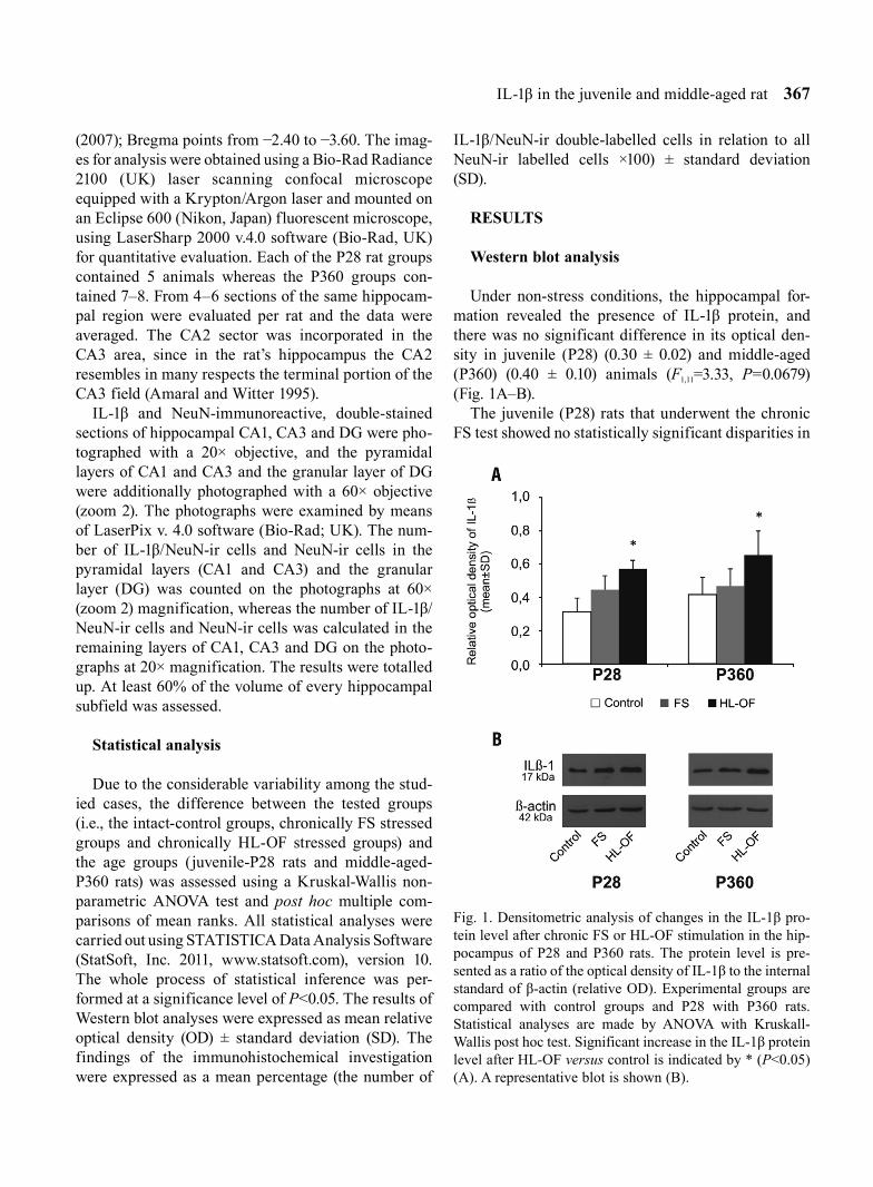
Percentage of IL-1β/NeuN-ir cells in the hippocampal structures (CA1, CA3 and DG) of P28 and P360 control rats
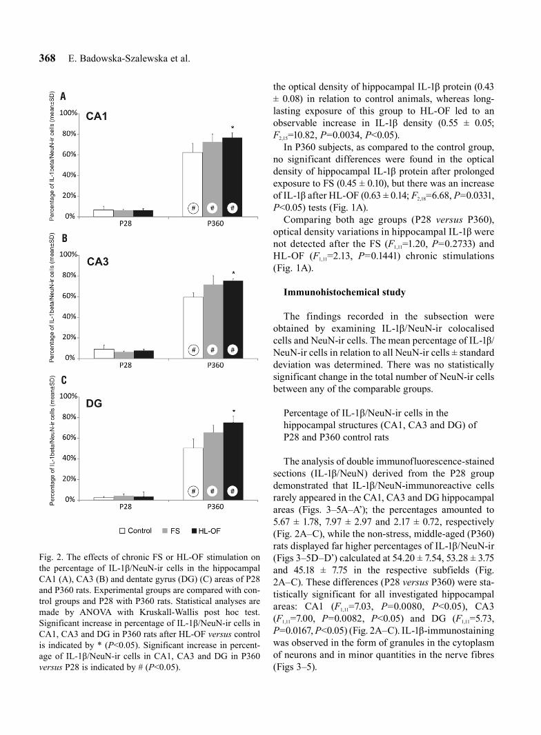
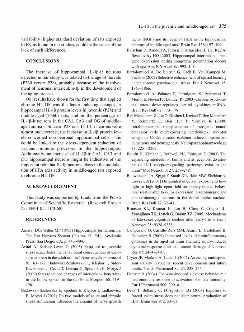
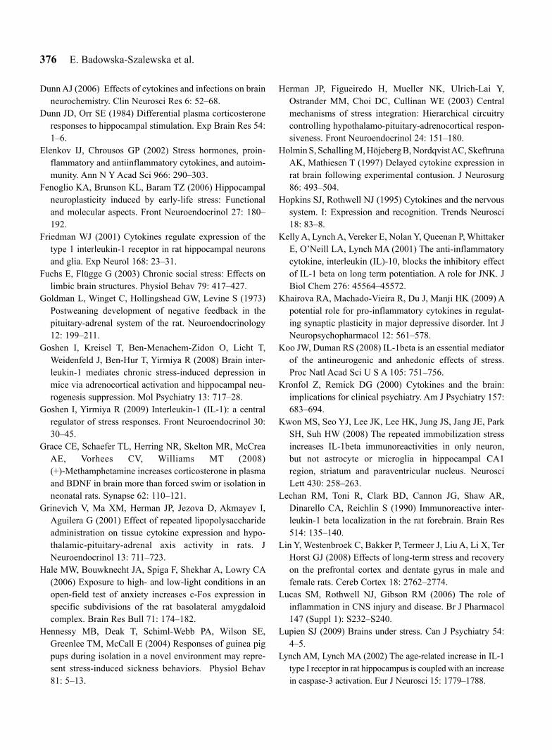
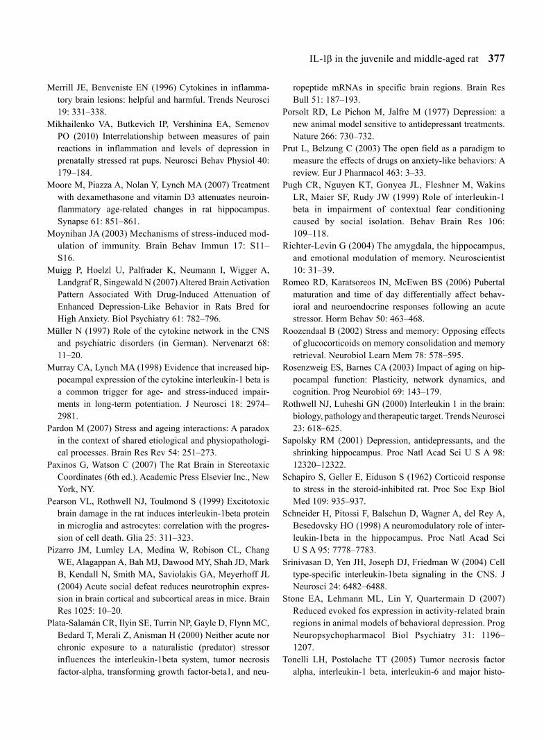
The analysis of double immunofluorescence-stained sections (IL-1β/NeuN) derived from the P28 group demonstrated that IL-1β/NeuN-immunoreactive cells rarely appeared in the CA1, CA3 and DG hippocampal areas (Figs. 3–5A–A’); the percentages amounted to 5.67 ± 1.78, 7.97 ± 2.97 and 2.17 ± 0.72, respectively (Fig. 2A–C), while the non-stress, middle-aged (P360) rats displayed far higher percentages of IL-1β/NeuN-ir (Figs 3–5D–D’) calculated at 54.20 ± 7.54, 53.28 ± 3.75 and 45.18 ± 7.75 in the respective subfields (Fig. 2A–C). These differences (P28 versus P360) were sta-tistically significant for all investigated hippocampal areas: CA1 (F1,11=7.03, P=0.0080, P<0.05), CA3 (F1,11=7.00, P=0.0082, P<0.05) and DG (F1,11=5.73, P=0.0167, P<0.05) (Fig. 2A–C). IL-1β-immunostaining was observed in the form of granules in the cytoplasm of neurons and in minor quantities in the nerve fibres (Figs 3–5).
Fig. 2. The effects of chronic FS or HL-OF stimulation on the percentage of IL-1β/NeuN-ir cells in the hippocampal CA1 (A), CA3 (B) and dentate gyrus (DG) (C) areas of P28 and P360 rats. Experimental groups are compared with con-trol groups and P28 with P360 rats. Statistical analyses are made by ANOVA with Kruskall-Wallis post hoc test. Significant increase in percentage of IL-1β/NeuN-ir cells in CA1, CA3 and DG in P360 rats after HL-OF versus control is indicated by * (P<0.05). Significant increase in percent-age of IL-1β/NeuN-ir cells in CA1, CA3 and DG in P360 versus P28 is indicated by # (P<0.05).
IL-1β in the juvenile and middle-aged rat 369
Fig. 3. IL-1β-ir cells (red), NeuN-ir cells (green) and double immunostaining IL-1β/NeuN-ir cells (yellow-orange; arrow) in the CA1 hippocampal subregion (A–F) and in the pyramidal layer of the CA1 (A’–F’) of juvenile (P28) (A, A’ – C, C’) and middle-aged (P360) (D, D’ – F, F’) rats from the control groups (A, A’, D, D’) and from the groups exposed to chronic FS (B, B’, E, E’) or chronic HL-OF (C, C’, F, F’) stress. Representative photographs were obtained at 20× (A–F) and 60× mag-nification (A’–F’).
370 E. Badowska-Szalewska et al.
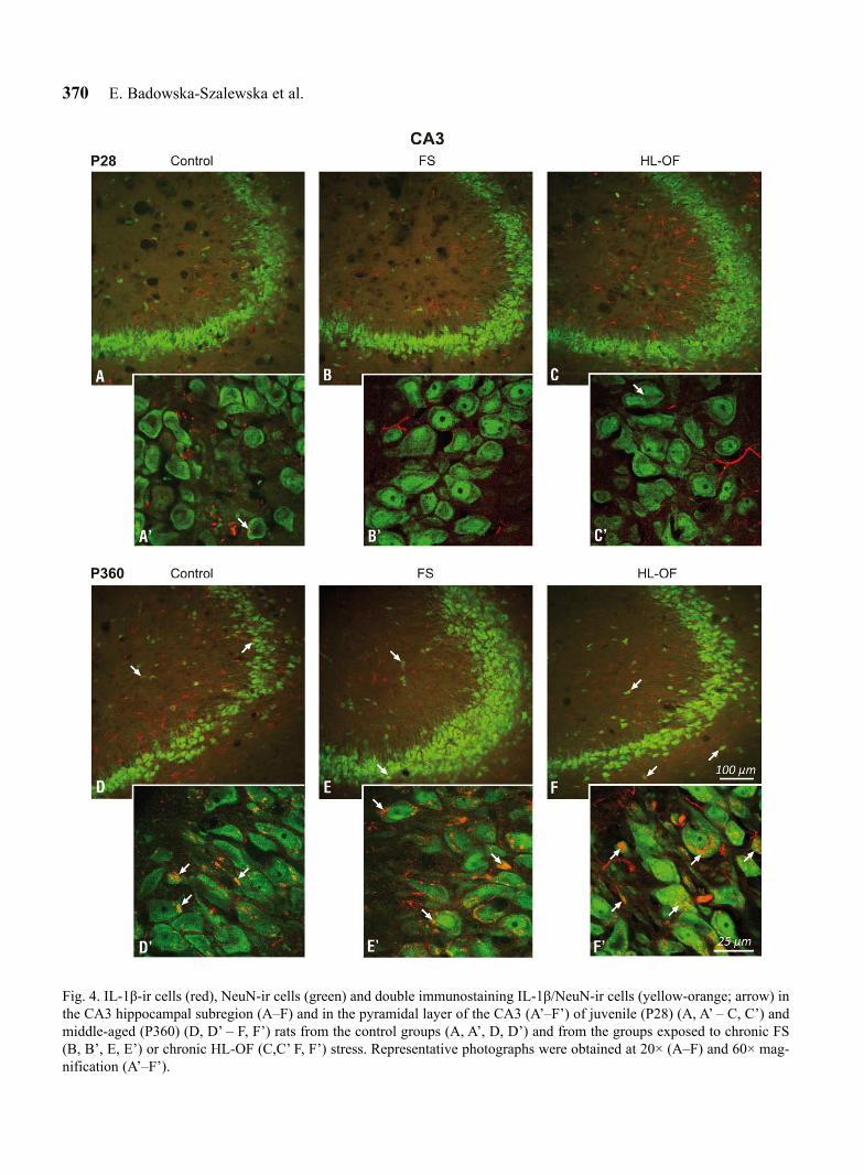
Fig. 4. IL-1β-ir cells (red), NeuN-ir cells (green) and double immunostaining IL-1β/NeuN-ir cells (yellow-orange; arrow) in the CA3 hippocampal subregion (A–F) and in the pyramidal layer of the CA3 (A’–F’) of juvenile (P28) (A, A’ – C, C’) and middle-aged (P360) (D, D’ – F, F’) rats from the control groups (A, A’, D, D’) and from the groups exposed to chronic FS (B, B’, E, E’) or chronic HL-OF (C,C’ F, F’) stress. Representative photographs were obtained at 20× (A–F) and 60× mag-nification (A’–F’).
IL-1β in the juvenile and middle-aged rat 371
The influence of chronic exposure to FS or HL-OF stimulation on the percentage of IL-1β/NeuN-ir cells in the CA1, CA3 and DG of juvenile (P28) rats
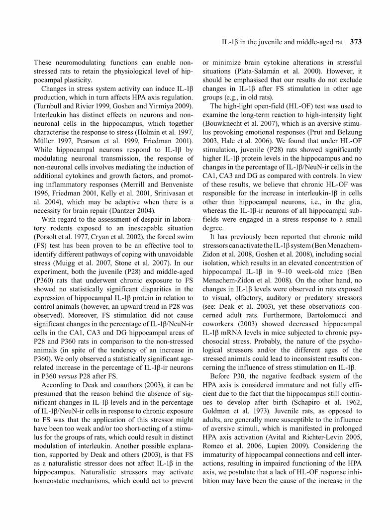
Long-lasting exposure of P28 to FS or HL-OF stress stimulation had no significant effect on the percentage of IL-1β/NeuN-ir cells in all hippocampal areas: CA1 (FS: 5.39 ± 0.58; HL-OF: 6.52 ± 0.99), CA3 (FS: 5.60 ± 0.42; HL-OF: 5.26 ± 1.07) and DG (FS: 3.67 ± 1.46; HL-OF: 5.68 ± 3.40) (Fig. 2A–C), where the number of IL-1β/NeuN-ir cells after the FS and HL-OF tests was low in all hippocampal subfields (Figs 3–5A–A’, 3–5B–B’, 3–5C–C’).
The influence of chronic exposure to the FS or HL-OF tests on the percentage of IL-1β/NeuN-ir cells in the hippocampal CA1, CA3 and DG of middle-aged (P360) rats
Following the chronic FS test, middle-aged (P360) rats showed no statistically significant alterations (despite a tendency towards growth) in the percentage of IL-1β/NeuN-ir cells in the CA1 (63.13 ± 6.67), CA3 (64.32 ± 7.58) and DG (58.57 ± 6.40) hippocampal sub-fields (Figs 2A–C, 3–5D–D’, 3–5E–E’). After apply-ing the chronic HL-OF stress stimulus, however, this percentage significantly increased in CA1 (66.67 ± 3.94; F2,22=7.90, P=0.0175, P<0.05), in CA3 (67.58 ± 1.72; F2,22=13.59, P=0.0007, P<0.05) and in DG (67.38 ± 5.66; F2,22=14.38, P=0.0005, P<0.05) when compared with the control rats (Figs 2A–C, 3–5D–D’, 3–5F–F’).
A juxtaposition of the percentage of IL-1β/NeuN-ir cells in hippocampal CA1, CA3 and DG after FS or HL-OF stress stimulation in relation to age (P28 versus P360)
FS and HL-OF stimulations were found to produce statistically significant age-related changes in the per-centage of IL-1β/NeuN-ir cells in the CA1, CA3 and DG subfields. FS resulted in an increase in the per-centage, which was higher in the CA1 (F1,12=7.41, P=0.0065, P<0.05), CA3 (F1,12=7.44, P=0.0064, P<0.05) and DG (F1,10=4.36, P=0.0367, P<0.05) of middle-aged (P360) animals than in the corresponding subfields of juvenile (P28) subjects (Figs 2A–C, 3–5B–B’, 3–5E–E’). HL-OF was associated with an increase in the percentage of IL-1β/NeuN-ir, which was higher
(F1,11=7.00, P=0.0082, P<0.05) in the hippocampal CA1, CA3 and DG of P360 animals than in the corre-sponding subregions of P28 rats (Figs 2A–C, 3–5C–C’, 3–5F–F’).
DISCUSSION
In the non-stress groups, no significant differences in IL-1β protein levels were reported. However, an increase in the percentage of IL-1β/NeuN-ir cells in hippocampal CA1, CA3 and DG of middle-aged (P360) rats was observed, as compared to juvenile (P28) animals. Furthermore, in contrast to P360 rats, there were only a few IL-1β/NeuN-ir cells in P28 rats. Thus, it can be assumed that in juvenile rats mainly non-neuronal (immune cells, glia) interleukin-1β per-forms an important physiological role in the function-ing of the hippocampus, whereas in middle-aged ani-mals it is also the interleukin present in neurons that plays a significant part.
IL-1β protein expression and IL-1β-immunoreactive neurons have previously been detected in the hip-pocampus under physiological conditions (Lechan et al. 1990, Friedman 2001). According to Moore and coworkers (2007), the increase with age in the hip-pocampal concentration of IL-1β can be correlated with age-related deficits in synaptic and cognitive functions. However, it is worth mentioning that Balschun and colleagues (2003) observed that basal IL-1β gene expression was only slightly reduced in middle-aged rats when compared to young ones (3 months). Furthermore, the authors believe that age-dependent deterioration of long-term potentiation (LTP) does not appear to manifest in rats before the age of approximately 20 months.
With respect to this observation, we have come to the conclusion that our observation of no significant increase in the IL-1β protein level in P360 rats and considerable increase in the percentage of IL-1β-ir neurons in middle-aged rats (when compared to P28 animals) were due to some age-related shift of IL-1β-production from glia to neurons. This may correspond to the role of IL-1β in hippocampal neurons. This role includes, for example, regulation of neuronal survival, control of synaptic activity and provides metabolic support for the maintenance of LTP (stimulate glucose uptake, release of certain neurotransmitters and modu-lators) (see: Vitkovic et al. 2000, Friedman 2001, Srinivasan et al. 2004, Viviani and Boraso 2011).
372 E. Badowska-Szalewska et al.
Fig. 5. IL-1β-ir cells (red), NeuN-ir cells (green) and double immunostaining IL-1β/NeuN-ir cells (yellow-orange; arrow) in the hippocampal dentate gyrus (DG) (A–F) and in the granular layer of the DG (A’–F’) of juvenile (P28) (A, A’ – C, C’) and middle-aged (P360) (D, D’ – F, F’) rats from the control groups (A,A’,D,D’) and from the groups exposed to chronic FS (B, B’, E, E’) or chronic HL-OF (C, C’, F, F’) stress. Representative photographs were obtained at 20× (A–F) and 60× mag-nification (A’–F’).
IL-1β in the juvenile and middle-aged rat 373
These neuromodulating functions can enable non-stressed rats to retain the physiological level of hip-pocampal plasticity.
Changes in stress system activity can induce IL-1β production, which in turn affects HPA axis regulation. (Turnbull and Rivier 1999, Goshen and Yirmiya 2009). Interleukin has distinct effects on neurons and non-neuronal cells in the hippocampus, which together characterise the response to stress (Holmin et al. 1997, Muller 1997, Pearson et al. 1999, Friedman 2001). While hippocampal neurons respond to IL-1β by modulating neuronal transmission, the response of non-neuronal cells involves mediating the induction of additional cytokines and growth factors, and promot-ing inflammatory responses (Merrill and Benveniste 1996, Friedman 2001, Kelly et al. 2001, Srinivasan et al. 2004), which may be adaptive when there is a necessity for brain repair (Dantzer 2004).
With regard to the assessment of despair in labora-tory rodents exposed to an inescapable situation (Porsolt et al. 1977, Cryan et al. 2002), the forced swim (FS) test has been proven to be an effective tool to identify different pathways of coping with unavoidable stress (Muigg et al. 2007, Stone et al. 2007). In our experiment, both the juvenile (P28) and middle-aged (P360) rats that underwent chronic exposure to FS showed no statistically significant disparities in the expression of hippocampal IL-1β protein in relation to control animals (however, an upward trend in P28 was observed). Moreover, FS stimulation did not cause significant changes in the percentage of IL-1β/NeuN-ir cells in the CA1, CA3 and DG hippocampal areas of P28 and P360 rats in comparison to the non-stressed animals (in spite of the tendency of an increase in P360). We only observed a statistically significant age-related increase in the percentage of IL-1β-ir neurons in P360 versus P28 after FS.
According to Deak and coauthors (2003), it can be presumed that the reason behind the absence of sig-nificant changes in IL-1β levels and in the percentage of IL-1β/NeuN-ir cells in response to chronic exposure to FS was that the application of this stressor might have been too weak and/or too short-acting of a stimu-lus for the groups of rats, which could result in distinct modulation of interleukin. Another possible explana-tion, supported by Deak and others (2003), is that FS as a naturalistic stressor does not affect IL-1β in the hippocampus. Naturalistic stressors may activate homeostatic mechanisms, which could act to prevent
or minimize brain cytokine alterations in stressful situations (Plata-Salamán et al. 2000). However, it should be emphasised that our results do not exclude changes in IL-1β after FS stimulation in other age groups (e.g., in old rats).
The high-light open-field (HL-OF) test was used to examine the long-term reaction to high-intensity light (Bouwknecht et al. 2007), which is an aversive stimu-lus provoking emotional responses (Prut and Belzung 2003, Hale et al. 2006). We found that under HL-OF stimulation, juvenile (P28) rats showed significantly higher IL-1β protein levels in the hippocampus and no changes in the percentage of IL-1β/NeuN-ir cells in the CA1, CA3 and DG as compared with controls. In view of these results, we believe that chronic HL-OF was responsible for the increase in interleukin-1β in cells other than hippocampal neurons, i.e., in the glia, whereas the IL-1β-ir neurons of all hippocampal sub-fields were engaged in a stress response to a small degree.
It has previously been reported that chronic mild stressors can activate the IL-1β system (Ben Menachem-Zidon et al. 2008, Goshen et al. 2008), including social isolation, which results in an elevated concentration of hippocampal IL-1β in 9–10 week-old mice (Ben Menachem-Zidon et al. 2008). On the other hand, no changes in IL-1β levels were observed in rats exposed to visual, olfactory, auditory or predatory stressors (see: Deak et al. 2003), yet these observations con-cerned adult rats. Furthermore, Bartolomucci and coworkers (2003) showed decreased hippocampal IL-1β mRNA levels in mice subjected to chronic psy-chosocial stress. Probably, the nature of the psycho-logical stressors and/or the different ages of the stressed animals could lead to inconsistent results con-cerning the influence of stress stimulation on IL-1β.
Before P30, the negative feedback system of the HPA axis is considered immature and not fully effi-cient due to the fact that the hippocampus still contin-ues to develop after birth (Schapiro et al. 1962, Goldman et al. 1973). Juvenile rats, as opposed to adults, are generally more susceptible to the influence of aversive stimuli, which is manifested in prolonged HPA axis activation (Avital and Richter-Levin 2005, Romeo et al. 2006, Lupien 2009). Considering the immaturity of hippocampal connections and cell inter-actions, resulting in impaired functioning of the HPA axis, we postulate that a lack of HL-OF response inhi-bition may have been the cause of the increase in the
374 E. Badowska-Szalewska et al.
IL-1β levels in juvenile rats. Similarly, the aforemen-tioned functional immaturity of hippocampal neurons and their intercellular (synaptic) connections were probably the reason for the lack of changes in the per-centage of IL-1β/NeuN-ir cells in the P28 rat CA1, CA3 and DG following chronic HL-OF.
It should also be determined if there was a correla-tion between the separation of juvenile animals from their mothers and IL-1β protein levels in the hip-pocampus. In our study, each experiment required separation from the mother for 15 minutes. Pizarro and colleagues (2004) demonstrated that the mother appears to regulate stress responsiveness in the infant by keep-ing the HPA axis relatively “unresponsive” to stimula-tion and by suppressing HPA axis activity when it was activated. What is more, short daily periods (15 min-utes) of separation from the mother have positive effects on hippocampal functioning (Fenoglio et al. 2006). In their study, Hennessy and others (2004) found that isolation of guinea pig pups for 90 or 180 minutes did not affect hippocampal levels of IL-1β. On these grounds we can postulate that the short-term (15 min) maternal separation of juvenile rats during the HL-OF tests did not affect the results of our study.
It is well documented that non-neuronal (e.g., glial) IL-1β promotes inflammatory responses (Merrill and Benveniste 1996, Friedman 2001, Kelly et al. 2001), so we suppose that the increase in IL-1β protein levels after chronic HL-OF could be associated with inflam-matory or immunological changes in hippocampal structures. These could result in cell damage or impaired hippocampal cytogenesis. Another possibili-ty is that the increase in IL-1β protein expression fol-lowing prolonged HL-OF could reflect a compensatory mechanism aimed at counteracting neuronal damage in the developing hippocampal structures by inducing synthesis and stimulating the release of a variety of cytokines and growth factors. Consequently, IL-1β can participate in promoting hippocampal cell repair (Hophins and Rothwell 1995, Rothwell and Luheshi 2000, Vitkovic et al. 2000, Kelly et al. 2001). Thus, we suppose that both of the above-mentioned impacts of IL-1β increase in the hippocampus of juvenile rats are probable.
The significant increase of the hippocampal IL-1β levels and the percentage of IL-1β/NeuN-ir cells in the CA1, CA3 and DG subfields, which are believed to be responsible for the inhibition of the HPA axis (Dunn and Orr 1984, Herman et al. 2003), could confirm that
the IL-1β present both in non-neuronal cells and in neurons may play an important role in the hippocam-pus in middle-aged (P360) rats during chronic HL-OF. Some researchers claim that under repeated stress con-ditions IL-1β protein expression in the hippocampus increases with age (Murray and Lynch, 1998). Moreover, an increase in the number of IL-1β-ir neu-rons was noted in adult mice exposed to repeated immobilization stress by Kwon and colleagues (2008) in the hippocampal CA1.
The aging process involves the disturbance of all mechanisms that participate in the HPA axis stress response, which is probably caused by the variable production and secretion of the neurohormones par-ticipating in the stress response (Roozendaal 2002, Rosenzweig and Barnes 2003, Brunson et al. 2005, Lupien 2009). However, little is known about the influ-ence of chronic stress, particularly in the context of the role that IL-1β plays in the hippocampus at the middle-aged stage. This is a period of life when some changes in the function of the HPA axis probably start to occur, but the intensity and effects of those changes are observed with advancing age. Thus, we assume that the increase in IL-1β may indicate early disturbances in the normal functioning of hippocampal structures in one-year-old rats during chronic HL-OF stimula-tion.
There are contradictory reports of the effects of increased IL-1β expression in neurons. Some scientists argue that a slight increase in IL-1β may contribute to maintaining physiological processes (Vitkovic et al. 2000), such as the regulation of neuronal function (e.g., maintenance of LTP can improve learning and memory processes) (Schneider et al. 1998, Friedman 2001) and reduction of neuronal cell death (Schneider et al. 1998, Campuzanoo et al. 2009) by the induction of neuropro-tective growth factors (Lucas et al. 2006). Others sup-port the notion that an increase of IL-1β in neurons may be pathological (Koo and Duman 2008), causing neu-ronal injury by increasing calcium entry and reactive oxygen production within neurons, which are strongly involved in neuronal damage (Viviani et al. 2003, Viviani and Boraso 2011). However, we are unable to assess the consequences of the increase in number of IL-1β neurons in one-year-old rats after HL-OF.
Finally, there were no clear differences in the per-centage of IL-1β/NeuN-ir cells in CA1 and CA3 hip-pocampal areas in either type of stress stimulation (FS versus HL-OF) in P360 rats. The greater individual
IL-1β in the juvenile and middle-aged rat 375
variability (higher standard deviation) of rats exposed to FS, as found in our studies, could be the cause of the lack of such differences.
CONCLUSIONS
The increase of hippocampal IL-1β-ir neurons detected in our study was related to the age of the rats (P360 versus P28), probably because of the involve-ment of neuronal interleukin-1β in the development of the aging process.
Our results have shown for the first time that applied chronic HL-OF was the factor inducing changes in hippocampal IL-1β protein levels in juvenile (P28) and middle-aged (P360) rats, and in the percentage of IL-1β-ir neurons in the CA1, CA3 and DG of middle-aged animals. Since in P28 rats, IL-1β-ir neurons were almost undetectable, the increase in IL-1β protein lev-els concerned non-neuronal hippocampal cells. This could be linked to the stress-dependent induction of various immune processes in the hippocampus. Additionally, an increase of IL-1β-ir CA1, CA3 and DG hippocampal neurons might be indicative of the important role that IL-1β neurons plays in the modula-tion of HPA axis activity in middle-aged rats exposed to chronic HL-OF.
ACKNOWLEDGEMENT
This study was supported by funds from the Polish Committee of Scientific Research (Research Project No. N401 011 31/0168).
REFERENCES
Amaral DG, Witter MP (1995) Hippocampal formation. In: The Rat Nervous System (Paxinos G, Ed.). Academic Press, San Diego, CA, p. 443–494.
Avital A, Richter–Levin G (2005) Exposure to juvenile stress exacerbates the behavioural consequences of expo-sure to stress in the adult rat. Int J Neuropsychopharmacol 8: 163–173. Badowska-Szalewska E, Klejbor I, Sidor-Kaczmarek J, Cecot T, Lietzau G, Spodnik JH, Moryś J (2009) Stress-induced changes of interleukin-1beta with-in the limbic system in the rat. Folia Morphol 68: 119–128.
Badowska-Szalewska E, Spodnik E, Klejbor I, Ludkiewicz B, Moryś J (2011) Do two models of acute and chronic stress stimulation influence the amount of nerve growth
factor (NGF) and its receptor TrkA in the hippocampal neurons of middle aged rats? Brain Res 1384: 97–109.
Balschun D, Randolf A, Pitossi F, Schneider H, Del Rey A, Besedovsky HO (2003) Hippocampal interleukin-1 beta gene expression during long-term potentiation decays with age. Ann N Y Acad Sci 992: 1–8.
Bartolomucci A, De Biurrun G, Czéh B, Van Kampen M, Fuchs E (2002) Selective enhancement of spatial learning under chronic psychosocial stress. Eur J Neurosci 15: 1863–1866.
Bartolomucci A, Palanza P, Parmigiani S, Pederzani T, Merlot E, Neveu PJ, Dantzer R (2003) Chronic psychoso-cial stress down-regulates central cytokines mRNA. Brain Res Bull 62: 173–178.
Ben Menachem-Zidon O, Goshen I, Kreisel T, Ben Menahem Y, Reinhartz E, Ben Hur T, Yirmiya R (2008) Intrahippocampal transplantation of transgenic neural precursor cells overexpressing interleukin-1 receptor antagonist blocks chronic isolation-induced impairment in memory and neurogenesis. Neuropsychopharmacology 33: 2251–2262.
Boutin H, Kimber I, Rothwell NJ, Pinteaux E (2003) The expanding interleukin-1 family and its receptors: do alter-native IL-1 receptor/signaling pathways exist in the brain? Mol Neurobiol 27: 239–248.
Bouwknecht JA, Spiga F, Staub DR, Hale MW, Shekhar A, Lowry CA (2007) Differential effects of exposure to low-light or high-light open-field on anxiety-related behav-iors: relationship to c-Fos expression in serotonergic and non-serotonergic neurons in the dorsal raphe nucleus. Brain Res Bull 72: 32–43.
Brunson KL, Kramar E, Lin B, Chen Y, Colgin LL, Yanagihara TK, Lynch G, Baram TZ (2005) Mechanisms of late-onset cognitive decline after early-life stress. J Neurosci 25: 9328–9338.
Campuzano O, Castillo-Ruiz MM, Acarin L, Castellano B, Gonzalez B (2009) Increased levels of proinflammatory cytokines in the aged rat brain attenuate injury-induced cytokine response after excitotoxic damage. J Neurosci Res 87: 2484–2497.
Cryan JF, Markou A, Lucki I (2002) Assessing antidepres-sant activity in rodents: recent developments and future needs. Trends Pharmacol Sci 23: 238–245.
Dantzer R (2004) Cytokine-induced sickness behaviour: a neuroimmune response to activation of innate immunity. Eur J Pharmacol 500: 399–411.
Deak T, Bellamy C, D’Agostino LG (2003) Exposure to forced swim stress does not alter central production of IL-1. Brain Res 972: 53–63.
376 E. Badowska-Szalewska et al.
Dunn AJ (2006) Effects of cytokines and infections on brain neurochemistry. Clin Neurosci Res 6: 52–68.
Dunn JD, Orr SE (1984) Differential plasma corticosterone responses to hippocampal stimulation. Exp Brain Res 54: 1–6.
Elenkov IJ, Chrousos GP (2002) Stress hormones, proin-flammatory and antiinflammatory cytokines, and autoim-munity. Ann N Y Acad Sci 966: 290–303.
Fenoglio KA, Brunson KL, Baram TZ (2006) Hippocampal neuroplasticity induced by early-life stress: Functional and molecular aspects. Front Neuroendocrinol 27: 180–192.
Friedman WJ (2001) Cytokines regulate expression of the type 1 interleukin-1 receptor in rat hippocampal neurons and glia. Exp Neurol 168: 23–31.
Fuchs E, Flugge G (2003) Chronic social stress: Effects on limbic brain structures. Physiol Behav 79: 417–427.
Goldman L, Winget C, Hollingshead GW, Levine S (1973) Postweaning development of negative feedback in the pituitary-adrenal system of the rat. Neuroendocrinology 12: 199–211.
Goshen I, Kreisel T, Ben-Menachem-Zidon O, Licht T, Weidenfeld J, Ben-Hur T, Yirmiya R (2008) Brain inter-leukin-1 mediates chronic stress-induced depression in mice via adrenocortical activation and hippocampal neu-rogenesis suppression. Mol Psychiatry 13: 717–28.
Goshen I, Yirmiya R (2009) Interleukin-1 (IL-1): a central regulator of stress responses. Front Neuroendocrinol 30: 30–45.
Grace CE, Schaefer TL, Herring NR, Skelton MR, McCrea AE, Vorhees CV, Williams MT (2008) (+)-Methamphetamine increases corticosterone in plasma and BDNF in brain more than forced swim or isolation in neonatal rats. Synapse 62: 110–121.
Grinevich V, Ma XM, Herman JP, Jezova D, Akmayev I, Aguilera G (2001) Effect of repeated lipopolysaccharide administration on tissue cytokine expression and hypo-thalamic-pituitary-adrenal axis activity in rats. J Neuroendocrinol 13: 711–723.
Hale MW, Bouwknecht JA, Spiga F, Shekhar A, Lowry CA (2006) Exposure to high- and low-light conditions in an open-field test of anxiety increases c-Fos expression in specific subdivisions of the rat basolateral amygdaloid complex. Brain Res Bull 71: 174–182.
Hennessy MB, Deak T, Schiml-Webb PA, Wilson SE, Greenlee TM, McCall E (2004) Responses of guinea pig pups during isolation in a novel environment may repre-sent stress-induced sickness behaviors. Physiol Behav 81: 5–13.
Herman JP, Figueiredo H, Mueller NK, Ulrich-Lai Y, Ostrander MM, Choi DC, Cullinan WE (2003) Central mechanisms of stress integration: Hierarchical circuitry controlling hypothalamo-pituitary-adrenocortical respon-siveness. Front Neuroendocrinol 24: 151–180.
Holmin S, Schalling M, Höjeberg B, Nordqvist AC, Skeftruna AK, Mathiesen T (1997) Delayed cytokine expression in rat brain following experimental contusion. J Neurosurg 86: 493–504.
Hopkins SJ, Rothwell NJ (1995) Cytokines and the nervous system. I: Expression and recognition. Trends Neurosci 18: 83–8.
Kelly A, Lynch A, Vereker E, Nolan Y, Queenan P, Whittaker E, O’Neill LA, Lynch MA (2001) The anti-inflammatory cytokine, interleukin (IL)-10, blocks the inhibitory effect of IL-1 beta on long term potentiation. A role for JNK. J Biol Chem 276: 45564–45572.
Khairova RA, Machado-Vieira R, Du J, Manji HK (2009) A potential role for pro-inflammatory cytokines in regulat-ing synaptic plasticity in major depressive disorder. Int J Neuropsychopharmacol 12: 561–578.
Koo JW, Duman RS (2008) IL-1beta is an essential mediator of the antineurogenic and anhedonic effects of stress. Proc Natl Acad Sci U S A 105: 751–756.
Kronfol Z, Remick DG (2000) Cytokines and the brain: implications for clinical psychiatry. Am J Psychiatry 157: 683–694.
Kwon MS, Seo YJ, Lee JK, Lee HK, Jung JS, Jang JE, Park SH, Suh HW (2008) The repeated immobilization stress increases IL-1beta immunoreactivities in only neuron, but not astrocyte or microglia in hippocampal CA1 region, striatum and paraventricular nucleus. Neurosci Lett 430: 258–263.
Lechan RM, Toni R, Clark BD, Cannon JG, Shaw AR, Dinarello CA, Reichlin S (1990) Immunoreactive inter-leukin-1 beta localization in the rat forebrain. Brain Res 514: 135–140.
Lin Y, Westenbroek C, Bakker P, Termeer J, Liu A, Li X, Ter Horst GJ (2008) Effects of long-term stress and recovery on the prefrontal cortex and dentate gyrus in male and female rats. Cereb Cortex 18: 2762–2774.
Lucas SM, Rothwell NJ, Gibson RM (2006) The role of inflammation in CNS injury and disease. Br J Pharmacol 147 (Suppl 1): S232–S240.
Lupien SJ (2009) Brains under stress. Can J Psychiatry 54: 4–5.
Lynch AM, Lynch MA (2002) The age-related increase in IL-1 type I receptor in rat hippocampus is coupled with an increase in caspase-3 activation. Eur J Neurosci 15: 1779–1788.
IL-1β in the juvenile and middle-aged rat 377
Merrill JE, Benveniste EN (1996) Cytokines in inflamma-tory brain lesions: helpful and harmful. Trends Neurosci 19: 331–338.
Mikhailenko VA, Butkevich IP, Vershinina EA, Semenov PO (2010) Interrelationship between measures of pain reactions in inflammation and levels of depression in prenatally stressed rat pups. Neurosci Behav Physiol 40: 179–184.
Moore M, Piazza A, Nolan Y, Lynch MA (2007) Treatment with dexamethasone and vitamin D3 attenuates neuroin-flammatory age-related changes in rat hippocampus. Synapse 61: 851–861.
Moynihan JA (2003) Mechanisms of stress-induced mod-ulation of immunity. Brain Behav Immun 17: S11–S16.
Muigg P, Hoelzl U, Palfrader K, Neumann I, Wigger A, Landgraf R, Singewald N (2007) Altered Brain Activation Pattern Associated With Drug-Induced Attenuation of Enhanced Depression-Like Behavior in Rats Bred for High Anxiety. Biol Psychiatry 61: 782–796.
Muller N (1997) Role of the cytokine network in the CNS and psychiatric disorders (in German). Nervenarzt 68: 11–20.
Murray CA, Lynch MA (1998) Evidence that increased hip-pocampal expression of the cytokine interleukin-1 beta is a common trigger for age- and stress-induced impair-ments in long-term potentiation. J Neurosci 18: 2974–2981.
Pardon M (2007) Stress and ageing interactions: A paradox in the context of shared etiological and physiopathologi-cal processes. Brain Res Rev 54: 251–273.
Paxinos G, Watson C (2007) The Rat Brain in Stereotaxic Coordinates (6th ed.). Academic Press Elsevier Inc., New York, NY.
Pearson VL, Rothwell NJ, Toulmond S (1999) Excitotoxic brain damage in the rat induces interleukin-1beta protein in microglia and astrocytes: correlation with the progres-sion of cell death. Glia 25: 311–323.
Pizarro JM, Lumley LA, Medina W, Robison CL, Chang WE, Alagappan A, Bah MJ, Dawood MY, Shah JD, Mark B, Kendall N, Smith MA, Saviolakis GA, Meyerhoff JL (2004) Acute social defeat reduces neurotrophin expres-sion in brain cortical and subcortical areas in mice. Brain Res 1025: 10–20.
Plata-Salamán CR, Ilyin SE, Turrin NP, Gayle D, Flynn MC, Bedard T, Merali Z, Anisman H (2000) Neither acute nor chronic exposure to a naturalistic (predator) stressor influences the interleukin-1beta system, tumor necrosis factor-alpha, transforming growth factor-beta1, and neu-
ropeptide mRNAs in specific brain regions. Brain Res Bull 51: 187–193.
Porsolt RD, Le Pichon M, Jalfre M (1977) Depression: a new animal model sensitive to antidepressant treatments. Nature 266: 730–732.
Prut L, Belzung C (2003) The open field as a paradigm to measure the effects of drugs on anxiety-like behaviors: A review. Eur J Pharmacol 463: 3–33.
Pugh CR, Nguyen KT, Gonyea JL, Fleshner M, Wakins LR, Maier SF, Rudy JW (1999) Role of interleukin-1 beta in impairment of contextual fear conditioning caused by social isolation. Behav Brain Res 106: 109–118.
Richter-Levin G (2004) The amygdala, the hippocampus, and emotional modulation of memory. Neuroscientist 10: 31–39.
Romeo RD, Karatsoreos IN, McEwen BS (2006) Pubertal maturation and time of day differentially affect behav-ioral and neuroendocrine responses following an acute stressor. Horm Behav 50: 463–468.
Roozendaal B (2002) Stress and memory: Opposing effects of glucocorticoids on memory consolidation and memory retrieval. Neurobiol Learn Mem 78: 578–595.
Rosenzweig ES, Barnes CA (2003) Impact of aging on hip-pocampal function: Plasticity, network dynamics, and cognition. Prog Neurobiol 69: 143–179.
Rothwell NJ, Luheshi GN (2000) Interleukin 1 in the brain: biology, pathology and therapeutic target. Trends Neurosci 23: 618–625.
Sapolsky RM (2001) Depression, antidepressants, and the shrinking hippocampus. Proc Natl Acad Sci U S A 98: 12320–12322.
Schapiro S, Geller E, Eiduson S (1962) Corticoid response to stress in the steroid-inhibited rat. Proc Soc Exp Biol Med 109: 935–937.
Schneider H, Pitossi F, Balschun D, Wagner A, del Rey A, Besedovsky HO (1998) A neuromodulatory role of inter-leukin-1beta in the hippocampus. Proc Natl Acad Sci U S A 95: 7778–7783.
Srinivasan D, Yen JH, Joseph DJ, Friedman W (2004) Cell type-specific interleukin-1beta signaling in the CNS. J Neurosci 24: 6482–6488.
Stone EA, Lehmann ML, Lin Y, Quartermain D (2007) Reduced evoked fos expression in activity-related brain regions in animal models of behavioral depression. Prog Neuropsychopharmacol Biol Psychiatry 31: 1196–1207.
Tonelli LH, Postolache TT (2005) Tumor necrosis factor alpha, interleukin-1 beta, interleukin-6 and major histo-
378 E. Badowska-Szalewska et al.
compatibility complex molecules in the normal brain and after peripheral immune challenge. Neurol Res 27: 679–684.
Turnbull AV, Rivier CL (1999) Regulation of the hypotha-lamic-pituitary-adrenal axis by cytokines: actions and mechanisms of action. Physiol Rev 79: 1–71.
Van Wijk N, Rijntjes E, Van De Heijning BJM (2008) Perinatal and chronic hypothyroidism impair behavioural development in male and female rats. Exp Physiol 93: 1199–1209.
Vitkovic L, Bockaert J, Jacque C (2000) “Inflammatory” cytokines: neuromodulators in normal brain? J Neurochem 74: 457–471.
Viviani B, Bartesaghi S, Gardoni F, Vezzani A, Behrens MM, Bartfai T, Binaglia M, Corsini E, Di Luca M, Galli CL, Marinovich M (2003) Interleukin-1beta enhances NMDA receptor-mediated intracellular calcium increase
through activation of the Src family of kinases. J Neurosci 23: 8692–8700.
Viviani B, Boraso M (2011) Cytokines and neuronal chan-nels: a molecular basis for age-related decline of neuronal function? Exp Gerontol 46: 199–206.
Yee BK, Zhu S, Mohammed AH, Feldon J (2007) Levels of neurotrophic factors in the hippocampus and amygdala correlate with anxiety- and fear-related behaviour in C57BL6 mice. J Neural Transm 114: 431–444.
Yirmiya R, Goshen I (2011) Immune modulation of learn-ing, memory, neural plasticity and neurogenesis. Brain Behav Immun 25: 181–213.
You Z, Luo C, Zhang W, Chen Y, He J, Zhao Q, Zuo R, Wu Y (2011) Pro- and anti-inflammatory cytokines expres-sion in rat’s brain and spleen exposed to chronic mild stress: involvement in depression. Behav Brain Res 225: 135–141.