Essential role of Src-family protein tyrosine kinases in NF-κB activation during B cell development
6
nature immunology • volume 4 no 3 • march 2003 • www.nature.com/natureimmunology A RTICLES 274 Kaoru Saijo 1† , Christian Schmedt 1† , I-hsin Su 1 , Hajime Karasuyama 2 , Clifford A. Lowell 3 , Michael Reth 4 ,Takahiro Adachi 4 *,Alina Patke 1 , Angela Santana 1 and Alexander Tarakhovsky 1 Published online 3 February 2003; doi:10.1038/ni893 The nature of signals that govern the development of immunoglobulin heavy chain-dependent B cells is largely unknown. Using mice deficient for the B cell-expressed Src-family protein tyrosine kinases (SFKs) Blk, Fyn and Lyn, we show an essential role of these kinases in pre-B cell receptor (pre-BCR)– mediated NF- κB activation and B cell development. This signaling defect is SFK specific, as a deficiency in Syk, which controls pre-B cell development, does not affect NF- κB induction. Impaired NF- κB induction was overcome by the activation of protein kinase C (PKC)- λ, thus suggesting the involvement of PKC- λ in pre-BCR–mediated SFK-dependent activation of NF- κB. Our data show the existence of a functionally distinct SFK signaling module responsible for pre-BCR–mediated NF- κB activation and B cell development. 1 Laboratory of Lymphocyte Signaling,The Rockefeller University, New York, NY 10021, USA. 2 Department of Immune Regulation,Tokyo Medical and Dental University Graduate School, 1-5-45 Yushima, Bunkyo-ku,Tokyo 113-8519, Japan. 3 Department of Laboratory Medicine, University of California, San Francisco, CA 94143, USA. 4Department of Molecular Immunology, Biology III, University of Freiburg and Max-Planck Institute of Immunology, Stuebeweg 51, 79108 Freiburg, Germany. *Present address: Department of Immunology, Medical Research Institute,Tokyo Medical and Dental University,Tokyo 113-8510, Japan. † These authors contributed equally to this work. Correspondence should be addressed to K.S. ([email protected]) or A.T. ([email protected]). Essential role of Src-family protein tyrosine kinases in NF-κB activation during B cell development Expression of the immunoglobulin heavy chain (IgH) on the surface of early B cell progenitors establishes the IgH-dependent signaling network that governs development of B cells in the bone marrow 1 . This signal-trans- ducing network starts to operate in pro-B cells, where IgH in association with V-preB and λ5 proteins form a surface-expressed, pre-B cell receptor (pre-BCR) 2 . The signals derived from the pre-BCR originate within a juxta-membrane signaling complex arranged around the IgH-associated scaffold proteins Igα and Igβ 3 . Both of these proteins possess several immuno-receptor tyrosine-based activation motifs (ITAM) that recruit and facilitate activation of the protein tyrosine kinase Syk 4 . Activated Syk phos- phorylates PLC-γ2, leading to the generation of second messengers, which are essential for protein kinase C (PKC) activation and calcium mobiliza- tion 5 . The essential role of the ITAM-Syk signaling circuit in B cell devel- opment has been underscored by grossly impaired pre-B cell development in the absence of Igα-ITAM 6 or Syk 7,8 . Another kinase activity associated with the pre-BCR is linked to Src- family protein tyrosine kinases (SFKs). B lineage cells express several distinct SFKs, such as the B cell-specific SFK Blk and the more widely expressed Lyn, Fyn, Hck, Fgr and Lck 9–11 . The biochemical evidence sug- gests involvement of SFKs in pre-BCR–mediated signaling. SFKs co- precipitate with the pre-BCR in cultured pre-B cell lines 12 and ex vivo cross-linking of the pre-BCR on isolated B cell progenitors results in tyrosine phosphorylation of several proteins 13 . On the basis of the possi- ble similarities between the pre-BCR and BCR signaling, two models of SFK involvement in the initiation of pre-BCR signaling could be consid- ered. The first model is founded on data that show the ability of SFKs to phosphorylate ITAMs on Igα-Igβ in vitro 14 . The subsequent recruitment and activation of Syk may therefore be regulated by SFKs. For pre-BCR signaling this model predicts a key role for SFKs in Syk activation and the initiation of signaling. The second model is based on data showing the apparently SFK-independent activation of Syk 15 . In this model, SFKs act in BCR signaling by phosphorylating target proteins such as Btk, Vav or CD19. Attempts to clarify the role of SFKs in B cell signaling and function have been hampered by the apparent redundancy among individual SFKs. Mice deficient for individual SFKs, Blk 16 , Lyn 17,18 , Fyn 19 , Hck 20 or Fgr 20 , do not show substantially altered patterns of B cell develop- ment in the bone marrow. Furthermore, the analysis of mice with vari- ous double- or triple-combined deficiencies of Fyn, Lyn, Fgr and Hck 21–23 did not reveal obvious defects in early B cell development. To overcome the redundancy among individual SFKs and to address the roles of SFKs in B cell development and function, we have generated mice deficient for Blk, Fyn and Lyn. These three SFKs are the most abundant in B cells and Blk is the only SFK specifically expressed in B lineage cells. Using Blk, Fyn and Lyn triple-deficient mice (hereafter referred to as SFK-deficient), we showed an essential role of Blk, Lyn and Fyn in pro- to pre-B cell development. This defect was not associ- ated with impaired tyrosine phosphorylation of known key signaling molecules such as Igα-Igβ and Syk. However, the absence of Blk, Fyn and Lyn completely abrogated pre-BCR–mediated NF-κB activation. SFKs seem to be connected to NF-κB activation through the atypical PKC-λ, which is hypo-phosphorylated in the absence of Blk, Fyn and © 2003 Nature Publishing Group http://www.nature.com/natureimmunology
Transcript of Essential role of Src-family protein tyrosine kinases in NF-κB activation during B cell development
nature immunology • volume 4 no 3 • march 2003 • www.nature.com/natureimmunology
ARTICLES
274
Kaoru Saijo1† , Christian Schmedt1† , I-hsin Su1, Hajime Karasuyama2, Clifford A. Lowell3,Michael Reth4,Takahiro Adachi4*,Alina Patke1,Angela Santana1 and Alexander Tarakhovsky1
Published online 3 February 2003; doi:10.1038/ni893
The nature of signals that govern the development of immunoglobulin heavy chain-dependent B cellsis largely unknown. Using mice deficient for the B cell-expressed Src-family protein tyrosine kinases(SFKs) Blk, Fyn and Lyn, we show an essential role of these kinases in pre-B cell receptor (pre-BCR)–mediated NF-κB activation and B cell development. This signaling defect is SFK specific, as adeficiency in Syk, which controls pre-B cell development, does not affect NF-κB induction. ImpairedNF-κB induction was overcome by the activation of protein kinase C (PKC)-λ , thus suggesting theinvolvement of PKC-λ in pre-BCR–mediated SFK-dependent activation of NF-κB. Our data show theexistence of a functionally distinct SFK signaling module responsible for pre-BCR–mediated NF-κBactivation and B cell development.
1Laboratory of Lymphocyte Signaling,The Rockefeller University, New York, NY 10021, USA. 2Department of Immune Regulation,Tokyo Medical and Dental UniversityGraduate School, 1-5-45 Yushima, Bunkyo-ku,Tokyo 113-8519, Japan. 3Department of Laboratory Medicine, University of California, San Francisco, CA 94143, USA.
4Department of Molecular Immunology, Biology III, University of Freiburg and Max-Planck Institute of Immunology, Stuebeweg 51, 79108 Freiburg, Germany. *Presentaddress: Department of Immunology, Medical Research Institute,Tokyo Medical and Dental University,Tokyo 113-8510, Japan. †These authors contributed equally to this
work. Correspondence should be addressed to K.S. ([email protected]) or A.T. ([email protected]).
Essential role of Src-family proteintyrosine kinases in NF-κB activation
during B cell development
Expression of the immunoglobulin heavy chain (IgH) on the surface ofearly B cell progenitors establishes the IgH-dependent signaling networkthat governs development of B cells in the bone marrow1. This signal-trans-ducing network starts to operate in pro-B cells, where IgH in associationwith V-preB and λ5 proteins form a surface-expressed, pre-B cell receptor(pre-BCR)2. The signals derived from the pre-BCR originate within ajuxta-membrane signaling complex arranged around the IgH-associatedscaffold proteins Igα and Igβ3. Both of these proteins possess severalimmuno-receptor tyrosine-based activation motifs (ITAM) that recruit andfacilitate activation of the protein tyrosine kinase Syk4. Activated Syk phos-phorylates PLC-γ2, leading to the generation of second messengers, whichare essential for protein kinase C (PKC) activation and calcium mobiliza-tion5. The essential role of the ITAM-Syk signaling circuit in B cell devel-opment has been underscored by grossly impaired pre-B cell developmentin the absence of Igα-ITAM6 or Syk7,8.
Another kinase activity associated with the pre-BCR is linked to Src-family protein tyrosine kinases (SFKs). B lineage cells express severaldistinct SFKs, such as the B cell-specific SFK Blk and the more widelyexpressed Lyn, Fyn, Hck, Fgr and Lck9–11. The biochemical evidence sug-gests involvement of SFKs in pre-BCR–mediated signaling. SFKs co-precipitate with the pre-BCR in cultured pre-B cell lines12 and ex vivocross-linking of the pre-BCR on isolated B cell progenitors results intyrosine phosphorylation of several proteins13. On the basis of the possi-ble similarities between the pre-BCR and BCR signaling, two models ofSFK involvement in the initiation of pre-BCR signaling could be consid-ered. The first model is founded on data that show the ability of SFKs to
phosphorylate ITAMs on Igα-Igβ in vitro14. The subsequent recruitmentand activation of Syk may therefore be regulated by SFKs. For pre-BCRsignaling this model predicts a key role for SFKs in Syk activation andthe initiation of signaling. The second model is based on data showingthe apparently SFK-independent activation of Syk15. In this model, SFKsact in BCR signaling by phosphorylating target proteins such as Btk, Vavor CD19.
Attempts to clarify the role of SFKs in B cell signaling and functionhave been hampered by the apparent redundancy among individualSFKs. Mice deficient for individual SFKs, Blk16, Lyn17,18, Fyn19, Hck20
or Fgr20, do not show substantially altered patterns of B cell develop-ment in the bone marrow. Furthermore, the analysis of mice with vari-ous double- or triple-combined deficiencies of Fyn, Lyn, Fgr andHck21–23 did not reveal obvious defects in early B cell development. Toovercome the redundancy among individual SFKs and to address theroles of SFKs in B cell development and function, we have generatedmice deficient for Blk, Fyn and Lyn. These three SFKs are the mostabundant in B cells and Blk is the only SFK specifically expressed in Blineage cells. Using Blk, Fyn and Lyn triple-deficient mice (hereafterreferred to as SFK-deficient), we showed an essential role of Blk, Lynand Fyn in pro- to pre-B cell development. This defect was not associ-ated with impaired tyrosine phosphorylation of known key signalingmolecules such as Igα-Igβ and Syk. However, the absence of Blk, Fynand Lyn completely abrogated pre-BCR–mediated NF-κB activation.SFKs seem to be connected to NF-κB activation through the atypicalPKC-λ, which is hypo-phosphorylated in the absence of Blk, Fyn and
©20
03 N
atu
re P
ub
lish
ing
Gro
up
h
ttp
://w
ww
.nat
ure
.co
m/n
atu
reim
mu
no
log
y
ARTICLES
www.nature.com/natureimmunology • march 2003 • volume 4 no 3 • nature immunology 275
Lyn and, if activated, can bypass the requirement for SFKs in pre-BCR–mediated NF-κB activation.
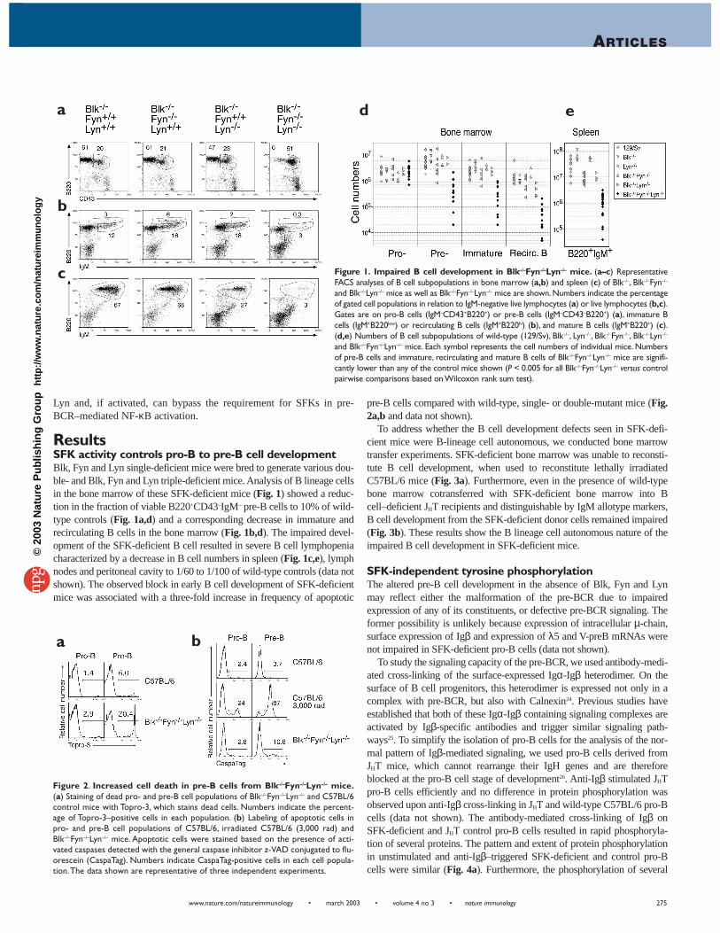
ResultsSFK activity controls pro-B to pre-B cell developmentBlk, Fyn and Lyn single-deficient mice were bred to generate various dou-ble- and Blk, Fyn and Lyn triple-deficient mice. Analysis of B lineage cellsin the bone marrow of these SFK-deficient mice (Fig. 1) showed a reduc-tion in the fraction of viable B220+CD43–IgM– pre-B cells to 10% of wild-type controls (Fig. 1a,d) and a corresponding decrease in immature andrecirculating B cells in the bone marrow (Fig. 1b,d). The impaired devel-opment of the SFK-deficient B cell resulted in severe B cell lymphopeniacharacterized by a decrease in B cell numbers in spleen (Fig. 1c,e), lymphnodes and peritoneal cavity to 1/60 to 1/100 of wild-type controls (data notshown). The observed block in early B cell development of SFK-deficientmice was associated with a three-fold increase in frequency of apoptotic
pre-B cells compared with wild-type, single- or double-mutant mice (Fig.2a,b and data not shown).
To address whether the B cell development defects seen in SFK-defi-cient mice were B-lineage cell autonomous, we conducted bone marrowtransfer experiments. SFK-deficient bone marrow was unable to reconsti-tute B cell development, when used to reconstitute lethally irradiatedC57BL/6 mice (Fig. 3a). Furthermore, even in the presence of wild-typebone marrow cotransferred with SFK-deficient bone marrow into Bcell–deficient JHT recipients and distinguishable by IgM allotype markers,B cell development from the SFK-deficient donor cells remained impaired(Fig. 3b). These results show the B lineage cell autonomous nature of theimpaired B cell development in SFK-deficient mice.
SFK-independent tyrosine phosphorylationThe altered pre-B cell development in the absence of Blk, Fyn and Lynmay reflect either the malformation of the pre-BCR due to impairedexpression of any of its constituents, or defective pre-BCR signaling. Theformer possibility is unlikely because expression of intracellular µ-chain,surface expression of Igβ and expression of λ5 and V-preB mRNAs werenot impaired in SFK-deficient pro-B cells (data not shown).
To study the signaling capacity of the pre-BCR, we used antibody-medi-ated cross-linking of the surface-expressed Igα-Igβ heterodimer. On thesurface of B cell progenitors, this heterodimer is expressed not only in acomplex with pre-BCR, but also with Calnexin24. Previous studies haveestablished that both of these Igα-Igβ containing signaling complexes areactivated by Igβ-specific antibodies and trigger similar signaling path-ways25. To simplify the isolation of pro-B cells for the analysis of the nor-mal pattern of Igβ-mediated signaling, we used pro-B cells derived fromJHT mice, which cannot rearrange their IgH genes and are thereforeblocked at the pro-B cell stage of development26. Anti-Igβ stimulated JHTpro-B cells efficiently and no difference in protein phosphorylation wasobserved upon anti-Igβ cross-linking in JHT and wild-type C57BL/6 pro-Bcells (data not shown). The antibody-mediated cross-linking of Igβ onSFK-deficient and JHT control pro-B cells resulted in rapid phosphoryla-tion of several proteins. The pattern and extent of protein phosphorylationin unstimulated and anti-Igβ–triggered SFK-deficient and control pro-Bcells were similar (Fig. 4a). Furthermore, the phosphorylation of several
Figure 2. Increased cell death in pre-B cells from Blk–/–Fyn–/–Lyn–/– mice.(a) Staining of dead pro- and pre-B cell populations of Blk–/–Fyn–/–Lyn–/– and C57BL/6control mice with Topro-3, which stains dead cells. Numbers indicate the percent-age of Topro-3–positive cells in each population. (b) Labeling of apoptotic cells inpro- and pre-B cell populations of C57BL/6, irradiated C57BL/6 (3,000 rad) andBlk–/–Fyn–/–Lyn–/– mice. Apoptotic cells were stained based on the presence of acti-vated caspases detected with the general caspase inhibitor z-VAD conjugated to flu-orescein (CaspaTag). Numbers indicate CaspaTag-positive cells in each cell popula-tion.The data shown are representative of three independent experiments.
a
b
a b
c
d e
Figure 1. Impaired B cell development in Blk–/–Fyn–/–Lyn–/– mice. (a–c) RepresentativeFACS analyses of B cell subpopulations in bone marrow (a,b) and spleen (c) of Blk–/–, Blk–/–Fyn–/–
and Blk–/–Lyn–/– mice as well as Blk–/–Fyn–/–Lyn–/– mice are shown. Numbers indicate the percentageof gated cell populations in relation to IgM-negative live lymphocytes (a) or live lymphocytes (b,c).Gates are on pro-B cells (IgM–CD43+B220+) or pre-B cells (IgM–CD43–B220+) (a), immature Bcells (IgM+B220low) or recirculating B cells (IgM+B220hi) (b), and mature B cells (IgM+B220+) (c).(d,e) Numbers of B cell subpopulations of wild-type (129/Sv), Blk–/–, Lyn–/–, Blk–/–Fyn–/–, Blk–/–Lyn–/–
and Blk–/–Fyn–/–Lyn–/– mice. Each symbol represents the cell numbers of individual mice. Numbersof pre-B cells and immature, recirculating and mature B cells of Blk–/–Fyn–/–Lyn–/– mice are signifi-cantly lower than any of the control mice shown (P < 0.005 for all Blk–/–Fyn–/–Lyn–/– versus controlpairwise comparisons based on Wilcoxon rank sum test).
©20
03 N
atu
re P
ub
lish
ing
Gro
up
h
ttp
://w
ww
.nat
ure
.co
m/n
atu
reim
mu
no
log
y
nature immunology • volume 4 no 3 • march 2003 • www.nature.com/natureimmunology
ARTICLES
276
substrates important for pre-BCR and BCR signaling was not impaired inthe absence of Blk, Fyn and Lyn. Phosphorylation of Syk at Tyr519 andTyr520, which is indicative of Syk activation27, was similar in anti-Igβ–stimulated SFK-deficient and JHT control pro-B cells. The anti-Igβ–induced phosphorylation of Btk, Vav and PLCγ-2 was also not alteredin SFK-deficient pro-B cells (Fig. 4b). Phosphorylation of Tyr482 andTyr513 in CD19 has been reported to be dependent on Lyn in mature Bcells28. However, the absence of Blk, Fyn and Lyn from pro-B cells did notaffect the phosphorylation of Tyr513 in CD19 (Fig. 4b).
SFK control pre-BCR induced NF-κB activationImpaired development, survival and activation of B cells have been previ-ously linked to changes in nuclear signaling mediated by NF-κB29.Therefore, we hypothesized that SFKs are involved in pre-BCR–mediatedactivation of NF-κB. The activation of NF-κB is initiated by activation ofthe IκB kinase (IKK) complex, which is composed of two catalytic sub-units, IKKα and IKKβ, and the regulatory subunit IKKγ30. BCR engage-ment activates IKKα and IKKβ followed by phosphorylation of the NF-κB–bound inhibitory protein IκB. Phosphorylation of IκB signals itsdegradation, thereby liberating NF-κB to translocate to the nucleus31. Theactivity of IKKα and IKKβ correlates directly with the degree of phos-phorylation of Ser180 and Ser181, which are localized within the kinasedomain activation loop of IKKα and IKKβ, respectively32. Using antibod-ies specific for IKKα and IKKβ phosphorylated at Ser180 and Ser181, wemeasured phosphorylation and activation of IKKα and IKKβ in JHT con-trol and SFK-deficient pro-B cells. IKKα and IKKβ were phosphorylatedin ex vivo isolated JHT control pro-B cells (Fig. 5a). Treatment of JHT con-trol pro-B cells with anti-Igβ increased phosphorylation of IKKβ, whereasphosphorylation of IKKα remained constant (Fig. 5a). In SFK-deficientpro-B cells, basal phosphorylation and anti-Igβ–induced IKKα and IKKβ
phosphorylation were substantially reduced compared with control cells(Fig. 5a). The lack of IKK phosphorylation in SFK-deficient cells corre-lated with the loss of IκBα phosphorylation and degradation (Fig. 5a). Noinducible NF-κB DNA binding activity was detected by electrophoreticmobility shift assay (EMSA) in nuclear lysates derived from anti-Igβ–stim-ulated SFK-deficient pro-B cells (Fig. 5b). To demonstrate specificity ofthe DNA binding activities observed, EMSA experiments were carried outwith specific antibodies to various NF-κB subunits. A RelA-p50 het-erodimer is induced in control JHT pro-B cells (Fig. 5c), whereas the DNAbinding activity present without activation in SFK-deficient pro-B cells(Fig. 5b) consisted of only p50 homodimer, but not RelA-p50 or other het-erodimers. These data demonstrate an essential role for Blk, Fyn and Lynin basal and Igβ-mediated NF-κB activation in pro-B cells.
Syk-independent pre-BCR–induced NF-κB activationThe loss of NF-κB activation in the absence of Blk, Fyn and Lyn raised aquestion about the specificity of the observed effect. Would other abla-tions of pre-BCR signaling essential for B cell development, like Syk defi-ciency7,8, also abrogate pre-BCR–induced NF-κB activation? To addressthis possibility, we studied NF-κB induction in pro-B cells derived fromSyk-deficient mice. Using standard techniques, Syk-deficient mice were
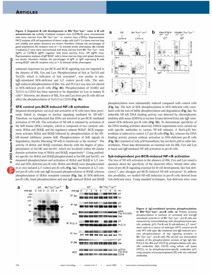
Figure 3. Impaired B cell development in Blk–/–Fyn–/–Lyn–/– mice is B cellautonomous. (a) Lethally irradiated recipient mice (C57BL/6) were reconstitutedwith bone marrow from Blk–/–Fyn–/–Lyn–/– or control mice (129/Sv). RepresentativeFACS analyses of B cell populations of donor origin cells (Ly9.1+) in bone marrow (topand middle) and spleen (bottom) are shown. Numbers indicate the percentages ofgated populations.All recipient mice (n = 5) showed similar phenotypes. (b) Lethallyirradiated JHT mice were reconstituted with bone marrow from Blk–/–Fyn–/–Lyn–/– mice(IgMb) or C57BL/6 (IgMb) together with bone marrow from 129 (IgMa) mice.Representative analyses of IgM+B220+ cells in bone marrow (top) and spleen (bottom)are shown. Numbers indicate the percentages of IgMa or IgMb expressing B cellsamong B220+ cells.All recipient mice (n = 3) showed similar phenotypes.
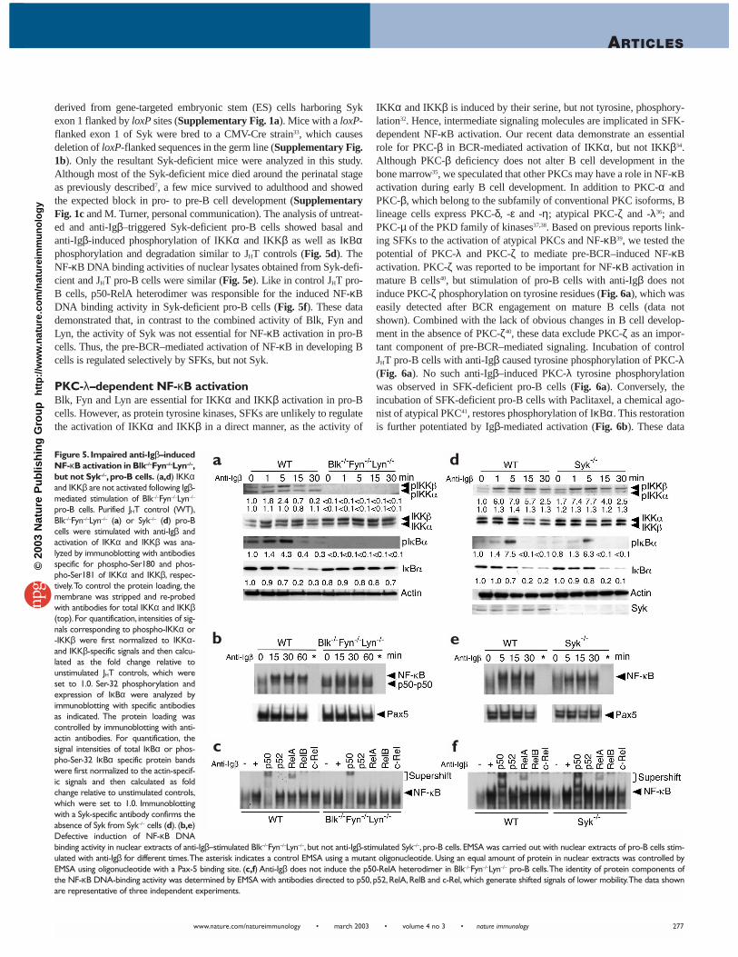
Figure 4. Igβ-mediated tyrosine phosphorylationin Blk–/–Fyn–/–Lyn–/– pro-B cells. (a) Protein tyrosinephosphorylation in extracts of untreated and anti-Igβstimulated controls or of Blk–/–Fyn–/–Lyn–/– pro-B cells wasmeasured by immunoblotting with phosphotyrosine-spe-cific antibody (pY). Pre-B and B cell-deficient JHT mice26
were used as a source of wild-type (WT) control pro-Bcells.WT, wild type. (b) Unaltered anti-Igβ–induced tyro-sine phosphorylation of key signaling proteins inBlk–/–Fyn–/–Lyn–/– pro-B cells. We carried out immunoblotanalysis of the phosphorylation content of Igα, Igβ, Syk,PLCγ-2,Vav, Btk and CD19 by phosphorylation-site–spe-cific antibodies (Syk, CD19) using whole cell lysate(WCL), or by phosphotyrosine-specific antibodies (pY)using samples immunoprecipitated (IP) with the indicatedantibodies.
a b
a b
©20
03 N
atu
re P
ub
lish
ing
Gro
up
h
ttp
://w
ww
.nat
ure
.co
m/n
atu
reim
mu
no
log
y
ARTICLES
www.nature.com/natureimmunology • march 2003 • volume 4 no 3 • nature immunology 277
derived from gene-targeted embryonic stem (ES) cells harboring Sykexon 1 flanked by loxP sites (Supplementary Fig. 1a). Mice with a loxP-flanked exon 1 of Syk were bred to a CMV-Cre strain33, which causesdeletion of loxP-flanked sequences in the germ line (Supplementary Fig.1b). Only the resultant Syk-deficient mice were analyzed in this study.Although most of the Syk-deficient mice died around the perinatal stageas previously described7, a few mice survived to adulthood and showedthe expected block in pro- to pre-B cell development (SupplementaryFig. 1c and M. Turner, personal communication). The analysis of untreat-ed and anti-Igβ–triggered Syk-deficient pro-B cells showed basal andanti-Igβ-induced phosphorylation of IKKα and IKKβ as well as IκBαphosphorylation and degradation similar to JHT controls (Fig. 5d). TheNF-κB DNA binding activities of nuclear lysates obtained from Syk-defi-cient and JHT pro-B cells were similar (Fig. 5e). Like in control JHT pro-B cells, p50-RelA heterodimer was responsible for the induced NF-κBDNA binding activity in Syk-deficient pro-B cells (Fig. 5f). These datademonstrated that, in contrast to the combined activity of Blk, Fyn andLyn, the activity of Syk was not essential for NF-κB activation in pro-Bcells. Thus, the pre-BCR–mediated activation of NF-κB in developing Bcells is regulated selectively by SFKs, but not Syk.
PKC-λ–dependent NF-κB activationBlk, Fyn and Lyn are essential for IKKα and IKKβ activation in pro-Bcells. However, as protein tyrosine kinases, SFKs are unlikely to regulatethe activation of IKKα and IKKβ in a direct manner, as the activity of
IKKα and IKKβ is induced by their serine, but not tyrosine, phosphory-lation32. Hence, intermediate signaling molecules are implicated in SFK-dependent NF-κB activation. Our recent data demonstrate an essentialrole for PKC-β in BCR-mediated activation of IKKα, but not IKKβ34.Although PKC-β deficiency does not alter B cell development in thebone marrow35, we speculated that other PKCs may have a role in NF-κBactivation during early B cell development. In addition to PKC-α andPKC-β, which belong to the subfamily of conventional PKC isoforms, Blineage cells express PKC-δ, -ε and -η; atypical PKC-ζ and -λ36; andPKC-µ of the PKD family of kinases37,38. Based on previous reports link-ing SFKs to the activation of atypical PKCs and NF-κB39, we tested thepotential of PKC-λ and PKC-ζ to mediate pre-BCR–induced NF-κBactivation. PKC-ζ was reported to be important for NF-κB activation inmature B cells40, but stimulation of pro-B cells with anti-Igβ does notinduce PKC-ζ phosphorylation on tyrosine residues (Fig. 6a), which waseasily detected after BCR engagement on mature B cells (data notshown). Combined with the lack of obvious changes in B cell develop-ment in the absence of PKC-ζ40, these data exclude PKC-ζ as an impor-tant component of pre-BCR–mediated signaling. Incubation of controlJHT pro-B cells with anti-Igβ caused tyrosine phosphorylation of PKC-λ(Fig. 6a). No such anti-Igβ–induced PKC-λ tyrosine phosphorylationwas observed in SFK-deficient pro-B cells (Fig. 6a). Conversely, theincubation of SFK-deficient pro-B cells with Paclitaxel, a chemical ago-nist of atypical PKC41, restores phosphorylation of IκBα. This restorationis further potentiated by Igβ-mediated activation (Fig. 6b). These data
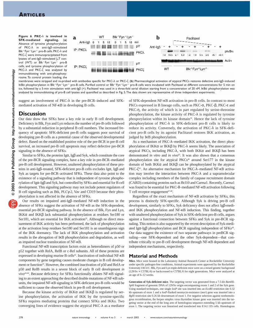
Figure 5. Impaired anti-Igβ–inducedNF-κB activation in Blk–/–Fyn–/–Lyn–/–,but not Syk–/–, pro-B cells. (a,d) IKKαand IKKβ are not activated following Igβ-mediated stimulation of Blk–/–Fyn–/–Lyn–/–
pro-B cells. Purified JHT control (WT),Blk–/–Fyn–/–Lyn–/– (a) or Syk–/– (d) pro-Bcells were stimulated with anti-Igβ andactivation of IKKα and IKKβ was ana-lyzed by immunoblotting with antibodiesspecific for phospho-Ser180 and phos-pho-Ser181 of IKKα and IKKβ, respec-tively.To control the protein loading, themembrane was stripped and re-probedwith antibodies for total IKKα and IKKβ(top). For quantification, intensities of sig-nals corresponding to phospho-IKKα or-IKKβ were first normalized to IKKα-and IKKβ-specific signals and then calcu-lated as the fold change relative tounstimulated JHT controls, which wereset to 1.0. Ser-32 phosphorylation andexpression of IκBα were analyzed byimmunoblotting with specific antibodiesas indicated. The protein loading wascontrolled by immunoblotting with anti-actin antibodies. For quantification, thesignal intensities of total IκBα or phos-pho-Ser-32 IκBα specific protein bandswere first normalized to the actin-specif-ic signals and then calculated as foldchange relative to unstimulated controls,which were set to 1.0. Immunoblottingwith a Syk-specific antibody confirms theabsence of Syk from Syk–/– cells (d). (b,e)Defective induction of NF-κB DNAbinding activity in nuclear extracts of anti-Igβ–stimulated Blk–/–Fyn–/–Lyn–/–, but not anti-Igβ-stimulated Syk–/–, pro-B cells. EMSA was carried out with nuclear extracts of pro-B cells stim-ulated with anti-Igβ for different times.The asterisk indicates a control EMSA using a mutant oligonucleotide. Using an equal amount of protein in nuclear extracts was controlled byEMSA using oligonucleotide with a Pax-5 binding site. (c,f) Anti-Igβ does not induce the p50-RelA heterodimer in Blk–/–Fyn–/–Lyn–/– pro-B cells.The identity of protein components ofthe NF-κB DNA-binding activity was determined by EMSA with antibodies directed to p50, p52, RelA, RelB and c-Rel, which generate shifted signals of lower mobility.The data shownare representative of three independent experiments.
a
b
c
d
e
f
©20
03 N
atu
re P
ub
lish
ing
Gro
up
h
ttp
://w
ww
.nat
ure
.co
m/n
atu
reim
mu
no
log
y
nature immunology • volume 4 no 3 • march 2003 • www.nature.com/natureimmunology
ARTICLES
278
suggest an involvement of PKC-λ in the pre-BCR–induced and SFK-mediated activation of NF-κB in developing B cells.
DiscussionOur data show that SFKs have a key role in early B cell development.Deficiency in Blk, Fyn and Lyn reduces the number of pre-B cells followedby a substantial reduction in peripheral B cell numbers. The increased fre-quency of apoptotic SFK-deficient pre-B cells suggests poor survival ofdeveloping pre-B cells as a potential cause of the observed developmentaldefect. Based on the established positive role of the pre-BCR in pre-B cellsurvival, an increased pre-B cell apoptosis may reflect defective pre-BCRsignaling in the absence of SFKs.
Similar to SFKs, the proteins Igα, Igβ and Syk, which constitute the coreof the pre-BCR signaling complex, have a key role in pre-BCR–mediatedpre-B cell development. However, unaltered phosphorylation of these pro-teins in anti-Igβ–treated, SFK-deficient pro-B cells excludes Igα, Igβ andSyk as targets for pre-BCR–activated SFKs. These data also point to theexistence of a signaling pathway that is independent of tyrosine phospho-rylation of Igα-Igβ and Syk, but controlled by SFKs and essential for B celldevelopment. This signaling pathway may not include potent regulators ofB cell signaling such as Btk, PLCγ-2, Vav and CD19 because their phos-phorylation is not affected in the absence of SFKs.
Our results on impaired anti-Igβ–mediated NF-κB induction in theabsence of SFKs suggest the activation of NF-κB as the SFK-dependent,essential pre-BCR signaling pathway. In the absence of Blk, Fyn and Lyn,IKKα and IKKβ lack substantial phosphorylation at residues Ser180 orSer181, which are essential for IKK activation32. Although no direct mea-surement of IKK activity has been performed, the lack of phosphorylationat the activation loop residues Ser180 and Ser181 is an unambiguous signof the IKK dormancy. The lack of IKK phosphorylation and activationresults in the abrogation of IκB phosphorylation and degradation, as wellas impaired nuclear translocation of NF-κB.
Functional NF-κB transcription factors exist as heterodimers of p50 orp52 together with RelA, RelB or c-Rel subunits. All of these proteins areexpressed in developing murine B cells42. Inactivation of individual NF-κBcomponents by gene targeting causes moderate changes in B cell develop-ment or function29. However, the combined inactivation of p50 and RelA orp50 and RelB results in a severe block of early B cell development invivo43,44. Because deficiency for SFKs functionally ablates NF-κB signal-ing to an extent approaching the described double mutations of NF-κB sub-units, the impaired NF-κB signaling in SFK-deficient pro-B cells would besufficient to cause the observed block in pre-B cell development.
Because the kinase activity of IKKα and IKKβ is regulated by ser-ine phosphorylation, the activation of IKK by the tyrosine-specificSFKs requires mediating proteins that connect SFKs and IKKs. Twoconverging lines of evidence suggest the atypical PKC-λ as a mediator
of SFK-dependent NF-κB activation in pro-B cells. In contrast to mostPKCs expressed in B lineage cells, such as PKC-α, PKC-β, PKC-ε andPKC-η, the activity of which is in part regulated by serine-threoninephosphorylation, the kinase activity of PKC-λ is regulated by tyrosinephosporylation within its kinase domain39. Hence the lack of tyrosinephosphorylation of PKC-λ in SFK-deficient pro-B cells is likely toreduce its activity. Conversely, the activation of PKC-λ in SFK-defi-cient pro-B cells by its agonist Paclitaxel restores IKK activation, asjudged by IκB phosphorylation.
As a mechanism of PKC-λ–mediated IKK activation, the direct phos-phorylation of IKKα or IKKβ by PKC-λ seems likely. The association ofatypical PKCs, including PKC-λ, with both IKKα and IKKβ has beendemonstrated in vitro and in vivo45. It was also shown that a consensusphosphorylation site for atypical PKCs46 around Ser177 in the kinasedomain of both IKKα and IKKβ can be phosphorylated by the atypicalPKC-ζ45. An alternative mechanism for PKC-λ–mediated NF-κB activa-tion may involve the interaction between PKC-λ and a supramolecularcomplex including members of the family of caspase recruitment domain(CARD) containing proteins such as Bcl10 and Carma1. Recently, Carma1was found to be essential for PKC-θ–mediated NF-κB activation followingT cell receptor engagement47,48.
Regardless of the exact mechanism of NF-κB activation by SFKs, thisprocess is distinctly SFK-specific. Although Syk is driving pre-B celldevelopment, similarly to SFKs, Syk deficiency does not affect Igβ-medi-ated IKK phosphorylation and NF-κB induction. This finding, togetherwith unaltered phosphorylation of Syk in SFK-deficient pro-B cells, arguesagainst a functional connection between SFKs and Syk in pre-BCR sig-naling. This notion is also supported by the recent description of Syk-medi-ated Igα-Igβ phosphorylation and BCR signaling independent of SFKs49.Our data suggest the existence of two separate pathways in preBCR sig-naling—one SFK-dependent and the other Syk-dependent—that con-tribute critically to pre-B cell development through NF-κB dependent andindependent mechanisms, respectively.
Material and MethodsMice. Mice were housed in the Laboratory Animal Research Center at Rockefeller Universityunder specific pathogen-free conditions. Animal experiments were approved by the RockefellerUniversity IACUC. Blk, Fyn and Lyn triple-deficient mice were on a mixed genetic background(129/Sv × C57BL/6) or backcrossed to C57BL/6 for eight generations. Mice were analyzed atan age of 6–12 weeks.
Generation of Syk-deficient mice. The targeting vector was generated from a 7.5-kb BamHI-SphI fragment of genomic DNA of 129/Sv origin encompassing exons 1 and 2 of the Syk gene.Using standard techniques, one single loxP site was inserted into an EcoRI restriction site 0.32kb upstream of exon 1 and a loxP-flanked neomycin-resistance (neor) gene was inserted into aBglII restriction site 0.26 kb downstream of exon 1. For negative selection against nonhomolo-gous recombination, the herpes simplex virus thymidine kinase gene was inserted into the tar-geting vector at the end of the long arm of homologous sequence extending 5 kb upstream ofexon 1. The targeting vector was linearized and transfected into E14.1 ES cells. Homologous
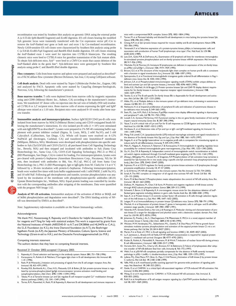
Figure 6. PKC-λ is involved inSFK-mediated signaling. (a)Absence of tyrosine phosphorylationof PKC-λ in anti-Igβ–stimulatedBlk–/–Fyn–/–Lyn–/– pro-B cells. PKC-λ andPKC-ζ were immunoprecipitated fromlysates of anti-Igβ–stimulated JHT con-trol (WT) or Blk–/–Fyn–/–Lyn–/– pro-Bcells, and tyrosine phosphorylation ofPKC-λ and PKC-ζ was analyzed byimmunoblotting with anti-phosphoty-rosine. To control protein loading, themembranes were stripped and re-probed with antibodies specific for PKC-λ or PKC-ζ. (b) Pharmacological activation of atypical PKCs restores defective anti-Igβ–inducedIκBα phosphorylation in Blk–/–Fyn–/–Lyn–/– pro-B cells. Purified control or Blk–/–Fyn–/–Lyn–/– pro-B cells were incubated with Paclitaxel at different concentrations for 5 min onice, followed by a 5-min stimulation with anti-Igβ (+). Paclitaxel was used in a three-fold serial dilution starting from a concentration of 20 nM. IκBα phosphorylation wasanalyzed by immunoblotting of pro-B cell lysates and quantified as described in Fig. 5.The data shown are representative of three independent experiments.
a b
©20
03 N
atu
re P
ub
lish
ing
Gro
up
h
ttp
://w
ww
.nat
ure
.co
m/n
atu
reim
mu
no
log
y
ARTICLES
www.nature.com/natureimmunology • march 2003 • volume 4 no 3 • nature immunology 279
recombination was tested by Southern blot analysis on genomic DNA using the external probeA (a 0.55-kb SphI-BamHI fragment) and EcoRI digestion. ES cell clones bearing the modifiedSyk genomic locus were transiently transfected with the Cre expression vector pIC-Cre toremove the loxP-flanked neor gene or both neor and exon 1 by Cre-mediated recombination.Newly G418-sensitive ES cell clones were characterized by Southern blot analysis using probeC (a 0.8-kb EcoRI-FspI fragment) and BamHI-XhoI double digestion. ES cell clones retainingthe loxP-flanked exon 1 were used for injection into C57BL/6 blastocysts. The resultingchimeric mice were bred to C57BL/6 mice for germline transmission of the Syk mutant allele.To obtain Syk-deficient mice, Sykfl/+ were bred to a CMV-Cre strain that causes deletion of theloxP-flanked allele in the germ line33. Syk-deficient mice were genotyped by Southern blotanalysis using probe C and BamHI-XhoI double digestion.
Flow cytometry. Cells from bone marrow and spleen were prepared and analyzed as described16
on a FACSCalibure flow cytometer (Becton Dickinson, San Jose, CA) using CellQuest software.
Cell death analysis. Dead cells were stained by Topro-3 (Molecular Probes Inc., Eugene, OR)and analyzed by FACS. Apoptotic cells were stained by CaspaTag (Intergen-Serologicals,Norcross, GA), following the manufacturer’s protocol.
Bone marrow transfer. T cells were depleted from bone marrow cells by magnetic separationusing anti-CD90 (Miltenyi Biotec Inc., Auburn, CA) according to the manufacturer’s instruc-tions. We transferred 107 donor cells via injection into the tail vein of lethally (950 rad) irradiat-ed C57BL/6 or JHT recipient mice. Bone marrow cells of strains expressing the IgMa and IgMb
allotype were mixed at a 1:10 ratio. Recipient mice were analyzed 8–10 weeks after bone mar-row transfer.
Immunoblot analysis and immunoprecipitation. Surface IgM–B220+CD43+pro-B cells wereisolated from bone marrow by MACS (Miltenyi Biotec) using anti-CD19 conjugated beads fol-lowing the manufacturer’s instructions. Ex vivo isolated pro-B cells (1 × 10⁶) were stimulatedwith anti-Igβ (HM79) as described24. Lysates were prepared in 1% NP-40 containing buffer sup-plement with protein inhibitor cocktail (Sigma, St. Louis, MO), 2 mM Na3VO4 and 5 nMCalyculinA (Calbiochem, San Diego, CA). Whole cell lysates were resolved on 4–12%NuPAGE gel (Invitrogen, Carlsbad, CA) and transferred onto Immobilon-P membrane(Millipore, Billerica, MA). Membranes were developed with phosphorylation site-specific anti-bodies to Syk (Tyr519/520) and CD19 (Tyr513; all purchased from Cell Signaling TechnologyInc., Beverly, MA) and then stripped and incubated with antibodies to Syk (Santa CruzBiotechnology Inc., Santa Cruz, CA), CD19 (Cell Signaling Technology), PKC-λ or PKC-ζ(BD Pharmingen, San Diego, CA) to control for loading. For immunoprecipitation, lysates werepre-cleared with protein-G-Sepharose (Amersham Biosciences Corp., Piscataway, NJ) for 30min, then incubated with antibodies to Btk, Vav, PLC-γ2, PKC-ζ (all from Santa CruzBiotechnology Inc.), PKC-λ (BD Pharmingen), Igα or Igβ (gifts from M. Clark) for 1 h at 4 °Cfollowed by a 30-min incubation with protein-G-Sepharose. Following immunoprecipitation,beads were washed five times with lysis buffer supplemented with 1 mM PMSF, 2 mM Na3VO4
and 10 mM NaF. Following gel electrophoresis and transfer, tyrosine phosphorylation was ana-lyzed by the incubation of the membrane with phosphotyrosine-specific antibodies (4G10;Upstate Biotechnologies Inc., Waltham, MA). Equal protein loading was controlled by incuba-tion with the corresponding antibodies after stripping of the membranes. Data were quantifiedwith the program NIH Image 1.62.
Analysis of NF-κB activation. Immunoblot analyses of the activation of IKKα or IKKβ, andof IκBα phosphorylation and degradation were described34. The DNA binding activity of NF-κB was determined by EMSA as described34.
Note: Supplementary information is available on the Nature Immunology website.
Acknowledgments
We thank M.C. Nussenzweig, K. Rajewsky and S. Desiderio for helpful discussions, M. Clarkfor reagents and Y.Yang for help with statistical analysis.This work is supported by grants fromthe National Institutes of Health (AI053545-01 to A.T. and DK58066 and HL54476 to C.A.L),the S.L.E. Foundation (to K.S.), the Irene Diamond foundation (to A.T.), the BoehringerIngelheim Fonds (to A.P.), the Japanese Ministry of Education, Culture, Sports, Science andTechnology (Grant-in-aid to H.K.), and the Deutsche Forschungsgemeinschaft (to M.R.).
Competing interests statementThe authors declare that they have no competing financial interests.
Received 21 October 2002; accepted 13 January 2003.
1. Rajewsky, K. Clonal selection and learning in the antibody system. Nature 381, 751–758 (1996).2. Karasuyama, H., Rolink,A. & Melchers, F. Surrogate light chain in B cell development. Adv. Immunol. 63,
1–41 (1996).3. Reth, M. & Wienands, J. Initiation and processing of signals from the B cell antigen receptor. Annu. Rev.
Immunol. 15, 453–479 (1997).4. Rowley, R.B., Burkhardt,A.L., Chao, H.G., Matsueda, G.R. & Bolen, J.B. Syk protein-tyrosine kinase is regu-
lated by tyrosine-phosphorylated Iga/Igb immunoreceptor tyrosine activation motif binding andautophosphorylation. J. Biol. Chem. 270, 11590–11594 (1995).
5. Takata, M. et al.Tyrosine kinases Lyn and Syk regulate B cell receptor-coupled Ca2+ mobilization throughdistinct pathways. EMBO J. 13, 1341–1349 (1994).
6. Torres, R.M., Flaswinkel, H., Reth, M. & Rajewsky, K.Aberrant B cell development and immune response in
mice with a compromised BCR complex. Science 272, 1802–1804 (1996).7. Turner, M. et al. Perinatal lethality and blocked B-cell development in mice lacking the tyrosine kinase Syk.
Nature 378, 298–302 (1995).8. Cheng,A.M. et al. Syk tyrosine kinase required for mouse viability and B-cell development. Nature 378,
303–306 (1995).9. Yamanashi,Y. et al. Selective expression of a protein-tyrosine kinase, p56lyn, in hematopoietic cells and
association with production of human T-cell lymphotropic virus type I. Proc. Natl.Acad. Sci. USA 86,6538–6542 (1989).
10. Law, D.A., Gold, M.R. & DeFranco,A.L. Examination of B lymphoid cell lines for membrane immunoglobu-lin-stimulated tyrosine phosphorylation and src-family tyrosine kinase mRNA expression. Mol. Immunol.29, 917–926 (1992).
11. Wechsler, R.J. & Monroe, J.G. Immature B lymphocytes are deficient in expression of the src-family kinas-es p59fyn and p55fgr1. J. Immunol. 154, 1919–1929 (1995).
12. Brouns, G.S. et al.The structure of the mu/pseudo light chain complex on human pre-B cells is consistentwith a function in signal transduction. Eur. J. Immunol. 23, 1088–1097 (1993).
13. Spanopoulou, E. et al. Functional immunoglobulin transgenes guide ordered B-cell differentiation in Rag-1-deficient mice. Genes Dev. 8, 1030–1042 (1994).
14. Johnson, S.A. et al. Phosphorylated immunoreceptor signaling motifs (ITAMs) exhibit unique abilities tobind and activate Lyn and Syk tyrosine kinases. J. Immunol. 155, 4596–4603 (1995).
15. Zoller, K.E., MacNeil, I.A. & Brugge, J.S. Protein tyrosine kinases Syk and ZAP-70 display distinct require-ments for Src family kinases in immune response receptor signal transduction. J. Immunol. 158,1650–1659 (1997).
16. Texido, G. et al.The B-cell-specific Src-family kinase Blk is dispensable for B-cell development and activa-tion. Mol. Cell. Biol. 20, 1227–1233 (2000).
17. Hibbs, M.L. et al. Multiple defects in the immune system of Lyn-deficient mice, culminating in autoimmunedisease. Cell 83, 301–311 (1995).
18. Nishizumi, H. et al. Impaired proliferation of peripheral B cells and indication of autoimmune disease inlyn-deficient mice. Immunity 3, 549–560 (1995).
19. Stein, P.L., Lee, H.M., Rich, S. & Soriano, P. pp59fyn mutant mice display differential signaling in thymocytesand peripheral T cells. Cell 70, 741–750 (1992).
20. Lowell, C.A., Soriano, P. & Varmus, H.E. Functional overlap in the src gene family: inactivation of hck and fgrimpairs natural immunity. Genes Dev. 8, 387–398 (1994).
21. Yasue,T. et al.A critical role of Lyn and Fyn for B cell responses to CD38 ligation and interleukin 5. Proc.Natl.Acad. Sci. USA 94, 10307–10312 (1997).
22. Horikawa, K. et al. Distinctive roles of Fyn and Lyn in IgD- and IgM-mediated signaling. Int. Immunol. 11,1441–1449 (1999).
23. Meng, F. & Lowell, C.A. Lipopolysaccharide (LPS)-induced macrophage activation and signal transduction inthe absence of Src-family kinases Hck, Fgr, and Lyn. J. Exp. Med. 185, 1661–1670 (1997).
24. Nagata, K. et al.The Iga/Igb heterodimer on m-negative proB cells is competent for transducing signals toinduce early B cell differentiation. Immunity 7, 559–570 (1997).
25. Maki, K., Nagata, K., Kitamura, F.,Takemori,T. & Karasuyama, H. Immunoglobulin b signaling regulates locusaccessibility for ordered immunoglobulin gene rearrangements. J. Exp. Med. 191, 1333–1340 (2000).
26. Gu, H., Zou,Y.R. & Rajewsky, K. Independent control of immunoglobulin switch recombination at individ-ual switch regions evidenced through Cre-loxP-mediated gene targeting. Cell 73, 1155–1164 (1993).
27. Zhang, J., Billingsley, M.L., Kincaid, R.L. & Siraganian, R.P. Phosphorylation of Syk activation loop tyrosines isessential for Syk function.An in vivo study using a specific anti-Syk activation loop phosphotyrosine anti-body. J. Biol. Chem. 275, 35442–35447 (2000).
28. Fujimoto, M. et al. CD19 regulates Src family protein tyrosine kinase activation in B lymphocytes throughprocessive amplification. Immunity 13, 47–57 (2000).
29. Li, Q. & Verma, I.M. NF-κB regulation in the immune system. Nat. Rev. Immunol. 2, 725–734 (2002).30. Israel,A.The IKK complex: an integrator of all signals that activate NF-κB? Trends Cell Biol. 10,
129–133 (2000).31. Karin, M. & Ben-Neriah,Y. Phosphorylation meets ubiquitination: the control of NF-κB activity. Annu. Rev.
Immunol. 18, 621–663 (2000).32. Delhase, M., Hayakawa, M., Chen,Y. & Karin, M. Positive and negative regulation of IκB kinase activity
through IKKβ subunit phosphorylation. Science 284, 309–313 (1999).33. Schwenk, F., Baron, U. & Rajewsky, K.A cre-transgenic mouse strain for the ubiquitous deletion of loxP-
flanked gene segments including deletion in germ cells. Nucleic Acids Res. 23, 5080–5081 (1995).34. Saijo, K. et al. Protein kinase C β controls nuclear factor κB activation in B cells through selective regula-
tion of the IκB kinase α. J. Exp. Med. 195, 1647–1652 (2002).35. Leitges, M. et al. Immunodeficiency in protein kinase Cβ-deficient mice. Science 273, 788–791 (1996).36. Mischak, H. et al. Expression of protein kinase C genes in hemopoietic cells is cell-type- and B cell-differ-
entiation stage specific. J. Immunol. 147, 3981–3987 (1991).37. Valverde,A.M., Sinnett-Smith, J.,Van Lint, J. & Rozengurt, E. Molecular cloning and characterization of pro-
tein kinase D: a target for diacylglycerol and phorbol esters with a distinctive catalytic domain. Proc. Natl.Acad. Sci. USA 91, 8572–8576 (1994).
38. Johannes, F.J., Prestle, J., Eis, S., Oberhagemann, P. & Pfizenmaier, K. PKCm is a novel, atypical member ofthe protein kinase C family. J. Biol. Chem. 269, 6140–6148 (1994).
39. Wooten, M.W.,Vandenplas, M.L., Seibenhener, M.L., Geetha,T. & Diaz-Meco, M.T. Nerve growth factorstimulates multisite tyrosine phosphorylation and activation of the atypical protein kinase C’s via a srckinase pathway. Mol. Cell. Biol. 21, 8414–8427 (2001).
40. Martin, P. et al. Role of ζ PKC in B-cell signaling and function. EMBO J. 21, 4049–4057 (2002).41. Lu,Y., Jamieson, L., Brasier,A.R. & Fields,A.P. NF-κB/RelA transactivation is required for atypical protein
kinase C ι-mediated cell survival. Oncogene 20, 4777–4792 (2001).42. Kistler, B., Rolink,A., Marienfeld, R., Neumann, M. & Wirth,T. Induction of nuclear factor-κB during primary
B cell differentiation. J. Immunol. 160, 2308–2317 (1998).43. Horwitz, B.H., Scott, M.L., Cherry, S.R., Bronson, R.T. & Baltimore, D. Failure of lymphopoiesis after adop-
tive transfer of NF-κB-deficient fetal liver cells. Immunity 6, 765–772 (1997).44. Weih, F. et al. p50-NF-κB complexes partially compensate for the absence of RelB: severely increased
pathology in p50(–/–)relB(–/–) double-knockout mice. J. Exp. Med. 185, 1359–1370 (1997).45. Lallena, M.J., Diaz-Meco, M.T., Bren, G., Paya, C.V. & Moscat, J.Activation of IκB kinase β by protein kinase
C isoforms. Mol. Cell. Biol. 19, 2180–2188 (1999).46. Yaffe, M.B. et al.A motif-based profile scanning approach for genome-wide prediction of signaling path-
ways. Nat. Biotechnol. 19, 348–353 (2001).47. Gaide, O. et al. CARMA1 is a critical lipid raft-associated regulator of TCR-induced NF-κB activation. Nat.
Immunol. 3, 836–843 (2002).48. Wang, D. et al.A requirement for CARMA1 in TCR-induced NF-κB activation. Nat. Immunol. 3,
830–835 (2002).49. Rolli,V. et al. Amplification of B cell antigen receptor signaling by a Syk/ITAM positive feedback loop. Mol.
Cell 10, 1057–1069 (2002).
©20
03 N
atu
re P
ub
lish
ing
Gro
up
h
ttp
://w
ww
.nat
ure
.co
m/n
atu
reim
mu
no
log
y