Differing roles of pyruvate dehydrogenase kinases...
11
RESEARCH ARTICLE Differing roles of pyruvate dehydrogenase kinases during mouse oocyte maturation Xiaojing Hou 1, *, Liang Zhang 1,2, *, Longsen Han 1 , Juan Ge 1 , Rujun Ma 1,2 , Xuesen Zhang 1 , Kelle Moley 3 , Tim Schedl 4 and Qiang Wang 1, ‡ ABSTRACT Pyruvate dehydrogenase kinases (PDKs) modulate energy homeostasis in multiple tissues and cell types, under various nutrient conditions, through phosphorylation of the α subunit (PDHE1α, also known as PDHA1) of the pyruvate dehydrogenase (PDH) complex. However, the roles of PDKs in meiotic maturation are currently unknown. Here, by undertaking knockdown and overexpression analysis of PDK paralogs (PDK1–PDK4) in mouse oocytes, we established the site-specificity of PDKs towards the phosphorylation of three serine residues (Ser232, Ser293 and Ser300) on PDHE1α. We found that PDK3-mediated phosphorylation of Ser293-PDHE1α results in disruption of meiotic spindle morphology and chromosome alignment and decreased total ATP levels, probably through inhibition of PDH activity. Unexpectedly, we discovered that PDK1 and PDK2 promote meiotic maturation, as their knockdown disturbs the assembly of the meiotic apparatus, without significantly altering ATP content. Moreover, phosphorylation of Ser232-PDHE1α was demonstrated to mediate PDK1 and PDK2 action in meiotic maturation, possibly through a mechanism that is distinct from PDH inactivation. These findings reveal that there are divergent roles of PDKs during oocyte maturation and indicate a new mechanism controlling meiotic structure. KEY WORDS: Pyruvate dehydrogenase kinase, Oocyte, Meiosis, Metabolism, Spindle INTRODUCTION Oocyte development is key in establishing female fertility. At around the time of birth, mammalian oocytes are arrested in prophase of the first meiotic division, which is also called the germinal vesicle stage. Following a stimulation by luteinizing hormone at puberty, immature fully-grown oocytes reinitiate meiosis, characterized by germinal vesicle breakdown (GVBD). With the chromatin condensation and microtubule organization, the oocytes proceed through the meiosis I (MI) division, extruding the first polar body (Pb1). Oocytes then become arrested at metaphase II (MII), waiting for fertilization (Jones and Lane, 2013; Wang and Sun, 2007). Accurate control of spindle assembly and chromosome alignment during meiotic maturation is necessary to produce a healthy oocyte. Errors in this process cause aneuploid eggs, which is a major cause of miscarriage and birth defects in humans (Hassold and Hunt, 2001). Balanced and efficient metabolism is crucial for the execution of oocyte and subsequent embryo development. Throughout the period of oocyte growth, pyruvate and oxygen consumption is markedly increased (Harris et al., 2009). Fully-grown oocytes have limited capacity to utilize glucose (Biggers et al., 1967; Saito et al., 1994; Zuelke and Brackett, 1992); instead, glucose first needs to be transferred to granulosa cells and then metabolized into pyruvate to support oocyte maturation (Eppig, 1976; Fagbohun and Downs, 1992). Oocyte-specific deletion of mouse PDHA1, the gene encoding the PDHE1α subunit of the pyruvate dehydrogenase (PDH) complex, results in compromised energetic status and meiotic defects in mouse oocytes (Johnson et al., 2007), further highlighting the importance of pyruvate metabolism during oocyte development. PDH is able to convert pyruvate into acetyl coenzyme A (acetyl- CoA) and thereby modulates the entry of glucose-derived carbons into the tricarboxylic acid (TCA) cycle. The TCA cycle provides an efficient means of producing ATP and many biosynthetic intermediates. The PDH complex is composed of three catalytic enzymes (E1, E2 and E3) and their regulatory proteins. The activity of PDH complex is under the control of two classes of enzymes, pyruvate dehydrogenase kinase (PDK) and pyruvate dehydrogenase phosphatase (PDP), through a reversible phosphorylation– dephosphorylation cycle (McFate et al., 2008; Sugden and Holness, 2006). Studies in somatic cells indicate that PDH activity is inhibited by site-specific phosphorylation at three serine residues of the E1 α subunit (PDHE1α): Ser232, Ser293 and Ser300 (Hitosugi et al., 2011; Korotchkina and Patel, 2001; Rardin et al., 2009). To date, four paralogous PDK genes (PDK1– PDK4) have been identified in mammals (Gudi et al., 1995; Tso et al., 2014). PDK1 expression is limited to heart and PDK3 is mainly found in testis, whereas PDK2 can be detected in many tissues. PDK4 is the most abundant of the PDKs in heart and skeletal muscle (Bowker-Kinley et al., 1998; Gudi et al., 1995; Korotchkina and Patel, 2001). These findings suggest that different PDK isoforms are utilized to meet the tissue- or cell-type-specific requirements for proper tuning of PDH activity. Some information is known about how PDK activity is regulated from biochemical or structural analysis and expression studies. For example, through binding to the inner lipoyl domain of the PDH E2 catalytic subunit, dihydrolipoyl acetyltransferase, PDKs have been showed to access their PDH-E2-bound PDH substrate. In the case of PDK2, ADP dissociation limits the PDH-E2-enhanced activity of PDK2, and pyruvate binding to the regulatory domain could further inhibit this process. NADH and acetyl-CoA stimulates PDK2 activity through a decrease in acetylation of lipoyl group and the resultant increase in ADP dissociation rate (for a review see Roche and Hiromasa, 2007). PDK4 is regulated, in part, through differences in gene transcription, as in a number of cell types the gene is transcriptionally inactive, through histones on the PDK4 gene being in the non-acetylated state (Kwon and Harris, 2004; Kwon et al., 2006). Received 5 December 2014; Accepted 28 April 2015 1 State Key Laboratory of Reproductive Medicine, Nanjing Medical University, Nanjing 210029, China. 2 College of Animal Science & Technology, Nanjing Agricultural University, Nanjing 210095, China. 3 Department of Obstetrics and Gynecology, Washington University School of Medicine, St Louis, MO 63110, USA. 4 Department of Genetics, Washington University School of Medicine, St Louis, MO 63110, USA. *These authors contributed equally to this work ‡ Author for correspondence ([email protected]) 2319 © 2015. Published by The Company of Biologists Ltd | Journal of Cell Science (2015) 128, 2319-2329 doi:10.1242/jcs.167049 Journal of Cell Science
Transcript of Differing roles of pyruvate dehydrogenase kinases...

RESEARCH ARTICLE
Differing roles of pyruvate dehydrogenase kinases during mouseoocyte maturationXiaojing Hou1,*, Liang Zhang1,2,*, Longsen Han1, Juan Ge1, Rujun Ma1,2, Xuesen Zhang1, Kelle Moley3,Tim Schedl4 and Qiang Wang1,‡
ABSTRACTPyruvate dehydrogenase kinases (PDKs) modulate energyhomeostasis in multiple tissues and cell types, under various nutrientconditions, through phosphorylation of the α subunit (PDHE1α, alsoknown as PDHA1) of the pyruvate dehydrogenase (PDH) complex.However, the roles of PDKs in meiotic maturation are currentlyunknown. Here, by undertaking knockdown and overexpressionanalysis of PDK paralogs (PDK1–PDK4) in mouse oocytes, weestablished the site-specificity of PDKs towards the phosphorylationof three serine residues (Ser232, Ser293andSer300) onPDHE1α.Wefound that PDK3-mediated phosphorylationofSer293-PDHE1α resultsindisruptionofmeiotic spindlemorphologyandchromosomealignmentand decreased total ATP levels, probably through inhibition of PDHactivity. Unexpectedly, we discovered that PDK1 and PDK2 promotemeiotic maturation, as their knockdown disturbs the assembly of themeiotic apparatus,without significantly alteringATPcontent.Moreover,phosphorylation of Ser232-PDHE1α was demonstrated to mediatePDK1 and PDK2 action in meiotic maturation, possibly through amechanism that is distinct fromPDH inactivation. These findings revealthat there are divergent roles of PDKs during oocyte maturation andindicate a new mechanism controlling meiotic structure.
KEY WORDS: Pyruvate dehydrogenase kinase, Oocyte, Meiosis,Metabolism, Spindle
INTRODUCTIONOocyte development is key in establishing female fertility. At aroundthe time of birth, mammalian oocytes are arrested in prophase of thefirst meiotic division, which is also called the germinal vesicle stage.Following a stimulation by luteinizing hormone at puberty, immaturefully-grown oocytes reinitiate meiosis, characterized by germinalvesicle breakdown (GVBD). With the chromatin condensation andmicrotubule organization, the oocytes proceed through the meiosis I(MI) division, extruding the first polar body (Pb1). Oocytes thenbecome arrested at metaphase II (MII), waiting for fertilization(Jones and Lane, 2013; Wang and Sun, 2007). Accurate control ofspindle assembly and chromosome alignment during meioticmaturation is necessary to produce a healthy oocyte. Errors in thisprocess cause aneuploid eggs, which is a major cause of miscarriageand birth defects in humans (Hassold and Hunt, 2001).
Balanced and efficient metabolism is crucial for the execution ofoocyte and subsequent embryo development. Throughout the periodof oocyte growth, pyruvate and oxygen consumption is markedlyincreased (Harris et al., 2009). Fully-grown oocytes have limitedcapacity to utilize glucose (Biggers et al., 1967; Saito et al., 1994;Zuelke and Brackett, 1992); instead, glucose first needs to betransferred to granulosa cells and then metabolized into pyruvate tosupport oocyte maturation (Eppig, 1976; Fagbohun and Downs,1992). Oocyte-specific deletion of mouse PDHA1, the geneencoding the PDHE1α subunit of the pyruvate dehydrogenase(PDH) complex, results in compromised energetic status andmeioticdefects in mouse oocytes (Johnson et al., 2007), further highlightingthe importance of pyruvate metabolism during oocyte development.
PDH is able to convert pyruvate into acetyl coenzyme A (acetyl-CoA) and thereby modulates the entry of glucose-derived carbonsinto the tricarboxylic acid (TCA) cycle. The TCA cycle provides anefficient means of producing ATP and many biosyntheticintermediates. The PDH complex is composed of three catalyticenzymes (E1, E2 and E3) and their regulatory proteins. The activityof PDH complex is under the control of two classes of enzymes,pyruvate dehydrogenase kinase (PDK) and pyruvate dehydrogenasephosphatase (PDP), through a reversible phosphorylation–dephosphorylation cycle (McFate et al., 2008; Sugden andHolness, 2006). Studies in somatic cells indicate that PDHactivity is inhibited by site-specific phosphorylation at threeserine residues of the E1 α subunit (PDHE1α): Ser232, Ser293and Ser300 (Hitosugi et al., 2011; Korotchkina and Patel, 2001;Rardin et al., 2009). To date, four paralogous PDK genes (PDK1–PDK4) have been identified in mammals (Gudi et al., 1995; Tsoet al., 2014). PDK1 expression is limited to heart and PDK3 ismainly found in testis, whereas PDK2 can be detected in manytissues. PDK4 is the most abundant of the PDKs in heart andskeletal muscle (Bowker-Kinley et al., 1998; Gudi et al., 1995;Korotchkina and Patel, 2001). These findings suggest that differentPDK isoforms are utilized to meet the tissue- or cell-type-specificrequirements for proper tuning of PDH activity. Some informationis known about how PDK activity is regulated from biochemical orstructural analysis and expression studies. For example, throughbinding to the inner lipoyl domain of the PDH E2 catalytic subunit,dihydrolipoyl acetyltransferase, PDKs have been showed to accesstheir PDH-E2-bound PDH substrate. In the case of PDK2, ADPdissociation limits the PDH-E2-enhanced activity of PDK2, andpyruvate binding to the regulatory domain could further inhibit thisprocess. NADH and acetyl-CoA stimulates PDK2 activity through adecrease in acetylation of lipoyl group and the resultant increase inADP dissociation rate (for a review see Roche and Hiromasa, 2007).PDK4 is regulated, in part, through differences in gene transcription,as in a number of cell types the gene is transcriptionally inactive,through histones on the PDK4 gene being in the non-acetylated state(Kwon and Harris, 2004; Kwon et al., 2006).Received 5 December 2014; Accepted 28 April 2015
1State Key Laboratory of Reproductive Medicine, Nanjing Medical University,Nanjing 210029, China. 2College of Animal Science & Technology, NanjingAgricultural University, Nanjing 210095, China. 3Department of Obstetrics andGynecology, Washington University School of Medicine, St Louis, MO 63110, USA.4Department of Genetics, Washington University School of Medicine, St Louis, MO63110, USA.*These authors contributed equally to this work
‡Author for correspondence ([email protected])
2319
© 2015. Published by The Company of Biologists Ltd | Journal of Cell Science (2015) 128, 2319-2329 doi:10.1242/jcs.167049
Journal
ofCe
llScience
The pivotal roles of PDKs in energy homeostasis in diversetissues and under various nutrient conditions have been extensivelyreported (Hitosugi et al., 2011; Jeoung and Harris, 2008; Kim et al.,2006; Michelakis et al., 2010; Takubo et al., 2013; Zhang et al.,2014). For example, PDK4 is involved in the fatty acid utilization inmuscle by interacting with cluster of differentiation 36 (CD36) andFoxO1 (Nahle et al., 2008). PDK2 is essential for the maintenanceof cell cycle quiescence and glycolytic metabolic status inhematopoietic stem cells (Takubo et al., 2013). It has recentlybeen found that metabolic perturbations result in significant meioticdefects in mouse oocytes (Luzzo et al., 2012; Wang et al., 2009; Wuet al., 2015), which might be through the action of PDKs on PDH.However, up to now, it is not known which PDKs are expressed andwhat are their functions in the maturation of the meiotic oocyte,even in the unperturbed metabolic state.In this study, by employing a morpholino (MO) knockdown
screen and overexpression analysis, we discovered distinct roles forPDK paralogs during mouse oocyte maturation, particularly incontrolling meiotic spindle structure and metabolic activity, andreport our findings below.
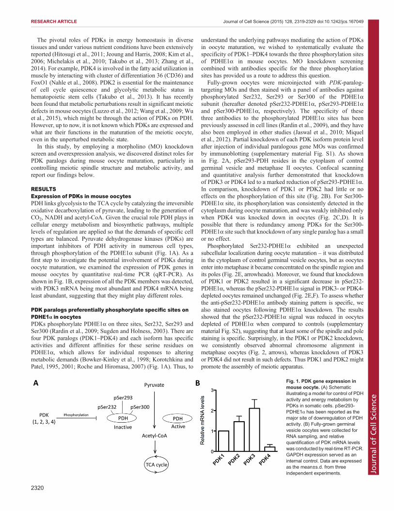
RESULTSExpression of PDKs in mouse oocytesPDH links glycolysis to the TCA cycle by catalyzing the irreversibleoxidative decarboxylation of pyruvate, leading to the generation ofCO2, NADH and acetyl-CoA. Given the crucial role PDH plays incellular energy metabolism and biosynthetic pathways, multiplelevels of regulation are applied so that the demands of specific celltypes are balanced. Pyruvate dehydrogenase kinases (PDKs) areimportant inhibitors of PDH activity in numerous cell types,through phosphorylation of the PDHE1α subunit (Fig. 1A). As afirst step to investigate the potential involvement of PDKs duringoocyte maturation, we examined the expression of PDK genes inmouse oocytes by quantitative real-time PCR (qRT-PCR). Asshown in Fig. 1B, expression of all the PDKmembers was detected,with PDK3 mRNA being most abundant and PDK4 mRNA beingleast abundant, suggesting that they might play different roles.
PDK paralogs preferentially phosphorylate specific sites onPDHE1α in oocytesPDKs phosphorylate PDHE1α on three sites, Ser232, Ser293 andSer300 (Rardin et al., 2009; Sugden and Holness, 2003). There arefour PDK paralogs (PDK1–PDK4) and each isoform has specificactivities and different affinities for these serine residues onPDHE1α, which allows for individual responses to alteringmetabolic demands (Bowker-Kinley et al., 1998; Korotchkina andPatel, 1995, 2001; Roche and Hiromasa, 2007) (Fig. 1A). Thus, to
understand the underlying pathways mediating the action of PDKsin oocyte maturation, we wished to systematically evaluate thespecificity of PDK1–PDK4 towards the three phosphorylation sitesof PDHE1α in mouse oocytes. MO knockdown screeningcombined with antibodies specific for the three phosphorylationsites has provided us a route to address this question.
Fully-grown oocytes were microinjected with PDK-paralog-targeting MOs and then stained with a panel of antibodies againstphosphorylated Ser232, Ser293 or Ser300 of the PDHE1αsubunit (hereafter denoted pSer232-PDHE1α, pSer293-PDHE1αand pSer300-PDHE1α, respectively). The specificity of thesethree antibodies to the phosphorylated PDHE1α sites has beenpreviously assessed in cell lines (Rardin et al., 2009), and they havealso been employed in other studies (Jaswal et al., 2010; Miquelet al., 2012). Partial knockdown of each PDK isoform protein levelafter injection of individual paralogous gene MOs was confirmedby immunoblotting (supplementary material Fig. S1). As shownin Fig. 2A, pSer293-PDH resides in the cytoplasm of controlgerminal vesicle and metaphase II oocytes. Confocal scanningand quantitative analysis further demonstrated that knockdownof PDK3 or PDK4 led to a marked reduction of pSer293-PDHE1α.In comparison, knockdown of PDK1 or PDK2 had little or noeffects on the phosphorylation of this site (Fig. 2B). For Ser300-PDHE1α site, its phosphorylation was consistently detected in thecytoplasm during oocyte maturation, and was weakly inhibited onlywhen PDK4 was knocked down in oocytes (Fig. 2C,D). It ispossible that there is redundancy among PDKs for the Ser300-PDHE1α site such that knockdown of any single paralog has a smallor no effect.
Phosphorylated Ser232-PDHE1α exhibited an unexpectedsubcellular localization during oocyte maturation – it was distributedin the cytoplasm of control germinal vesicle oocytes, but as oocytesenter into metaphase it became concentrated on the spindle region andits poles (Fig. 2E, arrowheads). Moreover, we found that knockdownof PDK1 or PDK2 resulted in a significant decrease in pSer232-PDHE1α, whereas the pSer232-PDHE1α signal in PDK3- or PDK4-depleted oocytes remained unchanged (Fig. 2E,F). To assess whetherthe anti-pSer232-PDHE1α antibody staining pattern is specific, wealso stained oocytes following PDHE1α knockdown. The resultsshowed that the pSer232-PDHE1α signal was reduced in oocytesdepleted of PDHE1α when compared to controls (supplementarymaterial Fig. S2), suggesting that at least some of the spindle and polestaining is specific. Surprisingly, in the PDK1 or PDK2 knockdown,we consistently observed abnormal chromosome alignment inmetaphase oocytes (Fig. 2, arrows), whereas knockdown of PDK3or PDK4 did not result in such defects. Thus PDK1 and PDK2 mightpromote the assembly of meiotic apparatus.
Fig. 1. PDK gene expression inmouse oocyte. (A) Schematicillustrating a model for control of PDHactivity and energy metabolism byPDKs in somatic cells. pSer293-PDHE1α has been reported as themajor site of downregulation of PDHactivity. (B) Fully-grown germinalvesicle oocytes were collected forRNA sampling, and relativequantification of PDK mRNA levelswas conducted by real-time RT-PCR.GAPDH expression served as aninternal control. Data are expressedas the mean±s.d. from threeindependent experiments.
2320
RESEARCH ARTICLE Journal of Cell Science (2015) 128, 2319-2329 doi:10.1242/jcs.167049
Journal
ofCe
llScience
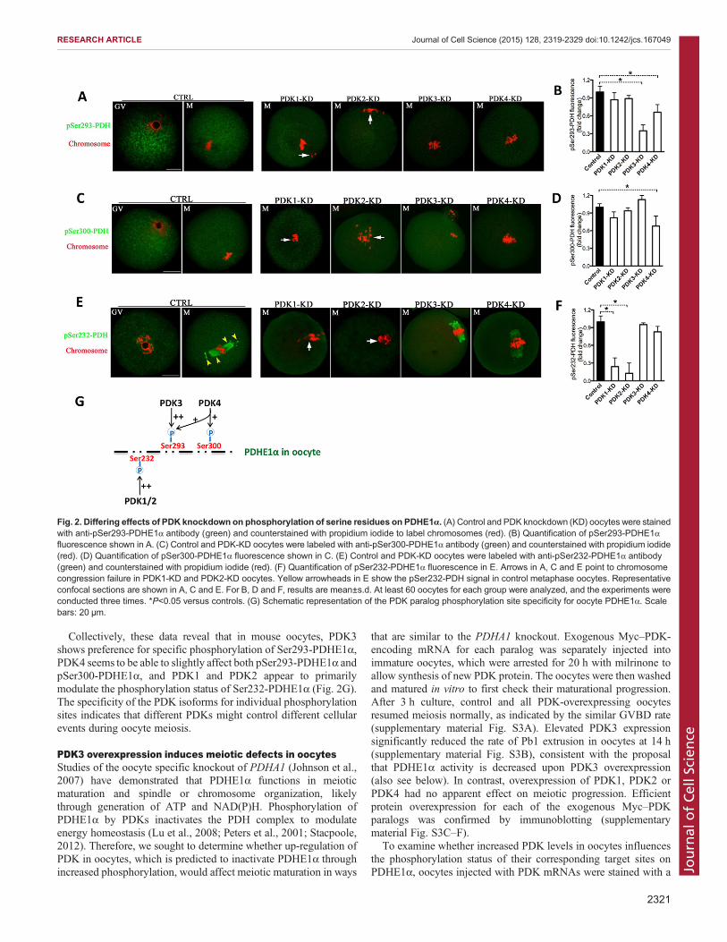
Collectively, these data reveal that in mouse oocytes, PDK3shows preference for specific phosphorylation of Ser293-PDHE1α,PDK4 seems to be able to slightly affect both pSer293-PDHE1α andpSer300-PDHE1α, and PDK1 and PDK2 appear to primarilymodulate the phosphorylation status of Ser232-PDHE1α (Fig. 2G).The specificity of the PDK isoforms for individual phosphorylationsites indicates that different PDKs might control different cellularevents during oocyte meiosis.
PDK3 overexpression induces meiotic defects in oocytesStudies of the oocyte specific knockout of PDHA1 (Johnson et al.,2007) have demonstrated that PDHE1α functions in meioticmaturation and spindle or chromosome organization, likelythrough generation of ATP and NAD(P)H. Phosphorylation ofPDHE1α by PDKs inactivates the PDH complex to modulateenergy homeostasis (Lu et al., 2008; Peters et al., 2001; Stacpoole,2012). Therefore, we sought to determine whether up-regulation ofPDK in oocytes, which is predicted to inactivate PDHE1α throughincreased phosphorylation, would affect meiotic maturation in ways
that are similar to the PDHA1 knockout. Exogenous Myc–PDK-encoding mRNA for each paralog was separately injected intoimmature oocytes, which were arrested for 20 h with milrinone toallow synthesis of new PDK protein. The oocytes were then washedand matured in vitro to first check their maturational progression.After 3 h culture, control and all PDK-overexpressing oocytesresumed meiosis normally, as indicated by the similar GVBD rate(supplementary material Fig. S3A). Elevated PDK3 expressionsignificantly reduced the rate of Pb1 extrusion in oocytes at 14 h(supplementary material Fig. S3B), consistent with the proposalthat PDHE1α activity is decreased upon PDK3 overexpression(also see below). In contrast, overexpression of PDK1, PDK2 orPDK4 had no apparent effect on meiotic progression. Efficientprotein overexpression for each of the exogenous Myc–PDKparalogs was confirmed by immunoblotting (supplementarymaterial Fig. S3C–F).
To examine whether increased PDK levels in oocytes influencesthe phosphorylation status of their corresponding target sites onPDHE1α, oocytes injected with PDK mRNAs were stained with a
Fig. 2. Differing effects of PDK knockdown on phosphorylation of serine residues on PDHE1α. (A) Control and PDK knockdown (KD) oocytes were stainedwith anti-pSer293-PDHE1α antibody (green) and counterstained with propidium iodide to label chromosomes (red). (B) Quantification of pSer293-PDHE1αfluorescence shown in A. (C) Control and PDK-KD oocytes were labeled with anti-pSer300-PDHE1α antibody (green) and counterstained with propidium iodide(red). (D) Quantification of pSer300-PDHE1α fluorescence shown in C. (E) Control and PDK-KD oocytes were labeled with anti-pSer232-PDHE1α antibody(green) and counterstained with propidium iodide (red). (F) Quantification of pSer232-PDHE1α fluorescence in E. Arrows in A, C and E point to chromosomecongression failure in PDK1-KD and PDK2-KD oocytes. Yellow arrowheads in E show the pSer232-PDH signal in control metaphase oocytes. Representativeconfocal sections are shown in A, C and E. For B, D and F, results are mean±s.d. At least 60 oocytes for each group were analyzed, and the experiments wereconducted three times. *P<0.05 versus controls. (G) Schematic representation of the PDK paralog phosphorylation site specificity for oocyte PDHE1α. Scalebars: 20 µm.
2321
RESEARCH ARTICLE Journal of Cell Science (2015) 128, 2319-2329 doi:10.1242/jcs.167049
Journal
ofCe
llScience
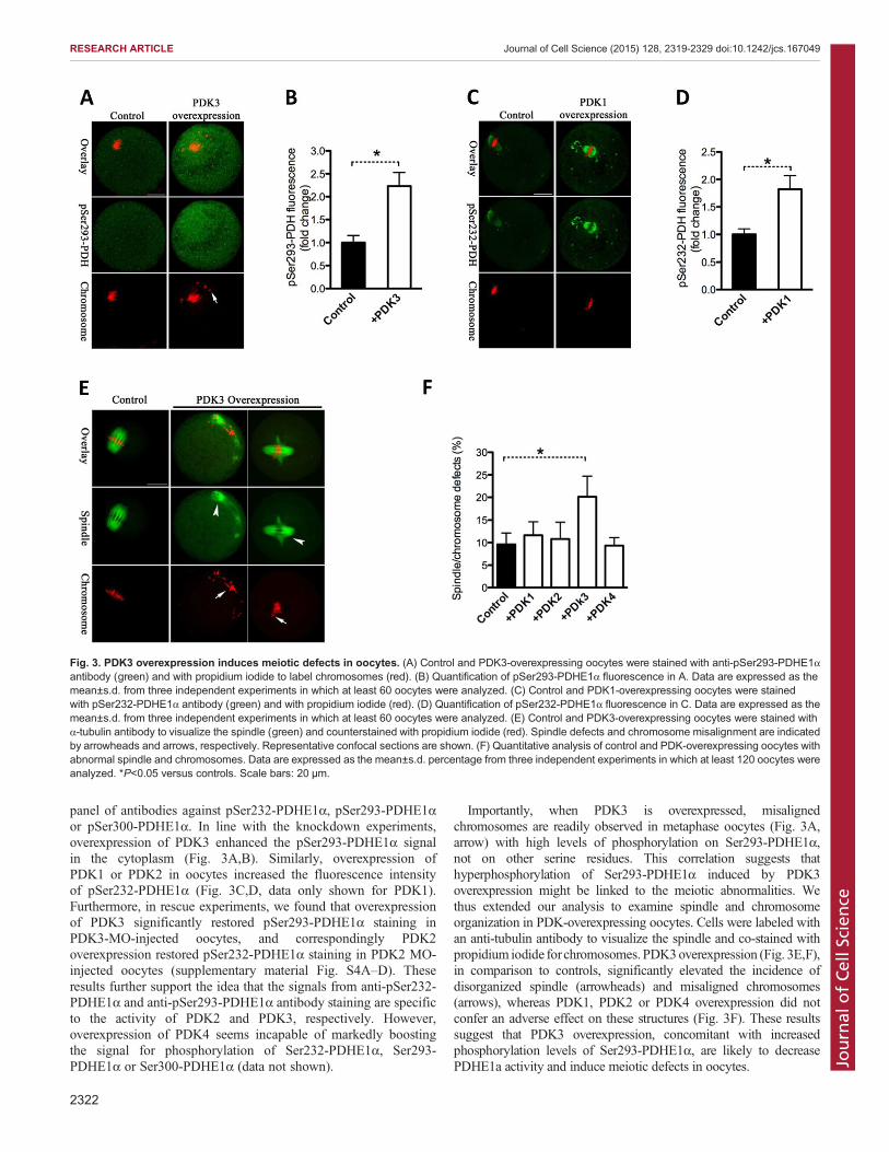
panel of antibodies against pSer232-PDHE1α, pSer293-PDHE1αor pSer300-PDHE1α. In line with the knockdown experiments,overexpression of PDK3 enhanced the pSer293-PDHE1α signalin the cytoplasm (Fig. 3A,B). Similarly, overexpression ofPDK1 or PDK2 in oocytes increased the fluorescence intensityof pSer232-PDHE1α (Fig. 3C,D, data only shown for PDK1).Furthermore, in rescue experiments, we found that overexpressionof PDK3 significantly restored pSer293-PDHE1α staining inPDK3-MO-injected oocytes, and correspondingly PDK2overexpression restored pSer232-PDHE1α staining in PDK2 MO-injected oocytes (supplementary material Fig. S4A–D). Theseresults further support the idea that the signals from anti-pSer232-PDHE1α and anti-pSer293-PDHE1α antibody staining are specificto the activity of PDK2 and PDK3, respectively. However,overexpression of PDK4 seems incapable of markedly boostingthe signal for phosphorylation of Ser232-PDHE1α, Ser293-PDHE1α or Ser300-PDHE1α (data not shown).
Importantly, when PDK3 is overexpressed, misalignedchromosomes are readily observed in metaphase oocytes (Fig. 3A,arrow) with high levels of phosphorylation on Ser293-PDHE1α,not on other serine residues. This correlation suggests thathyperphosphorylation of Ser293-PDHE1α induced by PDK3overexpression might be linked to the meiotic abnormalities. Wethus extended our analysis to examine spindle and chromosomeorganization in PDK-overexpressing oocytes. Cells were labeled withan anti-tubulin antibody to visualize the spindle and co-stained withpropidium iodide forchromosomes. PDK3overexpression (Fig. 3E,F),in comparison to controls, significantly elevated the incidence ofdisorganized spindle (arrowheads) and misaligned chromosomes(arrows), whereas PDK1, PDK2 or PDK4 overexpression did notconfer an adverse effect on these structures (Fig. 3F). These resultssuggest that PDK3 overexpression, concomitant with increasedphosphorylation levels of Ser293-PDHE1α, are likely to decreasePDHE1a activity and induce meiotic defects in oocytes.
Fig. 3. PDK3 overexpression induces meiotic defects in oocytes. (A) Control and PDK3-overexpressing oocytes were stained with anti-pSer293-PDHE1αantibody (green) and with propidium iodide to label chromosomes (red). (B) Quantification of pSer293-PDHE1α fluorescence in A. Data are expressed as themean±s.d. from three independent experiments in which at least 60 oocytes were analyzed. (C) Control and PDK1-overexpressing oocytes were stainedwith pSer232-PDHE1α antibody (green) and with propidium iodide (red). (D) Quantification of pSer232-PDHE1α fluorescence in C. Data are expressed as themean±s.d. from three independent experiments in which at least 60 oocytes were analyzed. (E) Control and PDK3-overexpressing oocytes were stained withα-tubulin antibody to visualize the spindle (green) and counterstained with propidium iodide (red). Spindle defects and chromosome misalignment are indicatedby arrowheads and arrows, respectively. Representative confocal sections are shown. (F) Quantitative analysis of control and PDK-overexpressing oocytes withabnormal spindle and chromosomes. Data are expressed as the mean±s.d. percentage from three independent experiments in which at least 120 oocytes wereanalyzed. *P<0.05 versus controls. Scale bars: 20 µm.
2322
RESEARCH ARTICLE Journal of Cell Science (2015) 128, 2319-2329 doi:10.1242/jcs.167049
Journal
ofCe
llScience
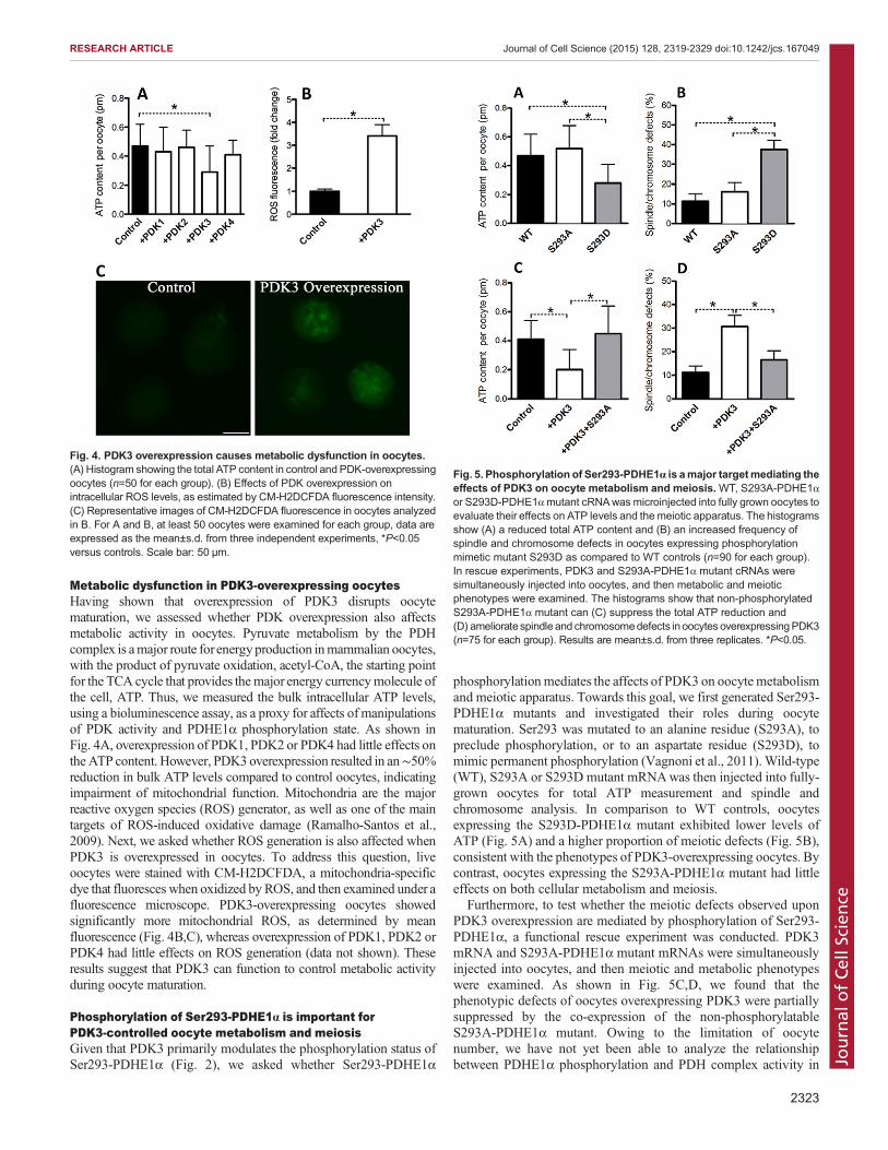
Metabolic dysfunction in PDK3-overexpressing oocytesHaving shown that overexpression of PDK3 disrupts oocytematuration, we assessed whether PDK overexpression also affectsmetabolic activity in oocytes. Pyruvate metabolism by the PDHcomplex is amajor route forenergy production inmammalian oocytes,with the product of pyruvate oxidation, acetyl-CoA, the starting pointfor the TCA cycle that provides themajor energy currencymolecule ofthe cell, ATP. Thus, we measured the bulk intracellular ATP levels,using a bioluminescence assay, as a proxy for affects of manipulationsof PDK activity and PDHE1α phosphorylation state. As shown inFig. 4A, overexpression of PDK1, PDK2 or PDK4 had little effects ontheATP content. However, PDK3 overexpression resulted in an∼50%reduction in bulk ATP levels compared to control oocytes, indicatingimpairment of mitochondrial function. Mitochondria are the majorreactive oxygen species (ROS) generator, as well as one of the maintargets of ROS-induced oxidative damage (Ramalho-Santos et al.,2009). Next, we asked whether ROS generation is also affected whenPDK3 is overexpressed in oocytes. To address this question, liveoocytes were stained with CM-H2DCFDA, a mitochondria-specificdye that fluoresces when oxidized by ROS, and then examined under afluorescence microscope. PDK3-overexpressing oocytes showedsignificantly more mitochondrial ROS, as determined by meanfluorescence (Fig. 4B,C), whereas overexpression of PDK1, PDK2 orPDK4 had little effects on ROS generation (data not shown). Theseresults suggest that PDK3 can function to control metabolic activityduring oocyte maturation.
Phosphorylation of Ser293-PDHE1α is important forPDK3-controlled oocyte metabolism and meiosisGiven that PDK3 primarily modulates the phosphorylation status ofSer293-PDHE1α (Fig. 2), we asked whether Ser293-PDHE1α
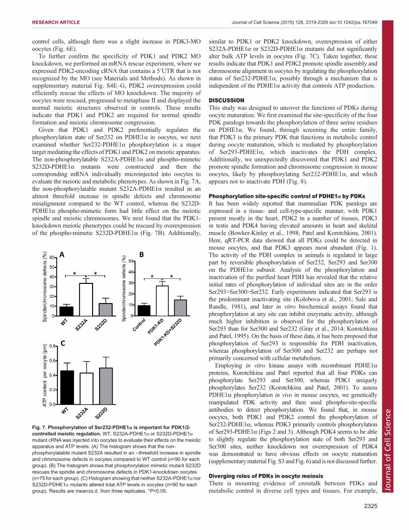
phosphorylationmediates the affects of PDK3 on oocytemetabolismand meiotic apparatus. Towards this goal, we first generated Ser293-PDHE1α mutants and investigated their roles during oocytematuration. Ser293 was mutated to an alanine residue (S293A), topreclude phosphorylation, or to an aspartate residue (S293D), tomimic permanent phosphorylation (Vagnoni et al., 2011). Wild-type(WT), S293A or S293D mutant mRNAwas then injected into fully-grown oocytes for total ATP measurement and spindle andchromosome analysis. In comparison to WT controls, oocytesexpressing the S293D-PDHE1α mutant exhibited lower levels ofATP (Fig. 5A) and a higher proportion of meiotic defects (Fig. 5B),consistent with the phenotypes of PDK3-overexpressing oocytes. Bycontrast, oocytes expressing the S293A-PDHE1α mutant had littleeffects on both cellular metabolism and meiosis.
Furthermore, to test whether the meiotic defects observed uponPDK3 overexpression are mediated by phosphorylation of Ser293-PDHE1α, a functional rescue experiment was conducted. PDK3mRNA and S293A-PDHE1α mutant mRNAs were simultaneouslyinjected into oocytes, and then meiotic and metabolic phenotypeswere examined. As shown in Fig. 5C,D, we found that thephenotypic defects of oocytes overexpressing PDK3 were partiallysuppressed by the co-expression of the non-phosphorylatableS293A-PDHE1α mutant. Owing to the limitation of oocytenumber, we have not yet been able to analyze the relationshipbetween PDHE1α phosphorylation and PDH complex activity in
Fig. 4. PDK3 overexpression causes metabolic dysfunction in oocytes.(A) Histogram showing the total ATP content in control and PDK-overexpressingoocytes (n=50 for each group). (B) Effects of PDK overexpression onintracellular ROS levels, as estimated by CM-H2DCFDA fluorescence intensity.(C) Representative images of CM-H2DCFDA fluorescence in oocytes analyzedin B. For A and B, at least 50 oocytes were examined for each group, data areexpressed as the mean±s.d. from three independent experiments, *P<0.05versus controls. Scale bar: 50 µm.
Fig. 5. Phosphorylation of Ser293-PDHE1α is amajor targetmediating theeffects of PDK3 on oocyte metabolism and meiosis. WT, S293A-PDHE1αor S293D-PDHE1αmutant cRNAwas microinjected into fully grown oocytes toevaluate their effects on ATP levels and the meiotic apparatus. The histogramsshow (A) a reduced total ATP content and (B) an increased frequency ofspindle and chromosome defects in oocytes expressing phosphorylationmimetic mutant S293D as compared to WT controls (n=90 for each group).In rescue experiments, PDK3 and S293A-PDHE1α mutant cRNAs weresimultaneously injected into oocytes, and then metabolic and meioticphenotypes were examined. The histograms show that non-phosphorylatedS293A-PDHE1α mutant can (C) suppress the total ATP reduction and(D) ameliorate spindle and chromosomedefects in oocytes overexpressingPDK3(n=75 for each group). Results are mean±s.d. from three replicates. *P<0.05.
2323
RESEARCH ARTICLE Journal of Cell Science (2015) 128, 2319-2329 doi:10.1242/jcs.167049
Journal
ofCe
llScience
mouse oocytes, although we assume that Ser293 phosphorylationinactivates PDH, as observed in somatic cells. Taken together, thesedata indicate that Ser293 is a major, if not unique, phosphorylationsite on PDHE1α, mediating the action of PDK3 on oocytemetabolism and meiotic maturation.
PDK1 and PDK2 promote spindle organization andchromosome congression in oocytes by phosphorylatingSer232-PDHE1αTwo of our observations would not be expected if PDK1- andPDK2-mediated Ser232-PDHE1α phosphorylation inhibited theactivity of the PDH complex: (1) that from the initial analysis ofPDK1 or PDK2 MO knockdown showing meiotic maturationdefects and (2) that overexpression of PDK1 or PDK2 did notinfluence oocyte maturation (in contrast to overexpression of PDK3described above). Therefore we explored the function of PDK1 andPDK2-mediated phosphorylation of Ser232-PDHE1α further.First, we individually knocked down PDK1–PDK4 by MO
injection, with a sham MO standard injected as control, and
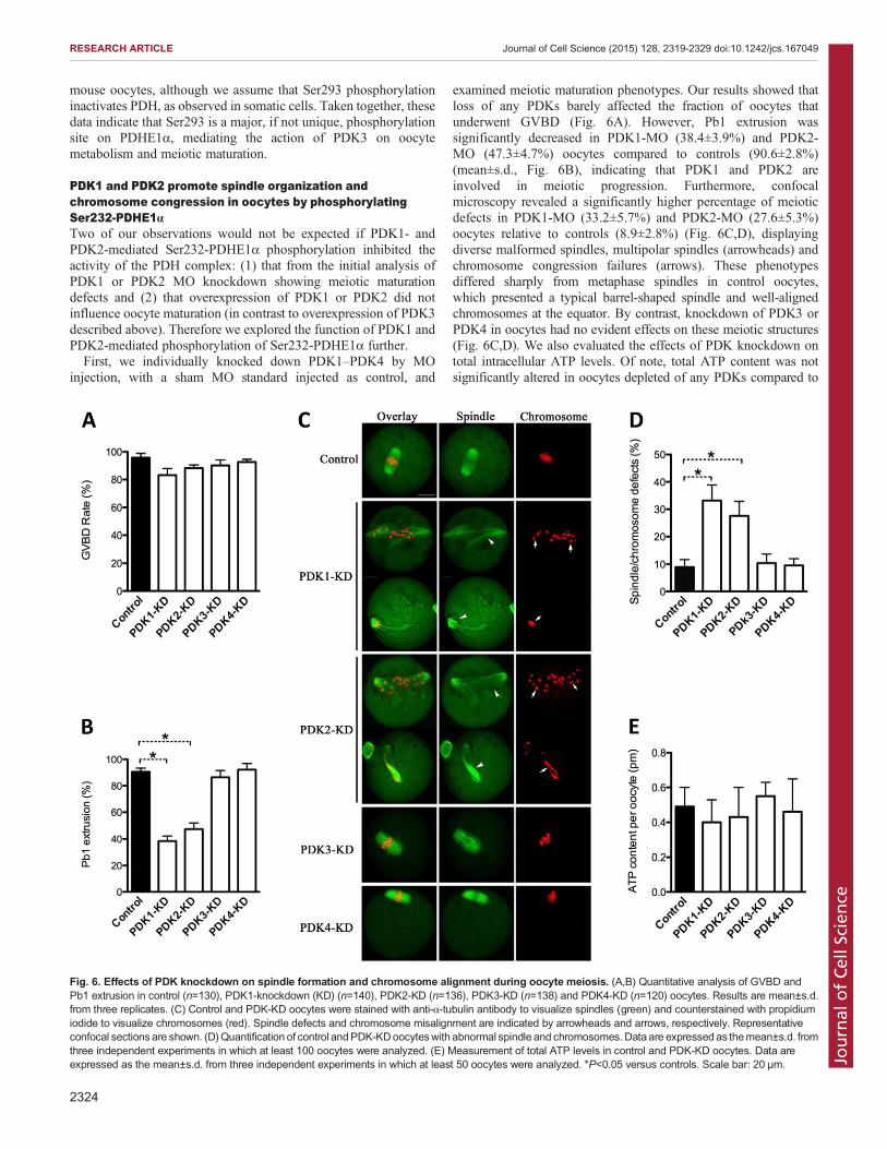
examined meiotic maturation phenotypes. Our results showed thatloss of any PDKs barely affected the fraction of oocytes thatunderwent GVBD (Fig. 6A). However, Pb1 extrusion wassignificantly decreased in PDK1-MO (38.4±3.9%) and PDK2-MO (47.3±4.7%) oocytes compared to controls (90.6±2.8%)(mean±s.d., Fig. 6B), indicating that PDK1 and PDK2 areinvolved in meiotic progression. Furthermore, confocalmicroscopy revealed a significantly higher percentage of meioticdefects in PDK1-MO (33.2±5.7%) and PDK2-MO (27.6±5.3%)oocytes relative to controls (8.9±2.8%) (Fig. 6C,D), displayingdiverse malformed spindles, multipolar spindles (arrowheads) andchromosome congression failures (arrows). These phenotypesdiffered sharply from metaphase spindles in control oocytes,which presented a typical barrel-shaped spindle and well-alignedchromosomes at the equator. By contrast, knockdown of PDK3 orPDK4 in oocytes had no evident effects on these meiotic structures(Fig. 6C,D). We also evaluated the effects of PDK knockdown ontotal intracellular ATP levels. Of note, total ATP content was notsignificantly altered in oocytes depleted of any PDKs compared to
Fig. 6. Effects of PDK knockdown on spindle formation and chromosome alignment during oocyte meiosis. (A,B) Quantitative analysis of GVBD andPb1 extrusion in control (n=130), PDK1-knockdown (KD) (n=140), PDK2-KD (n=136), PDK3-KD (n=138) and PDK4-KD (n=120) oocytes. Results are mean±s.d.from three replicates. (C) Control and PDK-KD oocytes were stained with anti-α-tubulin antibody to visualize spindles (green) and counterstained with propidiumiodide to visualize chromosomes (red). Spindle defects and chromosome misalignment are indicated by arrowheads and arrows, respectively. Representativeconfocal sections are shown. (D)Quantification of control andPDK-KDoocyteswith abnormal spindle and chromosomes.Data are expressed as themean±s.d. fromthree independent experiments in which at least 100 oocytes were analyzed. (E) Measurement of total ATP levels in control and PDK-KD oocytes. Data areexpressed as the mean±s.d. from three independent experiments in which at least 50 oocytes were analyzed. *P<0.05 versus controls. Scale bar: 20 µm.
2324
RESEARCH ARTICLE Journal of Cell Science (2015) 128, 2319-2329 doi:10.1242/jcs.167049
Journal
ofCe
llScience
control cells, although there was a slight increase in PDK3-MOoocytes (Fig. 6E).To further confirm the specificity of PDK1 and PDK2 MO
knockdown, we performed an mRNA rescue experiment, where weexpressed PDK2-encoding cRNA that contains a 5′UTR that is notrecognized by the MO (see Materials and Methods). As shown insupplementary material Fig. S4E–G, PDK2 overexpression couldefficiently rescue the effects of MO knockdown. The majority ofoocytes were rescued, progressed to metaphase II and displayed thenormal meiotic structures observed in controls. These resultsindicate that PDK1 and PDK2 are required for normal spindleformation and meiotic chromosome congression.Given that PDK1 and PDK2 preferentially regulates the
phosphorylation state of Ser232 on PDHE1α in oocytes, we nextexamined whether Ser232-PDHE1α phosphorylation is a majortarget mediating the effects of PDK1 and PDK2 onmeiotic apparatus.The non-phosphorylatable S232A-PDHE1α and phospho-mimeticS232D-PDHE1α mutants were constructed and then thecorresponding mRNA individually microinjected into oocytes toevaluate the meiotic and metabolic phenotypes. As shown in Fig. 7A,the non-phosphorylatable mutant S232A-PDHE1α resulted in analmost threefold increase in spindle defects and chromosomemisalignment compared to the WT control, whereas the S232D-PDHE1α phospho-mimetic form had little effect on the meioticspindle and meiotic chromosomes. We next found that the PDK1-knockdown meiotic phenotypes could be rescued by overexpressionof the phospho-mimetic S232D-PDHE1α (Fig. 7B). Additionally,
similar to PDK1 or PDK2 knockdown, overexpression of eitherS232A-PDHE1α or S232D-PDHE1α mutants did not significantlyalter bulk ATP levels in oocytes (Fig. 7C). Taken together, theseresults indicate that PDK1 and PDK2 promote spindle assembly andchromosome alignment in oocytes by regulating the phosphorylationstatus of Ser232-PDHE1α, possibly through a mechanism that isindependent of the PDHE1α activity that controls ATP production.
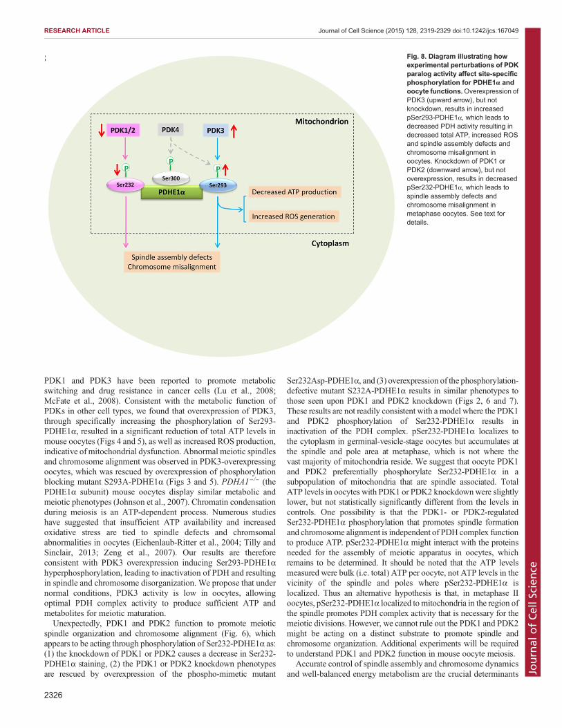
DISCUSSIONThis study was designed to uncover the functions of PDKs duringoocyte maturation. We first examined the site-specificity of the fourPDK paralogs towards the phosphorylation of three serine residueson PDHE1α. We found, through screening the entire family,that PDK3 is the primary PDK that functions in metabolic controlduring oocyte maturation, which is mediated by phosphorylationof Ser293-PDHE1α, which inactivates the PDH complex.Additionally, we unexpectedly discovered that PDK1 and PDK2promote spindle formation and chromosome congression in mouseoocytes, likely by phosphorylating Ser232-PDHE1α, and whichappears not to inactivate PDH (Fig. 8).
Phosphorylation site-specific control of PDHE1α by PDKsIt has been widely reported that mammalian PDK paralogs areexpressed in a tissue- and cell-type-specific manner, with PDK1present mostly in the heart, PDK2 in a number of tissues, PDK3in testis and PDK4 having elevated amounts in heart and skeletalmuscle (Bowker-Kinley et al., 1998; Patel and Korotchkina, 2001).Here, qRT-PCR data showed that all PDKs could be detected inmouse oocytes, and that PDK3 appears most abundant (Fig. 1).The activity of the PDH complex in animals is regulated in largepart by reversible phosphorylation of Ser232, Ser293 and Ser300on the PDHE1α subunit. Analysis of the phosphorylation andinactivation of the purified heart PDH has revealed that the relativeinitial rates of phosphorylation of individual sites are in the orderSer293>Ser300>Ser232. Early experiments indicated that Ser293 isthe predominant inactivating site (Kolobova et al., 2001; Sale andRandle, 1981), and later in vitro biochemical assays found thatphosphorylation at any site can inhibit enzymatic activity, althoughmuch higher inhibition is observed for the phosphorylation ofSer293 than for Ser300 and Ser232 (Gray et al., 2014; Korotchkinaand Patel, 1995). On the basis of these data, it has been proposed thatphosphorylation of Ser293 is responsible for PDH inactivation,whereas phosphorylation of Ser300 and Ser232 are perhaps notprimarily concerned with cellular metabolism.
Employing in vitro kinase assays with recombinant PDHE1αproteins, Korotchkina and Patel reported that all four PDKs canphosphorylate Ser293 and Ser300, whereas PDK1 uniquelyphosphorylates Ser232 (Korotchkina and Patel, 2001). To assessPDHE1α phosphorylation in vivo in mouse oocytes, we geneticallymanipulated PDK activity and then used phospho-site-specificantibodies to detect phosphorylation. We found that, in mouseoocytes, both PDK1 and PDK2 control the phosphorylation ofSer232-PDHE1α, whereas PDK3 primarily controls phosphorylationof Ser293-PDHE1α (Figs 2 and 3). Although PDK4 seems to be ableto slightly regulate the phosphorylation state of both Ser293 andSer300 sites, neither knockdown nor overexpression of PDK4was demonstrated to have obvious effects on oocyte maturation(supplementarymaterial Fig. S3 andFig. 6) and is not discussed further.
Diverging roles of PDKs in oocyte meiosisThere is mounting evidence of crosstalk between PDKs andmetabolic control in diverse cell types and tissues. For example,
Fig. 7. Phosphorylation of Ser232-PDHE1α is important for PDK1/2-controlled meiotic regulation. WT, S232A-PDHE1α or S232D-PDHE1αmutant cRNAwas injected into oocytes to evaluate their effects on the meioticapparatus and ATP levels. (A) The histogram shows that the non-phosphorylatable mutant S232A resulted in an ∼threefold increase in spindleand chromosome defects in oocytes compared to WT control (n=90 for eachgroup). (B) The histogram shows that phosphorylation mimetic mutant S232Drescues the spindle and chromosome defects in PDK1-knockdown oocytes(n=75 for each group). (C) Histogram showing that neither S232A-PDHE1α norS232D-PDHE1α mutants altered total ATP levels in oocytes (n=90 for eachgroup). Results are mean±s.d. from three replicates. *P<0.05.
2325
RESEARCH ARTICLE Journal of Cell Science (2015) 128, 2319-2329 doi:10.1242/jcs.167049
Journal
ofCe
llScience
PDK1 and PDK3 have been reported to promote metabolicswitching and drug resistance in cancer cells (Lu et al., 2008;McFate et al., 2008). Consistent with the metabolic function ofPDKs in other cell types, we found that overexpression of PDK3,through specifically increasing the phosphorylation of Ser293-PDHE1α, resulted in a significant reduction of total ATP levels inmouse oocytes (Figs 4 and 5), as well as increased ROS production,indicative of mitochondrial dysfunction. Abnormal meiotic spindlesand chromosome alignment was observed in PDK3-overexpressingoocytes, which was rescued by overexpression of phosphorylationblocking mutant S293A-PDHE1α (Figs 3 and 5). PDHA1−/− (thePDHE1α subunit) mouse oocytes display similar metabolic andmeiotic phenotypes (Johnson et al., 2007). Chromatin condensationduring meiosis is an ATP-dependent process. Numerous studieshave suggested that insufficient ATP availability and increasedoxidative stress are tied to spindle defects and chromsomalabnormalities in oocytes (Eichenlaub-Ritter et al., 2004; Tilly andSinclair, 2013; Zeng et al., 2007). Our results are thereforeconsistent with PDK3 overexpression inducing Ser293-PDHE1αhyperphosphorylation, leading to inactivation of PDH and resultingin spindle and chromosome disorganization. We propose that undernormal conditions, PDK3 activity is low in oocytes, allowingoptimal PDH complex activity to produce sufficient ATP andmetabolites for meiotic maturation.Unexpectedly, PDK1 and PDK2 function to promote meiotic
spindle organization and chromosome alignment (Fig. 6), whichappears to be acting through phosphorylation of Ser232-PDHE1α as:(1) the knockdown of PDK1 or PDK2 causes a decrease in Ser232-PDHE1α staining, (2) the PDK1 or PDK2 knockdown phenotypesare rescued by overexpression of the phospho-mimetic mutant
Ser232Asp-PDHE1α, and (3) overexpression of the phosphorylation-defective mutant S232A-PDHE1α results in similar phenotypes tothose seen upon PDK1 and PDK2 knockdown (Figs 2, 6 and 7).These results are not readily consistent with a model where the PDK1and PDK2 phosphorylation of Ser232-PDHE1α results ininactivation of the PDH complex. pSer232-PDHE1α localizes tothe cytoplasm in germinal-vesicle-stage oocytes but accumulates atthe spindle and pole area at metaphase, which is not where thevast majority of mitochondria reside. We suggest that oocyte PDK1and PDK2 preferentially phosphorylate Ser232-PDHE1α in asubpopulation of mitochondria that are spindle associated. TotalATP levels in oocytes with PDK1 or PDK2 knockdownwere slightlylower, but not statistically significantly different from the levels incontrols. One possibility is that the PDK1- or PDK2-regulatedSer232-PDHE1α phosphorylation that promotes spindle formationand chromosome alignment is independent of PDH complex functionto produce ATP. pSer232-PDHE1α might interact with the proteinsneeded for the assembly of meiotic apparatus in oocytes, whichremains to be determined. It should be noted that the ATP levelsmeasured were bulk (i.e. total) ATP per oocyte, not ATP levels in thevicinity of the spindle and poles where pSer232-PDHE1α islocalized. Thus an alternative hypothesis is that, in metaphase IIoocytes, pSer232-PDHE1α localized to mitochondria in the region ofthe spindle promotes PDH complex activity that is necessary for themeiotic divisions. However, we cannot rule out the PDK1 and PDK2might be acting on a distinct substrate to promote spindle andchromosome organization. Additional experiments will be requiredto understand PDK1 and PDK2 function in mouse oocyte meiosis.
Accurate control of spindle assembly and chromosome dynamicsand well-balanced energy metabolism are the crucial determinants
Fig. 8. Diagram illustrating howexperimental perturbations of PDKparalog activity affect site-specificphosphorylation for PDHE1α andoocyte functions.Overexpression ofPDK3 (upward arrow), but notknockdown, results in increasedpSer293-PDHE1α, which leads todecreased PDH activity resulting indecreased total ATP, increased ROSand spindle assembly defects andchromosome misalignment inoocytes. Knockdown of PDK1 orPDK2 (downward arrow), but notoverexpression, results in decreasedpSer232-PDHE1α, which leads tospindle assembly defects andchromosome misalignment inmetaphase oocytes. See text fordetails.
2326
RESEARCH ARTICLE Journal of Cell Science (2015) 128, 2319-2329 doi:10.1242/jcs.167049
Journal
ofCe
llScience
for oocyte quality. This study uncovers the different roles of PDKisoforms in these processes, providing a newmechanism controllingoocyte maturation. As any errors during meiotic division could leadto pregnancy loss and infertility, our findings are not only offundamental scientific interest but also vital for prevention of birthdefects and treatment of reproductive disease in women.
MATERIALS AND METHODSAll chemicals and culture media were purchased from Sigma (St Louis, MO)unless stated otherwise.
MiceICR mice were used in all experiments. All experiments were approved bythe Animal Care and Use Committee of Nanjing Medical University andwere performed in accordance with institutional guidelines.
AntibodiesRabbit polyclonal anti-PDK1 and rabbit polyclonal anti-Myc antibodieswere purchased from Abcam (Cambridge, MA; catalog numbers ab92959and ab9106); rabbit polyclonal anti-PDK2, rabbit polyclonal anti-PDK3 andrabbit polyclonal anti-PDK4 antibodies were purchased from NovusBiologicals (Concord, MA; catalog numbers NBP1-87307, NBP1-32581and NBP1-07047); rabbit polyclonal anti-PDHE1α (pSer232), rabbitpolyclonal anti-PDHE1α (pSer293), and rabbit polyclonal anti-PDHE1α(pSer300) antibodies were purchased from EMD Chemicals (Beverly, MA;catalog numbers AP1063, AP1062 and AP1064); mouse monoclonal anti-β-actin and mouse monoclonal anti-α-tubulin antibodies conjugated toFITC were purchased from Sigma (catalog numbers A5441 and F2168).FITC-conjugated goat anti-rabbit-IgG antibody was purchased fromThermo Fisher Scientific (Rockford, IL).
Oocyte collection and cultureTo collect fully-grown germinal vesicle oocytes, mice were superovulatedwith 5 IU pregnant mares serum gonadotropin (PMSG) by intraperitonealinjection, and 48 h later, cumulus-enclosed oocytes were obtained bymanual rupturing of antral ovarian follicles. To collect denuded oocytes,cumulus cells were removed by repeatedly pipetting. For in vitromaturation,oocytes were cultured inM2 medium under mineral oil at 37°C in a 5% CO2
incubator.
Plasmid construction and mRNA synthesisTotal RNA was extracted from 100 denuded oocytes using an ArcturusPicoPure RNA isolation kit (Applied Biosystems, CA), and the cDNA wasgenerated with QIAquick PCR purification kit (Qiagen, Germany). PCRproducts were purified, digested with FseI and AscI (NEB Inc, MA), andthen cloned into the pCS2+ vector with six Myc tags. The pCS2+ vectorsencoding the Myc–PDHE1α substitution mutants S232A, S232D, S293Aand S293D, were generated with the use of a QuikChange site-directedmutagenesis kit (Stratagene). The related primer sequences can be found insupplementary material Tables S2, S3.
For the synthesis of mRNA, the PDK and PDHE1α-pCS2+ plasmids werelinearized by NotI. Capped cRNAs were made using in vitro transcriptionwith SP6 mMESSAGE mMACHINE (Ambion, CA) according to themanufacturer’s instructions, and then purified with an RNeasy Micro kit(Qiagen). Synthesized RNA was aliquoted and stored at −80°C.
PDK knockdown and overexpressionMicroinjections of MO or mRNA, with a Narishige microinjector, wereused to knock down or overexpress proteins in mouse oocytes, respectively.For overexpression experiments, 10 pl cRNA solution (10 ng/µl) wasinjected into cytoplasm of germinal vesicle oocytes. The same amount ofRNase-free PBS was injected as control. For knockdown experiments, MOsagainst each PDK were designed by Gene Tools (Philomath, OR) throughtheir site selection procedure, to target the initiation of translation and werethus complementary to the 5′UTR or N-terminal coding sequences; PDK2-MO and PDK3-MO targeted the 5′UTR of mRNA, whereas PDK1-MO and
PDK4-MO targeted coding sequence because there were not appropriateoligonucleotides that were specific for their 5′UTR. MOs were diluted withwater to give a stock concentration of 1 mM, and then 2.5 pl MO solutionwas injected into oocytes. A non-targeting MO was injected as a control.
For rescue experiments using PDK2 or PDK3 cRNA, 10 pl cRNAsolution (10 ng/µl) was microinjected into oocytes 2 h after MO injection.The PDK2 and PDK3 cRNAs contain a 5′UTR that is not recognized by theMO: PDK1-MO (CDS), 5′-TTGTTGCTCCTCGATTAGCCATGTC-3′;PDK2-MO (UTR), 5′-GGCTTGGATCTCGCTGCTGTCCC-3′; PDK3-MO (UTR), 5′-CCTCACTAGAGCAGTGTGCAGATCC-3′; PDK4-MO (CDS), 5′-CATCACGAAGCGGGCTGCCTTCATC-3′; CTRL-MO,5′-CCTCTTACCTCAGTTACAATTTATA-3′.
After injections, oocytes were arrested at germinal vesicle stage in M2medium supplemented with 2.5 µM milrinone, a phosphodiesteraseinhibitor that blocks meiotic maturation, for 20 h, to allow time for eitherMO-mediated knockdown of gene-specific mRNA translation or to permitoverexpression. Then oocytes werewashed three times in milrinone-freeM2medium and cultured for in vitro maturation.
Western blottingA pool of ∼100–150 denuded oocytes was lysed in Laemmli sample buffercontaining protease inhibitor and then subjected to SDS-PAGE. Theseparated proteins were transferred onto a PVDF membrane. Membraneswere blocked in TBS containing 0.1% Tween 20 and 5% low-fat dried milkpowder for 1 h and then incubated with primary antibodies. After multiplewashes in TBS containing 0.1% Tween 20 and incubation with horseradishperoxidase (HRP)-conjugated secondary antibodies, the protein bands werevisualized using an ECL Plus Western Blotting Detection System. Themembrane was then washed and reblotted with anti-β-actin (1:5000)antibody as a loading control. All western blot experiments were repeated atleast three times.
Quantitative real-time PCRqRT-PCR was performed as described previously (Ma et al., 2013). TotalRNA was isolated from 50 oocytes using an RNAqueous-Micro Kit(Ambion, TX) following the manufacturer’s protocols. The cDNA wasquantified by qRT-PCR using an ABI Stepone Plus Real-time PCR system(Applied Biosystems, CA). The fold change in gene expression wascalculated using the ΔΔCt method with the house keeping gene,glyceraldehydes-3-phosphate dehydrogenase (GAPDH), as the internalcontrol. The result was expressed as fold change relative to PDK1. Primersequences are listed in supplementary material Table S1.
Determination of total ATP contentTotal ATP content in pools of 10–20 oocytes was determined using thebioluminescent somatic cell assay kit (Sigma), following the proceduredescribed by Combelles and Albertini (2003) and the manufacturer’srecommendations. A six-point standard curve (0, 0. 1, 0.5, 1.0, 10, and50 pmol of ATP) was generated in each assay and the ATP content wascalculated by using the formula derived from the linear regression of thestandard curve.
Evaluation of intracellular ROSTo detect intercellular ROS in living oocytes, CM-H2DCFDA fromInvitrogen was used. CM-H2DCFDA was prepared in DMSO prior toloading. Oocytes were incubated with 5 μM CM-H2DCFDA for 30 min,and then immediately observed under a fluorescence microscope (AxioVert,Zeiss, Germany).
Immunofluorescence and confocal microscopyOocytes were fixed with 4% paraformaldehyde for 30 min and thenpermeabilized with 0.5% Triton X-100 for 20 min. Following blockingin 1% BSA-supplemented PBS for 1 h, samples were incubated overnight at4°C with primary antibody, and then at room temperature for 1 h withsecondary antibody. Chromosomes were evaluated by staining withpropidium iodide for 10 min. After washes in PBS, oocyte samples weremounted on anti-fade medium (Vectashield, Burlingame, CA, USA) and
2327
RESEARCH ARTICLE Journal of Cell Science (2015) 128, 2319-2329 doi:10.1242/jcs.167049
Journal
ofCe
llScience
examined under a Laser Scanning Confocal Microscope (LSM 710, Zeiss,Germany) equipped with 40× or 63× oil objectives.
To quantify the intensity of fluorescence, Image J software (NIH) wasused as previously described (Wang et al., 2012). For quantification ofoocytes with meiotic defects, the gross morphology of spindle andchromosomes was assessed. The metaphase II spindle was classified asnormal if it displayed the typical barrel shape. The spindle was scored asabnormal if it displayed defects, such as single or multiple poles, prominentasters and other malformations. The alignment of chromosomes was definedas normal if they tightly lay on the metaphase plate or abnormal if one ormore bivalent was displaced from the metaphase plate.
Statistical analysisData are presented as mean±s.d., unless otherwise indicated. Differencesbetween two groups were analyzed by Student’s t-test. Multiplecomparisons between more than two groups were analyzed by one-wayANOVA test using Prism 5.0. P<0.05 was considered to be significant.
Competing interestsThe authors declare no competing or financial interests.
Author contributionsX.H., X.Z., T.S. and Q.W. designed research; X.H., L.Z., L.H., J.G. and R.M.performed research; X.H., K.M., T.S. and Q.W. analyzed data; K.M., T.S. and Q.W.wrote the paper.
FundingThis work was supported by National Key Scientific Research Projects [grantnumber 2014CB943200]; and National Natural Science Foundation of China [grantnumber 31271541 and 31301181]. K.M. was supported by the American DiabetesAssociation [grant number 76274]. T.S. was supported by the National Institutesof Health [grant number R01 GM100756]. Deposited in PMC for release after12 months.
Supplementary materialSupplementary material available online athttp://jcs.biologists.org/lookup/suppl/doi:10.1242/jcs.167049/-/DC1
ReferencesBiggers, J. D., Whittingham, D. G. and Donahue, R. P. (1967). The pattern ofenergy metabolism in the mouse oocyte and zygote. Proc. Natl. Acad. Sci. USA58, 560-567.
Bowker-Kinley, M. M., Davis, W. I., Wu, P., Harris, R. A. and Popov, K. M. (1998).Evidence for existence of tissue-specific regulation of the mammalian pyruvatedehydrogenase complex. Biochem. J. 329, 191-196.
Combelles, C. M. H. and Albertini, D. F. (2003). Assessment of oocyte qualityfollowing repeatedgonadotropin stimulation in themouse.Biol. Reprod.68, 812-821.
Eichenlaub-Ritter, U., Vogt, E., Yin, H. and Gosden, R. (2004). Spindles,mitochondria and redox potential in ageing oocytes. Reprod. Biomed. Online 8,45-58.
Eppig, J. J. (1976). Analysis of mouse oogenesis in vitro. Oocyte isolation and theutilization of exogenous energy sources by growing oocytes. J. Exp. Zool. 198,375-381.
Fagbohun, C. F. and Downs, S. M. (1992). Requirement for glucose in ligand-stimulated meiotic maturation of cumulus cell-enclosed mouse oocytes.J. Reprod. Fertil. 96, 681-697.
Gray, L. R., Tompkins, S. C. and Taylor, E. B. (2014). Regulation of pyruvatemetabolism and human disease. Cell. Mol. Life. Sci. 71, 2577-2604.
Gudi, R., Melissa, M. B.-K., Kedishvili, N. Y., Zhao, Y. and Popov, K. M. (1995).Diversity of the pyruvate dehydrogenase kinase gene family in humans. J. Biol.Chem. 270, 28989-28994.
Harris, S. E., Leese, H. J., Gosden, R. G. and Picton, H. M. (2009). Pyruvate andoxygen consumption throughout the growth and development of murine oocytes.Mol. Reprod. Dev. 76, 231-238.
Hassold, T. and Hunt, P. (2001). To err (meiotically) is human: the genesis ofhuman aneuploidy. Nat. Rev. Genet. 2, 280-291.
Hitosugi, T., Fan, J., Chung, T.-W., Lythgoe, K., Wang, X., Xie, J., Ge, Q., Gu, T.-L.,Polakiewicz, R. D., Roesel, J. L. et al. (2011). Tyrosine phosphorylation ofmitochondrial pyruvate dehydrogenase kinase 1 is important for cancer metabolism.Mol. Cell 44, 864-877.
Jaswal, J. S., Lund, C. R., Keung, W., Beker, D. L., Rebeyka, I. M. andLopaschuk, G. D. (2010). Isoproterenol stimulates 5′-AMP-activated proteinkinase and fatty acid oxidation in neonatal hearts. Am. J. Physiol. Heart Circ.Physiol. 299, H1135-H1145.
Jeoung, N. H. and Harris, R. A. (2008). Pyruvate dehydrogenase kinase-4deficiency lowers blood glucose and improves glucose tolerance in diet-inducedobese mice. Am. J. Physiol. Endocrinol. Metab. 295, E46-E54.
Johnson, M. T., Freeman, E. A., Gardner, D. K. and Hunt, P. A. (2007). Oxidativemetabolism of pyruvate is required for meiotic maturation of murine oocytes invivo. Biol. Reprod. 77, 2-8.
Jones, K. T. and Lane, S. I. R. (2013). Molecular causes of aneuploidy inmammalian eggs. Development 140, 3719-3730.
Kim, J.-W., Tchernyshyov, I., Semenza, G. L. and Dang, C. V. (2006). HIF-1-mediated expression of pyruvate dehydrogenase kinase: a metabolic switchrequired for cellular adaptation to hypoxia. Cell Metab. 3, 177-185.
Kolobova, E., Tuganova, A., Boulatnikov, I. and Popov, K. M. (2001). Regulationof pyruvate dehydrogenase activity through phosphorylation at multiple sites.Biochem. J. 358, 69-77.
Korotchkina, L. G. and Patel, M. S. (1995). Mutagenesis studies of thephosphorylation sites of recombinant human pyruvate dehydrogenase. Site-specific regulation. J. Biol. Chem. 270, 14297-14304.
Korotchkina, L. G. and Patel, M. S. (2001). Site specificity of four pyruvatedehydrogenase kinase isoenzymes toward the three phosphorylation sites ofhuman pyruvate dehydrogenase. J. Biol. Chem. 276, 37223-37229.
Kwon, H.-S. and Harris, R. A. (2004). Mechanisms responsible for regulation ofpyruvate dehydrogenase kinase 4 gene expression. Adv. Enzyme Regul. 44,109-121.
Kwon, H.-S., Huang, B., Ho Jeoung, N., Wu, P., Steussy, C. N. and Harris, R. A.(2006). Retinoic acids and trichostatin A (TSA), a histone deacetylase inhibitor,induce human pyruvate dehydrogenase kinase 4 (PDK4) gene expression.Biochim. Biophys. Acta 1759, 141-151.
Lu, C.-W., Lin, S.-C., Chen, K.-F., Lai, Y.-Y. and Tsai, S.-J. (2008). Induction ofpyruvate dehydrogenase kinase-3 by hypoxia-inducible factor-1 promotesmetabolic switch and drug resistance. J. Biol. Chem. 283, 28106-28114.
Luzzo, K. M., Wang, Q., Purcell, S. H., Chi, M., Jimenez, P. T., Grindler, N.,Schedl, T. and Moley, K. H. (2012). High fat diet induced developmental defectsin the mouse: oocyte meiotic aneuploidy and fetal growth retardation/braindefects. PLoS ONE 7, e49217.
Ma, J., Flemr, M., Strnad, H., Svoboda, P. and Schultz, R. M. (2013). Maternallyrecruited DCP1A and DCP2 contribute to messenger RNA degradation duringoocyte maturation and genome activation in mouse. Biol. Reprod. 88, 11.
McFate, T., Mohyeldin, A., Lu, H., Thakar, J., Henriques, J., Halim, N. D., Wu, H.,Schell, M. J., Tsang, T. M., Teahan, O. et al. (2008). Pyruvate dehydrogenasecomplex activity controls metabolic and malignant phenotype in cancer cells.J. Biol. Chem. 283, 22700-22708.
Michelakis, E. D., Sutendra, G., Dromparis, P., Webster, L., Haromy, A., Niven,E., Maguire, C., Gammer, T. L., Mackey, J. R., Fulton, D. et al. (2010). Metabolicmodulation of glioblastoma with dichloroacetate. Sci. Transl. Med. 2, 31ra34.
Miquel, E., Cassina, A., Martınez-Palma, L., Bolatto, C., Trıas, E., Gandelman,M., Radi, R., Barbeito, L. and Cassina, P. (2012). Modulation of astrocyticmitochondrial function by dichloroacetate improves survival and motorperformance in inherited amyotrophic lateral sclerosis. PLoS ONE 7, e34776.
Nahle, Z., Hsieh, M., Pietka, T., Coburn, C. T., Grimaldi, P. A., Zhang, M. Q., Das,D. andAbumrad, N. A. (2008). CD36-dependent regulation of muscle FoxO1 andPDK4 in the PPAR delta/beta-mediated adaptation to metabolic stress. J. Biol.Chem. 283, 14317-14326.
Patel, M. S. and Korotchkina, L. G. (2001). Regulation of mammalian pyruvatedehydrogenase complex by phosphorylation: complexity of multiplephosphorylation sites and kinases. Exp. Mol. Med. 33, 191-197.
Peters, S. J., Harris, R. A., Wu, P., Pehleman, T. L., Heigenhauser, G. J. andSpriet, L. L. (2001). Human skeletal muscle PDH kinase activity and isoformexpression during a 3-day high-fat/low-carbohydrate diet. Am. J. Physiol.Endocrinol. Metab. 281, E1151-E1158.
Ramalho-Santos, J., Varum, S., Amaral, S., Mota, P. C., Sousa, A. P. andAmaral, A. (2009). Mitochondrial functionality in reproduction: from gonads andgametes to embryos and embryonic stem cells. Hum. Reprod. Update 15,553-572.
Rardin, M. J., Wiley, S. E., Naviaux, R. K., Murphy, A. N. and Dixon, J. E. (2009).Monitoring phosphorylation of the pyruvate dehydrogenase complex. Anal.Biochem. 389, 157-164.
Roche, T. E. and Hiromasa, Y. (2007). Pyruvate dehydrogenase kinase regulatorymechanisms and inhibition in treating diabetes, heart ischemia, and cancer. Cell.Mol. Life Sci. 64, 830-849.
Saito, T., Hiroi, M. and Kato, T. (1994). Development of glucose utilization studiedin single oocytes and preimplantation embryos from mice. Biol. Reprod. 50,266-270.
Sale, G. J. and Randle, P. J. (1981). Analysis of site occupancies in [32P]phosphorylated pyruvate dehydrogenase complexes by aspartyl-prolyl cleavageof tryptic phosphopeptides. Eur. J. Biochem. 120, 535-540.
Stacpoole, P. W. (2012). The pyruvate dehydrogenase complex as a therapeutictarget for age-related diseases. Aging Cell 11, 371-377.
Sugden, M. C. and Holness, M. J. (2003). Recent advances in mechanismsregulating glucose oxidation at the level of the pyruvate dehydrogenase complexby PDKs. Am. J. Physiol. Endocrinol. Metab. 284, E855-E862.
2328
RESEARCH ARTICLE Journal of Cell Science (2015) 128, 2319-2329 doi:10.1242/jcs.167049
Journal
ofCe
llScience
Sugden, M. C. and Holness, M. J. (2006). Mechanisms underlying regulation of theexpression and activities of the mammalian pyruvate dehydrogenase kinases.Arch. Physiol. Biochem. 112, 139-149.
Takubo,K., Nagamatsu,G., Kobayashi, C. I., Nakamura-Ishizu,A., Kobayashi, H.,Ikeda, E., Goda, N., Rahimi, Y., Johnson, R. S., Soga, T. et al. (2013).Regulation of glycolysis by Pdk functions as a metabolic checkpoint for cell cyclequiescence in hematopoietic stem cells. Cell Stem Cell 12, 49-61.
Tilly, J. L. and Sinclair, D. A. (2013). Germline energetics, aging, and femaleinfertility. Cell Metab. 17, 838-850.
Tso, S.-C., Qi, X., Gui, W.-J., Wu, C.-Y., Chuang, J. L., Wernstedt-Asterholm, I.,Morlock, L. K., Owens, K. R., Scherer, P. E., Williams, N. S. et al.(2014). Structure-guided development of specific pyruvate dehydrogenasekinase inhibitors targeting the ATP-binding pocket. J. Biol. Chem. 289,4432-4443.
Vagnoni, A., Rodriguez, L., Manser, C., De Vos, K. J. and Miller, C. C. J. (2011).Phosphorylation of kinesin light chain 1 at serine 460 modulates binding andtrafficking of calsyntenin-1. J. Cell Sci. 124, 1032-1042.
Wang, Q. and Sun, Q.-Y. (2007). Evaluation of oocyte quality: morphological,cellular and molecular predictors. Reprod. Fertil. Dev. 19, 1-12.
Wang, Q., Ratchford, A. M., Chi, M. M.-Y., Schoeller, E., Frolova, A., Schedl, T.and Moley, K. H. (2009). Maternal diabetes causes mitochondrial
dysfunction and meiotic defects in murine oocytes. Mol. Endocrinol. 23,1603-1612.
Wang, Q., Chi, M. M. andMoley, K. H. (2012). Live imaging reveals the link betweendecreased glucose uptake in ovarian cumulus cells and impaired oocyte quality infemale diabetic mice. Endocrinology 153, 1984-1989.
Wu, L. L., Russell, D. L., Wong, S. L., Chen, M., Tsai, T.-S., St John, J. C.,Norman, R. J., Febbraio, M. A., Carroll, J. and Robker, R. L. (2015).Mitochondrial dysfunction in oocytes of obese mothers: transmission tooffspring and reversal by pharmacological endoplasmic reticulum stressinhibitors. Development 142, 681-691.
Zeng, H.-t., Ren, Z., Yeung, W. S. B., Shu, Y.-m., Xu, Y.-w., Zhuang, G.-l. andLiang, X.-y. (2007). Low mitochondrial DNA and ATP contents contribute to theabsence of birefringent spindle imaged with PolScope in in vitro matured humanoocytes. Hum. Reprod. 22, 1681-1686.
Zhang, S., Hulver, M. W., McMillan, R. P., Cline, M. A. and Gilbert, E. R. (2014).The pivotal role of pyruvate dehydrogenase kinases in metabolic flexibility. Nutr.Metab. (Lond.) 11, 10.
Zuelke, K. A. and Brackett, B. G. (1992). Effects of luteinizing hormone on glucosemetabolism in cumulus-enclosed bovine oocytes matured in vitro. Endocrinology131, 2690-2696.
2329
RESEARCH ARTICLE Journal of Cell Science (2015) 128, 2319-2329 doi:10.1242/jcs.167049
Journal
ofCe
llScience