Guidelines on Renal Cell Carcinoma - ΙΜΟΠ · 7.3 Drugs targeting VEGF, including other receptor...
56
Guidelines on Renal Cell Carcinoma B. Ljungberg (chair), K. Bensalah, A. Bex, S. Canfield, S. Dabestani, F. Hofmann, M. Hora, M.A. Kuczyk, T. Lam, L. Marconi, A.S. Merseburger, P.F.A. Mulders, M. Staehler, A. Volpe © European Association of Urology 2013
Transcript of Guidelines on Renal Cell Carcinoma - ΙΜΟΠ · 7.3 Drugs targeting VEGF, including other receptor...

Guidelines on
Renal CellCarcinoma
B. Ljungberg (chair), K. Bensalah, A. Bex, S. Canfield,S. Dabestani, F. Hofmann, M. Hora, M.A. Kuczyk, T. Lam,
L. Marconi, A.S. Merseburger, P.F.A. Mulders, M. Staehler, A. Volpe
© European Association of Urology 2013
2 RENAL CELL CARCINOMA - UPDATE MARCH 2013
TABLE OF CONTENTS pAgE1. METHODOLOGY 5 1.1 Introduction 5 1.2 Methodology 5 1.2.1 Data identification 5 1.3 Level of evidence and grade of recommendation 6 1.4 Publication history 7 1.5 Future goals 7 1.6 Potential conflict of interest statement 7 1.5 References 7
2. EPIDEMIOLOGY AND ETIOLOGY 8 2.1 Conclusion 8 2.2 Recommendation 8 2.3 References 8
3. DIAGNOSIS AND STAGING 9 3.1 Symptoms 9 3.1.1 Physical examination 10 3.1.2 Laboratory findings 10 3.2 Imaging investigations 10 3.2.1 Presence of enhancement 10 3.2.2 CT or MRI 10 3.2.3 Other investigations 11 3.2.4 Radiographic investigations for metastatic RCC 11 3.2.5 Bosniak classification of renal cystic masses 11 3.3 Renal tumour biopsy (42-111) 12 3.4 Histological diagnosis 13 3.5 Conclusions 13 3.6 Recommendations 14 3.7 References 14
4. CLASSIFICATION AND PROGNOSTIC FACTORS 20 4.1 Classification 20 4.2 Prognostic factors 21 4.2.1 Anatomical factors 21 4.2.2 Histological factors 21 4.2.3 Clinical factors 22 4.2.4 Molecular factors 22 4.2.5 Prognostic systems and nomograms 22 4.3 Conclusions 22 4.4 Recommendations 22 4.5 References 24
5. OTHER RENAL TUMOURS 26 5.1 Bellini duct carcinoma (collecting-duct carcinoma) 26 5.2 Renal medullary carcinoma 26 5.3 Sarcomatoid RCC 26 5.4 Unclassified RCC 27 5.5 Multilocular cystic RCC 27 5.6 Papillary adenoma 27 5.7 Translocation carcinoma (MITF/TFE family translocation-associated carcinoma) 27 5.8 Mucinous tubular and spindle cell carcinoma 27 5.9 Carcinoma associated with end-stage renal disease 27 5.10 Metanephric tumours 27 5.11 Renal epithelial and stromal tumours 28 5.12 Oncocytoma 28 5.13 Hereditary kidney tumours 28
RENAL CELL CARCINOMA - UPDATE MARCH 2013 3
5.14 Mesenchymal tumours 28 5.14.1 Angiomyolipoma 28 5.15 New histological entities 28 5.16 Summary 29 5.17 Recommendations 30 5.18 References 30
6. TREATMENT OF LOCALISED RCC AND LOCAL TREATMENT OF METASTATIC RCC 33 6.1 Main comparisons 33 6.1.1 Surgery versus non-surgical treatment 33 6.1.2 Nephron-sparing surgery versus radical nephrectomy 33 6.1.3 Associated procedures 34 6.1.3.1 Adrenalectomy 34 6.1.3.2 Lymph node dissection 34 6.1.3.3 Embolisation 34 6.2 Techniques of radical and partial nephrectomy 35 6.2.1 Techniques of radical nephrectomy 35 6.2.2 Techniques of partial nephrectomy and minimally invasive ablative procedures 35 6.3 Therapeutic approaches as alternatives to surgery 36 6.3.1 Surveillance 36 6.4 Adjuvant therapy 37 6.5 Surgical treatment of metastatic RCC (tumour nephrectomy or cytoreductive nephrectomy) 37 6.6 Surgical resection of metastases in metastatic RCC 37 6.7 Radiotherapy for metastases in metastatic RCC 38 6.8 References 38
7. SYSTEMIC THERAPY FOR METASTATIC RCC 43 7.1 Chemotherapy 43 7.1.1 Conclusion and recommendation 43 7.2 Immunotherapy 43 7.2.1 Interferon alpha as monotherapy and combined with bevacizumab 43 7.2.2 Interleukin-2 43 7.2.3 Vaccines and targeted immunotherapy 44 7.2.4 Conclusions 44 7.2.5 Recommendations 44 7.3 Drugs targeting VEGF, including other receptor kinases and mammalian target of
rapamycin (mTOR) 44 7.3.1 Tyrosine kinase inhibitors 45 7.3.1.1 Sorafenib 45 7.3.1.2 Sunitinib 45 7.3.1.3 Pazopanib 46 7.3.1.4 Axitinib 46 7.3.1.5 Tivozanib 46 7.3.2 Monoclonal antibody against circulating VEGF 46 7.3.2.1 Bevacizumab monotherapy and combined with interferon alpha 46 7.3.3 Mammalian target of rapamycin (mTOR) inhibitors 47 7.3.3.1 Temsirolimus 47 7.3.3.2 Everolimus 47 7.3.4 Sequencing targeted therapy 47 7.3.5 Combination of targeted agents 47 7.3.6 Non-clear cell renal cancer 47 7.3.7 Conclusions 48 7.3.8 Recommendations for systemic therapy for mRCC 49 7.4 References 49
8. FOLLOW-UP AFTER RADICAL OR PARTIAL NEPHRECTOMY OR ABLATIVE THERAPIES FOR RCC 52
8.1 Introduction 52 8.2 Which investigations for which patients, and when? 52
4 RENAL CELL CARCINOMA - UPDATE MARCH 2013
8.3 Conclusions and recommendations for surveillance following radical or partial nephrectomy or ablative therapies for RCC 53
8.4 References 53
9. ABBREVIATIONS USED IN THE TEXT 56
RENAL CELL CARCINOMA - UPDATE MARCH 2013 5
1. METHODOLOgY1.1 IntroductionThe European Association of Urology (EAU) Renal Cell Cancer (RCC) Guidelines Panel has compiled these clinical guidelines to provide urologists with evidence-based information and recommendations for the management of renal cell cancer. The RCC panel is an international group consisting of 10 clinicians with particular expertise in this field of urological care. The guideline update methodology is detailed below, but for a substantial portion of the text the evidence base has been upgraded. The aim is to progress this further in the years to come. Without the inspiration and practical assistance provided by Prof. James N’Dow, this would have been unattainable. We owe him and his UCAN team (Urological Cancer Charity, Scotland) a debt of gratitude. In the course of 2012, Dr. Thomas Lam joined our efforts and his support of the review team at his home institution (Aberdeen University Hospital), and in particular of the three young urologists who joined the RCC panel last year (Dr. Saeed Dabestani, Dr. Fabian Hofmann and Dr. Lorenzo Marconi), has been invaluable. Drs. Dabestani, Hofmann and Marconi have taken on the data management of the systematic reviews underpinning this 2013 publication. For this 2013 update, the Panel did not manage to complete all systematic reviews in a timely fashion. As a result, sections of the document have been updated following a structured literature assessment. The focus for 2014 is to proceed with the systematic review, aiming for the complete guidelines document to be based on a uniformly high level of data work-up.
1.2 Methodology1.2.1 Data identificationAll chapters of the 2013 RCC Guidelines publication have been updated. As mentioned above, the consistency of the data work-up will differ between sections. An overview is presented in Table 1.
Table 1: Description of update and summary of review methodology
Chapter Brief description of review methodologyIntroduction Not applicableEpidemiology and etiology The chapter has been updated using a structured data
assessmentDiagnosis and staging The chapter has been updated using a systematic review on
tumour biopsy and a traditional narrative review for the other aspects of diagnosis and staging
Classification and prognostic factors The chapter has been updated using a structured data assessment
Other renal tumors The chapter has been updated using a traditional narrative review
Treatment of localised disease The chapter has been updated using a systematic reviewSystemic therapy for metastatic disease The chapter has been updated using a mixed methods
approach. Literature searching, study identification and data abstraction were carried out using systematic review methodology, with 54 studies being deemed eligible for inclusion. Ten of the most important and influential studies, as determined by consensus, were data-abstracted and the review was based on these 10 studies
Surveillance following radical or partial nephrectomy or ablative therapies
The chapter has been updated using a traditional narrative review
For the parts of the guideline that have been updated by way of a systematic review, the review methodology is outlined in detail elsewhere (1). In brief, a systematic review of the literature was conducted in accordance with the Preferred Reporting Items for Systematic Reviews and Meta-Analyses (PRISMA) guidelines (2). Important topics and questions were prioritised by the panel for the present update. Elements for inclusion and exclusion, including patient population, intervention, comparison, outcomes, study design, and search terms and restrictions were developed using an iterative process involving all members of the panel, to achieve consensus. Individual literature searches were conducted separately for each update question, and in most instances the search was conducted up to the end of September 2012. Two independent reviewers screened abstracts and full texts, carried out data abstraction and assessed risk of bias. The results were presented in
6 RENAL CELL CARCINOMA - UPDATE MARCH 2013
tables showing baseline characteristics and summaries of findings. A narrative synthesis of the evidence was produced. The remaining parts of the guideline have been updated using a traditional narrative review strategy. Structured literature searches using an expert consultant were designed. Searches were carried out in the Cochrane Database of Systematic Reviews, the Cochrane Library of Controlled Clinical Trials and Medline and Embase on the Dialog-Datastar platform. The controlled terminology of the respective databases was used, and both MesH and Emtree were analysed for relevant entry terms. The search strategies covered the last 3 years. An update search was carried out before the publication of this document. Other data sources were also consulted, such as the Database of Abstracts of Reviews of Effectiveness (DARE), as well as relevant reference lists from other guidelines producers such as the National Institute for Clinical Excellence (NICE) and the American Urological Association (AUA). Most reviewed studies are retrospective analyses that include some larger multicentre studies and well-designed controlled studies. As only a few randomised controlled trials are available, there is a certain lack of data with a strong evidence base. Conversely, in the systemic treatment of metastasised RCC, a number of randomised studies have been performed, resulting in highly evidence-based recommendations.
1.3 Level of evidence and grade of recommendationReferences in the text have been assessed according to their level of scientific evidence (Table 1), and guideline recommendations have been graded (Table 3) according to the Oxford Centre for Evidence-based Medicine Levels of Evidence (3). Grading aims to provide transparency between the underlying evidence and the recommendation given.
Table 2: Level of evidence*
Level Type of evidence1a Evidence obtained from meta-analysis of randomised trials.1b Evidence obtained from at least one randomised trial.2a Evidence obtained from one well-designed controlled study without randomisation.2b Evidence obtained from at least one other type of well-designed quasi-experimental study.3 Evidence obtained from well-designed non-experimental studies, such as comparative studies,
correlation studies and case reports.4 Evidence obtained from expert committee reports or opinions or clinical experience of respected
authorities.
* Adapted from (3).
It should be noted that when recommendations are graded, the link between the level of evidence (LE) and the grade of recommendation (GR) is not directly linear. The availability of randomised controlled trials (RCTs) may not necessarily translate into a grade A recommendation when there are methodological limitations or disparities in the published results. Conversely, an absence of a high level of evidence does not necessarily preclude a grade A recommendation if there is overwhelming clinical experience and consensus. There may be exceptional situations in which corroborating studies cannot be performed, perhaps for ethical or other reasons, and in this case unequivocal recommendations are considered helpful. Whenever this occurs, it is indicated in the text as “upgraded based on panel consensus.” The quality of the underlying scientific evidence - although a very important factor - has to be balanced against benefits and burdens, values and preferences, and costs when a grade is assigned (4-6). The EAU Guidelines Office does not perform structured cost assessments, nor can it address local/national preferences in a systematic fashion. But whenever these data are available, the expert panel will include the information.
RENAL CELL CARCINOMA - UPDATE MARCH 2013 7
Table 3: grade of recommendation*
grade Nature of recommendationsA Based on clinical studies of good quality and consistency that addressed the specific
recommendations, including at least one randomised trial.B Based on well-conducted clinical studies, but without randomised clinical trials.C Made despite the absence of directly applicable clinical studies of good quality.
* Adapted from (3).
1.4 publication historyThe EAU Renal Cell Cancer Guidelines were first published in 2000, with subsequent updates in 2001 (limited update), 2002 (limited update), and 2006 (full update), and partial updates in 2007, 2008, 2009, and 2010. This current 2013 printing presents a full-text update. A quick reference guide presenting the main findings of the Renal Cell Cancer Guidelines is also available (Pocket Guidelines), as well as a number of scientific publications in the EAU journal, European Urology (7-9). All of the texts can be viewed and downloaded for personal use at the society’s web site: http://www.uroweb.org/guidelines/online-guidelines/. The RCC panel recognises that there is a constant need to reevaluate the published evidence for this particular topic, but the next update, scheduled for 2014, will focus on covering sections with systematic reviews that could not be completed for the current printing.
1.5 Future goalsIn addition to the systematic review, a number of other goals need to be taken into account. These include patient-derived needs, as well as recommendations requested by the ordinary urologist. We will be introducing such thoughts in the coming updates.
1.6 potential conflict of interest statementThe members of the expert panel have submitted potential conflict of interest statements, which can be viewed on the EAU web site: http://www.uroweb.org/guidelines/.
1.5 References 1. Dabestani S, Hofmann F, Marconi L, et al. EAU Renal Cell Cancer Guideline Panel. Systematic review
methodology for the EAU RCC Guideline update 2013. http://www.uroweb.org/gls/refs/Systematic_methodology_RCC_2013_update.pdf
2. Moher D, Liberati A, Tetzlaff J, et al. Preferred reporting items for systematic reviews and meta-analyses: the PRISMA statement. J Clin Epidemiol 2009 Oct;62(10):1006-12. [no abstract available]http://www.ncbi.nlm.nih.gov/pubmed/19631508
3. Oxford Centre for Evidence-based Medicine Levels of Evidence (May 2001). Produced by Bob Phillips, Chris Ball, Dave Sackett, Doug Badenoch, Sharon Straus, Brian Haynes, Martin Dawes since November 1998. Produced by Updated by Jeremy Howick March 2009.http://www.cebm.net/index.aspx?o=1025 [Access date February 2013]
4. Atkins D, Best D, Briss PA, et al; GRADE Working Group. Grading quality of evidence and strength of recommendations. BMJ 2004 Jun 19;328(7454):1490.http://www.ncbi.nlm.nih.gov/pubmed/15205295
5. Guyatt GH, Oxman AD, Vist GE, et al. GRADE: an emerging consensus on rating quality of evidence and strength of recommendations. BMJ 2008 Apr;336(7650):924-6.http://www.ncbi.nlm.nih.gov/pubmed/18436948
6. Guyatt GH, Oxman AD, Kunz R, et al; GRADE Working Group. Going from evidence to recommendations. BMJ 2008 May 10;336(7652):1049-51.http://www.ncbi.nlm.nih.gov/pmc/articles/PMC2376019/?tool=pubmed
7. Mickish G, Carballido J, Hellsten S, et al. EAU Guidelines on Renal Cell Cancer. Eur Urol 2001 Sep; 40(3):252-55.http://www.ncbi.nlm.nih.gov/pubmed/11684839
8. Ljungberg B, Hanbury DC, Kuczyk M, et al. EAU Renal Cell Carcinoma Guidelines. Eur Urol 2007 Jun;51(6):1502-10.http://www.ncbi.nlm.nih.gov/pubmed/17408850
8 RENAL CELL CARCINOMA - UPDATE MARCH 2013
9. Ljungberg B, Cowan C., Hanbury DC, et al. EAU Guidelines on Renal Cell Carcinoma; The 2010 Update. Eur Urol 2010 Sep;58(3):398-406.http://www.ncbi.nlm.nih.gov/pubmed/20633979
2. EpIDEMIOLOgY AND ETIOLOgYRenal cell carcinoma (RCC) represents 2-3% of all cancers with an age-standardised rate incidence of 5.8 and mortality of 1.4 per 100,000, respectively, in more developed areas (1). The highest incidence all over the world is in the Czech Republic, where in 2010 the incidence rate was 14.62 and mortality 5.17 (age-standardised rate/world per 100,000) (2). Generally, during the last two decades and until recently, there has been an annual increase of about 2% in the incidence both worldwide and in Europe, although in Denmark and Sweden a continuing decrease has been observed (3). In 2008, it was estimated that there were 88,400 new cases of RCC and 39,300 kidney cancer-related deaths in the European Union (4). In Europe, the overall mortality rates for RCC increased up until the early 1990s, with rates generally stabilising in the following years, but increasing again in recent years (5). There has been a decrease in the mortality since the 1980s in Scandinavian countries and since the early 1990s in France, Germany, Austria, the Netherlands, and Italy. However, in some European countries (Croatia, Estonia, Greece, Ireland, Slovakia), the mortality rates are still showing an upward trend, with increasing rates (5). The mortality rate in Europe is 14,500 in females and 24,800 in males (both sexes 39,300) (4). Renal cell carcinoma is the commonest solid lesion in the kidney and accounts for approximately 90% of all kidney malignancies. It includes different types, with specific histopathological and genetic characteristics (6). There is a 1.5:1.0 predominance of men over women, with the peak incidence occurring between the ages of 60 and 70. Etiological factors include lifestyle factors such as smoking, obesity, and hypertension (7-11). Obesity is a controversial issue, as there have been reports showing a better prognosis for obese patients suffering from renal cell cancer (12) Having a first-degree relative with kidney cancer is also associated with an increased risk of RCC (13,14). The most effective prophylaxis is to avoid cigarette smoking and obesity. As tumours are detected more frequently using imaging techniques such as ultrasound and computed tomography (CT), the numbers of RCCs diagnosed incidentally has increased. These tumours are more often smaller and at a lower stage (15-17).
2.1 ConclusionSeveral verified risk factors have been identified, including smoking, obesity, and hypertension. Cigarette smoking is a definite risk factor for RCC (LE: 2a).
2.2 Recommendation
gRThe most important methods for primary prevention of RCC are to eliminate cigarette smoking and avoid obesity.
B
2.3 References1. Jemal A, Bray F, Center MM, et al. Global cancer statistics. CA Cancer J Clin. 2011 Mar-
Apr;61(2):69-90.http://www.ncbi.nlm.nih.gov/pubmed/21296855
2. Ferlay J, Parkin DM, Steliarova-Foucher E. Estimates of cancer incidence and mortality in Europe in 2008. Eur J Cancer 2010 Mar;46(4):765-81.http://www.ncbi.nlm.nih.gov/pubmed/20116997
3. Lindblad P. Epidemiology of renal cell carcinoma. Scand J Surg 2004;93(2):88-96.http://www.ncbi.nlm.nih.gov/pubmed/15285559
4. Ferlay J, S.H., Bray F, Forman D, et al. GLOBOCAN 2008 v1.2, Cancer Incidence and MortalityWorldwide: IARC CancerBase No. 10 2010, International Agency for Research on Cancer: Lyon,France.http://www.iarc.fr/en/publications/eresources/cancerbases/index.php
5. Levi F, Ferlay J, Galeone C, et al. The changing pattern of kidney cancer incidence and mortality in Europe. BJU Int 2008 Apr;101(8):949-58.http://www.ncbi.nlm.nih.gov/pubmed/18241251
RENAL CELL CARCINOMA - UPDATE MARCH 2013 9
6. Kovacs G, Akhtar M, Beckwith BJ, et al. The Heidelberg classification of renal cell tumours. J Pathol 1997 Oct;183(2):131-3.http://www.ncbi.nlm.nih.gov/pubmed/9390023
7. Lipworth L, Tarone RE, McLaughlin JK. The epidemiology of renal cell carcinoma. J Urol 2006; Dec;176(6 Pt 1):2353-8.http://www.ncbi.nlm.nih.gov/pubmed/17085101
8. International Agency for Research on cancer (IARC). WHO IARC monographs. Vol. 83, 2004. Available at: http://monographs.iarc.fr/ENG/Monographs/vol83/index.php [Accessed January 2012].
9. Bergstrom A, Hsieh CC, Lindblad P, et al. Obesity and renal cell cancer-a quantitative review. Br J Cancer 2001 Sep;85(7):984-90.http://www.ncbi.nlm.nih.gov/pubmed/11592770
10. Pischon T, Lahmann PH, Boeing H, et al. Body size and risk of renal cell carcinoma in the European Prospective Investigation into Cancer and Nutrition (EPIC). Int J Cancer 2006 Feb;118(3):728-38.http://www.ncbi.nlm.nih.gov/pubmed/16094628
11. Weikert S, Boeing H, Pischon T, et al. Blood pressure and risk of renal cell carcinoma in the European prospective investigation into cancer and nutrition. Am J Epidemiol 2008 Feb;167(4):438-46.http://www.ncbi.nlm.nih.gov/pubmed/18048375
12. Waalkes S, Merseburger AS, Kramer MW, et al. Obesity is associated with improved survival in patients with organ-confined clear-cell kidney cancer. Cancer Causes Control 2010 Nov;21(11): 1905-10.
13. Clague J, Lin J, Cassidy A, et al. Family history and risk of renal cell carcinoma: results from a case control study and systematic meta-analysis. Cancer Epidemiol Biomarkers Prev 2009 Mar;18(3):801-7.http://www.ncbi.nlm.nih.gov/pubmed/19240244
14. Gudbjartsson T, Jónasdóttir TJ, Thoroddsen A, et al. A population-based familial aggregation analysis indicates genetic contribution in a majority of renal cell carcinomas. Int J Cancer 2002 Aug;100(4): 476-9.http://www.ncbi.nlm.nih.gov/pubmed/12115533
15. Patard JJ, Rodriguez A, Rioux-Leclercq N, et al. Prognostic significance of the mode of detection in renal tumours. BJU Int 2002 Sep;90(4):358-63.http://www.ncbi.nlm.nih.gov/pubmed/12175389
16. Kato M, Suzuki T, Suzuki Y, et al. Natural history of small renal cell carcinoma: evaluation of growth rate, histological grade, cell proliferation and apoptosis. J Urol 2004 Sep;172(3):863-6.http://www.ncbi.nlm.nih.gov/pubmed/15310984
17. Tsui KH, Shvarts O, Smith RB, et al. Renal cell carcinoma: prognostic significance of incidentally detected tumors. J Urol 2000 Feb;163(2):426-30.http://www.ncbi.nlm.nih.gov/pubmed/10647646
3. DIAgNOSIS AND STAgINg3.1 SymptomsMany renal masses remain asymptomatic until the late stages of the disease. Currently, more than 50% of RCCs are detected incidentally when non-invasive imaging is used to investigate a variety of nonspecific symptoms and other abdominal diseases (1,2) (LE: 3). The classic triad of flank pain, gross hematuria, and palpable abdominal mass is now rare (6-10%) and correlates with aggressive histology and advanced disease (3,4) (LE: 3). Paraneoplastic syndromes are found in approximately 30% of patients with symptomatic RCCs (Table 4) (LE: 4). A few symptomatic patients present with symptoms caused by metastatic disease, such as bone pain or persistent cough (5) (LE: 3).

10 RENAL CELL CARCINOMA - UPDATE MARCH 2013
Table 4. Most common paraneoplastic syndromes
• Hypertension• Cachexia• Weight loss• Pyrexia• Neuromyopathy• Amyloidosis• Elevated erythrocyte sedimentation rate• Anemia• Abnormal liver function• Hypercalcemia• Polycythemia
3.1.1 Physical examinationPhysical examination has only a limited role in the diagnosis of RCC. However, the following findings should prompt radiological examinations:• Palpable abdominal mass;• Palpable cervical lymphadenopathy;• Nonreducing varicocele and bilateral lower extremity edema, that suggests venous involvement.
3.1.2 Laboratory findingsThe most commonly assessed laboratory parameters are serum creatinine, glomerular filtration rate (GFR), complete cell blood count, erythrocyte sedimentation rate, liver function study, alkaline phosphatase, lactate dehydrogenase (LDH), serum corrected calcium (6,7), coagulation study, and urinalysis (LE: 4). If there are central renal masses abutting or invading the collecting system, urinary cytology and possibly endoscopic assessment of the upper urinary tract should be considered in order to rule out the presence of urothelial cancer (LE: 4).Split renal function should be estimated using renal scintigraphy in the following situations (8,9) (LE: 2b):• When renal function is compromised, as indicated by an increased concentration of serum creatinine
or a significantly decreased GFR.• When renal function is clinically important - e.g., in patients with a solitary kidney or multiple or
bilateral tumours (as in the hereditary forms of RCC).
Renal scintigraphy is an additional diagnostic option in patients who are at risk of future renal impairment due to comorbid disorders - e.g., diabetes, severe hypertension, chronic pyelonephritis, renovascular disease, urinary stones, or renal polycystic disease.
3.2 Imaging investigationsMost renal tumours are diagnosed when abdominal ultrasonography (US) or computed tomography (CT) are carried out for other medical reasons (LE: 3) (1).Renal masses can be classified as solid or cystic on the basis of the imaging findings.
3.2.1 Presence of enhancementWith solid renal masses, the most important criterion for differentiating malignant lesions is the presence of enhancement (10) (LE: 3). The traditional approach for detecting and characterising renal masses is to use US, CT, or magnetic resonance imaging (MRI). Most renal masses can be diagnosed accurately using imaging alone. Contrast-enhanced US can be helpful in specific cases (e.g., chronic renal failure with a relative contraindication for iodinated or gadolinium contrast media, complex cystic masses, and differential diagnosis of peripheral vascular disorders such as infarction and cortical necrosis) (11-13) (LE: 3).
3.2.2 CT or MRIComputed tomography or MRI are used to characterise a renal mass. Imaging must be performed both before and after administration of intravenous contrast material in order to demonstrate enhancement. In CT imaging, enhancement in renal masses is determined by comparing Hounsfield unit (HU) readings before and after contrast administration. A change of 15 Hounsfield units or more is evidence of enhancement (14) (LE: 3). To maximise differential diagnosis and detection, the evaluation should include images from the nephrographic phase, as this phase provides the best depiction of renal masses, which typically do not enhance to the same degree as the renal parenchyma.
RENAL CELL CARCINOMA - UPDATE MARCH 2013 11
CT or MRI allow accurate diagnosis of RCC in most cases. However, CT and MRI features cannot reliably distinguish oncocytoma and fat-free angiomyolipoma from malignant renal neoplasms (15-18) (LE: 3).Abdominal CT provides information on:• Function and morphology of the contralateral kidney (19) (LE: 3);• Primary tumour extension (extrarenal spread);• Venous involvement;• Enlargement of locoregional lymph nodes;• Condition of the adrenal glands and liver (LE: 3).
Abdominal contrast-enhanced biphasic CT angiography is a useful tool in selected cases to obtain detailed information about the renal vascular supply (e.g., for segmental renal artery clamping during partial nephrectomy) (20,21). If the patient is allergic to CT contrast medium, MRI biphasic angiography (MRA) may be indicated, but this is less sensitive and accurate than CT angiography for detecting supernumerary vessels (22).If the results of CT are indeterminate, MRI may provide additional information in order to:• Demonstrate enhancement in renal masses (including solid enhancing nodular components in
complex cystic masses) (23);• Investigate locally advanced malignancy (24-26);• Investigate venous involvement if the extent of an inferior vena cava tumour thrombus is poorly
defined on CT scanning (24-27) (LE: 3). Doppler US is less accurate for identification of the extent of a venous tumour thrombus (26) (LE: 3).
MRI is indicated in patients who are allergic to intravenous CT contrast medium and in pregnancy without renal failure (25,28) (LE: 3). Advanced MRI techniques such as diffusion-weighted and perfusion-weighted imaging are being explored in the assessment of renal masses (29).
3.2.3 Other investigationsRenal arteriography and inferior venacavography only have a limited role in the work-up of selected patients with RCC (LE: 3). In patients with any sign of impaired renal function, an isotope renogram and total renal function evaluation should be considered in order to optimise treatment decision-making - e.g., the need to preserve renal function (8,9) (LE: 2a). The true value of positron-emission tomography (PET) in the diagnosis and follow-up of RCC remains to be determined, and PET is not currently a standard investigation (30) (LE: 3).
3.2.4 Radiographic investigations for metastatic RCCChest CT is the most accurate investigation for chest staging (31-35) (LE: 3). However, at the very least, routine chest radiography must be performed for metastatic evaluation, as a less accurate alternative to chest CT (LE: 3). There is a consensus that most bone and brain metastases are symptomatic at diagnosis, so that routine bone or brain imaging is not generally indicated (31,36,37) (LE: 3). However, bone scan, brain CT, or MRI may be used in presence of specific clinical or laboratory signs and symptoms (37-39) (LE: 3).
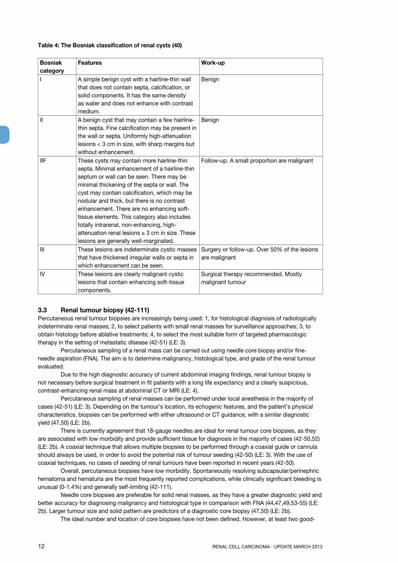
3.2.5 Bosniak classification of renal cystic massesFor the evaluation of renal cystic masses, the Bosniak classification classifies renal cysts into five categories based on their CT imaging appearance, in an attempt to predict the risk of malignancy (40,41) (LE: 3). The Bosniak system also advocates treatment for each category (Table 4).
12 RENAL CELL CARCINOMA - UPDATE MARCH 2013
Table 4: The Bosniak classification of renal cysts (40)
Bosniak category
Features Work-up
I A simple benign cyst with a hairline-thin wall that does not contain septa, calcification, or solid components. It has the same density as water and does not enhance with contrast medium.
Benign
II A benign cyst that may contain a few hairline-thin septa. Fine calcification may be present in the wall or septa. Uniformly high-attenuation lesions < 3 cm in size, with sharp margins but without enhancement.
Benign
IIF These cysts may contain more hairline-thin septa. Minimal enhancement of a hairline-thin septum or wall can be seen. There may be minimal thickening of the septa or wall. The cyst may contain calcification, which may be nodular and thick, but there is no contrast enhancement. There are no enhancing soft-tissue elements. This category also includes totally intrarenal, non-enhancing, high-attenuation renal lesions ≥ 3 cm in size. These lesions are generally well-marginated.
Follow-up. A small proportion are malignant
III These lesions are indeterminate cystic masses that have thickened irregular walls or septa in which enhancement can be seen.
Surgery or follow-up. Over 50% of the lesions are malignant
IV These lesions are clearly malignant cystic lesions that contain enhancing soft-tissue components.
Surgical therapy recommended. Mostly malignant tumour
3.3 Renal tumour biopsy (42-111) Percutaneous renal tumour biopsies are increasingly being used: 1, for histological diagnosis of radiologically indeterminate renal masses; 2, to select patients with small renal masses for surveillance approaches; 3, to obtain histology before ablative treatments; 4, to select the most suitable form of targeted pharmacologic therapy in the setting of metastatic disease (42-51) (LE: 3). Percutaneous sampling of a renal mass can be carried out using needle core biopsy and/or fine-needle aspiration (FNA). The aim is to determine malignancy, histological type, and grade of the renal tumour evaluated. Due to the high diagnostic accuracy of current abdominal imaging findings, renal tumour biopsy is not necessary before surgical treatment in fit patients with a long life expectancy and a clearly suspicious, contrast-enhancing renal mass at abdominal CT or MRI (LE: 4). Percutaneous sampling of renal masses can be performed under local anesthesia in the majority of cases (42-51) (LE: 3). Depending on the tumour’s location, its echogenic features, and the patient’s physical characteristics, biopsies can be performed with either ultrasound or CT guidance, with a similar diagnostic yield (47,50) (LE: 2b). There is currently agreement that 18-gauge needles are ideal for renal tumour core biopsies, as they are associated with low morbidity and provide sufficient tissue for diagnosis in the majority of cases (42-50,52) (LE: 2b). A coaxial technique that allows multiple biopsies to be performed through a coaxial guide or cannula should always be used, in order to avoid the potential risk of tumour seeding (42-50) (LE: 3). With the use of coaxial techniques, no cases of seeding of renal tumours have been reported in recent years (42-50). Overall, percutaneous biopsies have low morbidity. Spontaneously resolving subcapsular/perinephric hematoma and hematuria are the most frequently reported complications, while clinically significant bleeding is unusual (0-1.4%) and generally self-limiting (42-111). Needle core biopsies are preferable for solid renal masses, as they have a greater diagnostic yield and better accuracy for diagnosing malignancy and histological type in comparison with FNA (44,47,49,53-55) (LE: 2b). Larger tumour size and solid pattern are predictors of a diagnostic core biopsy (47,50) (LE: 2b). The ideal number and location of core biopsies have not been defined. However, at least two good-
RENAL CELL CARCINOMA - UPDATE MARCH 2013 13
quality cores (nonfragmented, > 10 mm in length) should be obtained, and necrotic areas should be avoided in order to maximize the diagnostic yield (42,44,47,48,50) (LE: 4). Peripheral biopsies are preferable for larger tumours, to avoid areas of central necrosis (56) (LE: 2b). In recent series from experienced centers, core biopsies of solid renal tumours have shown a diagnostic yield of 78-97%, high specificity (98-100%), and high sensitivity (86-100%) for the diagnosis of malignancy (42-50,54,55,57-75) (LE: 2b). However, it should be noted that 2.5-22% of core biopsies are nondiagnostic (42-50,54,55,57-75) (LE: 2b). If a biopsy is nondiagnostic, but there are radiologic findings suspicious for malignancy, a further biopsy or surgical exploration should always be considered (LE: 4). Assessment of tumour grade on core biopsies is challenging. The accuracy of Fuhrman grading on biopsies is poor (43-75%), but it can be improved using a simplified two-tier system (high-grade vs. low grade) 42-50,54,55,57-75) (LE: 2b). Core biopsies have a low diagnostic yield for cystic renal masses and should not be recommended alone in these cases, unless areas with a solid pattern are present (Bosniak IV cysts) (47,50) (LE: 2b). Combined FNA and core biopsies can provide complementary results, especially for complex cystic lesions (49,55,57,58,73,76,77) (LE: 3).
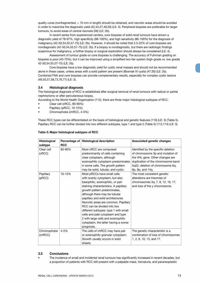
3.4 Histological diagnosisThe histological diagnosis of RCC is established after surgical removal of renal tumours with radical or partial nephrectomy or after percutaneous biopsy.According to the World Health Organization (112), there are three major histological subtypes of RCC:• Clear cell (cRCC, 80-90%)• Papillary (pRCC, 10-15%)• Chromophobe (chRCC, 4-5%)
These RCC types can be differentiated on the basis of histological and genetic features (110) (LE: 3) (Table 5). Papillary RCC can be further divided into two different subtypes, type 1 and type 2 (Table 5) (113,114) (LE: 3).
Table 5: Major histological subtypes of RCC
Histological subtype
percentage of RCC
Histological description Associated genetic changes
Clear cell (cRCC)
80-90% Most cRCC are composed predominantly of cells containing clear cytoplasm, although eosinophilic cytoplasm predominates in some cells. The growth pattern may be solid, tubular, and cystic.
Identified by the specific deletion of chromosome 3p and mutation of the VHL gene. Other changes are duplication of the chromosome band 5q22, deletion of chromosome 6q, 8p, 9p, and 14q.
Papillary (pRCC)
10-15% Most pRCCs have small cells with scanty cytoplasm, but also basophilic, eosinophilic, or pail-staining characteristics. A papillary growth pattern predominates, although there may be tubular papillary and solid architectures. Necrotic areas are common. Papillary RCC can be divided into two different subtypes: type 1 with small cells and pale cytoplasm and type 2 with large cells and eosinophilic cytoplasm, the latter having a worse prognosis.
The most consistent genetic alterations are trisomies of chromosomes 3q, 7, 8, 12, 16, 17, and loss of the y chromosome.
Chromophobe (chRCC)
4-5% The cells of chRCC may have pail or eosinophilic granular cytoplasm. Growth usually occurs in solid sheets.
The genetic characteristic is a combination of loss of chromosomes 1, 2, 6, 10, 13, and 17.
3.5 Conclusions• The incidence of small and incidental renal tumours has significantly increased in recent decades, but
a proportion of patients with RCC still present with a palpable mass, hematuria, and paraneoplastic
14 RENAL CELL CARCINOMA - UPDATE MARCH 2013
and metastatic symptoms (LE: 3). Appropriate staging of RCC requires abdominal CT or MRI and chest imaging (LE: 3). Chest CT is the most sensitive approach for detecting lung metastases, but at least a chest radiograph should be performed for chest staging. There is no role for routine bone scanning or brain CT or MRI in the standard clinical work-up of asymptomatic patients.
• Percutaneous renal tumour biopsies are increasingly being used: 1, to establish the diagnosis of radiologically indeterminate renal masses; 2, to obtain histology of incidentally detected renal masses in patients who are candidates for nonsurgical treatment (active surveillance, ablative therapies); and 3, to select the most suitable targeted therapy for metastatic renal tumours.
3.6 Recommendations
gRIn a patient with one or more suspicious laboratory or physical findings, the possible presence of RCC should be suspected.
B
Contrast-enhanced abdominal CT and MRI are recommended for the work-up of patients with RCC. These are the most appropriate imaging modalities for renal tumour staging prior to surgery.
A
A chest CT is most sensitive for assessment of the lung, but at least a plain chest radiograph should be taken for clinical staging.
A
In patients at risk for bone metastases (raised alkaline phosphatase level or bone pain), further evaluation with a bone scan is needed.
A
Evaluation of renal function is recommended before treatment decision in any patient in whom renal impairment is suspected.
B
Percutaneous biopsy is always required before ablative therapy and systemic therapy without previous pathology.
A
Percutaneous biopsy is recommended in active surveillance strategies in order to stratify the follow-up according to tumour histology.
B
When biopsy is indicated, good-quality needle cores should be obtained with a coaxial technique in order to increase the safety of the procedure and maximise its diagnostic yield.
B
3.7 References1. Jayson M, Sanders H. Increased incidence of serendipitously discovered renal cell carcinoma. Urology
1998 Feb;51(2):203-5.http://www.ncbi.nlm.nih.gov/pubmed/9495698
2. Novara G, Ficarra V, Antonelli A, et al. Validation of the 2009 TNM version in a large multi-institutional cohort of patients treated for renal cell carcinoma: are further improvements needed? Eur Urol 2010 Oct;58(4):588-95.http://www.ncbi.nlm.nih.gov/pubmed/20674150
3. Lee CT, Katz J, Fearn PA, et al. Mode of presentation of renal cell carcinoma provides prognostic information. Urol Oncol 2002 Jul-Aug;7(4):135-40.http://www.ncbi.nlm.nih.gov/pubmed/12474528
4. Patard JJ, Leray E, Rodriguez A, et al. Correlation between symptom graduation, tumor characteristics and survival in renal cell carcinoma. Eur Urol 2003 Aug;44(2):226-32.http://www.ncbi.nlm.nih.gov/pubmed/12875943
5. Kim HL, Belldegrun AS, Freitas DG, et al. Paraneoplastic signs and symptoms of renal cell carcinoma: implications for prognosis. J Urol 2003 Nov;170(5):1742-6.http://www.ncbi.nlm.nih.gov/pubmed/14532767
6. Motzer RJ, Bacik J, Murphy BA, et al. Interferon-alfa as a comparative treatment for clinical trials ofnew therapies against advanced renal cell carcinoma. J Clin Oncol 2002 Jan 1;20(1):289-96.http://www.ncbi.nlm.nih.gov/pubmed/11773181
7. Sufrin G, Chasan S, Golio A, et al. Paraneoplastic and serologic syndromes of renal adenocarcinoma. Semin Urol 1989 Aug;7(3):158-71.http://www.ncbi.nlm.nih.gov/pubmed/2690260
8. Uzzo RG, Novick AC. Nephron sparing surgery for renal tumors: indications, techniques and outcomes. J Urol 2001 Jul;66(1):6-18.http://www.ncbi.nlm.nih.gov/pubmed/11435813
9. Huang WC, Levey AS, Serio AM, et al. Chronic kidney disease after nephrectomy in patients with renal cortical tumours: a retrospective cohort study. Lancet Oncol 2006 Sep;7(9):735-40.http://www.ncbi.nlm.nih.gov/pubmed/16945768
RENAL CELL CARCINOMA - UPDATE MARCH 2013 15
10. Israel GM, Bosniak MA. How I do it: evaluating renal masses. Radiology 2005 Aug;236(2):441-50.http://www.ncbi.nlm.nih.gov/pubmed/16040900
11. Fan L, Lianfang D, Jinfang X, et al. Diagnostic efficacy of contrast-enhanced ultrasonography in solid renal parenchymal lesions with maximum diameters of 5 cm. J Ultrasound Med 2008 Jun;27(6): 875-85.http://www.ncbi.nlm.nih.gov/pubmed/18499847
12. Correas JM, Tranquart F, Claudon M. [Guidelines for contrast enhanced ultrasound (CEUS)-update 2008.] J Radiol 2009 Jan;90(1 Pt 2):123-38. [Article in French]http://www.ncbi.nlm.nih.gov/pubmed/19212280
13. Mitterberger M, Pelzer A, Colleselli D, et al. Contrast-enhanced ultrasound for diagnosis of prostate cancer and kidney lesions. Eur J Radiol 2007 Nov;64(2):231-8.http://www.ncbi.nlm.nih.gov/pubmed/17881175
14. Israel GM, Bosniak MA. Pitfalls in renal mass evaluation and how to avoid them. Radiographics 2008 Sep-Oct;28(5):1325-38.http://www.ncbi.nlm.nih.gov/pubmed/18794310
15. Choudhary S, Rajesh A, Mayer NJ, et al. Renal oncocytoma: CT features cannot reliably distinguish oncocytoma from other renal neoplasms. Clin Radiol 2009 May;64(5):517-22.http://www.ncbi.nlm.nih.gov/pubmed/19348848
16. Rosenkrantz AB, Hindman N, Fitzgerald EF, et al. MRI features of renal oncocytoma and chromophobe renal cell carcinoma. AJR Am J Roentgenol 2010 Dec;195(6):W421-7.http://www.ncbi.nlm.nih.gov/pubmed/21098174
17. Hindman N, Ngo L, Genega EM, et al. Angiomyolipoma with minimal fat: can it be differentiated from clear cell renal cell carcinoma by using standard MR techniques? Radiology 2012 Nov;265(2):468-77.http://www.ncbi.nlm.nih.gov/pubmed/23012463
18. Pedrosa I, Sun MR, Spencer M, et al. MR imaging of renal masses: correlation with findings at surgery and pathologic analysis. Radiographics 2008 Jul-Aug;28(4):985-1003.http://www.ncbi.nlm.nih.gov/pubmed/18635625
19. Gong IH, Hwang J, Choi DK, et al. Relationship among total kidney volume, renal function and age. J Urol 2012 Jan;187(1):344-9.http://www.ncbi.nlm.nih.gov/pubmed/22099987
20. Ferda J, Hora M, Hes O, et al. Assessment of the kidney tumor vascular supply by two-phase MDCTangiography. Eur J Radiol 2007 May;62(2):295-301.http://www.ncbi.nlm.nih.gov/pubmed/17324548
21. Shao P, Tang L, Li P, et al. Precise segmental renal artery clamping under the guidance of dual-source computed tomography angiography during laparoscopic partial nephrectomy. Eur Urol 2012 Dec;62(6):1001-8.http://www.ncbi.nlm.nih.gov/pubmed/22695243
22. Hora M, Stránský P, Trávnícek I, et al. Three-tesla MRI biphasic angiography: a method for preoperative assessment of the vascular supply in renal tumours-a surgical perspective. World J Urol. 2012 Apr 19. [Epub ahead of print]http://www.ncbi.nlm.nih.gov/pubmed/22527675
23. Adey GS, Pedrosa I, Rofsky NM, et al. Lower limits of detection using magnetic resonance imaging for solid components in cystic renal neoplasms. Urology 2008 Jan;71(1):47-51.http://www.ncbi.nlm.nih.gov/pubmed/18242363
24. Janus CL, Mendelson DS. Comparison of MRI and CT for study of renal and perirenal masses. Crit Rev Diagn Imaging 1991;32(2):69-118.http://www.ncbi.nlm.nih.gov/pubmed/1863349
25. Krestin GP, Gross-Fengels W, Marincek B. [The importance of magnetic resonance tomography in the diagnosis and staging of renal cell carcinoma.] Radiologe 1992;32(3):121-6. [Article in German]http://www.ncbi.nlm.nih.gov/pubmed/1565792
26. Mueller-Lisse UG, Mueller-Lisse UL. Imaging of advanced renal cell carcinoma. World J Urol 2010 Jun;28(3):253-61.http://www.ncbi.nlm.nih.gov/pubmed/20458484
27. Kabala JE, Gillatt DA, Persad RA, et al. Magnetic resonance imaging in the staging of renal cell carcinoma. Br J Radiol 1991;64(764):683-9.http://www.ncbi.nlm.nih.gov/pubmed/1884119
28. Putra LG, Minor TX, Bolton DM, et al. Improved assessment of renal lesions in pregnancy with magnetic resonance imaging. Urology 2009 Sep;74(3):535-9.http://www.ncbi.nlm.nih.gov/pubmed/19604560
16 RENAL CELL CARCINOMA - UPDATE MARCH 2013
29. Giannarini G, Petralia G, Thoeny HC. Potential and limitations of diffusion-weighted magnetic resonance imaging in kidney, prostate, and bladder cancer including pelvic lymph node staging: a critical analysis of the literature. Eur Urol 2012 Feb;61(2):326-40.http://www.ncbi.nlm.nih.gov/pubmed/22000497
30. Park JW, Jo MK, Lee HM. Significance of 18F-fluorodeoxyglucose positron-emission tomography/computed tomography for the postoperative surveillance of advanced renal cell carcinoma. BJU Int 2009 Mar;103(5):615-9.http://www.ncbi.nlm.nih.gov/pubmed/19007371
31. Bechtold RE, Zagoria RJ. Imaging approach to staging of renal cell carcinoma. Urol Clin North Am 1997;24(3):507-22.http://www.ncbi.nlm.nih.gov/pubmed/9275976
32. Heidenreich A, Ravery V. European Society of Oncological Urology. Preoperative imaging in renal cell cancer. World J Urol 2004;22(5):307-15.http://www.ncbi.nlm.nih.gov/pubmed/15290202
33. Sheth S, Scatarige JC, Horton KM, et al. Current concepts in the diagnosis and management of renal cell carcinoma: role of multidetector CT and three-dimensional CT. Radiographics 2001;21 Spec No:S237-54.http://www.ncbi.nlm.nih.gov/pubmed/11598260
34. Miles KA, London NJ, Lavelle JM, et al. CT staging of renal carcinoma: a prospective comparison of three dynamic computed tomography techniques. Eur J Radiol 1991;13(1):37-42.http://www.ncbi.nlm.nih.gov/pubmed/1889427
35. Lim DJ, Carter MF. Computerized tomography in the preoperative staging for pulmonary metastases in patients with renal cell carcinoma. J Urol 1993;150(4):1112-4.http://www.ncbi.nlm.nih.gov/pubmed/8371366
36. Koga S, Tsuda S, Nishikido M, et al. The diagnostic value of bone scan in patients with renal cell carcinoma. J Urol 2001 Dec;166(6):2126-8.http://www.ncbi.nlm.nih.gov/pubmed/11696720
37. Marshall ME, Pearson T, Simpson W, et al. Low incidence of asymptomatic brain metastases in patients with renal cell carcinoma. Urology 1990 Oct;36(4):300-2.http://www.ncbi.nlm.nih.gov/pubmed/2219605
38. Henriksson C, Haraldsson G, Aldenborg F, et al. Skeletal metastases in 102 patients evaluated before surgery for renal cell carcinoma. Scand J Urol Nephrol 1992;26(4):363-6.http://www.ncbi.nlm.nih.gov/pubmed/1292074
39. Seaman E, Goluboff ET, Ross S, et al. Association of radionuclide bone scan and serum alkaline phosphatase in patients with metastatic renal cell carcinoma. Urol 1996;48(5):692-5.http://www.ncbi.nlm.nih.gov/pubmed/8911510
40. Warren KS, McFarlane J. The Bosniak classification of renal cystic masses. BJU Int 2005 May;95(7):939-42.http://www.ncbi.nlm.nih.gov/pubmed/15839908
41. Bosniak MA. The use of the Bosniak classification system for renal cysts and cystic tumors. J Urol 1997 May;157(5):1852-3.http://www.ncbi.nlm.nih.gov/pubmed/9112545
42. Neuzillet Y, Lechevallier E, Andre M, et al. Accuracy and clinical role of fine needle percutaneous biopsy with computerized tomography guidance of small (less than 4.0 cm) renal masses. J Urol 2004 May;171(5):1802-5.http://www.ncbi.nlm.nih.gov/pubmed/15076280
43. Shannon BA, Cohen RJ, de Bruto H, et al. The value of preoperative needle core biopsy for diagnosing benign lesions among small, incidentally detected renal masses. J Urol 2008 Oct;180(4):1257-61; discussion 1261.http://www.ncbi.nlm.nih.gov/pubmed/18707712
44. Schmidbauer J, Remzi M, Memarsadeghi M, et al. Diagnostic accuracy of computed tomography-guided percutaneous biopsy of renal masses. Eur Urol 2008 May;53(5):1003-11.http://www.ncbi.nlm.nih.gov/pubmed/18061339
45. Lebret T, Poulain JE, Molinie V, et al. Percutaneous core biopsy for renal masses: indications, accuracy and results. J Urol 2007 Oct;178(4 Pt 1):1184-8; discussion 1188.http://www.ncbi.nlm.nih.gov/pubmed/17698122
46. Maturen KE, Nghiem HV, Caoili EM, et al. Renal mass core biopsy: accuracy and impact on clinical management. AJR Am J Roentgenol 2007 Feb;188(2):563-70.http://www.ncbi.nlm.nih.gov/pubmed/17242269
RENAL CELL CARCINOMA - UPDATE MARCH 2013 17
47. Volpe A, Mattar K, Finelli A, et al. Contemporary results of percutaneous biopsy of 100 small renal masses: a single center experience. J Urol 2008 Dec;180(6):2333-7.http://www.ncbi.nlm.nih.gov/pubmed/18930274
48. Wang R, Wolf JS Jr, Wood DP Jr, et al. Accuracy of percutaneous core biopsy in management of small renal masses. Urology 2009 Mar;73(3):586-90; discussion 590-1.http://www.ncbi.nlm.nih.gov/pubmed/19118884
49. Veltri A, Garetto I, Tosetti I, et al. Diagnostic accuracy and clinical impact of imaging-guided needle biopsy of renal masses. Retrospective analysis on 150 cases. Eur Radiol 2011 Feb;21(2):393-401.http://www.ncbi.nlm.nih.gov/pubmed/20809129
50. Leveridge MJ, Finelli A, Kachura JR, et al. Outcomes of small renal mass needle core biopsy, nondiagnostic percutaneous biopsy, and the role of repeat biopsy. Eur Urol 2011 Sep;60(3):578-84.http://www.ncbi.nlm.nih.gov/pubmed/21704449
51. Abel EJ, Culp SH, Matin SF, et al. Percutaneous biopsy of primary tumor in metastatic renal cell carcinoma to predict high risk pathological features: comparison with nephrectomy assessment. J Urol 2010 Nov;184(5):1877-81.http://www.ncbi.nlm.nih.gov/pubmed/20850148
52. Breda A, Treat EG, Haft-Candell L, et al. Comparison of accuracy of 14-, 18- and 20-G needles in ex-vivo renal mass biopsy: a prospective, blinded study. BJU Int 2010 Apr;105(7):940-5.http://www.ncbi.nlm.nih.gov/pubmed/19888984
53. Aribas BK, Arda K, Aktas E, et al. Percutaneous US-guided needle biopsies of solid renal masses. Neoplasma 2011;58(2):146-52.http://www.ncbi.nlm.nih.gov/pubmed/21275465
54. Beland MD, Mayo-Smith WW, Dupuy DE, et al. Diagnostic yield of 58 consecutive imaging-guided biopsies of solid renal masses: should we biopsy all that are indeterminate? AJR Am J Roentgenol 2007 Mar;188(3):792-7.http://www.ncbi.nlm.nih.gov/pubmed/17312070
55. Li G, Cuilleron M, Zhao A, et al. Combination of core biopsy and fine-needle aspiration increases diagnostic rate for small solid renal tumors. Anticancer Res 2012 Aug;32(8):3463-6.http://www.ncbi.nlm.nih.gov/pubmed/22843931
56. Wunderlich H, Hindermann W, Al Mustafa AM, et al. The accuracy of 250 fine needle biopsies of renal tumors. J Urol 2005 Jul;174(1):44-6.http://www.ncbi.nlm.nih.gov/pubmed/15947574
57. Parks GE, Perkins LA, Zagoria RJ, et al. Benefits of a combined approach to sampling of renal neoplasms as demonstrated in a series of 351 cases. Am J Surg Pathol 2011 Jun;35(6):827-35.http://www.ncbi.nlm.nih.gov/pubmed/21552112
58. Wood BJ, Khan MA, McGovern F, et al. Imaging guided biopsy of renal masses: indications, accuracy and impact on clinical management. J Urol 1999 May;161(5):1470-4.http://www.ncbi.nlm.nih.gov/pubmed/10210375
59. Blumenfeld AJ, Guru K, Fuchs GJ, et al. Percutaneous biopsy of renal cell carcinoma underestimates nuclear grade. Urology 2010 Sep;76(3):610-3.http://www.ncbi.nlm.nih.gov/pubmed/20163843
60. Caoili EM, Bude RO, Higgins EJ, et al. Evaluation of sonographically guided percutaneous core biopsy of renal masses. AJR Am J Roentgenol 2002 Aug;179(2):373-8.http://www.ncbi.nlm.nih.gov/pubmed/12130435
61. Chyhrai A, Sanjmyatav J, Gajda M, et al. Multi-colour FISH on preoperative renal tumour biopsies to confirm the diagnosis of uncertain renal masses. World J Urol 2010 Jun;28(3):269-74.http://www.ncbi.nlm.nih.gov/pubmed/20390284
62. Eshed I, Elias S, Sidi AA. Diagnostic value of CT-guided biopsy of indeterminate renal masses. Clin Radiol 2004 Mar;59(3):262-7.http://www.ncbi.nlm.nih.gov/pubmed/15037139
63. Hara I, Miyake H, Hara S, et al. Role of percutaneous image-guided biopsy in the evaluation of renal masses. Urol Int 2001;67(3):199-202.http://www.ncbi.nlm.nih.gov/pubmed/11598445
64. Lechevallier E, André M, Barriol D, et al. Fine-needle percutaneous biopsy of renal masses with helical CT guidance. Radiology 2000 Aug;216(2):506-10.http://www.ncbi.nlm.nih.gov/pubmed/10924578
65. Rybicki FJ, Shu KM, Cibas ES, et al. Percutaneous biopsy of renal masses: sensitivity and negative predictive value stratified by clinical setting and size of masses. AJR Am J Roentgenol 2003 May;180(5):1281-7.http://www.ncbi.nlm.nih.gov/pubmed/12704038
18 RENAL CELL CARCINOMA - UPDATE MARCH 2013
66. Sofikerim M, Tatlisen A, Canoz O, et al. What is the role of percutaneous needle core biopsy in diagnosis of renal masses? Urology 2010 Sep;76(3):614-8.http://www.ncbi.nlm.nih.gov/pubmed/20110106
67. Somani BK, Nabi G, Thorpe P, et al. Image-guided biopsy-diagnosed renal cell carcinoma: critical appraisal of technique and long-term follow-up. Eur Urol 2007 May;51(5):1289-95; discussion 1296-7.http://www.ncbi.nlm.nih.gov/pubmed/17081679
68. Tan HJ, Jacobs BL, Hafez KS, et al. Understanding the role of percutaneous biopsy in the management of patients with a small renal mass. Urology 2012 Feb;79(2):372-7.http://www.ncbi.nlm.nih.gov/pubmed/22310755
69. Vasudevan A, Davies RJ, Shannon BA, et al. Incidental renal tumours: the frequency of benign lesions and the role of preoperative core biopsy. BJU Int 2006 May;97(5):946-9.http://www.ncbi.nlm.nih.gov/pubmed/16643475
70. Thuillier C, Long JA, Lapouge O, et al. [Value of percutaneous biopsy for solid renal tumours less than 4 cm in diameter based on a series of 53 cases]. Prog Urol 2008 Jul;18(7):435-9. [Article in French].http://www.ncbi.nlm.nih.gov/pubmed/18602603
71. Shah RB, Bakshi N, Hafez KS, et al. Image-guided biopsy in the evaluation of renal mass lesions in contemporary urological practice: indications, adequacy, clinical impact, and limitations of the pathological diagnosis. Hum Pathol 2005 Dec;36(12):1309-15.http://www.ncbi.nlm.nih.gov/pubmed/16311125
72. Reichelt O, Gajda M, Chyhrai A, et al. Ultrasound-guided biopsy of homogenous solid renal masses. Eur Urol 2007 Nov;52(5):1421-6.http://www.ncbi.nlm.nih.gov/pubmed/17306920
73. Torp-Pedersen S, Juul N, Larsen T, et al. US-guided fine needle biopsy of solid renal masses--comparison of histology and cytology. Scand J Urol Nephrol Suppl 1991;137:41-3.http://www.ncbi.nlm.nih.gov/pubmed/1947839
74. Jaff A, Molinié V, Mellot F, et al. Evaluation of imaging-guided fine-needle percutaneous biopsy of renal masses. Eur Radiol 2005 Aug;15(8):1721-6.http://www.ncbi.nlm.nih.gov/pubmed/15627185
75. Kroeze SG, Huisman M, Verkooijen HM, et al. Real-time 3D fluoroscopy-guided large core needle biopsy of renal masses: a critical early evaluation according to the IDEAL recommendations. Cardiovasc Intervent Radiol 2012 Jun;35(3):680-5.http://www.ncbi.nlm.nih.gov/pubmed/21822769
76. Harisinghani MG, Maher MM, Gervais DA, et al. Incidence of malignancy in complex cystic renal masses (Bosniak category III): should imaging-guided biopsy precede surgery? AJR Am J Roentgenol 2003 Mar;180(3):755-8.http://www.ncbi.nlm.nih.gov/pubmed/12591691
77. Lang EK, Macchia RJ, Gayle B, et al. CT-guided biopsy of indeterminate renal cystic masses (Bosniak 3 and 2F): accuracy and impact on clinical management. Eur Radiol 2002 Oct;12(10):2518-24.http://www.ncbi.nlm.nih.gov/pubmed/12271393
78. Al Nazer M, Mourad WA. Successful grading of renal-cell carcinoma in fine-needle aspirates. Diagn Cytopathol 2000 Apr;22(4):223-6.http://www.ncbi.nlm.nih.gov/pubmed/10787141
79. Amendola MA, Bree RL, Pollack HM, et al. Small renal cell carcinomas: resolving a diagnostic dilemma. Radiology 1988 Mar;166(3):637-41.http://www.ncbi.nlm.nih.gov/pubmed/3277239
80. Andonian S, Okeke Z, Okeke DA, et al. Number of needle passes does not correlate with the diagnostic yield of renal fine needle aspiration cytology. J Endourol 2008 Oct;22(10):2377-80.http://www.ncbi.nlm.nih.gov/pubmed/18937600
81. Bielsa Gali O, Arango Toro O, Cortadellas Angel R, et al. [The preoperative diagnosis of complex renal cystic masses]. Arch Esp Urol 1999 Jan-Feb;52(1):19-25. [Article in Spanish].http://www.ncbi.nlm.nih.gov/pubmed/10101883
82. Bishop JA, Hosler GA, Kulesza P, et al. Diagn Cytopathol 2011 Mar;39(3):168-71. Fine-needle aspiration of renal cell carcinoma: is accurate Fuhrman grading possible on cytologic material?http://www.ncbi.nlm.nih.gov/pubmed/21319316
83. Brierly RD, Thomas PJ, Harrison NW, et al. Evaluation of fine-needle aspiration cytology for renal masses. BJU Int 2000 Jan;85(1):14-8.http://www.ncbi.nlm.nih.gov/pubmed/10619937
RENAL CELL CARCINOMA - UPDATE MARCH 2013 19
84. Cajulis RS, Katz RL, Dekmezian R, et al. Fine needle aspiration biopsy of renal cell carcinoma. Cytologic parameters and their concordance with histology and flow cytometric data. Acta Cytol 1993 May-Jun;37(3):367-72.http://www.ncbi.nlm.nih.gov/pubmed/8498138
85. Campbell SC, Novick AC, Herts B, et al. Prospective evaluation of fine needle aspiration of small, solid renal masses: accuracy and morbidity. Urology 1997 Jul;50(1):25-9.http://www.ncbi.nlm.nih.gov/pubmed/9218014
86. Civardi G, Cavanna L, Fornari F, et al. [Echographically guided, percutaneous, fine-needle puncture in the diagnosis of renal masses suspected of malignancy]. Recenti Prog Med 1986 Sep;77(9):420-2. [Article in Italian]. [No abstract available].http://www.ncbi.nlm.nih.gov/pubmed/3797794
87. Cristallini EG, Paganelli C, Bolis GB. Role of fine-needle aspiration biopsy in the assessment of renal masses. Diagn Cytopathol 1991;7(1):32-5.http://www.ncbi.nlm.nih.gov/pubmed/2026081
88. Elder DD, Orell SR, Sage MR, et al. The diagnosis and local staging of renal cancer--an appraisal. Aust N Z J Surg 1984 Jun;54(3):219-21.http://www.ncbi.nlm.nih.gov/pubmed/6590018
89. García-Solano J, Acosta-Ortega J, Pérez-Guillermo M, et al. Solid renal masses in adults: image-guided fine-needle aspiration cytology and imaging techniques--”two heads better than one?”. Diagn Cytopathol 2008 Jan;36(1):8-12.http://www.ncbi.nlm.nih.gov/pubmed/18064683
90. Haubek A, Lundorf E, Lauridsen KN. Diagnostic strategy in renal mass lesions. Scand J Urol Nephrol Suppl 1991;137:35-9.http://www.ncbi.nlm.nih.gov/pubmed/1947838
91. Izumi K, Narimoto K, Sugimoto K, et al. The role of percutaneous needle biopsy in differentiation of renal tumors. Jpn J Clin Oncol 2010 Nov;40(11):1081-6.http://www.ncbi.nlm.nih.gov/pubmed/20601495
92. Johnson PT, Nazarian LN, Feld RI, et al. Sonographically guided renal mass biopsy: indications and efficacy. J Ultrasound Med 2001 Jul;20(7):749-53; quiz 755.http://www.ncbi.nlm.nih.gov/pubmed/11444733
93. Juul N, Torp-Pedersen S, Grønvall S, et al. Ultrasonically guided fine needle aspiration biopsy of renal masses. J Urol 1985 Apr;133(4):579-81.http://www.ncbi.nlm.nih.gov/pubmed/3884841
94. Karp W, Ekelund L. Ultrasound, angiography and fine needle aspiration biopsy in diagnosis of renal neoplasms. Acta Radiol Diagn (Stockh) 1979;20(4):649-59.http://www.ncbi.nlm.nih.gov/pubmed/525406
95. Kelley CM, Cohen MB, Raab SS. Utility of fine-needle aspiration biopsy in solid renal masses. Diagn Cytopathol 1996 Feb;14(1):14-9.http://www.ncbi.nlm.nih.gov/pubmed/8834071
96. Leiman G. Audit of fine needle aspiration cytology of 120 renal lesions. Cytopathology 1990;1(2): 65-72.http://www.ncbi.nlm.nih.gov/pubmed/2102349
97. Li G, Cuilleron M, Cottier M, et al. The use of MN/CA9 gene expression in identifying malignant solid renal tumors. Eur Urol 2006 Feb;49(2):401-5.http://www.ncbi.nlm.nih.gov/pubmed/16387417
98. Masoom S, Venkataraman G, Jensen J, et al. Renal FNA-based typing of renal masses remains a useful adjunctive modality: evaluation of 31 renal masses with correlative histology. Cytopathology 2009 Feb;20(1):50-5.http://www.ncbi.nlm.nih.gov/pubmed/18476991
99. Mignon F, Mesurolle B, Ariche-Cohen M, et al. [Value of CT guided renal biopsies: retrospective review of 67 cases]. J Radiol 2001 Aug;82(8):907-11. [Article in French]http://www.ncbi.nlm.nih.gov/pubmed/11604686
100. Mondal A, Ghosh E. Fine needle aspiration cytology (FNAC) in the diagnosis of solid renal masses--a study of 92 cases. Indian J Pathol Microbiol 1992 Oct;35(4):333-9.http://www.ncbi.nlm.nih.gov/pubmed/1344223
101. Nadel L, Baumgartner BR, Bernardino ME. Percutaneous renal biopsies: accuracy, safety, and indications. Urol Radiol 1986;8(2):67-71.http://www.ncbi.nlm.nih.gov/pubmed/3024375

20 RENAL CELL CARCINOMA - UPDATE MARCH 2013
102. Niceforo J, Coughlin BF. Diagnosis of renal cell carcinoma: value of fine-needle aspiration cytology in patients with metastases or contraindications to nephrectomy. AJR Am J Roentgenol 1993 Dec;161(6):1303-5.http://www.ncbi.nlm.nih.gov/pubmed/8249747
103. Orell SR, Langlois SL, Marshall VR. Fine needle aspiration cytology in the diagnosis of solid renal and adrenal masses. Scand J Urol Nephrol 1985;19(3):211-6.http://www.ncbi.nlm.nih.gov/pubmed/3906863
104. Pilotti S, Rilke F, Alasio L, Garbagnati F. The role of fine needle aspiration in the assessment of renal masses. Acta Cytol 1988 Jan-Feb;32(1):1-10.http://www.ncbi.nlm.nih.gov/pubmed/3336946
105. Renshaw AA, Lee KR, Madge R, et al. Accuracy of fine needle aspiration in distinguishing subtypes of renal cell carcinoma. Acta Cytol 1997 Jul-Aug;41(4):987-94.http://www.ncbi.nlm.nih.gov/pubmed/9250289
106. Richter F, Kasabian NG, Irwin RJ Jr, et al. Accuracy of diagnosis by guided biopsy of renal mass lesions classified indeterminate by imaging studies. Urology 2000 Mar;55(3):348-52.http://www.ncbi.nlm.nih.gov/pubmed/10699608
107. Strojan Fležar M, Gutnik H, Jeruc J, et al. Typing of renal tumors by morphological and immunocytochemical evaluation of fine needle aspirates. Virchows Arch 2011 Dec;459(6):607-14.http://www.ncbi.nlm.nih.gov/pubmed/22052200
108. Tikkakoski T, Päivänsalo M, Apaja-Sarkkinen M, et al. Ultrasound-guided aspiration cytology of renal expansions. Rontgenblatter 1990 Dec;43(12):502-6.http://www.ncbi.nlm.nih.gov/pubmed/2287877
109. Todd TD, Dhurandhar B, Mody D, et al. Fine-needle aspiration of cystic lesions of the kidney. Morphologic spectrum and diagnostic problems in 41 cases. Am J Clin Pathol 1999 Mar;111(3): 317-28.http://www.ncbi.nlm.nih.gov/pubmed/10078106
110. Truong LD, Todd TD, Dhurandhar B, et al. Fine-needle aspiration of renal masses in adults: analysis of results and diagnostic problems in 108 cases. Diagn Cytopathol 1999 Jun;20(6):339-49.http://www.ncbi.nlm.nih.gov/pubmed/10352906
111. Zardawi IM. Renal fine needle aspiration cytology. Acta Cytol 1999 Mar-Apr;43(2):184-90.http://www.ncbi.nlm.nih.gov/pubmed/10097707
112. Eble JN, Sauter G, Epstein JI, et al (eds). In: Pathology and genetics of tumours of the urinary systemand male genital organs. World Health Organization Classification of Tumours. Lyon: IARC Press, 2004.
113. Pignot G, Elie C, Conquy S, et al. Survival analysis of 130 patients with papillary renal cell carcinoma: prognostic utility of type 1 and type 2 subclassification. Urology 2007 Feb;69(2):230-5.http://www.ncbi.nlm.nih.gov/pubmed/17275070
114. Delahunt B, Eble JN, McCredie MR, et al. Morphologic typing of papillary renal cell carcinoma: comparison of growth kinetics and patient survival in 66 cases. Hum Pathol 2001 Jun;32(6):590-5.http://www.ncbi.nlm.nih.gov/pubmed/11431713
4. CLASSIFICATION AND pROgNOSTIC FACTORS4.1 ClassificationThe TNM classification system is generally recommended for clinical and scientific use (1). However, thesystem requires continuous improvements (2). The latest version of the TNM classification was published in 2010 (Table 6). The prognostic value of the 2010 TNM classification has been confirmed in both single and multi-institution studies (3,4). However, some uncertainties remain:• The sub-classification of T1 tumours using a cut-off of 4 cm might not be optimal with the widening of
nephron-sparing surgery for localised cancer. • The value of size stratification of T2 tumours has been questioned (5).• Since the 2002 version of the TNM classification, tumours with renal sinus fat invasion have been
classified as pT3a. However, accumulating data suggest that renal sinus fat invasion carries a worse prognosis than perinephric fat invasion and therefore should not be included in the same pT3a stage group (LE: 3) (6-8).
• Some substages of the classification (pT2b, pT3a, pT3c and pT4) may overlap (4).• The accuracy of the N1-N2 sub-classification has been questioned (9) (LE: 3). For adequate M staging
RENAL CELL CARCINOMA - UPDATE MARCH 2013 21
of patients with RCC, accurate preoperative imaging (currently, chest and abdominal CT) should be performed (10,11) (LE: 4).
4.2 prognostic factorsFactors influencing prognosis can be classified into: anatomical, histological, clinical, and molecular.
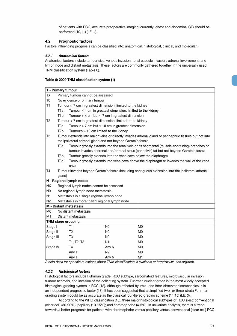
4.2.1 Anatomical factorsAnatomical factors include tumour size, venous invasion, renal capsule invasion, adrenal involvement, and lymph node and distant metastasis. These factors are commonly gathered together in the universally used TNM classification system (Table 6).
Table 6: 2009 TNM classification system (1)
T - primary tumourTX Primary tumour cannot be assessedT0 No evidence of primary tumourT1 Tumour < 7 cm in greatest dimension, limited to the kidney
T1a Tumour < 4 cm in greatest dimension, limited to the kidneyT1b Tumour > 4 cm but < 7 cm in greatest dimension
T2 Tumour > 7 cm in greatest dimension, limited to the kidneyT2a Tumour > 7 cm but < 10 cm in greatest dimensionT2b Tumours > 10 cm limited to the kidney
T3 Tumour extends into major veins or directly invades adrenal gland or perinephric tissues but not into the ipsilateral adrenal gland and not beyond Gerota’s fascia
T3a Tumour grossly extends into the renal vein or its segmental (muscle-containing) branches or tumour invades perirenal and/or renal sinus (peripelvic) fat but not beyond Gerota’s fascia
T3b Tumour grossly extends into the vena cava below the diaphragm T3c Tumour grossly extends into vena cava above the diaphragm or invades the wall of the vena
cavaT4 Tumour invades beyond Gerota’s fascia (including contiguous extension into the ipsilateral adrenal
gland)N - Regional lymph nodesNX Regional lymph nodes cannot be assessedN0 No regional lymph node metastasisN1 Metastasis in a single regional lymph nodeN2 Metastasis in more than 1 regional lymph nodeM - Distant metastasisM0 No distant metastasisM1 Distant metastasisTNM stage groupingStage I T1 N0 M0Stage II T2 N0 M0Stage III T3 N0 M0
T1, T2, T3 N1 M0Stage IV T4 Any N M0
Any T N2 M0Any T Any N M1
A help desk for specific questions about TNM classification is available at http://www.uicc.org/tnm.
4.2.2 Histological factorsHistological factors include Fuhrman grade, RCC subtype, sarcomatoid features, microvascular invasion, tumour necrosis, and invasion of the collecting system. Fuhrman nuclear grade is the most widely accepted histological grading system in RCC (12). Although affected by intra- and inter-observer discrepancies, it is an independent prognostic factor (13). It has been suggested that a simplified two- or three-strata Fuhrman grading system could be as accurate as the classical four-tiered grading scheme (14,15) (LE: 3). According to the WHO classification (16), three major histological subtypes of RCC exist: conventional (clear cell) (80-90%); papillary (10-15%); and chromophobe (4-5%). In univariate analysis, there is a trend towards a better prognosis for patients with chromophobe versus papillary versus conventional (clear cell) RCC
22 RENAL CELL CARCINOMA - UPDATE MARCH 2013
(17,18). However, the prognostic information provided by the RCC subtype is lost when stratified to tumour stage (18,19) (LE: 3). Among papillary RCCs, two subgroups with different outcomes have been identified (20): Type 1 are low-grade tumours with a chromophilic cytoplasm and a favourable prognosis. Type 2 are mostly high-grade tumours with an eosinophilic cytoplasm and a great propensity for developing metastases (LE: 3). RCC with Xp 11.2 translocation has been associated with a poor prognosis (21). Its incidence is low but should be systematically addressed in young patients. The RCC type classification has been confirmed at the molecular level by cytogenetic and genetic analyses (22-24) (LE: 2b).
4.2.3 Clinical factorsClinical factors include patient performance status, localised symptoms, cachexia, anaemia, and platelet count(25-28) (LE: 3).
4.2.4 Molecular factorsNumerous molecular markers being investigated, including: carbonic anhydrase IX (CaIX), vascular endothelialgrowth factor (VEGF), hypoxia-inducible factor (HIF), Ki67 (proliferation), p53, PTEN (phosphatase and tensinhomolog) (cell cycle), E-cadherin, C-reactive protein (CRP), osteopontin (29) and CD44 (cell adhesion) (30,31) (LE: 3). To date, none of these markers has been shown to improve the predictive accuracy of current prognostic systems and their use is therefore not recommended in routine practice. Finally, even though gene expression profiling seems a promising method, it has not helped so far to identify new relevant prognostic factors (32).
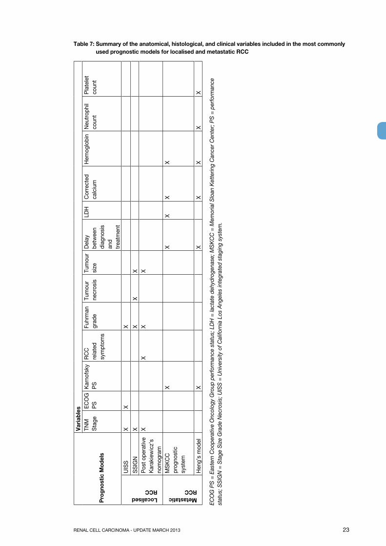
4.2.5 Prognostic systems and nomogramsPostoperative prognostic systems and nomograms that combine independent prognostic factors have been developed and externally validated (33-39). These systems may be more accurate than TNM stage or Fuhrman grade alone for predicting survival (LE: 3). An important advantage of nomograms is their ability to measure predictive accuracy (PA), which enables all new predictive parameters to be objectively evaluated. Before being adopted, every new prognostic variable or system should be able to demonstrate that its PA is superior to conventional postoperative histo-prognostic schemes (40). Recently, new preoperative nomograms with excellent PAs have been designed (41,42). Table 7 summarises the current most relevant prognostic systems.
4.3 Conclusions
LEIn patients with RCC, TNM stage, nuclear grade according to Fuhrman, and RCC subtype (WHO, 2004; [21]), should be performed because they contribute important prognostic information.
2
Prognostic systems should currently be used in a metastatic setting and are still investigational in localised disease.
2
4.4 Recommendations
gRThe current TNM classification system is recommended because it has consequences for prognosis and therapy.
B
The Fuhrman grading system and classification of RCC subtype should be used. BA stratification system should be used in a metastatic setting for selecting the appropriate first-line treatment.
B
In localised disease, the use of integrated prognostic systems or nomograms is not routinely recommended, even though these systems can provide a rationale for enrolling patients into clinical trials.
B
No molecular prognostic marker is currently recommended for routine clinical use. B
RENAL CELL CARCINOMA - UPDATE MARCH 2013 23
Table 7: Summary of the anatomical, histological, and clinical variables included in the most commonly used prognostic models for localised and metastatic RCC
pro
gnos
tic M
odel
s
Var
iabl
esTN
M
Sta
geEC
OG
P
SK
arno
fsky
PS
RC
C
rela
ted
sym
ptom
s
Fuhr
man
gr
ade
Tum
our
necr
osis
Tum
our
size
Del
ay
betw
een
diag
nosi
s an
d tr
eatm
ent
LDH
Cor
rect
ed
calc
ium
Hem
oglo
bin
Neu
trop
hil
coun
tP
late
let
coun
t
Localised RCC
UIS
SX
XX
SS
IGN
XX
XX
Pos
t ope
rativ
e K
arak
iew
icz’
s no
mog
ram
XX
XX
Metastatic RCC
MS
KC
C
prog
nost
ic
syst
em
XX
XX
X
Hen
g’s
mod
elX
XX
XX
X
ECO
G P
S =
Eas
tern
Coo
pera
tive
Onc
olog
y G
roup
per
form
ance
sta
tus;
LD
H =
lact
ate
dehy
drog
enas
e; M
SK
CC
= M
emor
ial S
loan
Ket
terin
g C
ance
r Cen
ter;
PS
= p
erfo
rman
ce
stat
us; S
SIG
N =
Sta
ge S
ize
Gra
de N
ecro
sis;
UIS
S =
Uni
vers
ity o
f Cal
iforn
ia L
os A
ngel
es in
tegr
ated
sta
ging
sys
tem
.
24 RENAL CELL CARCINOMA - UPDATE MARCH 2013
4.5 References1. Sobin LH, Gospodariwicz M, Wittekind C (eds). TNM classification of malignant tumors. UICC
International Union Against Cancer. 7th edn. Wiley-Blackwell, 2009: pp. 255-257.http://www.uicc.org/tnm
2. Gospodarowicz MK, Miller D, Groome PA, et al. The process for continuous improvement of the TNM classification. Cancer 2004 Jan;100(1):1-5.http://www.ncbi.nlm.nih.gov/pubmed/14692017
3. Kim SP, Alt AL, Weight CJ, et al. Independent validation of the 2010 American Joint Committee on Cancer TNM classification for renal cell carcinoma: results from a large, single institution cohort. J Urol 2011 Jun;185(6):2035-9.http://www.ncbi.nlm.nih.gov/pubmed/21496854
4. Novara G, Ficarra V, Antonelli A, et al; SATURN Project-LUNA Foundation. Validation of the 2009 TNM version in a large multi-institutional cohort of patients treated for renal cell carcinoma: are further improvements needed? Eur Urol 2010 Oct;58(4):588-95http://www.ncbi.nlm.nih.gov/pubmed/20674150
5. Waalkes S, Becker F, Schrader AJ, et al. Is there a need to further subclassify pT2 renal cell cancers as implemented by the revised 7th TNM version? Eur Urol 2011 Feb;59(2):258-63. http://www.ncbi.nlm.nih.gov/pubmed/21030143
6. Bertini R, Roscigno M, Freschi M, et al. Renal sinus fat invasion in pT3a clear cell renal cell carcinoma affects outcomes of patients without nodal involvement or distant metastases. J Urol 2009 May;181(5):2027-32.http://www.ncbi.nlm.nih.gov/pubmed/19286201
7. Poon SA, Gonzalez JR, Benson MC, et al. Invasion of renal sinus fat is not an independent predictor of survival in pT3a renal cell carcinoma. BJU Int 2009 Jun;103(12):1622-5.http://www.ncbi.nlm.nih.gov/pubmed/19154464
8. Bedke J, Buse S, Pritsch M, et al. Perinephric and renal sinus fat infiltration in pT3a renal cell carcinoma: possible prognostic differences. BJU Int 2009 May;103(10):1349-54.http://www.ncbi.nlm.nih.gov/pubmed/19076147
9. Terrone C, Cracco F, Porpiglia F, et al. Reassessing the current TNM lymph node staging for renal cell carcinoma. Eur Urol 2006 Feb;49(2):324-31.http://www.ncbi.nlm.nih.gov/pubmed/16386352
10. Heidenreich A, Ravery V; European Society of Oncological Urology. Preoperative imaging in renal cell cancer. World J Urol 2004 Nov;22(5):307-15.http://www.ncbi.nlm.nih.gov/pubmed/15290202
11. Sheth S, Scatarige JC, Horton KM, et al. Current concepts in the diagnosis and management of renal cell carcinoma: role of multidetector CT and three-dimensional CT. Radiographics 2001 Oct;21. Spec No:S237-54.http://www.ncbi.nlm.nih.gov/pubmed/11598260
12. Fuhrman SA, Lasky LC, Limas C. Prognostic significance of morphologic parameters in renal cell carcinoma. Am J Surg Pathol 1982 Oct;6(7):655-63.http://www.ncbi.nlm.nih.gov/pubmed/7180965
13. Lang H, Lindner V, de Fromont M, et al. Multicenter determination of optimal interobserver agreement using the Fuhrman grading system for renal cell carcinoma: assessment of 241 patients with > 15-year follow-up. Cancer 2005 Feb;103(3):625-9.http://www.ncbi.nlm.nih.gov/pubmed/15611969
14. Rioux-Leclercq N, Karakiewicz PI, Trinh QD, et al. Prognostic ability of simplified nuclear grading of renal cell carcinoma. Cancer 2007 Mar;109(5):868-74.http://www.ncbi.nlm.nih.gov/pubmed/17262800
15. Sun M, Lughezzani G, Jeldres C, et al. A proposal for reclassification of the Fuhrman grading system in patients with clear cell renal cell carcinoma. Eur Urol 2009 Nov;56(5):775-81.http://www.ncbi.nlm.nih.gov/pubmed/19573980
16. Eble JN, Sauter G, Epstein JI, et al (eds). In: Pathology and genetics of tumours of the urinary system and male genital organs. World Health Organization Classification of Tumours. Lyons: IARC Press, 2004, p. 7.
17. Cheville JC, Lohse CM, Zincke H, et al. Comparisons of outcome and prognostic features among histological subtypes of renal cell carcinoma. Am J Surg Pathol 2003 May;27(5):612-24.http://www.ncbi.nlm.nih.gov/pubmed/12717246
18. Patard JJ, Leray E, Rioux-Leclercq N, et al. Prognostic value of histological subtypes in renal cell carcinoma: a multicenter experience. J Clin Oncol 2005 Apr;23(12):2763-71.http://www.ncbi.nlm.nih.gov/pubmed/15837991
RENAL CELL CARCINOMA - UPDATE MARCH 2013 25
19. Capitanio U, Cloutier V, Zini L, et al. A critical assessment of the prognostic value of clear cell, papillary and chromophobe histological subtypes in renal cell carcinoma: a population-based study. BJU Int 2009 Jun;103(11):1496-500.http://www.ncbi.nlm.nih.gov/pubmed/19076149
20. Delahunt B, Eble JN, McCredie MR, et al. Morphologic typing of papillary renal cell carcinoma: comparison of growth kinetics and patient survival in 66 cases. Hum Pathol 2001 Jun;32(6):590-5.http://www.ncbi.nlm.nih.gov/pubmed/11431713
21. Klatte T, Streubel B, Wrba F, et al. Renal cell carcinoma associated with transcription factor E3 expression and Xp11.2 translocation: incidence, characteristics, and prognosis. Am J Clin Pathol 2012 May;137(5):761-8.http://www.ncbi.nlm.nih.gov/pubmed/22523215
22. Yang XJ, Tan MH, Kim HL, et al. A molecular classification of papillary renal cell carcinoma. Cancer Res 2005 Jul;65(13):5628-37.http://www.ncbi.nlm.nih.gov/pubmed/15994935
23. Linehan WM, Vasselli J, Srinivasan R, et al. Genetic basis of cancer of the kidney: disease specific approaches to therapy. Clin Cancer Res 2004;10(18 Pt 2):6282S-9S.http://www.ncbi.nlm.nih.gov/pubmed/15448018
24. Furge KA, Tan MH, Dykema K, et al. Identification of deregulated oncogenic pathways in renal cell carcinoma: an integrated oncogenomic approach based on gene expression profiling. Oncogene 2007 Feb;26(9):1346-50.http://www.ncbi.nlm.nih.gov/pubmed/17322920
25. Bensalah K, Leray E, Fergelot P, et al. Prognostic value of thrombocytosis in renal cell carcinoma. J Urol 2006 Mar;175(3 Pt 1):859-63.http://www.ncbi.nlm.nih.gov/pubmed/16469566
26. Kim HL, Belldegrun AS, Freitas DG, et al. Paraneoplastic signs and symptoms of renal cell carcinoma: implications for prognosis. J Urol 2003 Nov;170(5):1742-6.http://www.ncbi.nlm.nih.gov/pubmed/14532767
27. Kim HL, Han KR, Zisman A, et al. Cachexia-like symptoms predict a worse prognosis in localized T1 renal cell carcinoma. J Urol 2004 May;171(5):1810-3.http://www.ncbi.nlm.nih.gov/pubmed/15076282
28. Patard JJ, Leray E, Cindolo L, et al. Multi-institutional validation of a symptom based classification for renal cell carcinoma. J Urol 2004 Sep;172(3):858-62.http://www.ncbi.nlm.nih.gov/pubmed/15310983
29. Sim SH, Messenger MP, Gregory WM, et al. Prognostic utility of pre-operative circulating osteopontin, carbonic anhydrase IX and CRP in renal cell carcinoma. Br J Cancer 2012 Sep 25;107(7):1131-7http://www.ncbi.nlm.nih.gov/pubmed/22918393
30. Sabatino M, Kim-Schulze S, Panelli MC, et al. Serum vascular endothelial growth factor and fibronectin predict clinical response to high-dose interleukin-2 therapy. J Clin Oncol 2009 Jun;27(16):2645-52.http://www.ncbi.nlm.nih.gov/pubmed/19364969
31. Li G, Feng G, Gentil-Perret A, et al. Serum carbonic anhydrase 9 level is associated with postoperative recurrence of conventional renal cell cancer. J Urol 2008 Aug;180(2):510-3; discussion 513-4.http://www.ncbi.nlm.nih.gov/pubmed/18550116
32. Zhao H, Ljungberg B, Grankvist K, et al. Gene expression profiling predicts survival in conventional renal cell carcinoma. PLoS Med 2006 Jan;3(1):e13.http://www.ncbi.nlm.nih.gov/pubmed/16318415
33. Sorbellini M, Kattan MW, Snyder ME, et al. A postoperative prognostic nomogram predicting recurrence for patients with conventional clear cell renal cell carcinoma. J Urol 2005 Jan;173(1):48-51.http://www.ncbi.nlm.nih.gov/pubmed/15592023
34. Zisman A, Pantuck AJ, Dorey F, et al. Improved prognostication of renal cell carcinoma using an integrated staging system. J Clin Oncol 2001 Mar;19(6):1649-57.http://www.ncbi.nlm.nih.gov/pubmed/11250993
35. Frank I, Blute ML, Cheville JC, et al. An outcome prediction model for patients with clear cell renal cell carcinoma treated with radical nephrectomy based on tumor stage, size, grade and necrosis: the SSIGN score. J Urol 2002 Dec;168(6):2395-400.http://www.ncbi.nlm.nih.gov/pubmed/12441925
36. Leibovich BC, Blute ML, Cheville JC, et al. Prediction of progression after radical nephrectomy for patients with clear cell renal cell carcinoma: a stratification tool for prospective clinical trials. Cancer 2003 Apr;97(7):1663-71.http://www.ncbi.nlm.nih.gov/pubmed/12655523
26 RENAL CELL CARCINOMA - UPDATE MARCH 2013
37. Patard JJ, Kim HL, Lam JS, et al. Use of the University of California Los Angeles integrated staging system to predict survival in renal cell carcinoma: an international multicenter study. J Clin Oncol 2004 Aug;22(16):3316-22.http://www.ncbi.nlm.nih.gov/pubmed/15310775
38. Karakiewicz PI, Briganti A, Chun FK, et al. Multi-institutional validation of a new renal cancer-specific survival nomogram. J Clin Oncol 2007 Apr;25(11):1316-22.http://www.ncbi.nlm.nih.gov/pubmed/17416852
39. Zigeuner R, Hutterer G, Chromecki T, et al. External validation of the Mayo Clinic stage, size, grade, and necrosis (SSIGN) score for clear-cell renal cell carcinoma in a single European centre applying routine pathology. Eur Urol 2010 Jan;57(1):102-9.http://www.ncbi.nlm.nih.gov/pubmed/19062157
40. Isbarn H, Karakiewicz PI. Predicting cancer-control outcomes in patients with renal cell carcinoma. Curr Opin Urol 2009 May;19(3):247-57.http://www.ncbi.nlm.nih.gov/pubmed/19325492
41. Raj GV, Thompson RH, Leibovich BC, et al. Preoperative nomogram predicting 12-year probability of metastatic renal cancer. J Urol 2008 Jun;179(6):2146-51; discussion 2151.http://www.ncbi.nlm.nih.gov/pubmed/18423735
42. Karakiewicz PI, Suardi N, Capitanio U, et al. A preoperative prognostic model for patients treated with nephrectomy for renal cell carcinoma. Eur Urol 2009 Feb; 55(2):287-95.http://www.ncbi.nlm.nih.gov/pubmed/18715700
5. OTHER RENAL TUMOURSDetailed morphological studies, which use contemporary immunohistochemical and molecular techniques, have resulted in the current classification of renal epithelial neoplasms, as outlined in the 2004 WHO monograph (1). A revised histopathological classification is expected in 2013. The common clear cell renal carcinoma (cRCC), papillary RCC (pRCC) and chromophobe RCC (chRCC) types account for 85-90% of renal malignancies. The remaining 10-15% of renal tumours includes a variety of uncommon, sporadic, and familial carcinomas, some of which have recently been described, and a group of unclassified carcinomas.
5.1 Bellini duct carcinoma (collecting-duct carcinoma)Collecting-duct carcinoma is a very rare type of RCC, often presenting at an advanced stage of disease. Up to 40% of patients have metastatic spread at initial presentation and most patients die within 1-3 years from the time of primary diagnosis. The hazard ratio in cancer specific survival is in comparison with cRCC 4.49 (2). To date, the largest case series (n = 81) to consider outcome showed that regional lymph node metastases were present in 44% of patients at diagnosis and distant metastases were present in 32%. The survival rate was 48% at 5 years and 14% at 10 years (3-5). Median survival was 30 months (6). Response to targeted therapies was poor (7).
5.2 Renal medullary carcinomaRenal medullary carcinoma is a devastating malignancy that primarily affects young black men with sickle cell trait. However, case reports in white and Hispanic patients without sickle cell trait have emerged (3). Renal medullary carcinoma is considered to be a subtype of collecting duct carcinoma (8). It is extremely rare; comprising approximately 2% of all primary renal tumours in young people aged 10 to 20 years. Metastatic disease is seen at presentation in 95% of patients (3,9,10). Median survival is 5 months (6). Surgical intervention alone is inadequate (9), systemic therapy is not defined, different regimes of chemotherapy are used, and the tumour is radiosensitive. Due to the rarity of this tumour type, it is unlikely that a randomised trial can be carried out in a timely fashion (11).
5.3 Sarcomatoid RCC Sarcomatoid RCC represents high-grade transformation in different RCC types, without being a distinct histological entity. Sarcomatoid changes in RCC carry a worse prognosis (12). The hazard ratio in cancer specific survival is in comparison with cRCC (2). Metastatic sarcomatoid RCC is associated with a poor response to systemic therapy. Sunitinib treatment resulted in a modest response rate (13). The combination of gemcitabine and doxorubicin could also be an option (14). (LE: 3) (GR: C).
RENAL CELL CARCINOMA - UPDATE MARCH 2013 27
5.4 Unclassified RCCUnclassified RCC is a diagnostic category for RCC that cannot be assigned to any other category of RCC-type carcinoma (1).
5.5 Multilocular cystic RCCThere are no strict histopathological criteria for this subtype. In the WHO 2004 classification (1), multilocular cystic RCC is an independent entity, but it is essentially a well-differentiated clear cell RCC (15). This subtype accounts for up to approximately 3.5% of surgically treated kidney tumours (16). To date, metastases of this tumour have not been described (16,17). According to the Bosniak classification, which is based on imaging criteria, multilocular cystic RCC presents as a Bosniak type II or III cystic lesion (18-20). However, this type of Bosniak lesion can also be due to a mixed epithelial and stromal tumour of the kidney (MESTK), a cystic nephroma (both see section 5.11), or a multilocular cyst, all of which are benign lesions. In many cases, a pre-operative biopsy and intra-operative frozen-section analysis does not lead to a correct diagnosis. Fortunately, all these tumours are treated with the same operative strategy. For this reason, if technically feasible, a nephron-sparing procedure is the technique of choice for a complex multicystic renal mass when enhanced density is observed (LE: 3) (GR: B) (15-17, 19,20).
5.6 papillary adenomaPapillary adenomas are tumours with papillary or tubular architecture of low nuclear grade and are 5 mm in diameter or smaller (1). Because they are so small, they are only found incidentally in a nephrectomy specimen.
5.7 Translocation carcinoma (MITF/TFE family translocation-associated carcinoma)Renal translocation carcinomas are uncommon tumours, which usually occur in children and young adults. Most translocation carcinomas (about 90%) involve the transcription factor E3 (TFE3) located on Xp11.2 and seem to follow a relatively indolent course, despite often being at an advanced stage at presentation, however, the clinical course is most aggressive in adults (3). Basically, there are 2 well-defined subtypes (ASPL/TFE3 and PRCC/TFE 3). VEGF-targeted agents appear to demonstrate some efficacy (21,22). Another rare group of RCCs that show a translocation [t(6; 11) (p21; q12)] has also been reported (3,23). A case report with a metastatic course and a partial response to sunitinib malate was described (24).
5.8 Mucinous tubular and spindle cell carcinomaThis tumour is associated with the loop of Henle. Most mucinous tubular and spindle-cell carcinomas behave in a low-grade fashion (1,3,25).
5.9 Carcinoma associated with end-stage renal disease Acquired cystic disease-associated renal cell carcinoma, clear cell papillary RCC.Cystic degenerative changes (acquired cystic kidney disease [ACKD]) and a higher incidence of RCC are typical features of ESKD (end-stage kidney disease). The incidence of ACKD is about 50% in patients undergoing dialysis, but also depends on the duration of dialysis, gender (three times more common in men), and the diagnostic criteria of the method of evaluation. RCCs of native end-stage kidneys are found in about 4% of patients. The lifetime risk of developing RCCs is at least 10 times higher than that in the general population. Compared with sporadic RCCs, the RCCs associated with ESKD and ACKD are characterised by multicentricity and bilaterality, are found in younger patients (mostly male), and have a less aggressive behaviour (26, 27). A relatively indolent outcome of tumours in ESKD is due only to the mode of diagnosis and not to specific ESKD-related molecular pathways still to be determined (27). RCC arising in native kidneys of transplant patients seems to exhibit many favourable clinical, pathological and outcome features compared with those diagnosed in dialysis-only patients. Further research is needed to determine whether this is due to particular molecular pathways or to biases in relation to mode of diagnosis (28). Although the histological spectrum of tumours within ACKD is similar to that in sporadic RCC, the most predominant form is pRCC, being found in 41-71% of ACKD-associated RCC versus 10% in sporadic RCC. The remaining tumours are mostly cRCC (3,26,27). Tickoo et al. (29) described two new renal tumours associated with ESKD: ‘acquired cystic disease-associated RCC‘ and ‘clear-cell pRCC‘. To date, these two entities are under conscientious discussion. Clear cell (tubulo) pRCC has been reported in otherwise normal kidneys as well, and has low potential for malignancy (30, 31). The existence of ACKD-associated RCC is in dispute (27). Patients with ESKD should undergo an annual ultrasound evaluation of the kidneys. Minimally invasive radical nephrectomy can be performed safely in these patients (32).
5.10 Metanephric tumoursMetanephric tumours are divided into metanephric adenoma, adenofibroma, and metanephric stromal tumour. These are very rare benign tumours and surgical excision is sufficient (1).
28 RENAL CELL CARCINOMA - UPDATE MARCH 2013
5.11 Renal epithelial and stromal tumoursRenal epithelial and stromal tumours (REST) is a new concept that brings together two benign mixed mesenchymal and epithelial tumours: cystic nephroma and mixed epithelial and stromal tumours (33). Imaging studies have revealed that most REST cystic lesions are Bosniak type III and less frequently Bosniak type II or IV (18,20). Although aggressive behaviour has been reported in very few cases, both neoplasms are generally considered to be benign and surgical excision is curative (33).
5.12 OncocytomaRenal oncocytomas are benign tumours (1) that comprise about 3-7% of all renal tumours (34). Imaging characteristics alone are unreliable when differentiating between oncocytoma and RCC. Histopathological diagnosis remains the reference standard (35, 36). Although only a percutaneous biopsy can lead to a preoperative diagnosis, it has a low specificity for oncocytoma because oncocytotic cells are also found in cRCC, (the granular-cell variant of RCC), and in the eosinophilic variant of pRCC (type 2) and the oncocytic variant of pRCC. ‘Watchful waiting’ can be considered in selected cases of histologically verified oncocytoma. Alternative management includes partial nephrectomy and minimally invasive approaches. (LE: 3) (GR: C) (37,38).
5.13 Hereditary kidney tumoursHereditary kidney tumours can be found as part of the following entities: Von Hippel-Lindau syndrome, hereditary pRCC, Birt-Hogg-Dubé syndrome (see Hybrid oncocytoma-chromophobe carcinoma), hereditary leiomyomatosis and renal cell cancer (HLRCC), tuberous sclerosis, and constitutional chromosome 3 translocation (1,39).
5.14 Mesenchymal tumoursMesenchymal tumours include different types of benign tumours and sarcomas and are relatively rare, except for angiomyolipoma.
5.14.1 AngiomyolipomaAngiomyolipoma (AML) is a benign mesenchymal tumour composed of a variable proportion of adipose tissue, spindle and epithelioid smooth muscle cells, and abnormal thick-walled blood vessels. It can occur sporadically, and is four times more likely in women. It also occurs in tuberous sclerosis (TS), when it is multiple, bilateral, larger, and likely to cause spontaneous haemorrhage. It accounts for approximately 1% of surgically removed tumours. Ultrasound, CT, and MRI often lead to diagnosis due to the presence of adipose tissue. Biopsy is rarely useful. Pre-operatively, it may be difficult to differentiate between tumours composed predominantly of smooth muscle cells and epithelial tumours. AML can be found in TS in lymph nodes, but it is not metastatic disease, but disease with a multicentric genesis. AML can be due to angiotropic-type growth involved in the renal vein even the inferior vena cava. AML with involvement of lymph nodes and tumorous thrombus is benign. Only epithelioid AML is a potentially malignant variant of AML (1, 40). AML is associated with a slow and consistent growth rate (0.088 cm/year), and typically has minimal morbidity (41). The main complications of renal AML are retroperitoneal bleeding or bleeding into the urinary collection system, which can be life-threatening (42). The bleeding tendency is related to the angiogenic component of the tumour that includes irregular and aneurysmatic blood vessels (42). The major risk factors for bleeding are tumour size, grade of the angiogenic component of the tumour, and the presence of tuberous sclerosis (42,43). Primary indications for intervention include symptoms such as pain, bleeding, or suspected malignancy.
Prophylactic intervention is justified for:• large tumours (the recommended threshold of intervention does not exist, the formerly recommended
size of > (3) 4 cm wide is disputed) (41, 42, 44);• females of childbearing age;• patients in whom follow-up or access to emergency care may be inadequate (43) (LE: 3) (GR: C).
Most cases of AML can be managed by conservative nephron-sparing approaches, although some cases of AML may require complete nephrectomy (43) (LE: 3). Of the standard surgical interventions, selective arterial embolisation (SAE) and radiofrequency ablation (RFA) can be used (41, 42, 44). Although SAE is effective at controlling haemorrhage in the acute setting, it has limited value in the longer-term management of AML (45). Clinical trials of medical management with m-TOR inhibitors are ongoing (46) and sirolimus can be combined with deferred surgery (47).
5.15 New histological entitiesNew histological entities have recently been described, for which there currently is very little clinical data. Some
RENAL CELL CARCINOMA - UPDATE MARCH 2013 29
of these entities are supposed to be included in a new ongoing histopathological classification. These entities include:• Hybrid oncocytoma-chromophobe RCC Hybrid oncocytic/chromophobe tumours (HOCT) of the kidney have been described for the first time
in patients with Birt-Hogg-Dubé syndrome (a rare autosomal dominant syndrome characterised by skin hamartomas and multiple renal tumours) in association with renal oncocytosis. A sporadic variant also exists. The tumours seem to behave indolently as no evidence of malignant behaviour has been documented to date. However, these tumours could have a low malignant potential and patients should be followed-up as chromophobe RCC (48, 49).
• Oncocytic papillary renal cell carcinoma - type 3 This tumour could be termed a pRCC type 3. In comparison with pRCC type I and II, it has no
pseudocapsule, no massive necroses, and extrarenal growth is relatively rare. The malignant potential is low (50).
• Tubulocystic renal cell carcinoma (TCRCC) This occurs predominantly in men over a wide age range. There is a possible relationship to pRCC. It
frequently displays a cystic component which may result in a radiological classification of Bosniak III or IV. TCRCC has definite malignant potential (51).
• thyroid-like follicular carcinoma of the kidney (52); rare tumour closely mimicking well-differentiated thyroid follicular neoplasms.
• RCC associated with neuroblastoma (1); extremely rare, morphologically heterogeneous entity.• Renal angiomyoadenomatous tumour (53); the relation with clear cell pRCC (see above 5.9) is
discussed (30,31,54).
Table 8: Summary of other renal tumours with an indication of malignant potential and recommendation for treatment (gR: C)
Entity Malignant potential Treatment of localised tumourSarcomatoid variants of RCC High SurgeryMultilocular clear cell RCC Low, no metastasis Surgery, NSS*Carcinoma of the collecting ducts of Bellini High, very aggressive Surgery, in M+ discussable Renal medullary carcinoma High, very aggressive SurgeryTranslocation carcinoma Intermediate Surgery, NSSMucinous tubular and spindle cell carcinoma
Intermediate Surgery, NSS
Carcinoma associated with end-stage renal disease
Variable Surgery
Metanephric tumours Benign Surgery, NSSRenal epithelial and stromal tumours (REST)
Low Surgery, NSS
Oncocytoma Benign Observation/surgery, NSSHereditary kidney tumours High Surgery, NSSAngiomyolipoma Benign Consider treatment only in very
well selected patientsUnclassified RCC Variable Surgery, NSS
*NSS = nephron-sparing surgery.
5.16 SummaryA variety of renal tumours exists, of which about 15% are benign. All kidney lesions have to be examined (e.g. imaging, biopsy, etc.) and judged regarding the likelihood of malignant behaviour.
30 RENAL CELL CARCINOMA - UPDATE MARCH 2013
5.17 Recommendations
LE gRExcept for angiomyolipomas, most of these less common renal tumours cannot be differentiated from RCC on the basis of radiology and should therefore be treated in the same way as RCC.
3 C
Bosniak cysts > type III should be treated surgically. When possible, a nephron-sparing procedure should be performed in Bosniak type III.
3 C
In oncocytomas verified on biopsy, follow-up is an option. 3 CIn angiomyolipomas, treatment (surgery, thermal ablation, and selective arterial embolisation) can be considered in only very well selected cases. A nephron-sparing procedure is preferred.
3 C
In advanced uncommon types of renal tumours, a standardised oncological treatment approach does not exist.
4 C
5.18 References1. Eble JN, Sauter G, Epstein JI, et al (eds). In: Pathology and genetics of tumours of the urinary system
and male genital organs. World Health Organization Classification of Tumours. Lyon: IARC Press, 2004: p. 9-87.
2. Keegan KA, Schupp CW, Chamie K, et al. Histopathology of surgically treated renal cell carcinoma: survival differences by subtype and stage. J Urol 2012 Aug;188(2):391-7.http://www.ncbi.nlm.nih.gov/pubmed/22698625
3. Srigley JR, Delahunt B. Uncommon and recently described renal carcinomas. Mod Pathol 2009 Jun;22 Suppl 2:S2-S23.http://www.ncbi.nlm.nih.gov/pubmed/19494850
4. Tokuda N, Naito S, Matsuzaki O, et al. Collecting duct (Bellini duct) renal cell carcinoma in Japan: a nationwide survey in Japan. J Urol 2006 Jul;176(1):40-3. http://www.ncbi.nlm.nih.gov/pubmed/16753362
5. Karakiewicz PI, Trinh QD, Rioux-Leclercq N, et al. Collecting duct renal cell carcinoma: a matched analysis of 41 cases. Eur Urol 2007 Oct;52(4):1140-5. http://www.ncbi.nlm.nih.gov/pubmed/17336449
6. Abern MR, Tsivian M, Polascik TJ, et al. Characteristics and outcomes of tumors arising from the distal nephron. Urology 2012 Jul;80(1):140-6.http://www.ncbi.nlm.nih.gov/pubmed/22626576
7. Husillos A, Herranz-Amo F, Subirá D, et al. [Collecting duct renal cell carcinoma]. Actas Urol Esp 2011 Jun;35(6):368-71. [Article in Spanish].http://www.ncbi.nlm.nih.gov/pubmed/21450372
8. Gupta R, Billis A, Shah RB, et al. Carcinoma of the collecting ducts of Bellini and renal medullary carcinoma: clinicopathologic analysis of 52 cases of rare aggressive subtypes of renal cell carcinoma with a focus on their interrelationship. Am J Surg Pathol 2012 Sep;36(9):1265-78.http://www.ncbi.nlm.nih.gov/pubmed/22895263
9. Hakimi AA, Koi PT, Milhoua PM, et al. Renal medullary carcinoma: the Bronx experience. Urology 2007 Nov;70(5):878-82. http://www.ncbi.nlm.nih.gov/pubmed/18068443
10. Watanabe IC, Billis A, Guimarães MS, et al. Renal medullary carcinoma: report of seven cases from Brazil. Mod Pathol 2007 Sep;20(9):914-20.http://www.ncbi.nlm.nih.gov/pubmed/17643096
11. Walsh AM, Fiveash JB, Reddy AT, et al. Response to radiation in renal medullary carcinoma. Rare Tumors. 2011 Jul 11;3(3):e32. http://www.ncbi.nlm.nih.gov/pubmed/22066039
12. de Peralta-Venturina M, Moch H, Amin M, et al. Sarcomatoid differentiation in renal cell carcinoma: a study of 101 cases. Am J Surg Pathol 2001 Mar;25(3):275-84. http://www.ncbi.nlm.nih.gov/pubmed/11224597
13. Molina AM, Tickoo SK, Ishill N, et al. Sarcomatoid-variant renal cell carcinoma: treatment outcome and survival in advanced disease. Am J Clin Oncol 2011 Oct;34(5):454-9.http://www.ncbi.nlm.nih.gov/pubmed/21127411
14. Roubaud G, Gross-Goupil M, Wallerand H, et al. Combination of gemcitabine and doxorubicin in rapidly progressive metastatic renal cell carcinoma and/or sarcomatoid renal cell carcinoma. Oncology 2011;80(3-4):214-8.http://www.ncbi.nlm.nih.gov/pubmed/21720184
RENAL CELL CARCINOMA - UPDATE MARCH 2013 31
15. Kuroda N, Ohe C, Mikami S, et al. Multilocular cystic renal cell carcinoma with focus on clinical and pathobiological aspects. Histol Histopathol 2012 Aug;27(8):969-74.http://www.ncbi.nlm.nih.gov/pubmed/22763870
16. Webster WS, Thompson RH, Cheville JC, et al. Surgical resection provides excellent outcomes for patients with cystic clear cell renal cell carcinoma. Urology 2007 Nov;70(5):900-4. Discussion 904.http://www.ncbi.nlm.nih.gov/pubmed/18068445
17. Gong K, Zhang N, He Z, et al. Multilocular cystic renal cell carcinoma: an experience of clinical management for 31 cases. J Cancer Res Clin Oncol 2008 Apr;134(4):433-7. http://www.ncbi.nlm.nih.gov/pubmed/17846788
18. Israel GM, Bosniak MA. An update of the Bosniak renal cyst classification system. Urology 2005 Sep;66(3):484-8. [no abstract available].http://www.ncbi.nlm.nih.gov/pubmed/16140062
19. Limb J, Santiago L, Kaswick J, et al. Laparoscopic evaluation of indeterminate renal cysts: long-term follow-up. J Endourol 2002 Mar;16(2):79-82.http://www.ncbi.nlm.nih.gov/pubmed/11962559
20. Hora M, Hes O, Michal M, et al. Extensively cystic renal neoplasms in adults (Bosniak classification II or III)-possible ‘common’ histological diagnoses: multilocular cystic renal cell carcinoma, cystic nephroma, and mixed epithelial and stromal tumor of the kidney. Int Urol Nephrol 2005 Dec;37(4): 743-50. http://www.ncbi.nlm.nih.gov/pubmed/16362592
21. Choueiri TK, Lim ZD, Hirsch MS, et al. Vascular endothelial growth factor-targeted therapy for the treatment of adult metastatic Xp11.2 translocation renal cell carcinoma. Cancer 2010 Nov 15;116(22):5219-25.http://www.ncbi.nlm.nih.gov/pubmed/20665500
22. Liu Y, Xu B, Chen F, et al. Recent Advances in Renal Cell Carcinoma Associated with Xp11.2 Translocations/TFE Gene Fusions. N A J Med Sci 2012;5(1): 43-7. http://www.najms.net/v5i1toc/
23. Hora M, Hes O, Ürge T, et al. A Distinctive translocation carcinoma of the kidney [‘rosette-like forming’, t(6;11), HMB45 positive renal tumor]. Int Urol Nephrol 2009 Sep;41(3):553-7. http://www.ncbi.nlm.nih.gov/pubmed/18998233
24. Ishihara A, Yamashita Y, Takamori H, et al. Renal carcinoma with (6;11)(p21;q12) translocation: report of an adult case. Pathol Int 2011 Sep;61(9):539-45. http://www.ncbi.nlm.nih.gov/pubmed/21884304
25. Hes O, Hora M, Perez-Montiel DM, et al. Spindle and cuboidal renal cell carcinoma, a tumour having frequent association with nephrolithiasis: report of 11 cases including a case with hybrid conventional renal cell carcinoma/spindle and cuboidal renal cell carcinoma components. Histopathol 2002 Dec; 41:549-55. http://www.ncbi.nlm.nih.gov/pubmed/12460208
26. Hora M, Hes O, Reischig T, et al. Tumours in end-stage kidney. Transplant Proc 2008 Dec;40(10): 3354-8. http://www.ncbi.nlm.nih.gov/pubmed/19100388
27. Neuzillet Y, Tillou X, Mathieu R, et al; Comité de Transplantation de l’Association Française d’Urologie; Comité de Cancérologie de l’Association Française d’Urologie. Renal cell carcinoma (RCC) in patients with end-stage renal disease exhibits many favourable clinical, pathologic, and outcome features compared with RCC in the general population. Eur Urol 2011 Aug;60(2):366-73.http://www.ncbi.nlm.nih.gov/pubmed/21377780
28. Gigante M, Neuzillet Y, Patard JJ, et al. The members of the Comité de Cancerologie de l’Association Française d’Urologie (CCAFU) and Comité de Transplantation de l’Association Française d’Urologie (CTAFU). Renal cell carcinoma (RCC) arising in native kidneys of dialyzed and transplant patients: are they different entities? BJU Int 2012 Jun 22.http://www.ncbi.nlm.nih.gov/pubmed/22726451
29. Tickoo SK, dePeralta-Venturina MN, Harik LR, et al. Spectrum of epithelial neoplasm in end-stage renal disease: an experience from 66 tumor-bearing kidneys with emphasis on histological pattern distinct from those in sporadic adult renal neoplasia. Am J Surg Pathol 2006 Feb;30(2):141-53. http://www.ncbi.nlm.nih.gov/pubmed/16434887
30. Aydin H, Chen L, Cheng L, et al. Clear cell tubulopapillary renal cell carcinoma: a study of 36 distinctive low-grade epithelial tumors of the kidney. Am J Surg Pathol 2010 Nov;34(11):1608-21. http://www.ncbi.nlm.nih.gov/pubmed/20924276
32 RENAL CELL CARCINOMA - UPDATE MARCH 2013
31. Adam J, Couturier J, Molinié V, et al. Clear-cell papillary renal cell carcinoma: 24 cases of a distinct low-grade renal tumour and a comparative genomic hybridization array study of seven cases. Histopathology 2011 Jun;58(7):1064-71. http://www.ncbi.nlm.nih.gov/pubmed/21707708
32. Klatte T, Marberger M. Renal cell carcinoma of native kidneys in renal transplant patients. Curr Opin Urol. 2011 Sep;21(5):376-9. http://www.ncbi.nlm.nih.gov/pubmed/21730855
33. Montironi R, Mazzucchelli R, Lopez-Beltran A, et al. Cystic nephroma and mixed epithelial and stromal tumour of the kidney: opposite ends of the spectrum of the same entity? Eur Urol 2008 Dec;54(6):1237-46. http://www.ncbi.nlm.nih.gov/pubmed/18006141
34. Kuroda N, Toi M, Hiroi M, et al. Review of renal oncocytoma with focus on clinical and pathobiological aspects. Histol Histopathol 2003 Jul;18(3):935-42. http://www.ncbi.nlm.nih.gov/pubmed/12792905
35. Choudhary S, Rajesh A, Mayer NJ, et al. Renal oncocytoma: CT features cannot reliably distinguish oncocytoma from other renal neoplasms. Clin Radiol 2009 May;64(5):517-22.http://www.ncbi.nlm.nih.gov/pubmed/19348848
36. Bird VG, Kanagarajah P, Morillo G, et al. Differentiation of oncocytoma and renal cell carcinoma in small renal masses (<4 cm): the role of 4-phase computerized tomography. World J Urol 2011 Dec;29(6):787-92.http://www.ncbi.nlm.nih.gov/pubmed/20717829
37. Kurup AN, Thompson RH, Leibovich BC, et al. Renal oncocytoma growth rates before intervention. BJU Int 2012 Nov; 110(10):1444-8.http://www.ncbi.nlm.nih.gov/pubmed/22520366
38. Kawaguchi S, Fernandes KA, Finelli A, et al. Most renal oncocytomas appear to grow: observations of tumor kinetics with active surveillance. J Urol 2011 Oct;186(4):1218-22.http://www.ncbi.nlm.nih.gov/pubmed/21849182
39. Sanz-Ortega J, Olivier C, Pérez Segura P, et al. [Hereditary renal cancer]. Actas Urol Esp 2009 Feb;33(2):127-33. [Article in Spanish].http://www.ncbi.nlm.nih.gov/pubmed/19418834
40. Nese N, Martignoni G, Fletcher CD, et al. Pure epithelioid PEComas (so-called epithelioid angiomyolipoma) of the kidney: A clinicopathologic study of 41 cases: detailed assessment of morphology and risk stratification. Am J Surg Pathol 2011 Feb;35(2):161-76.http://www.ncbi.nlm.nih.gov/pubmed/21263237
41. Mues AC, Palacios JM, Haramis G, et al. Contemporary experience in the management of angiomyolipoma. J Endourol 2010 Nov;24(11):1883-6.http://www.ncbi.nlm.nih.gov/pubmed/20919915
42. Ramon J, Rimon U, Garniek A, et al. Renal angiomyolipoma: long-term results following selective arterial embolization. Eur Urol 2009 May;55(5):1155-61. http://www.ncbi.nlm.nih.gov/pubmed/18440125
43. Nelson CP, Sanda MG. Contemporary diagnosis and management of renal angiomyolipoma. J Urol 2002 Oct;168 (4 Pt 1):1315-25. http://www.ncbi.nlm.nih.gov/pubmed/12352384
44. Oesterling JE, Fishamn EK, Goldman SM, et al. The management of renal angiomyolipoma. J Urol 1986 Jun;135(6):1121-4. [No abstract available].http://www.ncbi.nlm.nih.gov/pubmed/3520013
45. Sooriakumaran P, Gibbs P, Coughlin G, et al. Angiomyolipomata: challenges, solutions, and future prospects based on over 100 cases treated. BJU Int 2010 Jan;105(1):101-6.http://www.ncbi.nlm.nih.gov/pubmed/19493268
46. Davies DM, de Vries PJ, Johnson SR, et al. Sirolimus therapy for angiomyolipoma in tuberous sclerosis and sporadic lymphangioleiomyomatosis: a phase 2 trial. Clin Cancer Res 2011 Jun;17(12):4071-81.http://www.ncbi.nlm.nih.gov/pubmed/21525172
47. Staehler M, Sauter M, Helck A, et al. Nephron-sparing resection of angiomyolipoma after sirolimus pretreatment in patients with tuberous sclerosis. Int Urol Nephrol 2012 Dec;44(6):1657-61.http://www.ncbi.nlm.nih.gov/pubmed/23054313
48. Petersson F, Gatalica Z, Grossmann P, et al. Sporadic hybrid oncocytic/chromophobe tumor of the kidney: a clinicopathologic, histomorphologic, immunohistochemical, ultrastructural, and molecular cytogenetic study of 14 cases. Virchows Arch 2010 Apr;456(4):355-65.http://www.ncbi.nlm.nih.gov/pubmed/20300772
RENAL CELL CARCINOMA - UPDATE MARCH 2013 33
49. Waldert M, Klatte T, Haitel A, et al. Hybrid renal cell carcinomas containing histopathologic features of chromophobe renal cell carcinomas and oncocytomas have excellent oncologic outcomes. Eur Urol 2010 Apr;57(4):661-5.http://www.ncbi.nlm.nih.gov/pubmed/19477583
50. Urge T, Hes O, Ferda J, et al. Typical signs of oncocytic papillary renal cell carcinoma in everyday clinical praxis. World J Urol 2010 Aug; 28(4): 513-7.http://www.ncbi.nlm.nih.gov/pubmed/20454896
51. Hora M, Urge T, Eret V, et al. Tubulocystic renal carcinoma: a clinical perspective. World J Urol 2011 Jun;29(3):349-54.http://www.ncbi.nlm.nih.gov/pubmed/21107846
52. Amin MB, Gupta R, Ondrej H, et al. Primary thyroid-like follicular carcinoma of the kidney: report of 6 cases of a histologically distinctive adult renal epithelial neoplasm. Am J Surg Pathol 2009 Mar;33(3):393-400.http://www.ncbi.nlm.nih.gov/pubmed/19047894
53. Michal M, Hes O, Nemcová J, et al. Renal angiomyoadenomatous tumor: morphologic, immunohistochemical and molecular genetic study of a new entity. Virchows Arch 2009 Jan;454(1): 89-99.http://www.ncbi.nlm.nih.gov/pubmed/19020896
54. Michal M, Hes O, Kuroda N, et al. Difference between RAT and clear cell papillary renal cell carcinoma/clear renal cell carcinoma. Virchows Arch 2009 Jun;454(6):719.http://www.ncbi.nlm.nih.gov/pubmed/19471960
6. TREATMENT OF LOCALISED RCC AND LOCAL TREATMENT OF METASTATIC RCCA systematic review underpins the findings of sections 6.1 – 6.2. This review included all relevant published literature comparing surgical management of localised RCC (T1-2N0M0) (1,2). Randomised or quasi-randomised controlled trials (RCTs) were included. However, due to the very limited number of RCTs, non-randomised studies (NRS), prospective observational studies with controls, retrospective matched-pair studies, and comparative studies from well-defined registries databases were also included. Studies with no comparator group (for example, case series), unmatched retrospective studies, and chart reviews were excluded due to their inherent risk of selection bias. The systematic review methodology has been reported in detail elsewhere (1,2).For sections 6.3 – 6.5, a traditional narrative review was performed (see Chapter 1). For sections 6.6 – 6.7, a systematic review and narrative synthesis of the evidence was performed (see Chapter 1).
6.1 Main comparisons6.1.1 Surgery versus non-surgical treatmentOne matched pair study (derived from the SEER database) compared surgery for small renal masses (< 4 cm) with non-surgical management (3). Included were pT1a patients who were assigned to either observation or active surveillance. The analysis showed that surgical therapy had a significant 5 year cancer-specific mortality benefit over non-surgical intervention. However, even though this study was matched, it is marked by allocation bias; the patients assigned to the surveillance arm were older and likely more frail and less suitable candidates for surgery. There was no comparative study addressing this comparison in terms of perioperative and QoL outcomes.
6.1.2 Nephron-sparing surgery versus radical nephrectomyBased on the available oncological and QoL outcomes, the current evidence suggests that localised renal cancers are best managed by nephron-sparing surgery (partial nephrectomy) rather than by radical nephrectomy, irrespective of the surgical approach. When open partial nephrectomy was compared to open radical nephrectomy the estimated cancer-specific survival rates (CSS) at 5 years were comparable. (4-7). A number of studies compared partial against radical nephrectomy, either performed by an open or laparoscopic approach for renal carcinoma (< 4 cm) (8-11). These studies showed that radical nephrectomy was associated with increased mortality from any cause after adjusting for patient characteristics. In studies analysing RCCs 4-7 cm no differences were shown
34 RENAL CELL CARCINOMA - UPDATE MARCH 2013
for CSS between partial nephrectomy and radical nephrectomy (11-16). Also when laparoscopic partial nephrectomy and laparoscopic radical nephrectomy was compared in RCCs > 4 cm there was no difference in overall survival (OS), CSS and recurrence-free survival rates (RFS) (17). In a number of studies various aspects of QoL and safety were compared for open partial and open radical nephrectomy (4-7,18-20). No difference in length of hospital stay (5,6,20), blood transfusions (5,18,20), or mean blood loss was found (5,20). In general, complication rates are inconsistently reported in NRS, and no clear conclusions in favour of one intervention over another can be drawn (21). The mean operative time was longer for the open partial group (20) but others found no such difference (22). Three studies consistently reported worse renal function after radical nephrectomy compared to partial nephrectomy (4,7,18). A greater proportion of patients had impaired postoperative renal function after radical nephrectomy after adjustment for diabetes, hypertension and age (7). One database review compared open partial with laparoscopic radical nephrectomy in RCCs 4-7 cm (13). After partial nephrectomy, the mean increase of post-operative creatinine levels was significantly lower. When laparoscopic partial nephrectomy was compared to laparoscopic radical nephrectomy, the estimated GFR in the nephron-sparing group decreased less as compared to the radical nephrectomy group which showed a significantly greater proportion of patients with a 2-stage increase in the CKD stage (17). Another database review (23) compared laparoscopic partial with laparoscopic radical nephrectomy for RCCs > 4 cm in size. The laparoscopic radical nephrectomy group had a significantly greater decrease in estimated GFR and a greater proportion of patients with a CKD 2 stage. Two studies reported QoL post-surgery for RCC. Patients who underwent partial nephrectomy reported better scores, in many aspects of quality of life (19). Those who underwent radical nephrectomy reported a higher degree of fear associated with living with only one kidney. Regardless of the intervention, patients with RCCs < 4 cm and a normal contralateral kidney showed the highest QoL scores after treatment, which matched their pre-diagnosis scores. Patients who had higher complications rates had lower QoL scores (5). No comparative studies were identified reporting on oncological outcomes for minimally invasive ablative procedures compared with radical nephrectomy. Patient and tumour characteristics permitting, the current oncological outcomes evidence base suggests that localised RCCs are best managed by NSS rather than by radical nephrectomy irrespective of surgical approach. Where open surgery is deemed necessary, the oncological outcomes following open NSS are at least as good as open radical nephrectomy and should be the preferred option when technically feasible. However, in some patients with localised RCC, NSS is not suitable because of: • locally advanced tumour growth.• partial resection is not technically feasible because the tumour is in an unfavourable location.• significant deterioration of a patient’s general health.
In these situations, the curative therapy remains radical nephrectomy, which includes removal of the tumour-bearing kidney. Complete resection of the primary tumour by either open or laparoscopic surgery offers a reasonable chance of curing the disease.
6.1.3 Associated procedures6.1.3.1 AdrenalectomyOne prospective NRS compared the outcomes of radical or partial nephrectomy with, or without, ipsilateral adrenalectomy (24). On multivariate analysis, upper pole location was not predictive of adrenal involvement but tumour size proved significant. There was no difference in overall survival (OS) at 5 or 10 years, with, or without, adrenalectomy. Adrenalectomy was justified using criteria, based on radiographic and intra-operative findings. Only 48 of 2,065 patients underwent concurrent ipsilateral adrenalectomy of which 42 were for benign lesions.
6.1.3.2 Lymph node dissectionAn extended or radical lymph node dissection does not appear to improve long-term survival following tumour nephrectomy (25). Thus, for staging purposes, lymph node dissection can be limited to the hilar region. In patients with palpable or CT-detected enlarged lymph nodes, resection of the affected lymph nodes should be performed to obtain adequate staging information.
6.1.3.3 EmbolisationBefore a routine nephrectomy, there is no benefit in performing tumour embolisation (26,27). In patients who are unfit for surgery, or who present with non-resectable disease, embolisation can control symptoms such as gross haematuria or flank pain (28-30). Embolisation prior to the resection of hypervascular bone or spinal metastases can reduce intra-operative blood loss (31). In selected patients with painful bone or paravertebral
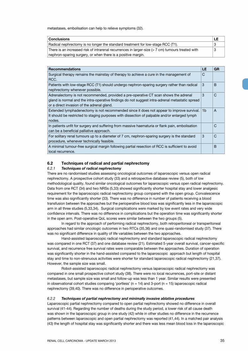
RENAL CELL CARCINOMA - UPDATE MARCH 2013 35
metastases, embolisation can help to relieve symptoms (32).
Conclusions LERadical nephrectomy is no longer the standard treatment for low-stage RCC (T1). 3There is an increased risk of intrarenal recurrences in larger-size (> 7 cm) tumours treated with nephron-sparing surgery, or when there is a positive margin.
3
Recommendations LE gRSurgical therapy remains the mainstay of therapy to achieve a cure in the management of RCC.
C
Patients with low-stage RCC (T1) should undergo nephron-sparing surgery rather than radical nephrectomy whenever possible.
3 B
Adrenalectomy is not recommended, provided a pre-operative CT scan shows the adrenal gland is normal and the intra-operative findings do not suggest intra-adrenal metastatic spread or a direct invasion of the adrenal gland.
3 C
Extended lymphadenectomy is not recommended since it does not appear to improve survival. It should be restricted to staging purposes with dissection of palpable and/or enlarged lymph nodes.
1b A
In patients unfit for surgery and suffering from massive haematuria or flank pain, embolisation can be a beneficial palliative approach.
C
For solitary renal tumours up to a diameter of 7 cm, nephron-sparing surgery is the standard procedure, whenever technically feasible.
3 C
A minimal tumour-free surgical margin following partial resection of RCC is sufficient to avoid local recurrence.
B
6.2 Techniques of radical and partial nephrectomy6.2.1 Techniques of radical nephrectomyThere are no randomised studies assessing oncological outcomes of laparoscopic versus open radical nephrectomy. A prospective cohort study (33) and a retrospective database review (5), both of low methodological quality, found similar oncological outcomes for laparoscopic versus open radical nephrectomy. Data from one RCT (34) and two NRSs (5,33) showed significantly shorter hospital stay and lower analgesic requirement for the laparoscopic radical nephrectomy group compared with the open group. Convalescence time was also significantly shorter (33). There was no difference in number of patients receiving a blood transfusion between the approaches but the perioperative blood loss was significantly less in the laparoscopic arm in all three studies (5,33,34). Surgical complications were marked by low event rates and very wide confidence intervals. There was no difference in complications but the operation time was significantly shorter in the open arm. Post-operative QoL scores were similar between the two groups (5). In regard to the approach of performing radical nephrectomy, both retroperitoneal or transperitoneal approaches had similar oncologic outcomes in two RTCs (35,36) and one quasi-randomised study (37). There was no significant difference in quality of life variables between the two approaches. Hand-assisted laparoscopic radical nephrectomy and standard laparoscopic radical nephrectomy was compared in one RCT (37) and one database review (21). Estimated 5-year overall survival, cancer-specific survival, and recurrence free survival rates were comparable between the approaches. Duration of operation was significantly shorter in the hand-assisted compared to the laparoscopic approach but length of hospital stay and time to non-strenuous activities were shorter for standard laparoscopic radical nephrectomy (21,37). However, the sample size was small. Robot-assisted laparoscopic radical nephrectomy versus laparoscopic radical nephrectomy was compared in one small prospective cohort study (38). There were no local recurrences, port-site or distant metastases, but sample size was small and follow-up was less than 1 year. Similar results were presented in observational cohort studies comparing ‘portless’ (n = 14) and 3-port (n = 15) laparoscopic radical nephrectomy (39,40). There was no difference in perioperative outcomes.
6.2.2 Techniques of partial nephrectomy and minimally invasive ablative proceduresLaparoscopic partial nephrectomy compared to open partial nephrectomy showed no difference in overall survival (41-44). Regarding the number of deaths during the study period, a lower risk of all cause death was shown in the laparoscopic group in one study (42) while in other studies no difference in the recurrence patterns between laparoscopic and open partial nephrectomy was reported (41,44). In a matched pair analysis (43) the length of hospital stay was significantly shorter and there was less mean blood loss in the laparoscopic
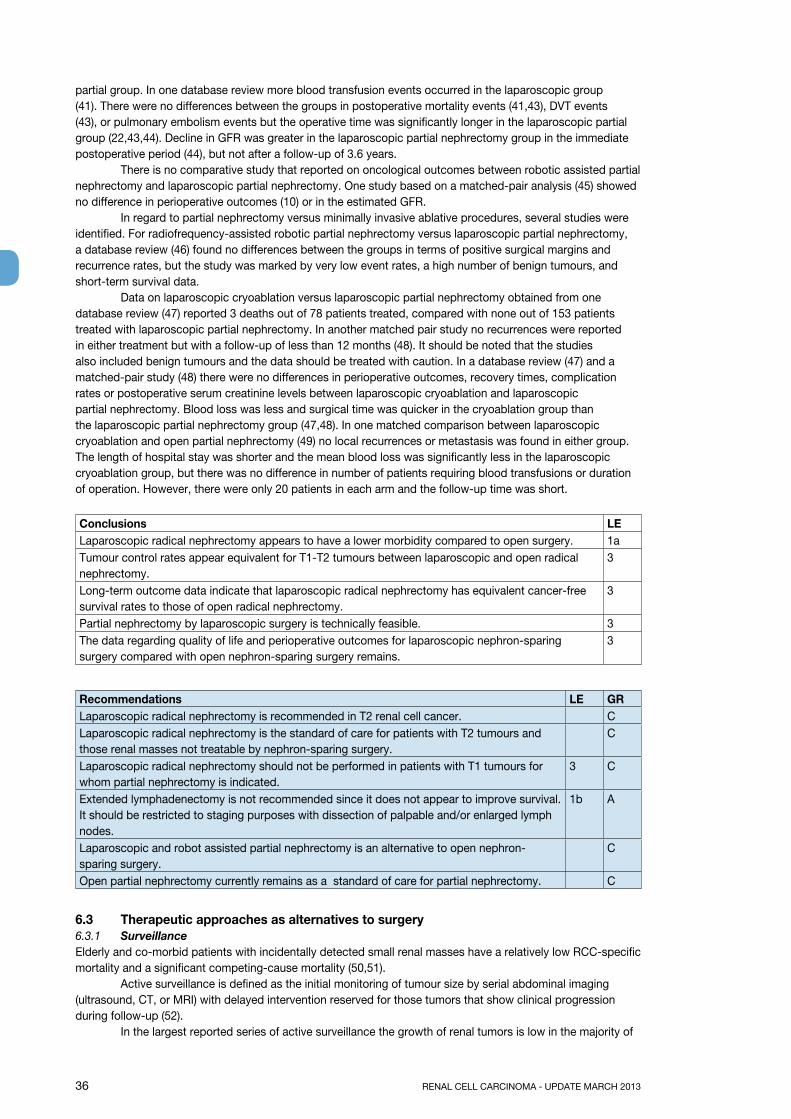
36 RENAL CELL CARCINOMA - UPDATE MARCH 2013
partial group. In one database review more blood transfusion events occurred in the laparoscopic group (41). There were no differences between the groups in postoperative mortality events (41,43), DVT events (43), or pulmonary embolism events but the operative time was significantly longer in the laparoscopic partial group (22,43,44). Decline in GFR was greater in the laparoscopic partial nephrectomy group in the immediate postoperative period (44), but not after a follow-up of 3.6 years. There is no comparative study that reported on oncological outcomes between robotic assisted partial nephrectomy and laparoscopic partial nephrectomy. One study based on a matched-pair analysis (45) showed no difference in perioperative outcomes (10) or in the estimated GFR. In regard to partial nephrectomy versus minimally invasive ablative procedures, several studies were identified. For radiofrequency-assisted robotic partial nephrectomy versus laparoscopic partial nephrectomy, a database review (46) found no differences between the groups in terms of positive surgical margins and recurrence rates, but the study was marked by very low event rates, a high number of benign tumours, and short-term survival data. Data on laparoscopic cryoablation versus laparoscopic partial nephrectomy obtained from one database review (47) reported 3 deaths out of 78 patients treated, compared with none out of 153 patients treated with laparoscopic partial nephrectomy. In another matched pair study no recurrences were reported in either treatment but with a follow-up of less than 12 months (48). It should be noted that the studies also included benign tumours and the data should be treated with caution. In a database review (47) and a matched-pair study (48) there were no differences in perioperative outcomes, recovery times, complication rates or postoperative serum creatinine levels between laparoscopic cryoablation and laparoscopic partial nephrectomy. Blood loss was less and surgical time was quicker in the cryoablation group than the laparoscopic partial nephrectomy group (47,48). In one matched comparison between laparoscopic cryoablation and open partial nephrectomy (49) no local recurrences or metastasis was found in either group. The length of hospital stay was shorter and the mean blood loss was significantly less in the laparoscopic cryoablation group, but there was no difference in number of patients requiring blood transfusions or duration of operation. However, there were only 20 patients in each arm and the follow-up time was short.
Conclusions LELaparoscopic radical nephrectomy appears to have a lower morbidity compared to open surgery. 1aTumour control rates appear equivalent for T1-T2 tumours between laparoscopic and open radical nephrectomy.
3
Long-term outcome data indicate that laparoscopic radical nephrectomy has equivalent cancer-free survival rates to those of open radical nephrectomy.
3
Partial nephrectomy by laparoscopic surgery is technically feasible. 3The data regarding quality of life and perioperative outcomes for laparoscopic nephron-sparing surgery compared with open nephron-sparing surgery remains.
3
Recommendations LE gRLaparoscopic radical nephrectomy is recommended in T2 renal cell cancer. CLaparoscopic radical nephrectomy is the standard of care for patients with T2 tumours and those renal masses not treatable by nephron-sparing surgery.
C
Laparoscopic radical nephrectomy should not be performed in patients with T1 tumours for whom partial nephrectomy is indicated.
3 C
Extended lymphadenectomy is not recommended since it does not appear to improve survival. It should be restricted to staging purposes with dissection of palpable and/or enlarged lymph nodes.
1b A
Laparoscopic and robot assisted partial nephrectomy is an alternative to open nephron-sparing surgery.
C
Open partial nephrectomy currently remains as a standard of care for partial nephrectomy. C
6.3 Therapeutic approaches as alternatives to surgery6.3.1 SurveillanceElderly and co-morbid patients with incidentally detected small renal masses have a relatively low RCC-specific mortality and a significant competing-cause mortality (50,51). Active surveillance is defined as the initial monitoring of tumour size by serial abdominal imaging (ultrasound, CT, or MRI) with delayed intervention reserved for those tumors that show clinical progression during follow-up (52). In the largest reported series of active surveillance the growth of renal tumors is low in the majority of
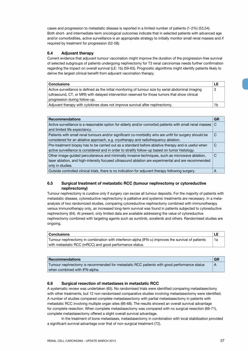
RENAL CELL CARCINOMA - UPDATE MARCH 2013 37
cases and progression to metastatic disease is reported in a limited number of patients (1-2%) (53,54).Both short- and intermediate-term oncological outcomes indicate that in selected patients with advanced age and/or comorbidities, active surveillance is an appropriate strategy to initially monitor small renal masses and if required by treatment for progression (52-58).
6.4 Adjuvant therapyCurrent evidence that adjuvant tumour vaccination might improve the duration of the progression-free survival of selected subgroups of patients undergoing nephrectomy for T3 renal carcinomas needs further confirmation regarding the impact on overall survival (LE: 1b) (59-63). Prognostic algorithms might identify patients likely to derive the largest clinical benefit from adjuvant vaccination therapy.
Conclusions LEActive surveillance is defined as the initial monitoring of tumour size by serial abdominal imaging (ultrasound, CT, or MRI) with delayed intervention reserved for those tumors that show clinical progression during follow-up.
3
Adjuvant therapy with cytokines does not improve survival after nephrectomy. 1b
Recommendations gRActive surveillance is a reasonable option for elderly and/or comorbid patients with small renal masses and limited life expectancy.
C
Patients with small renal tumours and/or significant co-morbidity who are unfit for surgery should be considered for an ablative approach, e.g. cryotherapy and radiofrequency ablation.
C
Pre-treatment biopsy has to be carried out as a standard before ablative therapy and is useful when active surveillance is considered and in order to stratify follow-up based on tumor histology.
C
Other image-guided percutaneous and minimally invasive techniques, such as microwave ablation, laser ablation, and high-intensity focused ultrasound ablation are experimental and are recommended only in studies.
C
Outside controlled clinical trials, there is no indication for adjuvant therapy following surgery. A
6.5 Surgical treatment of metastatic RCC (tumour nephrectomy or cytoreductive nephrectomy)
Tumour nephrectomy is curative only if surgery can excise all tumour deposits. For the majority of patients with metastatic disease, cytoreductive nephrectomy is palliative and systemic treatments are necessary. In a meta-analysis of two randomized studies, comparing cytoreductive nephrectomy combined with immunotherapy versus immunotherapy only, an increased long-term survival was found in patients subjected to cytoreductive nephrectomy (64). At present, only limited data are available addressing the value of cytoreductive nephrectomy combined with targeting agents such as sunitinib, sorafenib and others. Randomised studies are ongoing.
Conclusions LETumour nephrectomy in combination with interferon-alpha (IFN-α) improves the survival of patients with metastatic RCC (mRCC) and good performance status.
1a
Recommendations gRTumour nephrectomy is recommended for metastatic RCC patients with good performance status when combined with IFN-alpha.
A
6.6 Surgical resection of metastases in metastatic RCCA systematic review was undertaken (65). No randomised trials were identified comparing metastasectomy with other treatments, but 12 non-randomised comparative studies involving metastasectomy were identified. A number of studies compared complete metastasectomy with partial metastasectomy in patients with metastatic RCC involving multiple organ sites (66-68). The results showed an overall survival advantage for complete resection. When complete metastasectomy was compared with no surgical resection (69-71), complete metastasectomy offered a slight overall survival advantage. In the treatment of bone metastases, metastasectomy in combination with local stabilization provided a significant survival advantage over that of non-surgical treatment (72).
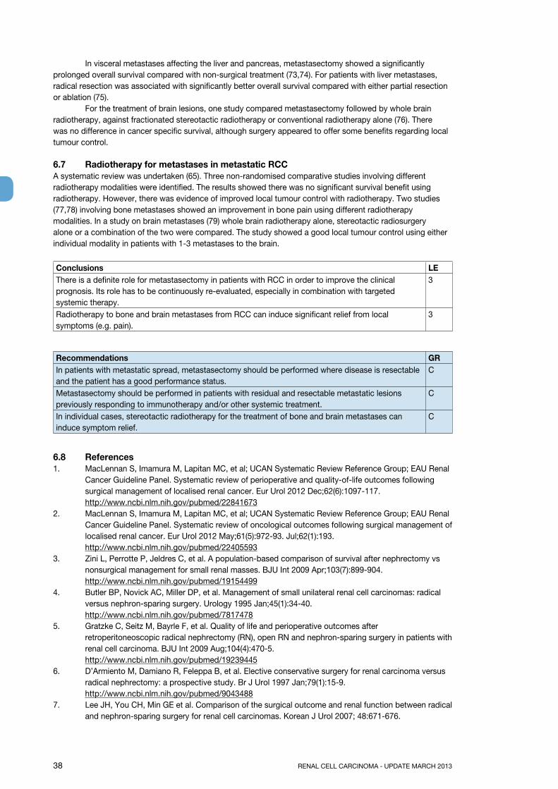
38 RENAL CELL CARCINOMA - UPDATE MARCH 2013
In visceral metastases affecting the liver and pancreas, metastasectomy showed a significantly prolonged overall survival compared with non-surgical treatment (73,74). For patients with liver metastases, radical resection was associated with significantly better overall survival compared with either partial resection or ablation (75). For the treatment of brain lesions, one study compared metastasectomy followed by whole brain radiotherapy, against fractionated stereotactic radiotherapy or conventional radiotherapy alone (76). There was no difference in cancer specific survival, although surgery appeared to offer some benefits regarding local tumour control.
6.7 Radiotherapy for metastases in metastatic RCCA systematic review was undertaken (65). Three non-randomised comparative studies involving different radiotherapy modalities were identified. The results showed there was no significant survival benefit using radiotherapy. However, there was evidence of improved local tumour control with radiotherapy. Two studies (77,78) involving bone metastases showed an improvement in bone pain using different radiotherapy modalities. In a study on brain metastases (79) whole brain radiotherapy alone, stereotactic radiosurgery alone or a combination of the two were compared. The study showed a good local tumour control using either individual modality in patients with 1-3 metastases to the brain.
Conclusions LEThere is a definite role for metastasectomy in patients with RCC in order to improve the clinical prognosis. Its role has to be continuously re-evaluated, especially in combination with targeted systemic therapy.
3
Radiotherapy to bone and brain metastases from RCC can induce significant relief from local symptoms (e.g. pain).
3
Recommendations gRIn patients with metastatic spread, metastasectomy should be performed where disease is resectable and the patient has a good performance status.
C
Metastasectomy should be performed in patients with residual and resectable metastatic lesions previously responding to immunotherapy and/or other systemic treatment.
C
In individual cases, stereotactic radiotherapy for the treatment of bone and brain metastases can induce symptom relief.
C
6.8 References1. MacLennan S, Imamura M, Lapitan MC, et al; UCAN Systematic Review Reference Group; EAU Renal
Cancer Guideline Panel. Systematic review of perioperative and quality-of-life outcomes following surgical management of localised renal cancer. Eur Urol 2012 Dec;62(6):1097-117. http://www.ncbi.nlm.nih.gov/pubmed/22841673
2. MacLennan S, Imamura M, Lapitan MC, et al; UCAN Systematic Review Reference Group; EAU Renal Cancer Guideline Panel. Systematic review of oncological outcomes following surgical management of localised renal cancer. Eur Urol 2012 May;61(5):972-93. Jul;62(1):193. http://www.ncbi.nlm.nih.gov/pubmed/22405593
3. Zini L, Perrotte P, Jeldres C, et al. A population-based comparison of survival after nephrectomy vs nonsurgical management for small renal masses. BJU Int 2009 Apr;103(7):899-904.http://www.ncbi.nlm.nih.gov/pubmed/19154499
4. Butler BP, Novick AC, Miller DP, et al. Management of small unilateral renal cell carcinomas: radical versus nephron-sparing surgery. Urology 1995 Jan;45(1):34-40.http://www.ncbi.nlm.nih.gov/pubmed/7817478
5. Gratzke C, Seitz M, Bayrle F, et al. Quality of life and perioperative outcomes after retroperitoneoscopic radical nephrectomy (RN), open RN and nephron-sparing surgery in patients with renal cell carcinoma. BJU Int 2009 Aug;104(4):470-5. http://www.ncbi.nlm.nih.gov/pubmed/19239445
6. D’Armiento M, Damiano R, Feleppa B, et al. Elective conservative surgery for renal carcinoma versus radical nephrectomy: a prospective study. Br J Urol 1997 Jan;79(1):15-9. http://www.ncbi.nlm.nih.gov/pubmed/9043488
7. Lee JH, You CH, Min GE et al. Comparison of the surgical outcome and renal function between radical and nephron-sparing surgery for renal cell carcinomas. Korean J Urol 2007; 48:671-676.
RENAL CELL CARCINOMA - UPDATE MARCH 2013 39
8. Huang WC, Elkin EB, Levey AS, et al. Partial nephrectomy versus radical nephrectomy in patients with small renal tumors--is there a difference in mortality and cardiovascular outcomes? J Urol 2009 Jan;181(1):55-61.http://www.ncbi.nlm.nih.gov/pubmed/19012918
9. Zini L, Perrotte P, Capitanio U, et al. Radical versus partial nephrectomy: effect on overall and noncancer mortality. Cancer 2009 Apr;115(7):1465-71.http://www.ncbi.nlm.nih.gov/pubmed/19195042
10. Thompson RH, Boorjian SA, Lohse CM, et al. Radical nephrectomy for pT1a renal masses may be associated with decreased overall survival compared with partial nephrectomy. J Urol 2008 Feb;179(2):468-71.http://www.ncbi.nlm.nih.gov/pubmed/18076931
11. Patard JJ, Bensalah KC, Pantuck AJ, et al. Radical nephrectomy is not superior to nephron sparing surgery in PT1B-PT2N0M0 renal tumours: A matched comparison analysis in 546 cases. Eur Urol Suppl 2008;7:194.
12. Thompson RH, Kaag M, Vickers A, et al. Contemporary use of partial nephrectomy at a tertiary care center in the United States. J Urol 2009 Mar;181(3):993-7.http://www.ncbi.nlm.nih.gov/pubmed/19150552
13. Dash A, Vickers AJ, Schachter LR, et al. Comparison of outcomes in elective partial vs radical nephrectomy for clear cell renal cell carcinoma of 4-7 cm. BJU Int 2006 May; 97(5):939-45.http://www.ncbi.nlm.nih.gov/pubmed/16643474
14. Weight CJ, Larson BT, Fergany AF, et al. Nephrectomy induced chronic renal insufficiency is associated with increased risk of cardiovascular death and death from any cause in patients with localized cT1b renal masses. J Urol 2010 Apr;183(4):1317-23.http://www.ncbi.nlm.nih.gov/pubmed/20171688
15. Crépel M, Jeldres C, Perrotte P, et al. Nephron-sparing surgery is equally effective to radical nephrectomy for T1BN0M0 renal cell carcinoma: a population-based assessment. Urology 2010 Feb;75(2):271-5.http://www.ncbi.nlm.nih.gov/pubmed/19962740
16. Patard JJ, Shvarts O, Lam JS, et al. Safety and efficacy of partial nephrectomy for all T1 tumors based on an international multicenter experience. J Urol 2004 Jun;171(6 Pt 1):2181-5.http://www.ncbi.nlm.nih.gov/pubmed/15126781
17. Simmons MN, Weight CJ, Gill IS. Laparoscopic radical versus partial nephrectomy for tumors >4 cm: intermediate-term oncologic and functional outcomes. Urology 2009 May;73(5):1077-82. http://www.ncbi.nlm.nih.gov/pubmed/19394509
18. Van Poppel H, Da Pozzo L, Albrecht W, et al. A prospective randomized EORTC intergroup phase 3 study comparing the complications of elective nephron-sparing surgery and radical nephrectomy for low-stage renal cell carcinoma. Eur Urol 2007 Jun; 51(6):1606-15.http://www.ncbi.nlm.nih.gov/pubmed/17140723
19. Poulakis V, Witzsch U, de Vries R, et al. Quality of life after surgery for localized renal cell carcinoma: comparison between radical nephrectomy and nephron-sparing surgery. Urology 2003 Nov;62(5): 814-20.http://www.ncbi.nlm.nih.gov/pubmed/14624900
20. Shekarriz B, Upadhyay J, Shekarriz H, et al. Comparison of costs and complications of radical and partial nephrectomy for treatment of localized renal cell carcinoma. Urology 2002 Feb; 59(2):211-15.http://www.ncbi.nlm.nih.gov/pubmed/11834387
21. Gabr AH, Gdor Y, Strope SA, et al. Approach and specimen handling do not influence oncological perioperative and long-term outcomes after laparoscopic radical nephrectomy. J Urol 2009 Sep;182(3):874-80.http://www.ncbi.nlm.nih.gov/pubmed/19616234
22. Imamura M, MacLennan S, Lapitan MC, et al. Systematic review of the clinical effectiveness of surgical management for localised renal cell carcinoma. University of Aberdeen, Academic Urology Unit ,2011. Aberdeen, UK. Available from: http://www.uroweb.org/?id=217&tyid=1&oid=4.
23. Simmons MN, Chung BI, Gill IS. Perioperative efficacy of laparoscopic partial nephrectomy for tumors larger than 4 cm. Eur Urol 2009 Jan;55(1):199-208.http://www.ncbi.nlm.nih.gov/pubmed/18684555
24. Lane BR, Tiong HY, Campbell SC, et al. Management of the adrenal gland during partial nephrectomy. J Urol 2009 Jun; 181(6):2430-6.http://www.ncbi.nlm.nih.gov/pubmed/19371896
40 RENAL CELL CARCINOMA - UPDATE MARCH 2013
25. Blom JH, Van Poppel H, Maréchal JM, et al. Radical Nephrectomy with and without Lymph-Node Dissection: Final Results of European Organization for Research and Treatment of Cancer (EORTC) Randomized Phase 3 Trial 30881. Eur Urol 2009 Jan;55(1):28-34.http://www.ncbi.nlm.nih.gov/pubmed/18848382
26. May M, Brookman-Amissah S, Pflanz S, et al. Pre-operative renal arterial embolisation does not provide survival benefit in patients with radical nephrectomy for renal cell carcinoma. Br J Radiol 2009 Aug;82(981):724-31. http://www.ncbi.nlm.nih.gov/pubmed/19255117
27. Subramanian VS, Stephenson AJ, Goldfarb DA, et al. Utility of preoperative renal artery embolization for management of renal tumors with inferior vena caval thrombi. Urology 2009 Jul:74(1):154-9. http://www.ncbi.nlm.nih.gov/pubmed/19428069
28. Maxwell NJ, Saleem Amer N, Rogers E, et al. Renal artery embolization in the palliative treatment of renal carcinoma. Br J Radiol 2007 Feb;80(950):96-102. http://www.ncbi.nlm.nih.gov/pubmed/17495058
29. Hallscheidt P, Besharati S, Noeldge G, et al. Preoperative and palliative embolization of renal cell carcinomas: follow-up of 49 patients. Rofo 2006 Apr;178(4):391-9.http://www.ncbi.nlm.nih.gov/pubmed/16612730
30. Lamb GW, Bromwich EJ, Vasey P, et al. Management of renal masses in patients medically unsuitable for nephrectomy–natural history, complications and outcome. Urology 2004 Nov;64(5):909-13. http://www.ncbi.nlm.nih.gov/pubmed/15533476
31. Kickuth R, Waldherr C, Hoppe H, et al. Interventional management of hypervascular osseous metastasis: role of embolotherapy before orthopedic tumor resection and bone stabilization. AJR Am J Roentgenol 2008 Dec;191(6):W240-7. http://www.ncbi.nlm.nih.gov/pubmed/19020210.
32. Forauer AR, Kent E, Cwikiel W, et al. Selective palliative transcatheter embolization of bony metastases from renal cell carcinoma. Acta Oncol 2007;46(7):1012-18. http://www.ncbi.nlm.nih.gov/pubmed/17851849
33. Hemal AK, Kumar A, Kumar R, et al. Laparoscopic versus open radical nephrectomy for large renal tumors: a long-term prospective comparison. J Urol 2007 Mar;177(3):862-6. http://www.ncbi.nlm.nih.gov/pubmed/17296361
34. Peng B, Zheng J-H, Xu D-F, Ren J-Z. Retroperitoneal laparoscopic nephrectomy and open nephrectomy for radical treatment of renal cell carcinoma: A comparison of clinical outcomes. Academic Journal of Second Military Medical University 2006;27:1167-9.
35. Desai MM, Strzempkowski B, Matin SF, et al. Prospective randomized comparison of transperitoneal versus retroperitoneal laparoscopic radical nephrectomy. J Urol 2005 Jan;173(1):38-41. http://www.ncbi.nlm.nih.gov/pubmed/15592021
36. Nambirajan T, Jeschke S, Al-Zahrani H et al. Prospective, randomized controlled study: transperitoneal laparoscopic versus retroperitoneoscopic radical nephrectomy. Urology 2004 Nov;64(5):919-24.http://www.ncbi.nlm.nih.gov/pubmed/15533478
37. Nadler RB, Loeb S, Clemens JQ et al. A prospective study of laparoscopic radical nephrectomy for T1 tumors--is transperitoneal, retroperitoneal or hand assisted the best approach? J Urol 2006 Apr;175(4):1230-3.http://www.ncbi.nlm.nih.gov/pubmed/16515966
38. Hemal AK, Kumar A. A prospective comparison of laparoscopic and robotic radical nephrectomy for T1-2N0M0 renal cell carcinoma. World J Urol 2009 Feb;27(1):89-94.http://www.ncbi.nlm.nih.gov/pubmed/18704439
39. Soga N, Kato M, Masui S, et al. Comparison of radical nephrectomy techniques in one center: Minimal incision portless endoscopic surgery versus laparoscopic surgery. Int J Urol 2008 Oct; 15(11): 1018-21. http://www.ncbi.nlm.nih.gov/pubmed/19138194
40. Park Y, Lee S, Ku J et al. Laparoendoscopic single-site radical nephrectomy for localized renal cell carcinoma: comparison with conventional laparoscopic surgery. J Endourol 2009; 23(Suppl):A19.
41. Gill IS, Kavoussi LR, Lane BR, et al. Comparison of 1,800 laparoscopic and open partial nephrectomies for single renal tumors. J Urol 2007 Jul;178(1):41-6.http://www.ncbi.nlm.nih.gov/pubmed/17574056
42. Lane BR, Gill IS. 7-year oncological outcomes after laparoscopic and open partial nephrectomy. J Urol 2010 Feb;183(2):473-9.http://www.ncbi.nlm.nih.gov/pubmed/20006866

RENAL CELL CARCINOMA - UPDATE MARCH 2013 41
43. Gong EM, Orvieto MA, Zorn KC, et al. Comparison of laparoscopic and open partial nephrectomy in clinical T1a renal tumors. J Endourol 2008 May;22(5):953-7.http://www.ncbi.nlm.nih.gov/pubmed/18363510
44. Marszalek M, Meixl H, Polajnar M, et al. Laparoscopic and open partial nephrectomy: a matched-pair comparison of 200 Patients. Eur Urol 2009 May;55(5):1171-8.http://www.ncbi.nlm.nih.gov/pubmed/19232819
45. Aron M, Koenig P, Kaouk JH, et al. Robotic and laparoscopic partial nephrectomy: a matched-pair comparison from a high-volume centre. BJU Int 2008 Jul;102(1):86-92. http://www.ncbi.nlm.nih.gov/pubmed/18336600
46. Wu SD, Viprakasit DP, Cashy J, et al. Radiofrequency ablation-assisted robotic laparoscopic partial nephrectomy without renal hilar vessel clamping versus laparoscopic partial nephrectomy: a comparison of perioperative outcomes. J Endourol 2010 Mar;24(3):385-91.http://www.ncbi.nlm.nih.gov/pubmed/20334556
47. Desai MM, Aron M, Gill IS. Laparoscopic partial nephrectomy versus laparoscopic cryoablation for the small renal tumor. Urology 2005 Nov;66(5 Suppl):23-8.http://www.ncbi.nlm.nih.gov/pubmed/16194703
48. O’Malley RL, Berger AD, Kanofsky JA, et al. A matched-cohort comparison of laparoscopic cryoablation and laparoscopic partial nephrectomy for treating renal masses. BJU Int 2007 Feb;99(2):395-8.http://www.ncbi.nlm.nih.gov/pubmed/17092288
49. Ko YH, Park HS, Moon du G, et al. A matched-cohort comparison of laparoscopic renal cryoablation using ultra-thin cryoprobes with open partial nephrectomy for the treatment of small renal cell carcinoma. Cancer Res Treat 2008 Dec;40(4):184-9.http://www.ncbi.nlm.nih.gov/pubmed/19688128
50. Lane BR, Abouassaly R, Gao T, Et al. Active treatment of localized renal tumors may not impact overall survival in patients aged 75 years or older. Cancer 2010 Jul;116(13):3119-26.http://www.ncbi.nlm.nih.gov/pubmed/20564627
51. Hollingsworth JM, Miller DC, Daignault S, et al. Five-year survival after surgical treatment for kidney cancer: a population-based competing risk analysis. Cancer 2007 May;109(9):1763-8.http://www.ncbi.nlm.nih.gov/pubmed/17351954
52. Volpe A, Panzarella T, Rendon RA, et al. The natural history of incidentally detected small renal masses.Cancer 2004 Feb;100(4):738-45.http://www.ncbi.nlm.nih.gov/pubmed/14770429
53. Jewett MA, Mattar K, Basiuk J, et al. Active surveillance of small renal masses: progression patterns of early stage kidney cancer. Eur Urol 2011 Jul;60(1):39-44.http://www.ncbi.nlm.nih.gov/pubmed/21477920
54. Smaldone MC, Kutikov A, Egleston BL, et al. Small renal masses progressing to metastases under active surveillance: a systematic review and pooled analysis. Cancer 2011 Feb;118(4):997-1006.http://www.ncbi.nlm.nih.gov/pubmed/21766302
55. Abou Youssif T, Kassouf W, Steinberg J, et al. Active surveillance for selected patients with renal masses: updated results with long-term follow-up. Cancer 2007 Sep;110(5):1010-4.http://www.ncbi.nlm.nih.gov/pubmed/17628489
56. Abouassaly R, Lane BR, Novick AC. Active surveillance of renal masses in elderly patients. J Urol 2008 Aug;180(2):505-8; discussion 8-9.http://www.ncbi.nlm.nih.gov/pubmed/18550113
57. Crispen PL, Viterbo R, Boorjian SA, et al. Natural history, growth kinetics, and outcomes of untreated clinically localized renal tumors under active surveillance. Cancer 2009 Jul;115(13):2844-52.http://www.ncbi.nlm.nih.gov/pubmed/19402168
58. Rosales JC, Haramis G, Moreno J, et al. Active surveillance for renal cortical neoplasms. J Urol 2010 May;183(5):1698-702. http://www.ncbi.nlm.nih.gov/pubmed/20299038
59. Galligioni E, Quaia M, Merlo A, et al. Adjuvant immunotherapy treatment of renal carcinoma patients with autologous tumor cells and bacillus Calmette-Guerin: five-year results of a prospective randomized study. Cancer 1996 Jun;77(12):2560-6. http://www.ncbi.nlm.nih.gov/pubmed/8640706
60. Figlin RA, Thompson JA, Bukowski RM, et al. Multicenter, randomized, phase III trial of CD8(+) tumor-infiltrating lymphocytes in combination with recombinant interleukin-2 in metastatic renal cell carcinoma. J Clin Oncol 1999 Aug;17(8):2521-9. http://www.ncbi.nlm.nih.gov/pubmed/10561318
42 RENAL CELL CARCINOMA - UPDATE MARCH 2013
61. Clark JI, Atkins MB, Urba WJ, et al. Adjuvant high-dose bolus interleukin-2 for patients with high-risk renal cell carcinoma: a cytokine working group randomized trial. J Clin Oncol 2003 Aug;21(16):3133-40. http://www.ncbi.nlm.nih.gov/pubmed/12810695.
62. Atzpodien J, Schmitt E, Gertenbach U, et al; German Cooperative Renal Carcinoma Chemo-Immunotherapy Trials Group (DGCIN). Adjuvant treatment with interleukin-2- and interferon-alpha2a-based chemoimmunotherapy in renal cell carcinoma post tumour nephrectomy: results of a prospectively randomised trial of the German Cooperative Renal Carcinoma Chemoimmunotherapy Group (DGCIN). Br J Cancer 2005 Mar;92(5):843-6. http://www.ncbi.nlm.nih.gov/pubmed/15756254
63. Jocham D, Richter A, Hoffmann L, et al. Adjuvant autologous renal tumour cell vaccine and risk of tumour progression in patients with renal-cell carcinoma after radical nephrectomy: phase III, randomised controlled trial. Lancet 2004 Feb;363(9409):594-9. http://www.ncbi.nlm.nih.gov/pubmed/14987883
64. Flanigan RC, Mickisch G, Sylvester R, et al. Cytoreductive nephrectomy in patients with metastatic renal cancer: a combined analysis. J Urol 2004 Mar;171(3):1071-6. http://www.ncbi.nlm.nih.gov/pubmed/14767273
65. Dabestani S, Hofmann F, Marconi L, et al, EAU Renal Cell Carcinoma Guideline Panel. Systematic review methodology for the EAU RCC Guideline 2013 update.http://www.uroweb.org/gls/refs/Systematic_methodology_RCC_2013_update.pdf
66. Alt AL, Boorjian SA, Lohse CM, et al. Survival after complete surgical resection of multiple metastases from renal cell carcinoma. Cancer 2011 Jul;117(13):2873-82.http://www.ncbi.nlm.nih.gov/pubmed/21692048
67. Russo P, Synder M, Vickers A, et al. Cytoreductive nephrectomy and nephrectomy/complete metastasectomy for metastatic renal cancer. ScientificWorldJournal. 2007 Feb;7:768-78.http://www.ncbi.nlm.nih.gov/pubmed/17619759
68. Steiner T, Kirchner H, Siebels M, et al. The role of surgery in clinical management of patients with metastatic papillary renal cell carcinoma. J Cancer Res Clin Oncol 2010 Jun;136(6):905-10. http://www.ncbi.nlm.nih.gov/pubmed/20012752
69. Brinkmann OA, Semik M, Gosherger G, et al. The role of residual tumor resection in patients with metastatic renal cell carcinoma and partial remission following immunotherapy. Eur Urol 2007; Suppl 6:641-5.
70. Lee SE, Kwak C, Byun SS, et al. Metastatectomy prior to immunochemotherapy for metastatic renal cell carcinoma. Urol Int 2006;76(3):256-63. http://www.ncbi.nlm.nih.gov/pubmed/16601390
71. Kwak C, Park YH, Jeong CW, et al. Metastasectomy without systemic therapy in metastatic renal cell carcinoma: comparison with conservative treatment. Urol Int 2007;79(2):145-51.http://www.ncbi.nlm.nih.gov/pubmed/ 17851285
72. Fuchs B, Trousdale RT, Rock MG. Solitary bony metastasis from renal cell carcinoma: significance of surgical treatment. Clin Orthop Relat Res 2005 Feb;(431):187-92. http://www.ncbi.nlm.nih.gov/pubmed/15685074
73. Zerbi A, Ortolano E, Balzano G, et al. Pancreatic metastasis from renal cell carcinoma: which patients benefit from surgical resection? Ann Surg Oncol 2008 Apr;15(4):1161-8. http://www.ncbi.nlm.nih.gov/pubmed/18196343
74. Staehler MD, Kruse J, Haseke N, et al. Liver resection for metastatic disease prolongs survival in renal cell carcinoma: 12-year results from a retrospective comparative analysis. World J Urol 2010 Aug;28(4):543-7. http://www.ncbi.nlm.nih.gov/pubmed/20440505
75. Ruys AT, Tanis PJ, Iris ND, vet al. Surgical treatment of renal cell cancer liver metastases: a population-based study. Ann Surg Oncol 2011 Jul;18(7):1932-8.http://www.ncbi.nlm.nih.gov/pubmed/21347794
76. Ikushima H, Tokuuye K, Sumi M, et al. Fractionated stereotactic radiotherapy of brain metastases from renal cell carcinoma. Int J Radiat Oncol Biol Phys 2000 Dec;48(5):1389-93. http://www.ncbi.nlm.nih.gov/pubmed/11121638
77. Hunter GK, Balagamwala EH, Koyfman SA, et al. The efficacy of external beam radiotherapy and stereotactic body radiotherapy (SBRT) for painful spinal metastases from renal cell carcinoma. International Journal of Radiation Oncology Biology Physics 2011;81(Suppl 2):S649-S650.
78. Zelefsky MJ, Greco C, Motzer R, et al. Tumor control outcomes after hypofractionated and single-dose stereotactic image-guided intensity-modulated radiotherapy for extracranial metastases from renal cell carcinoma. Int J Radiat Oncol Biol Phys 2012 Apr;82(5):1744-8. http://www.ncbi.nlm.nih.gov/pubmed/21596489
RENAL CELL CARCINOMA - UPDATE MARCH 2013 43
79. Fokas E, Henzel M, Hamm K, et al. Radiotherapy for brain metastases from renal cell cancer: should whole-brain radiotherapy be added to stereotactic radiosurgery?: analysis of 88 patients. Strahlenther Onkol 2010 Apr;186(4):210-7.. http://www.ncbi.nlm.nih.gov/pubmed/20165820
7. SYSTEMIC THERApY FOR METASTATIC RCC7.1 ChemotherapySince RCCs develop from the proximal tubules, they have high levels of expression of the multiple-drug resistance protein, P-glycoprotein, and are therefore resistant to most forms of chemotherapy. Chemotherapy appears to be moderately effective only if 5-fluorouracil (5-FU) is combined with immunotherapeutic agents (1). However, in a prospective randomised study, interferon-alpha (IFN-α) showed equivalent efficacy to a combination of IFN-α + interleukin-2 (IL-2) + 5-FU (2).
7.1.1 Conclusion and recommendation
Conclusion LE5-FU in combination with immunotherapy is equivalent in efficacy to monotherapy with IFN-α in patients with mRCC.
1b
Recommendation gRIn patients with clear-cell mRCC, chemotherapy as monotherapy should not be considered effective in patients with mRCC.
B
7.2 Immunotherapy7.2.1 Interferon alpha as monotherapy and combined with bevacizumabInterferon alpha has been shown in randomised studies to be superior in relation to survival to hormonal therapy in patients with mRCC (3). IFN-α provided a response rate of 6-15%, together with a 25% decrease in the risk for tumour progression and a modest survival benefit of 3-5 months in comparison with a placebo equivalent (4,5). The positive effect of IFN-α is particularly apparent in mRCC patients with clear cell histology, good-risk Motzer criteria, and lung metastases only (5). In a prospective randomised study, IFN-α showed equivalent efficacy to a combination of IFN-α + IL2 + 5-FU (2). The moderate efficacy of immunotherapy was also confirmed in a Cochrane meta-analysis including 42 eligible studies (6). A combination of bevacizumab + IFN-α was recently shown to be associated with increased response rates and better progression-free survival in first-line therapy in comparison with IFN-α monotherapy (7). All recent randomized studies comparing anti-angiogenic drugs in a first-line setting to IFN-α monotherapy have shown superiority for either sunitinib, bevacizumab + IFN-α, or temsirolimus (7-10).
Table 9: Memorial Sloan-Kettering Cancer Center (Motzer) criteria for predicting survival in patients with advanced RCC treated with interferon alpha, depending on the presence or absence of five distinct risk factors (4)
Risk factors* Cut-off point usedKarnofsky performance status < 80Time from diagnosis to treatment with IFN-α < 12 monthsHemoglobin < Lower limit of laboratory reference rangeLactate dehydrogenase > 1.5 times the upper limit of laboratory rangeCorrected serum calcium > 10.0 mg/dL (2.4 mmol/L)
* Favourable (low) risk, no risk factors; intermediate risk, one or two risk factors; poor (high) risk, three or more risk factors.
7.2.2 Interleukin-2Interleukin-2 (IL-2) has been used to treat mRCC since 1985, with response rates ranging from 7% to 27% (10-12). The optimal IL-2 regimen is not clear, but long-term (> 10 years) complete responses have been achieved
44 RENAL CELL CARCINOMA - UPDATE MARCH 2013
with high-dose bolus IL-2 in a randomised phase III study (13). The toxicity of IL-2 is substantially greater than that of IFN-α. Only clear cell-type RCC responds to immunotherapy. Interleukin-2 has not been validated in controlled randomised studies in comparison with best supportive care (5).
7.2.3 Vaccines and targeted immunotherapyNo recommendations can be made. An earlier phase III trial of vaccine therapy with tumour antigen 5T4 in combination with the first-line standard of care (either sunitinib, interleukin-2, or interferon alpha) failed to demonstrate any survival benefit in comparison with placebo and the first-line standard of care (14). Several phase III vaccination studies are ongoing. Targeted immunotherapy with programmed death-1 ligand (PD-1L), which has shown efficacy and acceptable toxicity in patients with RCC (15), is currently under investigation in a phase II trial in comparison with everolimus in patients in whom anti-angiogenic therapy previously failed.
7.2.4 Conclusions
LEInterferon-alpha monotherapy is no longer recommended as first-line therapy for mRCC. 1bInterferon alpha monotherapy still has a role only in selected cases (good performance status, clear cell type, lung metastases only).
2
Interleukin-2 has more side effects than INF-α. 2-3High-dose IL-2 is associated with durable complete responses in a limited number of patients. 1bInterleukin-2 can be considered as monotherapy in selected patients with a good prognosis profile. 1bA combination of bevacizumab and IFN-α is more effective than IFN-α in treatment-naïve, low-risk and intermediate-risk tumours.
1b
Vaccination therapy with tumour antigen 5T4 showed no survival benefit over the first-line standard of care.
1b
7.2.5 Recommendations
gRMonotherapy with IFN-α or high-dose bolus IL-2 can only be recommended as a first-line treatment for mRCC in selected patients with clear cell histology and good prognostic factors.
A
Bevacizumab + IFN-α is recommended as first-line therapy in low-risk and intermediate-risk patients. BOnly selected patients with mRCC who have a good risk profile and clear cell subtype histology show clinical benefit from immunotherapy with IL-2.Cytokine combinations, with or without additional chemotherapy, do not improve the overall survival in comparison with monotherapy.
A
7.3 Drugs targeting VEgF, including other receptor kinases and mammalian target of rapamycin (mTOR)
Recent advances in molecular biology have led to the development of several novel agents for the treatment of mRCC (Table 11). In sporadic clear cell RCC, hypoxia-inducible factor (HIF) accumulation due to von Hippel-Lindau (VHL) inactivation results in overexpression of vascular endothelial growth factor (VEGF) and platelet-derived growth factor (PDGF), both of which promote neoangiogenesis (16-18). This process substantially contributes to the development and progression of RCC. At present, several targeting drugs have been approved both in the USA and in Europe for the treatment of mRCC:• Sorafenib (Nexavar®)• Sunitinib (Sutent®)• Bevacizumab (Avastin®) combined with IFN-α• Pazopanib (Votrient®)• Temsirolimus (Torisel®)• Everolimus (Afinitor®)• Axitinib (Inlyta®)
New agents targeting angiogenesis are under investigation, as well as combinations of these new agents with each other or with cytokines. One of the new agents targeting angiogenesis, tivozanib, has been investigated in a phase III trial and is currently not approved. Evidence-based data for this drug are presented below. Most published trials have selected for clear cell carcinoma subtypes, and consequently no evidence-based
RENAL CELL CARCINOMA - UPDATE MARCH 2013 45
recommendations can be given for non-clear cell subtypes. In the major phase III trials leading to registration of the approved targeted agents, patients were stratified according to the Memorial Sloan-Kettering Cancer Center (MSKCC) risk model, as published in 2002 (3) (Table 9). Since the MSKCC criteria were established in the era of cytokines, an international database consortium has established and validated a risk model (the Database Consortium Model, DCM) which may yield a more accurate prognosis for patients treated in the era of targeted therapy. In the DCM, neutrophilia and thrombocytosis are added to the MSKCC risk factors. By contrast, lactate dehydrogenase (LDH) is omitted from the factors associated with the prognosis (19). The DCM has recently been used to establish data on conditional survival that can be used to counsel patients (20). The DCM has been validated and compared with the risk model of the Cleveland Clinic Foundation (CCF), the French model, MSKCC model, and the International Kidney Cancer Working Group (IKCWG) model. The DCM showed a concordance level of 0.66, which did not differ from the other models, indicating that a ceiling has been reached for clinical risk models for predicting the prognosis based solely on clinical factors. However, the reported versus predicted number of deaths at 2 years was most similar in the DCM in comparison with the other models (21). The DCM has been externally validated for use in the era of targeted therapy (21).
Table 10: Median overall survival and percentage of patients surviving 2 years treated in the era of targeted therapy per DCM risk group, based on the publications by Heng et al. (19,21)
Database Consortium Model *,**
patients**Median OS*
(months)2-y OS (95% CI) **n %
Favorable 157 18 43.2 75% (65-82%)Intermediate 440 52 22.5 53% (46-59%)Poor 252 30 7.8 7% (2-16%)
* Based on (21); ** based on (19); CI = confidence intervals; OS = overall survival.
7.3.1 Tyrosine kinase inhibitors
7.3.1.1 SorafenibSorafenib is an oral multikinase inhibitor with activity against Raf-1 serine/threonine kinase, B-Raf, vascular endothelial growth factor receptor-2 (VEGFR-2), platelet-derived growth factor receptor (PDGFR), FMS-like tyrosine kinase 3 (FLT-3), and c-KIT. A phase III trial compared sorafenib and placebo after failure of prior systemic immunotherapy or in patients unfit for immunotherapy. The trial reported a 3-month improvement in progression-free survival in favor of sorafenib (22). Survival appears to improve in patients crossed over from placebo to sorafenib treatment (23).
7.3.1.2 SunitinibSunitinib is an oxindol tyrosine kinase (TK) inhibitor. It selectively inhibits PDGFR, VEGFR, c-KIT, and FLT-3 and has antitumour and anti-angiogenic activity. Phase II trials with sunitinib as second-line monotherapy in patients with mRCC demonstrated a partial response in 34-40% of patients and stable disease > 3 months in 27-29% of patients (24). In a pivotal phase III trial of first-line monotherapy comparing treatment with sunitinib versus IFN-α, sunitinib achieved a longer progression-free survival than IFN-α (11 versus 5 months; P < 0.000001). The results suggested that monotherapy with IFN-α was inferior to sunitinib in low-risk and intermediate-risk patients with mRCC (25). The overall survival was 26.4 and 21.8 months in the sunitinib and IFN-α arms, respectively (P = 0.05) (25). In patients crossed over from IFN-α to sunitinib (n = 25), median survival times were 26.4 versus 20.0 months for sunitinib and IFN-α, respectively (P = 0.03). In patients who did not receive any post-study treatment, the median overall survival reached 28.1 months in the sunitinib group versus 14.1 months in the IFN-α group (P = 0.003). In a recent randomised phase II trial including 292 patients, sunitinib 50 mg/day (4 weeks on / 2 weeks off) was compared with a continuous uninterrupted dosage of sunitinib 37.5 mg/day in patients with metastatic clear cell renal carcinoma (26). The median time to progression with sunitinib 50 mg (4/2) (n = 146) was 9.9 months, compared with 7.1 months for 37.5 mg/day continuous dosing (n = 146). The overall response rate was 32% for 50 mg (4/2) versus 28% for 37.5 mg continuous dosing. No significant differences were observed with regard to overall survival (23.1 vs. 23.5 months; P = 0.615), commonly reported adverse events, or patient-reported kidney cancer symptoms. Because of the statistically nonsignificant but numerically longer time to progression with the standard 50 mg (4/2) dosage, the authors recommended adherence to this regimen.
46 RENAL CELL CARCINOMA - UPDATE MARCH 2013
7.3.1.3 PazopanibPazopanib is an oral angiogenesis inhibitor that targets VEGFR, PDGFR, and c-KIT. In a prospective randomized trial of pazopanib versus placebo in treatment-naïve mRCC patients and cytokine-treated patients, there was a significant improvement in the progression-free survival and tumour response (9.2 vs 4.2 months) (27). The trial showed significant results that established pazopanib as a first-line option. Since the initial phase III study involved a substantially smaller number of patients than in phase III studies of other targeted agents, the recommendation was to use pazopanib as second option in first-line treatment. Recently, the results of a randomized phase III non-inferiority trial comparing pazopanib with sunitinib (COMPARZ) showed no significant differences in the outcome parameters, with different toxicity profiles for the two drugs. With a very short follow-up period, these data are not yet mature, particularly with regard to remission. One major shortcoming of the COMPARZ trial is the fact that the study recruited almost one-third of its patients in Asia. Given the fact that there are ethnic differences in side effect profiles, the overall assessment of this trial remains unstable and further interpretation of any subgroups is almost impossible. Full publication is expected, but COMPARZ has established pazopanib as a first-line treatment option.
7.3.1.4 AxitinibAxitinib is an oral selective second-generation inhibitor of VEGFR-1, -2, and -3 that blocks VEGFR receptors at subnanomolar drug concentrations with minimal inhibition of other targets. It has a short half-life. In the AXIS trial (a randomized phase III trial of axitinib versus sorafenib in patients in whom previous cytokine treatment or targeted agents had failed), the sample size calculation was based on a 40% improvement in the median progression-free survival PFS from 5 months to 7 months in patients randomly assigned to receive axitinib (28). Sorafenib was chosen as the comparator because at the time the trial was designed there was no standard for second-line treatment after failure of a previous VEGF targeted therapy. With 723 patients included, the overall median progression-free survival was 6.7 months for patients in the axitinib group in comparison with 4.7 months for those in the sorafenib group (hazard ratio [HR] 0.67; 95% CI, 0.54 to 0.81). However, the difference in PFS was greatest in the patients in whom cytokine treatment had failed. For those in whom sunitinib had failed (n = 194 axitinib and n = 195 sorafenib), axitinib led to a PFS of 4.8 months (95% CI, 4.5 to 6.4) versus 3.4 months (95% CI, 2.6 to 4.7) for sorafenib. In the AXIS trial, axitinib showed greater than or equal to grade 3 toxicity for diarrhea in 11%, hypertension in 16%, and fatigue in 11%. Across all grades, nausea was recorded in 32%, vomiting in 24%, and asthenia in 21%. Overall survival (OS) was a secondary end point of the trial, but these data were not mature at the time of publication. However, since crossover was not allowed in this trial comparing two active VEGFR inhibitors, the data have in the meantime been analyzed and showed no significant differences between axitinib and sorafenib in second-line treatment (29).
7.3.1.5 TivozanibTivozanib is an oral selective tyrosine kinase inhibitor targeting all three VEGF receptors. It has a long half-life. Tivozanib showed activity and tolerability in a phase II discontinuation trial. The overall response rate was 24% (95% CI, 19% to 30%), and the median PFS was 11.7 months (95% CI, 8.3 to 14.3 months) in the trial population. The most common grade 3 and 4 treatment-related adverse event was hypertension (12%) (30). The results of a phase III trial of tivozanib versus sorafenib in treatment-naïve mRCC patients or those having received one prior systemic treatment excluding VEGF targeted therapy or mTOR inhibitors were reported at the American Society of Clinical Oncology (ASCO) meeting in 2012, and full publication is pending. For the 70% treatment-naïve patients enrolled, the median PFS was 12.7 months for tivozanib versus 9.1 months for sorafenib (HR 0.756; 95% CI, 0.580 to 0.985). For all patients, the objective response rates were 33% for tivozanib versus 23% for sorafenib. The most common adverse events (AEs) for tivozanib (all grades / > grade 3) were hypertension (46%/26%), diarrhea (22%/2%), fatigue (18%/5%), and neutropenia (10%/2%) (31). Full publication of this study is pending. If approved, tivozanib might be a tyrosine kinase inhibitor with effectiveness not inferior to that of sorafenib, as apparent in the groups of patients tested.
7.3.2 Monoclonal antibody against circulating VEGF7.3.2.1 Bevacizumab monotherapy and combined with interferon alphaBevacizumab is a humanized monoclonal antibody that binds isoforms of VEGF-A. Bevacizumab 10 mg/kg every 2 weeks in patients refractory to immunotherapy was associated with an increase in the overall response (10%) and in the progression-free survival in comparison with placebo (27). A double-blind phase III trial (AVOREN) (n = 649) in patients with mRCC compared bevacizumab + IFN-α with IFN-α monotherapy (7). The median overall response was 31% in the bevacizumab + IFN-α group versus 13% in the group receiving only IFN-α (P < 0.0001). The median progression-free survival increased significantly from 5.4 months with IFN-α to 10.2 months with bevacizumab + IFN-α (P < 0.0001), but only in low-risk and intermediate-risk patients. No benefit was seen in high-risk patients. In a recent update, the median OS in the AVOREN trial, which allowed
RENAL CELL CARCINOMA - UPDATE MARCH 2013 47
crossover after progression, was 23.3 months for bevacizumab-IFN-α versus 21.3 months for IFN-α alone (P < 0.336) (32).A similarly designed trial (CALGB 90206), including 732 patients (33,34), of bevacizumab (10 mg/kg intravenously every 2 weeks) plus IFN (9 million units subcutaneously three times weekly) versus IFN (9 million units subcutaneously three times weekly) showed a median PFS of 8.5 months for the combination versus 5.2 months for IFN-α alone. The median OS with a crossover design was 18.3 months for the combination versus 17.4 months for IFN alpha alone. Bevacizumab plus IFN-α had a higher objective response rate (ORR) in comparison with IFN (25.5%: 95% CI, 20.9% to 30.6%; vs. 13.1%: 95% CI, 9.5% to 17.3%); P < 0.0001). The overall toxicity was greater for bevacizumab plus IFN-α, with significantly more grade 3 hypertension (9% vs. 0%), anorexia (17% vs. 8%), fatigue (35% vs. 28%), and proteinuria (13% vs. 0%).
7.3.3 Mammalian target of rapamycin (mTOR) inhibitors7.3.3.1 TemsirolimusTemsirolimus is a specific inhibitor of mammalian target of rapamycin (mTOR) (35). Patients with high-risk mRCC were randomly assigned in a phase III trial (NCT00065468) to receive first-line treatment with temsirolimus or IFN-α monotherapy, or a combination. In the temsirolimus group, the overall survival was 10.9 months versus 7.3 months in the IFN-α group (P < 0.0069). However, the overall survival in the temsirolimus + IFN-α group was not significantly improved (9).
7.3.3.2 EverolimusEverolimus is an oral mTOR inhibitor. A phase III study (RECORD-1) compared everolimus plus best supportive care (BSC) versus placebo plus BSC in patients in whom previous anti-VEGFR treatment had failed. The median progression-free survival was 4 months with everolimus versus 1.9 months with placebo (P < 0.001). In the RECORD-1 trial, 124 patients (46%) had received sunitinib as the only previous systemic treatment, with a PFS of 4.0 months (95% CI, 3.7 to 5.5 months). Comparison with the AXIS data is complicated by the fact that in the RECORD-1 trial, 53% of the patients with progression after previous targeted therapy had at least more than one previous treatment, often cytokines prior to tyrosine kinase inhibitors (TKIs). In addition, the PFS analysis in this trial was not specifically carried out for previous sunitinib treatment (16,36).
7.3.4 Sequencing targeted therapyCurrently, no recommendations can be given as to the best sequence of targeted therapy. The AXIS trial is the only recent randomized phase III superiority trial comparing two TKIs after failure of a prior TKI. The results and interpretation are described under 7.3.1.3 above. For the subgroup of patients treated previously with sunitinib, the difference in PFS did not reach statistical significance for axitinib versus sorafenib, and no difference in the OS was observed. Randomized phase III trials investigating the safety and efficacy of sorafenib followed by sunitinib versus sunitinib followed by sorafenib (SWITCH-I) and sequential pazopanib and sorafenib versus sorafenib and pazopanib (SWITCH-II) are ongoing.
7.3.5 Combination of targeted agentsNo recommendations can be made. At present, there have been no phase III trials reporting on a combination of two targeted agents versus monotherapy with a targeted agent. A previous randomised phase II study reported unacceptable toxicity (37). The TORAVA trial showed that the toxicity of a combination of temsirolimus and bevacizumab was much greater than anticipated and that it limited treatment continuation over time in comparison with either standard treatment with sunitinib or bevacizumab and IFN-α. In addition, clinical activity was low in comparison with the benefit expected from sequential use of each targeted therapy. This combination has not been further recommended or investigated. In a nonrandomized phase II trial, the combination of everolimus with bevacizumab was found to be effective with acceptable toxicity, except for grade 3/4 proteinuria in 25% of the patients (38). A randomised phase II trial of everolimus in combination with bevacizumab and IFN-α versus IFN-α alone is ongoing.
7.3.6 Non-clear cell renal cancerNo recommendations can be made at present. No phase III trials on systemic treatment of patients with non-clear cell carcinoma have been reported. A nonrandomized phase II trial in patients with papillary renal cancer who were treated with foretinib, a dual MET/VEGFR2 inhibitor, reported activity and acceptable toxicity with high response rates in patients with germline MET mutations (39). Patients should be treated in the framework of clinical trials. If a trial is not available, a decision can be made in consultation with the patient to perform treatment in line with clear cell renal cell carcinoma.
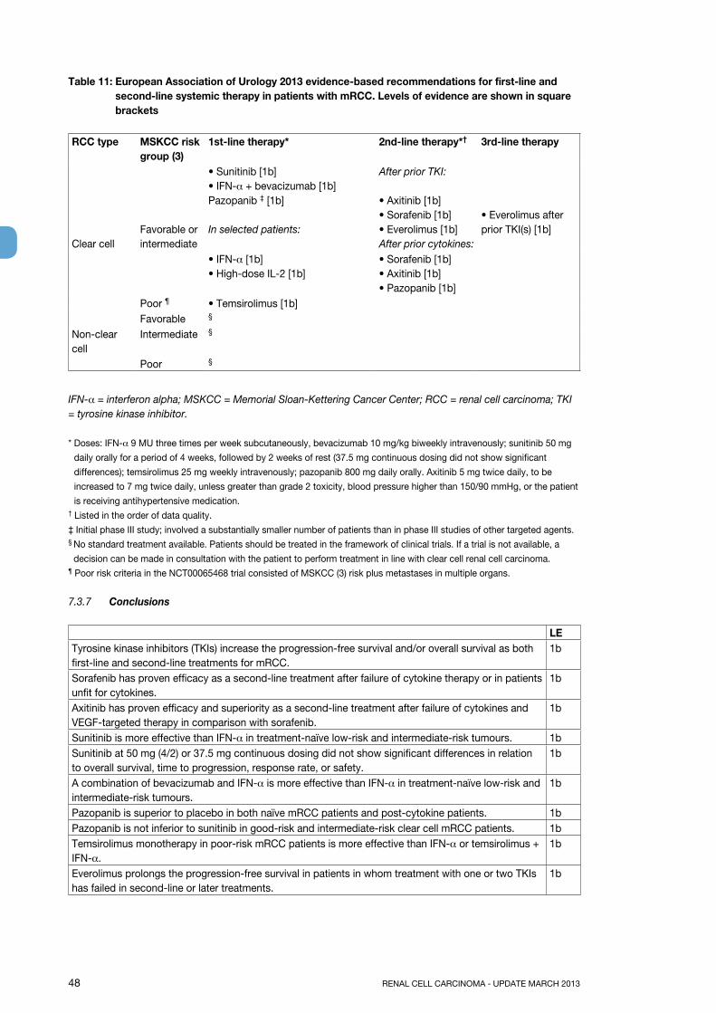
48 RENAL CELL CARCINOMA - UPDATE MARCH 2013
Table 11: European Association of Urology 2013 evidence-based recommendations for first-line and second-line systemic therapy in patients with mRCC. Levels of evidence are shown in square brackets
RCC type MSKCC risk group (3)
1st-line therapy* 2nd-line therapy*† 3rd-line therapy
Clear cellFavorable or intermediate
• Sunitinib [1b]• IFN-α + bevacizumab [1b]Pazopanib ‡ [1b]
In selected patients:
After prior TKI:
• Axitinib [1b]• Sorafenib [1b]• Everolimus [1b]After prior cytokines:
• Everolimus after prior TKI(s) [1b]
• IFN-α [1b]• High-dose IL-2 [1b]
• Sorafenib [1b]• Axitinib [1b]• Pazopanib [1b]
Poor ¶ • Temsirolimus [1b]Favorable §
Non-clear cell
Intermediate §
Poor §
IFN-α = interferon alpha; MSKCC = Memorial Sloan-Kettering Cancer Center; RCC = renal cell carcinoma; TKI = tyrosine kinase inhibitor.
* Doses: IFN-α 9 MU three times per week subcutaneously, bevacizumab 10 mg/kg biweekly intravenously; sunitinib 50 mg
daily orally for a period of 4 weeks, followed by 2 weeks of rest (37.5 mg continuous dosing did not show significant
differences); temsirolimus 25 mg weekly intravenously; pazopanib 800 mg daily orally. Axitinib 5 mg twice daily, to be
increased to 7 mg twice daily, unless greater than grade 2 toxicity, blood pressure higher than 150/90 mmHg, or the patient
is receiving antihypertensive medication.† Listed in the order of data quality.
‡ Initial phase III study; involved a substantially smaller number of patients than in phase III studies of other targeted agents.§ No standard treatment available. Patients should be treated in the framework of clinical trials. If a trial is not available, a
decision can be made in consultation with the patient to perform treatment in line with clear cell renal cell carcinoma.¶ Poor risk criteria in the NCT00065468 trial consisted of MSKCC (3) risk plus metastases in multiple organs.
7.3.7 Conclusions
LETyrosine kinase inhibitors (TKIs) increase the progression-free survival and/or overall survival as both first-line and second-line treatments for mRCC.
1b
Sorafenib has proven efficacy as a second-line treatment after failure of cytokine therapy or in patients unfit for cytokines.
1b
Axitinib has proven efficacy and superiority as a second-line treatment after failure of cytokines and VEGF-targeted therapy in comparison with sorafenib.
1b
Sunitinib is more effective than IFN-α in treatment-naïve low-risk and intermediate-risk tumours. 1bSunitinib at 50 mg (4/2) or 37.5 mg continuous dosing did not show significant differences in relation to overall survival, time to progression, response rate, or safety.
1b
A combination of bevacizumab and IFN-α is more effective than IFN-α in treatment-naïve low-risk and intermediate-risk tumours.
1b
Pazopanib is superior to placebo in both naïve mRCC patients and post-cytokine patients. 1bPazopanib is not inferior to sunitinib in good-risk and intermediate-risk clear cell mRCC patients. 1bTemsirolimus monotherapy in poor-risk mRCC patients is more effective than IFN-α or temsirolimus + IFN-α.
1b
Everolimus prolongs the progression-free survival in patients in whom treatment with one or two TKIs has failed in second-line or later treatments.
1b
RENAL CELL CARCINOMA - UPDATE MARCH 2013 49
The role of the new drugs is still under development and combination studies are ongoing. To date, no data are available indicating whether the new agents have a curative effect. These agents appear promising for stabilizing mRCC for a prolonged period of time. However, this promise has to be balanced against their toxicity profile and the patient’s quality of life. Anti-angiogenic monotherapy and its sequences have become the standard of care in mRCC treatment.
4
7.3.8 Recommendations for systemic therapy for mRCC
Recommendations gRSunitinib is recommended as first-line therapy in favorable-risk and intermediate-risk patients. ABevacizumab + IFN-α is recommended as first-line therapy in favourable-risk and intermediate-risk patients.
A
Sorafenib is recommended as a second-line treatment for mRCC after cytokine failure. APazopanib is recommended as first-line or after cytokine failure in favourable-risk and intermediate-risk patients.
A
Temsirolimus is recommended as first-line treatment in poor-risk patients. AEverolimus is recommended as second-line treatment after failure of tyrosine kinase inhibitors. AAxitinib is recommended as second-line treatment after failure of cytokines or tyrosine kinase inhibitors.
A
7.4 References1. Stadler WM, Huo D, George C, et al. Prognostic factors for survival with gemcitabine plus
5-fluorouracil based regimens for metastatic renal cancer. J Urol 2003 Oct;170(4 Pt 1):1141-5.http://www.ncbi.nlm.nih.gov/pubmed/14501711
2. Gore ME, Griffin CL, Hancock B, et al. Interferon alfa-2a versus combination therapy with interferon alfa-2a, interleukin-2, and fluorouracil in patients with untreated metastatic renal cell carcinoma (MRC RE04/EORTC GU 30012): an open-label randomised trial. Lancet 2010 Feb 20;375(9715):641-8.http://www.ncbi.nlm.nih.gov/pubmed/20153039
3. Medical Research Council Renal Cancer Collaborators. Interferon-alpha and survival in metastatic renal carcinoma: early results of a randomised controlled trial. Medical Research Council Renal Cancer Collaborators. Lancet 1999 Jan;353(9146):14-7.http://www.ncbi.nlm.nih.gov/pubmed/10023944
4. Motzer RJ, Bacik J, Murphy BA, et al. Interferon-alfa as a comparative treatment for clinical trials of new therapies against advanced renal cell carcinoma. J Clin Oncol 2002 Jan;20(1):289-96.http://www.ncbi.nlm.nih.gov/pubmed/11773181
5. Coppin C, Porzsolt F, Awa A, et al. Immunotherapy for advanced renal cell cancer. Cochrane Database Syst Rev 2005 Jan;(1):CD001425.http://www.ncbi.nlm.nih.gov/pubmed/15674877
6. Coppin C, Porzsolt F, Awa A, et al. Immunotherapy for advanced renal cell cancer. Cochrane Database Syst Rev 2005 Jan 25;(1):CD001425.http://www.ncbi.nlm.nih.gov/pubmed/15674877
7. Escudier B, Pluzanska A, Koralewski P, et al; AVOREN Trial investigators. Bevacizumab plus interferon alfa-2a for treatment of metastatic renal cell carcinoma: a randomised, double-blind phase III trial. Lancet 2007 Dec;370(9605):2103-11.http://www.ncbi.nlm.nih.gov/pubmed/18156031
8. Motzer RJ, Hutson TE, Tomczak P, et al. Sunitinib versus interferon alfa in metastatic renal-cell carcinoma. N Engl J Med 2007 Jan;356(2):115-24.http://www.ncbi.nlm.nih.gov/pubmed/17215529
9. Hudes G, Carducci M, Tomczak P, et al; Global ARCC Trial. Temsirolimus, interferon alfa, or both for advanced renal-cell carcinoma. N Engl J Med 2007 May;356(22):2271-81.http://www.ncbi.nlm.nih.gov/pubmed/17538086
10. Rosenberg SA, Lotze MT, Yang JC, et al. Prospective randomized trial of high-dose interleukin-2 alone or in conjunction with lymphokine-activated killer cells for the treatment of patients with advanced cancer. J Natl Cancer Inst 1993 Apr;21(85):622-32.http://www.ncbi.nlm.nih.gov/pubmed/8468720
11. Fyfe G, Fisher RI, Rosenberg SA, et al. Results of treatment of 255 patients with metastatic renal cell carcinoma who received high-dose recombinant interleukin-2 therapy. J Clin Oncol 1995 Mar;13(3):688-96.http://www.ncbi.nlm.nih.gov/pubmed/7884429
50 RENAL CELL CARCINOMA - UPDATE MARCH 2013
12. McDermott DF, Regan MM, Clark JI, et al. Randomized phase III trial of high-dose interleukin-2 versus subcutaneous interleukin-2 and interferon in patients with metastatic renal cell carcinoma. J Clin Oncol 2005 Jan;23(1):133-41.http://www.ncbi.nlm.nih.gov/pubmed/15625368
13. Yang JC, Sherry RM, Steinberg SM, et al. Randomized study of high-dose and low-dose interleukin-2 in patients with metastatic renal cancer. J Clin Oncol 2003 Aug;21(16):3127-32.http://www.ncbi.nlm.nih.gov/pubmed/12915604
14. Amato RJ, Hawkins RE, Kaufman HL, et al. Vaccination of metastatic renal cancer patients with MVA-5T4: a randomized, double-blind, placebo-controlled phase III study. Clin Cancer Res 2010 Nov;16(22):5539-47.http://www.ncbi.nlm.nih.gov/pubmed/20881001
15. Brahmer JR, Tykodi SS, Chow LQ, et al. Safety and activity of anti-PD-L1 antibody in patients with advanced cancer. N Engl J Med 2012 Jun 28;366(26):2455-65.http://www.ncbi.nlm.nih.gov/pubmed/22658128
16. Patel PH, Chadalavada RS, Chaganti RS, et al. Targeting von Hippel-Lindau pathway in renal cell carcinoma. Clin Cancer Res 2006 Dec;12(24):7215-20.http://www.ncbi.nlm.nih.gov/pubmed/17189392
17. Yang JC, Haworth L, Sherry RM, et al. A randomized trial of bevacizumab, an anti-vascular endothelial growth factor antibody, for metastatic renal cancer. N Engl J Med 2003 Jul;349(5):427-34.http://www.ncbi.nlm.nih.gov/pubmed/12890841
18. Patard JJ, Rioux-Leclercq N, Fergelot P. Understanding the importance of smart drugs in renal cell carcinoma. Eur Urol 2006 Apr;49(4):633-43.http://www.ncbi.nlm.nih.gov/pubmed/16481093
19. Heng DY, Xie W, Regan MM, et al. Prognostic factors for overall survival in patients with metastatic renal cell carcinoma treated with vascular endothelial growth factor-targeted agents: results from a large, multicenter study. J Clin Oncol 2009 Dec;27(34):5794-9.http://www.ncbi.nlm.nih.gov/pubmed/19826129
20. Harshman LC, Xie W, Bjarnason GA, et al. Conditional survival of patients with metastatic renal-cell carcinoma treated with VEGF-targeted therapy: a population-based study. Lancet Oncol 2012 Sep;13(9):927-35.http://www.ncbi.nlm.nih.gov/pubmed/22877847
21. Heng DY, Xie W, Regan MM, et al. External validation and comparison with other models of the International Metastatic Renal-Cell Carcinoma Database Consortium prognostic model: a population-based study. Lancet Oncol 2013 Feb;14(2):141-8.http://www.ncbi.nlm.nih.gov/pubmed/23312463
22. Escudier B, Eisen T, Stadler WM, et al; TARGET Study Group. Sorafenib in advanced clear-cell renal cell carcinoma. N Engl J Med 2007 Jan;356(2):125-34.http://www.ncbi.nlm.nih.gov/pubmed/17215530
23. Bellmunt J, Négrier S, Escudier B, et al. The medical treatment of metastatic renal cell cancer in the elderly: position paper of a SIOG Taskforce. Crit Rev Oncol Hematol 2009 Jan;69(1):64-72.http://www.ncbi.nlm.nih.gov/pubmed/18774306
24. Motzer RJ, Michaelson MD, Redman BG, et al. Activity of SU11248, a multitargeted inhibitor of vascular endothelial growth factor receptor and platelet-derived growth factor receptor, in patients with metastatic renal cell carcinoma. J Clin Oncol 2006 Jan;24(1):16-24.http://www.ncbi.nlm.nih.gov/pubmed/16330672
25. Figlin RA, Hutson TE, Tomczac P, et al. Overall survival with sunitinib versus interferon alfa as first-line treatment in metastatic renal-cell carcinoma. ASCO Annual Meeting Proceedings 2008. J Clin Oncol 2008;26(Suppl.):Abstr 5024.http://www.asco.org/ASCO/Abstracts+%26+Virtual+Meeting/Abstracts?&vmview=abst_detail_view&confID=55&abstractID=32895
26. Motzer RJ, Hutson TE, Olsen MR, et al. Randomized phase II trial of sunitinib on an intermittent versus continuous dosing schedule as first-line therapy for advanced renal cell carcinoma. J Clin Oncol 2012 Apr 20;30(12):1371-7.http://www.ncbi.nlm.nih.gov/pubmed/22430274
27. Sternberg CN, Davis ID, Mardiak J, et al. Pazopanib in locally advanced or metastatic renal cell carcinoma: results of a randomized phase iii trial. J Clin Oncol. 2010 Feb 20;28(6):1061-8.http://www.ncbi.nlm.nih.gov/pubmed/20100962
RENAL CELL CARCINOMA - UPDATE MARCH 2013 51
28. Rini BI, Escudier B, Tomczak P, et al. Comparative effectiveness of axitinib versus sorafenib in advanced renal cell carcinoma (AXIS): a randomised phase 3 trial. Lancet 2011 Dec 3;378(9807): 1931-9.http://www.ncbi.nlm.nih.gov/pubmed/22056247
29. Dror Michaelson M, Rini BI, Escudier BJ, et al. Phase III AXIS trial of axitinib versus sorafenib in metastatic renal cell carcinoma: Updated results among cytokine-treated patients. J Clin Oncol 30, 2012 (suppl; abstr 4546).http://www.asco.org/ASCOv2/Meetings/Abstracts?&vmview=abst_detail_view&confID=114&abstractID=94426
30. Nosov DA, Esteves B, Lipatov ON, et al. Antitumor activity and safety of tivozanib (AV-951) in a phase II randomized discontinuation trial in patients with renal cell carcinoma. J Clin Oncol 2012 May 10;30(14):1678-85.http://www.ncbi.nlm.nih.gov/pubmed/22493422
31. Motzer R, Nosov D, Eisen T, et al (2012). Tivozanib versus sorafenib as initial targeted therapy for patients with advanced renal cell carcinoma: Results from a phase III randomized, open-label, multicenter trial. J Clin Oncol 30: abstr 4501.http://www.asco.org/ASCOv2/Meetings/Abstracts?vmview=abst_detail_view&confID=114&abstractID=96560
32. Escudier B, Bellmunt J, Négrier S, et al. Phase III trial of bevacizumab plus interferon alfa-2a in patients with metastatic renal cell carcinoma (AVOREN): final analysis of overall survival. J Clin Oncol 2010 May 1;28(13):2144-50.http://www.ncbi.nlm.nih.gov/pubmed/20368553
33. Rini BI, Halabi S, Rosenberg JE, et al. Bevacizumab plus interferon alfa compared with interferon alfa monotherapy in patients with metastatic renal cell carcinoma: CALGB 90206. J Clin Oncol 2008 Nov 20;26(33):5422-8.http://www.ncbi.nlm.nih.gov/pubmed/18936475
34. Rini BI, Halabi S, Rosenberg JE, et al. Phase III trial of bevacizumab plus interferon alfa versus interferon alfa monotherapy in patients with metastatic renal cell carcinoma: final results of CALGB 90206. J Clin Oncol 2010 May 1;28(13):2137-43.http://www.ncbi.nlm.nih.gov/pubmed/20368558
35. Larkin JM, Eisen T. Kinase inhibitors in the treatment of renal cell carcinoma. Crit Rev Oncol Hematol 2006 Dec;60(3):216-26.http://www.ncbi.nlm.nih.gov/pubmed/16860997
36. Motzer RJ, Escudier B, Oudard S, et al; RECORD-1 Study Group. Efficacy of everolimus in advanced renal cell carcinoma: a double-blind, randomised, placebocontrolled phase III trial. Lancet 2008 Aug;372(9637):449-56.http://www.ncbi.nlm.nih.gov/pubmed/18653228
37. Négrier S, Gravis G, Pérol D, et al. Temsirolimus and bevacizumab, or sunitinib, or interferon alfa and bevacizumab for patients with advanced renal cell carcinoma (TORAVA): a randomised phase 2 trial. Lancet Oncol 2011 Jul;12(7):673-80.http://www.ncbi.nlm.nih.gov/pubmed/21664867
38. Hainsworth JD, Spigel DR, Burris HA 3rd, et al. Phase II trial of bevacizumab and everolimus in patients with advanced renal cell carcinoma. J Clin Oncol 2010 May 1;28(13):2131-6.http://www.ncbi.nlm.nih.gov/pubmed/20368560
39. Choueiri TK, Vaishampayan U, Rosenberg JE, et al. Phase II and Biomarker Study of the Dual MET/VEGFR2 Inhibitor Foretinib in Patients With Papillary Renal Cell Carcinoma. J Clin Oncol 2013 Jan 10;31(2):181-6.http://www.ncbi.nlm.nih.gov/pubmed/23213094
52 RENAL CELL CARCINOMA - UPDATE MARCH 2013
8. FOLLOW-Up AFTER RADICAL OR pARTIAL NEpHRECTOMY OR ABLATIVE THERApIES FOR RCC8.1 IntroductionSurveillance after treatment for renal cell carcinoma (RCC) allows the urologist to monitor or identify:• Postoperative complications• Renal function• Local recurrence after partial nephrectomy or ablative treatment• Recurrence in the contralateral or ipsilateral (after partial nephrectomy) kidney• Development of metastases
The method and timing of examinations have been the subject of many publications. There is no consensus on surveillance after treatment for RCC, and in fact there is no evidence that early versus later diagnosis of recurrences improves survival. However, follow-up is important in order to increase the information about RCC available, and it should be performed by the urologist, who should record the time that has elapsed up to a recurrence or the development of metastases. Postoperative complications and renal function are readily assessed by the patient’s history, physical examination, and measurement of serum creatinine and estimated glomerular filtration rate (eGFR). Repeated long-term monitoring of eGFR is indicated if there is impaired renal function before surgery, or postoperative deterioration. Renal function (1,2) and non-cancer survival (3-5) can be optimized by carrying out nephron-sparing surgery whenever possible for T1 and T2 tumours (6) (LE: 3). Tumour-bed recurrence is rare (2.9%), but early diagnosis is useful, since the most effective treatment is cytoreductive surgery (7,8). Recurrence in the contralateral kidney is also rare (1.2%) and is related to positive margins, multifocality, and grade (9) (LE: 3). The reason for carrying out surveillance is to identify local recurrences or metastases at an early stage. This is particularly important with ablative therapies such as cryotherapy and radiofrequency ablation (RFA). Although the local recurrence rate is higher than after conventional surgery, the patient may still be cured using repeat ablative therapy or radical nephrectomy (10) (LE: 3). In metastatic disease, more extended tumour growth can limit the opportunity for surgical resection, which is considered the standard therapy in cases of resectable and preferably solitary lesions. In addition, in clinical trials, an early diagnosis of tumour recurrence may enhance the efficacy of a systemic treatment if the tumour burden is low.
8.2 Which investigations for which patients, and when?Intensive radiological surveillance for all patients is unnecessary. For example, the outcome after surgery for T1a low-grade tumours is almost always excellent. It is therefore reasonable to stratify the follow-up, taking into account the risk of a recurrence or metastases developing. Although there is no randomized evidence, there have been large studies examining prognostic factors with long follow-up periods, from which some conclusions can be drawn (11-13) (LE: 4):• The sensitivity of chest radiography for small metastases is poor and ultrasound has limitations.
Surveillance should therefore not be based on these imaging modalities. With low-risk tumours, the surveillance intervals should be adapted relative to radiation exposure and benefit. Magnetic resonance imaging (MRI) can be used to reduce radiation exposure.
• When the risk of relapse is intermediate or high, computed tomography (CT) of the chest and abdomen is the investigation of choice, although the significant morbidity associated with the radiation exposure involved in repeated CT scans should be taken into account (14).
• Surveillance should also include clinical evaluation of renal function and cardiovascular risk factors.• Positron-emission tomography (PET) and PET-CT as well as bone scintigraphy are not the standard of
care in RCC surveillance, due to their limited specificity and sensitivity.Depending on the availability of effective new treatments, more strict follow-up schedules may be required, particularly as there is a higher local recurrence rate after cryotherapy and RFA. There is controversy over the optimal duration of follow-up. Some argue that follow-up with imaging is not cost-effective after 5 years; however, late metastases are more likely to be solitary and justify more aggressive therapy with curative intent. In addition, patients with tumours that develop in the contralateral kidney can be treated with nephron-sparing surgery if the tumours are detected when small. In addition, for tumours < 4 cm in size, there is no difference between partial and radical nephrectomy with regard to recurrences during the follow-up (15) (LE: 3). Several authors - notably Kattan, Liebovich, UCLA, and Karakiewicz (16-19) - have designed scoring systems and nomograms to quantify the likelihood of patients developing tumour recurrences, metastases, and subsequent death. These systems have been compared and validated (20) (LE: 2). Using prognostic variables,
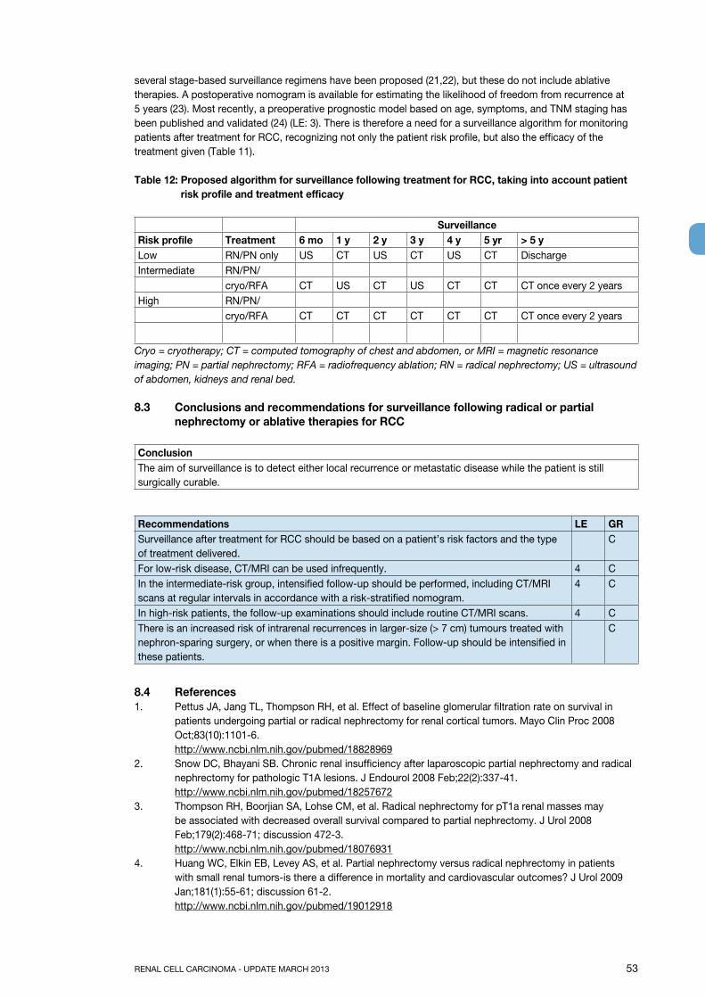
RENAL CELL CARCINOMA - UPDATE MARCH 2013 53
several stage-based surveillance regimens have been proposed (21,22), but these do not include ablative therapies. A postoperative nomogram is available for estimating the likelihood of freedom from recurrence at 5 years (23). Most recently, a preoperative prognostic model based on age, symptoms, and TNM staging has been published and validated (24) (LE: 3). There is therefore a need for a surveillance algorithm for monitoring patients after treatment for RCC, recognizing not only the patient risk profile, but also the efficacy of the treatment given (Table 11).
Table 12: proposed algorithm for surveillance following treatment for RCC, taking into account patient risk profile and treatment efficacy
SurveillanceRisk profile Treatment 6 mo 1 y 2 y 3 y 4 y 5 yr > 5 yLow RN/PN only US CT US CT US CT DischargeIntermediate RN/PN/
cryo/RFA CT US CT US CT CT CT once every 2 yearsHigh RN/PN/
cryo/RFA CT CT CT CT CT CT CT once every 2 years
Cryo = cryotherapy; CT = computed tomography of chest and abdomen, or MRI = magnetic resonance imaging; PN = partial nephrectomy; RFA = radiofrequency ablation; RN = radical nephrectomy; US = ultrasound of abdomen, kidneys and renal bed.
8.3 Conclusions and recommendations for surveillance following radical or partial nephrectomy or ablative therapies for RCC
ConclusionThe aim of surveillance is to detect either local recurrence or metastatic disease while the patient is still surgically curable.
Recommendations LE gRSurveillance after treatment for RCC should be based on a patient’s risk factors and the type of treatment delivered.
C
For low-risk disease, CT/MRI can be used infrequently. 4 CIn the intermediate-risk group, intensified follow-up should be performed, including CT/MRI scans at regular intervals in accordance with a risk-stratified nomogram.
4 C
In high-risk patients, the follow-up examinations should include routine CT/MRI scans. 4 CThere is an increased risk of intrarenal recurrences in larger-size (> 7 cm) tumours treated with nephron-sparing surgery, or when there is a positive margin. Follow-up should be intensified in these patients.
C
8.4 References1. Pettus JA, Jang TL, Thompson RH, et al. Effect of baseline glomerular filtration rate on survival in
patients undergoing partial or radical nephrectomy for renal cortical tumors. Mayo Clin Proc 2008 Oct;83(10):1101-6.http://www.ncbi.nlm.nih.gov/pubmed/18828969
2. Snow DC, Bhayani SB. Chronic renal insufficiency after laparoscopic partial nephrectomy and radical nephrectomy for pathologic T1A lesions. J Endourol 2008 Feb;22(2):337-41.http://www.ncbi.nlm.nih.gov/pubmed/18257672
3. Thompson RH, Boorjian SA, Lohse CM, et al. Radical nephrectomy for pT1a renal masses may be associated with decreased overall survival compared to partial nephrectomy. J Urol 2008 Feb;179(2):468-71; discussion 472-3.http://www.ncbi.nlm.nih.gov/pubmed/18076931
4. Huang WC, Elkin EB, Levey AS, et al. Partial nephrectomy versus radical nephrectomy in patients with small renal tumors-is there a difference in mortality and cardiovascular outcomes? J Urol 2009 Jan;181(1):55-61; discussion 61-2.http://www.ncbi.nlm.nih.gov/pubmed/19012918
54 RENAL CELL CARCINOMA - UPDATE MARCH 2013
5. Zini L, Perotte P, Capitanio U, et al. Radical versus partial nephrectomy: effect on overall and noncancer mortality. Cancer 2009 Apr;115(7):1465-71.http://www.ncbi.nlm.nih.gov/pubmed/19195042
6. Jeldres C, Patard JJ, Capitano U, et al. Partial versus radical nephrectomy in patients with adverse clinical or pathologic characteristics. Urology 2009 Jun;73(6):1300-5.http://www.ncbi.nlm.nih.gov/pubmed/19376568
7. Bruno JJ, Snyder ME, Motzer RJ, et al. Renal cell carcinoma local recurrences, impact of surgical treatment and concomitant metastasis on survival. BJU Int 2006 May;97(5):933-8.http://www.ncbi.nlm.nih.gov/pubmed/16643473
8. Sandhu SS, Symes A, A’Hern R, et al. Surgical excision of isolated renal-bed recurrence after radical nephrectomy for renal cell carcinoma. BJU Int 2005 Mar;95(4):522-5.http://www.ncbi.nlm.nih.gov/pubmed/15705072
9. Bani-Hani AH, Leibovich BC, Lohse CM, et al. Associations with contralateral recurrence following nephrectomy for renal cell carcinoma using a cohort of 2,352 patients. J Urol 2005 Feb;173(2);391-4.http://www.ncbi.nlm.nih.gov/pubmed/15643178
10. Matin SF, Ahrar K, Cadeddu JA, et al. Residual and recurrent disease following renal energy ablative therapy: a multi-institutional study. J Urol 2006 Nov;176(5):1973-7.http://www.ncbi.nlm.nih.gov/pubmed/17070224
11. Lam JS, Shvarts O, Leppert JT, et al. Renal cell carcinoma 2005: new frontiers in staging, prognostication and targeted molecular therapy. J Urol 2005 Jun;173(6):1853-62.http://www.ncbi.nlm.nih.gov/pubmed/15879764
12. Capitanio U, Cloutier V, Zini L, et al. A critical assessment of the value of clear cell, papillary and chromophobe histological subtypes in renal cell carcinoma: a population-based study. BJU Int 2009 Jun;103(11):1496-500.http://www.ncbi.nlm.nih.gov/pubmed/19076149
13. Scoll BJ, Wong YN, Egleston BL, et al. Age, tumor size and relative survival of patients with localized renal cell carcinoma: a surveillance, epidemiology and end results analysis. J Urol 2009 Feb;181(2) 506-11.http://www.ncbi.nlm.nih.gov/pubmed/19084868
14. Ionising Radiation (Medical Exposures) Regulations 2000. National Radiation Protection Board 2000. http://www.legislation.gov.uk/uksi/2000/1059/contents/made15. Patard JJ, Shvarts O, Lam JS, et al. Safety and efficacy of partial nephrectomy for all T1 tumors based
on an international multicenter experience. J Urol 2004 Jun;171(6 Pt 1):2181-5; quiz 2435. http://www.ncbi.nlm.nih.gov/pubmed/15126781
16. Kattan MW, Reuter V, Motzer RJ, et al. A postoperative prognostic nomogram for renal cell carcinoma. J Urol 2001 Jul;166(1):63-7.http://www.ncbi.nlm.nih.gov/pubmed/11435824
17. Lam JS, Shvarts O, Leppert JT, et al. Postoperative surveillance protocol for patients with localized and locally advanced renal cell carcinoma based on a validated prognosticated nomogram and risk group stratification system. J Urol 2005 Aug;174(2):466-72; discussion 472; quiz 801.http://www.ncbi.nlm.nih.gov/pubmed/16006866
18. Leibovich BC, Blute ML, Cheville JC, et al. Prediction of progression after radical nephrectomy for patients with clear cell renal cell carcinoma: a stratification tool for prospective clinical trials. Cancer 2003 Apr;97(7):1663-71.http://www.ncbi.nlm.nih.gov/pubmed/12655523
19. Karakiewicz PI, Briganti A, Chun FK, et al. Multi-institutional validation of a new renal cancer-specific survival nomogram. J Clin Oncol 2007 Apr;25(11):1316-22.http://www.ncbi.nlm.nih.gov/pubmed/17416852
20. Cindolo L, Patard JJ, Chiodini P, et al. Comparison of predictive accuracy of four prognostic models for nonmetastatic renal cell carcinoma after nephrectomy: a multicenter European study. Cancer 2005 Oct;104(7):1362-71.http://www.ncbi.nlm.nih.gov/pubmed/16116599
21. Skolarikos A, Alivizatos G, Laguna P, et al. A review on follow-up strategies for renal cell carcinoma after nephrectomy. Eur Urol 2007 Jun;51(6):1490-500; discussion 1501.http://www.ncbi.nlm.nih.gov/pubmed/17229521
22. Chin AI, Lam JS, Figlin RA, et al. Surveillance strategies for renal cell carcinoma patients following nephrectomy. Rev Urol 2006 Winter;8(1):1-7.http://www.ncbi.nlm.nih.gov/pubmed/16985554
RENAL CELL CARCINOMA - UPDATE MARCH 2013 55
23. Sorbellini M, Kattan MW, Snyder ME, et al. A postoperative nomogram predicting recurrence for patients with conventional clear cell renal cell carcinoma. J Urol 2005 Jan;173(1):48-51.http://www.ncbi.nlm.nih.gov/pubmed/15592023
24. Karakiewicz PI, Suardi N, Capitano U, et al. A preoperative prognostic model for patients treated with nephrectomy for renal cell carcinoma. Eur Urol 2009 Feb;55(2);287-95.http://www.ncbi.nlm.nih.gov/pubmed/18715700

56 RENAL CELL CARCINOMA - UPDATE MARCH 2013
9. ABBREVIATIONS USED IN THE TEXT This list is not comprehensive for the most common abbreviations
ACKD acquired cystic kidney diseaseAML Angiomyolipoma5FU 5-fluorouracilBSC best supportive careCaIX carbonic anhydrase IXcRCC clear cell renal carcinomachRCC chromophobe renal cell carcinomaCT computed tomographyESKD end-stage kidney diseaseFLT-3 FMS-like tyrosine kinase 3GR grade of recommendationHIF hypoxia inducible factorHIFU high-intensity focused ultrasoundHU Hounsfield unitIFN-alpha interferon-alphaIL-2 interleukin-2LE level of evidenceMESTK mixed epithelial and stromal tumour of the kidneymRCC metastatic renal cell carcinomaMRI magnetic resonance imagingmTOR mammalian target of rapamycinNSS nephron-sparing surgeryPA predictive accuracypRCC papillary renal cell carcinomaRCC renal cell carcinomaPDGF platelet-derived growth factorPDGFR platelet-derived growth factor receptorPET positron emission tomographyPTEN phosphatase and tensin homologREST Renal epithelial and stromal tumoursRF radiofrequencyRFA radiofrequency ablationSAE selective arterial embolisationTFE3 transcription factor E3TK tyrosine kinaseTKI Tyrosine kinase inhibitorsTNM Tumour Node MetastasisUS abdominal ultrasoundVEGF vascular endothelial growth factorVEGFR vascular endothelial growth factor receptorVHL von Hippel-LindauWHO World Health Organization
Conflict of interestAll members of the Renal Cell Cancer working group have provided disclosure statements of all relationships that they have that might be perceived as a potential source of a conflict of interest. This information is publically accessible through the European Association of Urology website. This guidelines document was developed with the financial support of the European Association of Urology. No external sources of funding and support have been involved. The EAU is a non-profit organisation and funding is limited to administrative assistance and travel and meeting expenses. No honoraria or other reimbursements have been provided.