EFSUMB Guidelines on Elastography · 2018-02-28 · •At interfaces or tissue layers there are...
24
EFSUMB Guidelines on Elastography: Part I: Βasic Principles & Technology Elena Drakonaki MD, PhD, PostDoc Consultant MSK Radiologist Independent Practice, Heraklion, Greece & Radcliff NHS Trust, Oxford, UK
Transcript of EFSUMB Guidelines on Elastography · 2018-02-28 · •At interfaces or tissue layers there are...
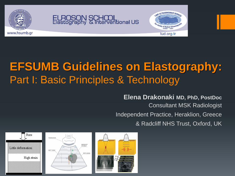
EFSUMB Guidelines on Elastography: Part I: Βasic Principles & Technology
Elena Drakonaki MD, PhD, PostDoc
Consultant MSK Radiologist
Independent Practice, Heraklion, Greece
& Radcliff NHS Trust, Oxford, UK
EUROSON SCHOOL 2014, Athens
www.drakonaki.gr
US Elastography
Rapidly evolving in recent years
Growing general interest, a lot of available techniques, level of scientific evidence
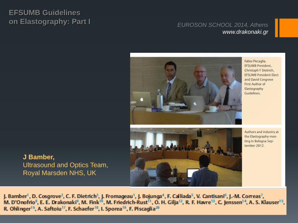
2011: steering committee selected European experts
Sept 2012: consensus meeting in Bologna
Supported by the industry (BK medical, Echosens, Esaote, GE, Hitachi Aloka, Philips, Siemens, Supersonic, Toshiba)
No influence on the content
EFSUMB Guidelines & Recommendation
on Elastography: Part I
EFSUMB
EUROSON SCHOOL 2014, Athens
www.drakonaki.gr
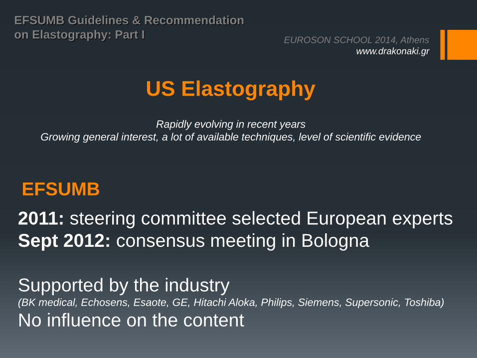
EFSUMB Guidelines
on Elastography: Part I
Ultraschall Med. 2013 Apr;34(2):169-84
Ultraschall Med. 2013 Jun;34(3):238-53 www.efsumb.org
EUROSON SCHOOL 2014, Athens
www.drakonaki.gr
EFSUMB Guidelines
on Elastography: Part I
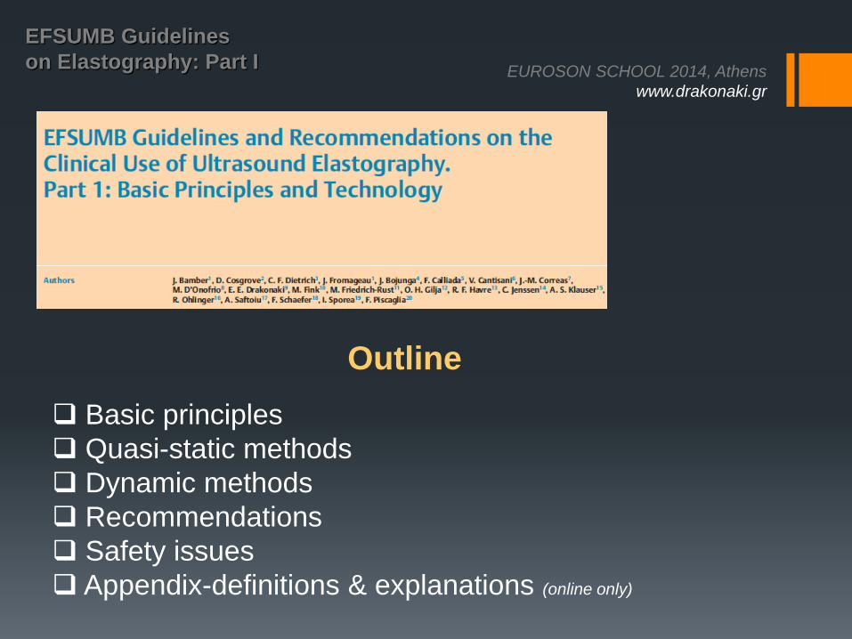
Outline
Basic principles
Quasi-static methods
Dynamic methods
Recommendations
Safety issues
Appendix-definitions & explanations (online only)
EUROSON SCHOOL 2014, Athens
www.drakonaki.gr
EFSUMB Guidelines
on Elastography: Part I
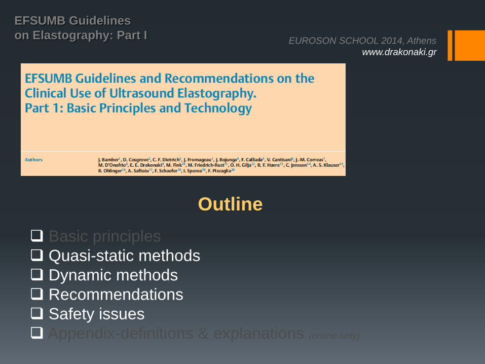
Outline
Basic principles
Quasi-static methods
Dynamic methods
Recommendations
Safety issues
Appendix-definitions & explanations (online only)
EUROSON SCHOOL 2014, Athens
www.drakonaki.gr
EFSUMB Guidelines
on Elastography: Part I
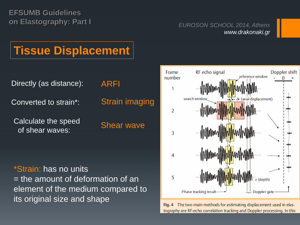
Tissue Displacement
Calculate the speed
of shear waves:
ARFI
Strain imaging
Shear wave
*Strain: has no units
= the amount of deformation of an
element of the medium compared to
its original size and shape
Directly (as distance):
Converted to strain*:
EUROSON SCHOOL 2014, Athens
www.drakonaki.gr
EFSUMB Guidelines
on Elastography: Part I
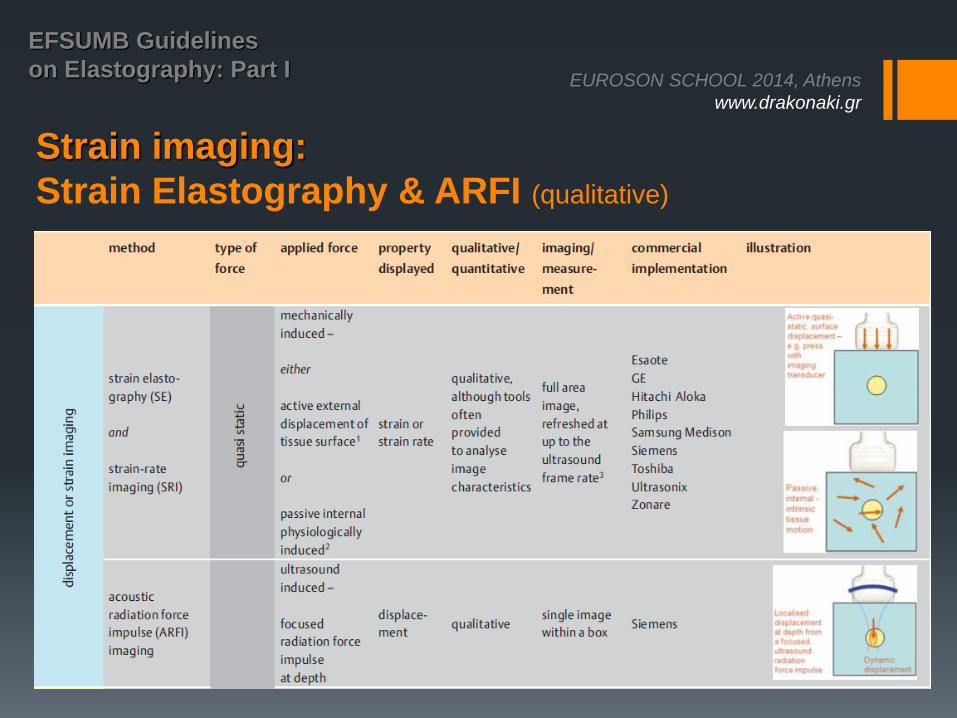
Strain imaging:
Strain Elastography & ARFI (qualitative)
EUROSON SCHOOL 2014, Athens
www.drakonaki.gr
EFSUMB Guidelines
on Elastography: Part I
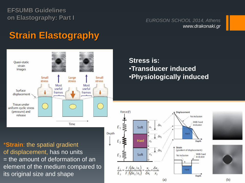
Strain Elastography
*Strain: the spatial gradient
of displacement, has no units
= the amount of deformation of an
element of the medium compared to
its original size and shape
Stress is:
•Transducer induced
•Physiologically induced
EUROSON SCHOOL 2014, Athens
www.drakonaki.gr
EFSUMB Guidelines
on Elastography: Part I
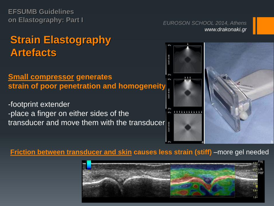
Strain Elastography
Artefacts
Small compressor generates
strain of poor penetration and homogeneity
-footprint extender
-place a finger on either sides of the
transducer and move them with the transducer
Friction between transducer and skin causes less strain (stiff) –more gel needed
EUROSON SCHOOL 2014, Athens
www.drakonaki.gr
EFSUMB Guidelines
on Elastography: Part I
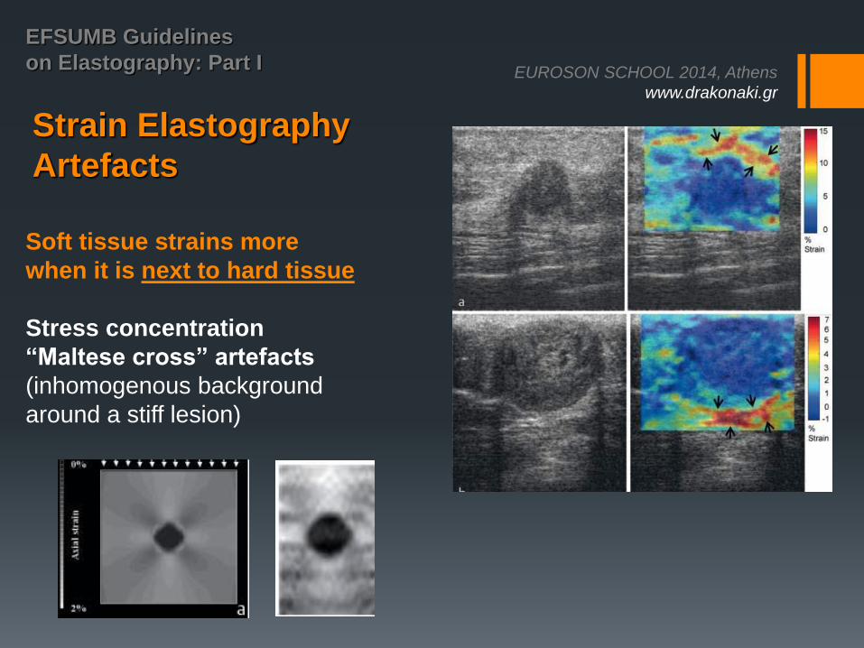
Strain Elastography
Artefacts
Soft tissue strains more
when it is next to hard tissue
Stress concentration
“Maltese cross” artefacts
(inhomogenous background
around a stiff lesion)
EUROSON SCHOOL 2014, Athens
www.drakonaki.gr
EFSUMB Guidelines
on Elastography: Part I
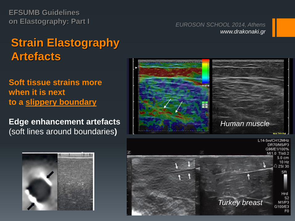
Strain Elastography
Artefacts
Soft tissue strains more
when it is next
to a slippery boundary
Edge enhancement artefacts
(soft lines around boundaries)
Turkey breast
Human muscle
EUROSON SCHOOL 2014, Athens
www.drakonaki.gr
EFSUMB Guidelines
on Elastography: Part I
Strain Elastography
Artefacts
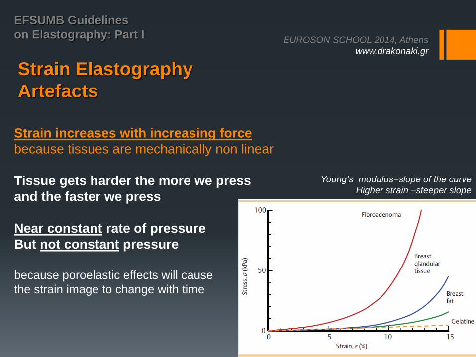
Strain increases with increasing force
because tissues are mechanically non linear
Tissue gets harder the more we press
and the faster we press
Near constant rate of pressure
But not constant pressure
because poroelastic effects will cause
the strain image to change with time
Young’s modulus=slope of the curve
Higher strain –steeper slope
EUROSON SCHOOL 2014, Athens
www.drakonaki.gr
EFSUMB Guidelines
on Elastography: Part I
Strain Elastography
Artefacts
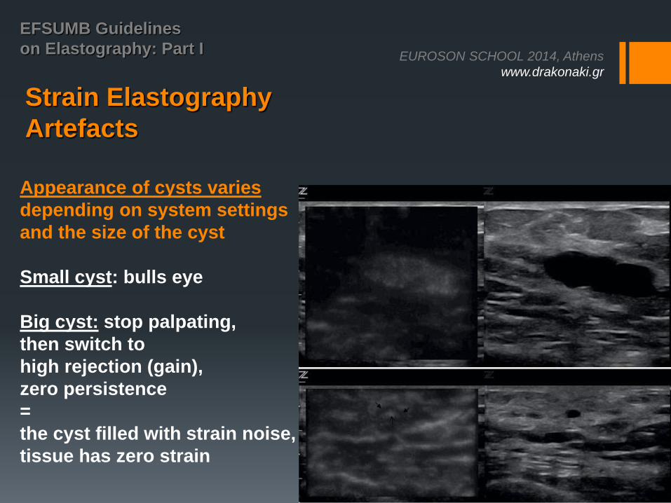
Appearance of cysts varies
depending on system settings
and the size of the cyst
Small cyst: bulls eye
Big cyst: stop palpating,
then switch to
high rejection (gain),
zero persistence
=
the cyst filled with strain noise,
tissue has zero strain
EUROSON SCHOOL 2014, Athens
www.drakonaki.gr
EFSUMB Guidelines
on Elastography: Part I
Strain Elastography
How to get good quality strain images:
▶ Close proximity to the transducer (< 3 –4cm)
▶ Near homogeneous tissue (e. g. liver)
▶ No anatomical planes that allow slipping
▶ Some distance to tissue boundaries
▶ No structures (e. g. large veins) that would damp the shear stress
▶ A broad stress source relative to the width of the imaged region
▶ A limited number of diagnostic targets
EUROSON SCHOOL 2014, Athens
www.drakonaki.gr
EFSUMB Guidelines
on Elastography: Part I
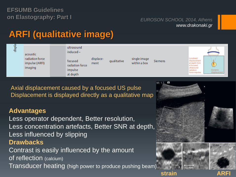
ARFI (qualitative image)
Advantages
Less operator dependent, Better resolution,
Less concentration artefacts, Better SNR at depth,
Less influenced by slipping
Drawbacks
Contrast is easily influenced by the amount
of reflection (calcium)
Transducer heating (high power to produce pushing beam)
strain ARFI
Axial displacement caused by a focused US pulse
Displacement is displayed directly as a qualitative map
EUROSON SCHOOL 2014, Athens
www.drakonaki.gr
EFSUMB Guidelines
on Elastography: Part I
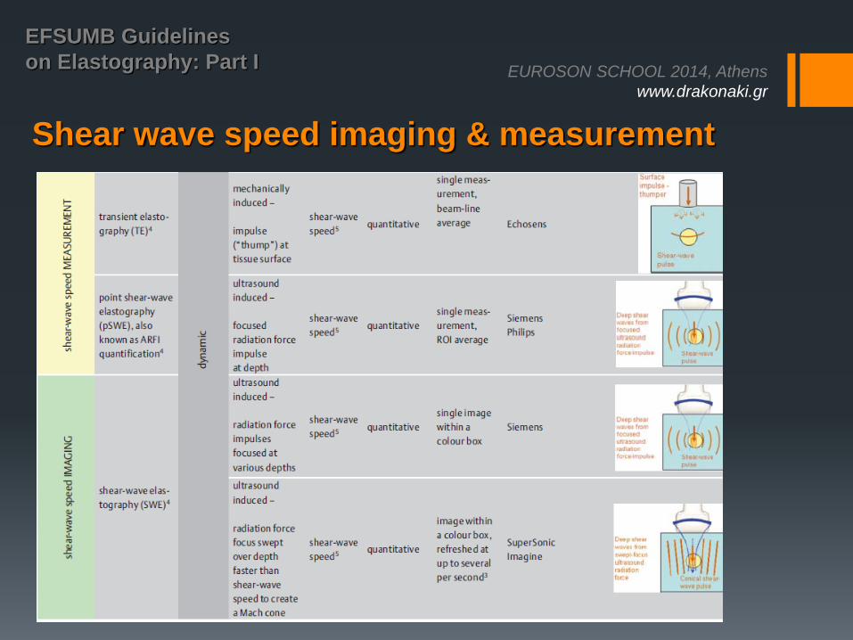
Shear wave speed imaging & measurement
EUROSON SCHOOL 2014, Athens
www.drakonaki.gr
EFSUMB Guidelines
on Elastography: Part I
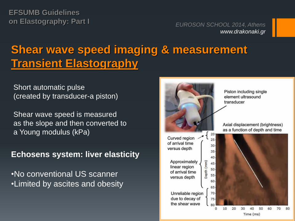
Shear wave speed imaging & measurement
Transient Elastography
Short automatic pulse
(created by transducer-a piston)
Shear wave speed is measured
as the slope and then converted to
a Young modulus (kPa)
Echosens system: liver elasticity
•No conventional US scanner
•Limited by ascites and obesity
EUROSON SCHOOL 2014, Athens
www.drakonaki.gr
EFSUMB Guidelines
on Elastography: Part I
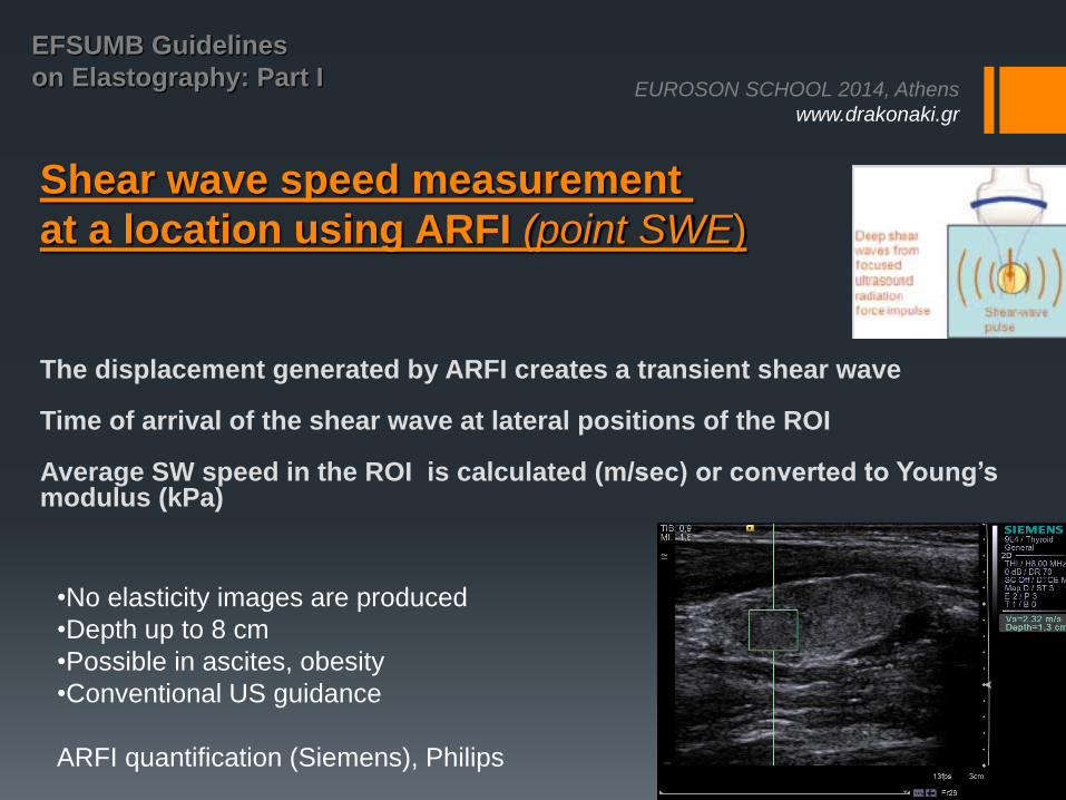
Shear wave speed measurement
at a location using ARFI (point SWE)
The displacement generated by ARFI creates a transient shear wave Time of arrival of the shear wave at lateral positions of the ROI Average SW speed in the ROI is calculated (m/sec) or converted to Young’s modulus (kPa)
•No elasticity images are produced
•Depth up to 8 cm
•Possible in ascites, obesity
•Conventional US guidance
ARFI quantification (Siemens), Philips
EUROSON SCHOOL 2014, Athens
www.drakonaki.gr
EFSUMB Guidelines
on Elastography: Part I
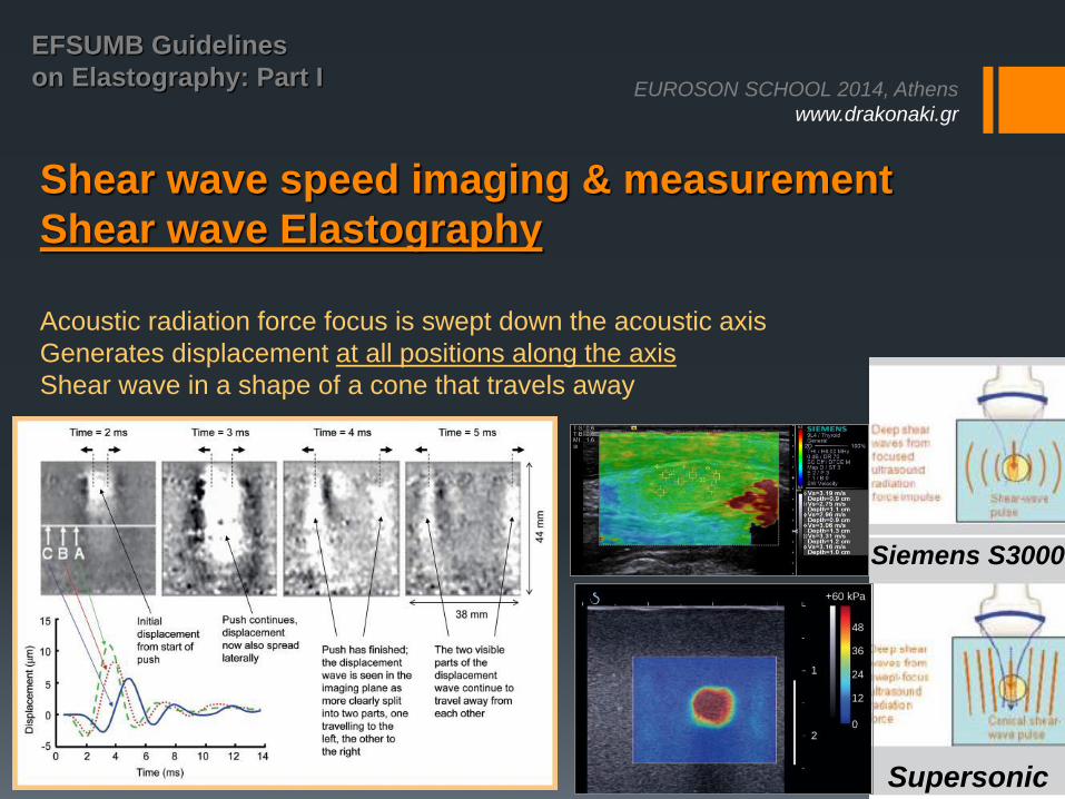
Shear wave speed imaging & measurement
Shear wave Elastography
Acoustic radiation force focus is swept down the acoustic axis
Generates displacement at all positions along the axis
Shear wave in a shape of a cone that travels away
Supersonic
Siemens S3000
EUROSON SCHOOL 2014, Athens
www.drakonaki.gr
EFSUMB Guidelines
on Elastography: Part I
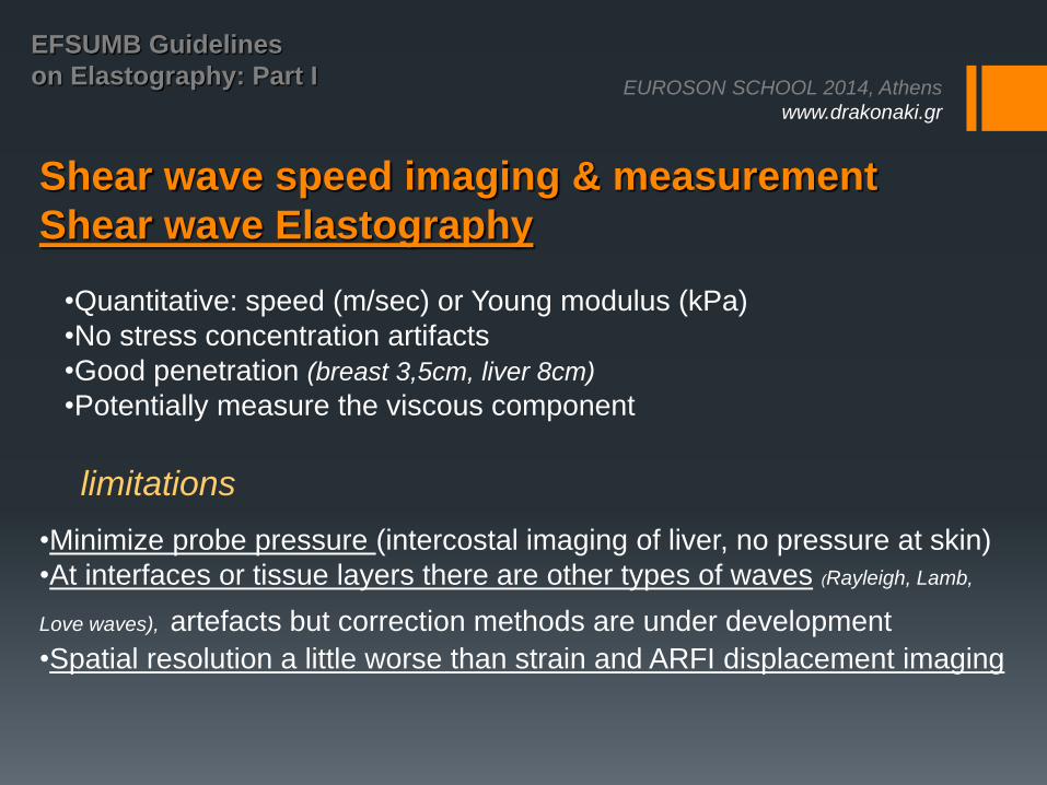
Shear wave speed imaging & measurement
Shear wave Elastography
•Minimize probe pressure (intercostal imaging of liver, no pressure at skin)
•At interfaces or tissue layers there are other types of waves (Rayleigh, Lamb,
Love waves), artefacts but correction methods are under development
•Spatial resolution a little worse than strain and ARFI displacement imaging
•Quantitative: speed (m/sec) or Young modulus (kPa)
•No stress concentration artifacts
•Good penetration (breast 3,5cm, liver 8cm)
•Potentially measure the viscous component
limitations
EUROSON SCHOOL 2014, Athens
www.drakonaki.gr
EFSUMB Guidelines
on Elastography: Part I
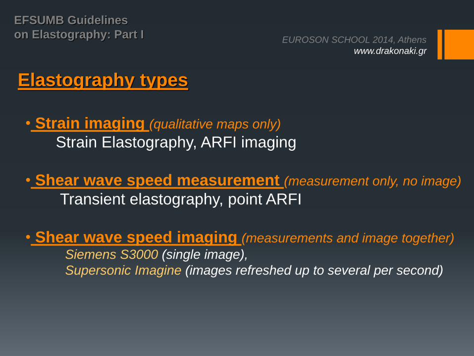
Elastography types
• Strain imaging (qualitative maps only)
Strain Elastography, ARFI imaging
• Shear wave speed measurement (measurement only, no image)
Transient elastography, point ARFI
• Shear wave speed imaging (measurements and image together)
Siemens S3000 (single image),
Supersonic Imagine (images refreshed up to several per second)
EUROSON SCHOOL 2014, Athens
www.drakonaki.gr
EFSUMB Guidelines
on Elastography: Part I
Elastography safety issues
Strain and shear wave:
identical safety issues as conventional US
ARFI:
higher TI but within AIUM limits,
safety issues similar to Doppler (eye, fetus etc)
EUROSON SCHOOL 2014, Athens
www.drakonaki.gr
EFSUMB Guidelines
on Elastography: Part I
Conclusion
Knowledge of physics and technology
Technology is expected to further develop
Improvements are expected in:
image quality
ease of use
quantification methods
tissue characteristics measurable (viscous component)
EUROSON SCHOOL 2014, Athens
www.drakonaki.gr
EFSUMB Guidelines
on Elastography: Part I
J Bamber,
Ultrasound and Optics Team,
Royal Marsden NHS, UK