Crystal Structure of the Calcium-Loaded Spherulin 3a Dimer Sheds Light on the Evolution Of the Eye...
10
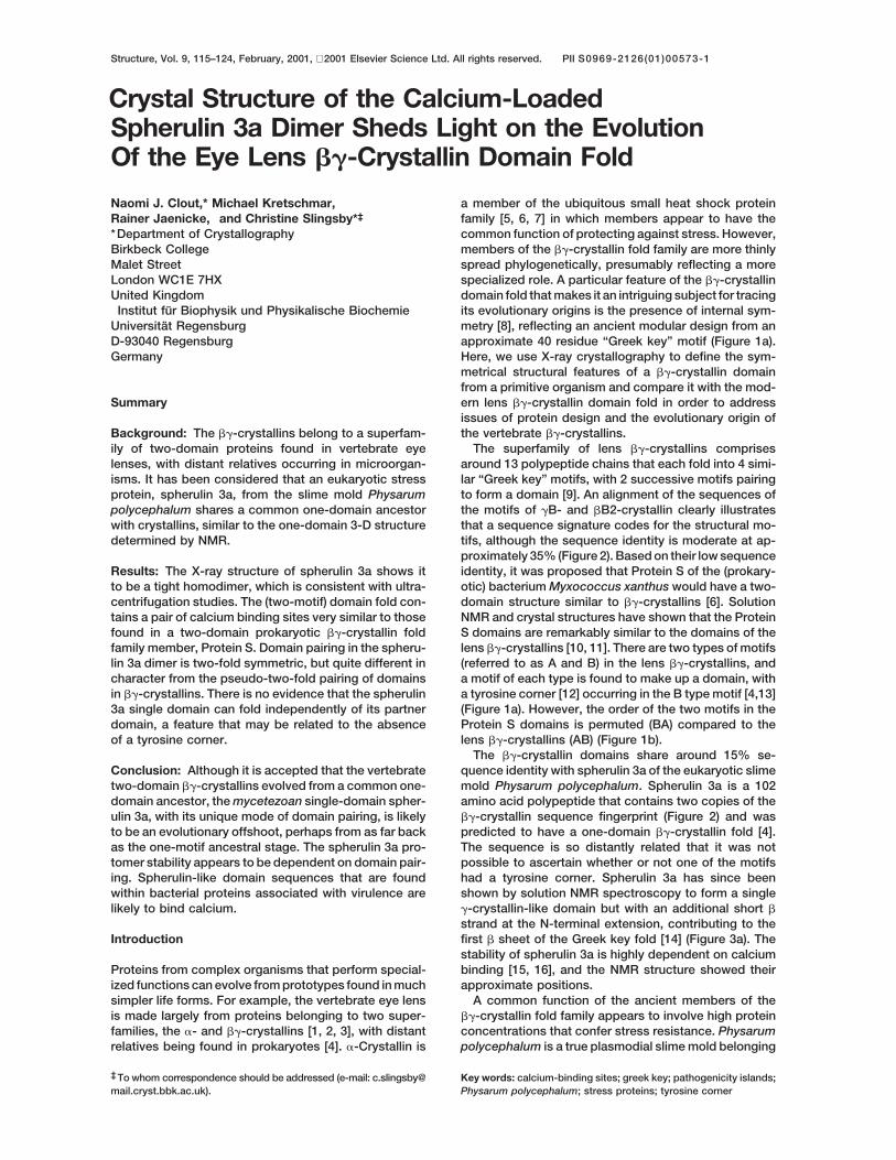
Structure, Vol. 9, 115–124, February, 2001, 2001 Elsevier Science Ltd. All rights reserved. PIIS0969-2126(01)00573-1 Crystal Structure of the Calcium-Loaded Spherulin 3a Dimer Sheds Light on the Evolution Of the Eye Lens bg-Crystallin Domain Fold a member of the ubiquitous small heat shock protein family [5, 6, 7] in which members appear to have the common function of protecting against stress. However, members of the bg-crystallin fold family are more thinly spread phylogenetically, presumably reflecting a more Naomi J. Clout,* Michael Kretschmar, Rainer Jaenicke, and Christine Slingsby* ‡ * Department of Crystallography Birkbeck College Malet Street London WC1E 7HX specialized role. A particular feature of the bg-crystallin domain fold that makes it an intriguing subject for tracing United Kingdom Institut fu ¨ r Biophysik und Physikalische Biochemie its evolutionary origins is the presence of internal sym- metry [8], reflecting an ancient modular design from an Universita ¨ t Regensburg D-93040 Regensburg approximate 40 residue “Greek key” motif (Figure 1a). Here, we use X-ray crystallography to define the sym- Germany metrical structural features of a bg-crystallin domain from a primitive organism and compare it with the mod- ern lens bg-crystallin domain fold in order to address Summary issues of protein design and the evolutionary origin of the vertebrate bg-crystallins. Background: The bg-crystallins belong to a superfam- ily of two-domain proteins found in vertebrate eye The superfamily of lens bg-crystallins comprises around 13 polypeptide chains that each fold into 4 simi- lenses, with distant relatives occurring in microorgan- isms. It has been considered that an eukaryotic stress lar “Greek key” motifs, with 2 successive motifs pairing to form a domain [9]. An alignment of the sequences of protein, spherulin 3a, from the slime mold Physarum polycephalum shares a common one-domain ancestor the motifs of gB- and bB2-crystallin clearly illustrates that a sequence signature codes for the structural mo- with crystallins, similar to the one-domain 3-D structure determined by NMR. tifs, although the sequence identity is moderate at ap- proximately 35% (Figure 2). Based on their low sequence identity, it was proposed that Protein S of the (prokary- Results: The X-ray structure of spherulin 3a shows it to be a tight homodimer, which is consistent with ultra- otic) bacterium Myxococcus xanthus would have a two- domain structure similar to bg-crystallins [6]. Solution centrifugation studies. The (two-motif) domain fold con- tains a pair of calcium binding sites very similar to those NMR and crystal structures have shown that the Protein S domains are remarkably similar to the domains of the found in a two-domain prokaryotic bg-crystallin fold family member, Protein S. Domain pairing in the spheru- lens bg-crystallins [10, 11]. There are two types of motifs (referred to as A and B) in the lens bg-crystallins, and lin 3a dimer is two-fold symmetric, but quite different in character from the pseudo-two-fold pairing of domains a motif of each type is found to make up a domain, with a tyrosine corner [12] occurring in the B type motif [4,13] in bg-crystallins. There is no evidence that the spherulin 3a single domain can fold independently of its partner (Figure 1a). However, the order of the two motifs in the Protein S domains is permuted (BA) compared to the domain, a feature that may be related to the absence of a tyrosine corner. lens bg-crystallins (AB) (Figure 1b). The bg-crystallin domains share around 15% se- quence identity with spherulin 3a of the eukaryotic slime Conclusion: Although it is accepted that the vertebrate two-domain bg-crystallins evolved from a common one- mold Physarum polycephalum. Spherulin 3a is a 102 amino acid polypeptide that contains two copies of the domain ancestor, the mycetezoan single-domain spher- ulin 3a, with its unique mode of domain pairing, is likely bg-crystallin sequence fingerprint (Figure 2) and was predicted to have a one-domain bg-crystallin fold [4]. to be an evolutionary offshoot, perhaps from as far back as the one-motif ancestral stage. The spherulin 3a pro- The sequence is so distantly related that it was not possible to ascertain whether or not one of the motifs tomer stability appears to be dependent on domain pair- ing. Spherulin-like domain sequences that are found had a tyrosine corner. Spherulin 3a has since been shown by solution NMR spectroscopy to form a single within bacterial proteins associated with virulence are likely to bind calcium. g-crystallin-like domain but with an additional short b strand at the N-terminal extension, contributing to the first b sheet of the Greek key fold [14] (Figure 3a). The Introduction stability of spherulin 3a is highly dependent on calcium binding [15, 16], and the NMR structure showed their Proteins from complex organisms that perform special- ized functions can evolve from prototypes found in much approximate positions. A common function of the ancient members of the simpler life forms. For example, the vertebrate eye lens is made largely from proteins belonging to two super- bg-crystallin fold family appears to involve high protein concentrations that confer stress resistance. Physarum families, the a- and bg-crystallins [1, 2, 3], with distant relatives being found in prokaryotes [4]. a-Crystallin is polycephalum is a true plasmodial slime mold belonging Key words: calcium-binding sites; greek key; pathogenicity islands; ‡ To whom correspondence should be addressed (e-mail: c.slingsby@ mail.cryst.bbk.ac.uk). Physarum polycephalum; stress proteins; tyrosine corner
-
Upload
naomi-j-clout -
Category
Documents
-
view
212 -
download
0
Transcript of Crystal Structure of the Calcium-Loaded Spherulin 3a Dimer Sheds Light on the Evolution Of the Eye...
Structure, Vol. 9, 115–124, February, 2001, 2001 Elsevier Science Ltd. All rights reserved. PII S0969-2126(01)00573-1
Crystal Structure of the Calcium-LoadedSpherulin 3a Dimer Sheds Light on the EvolutionOf the Eye Lens bg-Crystallin Domain Fold
a member of the ubiquitous small heat shock proteinfamily [5, 6, 7] in which members appear to have thecommon function of protecting against stress. However,members of the bg-crystallin fold family are more thinlyspread phylogenetically, presumably reflecting a more
Naomi J. Clout,* Michael Kretschmar,†Rainer Jaenicke,† and Christine Slingsby*‡
*Department of CrystallographyBirkbeck CollegeMalet StreetLondon WC1E 7HX specialized role. A particular feature of the bg-crystallin
domain fold that makes it an intriguing subject for tracingUnited Kingdom†Institut fur Biophysik und Physikalische Biochemie its evolutionary origins is the presence of internal sym-
metry [8], reflecting an ancient modular design from anUniversitat RegensburgD-93040 Regensburg approximate 40 residue “Greek key” motif (Figure 1a).
Here, we use X-ray crystallography to define the sym-Germanymetrical structural features of a bg-crystallin domainfrom a primitive organism and compare it with the mod-ern lens bg-crystallin domain fold in order to addressSummaryissues of protein design and the evolutionary origin ofthe vertebrate bg-crystallins.Background: The bg-crystallins belong to a superfam-
ily of two-domain proteins found in vertebrate eye The superfamily of lens bg-crystallins comprisesaround 13 polypeptide chains that each fold into 4 simi-lenses, with distant relatives occurring in microorgan-
isms. It has been considered that an eukaryotic stress lar “Greek key” motifs, with 2 successive motifs pairingto form a domain [9]. An alignment of the sequences ofprotein, spherulin 3a, from the slime mold Physarum
polycephalum shares a common one-domain ancestor the motifs of gB- and bB2-crystallin clearly illustratesthat a sequence signature codes for the structural mo-with crystallins, similar to the one-domain 3-D structure
determined by NMR. tifs, although the sequence identity is moderate at ap-proximately 35% (Figure 2). Based on their low sequenceidentity, it was proposed that Protein S of the (prokary-Results: The X-ray structure of spherulin 3a shows it
to be a tight homodimer, which is consistent with ultra- otic) bacterium Myxococcus xanthus would have a two-domain structure similar to bg-crystallins [6]. Solutioncentrifugation studies. The (two-motif) domain fold con-
tains a pair of calcium binding sites very similar to those NMR and crystal structures have shown that the ProteinS domains are remarkably similar to the domains of thefound in a two-domain prokaryotic bg-crystallin fold
family member, Protein S. Domain pairing in the spheru- lens bg-crystallins [10, 11]. There are two types of motifs(referred to as A and B) in the lens bg-crystallins, andlin 3a dimer is two-fold symmetric, but quite different in
character from the pseudo-two-fold pairing of domains a motif of each type is found to make up a domain, witha tyrosine corner [12] occurring in the B type motif [4,13]in bg-crystallins. There is no evidence that the spherulin
3a single domain can fold independently of its partner (Figure 1a). However, the order of the two motifs in theProtein S domains is permuted (BA) compared to thedomain, a feature that may be related to the absence
of a tyrosine corner. lens bg-crystallins (AB) (Figure 1b).The bg-crystallin domains share around 15% se-
quence identity with spherulin 3a of the eukaryotic slimeConclusion: Although it is accepted that the vertebratetwo-domain bg-crystallins evolved from a common one- mold Physarum polycephalum. Spherulin 3a is a 102
amino acid polypeptide that contains two copies of thedomain ancestor, the mycetezoan single-domain spher-ulin 3a, with its unique mode of domain pairing, is likely bg-crystallin sequence fingerprint (Figure 2) and was
predicted to have a one-domain bg-crystallin fold [4].to be an evolutionary offshoot, perhaps from as far backas the one-motif ancestral stage. The spherulin 3a pro- The sequence is so distantly related that it was not
possible to ascertain whether or not one of the motifstomer stability appears to be dependent on domain pair-ing. Spherulin-like domain sequences that are found had a tyrosine corner. Spherulin 3a has since been
shown by solution NMR spectroscopy to form a singlewithin bacterial proteins associated with virulence arelikely to bind calcium. g-crystallin-like domain but with an additional short b
strand at the N-terminal extension, contributing to thefirst b sheet of the Greek key fold [14] (Figure 3a). TheIntroductionstability of spherulin 3a is highly dependent on calciumbinding [15, 16], and the NMR structure showed theirProteins from complex organisms that perform special-
ized functions can evolve from prototypes found in much approximate positions.A common function of the ancient members of thesimpler life forms. For example, the vertebrate eye lens
is made largely from proteins belonging to two super- bg-crystallin fold family appears to involve high proteinconcentrations that confer stress resistance. Physarumfamilies, the a- and bg-crystallins [1, 2, 3], with distant
relatives being found in prokaryotes [4]. a-Crystallin is polycephalum is a true plasmodial slime mold belonging
Key words: calcium-binding sites; greek key; pathogenicity islands;‡ To whom correspondence should be addressed (e-mail: [email protected]). Physarum polycephalum; stress proteins; tyrosine corner
Structure116
Figure 1. Circular Permutation in the bg-Crystallin Fold Family Single Domain
(a) The N-terminal domain of bovine gB-crys-tallin (4GCR), with motif 1 (A type) shown indark blue and motif 2 (B type) shown in lightblue. The view is approximately perpendicu-lar to the pseudo-two-fold axis relating thetwo motifs. The c strand of one motif contrib-utes to the a, b, and d strands of the partnermotif to make a pair of b sheets. The tyrosinecorner residue is appended in light blue onthe B type motif.(b) The N-terminal domain of Protein S(1NPS), with motif 1 (B type) shown in darkblue and motif 2 (A type) shown in yellow. Thetyrosine corner residue is appended in darkblue on the B type motif. The figure wasdrawn using the program Setor [42].
to the mycetozoa, which are thought to be late emerging, grescens [22] have the same topology as bg-crystallindomain folds, although they may be examples of conver-multicellular eukaryotes more closely related to the ani-
mal-fungal clade than are green plants [17]. This crea- gent evolution [23]. Here, we show by X-ray crystallogra-phy that spherulin 3a and Protein S have very similarture has three lifestyles: microscopic amoeba; gigantic,
crawling, multi-nucleate plasmodium; and the fruiting two-fold symmetric calcium binding sites. As Protein Sis a well established member of the bg-crystallin foldbody sporangium. During stress, such as starvation and
darkness, the plasmodium can divide into smaller dehy- family, this further supports spherulin 3a being a bonafida single-domain member of this fold family. However,drated spherules, each containing several nuclei that
overexpress specific proteins, the most abundant of spherulin 3a does not exist as a single domain in solutionbut as a dimer with tight binding, as dissociation cannotwhich is spherulin 3a [18]. These proteins are encased
within a hard wall in the presence of calcium, which is be accomplished without full denaturation [15, 16, 24].For a one-domain crystallin to be the prototype of a bg-in sufficient amounts to occupy the two calcium binding
sites on the spherulin 3a domain [15]. Protein S also has crystallin ancestor, it would be expected to self-associ-ate to form a noncovalent dimer with a similar domaina stress connection. Upon starvation, cells differentiate
into highly durable myxospores that are resistant to des- interface as that between the N and C domains in thebg-crystallins. The spherulin 3a dimer was not revealediccation, heat, and ultraviolet radiation, enabling them to
be viable for years. They are protected by a multilayered by solution NMR spectroscopy. Here, we use X-ray crys-tallography to define the dimer structure and show thatspore coat consisting of polysaccharides and proteins
that includes the calcium binding Protein S. The process it is unlikely to be the prototype single-domain ancestorof the bg-crystallin fold family.of spore coat formation requires calcium [19, 20]. It
seems that the ancestral-like bg-crystallin domains usecalcium to stabilize the domain fold. Results and Discussion
Other one-domain structures have membershipclaims on the bg-crystallin fold family. The killer toxin The Domain Structure
The crystal structure of the spherulin 3a monomer con-from the yeast Williopsis mraki [21] and a metallopro-teinase inhibitor from the prokaryotic Streptomyces ni- firms the fold of the monomer found by solution NMR
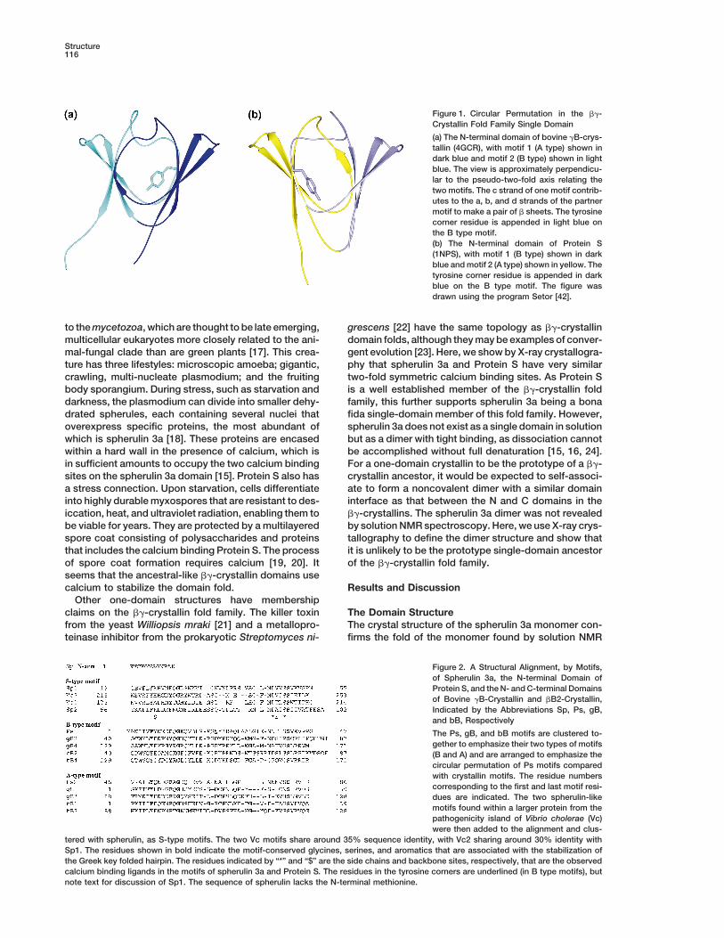
Figure 2. A Structural Alignment, by Motifs,of Spherulin 3a, the N-terminal Domain ofProtein S, and the N- and C-terminal Domainsof Bovine gB-Crystallin and bB2-Crystallin,Indicated by the Abbreviations Sp, Ps, gB,and bB, Respectively
The Ps, gB, and bB motifs are clustered to-gether to emphasize their two types of motifs(B and A) and are arranged to emphasize thecircular permutation of Ps motifs comparedwith crystallin motifs. The residue numberscorresponding to the first and last motif resi-dues are indicated. The two spherulin-likemotifs found within a larger protein from thepathogenicity island of Vibrio cholerae (Vc)were then added to the alignment and clus-
tered with spherulin, as S-type motifs. The two Vc motifs share around 35% sequence identity, with Vc2 sharing around 30% identity withSp1. The residues shown in bold indicate the motif-conserved glycines, serines, and aromatics that are associated with the stabilization ofthe Greek key folded hairpin. The residues indicated by “*” and “$” are the side chains and backbone sites, respectively, that are the observedcalcium binding ligands in the motifs of spherulin 3a and Protein S. The residues in the tyrosine corners are underlined (in B type motifs), butnote text for discussion of Sp1. The sequence of spherulin lacks the N-terminal methionine.
Structure of calcium loaded spherulin 3a dimer117
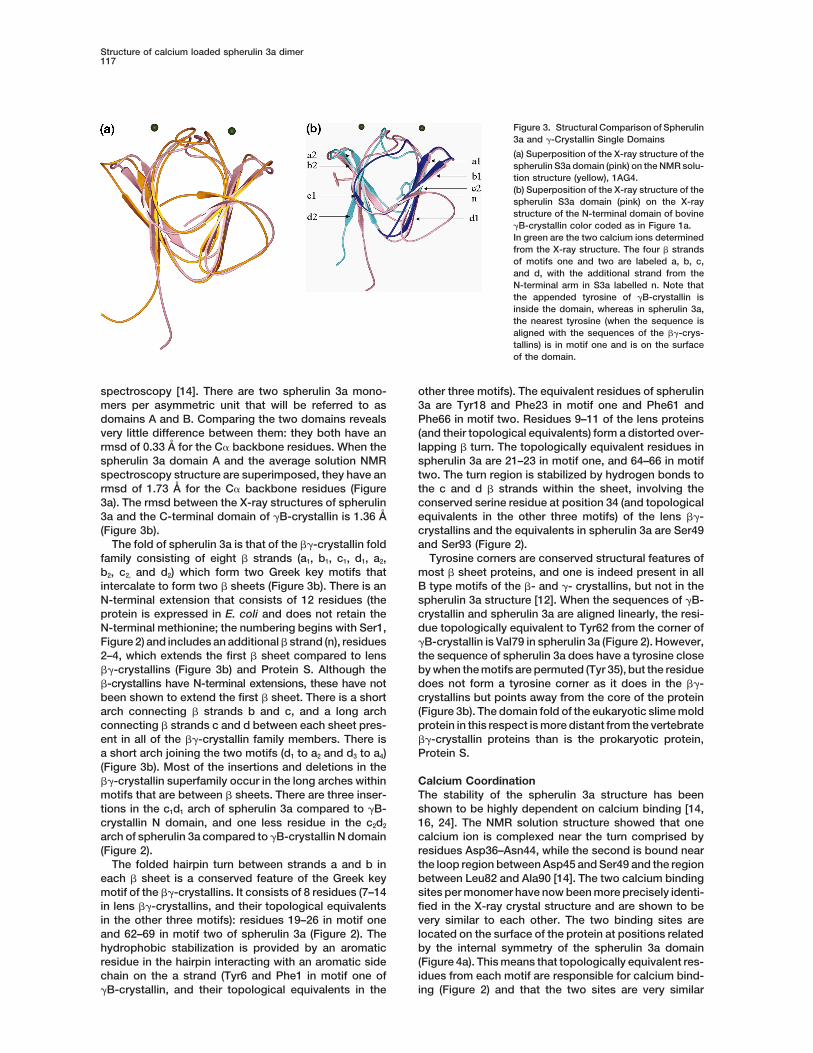
Figure 3. Structural Comparison of Spherulin3a and g-Crystallin Single Domains
(a) Superposition of the X-ray structure of thespherulin S3a domain (pink) on the NMR solu-tion structure (yellow), 1AG4.(b) Superposition of the X-ray structure of thespherulin S3a domain (pink) on the X-raystructure of the N-terminal domain of bovinegB-crystallin color coded as in Figure 1a.In green are the two calcium ions determinedfrom the X-ray structure. The four b strandsof motifs one and two are labeled a, b, c,and d, with the additional strand from theN-terminal arm in S3a labelled n. Note thatthe appended tyrosine of gB-crystallin isinside the domain, whereas in spherulin 3a,the nearest tyrosine (when the sequence isaligned with the sequences of the bg-crys-tallins) is in motif one and is on the surfaceof the domain.
spectroscopy [14]. There are two spherulin 3a mono- other three motifs). The equivalent residues of spherulin3a are Tyr18 and Phe23 in motif one and Phe61 andmers per asymmetric unit that will be referred to as
domains A and B. Comparing the two domains reveals Phe66 in motif two. Residues 9–11 of the lens proteins(and their topological equivalents) form a distorted over-very little difference between them: they both have an
rmsd of 0.33 A for the Ca backbone residues. When the lapping b turn. The topologically equivalent residues inspherulin 3a are 21–23 in motif one, and 64–66 in motifspherulin 3a domain A and the average solution NMR
spectroscopy structure are superimposed, they have an two. The turn region is stabilized by hydrogen bonds tothe c and d b strands within the sheet, involving thermsd of 1.73 A for the Ca backbone residues (Figure
3a). The rmsd between the X-ray structures of spherulin conserved serine residue at position 34 (and topologicalequivalents in the other three motifs) of the lens bg-3a and the C-terminal domain of gB-crystallin is 1.36 A
(Figure 3b). crystallins and the equivalents in spherulin 3a are Ser49and Ser93 (Figure 2).The fold of spherulin 3a is that of the bg-crystallin fold
family consisting of eight b strands (a1, b1, c1, d1, a2, Tyrosine corners are conserved structural features ofmost b sheet proteins, and one is indeed present in allb2, c2, and d2) which form two Greek key motifs that
intercalate to form two b sheets (Figure 3b). There is an B type motifs of the b- and g- crystallins, but not in thespherulin 3a structure [12]. When the sequences of gB-N-terminal extension that consists of 12 residues (the
protein is expressed in E. coli and does not retain the crystallin and spherulin 3a are aligned linearly, the resi-due topologically equivalent to Tyr62 from the corner ofN-terminal methionine; the numbering begins with Ser1,
Figure 2) and includes an additional b strand (n), residues gB-crystallin is Val79 in spherulin 3a (Figure 2). However,the sequence of spherulin 3a does have a tyrosine close2–4, which extends the first b sheet compared to lens
bg-crystallins (Figure 3b) and Protein S. Although the by when the motifs are permuted (Tyr 35), but the residuedoes not form a tyrosine corner as it does in the bg-b-crystallins have N-terminal extensions, these have not
been shown to extend the first b sheet. There is a short crystallins but points away from the core of the protein(Figure 3b). The domain fold of the eukaryotic slime moldarch connecting b strands b and c, and a long arch
connecting b strands c and d between each sheet pres- protein in this respect is more distant from the vertebratebg-crystallin proteins than is the prokaryotic protein,ent in all of the bg-crystallin family members. There is
a short arch joining the two motifs (d1 to a2 and d3 to a4) Protein S.(Figure 3b). Most of the insertions and deletions in thebg-crystallin superfamily occur in the long arches within Calcium Coordination
The stability of the spherulin 3a structure has beenmotifs that are between b sheets. There are three inser-tions in the c1d1 arch of spherulin 3a compared to gB- shown to be highly dependent on calcium binding [14,
16, 24]. The NMR solution structure showed that onecrystallin N domain, and one less residue in the c2d2
arch of spherulin 3a compared to gB-crystallin N domain calcium ion is complexed near the turn comprised byresidues Asp36–Asn44, while the second is bound near(Figure 2).
The folded hairpin turn between strands a and b in the loop region between Asp45 and Ser49 and the regionbetween Leu82 and Ala90 [14]. The two calcium bindingeach b sheet is a conserved feature of the Greek key
motif of the bg-crystallins. It consists of 8 residues (7–14 sites per monomer have now been more precisely identi-fied in the X-ray crystal structure and are shown to bein lens bg-crystallins, and their topological equivalents
in the other three motifs): residues 19–26 in motif one very similar to each other. The two binding sites arelocated on the surface of the protein at positions relatedand 62–69 in motif two of spherulin 3a (Figure 2). The
hydrophobic stabilization is provided by an aromatic by the internal symmetry of the spherulin 3a domain(Figure 4a). This means that topologically equivalent res-residue in the hairpin interacting with an aromatic side
chain on the a strand (Tyr6 and Phe1 in motif one of idues from each motif are responsible for calcium bind-ing (Figure 2) and that the two sites are very similargB-crystallin, and their topological equivalents in the
Structure118
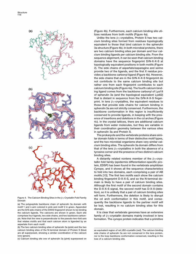
(Figure 4b). Furthermore, each calcium binding site uti-lizes residues from both motifs (Figure 4a).
Unlike the lens bg-crystallins, Protein S has two cal-cium binding sites formed from residues topologicallyequivalent to those that bind calcium in the spherulin3a structure (Figure 4b). In both microbial proteins, thereare two calcium binding sites per domain and four cal-cium binding ligands per calcium binding site. From thesequence alignment, it can be seen that calcium bindingdomains have the sequence fingerprint D/N-X-X-S attopologically equivalent positions in both motifs (Figure2). The side chains of aspartate/asparagine and serineprovide two of the ligands, and the first X residue pro-vides a backbone carbonyl ligand (Figure 4b). However,the side chains that are in the D/N-X-X-S fingerprint donot contribute to the same calcium binding site butrather one from each fingerprint contributes to eachcalcium binding site (Figure 4a). The fourth calcium bind-ing ligand comes from the backbone carbonyl of Lys19of spherulin 3a (and the topological equivalent Lys62)that is distant in sequence from the D/N-X-X-S finger-print. In lens bg-crystallins, the equivalent residues tothose that provide side chains for calcium binding inspherulin 3a are not strictly conserved. Furthermore, thebackbone conformation in this region is insufficientlyconserved to provide ligands, in keeping with the pres-ence of insertions and deletions in the cd arches (Figure4c). In the crystal lattices, there are additional calciumligands from water molecules, but they are variable intheir coordination geometry between the various sitesin spherulin 3a and Protein S.
The prokaryote and the vertebrate proteins share simi-lar domain folds in terms of their distinctive motif types,and the two microbial organisms share very similar cal-cium binding sites. The spherulin 3a domain differs fromthat of the lens bg-crystallins in both the absence of atyrosine corner and the presence of two distinct calciumbinding sites.
A distantly related nonlens member of the bg-crys-tallin fold family (epidermis differentiation-specific pro-tein, EDSP) has been found in the vertebrate amphibianCynops, and it shows all the sequence characteristicsto fold into two domains, each comprising a pair of ABmotifs [13]. The first two motifs each show the calciumbinding fingerprint D-X-X-S, and so the N-terminal do-main is likely to have a pair of calcium binding sites.Although the first motif of the second domain containsthe D-X-X-S signal, the second motif has D-X-X-(dele-tion), so it is unlikely that a pair of calcium binding siteswill form. Furthermore, the deletion is likely to disrupt
Figure 4. The Calcium Binding Sites in the bg-Crystallin Fold Family the cd arch conformation in this motif, and conse-Domain
quently the backbone ligands to the partner motif will(a) The polypeptide backbone chain of spherulin 3a domain with
be lost, resulting in no calcium binding sites for thismotif 1 and n-arm colored in pink and motif 2 in green. Appendeddomain.are the D/S side chains of the DXXS fingerprint shown to be binding
It is clear that vertebrate genomes have an extensivethe calcium ligands. The calciums are shown in green. Each sitecomprises four ligands, two side chains, and two backbone carbon- family of bg-crystallin domains mainly involved in lensyls. Note that the view is perpendicular to the pseudo-two-fold axis formation. The cynops protein indicates that a primitivethat relates motifs and that each calcium atom is liganded by anaspartate from each motif.(b) The two calcium binding sites of spherulin 3a (pink) and the twocalcium binding sites of the N-terminal domain of Protein S (blue) an equivalent region of rat bB2-crystallin (red). The calcium bindingare all superposed, showing a similar coordination of the calcium side chains of spherulin 3a are not conserved in the lens protein,ions (green). nor is the loop backbone conformation conserved, resulting in the(c) Calcium binding site one of spherulin 3a (pink) superposed on loss of a calcium binding site.
Structure of calcium loaded spherulin 3a dimer119
vertebrate possesses a relative that has a different func-tion and intriguingly appears to have a feature, calciumbinding, found in microbial bg-crystallin folds. Furtherevidence that links the vertebrate bg-crystallin fold tothe microbial forms came with a cDNA sequence fromthe lowest metazoan phylum, the marine sponge Geodiacydonium, which contains a sequence finger print thatsuggests a four-fold Greek key motif repeat. The Greekkey motif fingerprint is more conserved for the putativemotif pair of the C-terminal domain [25]. However, thereis little conservation of the calcium binding fingerprint,with each motif retaining only one of the calcium bindingside chains (motif three has the serine, and motif fourhas the aspartate). It is possible then that one calciumbinding site has been preserved. However, the cd archin motif three is quite short, and so it may not haveretained the backbone conformation to form even oneof the calcium binding sites. The presence of a calciumbinding site in the Geodia cydonium Greek key repeatingprotein would further substantiate the relationship be-tween the microbial (calcium binding) and vertebratebg-crystallin family members.
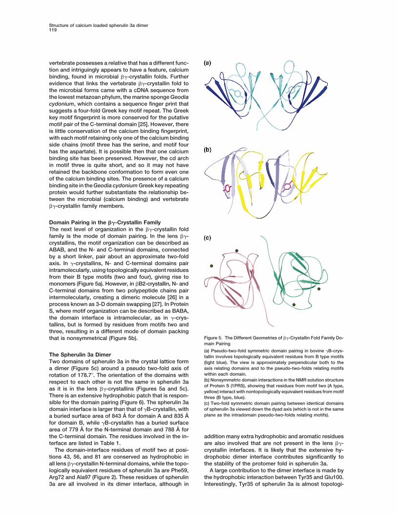
Domain Pairing in the bg-Crystallin FamilyThe next level of organization in the bg-crystallin foldfamily is the mode of domain pairing. In the lens bg-crystallins, the motif organization can be described asABAB, and the N- and C-terminal domains, connectedby a short linker, pair about an approximate two-foldaxis. In g-crystallins, N- and C-terminal domains pairintramolecularly, using topologically equivalent residuesfrom their B type motifs (two and four), giving rise tomonomers (Figure 5a). However, in bB2-crystallin, N- andC-terminal domains from two polypeptide chains pairintermolecularly, creating a dimeric molecule [26] in aprocess known as 3-D domain swapping [27]. In ProteinS, where motif organization can be described as BABA,the domain interface is intramolecular, as in g-crys-tallins, but is formed by residues from motifs two andthree, resulting in a different mode of domain packing
Figure 5. The Different Geometries of bg-Crystallin Fold Family Do-that is nonsymmetrical (Figure 5b).main Pairing
(a) Pseudo-two-fold symmetric domain pairing in bovine gB-crys-The Spherulin 3a Dimer tallin involves topologically equivalent residues from B type motifsTwo domains of spherulin 3a in the crystal lattice form (light blue). The view is approximately perpendicular both to the
axis relating domains and to the pseudo-two-folds relating motifsa dimer (Figure 5c) around a pseudo two-fold axis ofwithin each domain.rotation of 178.78. The orientation of the domains with(b) Nonsymmetric domain interactions in the NMR solution structurerespect to each other is not the same in spherulin 3aof Protein S (1PRS), showing that residues from motif two (A type,
as it is in the lens bg-crystallins (Figures 5a and 5c). yellow) interact with nontopologically equivalent residues from motifThere is an extensive hydrophobic patch that is respon- three (B type, blue).sible for the domain pairing (Figure 6). The spherulin 3a (c) Two-fold symmetric domain pairing between identical domains
of spherulin 3a viewed down the dyad axis (which is not in the samedomain interface is larger than that of gB-crystallin, withplane as the intradomain pseudo-two-folds relating motifs).a buried surface area of 843 A for domain A and 835 A
for domain B, while gB-crystallin has a buried surfacearea of 779 A for the N-terminal domain and 788 A forthe C-terminal domain. The residues involved in the in- addition many extra hydrophobic and aromatic residues
are also involved that are not present in the lens bg-terface are listed in Table 1.The domain-interface residues of motif two at posi- crystallin interfaces. It is likely that the extensive hy-
drophobic dimer interface contributes significantly totions 43, 56, and 81 are conserved as hydrophobic inall lens bg-crystallin N-terminal domains, while the topo- the stability of the protomer fold in spherulin 3a.
A large contribution to the dimer interface is made bylogically equivalent residues of spherulin 3a are Phe59,Arg72 and Ala97 (Figure 2). These residues of spherulin the hydrophobic interaction between Tyr35 and Glu100.
Interestingly, Tyr35 of spherulin 3a is almost topologi-3a are all involved in its dimer interface, although in
Structure120
Figure 6. The Extensive Hydrophobic Interface Between Domainsof the Spherulin s3a Homodimer
The appended side chains are listed in Table 1.
cally equivalent to Tyr62 of gB-crystallin when the se-quences are compared with the motifs permuted (Figure2). It is this residue that forms the tyrosine corners in Btype domains in lens bg-crystallins and Protein S (Fig-ures 1 and 5). It is interesting that the presence of one
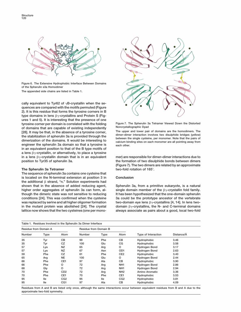
Figure 7. The Spherulin 3a Tetramer Viewed Down the Distortedtyrosine corner per domain is correlated with the foldingNoncrystallographic Dyadof domains that are capable of existing independentlyThe upper and lower pair of domains are the homodimers. The[28]. It may be that, in the absence of a tyrosine corner,dimer–dimer interaction involves two disulphide bridges (yellow)the stabilization of spherulin 3a is provided through thebetween the single cysteine, per monomer. Note that the pairs of
dimerization of the domains. It would be interesting to calcium binding sites on each monomer are all pointing away fromengineer the spherulin 3a domain so that a tyrosine is each other.in an equivalent position to that of the B type motifs ofa lens bg-crystallin, or alternatively, to place a tyrosinein a lens bg-crystallin domain that is in an equivalent mer) are responsible for dimer–dimer interactions due to
the formation of two disulphide bonds between dimersposition to Tyr35 of spherulin 3a.(Figure 7). The two dimers are related by an approximatetwo-fold rotation of 1658.The Spherulin 3a Tetramer
The sequence of spherulin 3a contains one cysteine thatis located on the N-terminal extension at position 3 in Conclusionthe additional b strand, “n.” Solution experiments hadshown that in the absence of added reducing agent, Spherulin 3a, from a primitive eukaryote, is a natural
single domain member of the bg-crystallin fold family.higher order aggregates of spherulin 3a can form, al-though the dimeric state was not sensitive to reducing It has been hypothesized that the one-domain spherulin
3a could be the prototype ancestor of the vertebrateconditions [24]. This was confirmed when the cysteinewas replaced by serine and all higher oligomer formation two-domain eye lens bg-crystallins [4, 14]. In lens two-
domain bg-crystallins, the N- and C-terminal domainsin the mutant protein was abolished [24]. The crystallattice now shows that the two cysteines (one per mono- always associate as pairs about a good, local two-fold
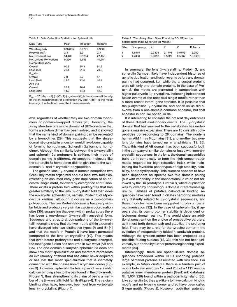
Table 1. Residues Involved in the Spherulin 3a Dimer Interface
Residue from Domain A Residue from Domain B
Number Type Atom Number Type Atom Type of Interaction Distance/A
35 Tyr CB 99 Phe CB Hydrophobic 3.4635 Tyr CZ 100 Glu CG Hydrophobic 3.5857 Lys NZ 65 Arg O Hydrogen Bond 3.1757 Lys NZ 67 Asn OD1 Hydrogen Bond 2.6359 Phe CZ 61 Phe CE2 Hydrophobic 3.4065 Arg NE 100 Glu O Hydrogen Bond 2.4466 Phe CE1 97 Ala CB Hydrophobic 3.9066 Phe O 72 Arg NH1 Hydrogen Bond 2.8668 Gly O 72 Arg NH1 Hydrogen Bond 2.9470 Phe CD2 72 Arg NH2 Amino Aromatic 3.3670 Phe CE1 70 Phe CE1 Hydrophobic 3.5395 Ile CG2 95 Ile CG2 Hydrophobic 3.8195 Ile CG1 97 Ala CB Hydrophobic 4.09
Residues from A and B are listed only once, although the same interactions occur between equivalent residues from B and A due to theapproximate two-fold symmetry.
Structure of calcium loaded spherulin 3a dimer121
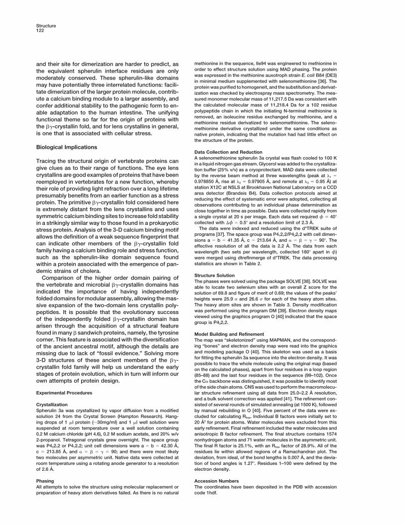
Table 3. The Heavy Atom Sites Found by SOLVE for theTable 2. Data Collection Statistics for Spherulin 3aSelenomethionine Spherulin 3a Mutant
Data Type Peak Inflection RemoteSite Occupancy X Y Z B factor
Wavelength/A 0.97885 0.9791 0.9500Resolution/A 2.3 2.3 2.3 1 1.1515 0.3338 0.1704 0.0753 15.000
2 1.2886 0.9652 0.5029 0.5362 18.3681No. Observations 54,400 57,284 27,155No. Unique Reflections 9,236 9,688 10,284Completeness/%Overall 90.9 95.3 91.2
In summary, the lens bg-crystallins, Protein S, andLast shell 72.4 91.6 70.8Rsym/% spherulin 3a most likely have independent histories ofOverall 7.0 5.7 5.1 genetic duplication and fusion events before any domainLast Shell 13.5 12.0 10.4 pairing had occurred, i.e., while the ancestral proteinsAve I/sI
were still only one-domain proteins. In the case of Pro-Overall 25.7 26.4 20.6tein S, the motifs are permuted in comparison withLast Shell 14.0 14.8 12.2higher eukaryotic bg-crystallins, indicating independent
Rsym 5 SS|I(h)i 2 ,I(h).|/S,I(h)., where I(h)i is the observed intensity fusion events of the ancestral single motifs rather thanof the ith measurement of a reflection (h), and ,I(h). is the mean
a more recent lateral gene transfer. It is possible thatintensity of reflection h over the i measurements.the b-crystallins, g-crystallins, and spherulin 3a did allevolve from a one-domain common ancestor, but thatancestor is not like spherulin 3a.
It is interesting to consider the present day outcomesaxis, regardless of whether they are two-domain mono-mers or domain-swapped dimers [26]. Recently, the of these distant evolutionary events. The bg-crystallin
domain that has survived to the vertebrates has under-X-ray structure of a single domain of bB2-crystallin thatforms a solution dimer has been solved, and it showed gone a massive expansion. There are 13 crystallin poly-
peptides corresponding to 26 domains. The nonlensthat the same kind of domain pairing can be recreatedby a homodimer [29]. This suggests that the single- human AIM 1 has 6 domains [31], and another two non-
lens domains have turned up in amphibians [13, 25].domain bg-crystallin ancestor would have been capableof forming homodimers. Spherulin 3a forms a homo- Thus, this kind of AB domain has been successful both
in the company of similar domains or fused with non-bg-dimer. Although the similarity between the bg-crystallinand spherulin 3a protomers is striking, their mode of crystallin sequences. In the lens, the domain assemblies
build up in complexity to form the high concentrationdomain pairing is different. An ancestral molecule likethe spherulin 3a homodimer did not give rise to the two- media required for high refractive index while main-
taining the favorable phenotypes of high stability, solu-domain b- and g-crystallin polypeptides.The generic lens bg-crystallin domain comprises two bility, and polydispersity. This success appears to have
been dependent on specific two-fold domain pairingGreek key motifs organized about a local two-fold axis,reflecting an assumed early gene duplication of an an- (but with variability in the connections), a route not fol-
lowed by the BA prototype, Protein S, where duplicationcestral single motif, followed by divergence and fusion.There exists a protein fold within prokaryotes that has was followed by nonisologous domain interactions (Fig-
ure 5). Families of putative calmodulin binding se-greater similarity to the lens bg-crystallin fold than doesthe eukaryotic spherulin 3a, namely, Protein S of Myxo- quences have been found in ciliates harboring modules
very distantly related to bg-crystallin sequences, andcoccus xanthus, although it occurs as a two-domainpolypeptide. The two Protein S domains have very simi- these modules have been suggested to play a role in
multimerisation [32]. In the case of spherulin 3a, it ap-lar folds and probably very similar calcium coordinationsites [30], suggesting that even within prokaryotes there pears that its own protomer stability is dependent on
isologous domain pairing. This would place an addi-has been a one-domain bg-crystallin ancestral form.Sequence and structural comparisons of the bg-crys- tional constraint on the choice of prospective partners,
as it must both domain pair and stabilize the protomertallin domains show that the two motifs within a domainhave diverged into two distinctive types (A and B) [4] fold. There may be a role for the tyrosine corner in the
evolution of independently folded b sandwich proteins.and that the motifs in Protein S have been permutedcompared to the lens bg-crystallins [10]. This means Although the tyrosine corner has been proposed as a
possible folding nucleus [12, 33], this has not been uni-that even before prokaryotes and eukaryotes diverged,the motif gene fusion has occurred in two ways (AB and versally supported by further protein engineering experi-
ments [34].BA). The one-domain eukaryotic spherulin 3a does notshow this motif specialization and therefore represents Interestingly, there are spherulin-like domain se-
quences embedded within ORFs encoding potentialan evolutionary offshoot that has either never acquiredor has lost this motif specialization that is intimately large bacterial proteins associated with virulence. For
example, in Vibrio cholerae there is a tandem pair ofconnected with the possession of a tyrosine corner (Fig-ure 3). However, spherulin 3a has a pair of very similar motifs between residues 175 and 253 of a 1111 residue
putative inner membrane protein (GenBank database,calcium binding sites to the pair found in the prokaryoticProtein S, thus strengthening its claim of being a mem- GI: 3,004,928) found within a pathogenicity island [35].
Like spherulin 3a, they are likely to have calcium bindingber of the bg-crystallin fold family (Figure 4). The calciumbinding sites have, however, been lost from vertebrate motifs and no tyrosine corner and so have been called
S type motifs (Figure 2). However, both their potentiallens bg-crystallins (Figure 4).
Structure122
methionine in the sequence, Ile94 was engineered to methionine inand their site for dimerization are harder to predict, asorder to effect structure solution using MAD phasing. The proteinthe equivalent spherulin interface residues are onlywas expressed in the methionine auxotroph strain E. coli B84 (DE3)moderately conserved. These spherulin-like domainsin minimal medium supplemented with selenomethionine [36]. The
may have potentially three interrelated functions: facili- protein was purified to homogeneit, and the substitution and derivat-tate dimerization of the larger protein molecule, contrib- ization was checked by electrospray mass spectrometry. The mea-
sured monomer molecular mass of 11,217.5 Da was consistent withute a calcium binding module to a larger assembly, andthe calculated molecular mass of 11,218.4 Da for a 102 residueconfer additional stability to the pathogenic form to en-polypeptide chain in which the initiating N-terminal methionine isable adaptation to the human intestine. The unifyingremoved, an isoleucine residue exchanged by methionine, and afunctional theme so far for the origin of proteins withmethionine residue derivatized to selenomethionine. The seleno-
the bg-crystallin fold, and for lens crystallins in general, methionine derivative crystallized under the same conditions asis one that is associated with cellular stress. native protein, indicating that the mutation had had little effect on
the structure of the protein.
Biological ImplicationsData Collection and ReductionA selenomethionine spherulin 3a crystal was flash cooled to 100 K
Tracing the structural origin of vertebrate proteins can in a liquid nitrogen gas stream. Glycerol was added to the crystalliza-give clues as to their range of functions. The eye lens tion buffer (25% v/v) as a cryoprotectant. MAD data were collectedcrystallins are good examples of proteins that have been by the reverse beam method at three wavelengths (peak at l1 5
0.978850 A, rise at l2 5 0.97905 A, and remote at l3 5 0.95 A) atreemployed in vertebrates for a new function, wherebystation X12C at NSLS at Brookhaven National Laboratory on a CCDtheir role of providing light refraction over a long lifetimearea detector (Brandeis B4). Data collection protocols aimed atpresumably benefits from an earlier function as a stressreducing the effect of systematic error were adopted, collecting all
protein. The primitive bg-crystallin fold considered here observations contributing to an individual phase determination asis extremely distant from the lens crystallins and uses close together in time as possible. Data were collected rapidly fromsymmetric calcium binding sites to increase fold stability a single crystal at 20 s per image. Each data set required φ 5 408
collected with Dφ 5 0.58 and a resolution limit of 2.3 A.in a strikingly similar way to those found in a prokaryoticThe data were indexed and reduced using the d*TREK suite ofstress protein. Analysis of the 3-D calcium binding motif
programs [37]. The space group was P41212/P43212 with cell dimen-allows the definition of a weak sequence fingerprint thatsions a 5 b 5 41.35 A, c 5 213.64 A, and a 5 b 5 g 5 908. The
can indicate other members of the bg-crystallin fold effective resolution of all the data is 2.2 A. The data from eachfamily having a calcium binding role and stress function, wavelength (two sets per wavelength, collected 1808 apart in φ)such as the spherulin-like domain sequence found were merged using dtreflnmerge of d*TREK. The data processing
statistics are shown in Table 2.within a protein associated with the emergence of pan-demic strains of cholera.
Structure SolutionComparison of the higher order domain pairing ofThe phases were solved using the package SOLVE [38]. SOLVE was
the vertebrate and microbial bg-crystallin domains has able to locate two selenium sites with an overall Z score for theindicated the importance of having independently solution of 69.8 and figure of merit of 0.69; the values of the peaks’folded domains for modular assembly, allowing the mas- heights were 25.9 s and 26.6 s for each of the heavy atom sites.
The heavy atom sites are shown in Table 3. Density modificationsive expansion of the two-domain lens crystallin poly-was performed using the program DM [39]. Electron density mapspeptides. It is possible that the evolutionary successviewed using the graphics program O [40] indicated that the spaceof the independently folded bg-crystallin domain hasgroup is P43212.
arisen through the acquisition of a structural featurefound in many b sandwich proteins, namely, the tyrosine Model Building and Refinementcorner. This feature is associated with the diversification The map was “skeletonized” using MAPMAN, and the correspond-
ing “bones” and electron density map were read into the graphicsof the ancient ancestral motif, although the details areand modeling package O [40]. This skeleton was used as a basismissing due to lack of “fossil evidence.” Solving morefor fitting the spherulin 3a sequence into the electron density. It was3-D structures of these ancient members of the bg-possible to trace the whole molecule using the original map (based
crystallin fold family will help us understand the early on the calculated phases), apart from four residues in a loop regionstages of protein evolution, which in turn will inform our (85–88) and the last four residues in the sequence (99–102). Onceown attempts of protein design. the Ca backbone was distinguished, it was possible to identify most
of the side chain atoms. CNS was used to perform the macromolecu-Experimental Procedures lar structure refinement using all data from 25.0–2.2 A resolution,
and a bulk solvent correction was applied [41]. The refinement con-sisted of several rounds of simulated annealing (at 1500 K), followedCrystallization
Spherulin 3a was crystallized by vapor diffusion from a modified by manual rebuilding in O [40]. Five percent of the data were ex-cluded for calculating Rfree. Individual B factors were initially set tosolution 24 from the Crystal Screen (Hampton Research). Hang-
ing drops of 1 ml protein (z30mg/ml) and 1 ml well solution were 20 A2 for protein atoms. Water molecules were excluded from thisearly refinement. Final refinement included the water molecules andsuspended at room temperature over a well solution containing
0.2 M calcium chloride (pH 4.6), 0.2 M sodium acetate, and 20% w/v anisotropic B factor refinement. The final structure contains 1574nonhydrogen atoms and 71 water molecules in the asymmetric unit.2-propanol. Tetragonal crystals grew overnight. The space group
was P43212 or P41212; unit cell dimensions were a 5 b 5 42.30 A, The final R factor is 25.1%, with an Rfree factor of 28.9%. All of theresidues lie within allowed regions of a Ramachandran plot. Thec 5 213.85 A, and a 5 b 5 g 5 90; and there were most likely
two molecules per asymmetric unit. Native data were collected at deviation, from ideal, of the bond lengths is 0.007 A, and the devia-tion of bond angles is 1.278. Residues 1–100 were defined by theroom temperature using a rotating anode generator to a resolution
of 2.6 A. electron density.
Accession NumbersPhasingAll attempts to solve the structure using molecular replacement or The coordinates have been deposited in the PDB with accession
code 1hdf.preparation of heavy atom derivatives failed. As there is no natural
Structure of calcium loaded spherulin 3a dimer123
Acknowledgments spherulin 3a from Physarum polycephalum by calcium binding.J. Mol. Biol. 289, 701–705.
17. Baldauf, S.L., and Doolittle, W.F. (1997). Origin and evolution ofDiffraction data for this study were collected at Brookhaven NationalLaboratory in the Biology Department single-crystal diffraction facil- the slime molds (Mycetozoa). Proc. Natl. Acad. Sci. USA 94,
12007–12012.ity at beamline X12C in the National Synchrotron Light Source. Thisfacility is supported by the United States Department of Energy 18. Bernier, F., Seligy, V.L., Pallotta, D., and Lemieux, G. (1986).
Changes in gene expression during spherulation in PhysarumOffices of Health and Environmental Research and of Basic EnergySciences, and by the National Institutes of Health National Center polycephalum. Biochem. Cell Biol. 64, 337–343.
19. Kaiser, D., Manoil, C.C., and Dworkin, M. (1979). Myxobacteria:for research sources. The spherulin 3a MAD data were collected,processed, and the phases solved on the Data Col 99 course orga- cell interactions, genetics and development. Annu. Rev. Micro-
biol. 33, 595–639.nized by Robert Sweet. Much gratitude is owed to all those involvedin teaching the course, including Robert Sweet and Jim Pflugrath. 20. Inouye, S., Inouye, M., McKeever, B., and Sharma, R. (1980).
Preliminary crystallographic data for protein S, a developmentMany thanks are owed to Jason Yano for his help during the DataCol99 course. Orval Bateman, David Moss, and Burkhard Rosinke are specific protein of Myxococcus xanthus. J. Mol. Chem. 255,
3713–3714.kindly acknowledged. The financial support of the Medical ResearchCouncil is gratefully acknowledged as is that of the Deutsche 21. Antuch, W, Guntert, P., and Wuthrich, K. (1996). Ancestral beta-
gamma-crystallin precursor structure in a yeast killer toxin. Nat.Forschungsgemeinschaft (grant ja78/33). The work has also beensupported by an EU BioMed grant (BMH4-CT98-3895). Struct. Biol. 3, 662–665.
22. Ohno, A., et al., and Kainosho, M. (1998). NMR structure of theStreptomyces metalloproteinase inhibitor, SMPI, isolated from
Received: September 5, 2000 Streptomyces nigrescens TK-23: another example of an ances-Revised: November 16, 2000 tral bg-crystallin precursor structure. J. Mol. Biol. 282, 421–433.Accepted: November 28, 2000 23. Clout, N.J., Slingsby, C., and Wistow, G.J. (1997). An eye on
crystallins. Nat. Struct. Biol. 4, 685.24. Kretschmar, M., Mayr, E.M., and Jaenicke, R. (1999). Homodi-References
meric spherulin 3a: a single-domain member of the bg-crystallinsuperfamily. Biol. Chem. 380, 89–94.1. Lubsen, N.H., Aarts, H.J.M., and Schoenmakers, J.G.G. (1988).
25. Krasko, A., Muller, I.M., and Muller, W.E.G. (1997). EvolutionaryThe evolution of lenticular proteins: the b- and g-crystallin superrelationships of the metazoan bg-crystallins, including that fromgene family. Prog. Biophys. Mol. Biol. 51, 47–76.the marine sponge Geodia cydonium. Proc. R. Soc. Lond. B.2. Wistow, G., and Piatigorsky, J. (1988). Lens crystallins - theBiol. Sci. 264, 1077–1084.evolution and expression of proteins for a highly specialized
26. Bax, B., et al., and Slingsby, C. (1990). X-ray analysis of beta-tissue. Annu. Rev. Biochem. 57, 479–504.B2-crystallin and evolution of oligomeric lens proteins. Nature3. de Jong, W.W., Lubsen, N.H., and Kraft, H.J. (1994). Molecular347, 776–780.evolution of the eye lens. Prog. Ret. Eye Res. 13, 391–442.
27. Bennett, M.J., Schlunegger, M.P., and Eisenberg, D. (1995). 3D4. Wistow, G. (1990). Evolution of a protein superfamily: relation-Domain swapping - a mechanism for oligomer assembly. Pro-ships between vertebrate lens crystallins and micro-organismtein Sci. 4, 2455–2468.dormancy proteins. J. Mol. Biol. 30, 140–145.
28. Mayr, E.-M., Jaenicke, R., and Glockshuber, R. (1994). Domain5. Ingolia, T., and Craig, E. (1982). Four small Drosophila heatinteractions and connecting peptides in lens crystallins. J. Mol.shock proteins are related to each other and to mammalianBiol. 235, 84–88.alpha-crystallin. Proc. Natl. Acad. Sci. USA 79, 2360–2364.
29. Clout, N.J., Basak, A., Wieligmann, K., Bateman, O.A., Jaenicke,6. Wistow, G., Summers, L., and Blundell, T. (1985). MyxococcusR., and Slingsby, C. (2000). The N-terminal domain of bB2-xanthus spore coat protein S may have a similar structure tocrystallin resembles the putative ancestral homodimer. J. Mol.vertebrate lens beta-gamma-crystallins. Nature 315, 771–773.Biol. 304, 253-257.7. de Jong, W.W., Caspers, G.J., and Leunissen, J.A.M. (1998).
30. Wenk, M., and Mayr, E.M. (1998). Myxococcus Xanthus sporeGenealogy of the a-crystallin – small heat shock protein super-coat Protein S, a stress induced member of the bg-crystallinfamily. Int. J. Biol. Macromol. 22, 151–162.superfamily, gains stability from binding of calcium ions. Eur.8. Blundell, T., et al., and Wistow, G. (1981). The molecular struc-J. Biochem. 255, 604–610.ture and stability of the eye lens: X-ray analysis of g-crystallin
31. Ray, M.E., Wistow, G., Su, Y.A., Meltzer, P.S., and Trent, J.M.II. Nature 289, 771–777.(1997). AIM1, a novel non-lens member of the bg-crystallin su-9. Slingsby, C., et al., and Bax, B. (1997). X-ray diffraction andperfamily, is associated with the control of tumorigenicity instructure of crystallins. Prog. Ret. Eye Res. 16, 3–29.human malignant melanoma. Proc. Natl. Acad. Sci. USA. 94,10. Bagby, S., Harvey, T., Eagle, S., Inouye, S., and Ikura, M. (1994).3229–3234.NMR-derived three-dimensional solution structure of Protein S
32. Chan, C.W.M., Saimi, Y., and Kung, C. (1999). A new multigenecomplexed with calcium. Structure 2, 107–122.family encoding calcium-dependent calmodulin-binding mem-11. Wenk, M., Baumgartner, R., Holak, T.A., Huber, R., Jaenicke,brane proteins of Paramecium tetraurelia. Gene 231, 21–32.R., and Mayr, E.M. (1999). The domains of Protein S from Myxoc-
33. Bagby, S., Go, S., Inouye, S., Ikura, M., and Chakrabartty, A.cocus xanthus: structure, stability and interactions. J. Mol. Biol.(1998). Equilibrium folding intermediates of a Greek-key beta-286, 1533–1545.barrel protein. J. Mol. Biol. 276, 669–681.12. Hemmingsen, J.M., Gernert, K.M., Richardson, J.S., and Rich-
34. Hamill, S.J., Cota, E., Chothia, C., and Clarke, J. (2000). Conser-ardson, D.C. (1994). The tyrosine corner: a feature of most Greekvation of folding and stability within a protein family: the tyrosinekey b-barrel proteins. Protein Sci. 3, 1927–1937.corner as an evolutionary cul-de-sac. J. Mol. Biol. 295, 641–649.13. Wistow, G., Jaworski, C., and Rao, P.V. (1995). A non-lens mem-
35. Karaolis, D.K.R., Johnson, J.A., Bailey, C., Boedeker, E.C.,ber of the bg-crystallin superfamily in a vertebrate, the amphib-Kaper, J.B., and Reeves, P.R. (1998). A Vibrio cholerae pathoge-ian Cynops. Exp. Eye Res. 61, 637–639.nicity island associated with epidemic and pandemic strains.14. Rosinke, B., Renner, C., Mayr, E., Rainer, J., and Holak, T. (1997).Proc. Natl. Acad. Sci. USA 95, 3134–3139.The solution structure of calcium loaded spherulin 3a. J. Mol.
36. Budisa, N., Steipe, B., Demange, P., Eckerskorn, C., Kellermann,Biol. 272, 1–11.J., and Huber, R. (1995). High-level biosynthetic substitution of15. Kretschmar, M., and Jaenicke, R. (1999). Stability of a homodi-methionine in proteins by its analogs 2-aminohexanoic acid,meric Ca21-binding member of the bg-crystallin superfamily:selenomethionine, telluromethionine and ethionine in E.coli. Eur.DSC measurements on spherulin 3a from Physarum polycepha-J. Biochem. 230, 788–796.lum. J. Mol. Biol. 291, 1147–1153.
37. Pflugrath, J.W. (1997). Diffraction data processing of electronic16. Kretschmar, M., Mayr, E.M., and Jaenicke, R. (1999). Kineticand thermodynamic stabilization of the bg-crystallin homolog detectors: theory and practice. Methods Enzymol. 276, 286–307.
Structure124
38. Terwilliger, T.C., and Berendzen, J. (1999). Automated MAD andMIR structure solution. Acta. Crystallogr. D 55, 849–861.
39. Cowtan, K.D. (1994). DM: an automated procedure for phaseimprovement by density modification. Joint CCP4 ESF-EACMBNewslett. Protein Crystallogr. 31, 34–38.
40. Jones, T., and Kjeldgaard, M. (1997). Electron-density mapinterpretation. Methods Enzymol. 277, 173–208.
41. Brunger, A.T., et al., and Warren, G.L. (1998). Crystallographyand NMR system: a new software suite for macromolecularstructure determination. Acta Crystallogr. D 54, 905–921.
42. Evans, S.V. (1993). Setor: hardware lighted three-dimensionalsolid model representations of macromolecules. J. Mol. Graph-ics 11, 134–138.
![arXiv:1705.10936v2 [cond-mat.str-el] 11 Sep 2017 · arXiv:1705.10936v2 [cond-mat.str-el] 11 Sep 2017 Dimer-Mott and charge-ordered insulating states in thequasi-one-dimensional organic](https://static.fdocument.org/doc/165x107/5f9cfc5e7ee0fa7ee112055e/arxiv170510936v2-cond-matstr-el-11-sep-2017-arxiv170510936v2-cond-matstr-el.jpg)