cis–trans Isomerization of β-casomorphin peptides bound to copper(II): integration of EPR and NMR...
6
2 PERKIN 252 J. Chem. Soc., Perkin Trans. 2, 2001, 252–257 DOI: 10.1039/b004964f This journal is © The Royal Society of Chemistry 2001 cis–trans Isomerization of -casomorphin peptides bound to copper(II): integration of EPR and NMR studies † Riccardo Basosi, Nicola D’Amelio, Elena Gaggelli, Rebecca Pogni and Gianni Valensin Department of Chemistry, University of Siena, Via A.Moro, Siena 53100, Italy Received (in Cambridge, UK) 21st June 2000, Accepted 17th January 2001 First published as an Advance Article on the web 16th February 2001 Copper complexes of β-casomorphin peptides (BCM-7, BCM-5, BCM-4) were investigated by EPR and NMR in DMSO-d 6 solutions. Speciation of copper among many of the possible isomers was apparent. Computer simulations of low and room temperature EPR allowed the number of co-ordinated nitrogens in the major species (2 for BCM-4 and BCM-5, 4 for BCM-7) to be inferred and a rotational correlation time of 0.18 ns at 298 K to be evaluated for all complexes. All isomers of BCM-4 and BCM-5 were shown to bind copper, but the resulting structures were strictly determined by the conformational state of 2 Pro. The trans, rather than the cis, conformation was shown to allow binding of the deprotonated 3 Phe-NH; the terminal amino and carboxylate groups provided the other binding groups in all cases. Structures were obtained by constrained molecular dynamics using copper–proton distances obtained from paramagnetic nuclear relaxation rates. In the case of BCM-7, only the cis-cis-trans and/or the cis-cis-cis isomers were not binding copper. The conformational state of each Pro was shown to drive formation of the copper–nitrogen bond within the immediately adjacent residue, leading to the complex having four co-ordinated nitrogens in the case of the trans-trans-trans isomer. β-Casomorphins (BCM) are opioid peptides originally identi- fied in enzymatic digests of the bovine milk protein β-casein (hence the name β-casein exorphins). 1 They share with other µ-receptor agonists the sequence Tyr-X-Phe at the N-terminus as opposed to the Tyr-X-X-Phe sequence typical of δ selec- tivity (e.g., enkephalins). The occurrence of proline in the sequence imposes structural constraints that may determine secondary structure, even in the case of short chain peptides 2 such as Tyr-Pro-Phe-Pro-NH 2 . 3 Another important feature is that peptidyl-proline bonds yield both cis and trans conformations that are relatively stable at temperatures above 0 C. 4 Sequences containing n prolines therefore exist as 2 n stable isomers, with the energy barrier being large enough (>20 kcal mol 1 ) 5 to lead to relatively slow cis– trans isomerization rates. A kinetic constant k = 6.6 × 10 5 s 1 has, for example, been calculated for Phe-Pro at 283 K and pH 8.4. 6 Whilst the four isomers of BCM-5 (Tyr-Pro-Phe-Pro-Gly) and BCM-4 (Tyr-Pro-Phe-Pro) have been isolated and charac- terized, 7 no report has appeared so far about the eight isomers of BCM-7 (Tyr-Pro-Phe-Pro-Gly-Pro-Ile). The interaction of copper with opioid peptides, as well as neuro-transmitters and neuro-modulators, has also been thoroughly investigated since the metal is unevenly distributed in the body; it is found at relatively high concentrations in the brain, although its function is still rather obscure. Interaction with BCM-5 and BCM-7 has been previously considered, 8 but this did not take the isomeric equilibrium into account. The present study was undertaken with a view to exploiting all of the advantages of EPR and NMR experiments, carried out in the same sample, for determining the ability of Cu() to co-ordinate all of the isomeric species of BCM-7 and the smaller BCM-5 and BCM-4 fragments. Experimental Bovine β-casomorphin peptides were purchased from Bachem † Magnified regions of the 1 H NMR spectrum of β-casomorphin are available as supplementary data. For direct electronic access see http://www.rsc.org/suppdata/p2/b0/b004964f/ and used without further purification. Solutions were made in [ 2 H 6 ]-DMSO 100% (DMSO-d 6 , Merck) and carefully deoxy- genated by a freezing–sealing–thawing cycle. A stock solution of Cu(ClO 4 ) 2 (Alpha Inorganics) in DMSO-d 6 was used for obtaining the desired copper concentration in the NMR and EPR samples. The X-band EPR spectra were obtained with a Bruker 200D SRC X-band spectrometer. Microwave frequencies were meas- ured with a XL Microwave Model 3120 counter (Jagmar, Krakow, Poland). The spectrometer was interfaced with a PS/2 Technical Instruments Hardware computer and the data acquired using the EPR data system CS-EPR produced by Stelar Inc., Mede, Italy. The spectra were preliminarily corrected for baseline drift using a simple procedure based on a cubic spline function. 9 The EPR simulation program COSMOS was written by us and is described in detail elsewhere. 10 It can cover virtually all of the dynamical conditions of copper complexes, from “fast tumbling” to “incipient slow motion”. The program used for simulation of frozen solution spectra was a modified version of that described by Pilbrow and co-workers. 11 Both programs allow the simultaneous occurrence of the two isotopes of copper to be considered. The best fit of the experimental spectra was found using the Simplex and Levenberg–Marquardt optimization procedures. The tolerances in the magnetic parameters during minimization were consistent with estimated experimental errors. Once a good simulation was obtained, input data was used to optimize magnetic parameters until the quality of the fit was invariant to further changes. NMR experiments were carried out at 14.1 T on a Bruker Avance DRX 600 spectrometer at controlled temperatures (±1 K). Chemical shifts were referenced to internal TMS. TOCSY experiments were acquired with total spin-locking times in the range 50–100 ms using an MLEV-17 mixing sequence. ROESY spectra were obtained at mixing times in the range 50–100 ms with the rf strength for the spin lock field at values lower than 3.5 kHz. Spin–lattice relaxation rates were measured with inver- sion recovery pulse sequences and calculated by exponential regression analysis of the recovery curves of the longitudinal Published on 16 February 2001. Downloaded by State University of New York at Stony Brook on 28/10/2014 08:39:26. View Article Online / Journal Homepage / Table of Contents for this issue
Transcript of cis–trans Isomerization of β-casomorphin peptides bound to copper(II): integration of EPR and NMR...
2PERKIN
252 J. Chem. Soc., Perkin Trans. 2, 2001, 252–257 DOI: 10.1039/b004964f
This journal is © The Royal Society of Chemistry 2001
cis–trans Isomerization of �-casomorphin peptides bound tocopper(II): integration of EPR and NMR studies†
Riccardo Basosi, Nicola D’Amelio, Elena Gaggelli, Rebecca Pogni and Gianni Valensin
Department of Chemistry, University of Siena, Via A.Moro, Siena 53100, Italy
Received (in Cambridge, UK) 21st June 2000, Accepted 17th January 2001First published as an Advance Article on the web 16th February 2001
Copper complexes of β-casomorphin peptides (BCM-7, BCM-5, BCM-4) were investigated by EPR and NMR inDMSO-d6 solutions. Speciation of copper among many of the possible isomers was apparent. Computer simulationsof low and room temperature EPR allowed the number of co-ordinated nitrogens in the major species (2 for BCM-4and BCM-5, 4 for BCM-7) to be inferred and a rotational correlation time of 0.18 ns at 298 K to be evaluated for allcomplexes. All isomers of BCM-4 and BCM-5 were shown to bind copper, but the resulting structures were strictlydetermined by the conformational state of 2Pro. The trans, rather than the cis, conformation was shown to allowbinding of the deprotonated 3Phe-NH; the terminal amino and carboxylate groups provided the other binding groupsin all cases. Structures were obtained by constrained molecular dynamics using copper–proton distances obtainedfrom paramagnetic nuclear relaxation rates. In the case of BCM-7, only the cis-cis-trans and/or the cis-cis-cis isomerswere not binding copper. The conformational state of each Pro was shown to drive formation of the copper–nitrogenbond within the immediately adjacent residue, leading to the complex having four co-ordinated nitrogens in the caseof the trans-trans-trans isomer.
β-Casomorphins (BCM) are opioid peptides originally identi-fied in enzymatic digests of the bovine milk protein β-casein(hence the name β-casein exorphins).1 They share with otherµ-receptor agonists the sequence Tyr-X-Phe at the N-terminusas opposed to the Tyr-X-X-Phe sequence typical of δ selec-tivity (e.g., enkephalins).
The occurrence of proline in the sequence imposes structuralconstraints that may determine secondary structure, even in thecase of short chain peptides 2 such as Tyr-Pro-Phe-Pro-NH2.
3
Another important feature is that peptidyl-proline bonds yieldboth cis and trans conformations that are relatively stable attemperatures above 0 �C.4 Sequences containing n prolinestherefore exist as 2n stable isomers, with the energy barrier beinglarge enough (>20 kcal mol�1) 5 to lead to relatively slow cis–trans isomerization rates. A kinetic constant k = 6.6 × 10�5 s�1
has, for example, been calculated for Phe-Pro at 283 K and pH8.4.6 Whilst the four isomers of BCM-5 (Tyr-Pro-Phe-Pro-Gly)and BCM-4 (Tyr-Pro-Phe-Pro) have been isolated and charac-terized,7 no report has appeared so far about the eight isomersof BCM-7 (Tyr-Pro-Phe-Pro-Gly-Pro-Ile).
The interaction of copper with opioid peptides, as wellas neuro-transmitters and neuro-modulators, has also beenthoroughly investigated since the metal is unevenly distributedin the body; it is found at relatively high concentrations in thebrain, although its function is still rather obscure. Interactionwith BCM-5 and BCM-7 has been previously considered,8 butthis did not take the isomeric equilibrium into account.
The present study was undertaken with a view to exploitingall of the advantages of EPR and NMR experiments, carriedout in the same sample, for determining the ability of Cu() toco-ordinate all of the isomeric species of BCM-7 and thesmaller BCM-5 and BCM-4 fragments.
ExperimentalBovine β-casomorphin peptides were purchased from Bachem
† Magnified regions of the 1H NMR spectrum of β-casomorphin areavailable as supplementary data. For direct electronic access seehttp://www.rsc.org/suppdata/p2/b0/b004964f/
and used without further purification. Solutions were made in[2H6]-DMSO 100% (DMSO-d6, Merck) and carefully deoxy-genated by a freezing–sealing–thawing cycle. A stock solutionof Cu(ClO4)2 (Alpha Inorganics) in DMSO-d6 was used forobtaining the desired copper concentration in the NMR andEPR samples.
The X-band EPR spectra were obtained with a Bruker 200DSRC X-band spectrometer. Microwave frequencies were meas-ured with a XL Microwave Model 3120 counter (Jagmar,Krakow, Poland). The spectrometer was interfaced with aPS/2 Technical Instruments Hardware computer and thedata acquired using the EPR data system CS-EPR producedby Stelar Inc., Mede, Italy. The spectra were preliminarilycorrected for baseline drift using a simple procedure based on acubic spline function.9
The EPR simulation program COSMOS was written by usand is described in detail elsewhere.10 It can cover virtually allof the dynamical conditions of copper complexes, from “fasttumbling” to “incipient slow motion”. The program used forsimulation of frozen solution spectra was a modified versionof that described by Pilbrow and co-workers.11 Both programsallow the simultaneous occurrence of the two isotopes ofcopper to be considered.
The best fit of the experimental spectra was found using theSimplex and Levenberg–Marquardt optimization procedures.The tolerances in the magnetic parameters during minimizationwere consistent with estimated experimental errors. Once agood simulation was obtained, input data was used to optimizemagnetic parameters until the quality of the fit was invariant tofurther changes.
NMR experiments were carried out at 14.1 T on a BrukerAvance DRX 600 spectrometer at controlled temperatures (±1K). Chemical shifts were referenced to internal TMS. TOCSYexperiments were acquired with total spin-locking times in therange 50–100 ms using an MLEV-17 mixing sequence. ROESYspectra were obtained at mixing times in the range 50–100 mswith the rf strength for the spin lock field at values lower than3.5 kHz. Spin–lattice relaxation rates were measured with inver-sion recovery pulse sequences and calculated by exponentialregression analysis of the recovery curves of the longitudinal
Publ
ishe
d on
16
Febr
uary
200
1. D
ownl
oade
d by
Sta
te U
nive
rsity
of
New
Yor
k at
Sto
ny B
rook
on
28/1
0/20
14 0
8:39
:26.
View Article Online / Journal Homepage / Table of Contents for this issue
J. Chem. Soc., Perkin Trans. 2, 2001, 252–257 253
magnetization components. Spin–spin relaxation rates weremeasured with Carr–Purcell–Meiboom–Gill pulse sequencesand calculated by exponential regression analysis of the decaycurves of the transverse magnetization components.
Molecular structures were generated using the HYPER-CHEM software package,12 as implemented on a Pentium-120MHz PC, by means of the ZINDO-1 semi-empiricalmethod (for charge calculations) and the MM� force field (formolecular mechanics and dynamics calculations).
Results and discussionPeptides have several possible candidates for binding groupsfor copper. In the absence of donor-containing side chains, themetal ion usually binds to the deprotonated terminal aminogroup and to the deprotonated amide nitrogens of the second,third and possibly fourth residues.13 The occurrence ofother complexes and even of macrocycles has, however, beenreported, and the speciation of copper as a function of pH,solvent, and concentration has been widely investigated. Thepresence of proline in position 2 determines a break pointto metal.13,14 Many possibilities have again been demon-strated 8,14,15 but no attention has so far been paid to the possiblerole of proline conformation.
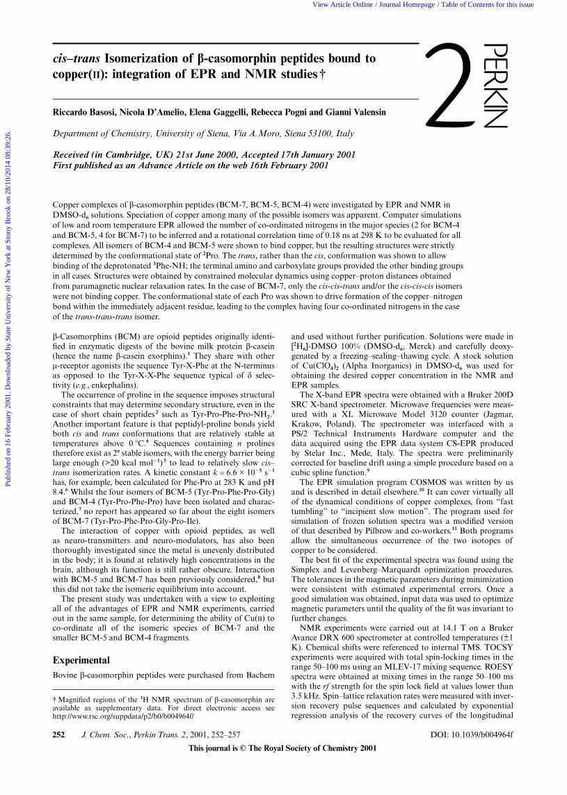
Low temperature EPR spectra were typical of square planarcopper complexes with all BCM derivatives. In the g|| regionthree of the four expected transitions were observed, with thefourth being masked in the g⊥ region. Contributions from thedifferent isotopes of copper were not resolved as it usuallyoccurs in the case of the relatively broad bandwidth ofnitrogen-bound copper. The best-fit magnetic parameters aresummarized in Table 1.
All the low temperature EPR spectra showed additionalpeaks in the g|| region that were interpreted in terms of thespeciation of copper among complexes having different mag-netic parameters. However, the absence of the typical midfieldtransition 16 and the large excess of the ligand ruled out thepresence of dimeric species and of solvated copper.
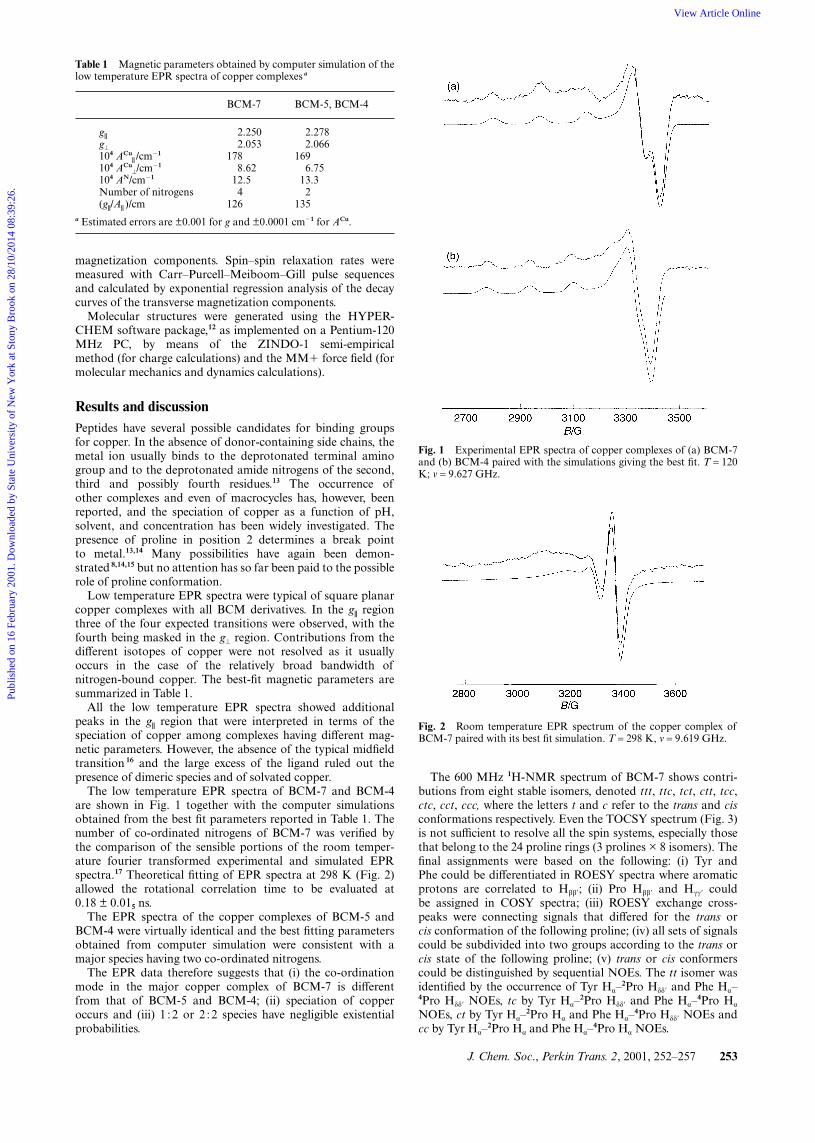
The low temperature EPR spectra of BCM-7 and BCM-4are shown in Fig. 1 together with the computer simulationsobtained from the best fit parameters reported in Table 1. Thenumber of co-ordinated nitrogens of BCM-7 was verified bythe comparison of the sensible portions of the room temper-ature fourier transformed experimental and simulated EPRspectra.17 Theoretical fitting of EPR spectra at 298 K (Fig. 2)allowed the rotational correlation time to be evaluated at0.18 ± 0.015 ns.
The EPR spectra of the copper complexes of BCM-5 andBCM-4 were virtually identical and the best fitting parametersobtained from computer simulation were consistent with amajor species having two co-ordinated nitrogens.
The EPR data therefore suggests that (i) the co-ordinationmode in the major copper complex of BCM-7 is differentfrom that of BCM-5 and BCM-4; (ii) speciation of copperoccurs and (iii) 1 : 2 or 2 :2 species have negligible existentialprobabilities.
Table 1 Magnetic parameters obtained by computer simulation of thelow temperature EPR spectra of copper complexes a
BCM-7 BCM-5, BCM-4
g||
g⊥
104 ACu|| /cm�1
104 ACu⊥/cm�1
104 AN/cm�1
Number of nitrogens(g||/A|| )/cm
2.2502.053
1788.62
12.54
126
2.2782.066
1696.75
13.32
135a Estimated errors are ±0.001 for g and ±0.0001 cm�1 for ACu.
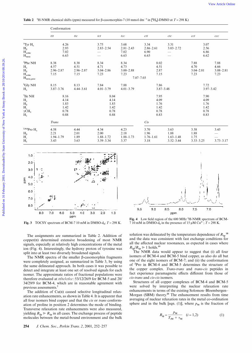
The 600 MHz 1H-NMR spectrum of BCM-7 shows contri-butions from eight stable isomers, denoted ttt, ttc, tct, ctt, tcc,ctc, cct, ccc, where the letters t and c refer to the trans and cisconformations respectively. Even the TOCSY spectrum (Fig. 3)is not sufficient to resolve all the spin systems, especially thosethat belong to the 24 proline rings (3 prolines × 8 isomers). Thefinal assignments were based on the following: (i) Tyr andPhe could be differentiated in ROESY spectra where aromaticprotons are correlated to Hββ�; (ii) Pro Hββ� and Hγγ� couldbe assigned in COSY spectra; (iii) ROESY exchange cross-peaks were connecting signals that differed for the trans orcis conformation of the following proline; (iv) all sets of signalscould be subdivided into two groups according to the trans orcis state of the following proline; (v) trans or cis conformerscould be distinguished by sequential NOEs. The tt isomer wasidentified by the occurrence of Tyr Hα–
2Pro Hδδ� and Phe Hα–4Pro Hδδ� NOEs, tc by Tyr Hα–
2Pro Hδδ� and Phe Hα–4Pro Hα
NOEs, ct by Tyr Hα–2Pro Hα and Phe Hα–
4Pro Hδδ� NOEs andcc by Tyr Hα–
2Pro Hα and Phe Hα–4Pro Hα NOEs.
Fig. 1 Experimental EPR spectra of copper complexes of (a) BCM-7and (b) BCM-4 paired with the simulations giving the best fit. T = 120K; ν = 9.627 GHz.
Fig. 2 Room temperature EPR spectrum of the copper complex ofBCM-7 paired with its best fit simulation. T = 298 K, ν = 9.619 GHz.
Publ
ishe
d on
16
Febr
uary
200
1. D
ownl
oade
d by
Sta
te U
nive
rsity
of
New
Yor
k at
Sto
ny B
rook
on
28/1
0/20
14 0
8:39
:26.
View Article Online
254 J. Chem. Soc., Perkin Trans. 2, 2001, 252–257
Table 2 1H-NMR chemical shifts (ppm) measured for β-casomorphin-7 (10 mmol dm�3 in [2H6]-DMSO at T = 298 K)
Conformation
ttt ttc tct tcc ctt ctc cct ccc
1Tyr Hα
Hβ
Hortho
Hmeta
4.262.937.026.63
3.752.83–2.54——
3.682.81–2.437.026.63
3.342.86–2.616.906.63
3.313.03–2.72——
3.072.566.866.62
3Phe NHHα
Hβ
Hortho
8.384.572.96–2.877.15
8.384.512.96–2.877.15
8.344.713.04–2.867.23
8.344.733.00–2.887.23
8.024.512.877.15
7.884.703.04–2.817.23
7.884.663.08–2.817.23
Hmeta, para 7.07–7.03
5Gly NHHα
8.153.87–3.76
8.134.44–3.61
7.844.01–3.79
7.884.01–3.79
7.863.87–3.48
7.723.97–3.42
7Ile NHHα
Hβ
Hγ
γCH3
Hδ
8.164.141.831.420.780.88
8.044.141.831.420.780.88
7.954.091.761.420.780.83
7.904.091.761.420.780.83
Trans Cis
2,4,6Pro Hα
Hβ
Hγ
Hδ
4.582.211.94–1.793.43
4.442.011.893.63
4.342.001.88–1.723.59–3.34
4.232.181.88–1.733.37
3.701.961.76–1.613.18
3.631.881.63–1.483.52–3.44
3.581.881.753.33–3.25
3.45——3.73–3.17
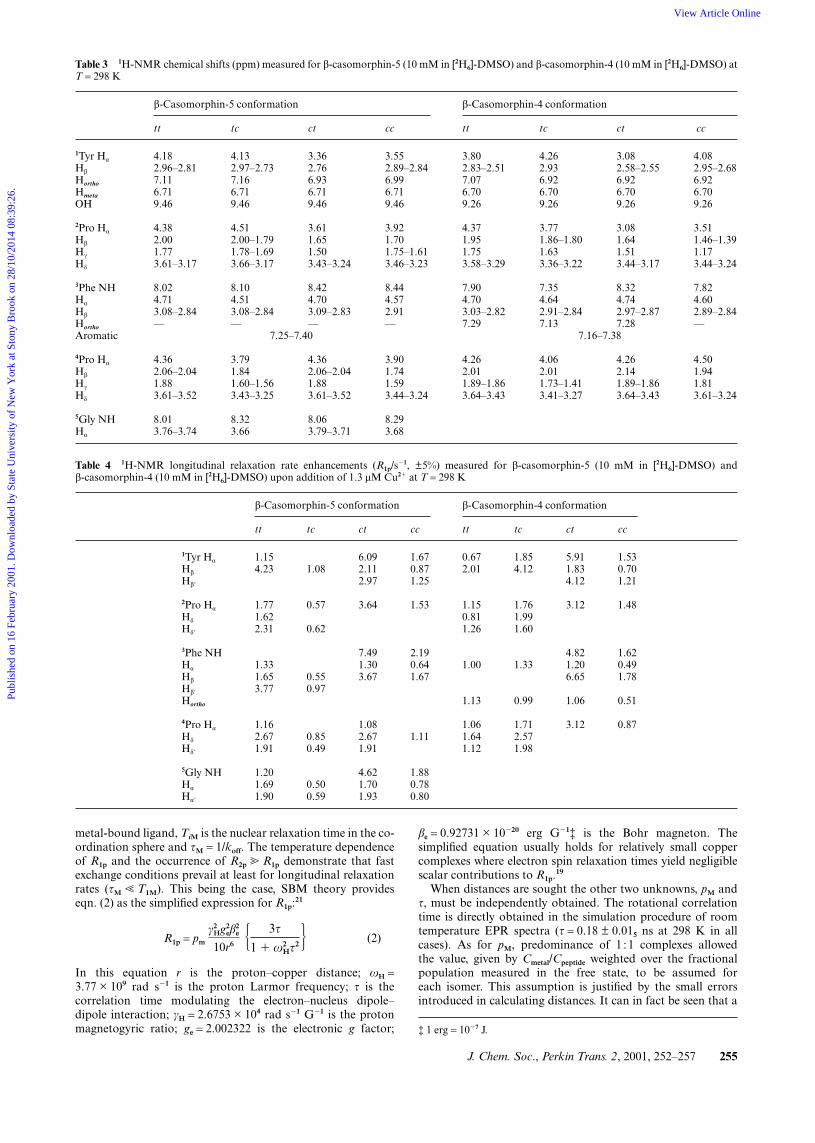
The assignments are summarized in Table 2. Addition ofcopper() determined extensive broadening of most NMRsignals, especially at relatively high concentrations of the metalion (Fig. 4). Interestingly, the hydroxy proton of tyrosine wassplit into at least two diversely broadened signals.
The NMR spectra of the smaller β-casomorphin fragmentswere completely assigned, as summarized in Table 3, by usingthe same delineated approach. In both cases it was possible todetect and integrate at least one set of resolved signals for eachisomer. The approximate ratios of fractional populations weretherefore evaluated at tt/tc/ct/cc : 53/12/26/9 for BCM-5 and 28/34/29/9 for BCM-4, which are in reasonable agreement withprevious assessments.7
The addition of Cu() caused selective longitudinal relax-ation rate enhancements, as shown in Table 4. It is apparent thatall four isomers bind copper and that the cis or trans conform-ation of proline in position 2 determines the mode of binding.Transverse relaxation rate enhancements were also measured,yielding R2p � R1p in all cases. The exchange process of peptidemolecules between the metal-bound environment and the bulk
Fig. 3 TOCSY spectrum of BCM-7 10 mM in DMSO-d6; T = 298 K.
solution was delineated by the temperature dependence of R1p18
and the data was consistent with fast exchange conditions forall the affected nuclear resonances, as expected in cases whereR2p/R1p � 1 holds.19
The NMR data would appear to suggest that (i) all fourisomers of BCM-4 and BCM-5 bind copper, as also do all butone of the eight isomers of BCM-7; and (ii) the conformationof 2Pro in BCM-4 and BCM-5 determines the structure ofthe copper complex. Trans-trans and trans-cis peptides infact experience paramagnetic effects different from those ofcis-trans and cis-cis isomers.
Structures of all copper complexes of BCM-4 and BCM-5were solved by interpreting the nuclear relaxation rateenhancements in terms of the existing Solomon–Bloembergen–Morgan (SBM) theory.20 The enhancement results from timeaveraging of nuclear relaxation rates in the metal co-ordinationsphere and in the bulk [eqn. (1)], where pM is the fraction of
Rip =pM
TiM � τM
(i = 1,2) (1)
Fig. 4 Low field region of the 600 MHz 1H-NMR spectrum of BCM-7 10 mM in DMSO-d6 in the presence of 15 µM Cu2�; T = 298 K.
Publ
ishe
d on
16
Febr
uary
200
1. D
ownl
oade
d by
Sta
te U
nive
rsity
of
New
Yor
k at
Sto
ny B
rook
on
28/1
0/20
14 0
8:39
:26.
View Article Online
J. Chem. Soc., Perkin Trans. 2, 2001, 252–257 255
Table 3 1H-NMR chemical shifts (ppm) measured for β-casomorphin-5 (10 mM in [2H6]-DMSO) and β-casomorphin-4 (10 mM in [2H6]-DMSO) atT = 298 K
β-Casomorphin-5 conformation β-Casomorphin-4 conformation
tt tc ct cc tt tc ct cc
1Tyr Hα
Hβ
Hortho
Hmeta
OH
2Pro Hα
Hβ
Hγ
Hδ
3Phe NHHα
Hβ
Hortho
4.182.96–2.817.116.719.46
4.382.001.773.61–3.17
8.024.713.08–2.84—
4.132.97–2.737.166.719.46
4.512.00–1.791.78–1.693.66–3.17
8.104.513.08–2.84—
3.362.766.936.719.46
3.611.651.503.43–3.24
8.424.703.09–2.83—
3.552.89–2.846.996.719.46
3.921.701.75–1.613.46–3.23
8.444.572.91—
3.802.83–2.517.076.709.26
4.371.951.753.58–3.29
7.904.703.03–2.827.29
4.262.936.926.709.26
3.771.86–1.801.633.36–3.22
7.354.642.91–2.847.13
3.082.58–2.556.926.709.26
3.081.641.513.44–3.17
8.324.742.97–2.877.28
4.082.95–2.686.926.709.26
3.511.46–1.391.173.44–3.24
7.824.602.89–2.84—
Aromatic 7.25–7.40 7.16–7.38
4Pro Hα
Hβ
Hγ
Hδ
5Gly NHHα
4.362.06–2.041.883.61–3.52
8.013.76–3.74
3.791.841.60–1.563.43–3.25
8.323.66
4.362.06–2.041.883.61–3.52
8.063.79–3.71
3.901.741.593.44–3.24
8.293.68
4.262.011.89–1.863.64–3.43
4.062.011.73–1.413.41–3.27
4.262.141.89–1.863.64–3.43
4.501.941.813.61–3.24
Table 4 1H-NMR longitudinal relaxation rate enhancements (R1p/s�1, ±5%) measured for β-casomorphin-5 (10 mM in [2H6]-DMSO) andβ-casomorphin-4 (10 mM in [2H6]-DMSO) upon addition of 1.3 µM Cu2� at T = 298 K
β-Casomorphin-5 conformation β-Casomorphin-4 conformation
tt tc ct cc tt tc ct cc
1Tyr Hα
Hβ
Hβ�
2Pro Hα
Hδ
Hδ�
3Phe NHHα
Hβ
Hβ�
Hortho
4Pro Hα
Hδ
Hδ�
5Gly NHHα
Hα�
1.154.23
1.771.622.31
1.331.653.77
1.162.671.91
1.201.691.90
1.08
0.57
0.62
0.550.97
0.850.49
0.500.59
6.092.112.97
3.64
7.491.303.67
1.082.671.91
4.621.701.93
1.670.871.25
1.53
2.190.641.67
1.11
1.880.780.80
0.672.01
1.150.811.26
1.00
1.13
1.061.641.12
1.854.12
1.761.991.60
1.33
0.99
1.712.571.98
5.911.834.12
3.12
4.821.206.65
1.06
3.12
1.530.701.21
1.48
1.620.491.78
0.51
0.87
metal-bound ligand, TiM is the nuclear relaxation time in the co-ordination sphere and τM = 1/koff. The temperature dependenceof R1p and the occurrence of R2p � R1p demonstrate that fastexchange conditions prevail at least for longitudinal relaxationrates (τM � T1M). This being the case, SBM theory provideseqn. (2) as the simplified expression for R1p:21
R1p = pm
γ2Hg2
eβ2e
10r6 � 3τ
1 � ω2Hτ2� (2)
In this equation r is the proton–copper distance; ωH =3.77 × 10⁹ rad s�1 is the proton Larmor frequency; τ is thecorrelation time modulating the electron–nucleus dipole–dipole interaction; γH = 2.6753 × 10⁴ rad s�1 G�1 is the protonmagnetogyric ratio; ge = 2.002322 is the electronic g factor;
βe = 0.92731 × 10�20 erg G�1‡ is the Bohr magneton. Thesimplified equation usually holds for relatively small coppercomplexes where electron spin relaxation times yield negligiblescalar contributions to R1p.19
When distances are sought the other two unknowns, pM andτ, must be independently obtained. The rotational correlationtime is directly obtained in the simulation procedure of roomtemperature EPR spectra (τ = 0.18 ± 0.015 ns at 298 K in allcases). As for pM, predominance of 1 :1 complexes allowedthe value, given by Cmetal/Cpeptide weighted over the fractionalpopulation measured in the free state, to be assumed foreach isomer. This assumption is justified by the small errorsintroduced in calculating distances. It can in fact be seen that a
‡ 1 erg = 10�7 J.
Publ
ishe
d on
16
Febr
uary
200
1. D
ownl
oade
d by
Sta
te U
nive
rsity
of
New
Yor
k at
Sto
ny B
rook
on
28/1
0/20
14 0
8:39
:26.
View Article Online

256 J. Chem. Soc., Perkin Trans. 2, 2001, 252–257
Table 5 Copper–proton distances (nm, ±8%) calculated from R1p values measured at 298 K (τ = 0.18 ± 0.015 ns)
β-Casomorphin-5 conformation β-Casomorphin-4 conformation
tt tc ct cc tt tc ct cc
1Tyr Hα
Hβ
Hβ�
2Pro Hα
Hδ
Hδ�
3Phe NHHα
Hβ
Hβ�
Hortho
4Pro Hα
Hδ
Hδ�
5Gly NHHα
Hα�
0.2860.230
0.2660.2700.255
0.2790.2690.235
0.2850.2480.263
0.2840.2680.263
0.225
0.250
0.247
0.2520.229
0.2340.257
0.2560.249
0.1920.2300.217
0.210
0.1860.2490.209
0.2570.2210.234
0.2020.2380.233
0.2010.2250.211
0.204
0.1920.2360.201
0.215
0.197
0.2280.227
0.2810.234
0.2570.2720.253
0.263
0.258
0.2600.2420.258
0.2450.214
0.2470.2420.251
0.259
0.272
0.2480.2320.242
0.1960.2390.209
0.219
0.2030.2560.193
0.2620.219
0.2030.2310.211
0.204
0.2010.2450.198
0.2440.223
change in pM from 0.5 to 0.1 would modify the calculated r bya factor of 1.3.
All proton–copper distances calculated by R1p values aresummarized in Table 5. The two β-casomorphin fragmentsbehave similarly, as also ratified by the absence of substantialdifferences in the EPR spectra. Consideration of the relativevalues of distances allows a subdivision of the four isomers ofboth peptides. The trans or cis conformation of 2Pro determinesthe binding mode; whereas the structure does not apparentlydepend upon the state of 4Pro.
The main observable difference is the absence of para-magnetic effects on 3Phe-NH in trans-trans or trans-cis isomers(Table 4). This suggests that this amide nitrogen is deprotonatedand bound to copper in these cases only.
Molecular models of the complexes were obtained in thefollowing way. A hydrated copper ion was added to an energy-minimized molecular model of each of the four isomers of
Fig. 5 Superimposed stick models of some low-energy structures ofthe copper complexes of (a) cis-cis BCM-4 and (b) trans-trans BCM-4.
either BCM-4 or BCM-5 and linked to the deprotonatedterminal amino group. The eight complexes were then subjectedto 25 ps restrained molecular dynamics at 300 K with theMM� force field. Five structures per ps were sampled over theMD run. It emerged that the trans-trans and trans-cis isomersonly allowed a favourable co-ordinating location of the amidenitrogen; whereas a copper–carboxylate interaction was evi-dent in all cases, in agreement with the EPR evidence of abinding mode independent of the availability of the amidenitrogen of glycine. Copper was therefore bound to the carb-oxylate of all isomers and to the deprotonated nitrogen oftt and tc isomers, and the restrained molecular dynamics pro-cedure was repeated. Some of the resulting structures areshown in Fig. 5. The RMSD value, calculated over the wholebackbone, was 0.08 nm and the maximum violations lay inthe range 0.02–0.04 nm. The average copper–proton dis-tances, measured in the five lowest energy structures in themolecular model, are compared to those obtained fromNMR relaxation analysis in Table 6 for two isomers ofBCM-4. The agreement is rather good, especially if it isremembered that distances determined from NMR data areaveraged over all the possible low- and high-energy conform-ational states in solution.
Table 6 Copper–proton distances within the metal complexes of thett and cc isomers of BCM-4 as measured (i) from paramagnetic relax-ation rates (rexp /nm) and (ii) in the five lowest energy structures of theminimized molecular model (⟨rmod⟩/nm)
tt cc
rexp ⟨rmod⟩ rexp ⟨rmod⟩
1Tyr Hα1Tyr Hβ1Tyr Hβ�2Pro Hα2Pro Hδ2Pro Hδ�3Phe NH3Phe Hα3Phe Hβ3Phe Hortho4Pro Hα4Pro Hδ4Pro Hδ�
0.2810.234
0.2570.2720.253
0.263
0.2580.2600.2420.258
0.2910.239
0.2650.2780.268
0.287
0.3260.2600.2460.269
0.2030.2310.2110.204
0.2010.2450.198
0.2440.223
0.2100.2860.2420.230
0.2100.2340.224
0.2680.247
Publ
ishe
d on
16
Febr
uary
200
1. D
ownl
oade
d by
Sta
te U
nive
rsity
of
New
Yor
k at
Sto
ny B
rook
on
28/1
0/20
14 0
8:39
:26.
View Article Online
J. Chem. Soc., Perkin Trans. 2, 2001, 252–257 257
As for BCM-7, the NMR data does not allow the para-magnetic contributions from isomeric complexes to be separ-ated. Almost all isomers apparently bind copper. However, athigher copper concentrations, when several signals are greatlybroadened (Fig. 4), Phe aromatic and Phe NH of ccc and/or cctisomers were virtually unaffected. It is therefore reasonable toconclude that these isomers either do not bind copper orelse bind in a completely different fashion. Interestingly, theTyr-OH was split into two components, with the low-field com-ponent being much more broadened than the other. A changein conformation is therefore apparent, with the folded tyrosinering being brought into proximity with one of the three Proresidues such that it may be affected by the cis or transconformation. Moreover, the two Tyr-OH protons interact dif-ferently with copper or, more probably, are variably broadenedby exchange with residual water molecules.
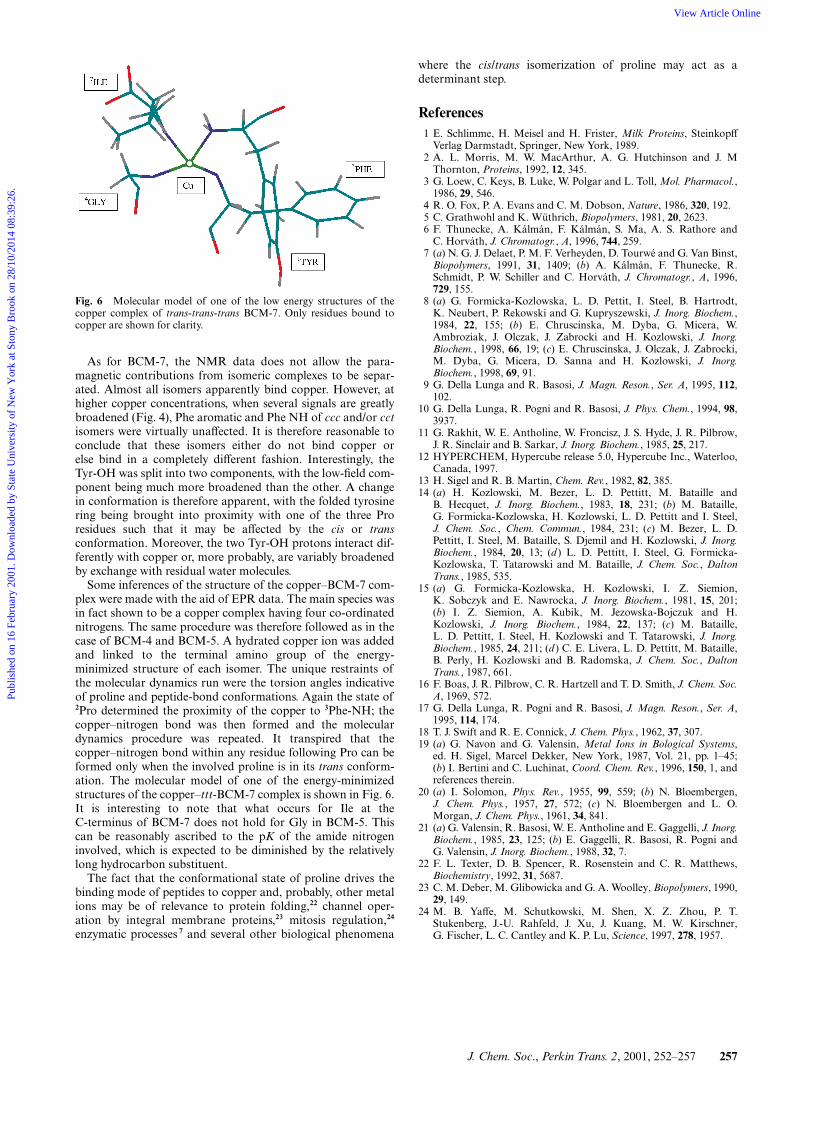
Some inferences of the structure of the copper–BCM-7 com-plex were made with the aid of EPR data. The main species wasin fact shown to be a copper complex having four co-ordinatednitrogens. The same procedure was therefore followed as in thecase of BCM-4 and BCM-5. A hydrated copper ion was addedand linked to the terminal amino group of the energy-minimized structure of each isomer. The unique restraints ofthe molecular dynamics run were the torsion angles indicativeof proline and peptide-bond conformations. Again the state of2Pro determined the proximity of the copper to 3Phe-NH; thecopper–nitrogen bond was then formed and the moleculardynamics procedure was repeated. It transpired that thecopper–nitrogen bond within any residue following Pro can beformed only when the involved proline is in its trans conform-ation. The molecular model of one of the energy-minimizedstructures of the copper–ttt-BCM-7 complex is shown in Fig. 6.It is interesting to note that what occurs for Ile at theC-terminus of BCM-7 does not hold for Gly in BCM-5. Thiscan be reasonably ascribed to the pK of the amide nitrogeninvolved, which is expected to be diminished by the relativelylong hydrocarbon substituent.
The fact that the conformational state of proline drives thebinding mode of peptides to copper and, probably, other metalions may be of relevance to protein folding,22 channel oper-ation by integral membrane proteins,23 mitosis regulation,24
enzymatic processes 7 and several other biological phenomena
Fig. 6 Molecular model of one of the low energy structures of thecopper complex of trans-trans-trans BCM-7. Only residues bound tocopper are shown for clarity.
where the cis/trans isomerization of proline may act as adeterminant step.
References1 E. Schlimme, H. Meisel and H. Frister, Milk Proteins, Steinkopff
Verlag Darmstadt, Springer, New York, 1989.2 A. L. Morris, M. W. MacArthur, A. G. Hutchinson and J. M
Thornton, Proteins, 1992, 12, 345.3 G. Loew, C. Keys, B. Luke, W. Polgar and L. Toll, Mol. Pharmacol.,
1986, 29, 546.4 R. O. Fox, P. A. Evans and C. M. Dobson, Nature, 1986, 320, 192.5 C. Grathwohl and K. Wüthrich, Biopolymers, 1981, 20, 2623.6 F. Thunecke, A. Kálmán, F. Kálmán, S. Ma, A. S. Rathore and
C. Horváth, J. Chromatogr., A, 1996, 744, 259.7 (a) N. G. J. Delaet, P. M. F. Verheyden, D. Tourwé and G. Van Binst,
Biopolymers, 1991, 31, 1409; (b) A. Kálmán, F. Thunecke, R.Schmidt, P. W. Schiller and C. Horváth, J. Chromatogr., A, 1996,729, 155.
8 (a) G. Formicka-Kozlowska, L. D. Pettit, I. Steel, B. Hartrodt,K. Neubert, P. Rekowski and G. Kupryszewski, J. Inorg. Biochem.,1984, 22, 155; (b) E. Chruscinska, M. Dyba, G. Micera, W.Ambroziak, J. Olczak, J. Zabrocki and H. Kozlowski, J. Inorg.Biochem., 1998, 66, 19; (c) E. Chruscinska, J. Olczak, J. Zabrocki,M. Dyba, G. Micera, D. Sanna and H. Kozlowski, J. Inorg.Biochem., 1998, 69, 91.
9 G. Della Lunga and R. Basosi, J. Magn. Reson., Ser. A, 1995, 112,102.
10 G. Della Lunga, R. Pogni and R. Basosi, J. Phys. Chem., 1994, 98,3937.
11 G. Rakhit, W. E. Antholine, W. Froncisz, J. S. Hyde, J. R. Pilbrow,J. R. Sinclair and B. Sarkar, J. Inorg. Biochem., 1985, 25, 217.
12 HYPERCHEM, Hypercube release 5.0, Hypercube Inc., Waterloo,Canada, 1997.
13 H. Sigel and R. B. Martin, Chem. Rev., 1982, 82, 385.14 (a) H. Kozlowski, M. Bezer, L. D. Pettitt, M. Bataille and
B. Hecquet, J. Inorg. Biochem., 1983, 18, 231; (b) M. Bataille,G. Formicka-Kozlowska, H. Kozlowski, L. D. Pettitt and I. Steel,J. Chem. Soc., Chem. Commun., 1984, 231; (c) M. Bezer, L. D.Pettitt, I. Steel, M. Bataille, S. Djemil and H. Kozlowski, J. Inorg.Biochem., 1984, 20, 13; (d ) L. D. Pettitt, I. Steel, G. Formicka-Kozlowska, T. Tatarowski and M. Bataille, J. Chem. Soc., DaltonTrans., 1985, 535.
15 (a) G. Formicka-Kozlowska, H. Kozlowski, I. Z. Siemion,K. Sobczyk and E. Nawrocka, J. Inorg. Biochem., 1981, 15, 201;(b) I. Z. Siemion, A. Kubik, M. Jezowska-Bojczuk and H.Kozlowski, J. Inorg. Biochem., 1984, 22, 137; (c) M. Bataille,L. D. Pettitt, I. Steel, H. Kozlowski and T. Tatarowski, J. Inorg.Biochem., 1985, 24, 211; (d ) C. E. Livera, L. D. Pettitt, M. Bataille,B. Perly, H. Kozlowski and B. Radomska, J. Chem. Soc., DaltonTrans., 1987, 661.
16 F. Boas, J. R. Pilbrow, C. R. Hartzell and T. D. Smith, J. Chem. Soc.A, 1969, 572.
17 G. Della Lunga, R. Pogni and R. Basosi, J. Magn. Reson., Ser. A,1995, 114, 174.
18 T. J. Swift and R. E. Connick, J. Chem. Phys., 1962, 37, 307.19 (a) G. Navon and G. Valensin, Metal Ions in Bological Systems,
ed. H. Sigel, Marcel Dekker, New York, 1987, Vol. 21, pp. 1–45;(b) I. Bertini and C. Luchinat, Coord. Chem. Rev., 1996, 150, 1, andreferences therein.
20 (a) I. Solomon, Phys. Rev., 1955, 99, 559; (b) N. Bloembergen,J. Chem. Phys., 1957, 27, 572; (c) N. Bloembergen and L. O.Morgan, J. Chem. Phys., 1961, 34, 841.
21 (a) G. Valensin, R. Basosi, W. E. Antholine and E. Gaggelli, J. Inorg.Biochem., 1985, 23, 125; (b) E. Gaggelli, R. Basosi, R. Pogni andG. Valensin, J. Inorg. Biochem., 1988, 32, 7.
22 F. L. Texter, D. B. Spencer, R. Rosenstein and C. R. Matthews,Biochemistry, 1992, 31, 5687.
23 C. M. Deber, M. Glibowicka and G. A. Woolley, Biopolymers, 1990,29, 149.
24 M. B. Yaffe, M. Schutkowski, M. Shen, X. Z. Zhou, P. T.Stukenberg, J.-U. Rahfeld, J. Xu, J. Kuang, M. W. Kirschner,G. Fischer, L. C. Cantley and K. P. Lu, Science, 1997, 278, 1957.
Publ
ishe
d on
16
Febr
uary
200
1. D
ownl
oade
d by
Sta
te U
nive
rsity
of
New
Yor
k at
Sto
ny B
rook
on
28/1
0/20
14 0
8:39
:26.
View Article Online


![The princess and the EPR pair - MITaram/talks/10-spread-princeton.pdfEPR pair. • Teleportation [BBCJPW93] is a method for sending one qubit using two classical bits and one EPR pair.](https://static.fdocument.org/doc/165x107/60bbd19f845cf921b57233ae/the-princess-and-the-epr-pair-mit-aramtalks10-spread-epr-pair-a-teleportation.jpg)