Chronic estradiol-17β exposure suppresses hypothalamic norepinephrine release and the...
9
www.elsevier.com/locate/brainres Available online at www.sciencedirect.com Research Report Chronic estradiol-17b exposure suppresses hypothalamic norepinephrine release and the steroid-induced luteinizing hormone surge: Role of nitration of tyrosine hydroxylase Badrinarayanan S. Kasturi a , Sheba M.J. MohanKumar b,c,d , Madhu P. Sirivelu c , Andrew C. Shin d , P.S. MohanKumar a,c,d, a Departments of Pathobiology & Diagnostic Investigation, College of Veterinary Medicine, Michigan State University, East Lansing, MI 48824, United States b Pharmacology & Toxicology, College of Veterinary Medicine, Michigan State University, East Lansing, MI 48824, United States c Graduate Programs in Comparative Medicine and Integrative Biology, College of Veterinary Medicine, Michigan State University, East Lansing, MI 48824, United States d Graduate Programs in Neuroscience, College of Veterinary Medicine, Michigan State University, East Lansing, MI 48824, United States article info Article history: Accepted 18 November 2012 Available online 26 November 2012 Keywords: Estrogen Medial preoptic area Tyrosine hydroxylase Nitric Oxide Interleukin-1b abstract Chronic exposure to estrogens is known to produce a variety of deleterious effects in women including breast and ovarian cancer and anovulation. In female rats, exposure to low levels of estradiol-17b (E2) decreases hypothalamic norepinephrine (NE) to suppress luteinizing hormone (LH) secretion and cause failure of ovulation. We hypothesized that E2 exposure most likely decreases NE release in the medial preoptic area (MPA) of the hypothalamus to produce this effect and that this may be due to E2-induced inflammatory changes in noradrenergic nuclei leading to nitration of an enzyme involved in NE synthesis. To test this, female Sprague Dawley rats were sham implanted or implanted with slow release E2 pellets (20 ng/day) for 30, 60 or 90 days (E30, E60 and E90 respectively). At the end of the treatment period, the rats were implanted with a push–pull cannula in the MPA, ovariectomized and steroid primied to induce a LH surge and subjected to push- pull perfusion. Perfusates were analyzed for NE levels using HPLC-EC. Blood samples collected simultaneously were analyzed for LH levels. We measured interleukin-1b (IL-1b) and nitrate levels in brainstem noradrenergic nuclei that innervate the MPA. In control animals, there was a marked increase in NE levels in response to steroid priming at 1600 h that was reduced in the E30 group, and completely abolished after 60 and 90 days of E2 exposure. LH profiles were similar to NE release profiles in control and E2-treated animals. We found that IL-1b levels increased in all three (A1, A2 and A6) noradrenergic nuclei with chronic E2 exposure, while nitrate levels increased only in the A6 region. There was an increase in the nitration of the NE synthesizing enzyme in the MPA in this group as well 0006-8993/$ - see front matter & 2012 Elsevier B.V. All rights reserved. http://dx.doi.org/10.1016/j.brainres.2012.11.031 Correspondence to: Department of Pathobiology & Diagnostic Investigation, College of Veterinary Medicine, B336 Life Sciences Building, Michigan State University, East Lansing, MI 48824, United States. Fax: þ1 517 353 8915. E-mail address: [email protected] (P.S. MohanKumar). brain research 1493 (2013) 90–98
Transcript of Chronic estradiol-17β exposure suppresses hypothalamic norepinephrine release and the...

Available online at www.sciencedirect.com
www.elsevier.com/locate/brainres
b r a i n r e s e a r c h 1 4 9 3 ( 2 0 1 3 ) 9 0 – 9 8
0006-8993/$ - see frohttp://dx.doi.org/10
�CorrespondenceBuilding, Michigan
E-mail address:
Research Report
Chronic estradiol-17b exposure suppresses hypothalamicnorepinephrine release and the steroid-inducedluteinizing hormone surge: Role of nitrationof tyrosine hydroxylase
Badrinarayanan S. Kasturia, Sheba M.J. MohanKumarb,c,d, Madhu P. Siriveluc,Andrew C. Shind, P.S. MohanKumara,c,d,�
aDepartments of Pathobiology & Diagnostic Investigation, College of Veterinary Medicine, Michigan State University, East Lansing,
MI 48824, United StatesbPharmacology & Toxicology, College of Veterinary Medicine, Michigan State University, East Lansing, MI 48824, United StatescGraduate Programs in Comparative Medicine and Integrative Biology, College of Veterinary Medicine, Michigan State University,
East Lansing, MI 48824, United StatesdGraduate Programs in Neuroscience, College of Veterinary Medicine, Michigan State University, East Lansing,
MI 48824, United States
a r t i c l e i n f o
Article history:
Accepted 18 November 2012
Chronic exposure to estrogens is known to produce a variety of deleterious effects in
women including breast and ovarian cancer and anovulation. In female rats, exposure to
Available online 26 November 2012
Keywords:
Estrogen
Medial preoptic area
Tyrosine hydroxylase
Nitric Oxide
Interleukin-1b
nt matter & 2012 Elsevie.1016/j.brainres.2012.11.0
to: Department of PathState University, East [email protected]
a b s t r a c t
low levels of estradiol-17b (E2) decreases hypothalamic norepinephrine (NE) to suppress
luteinizing hormone (LH) secretion and cause failure of ovulation. We hypothesized that E2
exposure most likely decreases NE release in the medial preoptic area (MPA) of the
hypothalamus to produce this effect and that this may be due to E2-induced inflammatory
changes in noradrenergic nuclei leading to nitration of an enzyme involved in NE
synthesis. To test this, female Sprague Dawley rats were sham implanted or implanted
with slow release E2 pellets (20 ng/day) for 30, 60 or 90 days (E30, E60 and E90 respectively).
At the end of the treatment period, the rats were implanted with a push–pull cannula in
the MPA, ovariectomized and steroid primied to induce a LH surge and subjected to push-
pull perfusion. Perfusates were analyzed for NE levels using HPLC-EC. Blood samples
collected simultaneously were analyzed for LH levels. We measured interleukin-1b (IL-1b)
and nitrate levels in brainstem noradrenergic nuclei that innervate the MPA. In control
animals, there was a marked increase in NE levels in response to steroid priming at 1600 h
that was reduced in the E30 group, and completely abolished after 60 and 90 days of E2
exposure. LH profiles were similar to NE release profiles in control and E2-treated animals.
We found that IL-1b levels increased in all three (A1, A2 and A6) noradrenergic nuclei with
chronic E2 exposure, while nitrate levels increased only in the A6 region. There was an
increase in the nitration of the NE synthesizing enzyme in the MPA in this group as well
r B.V. All rights reserved.31
obiology & Diagnostic Investigation, College of Veterinary Medicine, B336 Life Sciencessing, MI 48824, United States. Fax: þ1 517 353 8915.
.edu (P.S. MohanKumar).

b r a i n r e s e a r c h 1 4 9 3 ( 2 0 1 3 ) 9 0 – 9 8 91
probably contributing to reduced NE synthesis. This could be a possible mechanism by
which chronic E2 exposure decreases NE levels in the MPA to suppress the LH surge.
& 2012 Elsevier B.V. All rights reserved.
1. Introduction
The preovulatory luteinizing hormone (LH) surge that occurs
on the afternoon of proestrus is known to be essential for
inducing ovulation in rats (Knobil, 1994) and for estrous
cyclicity. The LH surge is produced due to the concerted
effort of a number of factors including estradiol-17b (E2) from
the ovaries. The gradual increase in serum E2 levels produces
a stimulatory effect on the brain stem, the hypothalamus and
the pituitary to promote the LH surge on the afternoon of
proestrus (Knobil, 1994). Although acute increases in E2
levels, as observed during estrous cycles, stimulates the LH
surge, chronic exposure to estrogenic compounds is known
to inhibit LH secretion (Armenti et al., 2008; Fernandez et al.,
2009; Laws et al., 2000). The mechanism behind this effect is
not clear.
During the estrous cycle, E2 influences the secretion of
several neurotransmitters in the brain (Mueller and Nistico,
1989). These neurotransmitters in turn, act on gonadotropin
releasing hormone (GnRH) neurons in the hypothalamus.
When these neurons are activated, GnRH is released from
their terminals, enters the portal circulation to stimulate
gonadotrophs in the anterior pituitary resulting in an
increase in LH secretion (Mueller and Nistico, 1989). Among
the many factors that are influenced by E2 to stimulate the
LH surge, we selected norepinephrine (NE) for the following
reasons. Several studies using NE synthesis blockers (Drouva
and Gallo, 1976), NE depletors (Kang et al., 1998), and
neurotoxic lesioning (Hancke and Wuttke, 1979; Simpkins
et al., 1979) have demonstrated a loss of the LH surge
indicating that NE plays an important role in this phenom-
enon. Moreover, GnRH neurons of the hypothalamus are
richly innervated by brainstem noradrenergic neurons
(Wright and Jennes, 1993) and these noradrenergic neurons
are also sensitive to E2 (Jennes et al., 1992; Kaba et al., 1983;
Liaw et al., 1992) and have estrogen receptors (Herbison,
1997). Further, removing the influence of E2 by ovariectomy
decreases NE levels in the hypothalamus and abolishes the
LH surge (Mohankumar and Mohankumar, 2004). Taken
together, these studies suggest that E2 most likely acts
through NE to stimulate GnRH neurons and thereby LH
secretion. In contrast to the acute stimulatory effects of E2
on LH secretion, we have observed that chronic exposure to
E2 suppresses LH secretion. This was accompanied by a
reduction in NE levels in hypothalamic areas that are rich
in GnRH neurons such as the MPA (Kasturi et al., 2009). The
reason for the reduction in NE levels is not clear but it is
possible that it could be due to reduced NE synthesis.
In a recent study, we provided evidence that chronic E2
exposure significantly decreased dopamine (DA) levels in the
tuberoinfundibular dopaminergic (TIDA) system of the
hypothalamus. TIDA neurons are located in the arcuate
nucleus and their terminals are located in the median
eminence. Chronic exposure to E2 induced an inflammatory
reaction in the arcuate nucleus that resulted in increased
generation of the cytokine interleukin-1beta (IL-1b) and nitric
oxide-related free radicals. This led to the nitration of
tyrosine hydroxylase (TH). Since TH is the rate-limiting
enzyme in catecholamine biosynthesis, nitration of this
enzyme led to reduced TH activity resulting in decreased
DA synthesis (Mohankumar et al., 2011). We hypothesized
that a similar phenomenon may be in operation in noradre-
nergic neurons as well since DA and NE share a common
biosynthetic pathway and TH is rate-limiting to the synthesis
of both DA and NE (Nagatsu et al., 1964) resulting in reduction
in NE in the MPA. TH positive neurons that innervate the MPA
are localized in three distinct nuclei in the brainstem viz. the
A1 (rostral ventrolateral medulla), A2 (nucleus tractus solitar-
ius) and A6 (locus coeruleus) nuclei (Moore and Bloom, 1979).
TH that is synthesized in the brainstem noradrenergic neu-
rons is probably transported to the MPA where it plays an
important role in NE synthesis. Thus, chronic estradiol
exposure-induced nitration of TH in brainstem noradrenergic
neurons might be responsible for the reduction in NE in
the MPA.
To investigate this possibility, young, intact Sprague-
Dawley rats were exposed to low levels of E2 for 30, 60 or 90
days. Simultaneous changes in NE levels in the hypothala-
mus and LH levels in the serum were monitored. IL-1b and
nitrate, a stable end product of nitric oxide (NO) metabolism,
were measured in brainstem noradrenergic nuclei that pro-
vide noradrenergic innervation to the MPA. Nitrated TH (NO-
TH) was measured in the MPA. Results from this study will
help us understand the possible molecular mechanisms by
which chronic E2 exposure decreases NE biosynthesis to
suppress LH secretion in female rats.
2. Results
2.1. Effects of chronic E2 treatment on estrous cyclicity
Effects of chronic estradiol exposure on estrous cycles are
shown in Fig. 1. More than 95% of the control animals showed
4–5 day regular estrous cycles. Exposure to E2 for 30 days did
not have any significant effect on estrous cyclicity (485% of
the animals showed regular estrous cycles). In contrast, after
60 and 90 days of E2 exposure the percentage of regular
cyclers declined to 35% and 20% respectively.
2.2. Location of the push–pull cannulae
Fig. 2 depicts the histological location of push–pull cannulae
in different treatment groups. Animals that did not have their
cannula in the MPA were excluded from analysis.

Fig. 1 – Effects of chronic E2 exposure on estrous cyclicity.
Animals were sham-implanted (control) or implanted with
20 ng/day of E2 pellets subcutaneously for 30 (E30), 60 (E60)
or 90 (E90) days.
Fig. 2 – Locations of push–pull cannula in the medial
preoptic area in animals from various groups are indicated
in the schematic representation. Sham-implanted animals
that were injected with either oil or estradiol and
progesterone (EþP) are indicated by open and closed circles
respectively. E-30 animals that were injected with oil and EP
are indicated by open and closed triangles respectively. E-60
animals treated with oil and EP are indicated by open and
closed rectangles respectively. E-90 animals treated with oil
and EP are indicated by open and closed pentagons
respectively. A1-P3 represents coronal sections in reference
to the Bregma. MPA: Medial preoptic area; StHy:
striohypothalamic nucleus; MPO: medial preoptic nucleus;
AVPO: anteroventral preoptic nucleus; Sch: suprachiasmatic
nucleus; OX: optic chiasm; SOX: supraoptic decussation; LA:
lateroanterior hypothalamic nucleus; AH: Anterior
hypothalamus; VMH: ventromedial hypothalamus.
Fig. 3 – (a) Norepinephrine (NE) release (mean7SE, pg/min)
in the MPA of OVX, sham-implanted animals (Panel A),
30- (Panel B), 60- (Panel C) and 90- days (Panel D) E2-treated
animals, that were OVX and treated with oil (n¼6) or EP
(n¼6). � Significant difference (po0.05) compared to 1300 h
in the same group and at all time points in the oil-treated
group. (b) Average NE release in all groups during the entire
period of observation. � indicates significant difference from
rest of the groups. (a) indicates significant decrease from
control EP but significant increase compared to the rest of
the groups. (b) indicates significant reduction when
compared to the control-oil group.
b r a i n r e s e a r c h 1 4 9 3 ( 2 0 1 3 ) 9 0 – 9 892
2.3. Effects of chronic E2 exposure on steroid-induced NErelease profiles in the MPA
NE release profiles (pg/min; mean7SE) in the MPA of sham-
implanted animals are shown in Fig. 3a, panel A. In the
OVXþoil group, NE release was 1.470.3 at 1300 h and
remained at that level during the rest of the observation
period. In contrast, in the OVXþEP group, NE release was
2.970.7 at 1300 h and increased gradually to reach a peak at
1500 h (9.273.5; po0.05) before decreasing to 4.972.5 at
1700 h.
Effects of chronic E2 exposure on NE release profiles in the E30,
E60 and E90 groups are shown in Fig. 3a, panels B–D. In the E30
group, NE levels after OVXþoil treatment were 1.170.06 at 1300 h
and did not change throughout the evening. In the E30 group
that was treated with OVXþEP, NE levels were 1.770.1 at 1300 h
and increased significantly to 3.570.7 at 1530 h (po0.05) and
reached peak levels of 5.371.3 at 1630 h (po0.05). The marked
difference in NE profiles between sham-implanted and the E30
group after EP treatment was that NE levels reached a peak at
b r a i n r e s e a r c h 1 4 9 3 ( 2 0 1 3 ) 9 0 – 9 8 93
1500 h in the former but reached peak levels only at 1630 h in the
latter. In contrast to the sham-implanted and E30 groups, NE
release in the MPA of E60 and E90 remained unaffected both in
the OVXþoil and OVXþEP groups.
Fig. 3b provides a comparison of average NE levels during
the entire observation period in all the groups. Average NE
levels (Mean7SE; pg/min) in the oil-treated sham-implanted
group was 1.3870.4 and was significantly different from the
EP treated group (5.7170.69; po0.0001). Average NE levels in
the E30 group treated with EP (2.5870.45) was significantly
lower than the shamþEP treated group (po0.01), but was
significantly higher than the E60þEP (0.9270.2) and the
E90þEP (0.6270.11) groups (po0.001). Average NE levels in
the E60þoil and E90þoil groups were lower than the
Shamþoil group.
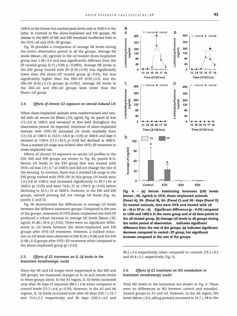
Fig. 4 – (a) Serum luteinizing hormone (LH) levels
(mean7SE, ng/ml) in OVX, sham-implanted animals
(Panel A), 30- (Panel B), 60- (Panel C) and 90- days (Panel D)
E2-treated animals, that were OVX and treated with oil
(n¼6) or EP (n¼6). � Significant difference (po0.05) compared
to 1300 and 1400 h in the same group and at all time points in
the oil-treated group. (b) Average LH levels in all groups during
the entire period of observation. � indicates significant
difference from the rest of the groups. (a) indicates significant
decrease compared to controlþEP group, but significant
increase compared to the rest of the groups.
2.4. Effects of chronic E2 exposure on steroid-induced LH
When sham-implanted animals were ovariectomized and trea-
ted with oil, serum LH (Mean7S.E; ng/ml; Fig. 4a, panel A) was
2.370.6 at 1300 h and remained at that level throughout the
observation period. As expected, treatment of sham-implanted
animals with OVXþEP increased LH levels markedly from
5.373.6 at 1300 h to 252.47126.0 (po0.05) at 1600 h and kept it
elevated at 1700 h (71.2742.5; po0.05) but declined at 1800 h.
Thus a marked LH surge was evident after OVXþEP treatment in
sham-implanted rats.
Effects of chronic E2 exposure on serum LH profiles in the
E30, E60 and E90 groups are shown in Fig. 4a, panels B–D.
Serum LH levels in the E30 group that was treated with
OVXþoil was 2.970.7 at 1300 h and did not change the rest of
the evening. In contrast, there was a marked LH surge in the
E30 group treated with OVXþEP. In this group, LH levels were
1.670.8 at 1300 h and increased significantly to 89.7764 at
1600 h (po0.05) and were 74.8737 at 1700 h (po0.05) before
declining to 32.5715 at 1800 h. However, in the E60 and E90
groups, steroid priming did not change LH levels (Fig. 4a,
panels C and D).
Fig. 4b demonstrates the differences in average LH levels
between the different treatment groups. Compared to the rest
of the groups, treatment of OVX sham-implanted rats with EP
produced a robust increase in average LH levels (Mean7SE;
ng/ml; 61.86739.4; po0.01). There were no significant differ-
ences in LH levels between the sham-implanted and E30
groups after OVXþEP treatment. However, a marked reduc-
tion in LH levels were observed in E60 (0.2470.08) and the E90
(1.0870.2) groups after OVXþEP treatment when compared to
the sham-implanted group (po0.01).
2.5. Effects of E2 treatment on IL-1b levels in thebrainstem noradrenergic nuclei
Since the NE and LH surges were suppressed in the E60 and
E90 groups, we measured changes in IL-1b and nitrate levels
in these groups alone. In the A1 region, IL-1b levels increased
only after 90 days of exposure (88.273.4) when compared to
control levels (75.774.6; po0.05). However, in the A2 and A6
regions, IL-1b levels increased both after 60 days (101.7712.7
and 73.675.5 respectively) and 90 days (106.274.9 and
88.273.4 respectively) when compared to controls (79.178.2
and 43.475.1 respectively; Fig. 5).
2.6. Effects of E2 treatment on NO metabolism inbrainstem noradrenergic nuclei
Total NO levels in the brainstem are shown in Fig. 6. There
were no differences in NO between control and estradiol-
treated groups in A1 and A2. However, in the A6 region, NO
levels (Mean7S.E; mM/mg protein) increased to 16.77.98 in the

Fig. 5 – Effects of varying durations of E2 exposure on IL-1b
levels in brainstem noradrenergic nuclei. Clear bars
represent the control group, gray bars represent the E-60
group and black bars represent the E-90 group. � indicates
po0.05 when compared to the corresponding control group.
Fig. 6 – Effects of chronic E2 exposure on total nitric oxide
levels measured as nitrate in the brainstem (B). Clear bars
represent the control group, gray bars represent the E-60
group and black bars represent the E-90 group. � indicates
po0.05 when compared to corresponding control group.
65Kd
65Kd
Fig. 7 – Effects of chronic E2 exposure on nitration
of tyrosine hydroxylase (TH) in the MPA. (A) and (B).
Representative Western blot showing immunoprecipitated
TH (A) and NO-TH (B) in the MPA of control, E-30, E-60 and
E-90 group. C. The ratio of NO-TH to TH in the MPA of
control, E-30, E-60 and E-90 group. � Significant difference
(po0.05) compared to control and E-30 groups.
b r a i n r e s e a r c h 1 4 9 3 ( 2 0 1 3 ) 9 0 – 9 894
E90 group when compared to the control group (12.371.3;
po0.05).
2.7. Effects of E2 treatment on nitration of TH in the MPA
Fig. 7A and B shows representative Western blots of TH and
NO-TH in the MPA of control and E2-treated rats. The ratio of
densities of NO-TH to TH is shown in Fig. 5C. As shown in the
figure, the ratio of NO-TH to TH increased in a duration-
dependent manner. In sham-implanted control animals the
ratio was 0.370.03. Treatment with E2 for 30 days did not
change the ratio significantly. However, treatment with E2 for
60 days increased the ratio of NO-TH to TH to 0.870.2 and
increased it further to 1.370.3 in animals treated with E2 for
90 days (po0.05).
3. Discussion
In this study, we demonstrate that exposure to low levels of
E2 on a chronic basis suppresses NE release in the MPA of the
hypothalamus upon steroid priming. This is accompanied by
a suppression of the LH surge in a duration dependent
manner. Chronic E2 exposure also increased IL-1b and nitrate
levels in specific noradrenergic nuclei that innervate the MPA.
There was a concurrent increase in the nitration of tyrosine
residues in TH, a modification that is known to inhibit the
activity of this enzyme. This reduction in TH activity most
probably contributes to decreased NE levels leading to a loss
of the LH surge. These results suggest that a novel phenom-
enon involving inflammation, free radicals and TH inactiva-
tion is in operation in brainstem noradrenergic neurons after
chronic E2 exposure. This could potentially contribute to loss
of estrous cyclicity in E2 exposed animals. Although these
results do not provide mechanistic evidence for the cascade
of events that are presented, it provides insight into a
possible sequence of events by which chronic E2 exposure
could interfere with the preovulatory LH surge and estrous
cyclicity.
E2 is an ovarian hormone that plays an important role in
the female reproductive system. Acute increases in serum
estrogen are critical for the stimulation of GnRH neurones to
trigger the LH surge that is essential for ovulation (Caraty
et al., 1995). On the other hand, chronic exposures to estro-
gens are believed to interfere with reproductive cycles (Biegel
b r a i n r e s e a r c h 1 4 9 3 ( 2 0 1 3 ) 9 0 – 9 8 95
et al., 1998; Jesionowska et al., 1990; Kasturi et al., 2009; Rosa
et al., 2003; Renner and Luine, 1986). Several studies in the
past have examined the effect of chronic estrogen exposure
on reproductive cycles and the LH surge. These studies
involved various estrogenic preparations such as ethinyl
estradiol, estradiol valerate, estradiol benzoate and estra-
diol-17b at doses ranging from a few micrograms to milli-
grams. The duration of exposure ranged from a few days to
several weeks (reviewed in (Karsch, 1987)). These studies
identified that acute exposure to estrogens initially sup-
pressed LH levels followed by stimulation of LH secretion.
On the other hand, long term exposures effectively sup-
pressed LH secretion (Karsch, 1987). In the present study,
we used a very low dose of E2 (20 ng/day) and exposed
animals to varying durations ranging from 30 to 90 days.
This dose was chosen because it produced serum estradiol
levels (�50 pg/ml) that were equivalent to those observed
during the morning of prestrous (Lapolt et al., 1986; Kasturi
et al., 2009) and it is much lower than the currently available
no observed adverse effect level for E2 (5 mg/kg) (Snyder et al.,
2008). It is also close to the acceptable daily intake levels
published by the Joint FAO/WHO committee (50 ng/kg) (Joint
FAO/WHO Expert Committee on Food Additives, 2004).
Results from our study indicate that even these low levels
of estrogen are capable of affecting estrous cycles after 60
days of exposure. We have observed that similar doses and
exposures of E2 result in significant increases in serum E2
(Kasturi et al., 2009; Mohankumar et al., 2011). This increase
in E2 levels could have a negative impact on LH secretion as
described below.
Chronic E2 treatment can interfere with LH secretion in
both intact animals and the ovariectomized, steroid-primed
rat model (Kasturi et al., 2009; Tsai and Legan, 2001; Tsai and
Legan, 2002). The mechanism by which E2 exposure sup-
presses LH secretion is not clear. Since GnRH neurons are
regulated by a variety of neurotransmitters and neuropep-
tides (Ciechanowska et al. 2010), E2 could affect one or more
of these mediators to suppress LH secretion. We chose to look
at the involvement of NE in this mechanism because NE is
known to be an important player in LH secretion
(Barraclough, 1983). We have found that similar doses of E2
as used in this study can indeed decrease NE concentrations
in the MPA (Kasturi et al., 2009). In the present study, we took
this a step further and examined the effect of low dose E2
exposure on NE release using push–pull perfusion of con-
scious, freely moving animals. We found that E2 exposure not
only decreases NE release in response to steroid priming in a
duration-dependent manner, it also suppresses basal NE
release in the oil-treated groups. While exposure for 30 days
had minimal effects on NE release, exposure for 60 and 90
days suppressed the basal and steroid-induced increase in NE
release that occurs at the time of the LH surge. These results
are supported by other studies that have observed reductions
in NE turnover (Hiemke et al., 1983) and release (Legan and
Callahan, 1999) after varying durations and doses of estrogen
exposure.
The reduction in NE levels observed in the E-60 and E-90
groups correlates well with the lack of a LH surge in these
animals. However, the mechanism by which E2 causes a
reduction in NE levels in the MPA is not clear. It is very likely
that the prolonged exposure to E2 causes de-sensitization or
downregulation of E2 receptors in the brainstem and/or
hypothalamus. We considered the possibility that chronic
E2 exposure may impair NE synthesis. There is evidence to
indicate that E2 treatment incites an inflammatory response
in the hypothalamus (Brawer et al., 1978). We have also
observed an increase in the arcuate nucleus of the hypotha-
lamus that contains dopamine neuronal cell bodies, after
similar exposures to E2 (Mohankumar et al., 2011). Therefore,
it is possible that the E2 exposure paradigms that we used in
this study could increase IL-1b levels in brainstem noradre-
nergic nuclei that contain cell bodies of noradernergic neu-
rons. Our results indicate that indeed, E2 exposure increased
IL-1b levels in all three noradrenergic nuclei. However, this
resulted in an increase in nitrate levels only in the A6 region.
The reason for this difference is not clear but it could relate to
differential IL-1 signaling (Sirivelu et al., 2012) or nitric oxide
synthesis in these nuclei. Since the exposures are chronic it is
likely that free radical scavenging mechanisms are activated
and these could affect the levels of nitrates in the brainstem
nuclei. The increase in IL-1b and nitric oxide-related free
radicals could affect noradrenergic activity in the MPA as
described below.
NE synthesis is under the tight control of TH which is the
rate-limiting enzyme in the synthesis of NE (Nagatsu et al.,
1964). Peroxynitrite, one of the products of nitric oxide
metabolism facilitates the formation of nitrotyrosine, which,
results in nitration of tyrosine residues in proteins (Huie and
Padmaja, 1993; Ischiropoulos et al., 1995). This is especially
true of proteins like TH that contain several tyrosine residues
in their structure (Ara et al., 1998). Since the tyrosine moieties
in TH are clustered around the active center of the enzyme,
nitration of these tyrosine residues is believed to cause steric
hindrance, resulting in reduced activity of this enzyme
(Imam et al., 2001) and decreased NE synthesis. Although
we did not measure TH activity, measurement of NO-TH
could provide insights into the possible reasons for chronic
E2-induced reduction in NE levels in the MPA. The results
from the present study clearly indicate that there is a
significant increase in the nitration of TH in the MPA of
E2-treated animals in a duration-dependent manner. It is
possible that TH gets nitrated in the brainstem and is later
transported to the MPA where it remains inactive. Nitration of
tyrosine residues is an irreversible process (Drew and
Leeuwenburgh, 2002). Since the E2 exposures were chronic,
it is likely that any new enzyme that is synthesized to make
up for the loss of enzyme activity will probably be nitrated
leading to continued suppression of NE synthesis. This
probably accounts for the higher levels of NO-TH observed
in the E-90 animals compared to E-60 and E-30 groups. As
more NO-TH is generated, specific systems are activated for
removal of nitrated proteins. NO-TH is supposed to be
selectively degraded in vivo by chymotrypsin (Souza et al.,
2000). Besides causing nitration of proteins, the NO that is
generated could cause nitration of NE also (Daveu et al., 1997),
leading to reduced binding to adrenergic receptors. Chronic
E2 exposures could interfere with these mechanisms as well
and these need to be investigated further.
In conclusion, the present study provides evidence that
chronic exposure to low levels of E2 is capable of suppressing
b r a i n r e s e a r c h 1 4 9 3 ( 2 0 1 3 ) 9 0 – 9 896
the steroid-induced LH surge in a duration-dependent man-
ner. This was accompanied by a reduction in NE release in the
MPA. The confidence in our results is increased by the fact
that both NE release and LH levels were measured in the
same animals. Our study also provides evidence for a novel
mechanism by which E2 exposure suppresses NE release in
the MPA. E2 exposure increased IL-1b and nitrate levels in the
brainstem noradrenergic nuclei with a concurrent increase in
the nitration of TH in the MPA. This could be responsible for
the reduction in TH activity, leading to decreased NE synth-
esis and LH secretion. Further studies are needed to deter-
mine the underlying mechanisms by which nitric oxide
metabolism are activated in the brain stem after E2 exposure.
4. Experimental procedure
4.1. Animals
Adult female Sprague-Dawley rats, around 3 months of age,
were obtained from Harlan Sprague-Dawley, Inc., (Indiana-
polis, IN, USA) and were housed in temperature (2372 1C) and
light-controlled (lights on from 0500 to 1900 h) animal rooms.
They were given food and water ad libitum. All the protocols
followed in this study were approved by the IACUC at
Michigan State University.
4.2. Treatment
Estrous cycles were monitored regularly and those animals
showing regular 4-day cycles were chosen for the experi-
ment. The animals were randomly divided into different
treatment groups (final n¼6/group for push–pull perfusion
studies and n¼7/group for measuring IL-1b, nitrate and NO-
TH). Animals in the control groups were sham-implanted and
those in the treatment groups were implanted subcuta-
neously with slow-release E2 pellets for 30 (E30 group), 60
(E60 group) or 90 (E90 group) days. The pellets were capable of
releasing E2 at the rate of 20 ng per day (Innovative Research
America, Sarasota, FL). We have observed previously that
serum E2 levels after 30, 60 and 90 days of exposure are about
35, 70 and 95 pg/ml respectively (Kasturi et al., 2009). Estrous
cyclicity was monitored daily as described before
(Mohankumar et al., 1994b). At the end of the treatment
period, animals used for IL-1b, nitrate and NO-TH measure-
ments were sacrificed at 1200 h when they were in the state
of estrus. Animals that were used for push–pull perfusion
were treated as described below.
4.3. Push–pull cannula implantation and perfusion
At the end of the treatment period (30, 60 or 90 days of
E2/sham treatment), animals were bilaterally ovariectomized
under pentobarbital anesthesia (Day-0). Simultaneously,
these animals were also implanted with a push–pull cannula
in the MPA as described previously (MohanKumar and
MohanKumar, 2004). The push–pull cannulae were con-
structed as described before (Mohankumar et al., 1991). The
animals were implanted with a cannula stereotaxically in the
MPA using the following coordinates: 8.5 mm ventral, 0.3 mm
posterior and 0.3 mm lateral to the bregma (MohanKumar
and MohanKumar, 2004). The cannula was secured with
dental cement and a 29-g stainless steel stylet was intro-
duced to prevent blockage due to gliosis. The animals were
allowed to recover for seven days. On the eighth day, the
animals were given 0.1 ml of either corn oil (OVXþoil) or E2
(30 mg, s.c.) at 1000 h. On the 9th day, they were implanted
with a jugular catheter between 3–5 pm. On the 10th day, (the
day of perfusion), animals in the OVXþoil group were given
0.1 ml of either corn oil while the remaining animals that had
received E2 earlier, were treated with progesterone (2 mg, s.c.;
OVXþEP) at 1000 h.
The animals were connected to a peristaltic pump and
perfusion was started around 1030 h as described previously
(Mohankumar et al., 1991, 1994b; MohanKumar and Quadri,
1993). Animals were conscious and freely-moving in the
perfusion cages. Artificial cerebrospinal fluid was used as
the perfusion medium at a flow rate of 10 ml/min. Perfusates
were collected from 1300–1700 h at 30 min intervals. Perfu-
sates were mixed with 0.5 M HClO4 at a ratio of 25:1 v/v and
stored at �70 1C until analyzed for NE concentrations using
HPLC-EC. Blood samples (200 ml) were collected from
1300–1800 h, at 1 h intervals through a jugular catheter.
Plasma samples were stored at �70 1C until they were
analyzed for LH concentrations by RIA. At the end of perfu-
sion, animals were sacrificed, their brains were removed and
frozen at �70 1C until sectioned for histological verification of
cannula location.
4.4. HPLC-EC
The following were the components of the HPLC-EC system
used: a phase II, 5 mm ODS reverse phase C-18 column
(Phenomenex, Torrance, CA, USA), a glassy carbon electrode,
a CTO-10 AT/VP column oven, a LC-10 AT/VP pump (Shimadzu,
Columbia, MD, USA), and a LC-4C amperometric detector (Bioa-
nalytical Systems, West Lafayette, IN, USA). The mobile phase
was made using nanopure water and it contained monochlor-
oacetic acid (14.14 g/l), sodium hydroxide (4.675 g/l), octane
sulfonic acid disodium salt (0.3 g/l), ethylenediamine tetraacetic
acid (0.25 g/l), acetonitrile (3.5%) and tetrahydrofuran (1.4%). The
mobile phase was filtered and degassed through a Milli-Q
purification system (Millipore, Bedford, MA, USA) and pumped
at a flow rate of 1.8 ml/min. The sensitivity of the detector was
1 nA full scale, and the potential of the working electrode was
0.65 V. The column was maintained at 37 1C. The perfusate (90 ml)
was mixed with 30 ml of the internal standard (0.05 M dihydroxy
benzylamine) and 100 ml of the mixture was injected into the
system. The HPLC system was capable of detecting o1 pg of NE.
NE levels in the perfusate were expressed as pg/min.
4.5. Brain sectioning
A cryostat (Slee, London, UK) was used to obtain serial brain
sections. Brains were sectioned at 40 mm thickness and
stained using cresyl violet to determine the location of the
cannula tip (Mohankumar et al., 1994b). Only those animals
with the cannula in the MPA were included in the study.
Brains from the groups used for measuring IL-1b, nitrate and
b r a i n r e s e a r c h 1 4 9 3 ( 2 0 1 3 ) 9 0 – 9 8 97
NO-TH were sectioned at 300 mm and the MPA and brainstem
noradrenergic nuclei were microdissected using Palkovits’
microdissection technique (Palkovits et al., 1975) with a
500 mm punch. Tissue punches were stored at �70 1C until
analyzed for IL-1b, nitrate and NO-TH as described below.
4.6. IL-1b ELISA
IL-1b levels were measured in control, E-60 and E-90 groups.
Tissue punches were homogenized in phosphate buffer (pH
7.4). 50 ml of the homogenate was used in duplicate for
measuring IL-1b using a commercial ELISA kit (Enzo Life
Sciences, Farmingdale, NY). Samples were assayed according
to the manufacturer’s directions. The sensitivity of the assay
was o12 pg/ml. IL-1b was expressed as pg/mg protein.
4.7. Measurement of total NO generation in brainstemnoradrenergic nuclei
NO levels were measured in control, E-60 and E-90 groups.
Total NO levels in the brainstem nuclei were measured using
a commercial kit that used the Griess reaction (Total nitric
oxide assay kit, Assay Designs Inc., Ann Arbor, MI). Since NO
is transient by nature and cannot be measured easily, the
Griess reaction uses nitrate reductase to convert NO ulti-
mately to nitrate that is measured photometrically. The
sensitivity of the assay was 0.625 mM/L. Total NO produced
is expressed as mM/mg protein.
4.8. Immunoprecipitation of tyrosine hydroxylase anddetection of nitrated-TH
TH was immunoprecipitated from tissue samples from the
MPA and subjected to western blotting as described pre-
viously (Ara et al., 1998; Mohankumar et al.). Briefly, MPA
samples were homogenized in cell lysis buffer and incubated
with Rabbit Anti-rat Tyrosine Hydroxylase antibody (0.5 mg;
Chemicon Intl., Temecula, CA.) overnight at 4 1C. Protein-A
Agarose slurry (100 mL) was added to the mixture and incu-
bated at 4 1C for 1.5 h. The mixture containing the
antigen–antibody complex was centrifuged at 14000 rpm for
10 min and TH was eluted using 30 mL of the elution buffer
(0.2 M glycine, pH 3). The supernatant containing TH was
divided into two equal halves and loaded onto two separate
20% SDS- polyacrylamide gel. After electrophoresis, the gels
were blotted onto nitrocellulose membranes, and one mem-
brane was probed with anti-rat tyrosine hydroxylase (1:1000
dilution, Chemicon Intl. Temecula,CA) and the other with
rabbit anti- nitrotyrosine, (1:1000 dilution, SIGMA, St. Louis,
MO). Bands were detected using 4-Chloro-1-Naphthol (Biorad,
Hercules, CA). TH and NO-TH concentrations were quantified
after densitometric scanning using a Kodak Digital Science
Image analysis system (Kodak, Rochester, NY). A ratio of pixel
intensities was used to express the ratio of TH to NO-TH.
4.9. LH-RIA
LH was measured in duplicates in the plasma by RIA using a
double antibody method as described before (MohanKumar
et al., 1994a). The standards (RP1) and antibody (Anti rLH-S11)
were obtained from Dr. A.F.Parlow, NHPP, NIDDK. The LH
tracer was obtained from Amersham Pharmacia Biotech
(Waukesha, WI, USA). Briefly, 40 ml of the serum was assayed
in duplicate. The first antibody was added at a dilution of
1:758,000. The intra-assay variability of the LH RIA was 5.2%.
LH levels were expressed as ng/ml.
4.10. Protein measurements for tissue samples
Protein concentrations in tissue homogenates of the MPA and
brainstem noradrenergic nuclei were determined using a
micro Bicinchoninic acid assay (Pierce, Rockford, IL). Prior to
the IL-1b and nitric oxide assays, tissue samples were homo-
genized in PBS and 20 ml aliquots were used for the protein
assay according to the manufacturer’s protocol.
4.11. Statistical analysis
Differences in NE release, and serum LH, profiles between
different treatment groups and across different time points
within each group were determined using two-way repeated
measures ANOVA followed by post hoc Fisher’s LSD test.
Differences in nitrate, IL-1b and the ratio of NO-TH to TH
were analyzed by one-way ANOVA followed by post hoc
Fisher’s LSD test.
Acknowledgment
The authors would like to thank Dr. Priya Balasubramanian
and Ms. Katrina Linning for their technical assistance. This
work was supported by NIH AG027697; NSF IBN0236385;
Equipment grant from the Companion Animal Fund, CVM;
and MSU AgBioResearch.
r e f e r e n c e s
Ara, J., et al., 1998. Inactivation of tyrosine hydroxylase bynitration following exposure to peroxynitrite and 1-methyl-4-phenyl-1,2,3,6-tetrahydropyridine (MPTP). Proc. Natl. Acad.Sci. 95, 7659–7663.
Armenti, A.E., et al., 2008. Developmental methoxychlor exposureaffects multiple reproductive parameters and ovarian follicu-logenesis and gene expression in adult rats. Toxicol. Appl.Pharmacol. 233, 286–296.
Barraclough, C.A., 1983. The role of catecholamines in theregulation of gonadotropin secretion. Acta Morphol. Hung.31, 101–115.
Biegel, L.B., et al., 1998. Effects of 17 beta-estradiol on serumhormone concentrations and estrous cycle in female Crl:CDBR rats: effects on parental and first generation rats. Toxicol.Sci. 44, 143–154.
Brawer, J.R., et al., 1978. Effects of a single injection of estradiolvalerate on the hypothalamic arcuate nucleus and on repro-ductive function in the female rat. Endocrinology 103,501–512.
Caraty, A., et al., 1995. The preovulatory gonadotrophin-releasinghormone surge: a neuroendocrine signal for ovulation. J.Reprod. Fertil. Suppl. 49, 245–255.
Ciechanowska, M., et al., 2010. Neuroendocrine regulation ofGnRH release and expression of GnRH and GnRH receptor

b r a i n r e s e a r c h 1 4 9 3 ( 2 0 1 3 ) 9 0 – 9 898
genes in the hypothalamus-pituitary unit in different physio-logical states. Reprod. Biol. 10, 85–124.
Daveu, C., et al., 1997. Oxidation and nitration of catecholaminesby nitrogen oxides derived from nitric oxide. Nitric Oxide 1,234–243.
Drew, B., Leeuwenburgh, C., 2002. Aging and the role of reactivenitrogen species. Ann. N. Y. Acad. Sci. 959, 66–81.
Drouva, S.V., Gallo, R.V., 1976. Catecholamine involvement inepisodic luteinizing hormone release in adult ovariectomizedrats. Endocrinology 99, 651–658.
Fernandez, M., et al., 2009. Neonatal exposure to bisphenol aalters reproductive parameters and gonadotropin releasinghormone signaling in female rats. Environ. Health Perspect.117, 757–762.
Francis, J., MohanKumar, S.M., MohanKumar, P.S., 2004. Leptininhibits norepinephrine efflux from the hypothalamusin vitro: role of gamma aminobutyric acid. Brain Res. 1021,286–291.
Hancke, J.L., Wuttke, W., 1979. Effects of chemical lesion of theventral noradrenergic bundle or the medial preoptic area onpreovulatory LH release in rats. Exp. Brain Res. 35, 127–134.
Herbison, A.E., 1997. Noradrenergic regulation of cyclic GnRHsecretion. Rev. Reprod. 2, 1–6.
Hiemke, C., et al., 1983. Effects of oestradiol benzoate andprogesterone on luteinizing hormone release and catechola-mine turnover in the preoptic-hypothalamic brain area ofovariectomized rats. J. Endocrinol. 97, 437–445.
Huie, R.E., Padmaja, S., 1993. The reaction of NO with superoxide.Free Radic Res. Commun. 18, 195–199.
Imam, S.Z., et al., 2001. Methamphetamine-induced dopaminer-gic neurotoxicity: role of peroxynitrite and neuroprotectiverole of antioxidants and peroxynitrite decomposition cata-lysts. Ann. N. Y. Acad. Sci. 939, 366–380.
Ischiropoulos, H., Duran, D., Horwitz, J., 1995. Peroxynitrite-mediated inhibition of DOPA synthesis in PC12 cells. J. Neuro-chem. 65, 2366–2372.
Jennes, L., et al., 1992. c-fos expression in noradrenergic A2neurons of the rat during the estrous cycle and after steroidhormone treatments. Brain Res. 586, 171–175.
Jesionowska, H., Karelus, K., Nelson, J.F., 1990. Effects of chronicexposure to estradiol on ovarian cyclicity in C57BL/6J mice:potentiation at low doses and only partial suppression at highdoses. Biol. Reprod. 43, 312–317.
Joint FAO/WHO Expert Committee on Food Additives, 2004.Summary of evaluations performed by the joint FAO/WHOExpert Committee on Food Additives (JECFA-1956-2004) FirstThrough sixty-third meetings. RFS 896-JECFA53/81. FAO/WHO.
Kaba, H., et al., 1983. Effects of estrogen on the excitability of neuronsprojecting from the noradrenergic A1 region to the preoptic andanterior hypothalamic area. Brain Res. 274, 156–159.
Kang, S.S., et al., 1998. Noradrenergic neurotoxin suppressesgonadotropin-releasing hormone (GnRH) and GnRH receptorgene expression in ovariectomized and steroid-treated rats. J.Neuroendocrinol. 10, 911–918.
Karsch, F.J., 1987. Central actions of ovarian steroids in thefeedback regulation of pulsatile secretion of luteinizing hor-mone. Annu. Rev. Physiol. 49, 365–382.
Kasturi, B.S., et al., 2009. Chronic exposure to low levels ofoestradiol-17beta affects oestrous cyclicity, hypothalamic nor-epinephrine and serum luteinising hormone in young intactrats. J. Neuroendocrinol. 21, 568–577.
Knobil, E.a.N.J.D., 1994. The Physiology of Reproduction 2, 633–639.Lapolt, P.S., et al., 1986. The relation of ovarian steroid levels in
young female rats to subsequent estrous cyclicity and repro-ductive function during aging. Biol. Reprod. 35, 1131–1139.
Laws, S.C., et al., 2000. Estrogenic activity of octylphenol, nonyl-phenol, bisphenol A and methoxychlor in rats. Toxicol. Sci. 54,154–167.
Legan, S.J., Callahan, W.H., 1999. Suppression of tonic luteinizinghormone secretion and norepinephrine release near the GnRHneurons by estradiol in ovariectomized rats. Neuroendocrinol-ogy 70, 237–245.
Liaw, J.J., et al., 1992. Changes in tyrosine hydroxylase mRNAlevels in medullary A1 and A2 neurons and locus coeruleusfollowing castration and estrogen replacement in rats. BrainRes. Mol. Brain Res. 13, 231–238.
Mohankumar, P.S., Thyagarajan, S., Quadri, S.K., 1991. Interleukin-1stimulates the release of dopamine and dihydroxypheny-lacetic acid from the hypothalamus in vivo. Life Sci. 48,925–930.
MohanKumar, P.S., Quadri, S.K., 1993. Systemic administration ofinterleukin-1 stimulates norepinephrine release in the para-ventricular nucleus. Life Sci. 52, 1961–1967.
MohanKumar, P.S., Meites, J., Quadri, S.K., 1994a. Deprenyl reducesserum prolactin concentrations in rats. Life Sci. 54, 841–845.
Mohankumar, P.S., Thyagarajan, S., Quadri, S.K., 1994b. Correla-tions of catecholamine release in the medial preoptic areawith proestrous surges of luteinizing hormone and prolactin:effects of aging. Endocrinology 135, 119–126.
Mohankumar, S.M., et al., 2011. Chronic estradiol exposureinduces oxidative stress in the hypothalamus to decreasehypothalamic dopamine and cause hyperprolactinemia. Am.J. Physiol. Regul. Integr. Comp. Physiol..
MohanKumar, S.M., MohanKumar, P.S., 2004. Aging alters nore-pinephrine release in the medial preoptic area in response tosteroid priming in ovariectomized rats. Brain Res. 1023, 24–30.
Moore, R.Y., Bloom, F.E., 1979. Central catecholamine neuronsystems: anatomy and physiology of the norepinephrine andepinephrine systems. Annu. Rev. Neurosci. 2, 113–168.
Mueller, E.E., Nistico, G., 1989. Brain Messengers and the Pituitary,Vol. Academic Press, New York.
Nagatsu, T., Levitt, M., Udenfriend, S., 1964. Tyrosine hydroxylase.The initial step in norepinephrine biosynthesis. J. Biol. Chem.239, 2910–2917.
Palkovits, M., et al., 1975. Effects of stress on catecholamines andtyrosine hydroxylase activity of individual hypothalamicnuclei. Neuroendocrinology 18, 144–153.
Renner, K., Luine, V., 1986. Analysis of temporal and dose-dependent effects of estrogen on monoamines in brain nuclei.Brain Res. 366, 64–71.
Rosa, E.S.A., et al., 2003. Prepubertal administration of estradiolvalerate disrupts cyclicity and leads to cystic ovarian mor-phology during adult life in the rat: role of sympatheticinnervation. Endocrinology 144, 4289–4297.
Simpkins, J.W., et al., 1979. Blockade of steroid-induced leuteinizinghormone release by selective depletion of anterior hypothalamicnorepinephrine activity. Endocrinology 104, 506–509.
Sirivelu, M.P., MohanKumar, P.S., MohanKumar, S.M., 2012. Differ-ential effects of systemic administration of interleukin-1 betaon brainstem noradrenergic nuclei. Life Sci. 90, 77–81.
Snyder, S.A., Trenholm, R.A., Snyder, E.M., Bruce, G.M., Pleus, R.C.,Hemming, J.D.C., 2008. Toxicological relevance of EDCs andpharmaceuticals in drinking water. Awwa Research Founda-tion, Denver, Colorado.
Souza, J.M., et al., 2000. Proteolytic degradation of tyrosinenitrated proteins. Arch. Biochem. Biophys. 380, 360–366.
Tsai, H.W., Legan, S.J., 2001. Chronic elevation of estradiol in youngovariectomized rats causes aging-like loss of steroid-inducedluteinizing hormone surges. Biol. Reprod. 64, 684–688.
Tsai, H.W., Legan, S.J., 2002. Loss of luteinizing hormone surgesinduced by chronic estradiol is associated with decreasedactivation of gonadotropin-releasing hormone neurons. Biol.Reprod. 66, 1104–1110.
Wright, D.E., Jennes, L., 1993. Origin of noradrenergic projectionsto GnRH perikarya-containing areas in the medial septum-diagonal band and preoptic area. Brain Res. 621, 272–278.