Non-neural tyrosine hydroxylase, via modulation of...
9
ARTICLE Non-neural tyrosine hydroxylase, via modulation of endocrine pancreatic precursors, is required for normal development of beta cells in the mouse pancreas Patricia Vázquez & Ana M. Robles & Flora de Pablo & Catalina Hernández-Sánchez Received: 8 January 2014 /Accepted: 1 July 2014 /Published online: 1 August 2014 # The Author(s) 2014. This article is published with open access at Springerlink.com Abstract Aims/hypothesis Apart from transcription factors, little is known about the molecules that modulate the proliferation and differentiation of pancreatic endocrine cells. The early expression of tyrosine hydroxylase (TH) in a subset of gluca- gon + cells led us to investigate whether catecholamines have a role in beta cell development. Methods We studied the immunohistochemical characteristics of TH-expressing cells in wild-type (Th +/+ ) mice during early pancreas development, and analysed the endocrine pancreas phenotype of TH-deficient (Th -/- ) mice. We also studied the effect of dopamine addition and TH-inhibition on insulin- producing cells in explant cultures. Results In the mouse pancreas at embryonic day (E)12.5– E13.5, the ∼10% of early glucagon + cells that co-expressed TH rarely proliferated and did not express the precursor mark- er neurogenin 3 at E13.5. The number of insulin + cells in the Th -/- embryonic pancreas was decreased as compared with wild-type embryos at E13.5. While no changes in pancreatic and duodenal homeobox 1 (PDX1) + -progenitor cell number were observed between groups at E12.5, the number of neurogenin 3 and NK2 homeobox 2 (NKX2.2)-expressing cells was reduced in Th -/- embryonic pancreas, an effect that occurred in parallel with increased expression of the transcrip- tional repressor Hes1. The potential role of dopamine as a pro- beta cell stimulus was tested by treating pancreas explants with this catecholamine, which resulted in an increase in total insulin content and insulin + cells relative to control explants. Conclusions/interpretation A non-neural catecholaminergic pathway appears to modulate the pancreatic endocrine precur- sor and insulin producing cell neogenesis. This finding may have important implications for approaches seeking to pro- mote the generation of beta cells to treat diabetes. Keywords Beta cells . Catecholamines . Dopamine . Glucagon . Insulin . Islet development . Neurogenin 3 . Tyrosine hydroxylase Abbreviations E Embryonic day NCC Neural crest cell NGN3 Neurogenin 3 NKX2.2 NK2 homeobox 2 PDX1 Pancreatic and duodenal homeobox 1 PHOX2b Paired-like homeobox 2b PTF1A Pancreas transcription factor 1 complex SOX10 SRY (sex determining region Y)-box 10 Introduction Studies of developmental biology have led to significant advances in our understanding of the generation of insulin- producing cells [1, 2]. However, further research is required to unravel the molecular mechanisms that underlie the processes of proliferation, differentiation and survival, which maintain Flora de Pablo and Catalina Hernández-Sánchez are joint senior authors. Electronic supplementary material The online version of this article (doi:10.1007/s00125-014-3341-6) contains peer-reviewed but unedited supplementary material, which is available to authorised users. P. Vázquez : A. M. Robles : F. de Pablo : C. Hernández-Sánchez (*) 3D (Development, Differentiation, Degeneration) Lab, Department of Cellular and Molecular Medicine, Centro de Investigaciones Biológicas (CSIC), Ramiro de Maeztu 9, 28040 Madrid, Spain e-mail: [email protected] P. Vázquez : F. de Pablo : C. Hernández-Sánchez Centro de Investigación Biomédica en Red de Diabetes y Enfermedades Metabólicas Asociadas (CIBERDEM) (ISCIII), Ministerio de Economía y Competitividad, Spain URL: http://www.ciberdem.org/ Diabetologia (2014) 57:2339–2347 DOI 10.1007/s00125-014-3341-6
Transcript of Non-neural tyrosine hydroxylase, via modulation of...
ARTICLE
Non-neural tyrosine hydroxylase, via modulation of endocrinepancreatic precursors, is required for normal development of betacells in the mouse pancreas
Patricia Vázquez & Ana M. Robles & Flora de Pablo &
Catalina Hernández-Sánchez
Received: 8 January 2014 /Accepted: 1 July 2014 /Published online: 1 August 2014# The Author(s) 2014. This article is published with open access at Springerlink.com
AbstractAims/hypothesis Apart from transcription factors, little isknown about the molecules that modulate the proliferationand differentiation of pancreatic endocrine cells. The earlyexpression of tyrosine hydroxylase (TH) in a subset of gluca-gon+ cells led us to investigate whether catecholamines have arole in beta cell development.Methods We studied the immunohistochemical characteristicsof TH-expressing cells in wild-type (Th+/+) mice during earlypancreas development, and analysed the endocrine pancreasphenotype of TH-deficient (Th−/−) mice. We also studied theeffect of dopamine addition and TH-inhibition on insulin-producing cells in explant cultures.Results In the mouse pancreas at embryonic day (E)12.5–E13.5, the ∼10% of early glucagon+ cells that co-expressedTH rarely proliferated and did not express the precursor mark-er neurogenin 3 at E13.5. The number of insulin+ cells in theTh−/− embryonic pancreas was decreased as compared withwild-type embryos at E13.5. While no changes in pancreaticand duodenal homeobox 1 (PDX1)+-progenitor cell numberwere observed between groups at E12.5, the number of
neurogenin 3 and NK2 homeobox 2 (NKX2.2)-expressingcells was reduced in Th−/− embryonic pancreas, an effect thatoccurred in parallel with increased expression of the transcrip-tional repressorHes1. The potential role of dopamine as a pro-beta cell stimulus was tested by treating pancreas explantswith this catecholamine, which resulted in an increase in totalinsulin content and insulin+ cells relative to control explants.Conclusions/interpretation A non-neural catecholaminergicpathway appears to modulate the pancreatic endocrine precur-sor and insulin producing cell neogenesis. This finding mayhave important implications for approaches seeking to pro-mote the generation of beta cells to treat diabetes.
Keywords Beta cells . Catecholamines . Dopamine .
Glucagon . Insulin . Islet development . Neurogenin 3 .
Tyrosine hydroxylase
AbbreviationsE Embryonic dayNCC Neural crest cellNGN3 Neurogenin 3NKX2.2 NK2 homeobox 2PDX1 Pancreatic and duodenal homeobox 1PHOX2b Paired-like homeobox 2bPTF1A Pancreas transcription factor 1 complexSOX10 SRY (sex determining region Y)-box 10
Introduction
Studies of developmental biology have led to significantadvances in our understanding of the generation of insulin-producing cells [1, 2]. However, further research is required tounravel the molecular mechanisms that underlie the processesof proliferation, differentiation and survival, which maintain
Flora de Pablo and Catalina Hernández-Sánchez are joint senior authors.
Electronic supplementary material The online version of this article(doi:10.1007/s00125-014-3341-6) contains peer-reviewed but uneditedsupplementary material, which is available to authorised users.
P. Vázquez :A. M. Robles : F. de Pablo :C. Hernández-Sánchez (*)3D (Development, Differentiation, Degeneration) Lab, Departmentof Cellular and Molecular Medicine, Centro de InvestigacionesBiológicas (CSIC), Ramiro de Maeztu 9, 28040 Madrid, Spaine-mail: [email protected]
P. Vázquez : F. de Pablo : C. Hernández-SánchezCentro de Investigación Biomédica en Red de Diabetes yEnfermedades Metabólicas Asociadas (CIBERDEM) (ISCIII),Ministerio de Economía y Competitividad, SpainURL: http://www.ciberdem.org/
Diabetologia (2014) 57:2339–2347DOI 10.1007/s00125-014-3341-6
the pool of pancreatic progenitor cells and lead to a maturebeta cell phenotype. These molecules include specific combi-nations of well-characterised transcription factors [3, 4] andless well known secreted signals [5–7]. In the present study,we set out to investigate the putative role of the catecholamin-ergic pathway in beta cell development.
Pancreas morphogenesis involves two overlapping phases.The primary transition, from embryonic day (E) 8.5/9 toE12.5, encompasses the specification of the pancreatic epithe-lium by the expression of PDX1 and PTF1A [8–10], theprotrusion of the pancreatic epithelium into the surroundingmesenchyme and the beginning of expansion and branching.The secondary transition, from E13.5 to E15.5, involves mas-sive differentiation of beta cells and continued expansion andbranching [11]. The initial stages of foregut endoderm pan-creatic specification and epithelial expansion are stronglyinfluenced by signals from neighbouring tissues, includingthe mesenchyme [5, 11, 12]. Growth factors known to influ-ence early pancreatic cell decisions include fibroblast growthfactors, Wnt and TGFβ family members [5, 6].
At E12.5, the pancreatic epithelium in the mouse is com-posed mainly of multipotent pancreatic progenitors that ex-press PDX1 and some early differentiated endocrine cellscontaining glucagon [13]. The PDX1+ progenitors are pro-gressively committed to more restricted fates by the combina-torial expression of several transcription factors [14]. Theactivation of neurogenin 3 (NGN3) expression in these pre-cursors triggers the specific gene regulatory networks thatdefine the endocrine programme [15, 16]. NGN3 plays anessential role at this stage, as evidenced by the impaireddevelopment of islets in NGN3-deficient mice [15]. Nkx2.2,one of the genes involved in this cascade, acts downstream ofNGN3 and is required for initial beta cell differentiation and,to a lesser extent, for the differentiation of other endocrinecells [17]. Notch signalling also greatly influences the transi-tion from a multipotent progenitor towards an endocrine pre-cursor [18, 19]. HES1, a downstream effector of the Notchpathway, represses the transcriptional activity of the Neurog3gene (which encodes NGN3), thus preventing premature en-docrine differentiation [20, 21].
In addition to the complex intrinsic network of pancreatictranscription factors, extrinsic signals act as modulators of cellprogenitor generation and lineage or fate decisions [7, 11]. Nofunction has been ascribed to catecholamines in pancreaticdevelopment, although the pioneering studies of Teitelmanand Lee [22] demonstrated the presence of a small subpopu-lation of cells expressing tyrosine hydroxylase (TH), the firstenzyme of the catecholaminergic pathway, in the mouse pan-creatic bud by E10. Catecholamines have recently been im-plicated in adult neurogenesis [23] and embryonichaematopoiesis [24]. Moreover, we have previously demon-strated that TH is required for heart morphogenesis and car-diomyocyte differentiation [25], broadening the spectrum of
neurohormonal effects of catecholamines to other functions indevelopment and differentiation. The aim of this study was toinvestigate a possible novel role of catecholamines in pancre-atic development.
Methods
Detailed methods, primer and probe sequences, and antibod-ies used are provided in the electronic supplementary material(ESM) Methods and ESM Tables 1–3. The sources of chem-ical substances are provided in ESM Table 4.
Mice and embryos All procedures involving animals wereapproved by the ethics committee of Centro de InvestigacionesBiológicas and were in accordance with the European Unionguidelines. The C57BL6/J TH heterozygote mouse strain waskindly provided by R. D. Palmiter (University of Washington,Seattle, WA, USA) [26], and was backcrossed with wild-typeCD1 mice for up to ten generations (for further details see theESM Methods).
Immunoblotting Pancreas samples of the indicated ages werepooled, homogenised and analysed by immunoblotting usingstandard procedures (for further details, see the ESMMethods).
Pancreas explant cultures E13.5 dorsal pancreatic buds werecultured on coverglasses coated with 25 mg/l collagen, in 24-well plates containing 1 ml of DMEM with 10% (vol./vol.)FBS, 1% (vol./vol.) penicillin/streptomycin and 1% (vol./vol.)glutamine. Where indicated, the cultured mediumwas supple-mented with 0.04 mmol/l dopamine or 1 mmol/l α-methyl-L-tyrosine. Explants were cultured for up to 5 days (after 24 h ofstabilisation) at 37°C and 5% CO2, and the medium wasrefreshed daily. For cell proliferation experiments, explantswere treated with 5 μmol/l BrdU. After culture, the explantswere processed for whole mount, tissue section or cytospin(for further details, see the ESM Methods).
Immunohistochemistry and TUNEL E12.5 and E13.5 embry-os were fixed overnight at 4°C with 4% (wt/vol.) PFA, em-bedded in paraffin, immunostained and TUNEL analysedusing standard procedures (for further details, see the ESMMethods).
RNA isolation and quantitative real-time PCR Total RNAfrom pancreas was extracted using TRIzol Reagent, andreverse transcription performed with random primers andSuperscript III enzyme (all from Life Technologies, Carls-bad, CA, USA) according to the manufacturer’s instruc-tions. Quantitative real-time PCR was performed in a 7900HT-Fast real-time PCR (Life Technologies) system with
2340 Diabetologia (2014) 57:2339–2347
Taqman Universal PCR Master Mix using Taqman assays(Life Technologies) or probes from the Universal ProbeLibrary (Roche, Mannheim, Germany).
In vivo BrdU labelling Pregnant mothers were i.p. injectedwith BrdU (100 mg/kg body weight) 2 h before sacrifice. Fordetails of BrdU detection and cell counting, see the ESMMethods.
ELISA Catecholamines were determined by ELISA (for fur-ther details, see the ESM Methods).
Images and statistical analysis Images were collected byconfocal microscopy (Leica TCS-SP5; Leica Microsystems,Wetzlar, Germany). For morphometric analysis, quantificationof the total and epithelial pancreatic area was performedin E-cadherin-stained sections using Image J 1.48v(http://imagej.nih.gov/ij). The number of cells expressing aspecific marker was determined as described in the ESMMethods.
Statistical analyses were performed using non-parametrictests (Mann–Whitney U test) for non-normally distributed datausing GraphPad Prism 5, version 5.01 (www.graphpad.com).Data from at least three independent experiments were analysedin each case, with p values <0.05 considered significant.
Results
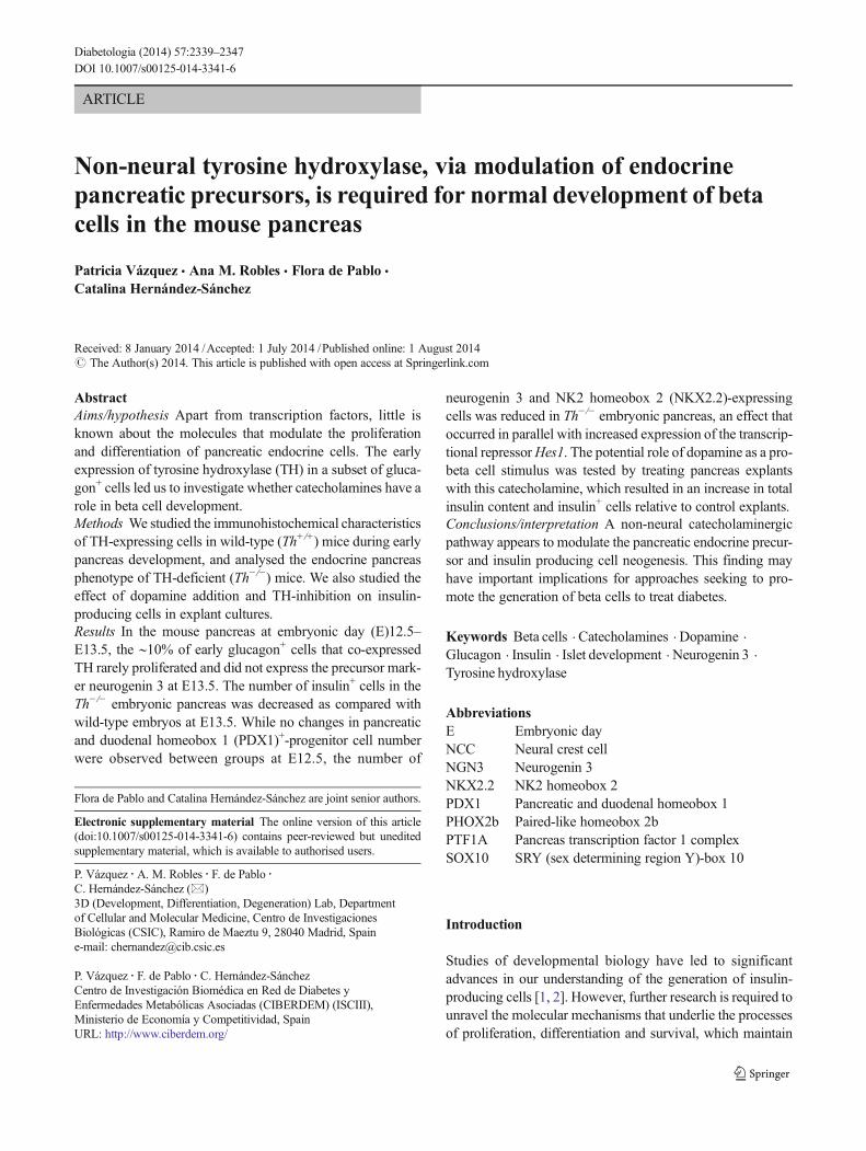
TH is expressed in a subset of early glucagon+ cells in thedeveloping mouse pancreas We first sought to better charac-terise the early TH-expressing cell population. Pancreatic ThmRNA was analysed in samples from E11.5 to E15.5, thelatter being the embryonic stage during which significantsympathetic fibre innervation begins. Levels of Th mRNAwere similar during the primary transition (E11.5–12.5) andthe beginning of the secondary transition (E13.5–14.5), butdecreased significantly at E15.5 (Fig. 1a), coinciding with themaximum expression of Neurog3 and around the peak of betacell differentiation. Immunoblotting of pancreas protein ex-tracts revealed a 60 kDa band corresponding to TH (Fig. 1b).The band specificity was confirmed by the absence of signalin pancreatic extracts from TH-deficient embryos (ESMFig. 1). TH protein levels were similar up to E15.5. Theapparent discrepancy between the mRNA and protein profilesfor TH could be due, at least in part, to the contribution of thesympathetic projections to the total content of TH protein.
Using co-immunofluorescence we further characterised thepopulation of TH-expressing cells. Glucagon-expressing cellsare one of the first pancreatic cells to differentiate. At E12.5(Fig. 1c) and E13.5, most TH-expressing cells (∼90%) co-expressed glucagon, although TH+ cells represented only asmall proportion (∼10%) of these early glucagon+ cells. At
Fig. 1 TH is expressed during early pancreas development in mice. (a)Quantitative real-time PCR of three to five pooled embryonic pancreases(E11.5 to E15.5). Levels of Th transcripts were normalised to 18S rRNAlevels and the data are presented on a logarithmic scale. Results representthe mean±SEM of at least three different pools for each embryonic stage.*p<0.05 vs E11.5. (b) TH immunoblot of protein extracts from fourpooled pancreases (E11.5 to E15.5). Adult adrenal gland (AG) extractwas included as a positive control andβ-actin as a loading control. As THis highly expressed in AG, only low amounts of AG protein were loaded,so AG β-actin was not detected. The blot is representative of threeindependent experiments. (c–g) Embryonic pancreatic sections. (c) Co-immunostaining for PDX1 (green), TH-rabbit (red) and glucagon (GCG,cyan). (d) Double immunostaining for E-cadherin (green) and TH-rabbit(red). (e) Double-immunostaining for BrdU (green) and TH-rabbit (red).Arrows indicate double-positive cells. (f) Double immunostaining forβIII-tubulin (green) and TH-mouse (red). (g) Double-immunostainingfor neurofilament (NF, green) and TH-rabbit (red). White X indicatesblood cells. Scale bar, 50 μm
Diabetologia (2014) 57:2339–2347 2341
later stages of development (E14.5 and E15.5), the degree ofco-expression tended to decrease (ESM Fig. 2a, b).
The TH+ cells shared features with the overall early gluca-gon population: they showed little co-localisation with PDX1[9, 19] (Fig. 1c); they localised in clusters within regions of thepancreatic epithelium with low E-cadherin expression [27](Fig. 1d); and rarely incorporated the mitotic marker BrdU(∼6%; Fig. 1e). These features are characteristics of hormone-expressing differentiated cells. Furthermore, we observed noco-expression of TH and the endocrine precursor markerNGN3 at E13.5 (see below).
TH+ cells did not express βIII-tubulin or neurofilament(neuronal markers) (Fig. 1f, g). They expressed neither SRY(sex determining region Y)-box 10 (SOX10) nor paired-likehomeobox 2b (PHOX2b)—both neural crest cell [NCC]markers (ESM Fig. 3a)—confirming their non-neural pheno-type. Moreover, at later stages of development (E14.5–15.5)when the first sympathetic projections (TH+ and βIII-tubulin+) were detected, the TH+ cell bodies did not expressβIII-tubulin (ESM Fig. 2c, d). Taken together, these resultsdemonstrate the endocrine character of early TH+ cells.
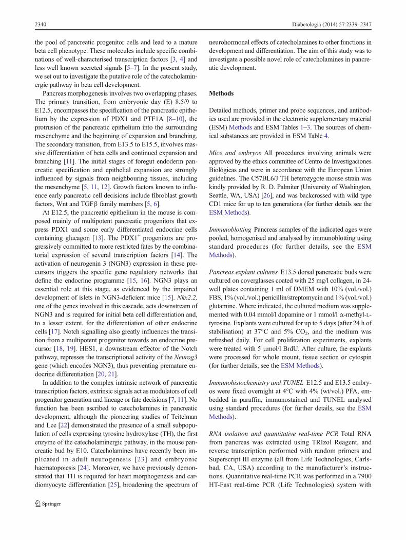
Beta cell number is modulated by the catecholaminergicpathway To date, no clear role has been ascribed to the earlyglucagon+ cells. Indeed, it has been suggested that this cellpopulation disappears during development and does not mea-surably contribute to the adult organ [28, 29]. To assess therole of TH in a subpopulation of the early glucagon+ cellsduring pancreas development we phenotypically characterisedthe pancreas of TH-deficient mice. As embryonic lethalitybegins to occur from E12.5 in Th−/− embryos [26], we studiedE13.5 embryos, the latest embryonic stage at which a signif-icant number of live Th−/− embryos can be collected. Mor-phometric analysis of the E12.5 and 13.5 pancreatic primordiaby E-cadherin immunostaining revealed no significant differ-ences in either the epithelial area or total (epithelium+mesen-chyme) pancreatic area between Th+/+ and Th−/− embryos(data not shown). At the beginning of the secondary transition,insulin-expressing cells begin to accumulate and the popula-tion increases exponentially until E15.5. We investigatedwhether the emerging population of insulin-expressing cellswas affected by the absence of TH expression. The number ofinsulin+ cells in the whole pancreas of E13.5 Th−/− embryoswas decreased relative to that in the whole pancreas of Th+/+
embryos. By contrast, we observed no differences betweengenotypes in the number of glucagon+ cells (Fig. 2a–c).Quantitative real-time PCR analysis confirmed lower expres-sion levels of both insulin genes (Ins1 and Ins2) in the E13.5Th−/− pancreatic primordium, without changes in GcgmRNAlevels (Fig. 2d–f).
To confirm the impact of TH deficiency on the final num-ber of insulin-expressing cells, we established an organotypicpancreas culture in which the mesenchyme was preserved.
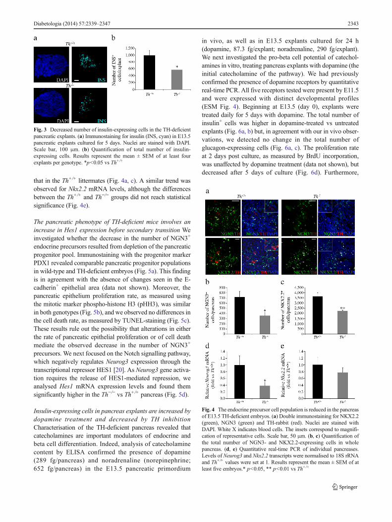
Beginning at E13.5, after 24 h of stabilisation of the culture(day 0), the pancreatic explants were cultured for 5 days. Thetotal number of insulin+ cells in the Th−/− explants at the endof the culture period was decreased compared with that in theTh+/+ explants (Fig. 3a, b).
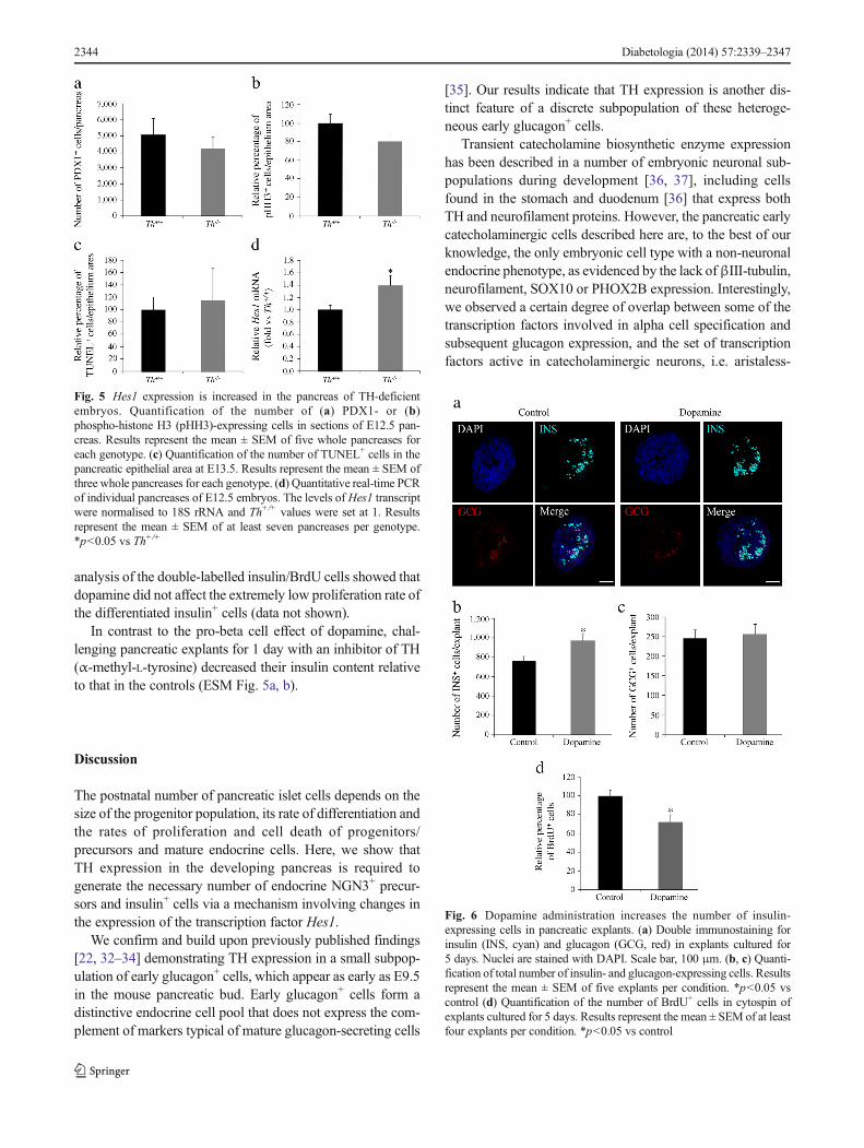
Lineage studies have established that all endocrine cells arederived from NGN3-expressing progenitors [16]. Further-more, the number of NGN3+ precursors during embryonicdevelopment determines the number of endocrine cells at birth[30]. We therefore analysed the NGN3+ cell population. Weobserved a decrease in the number of NGN3-expressing cellsin the E13.5 Th−/− vs Th+/+ pancreas (Fig. 4a, b). In agreementwith this finding, Neurog3mRNA levels in the pancreas werelower in mutant embryos than in Th+/+ littermates (Fig. 4d).NKX2.2 is a transcription factor required for the determina-tion of beta cell fate [17], and its expression is regulated byNGN3 [31]. We found that the number of cells expressingNKX2.2 in the Th−/− pancreas was decreased compared with
Fig. 2 The number of insulin-expressing cells is decreased in the pan-creas of TH-deficient E13.5 embryos. (a) Double immunostaining forinsulin (INS, cyan) and glucagon (GCG, red). Nuclei are stained withDAPI. Arrows andwhite X indicate insulin+ and blood cells, respectively.Scale bar, 50 μm. (b, c) Quantification of the total number of insulin- andglucagon-expressing cells in whole pancreas. (d–f) Quantitative real-timePCR of individual pancreases. Levels of the Ins1, Ins2 and Gcgtranscripts were normalised to 18S rRNA and Th+/+ values were setat 1. Results represent the mean ± SEM of at least five animals.*p<0.05vs Th+/+
2342 Diabetologia (2014) 57:2339–2347
that in the Th+/+ littermates (Fig. 4a, c). A similar trend wasobserved for Nkx2.2 mRNA levels, although the differencesbetween the Th+/+ and Th−/− groups did not reach statisticalsignificance (Fig. 4e).
The pancreatic phenotype of TH-deficient mice involves anincrease in Hes1 expression before secondary transition Weinvestigated whether the decrease in the number of NGN3+
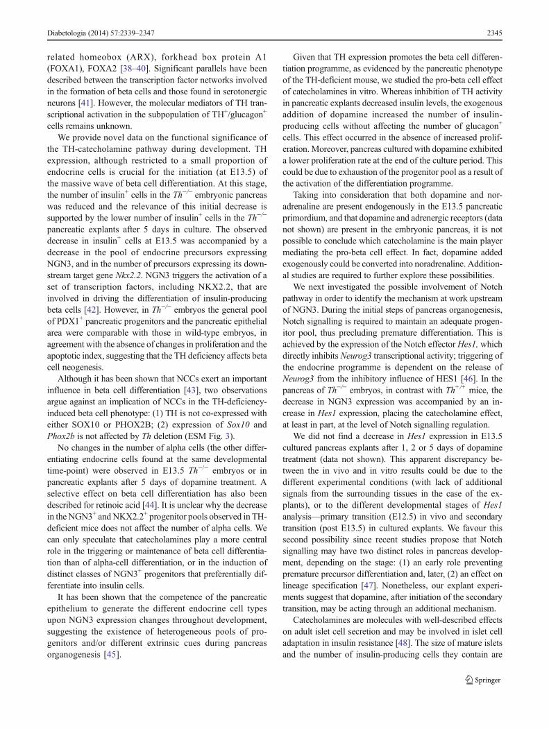
endocrine precursors resulted from depletion of the pancreaticprogenitor pool. Immunostaining with the progenitor markerPDX1 revealed comparable pancreatic progenitor populationsin wild-type and TH-deficient embryos (Fig. 5a). This findingis in agreement with the absence of changes seen in the E-cadherin+ epithelial area (data not shown). Moreover, thepancreatic epithelium proliferation rate, as measured usingthe mitotic marker phospho-histone H3 (pHH3), was similarin both genotypes (Fig. 5b), and we observed no differences inthe cell death rate, as measured by TUNEL-staining (Fig. 5c).These results rule out the possibility that alterations in eitherthe rate of pancreatic epithelial proliferation or of cell deathmediate the observed decrease in the number of NGN3+
precursors. We next focused on the Notch signalling pathway,which negatively regulates Neurog3 expression through thetranscriptional repressor HES1 [20]. As Neurog3 gene activa-tion requires the release of HES1-mediated repression, weanalysed Hes1 mRNA expression levels and found themsignificantly higher in the Th−/− vs Th+/+ pancreas (Fig. 5d).
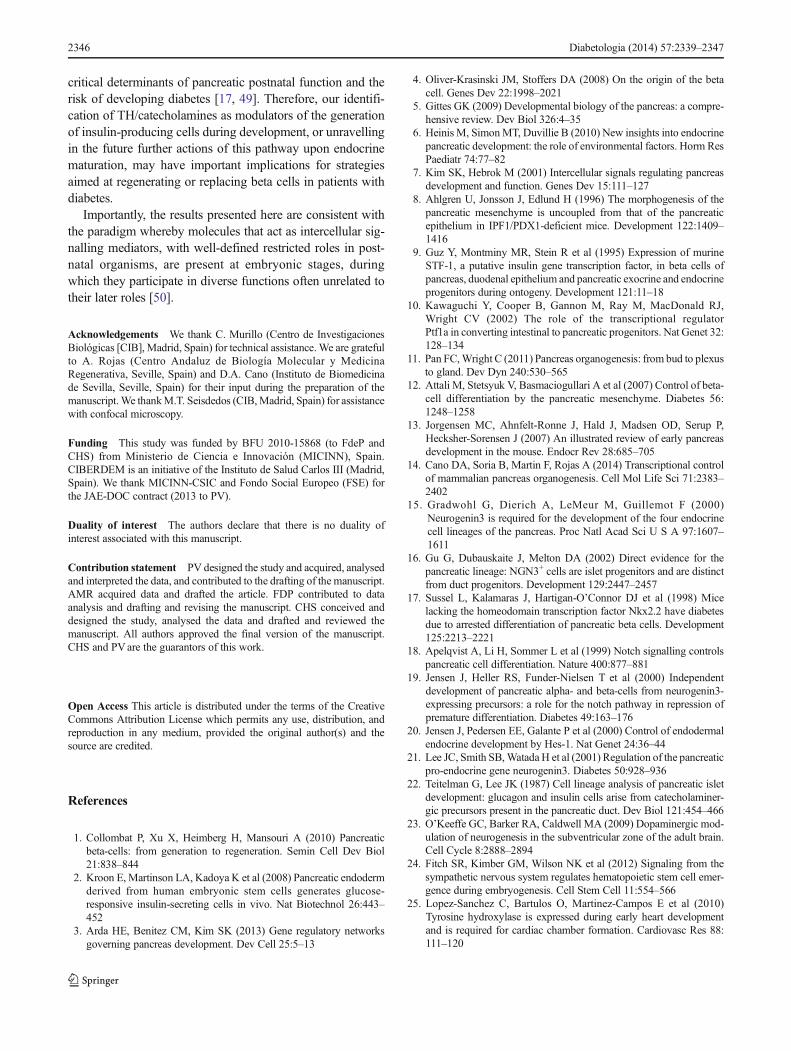
Insulin-expressing cells in pancreas explants are increased bydopamine treatment and decreased by TH inhibitionCharacterisation of the TH-deficient pancreas revealed thatcatecholamines are important modulators of endocrine andbeta cell differentiation. Indeed, analysis of catecholaminecontent by ELISA confirmed the presence of dopamine(289 fg/pancreas) and noradrenaline (norepinephrine;652 fg/pancreas) in the E13.5 pancreatic primordium
in vivo, as well as in E13.5 explants cultured for 24 h(dopamine, 87.3 fg/explant; noradrenaline, 290 fg/explant).We next investigated the pro-beta cell potential of catechol-amines in vitro, treating pancreas explants with dopamine (theinitial catecholamine of the pathway). We had previouslyconfirmed the presence of dopamine receptors by quantitativereal-time PCR. All five receptors tested were present by E11.5and were expressed with distinct developmental profiles(ESM Fig. 4). Beginning at E13.5 (day 0), explants weretreated daily for 5 days with dopamine. The total number ofinsulin+ cells was higher in dopamine-treated vs untreatedexplants (Fig. 6a, b) but, in agreement with our in vivo obser-vations, we detected no change in the total number ofglucagon-expressing cells (Fig. 6a, c). The proliferation rateat 2 days post culture, as measured by BrdU incorporation,was unaffected by dopamine treatment (data not shown), butdecreased after 5 days of culture (Fig. 6d). Furthermore,
Fig. 3 Decreased number of insulin-expressing cells in the TH-deficientpancreatic explants. (a) Immunostaining for insulin (INS, cyan) in E13.5pancreatic explants cultured for 5 days. Nuclei are stained with DAPI.Scale bar, 100 μm. (b) Quantification of total number of insulin-expressing cells. Results represent the mean ± SEM of at least fourexplants per genotype. *p<0.05 vs Th+/+
Fig. 4 The endocrine precursor cell population is reduced in the pancreasof E13.5 TH-deficient embryos. (a) Double immunostaining for NKX2.2(green), NGN3 (green) and TH-rabbit (red). Nuclei are stained withDAPI. White X indicates blood cells. The insets correspond to magnifi-cation of representative cells. Scale bar, 50 μm. (b, c) Quantification ofthe total number of NGN3- and NKX2.2-expressing cells in wholepancreas. (d, e) Quantitative real-time PCR of individual pancreases.Levels of Neurog3 and Nkx2.2 transcripts were normalised to 18S rRNAand Th+/+ values were set at 1. Results represent the mean ± SEM of atleast five embryos.* p<0.05, ** p<0.01 vs Th+/+
Diabetologia (2014) 57:2339–2347 2343
analysis of the double-labelled insulin/BrdU cells showed thatdopamine did not affect the extremely low proliferation rate ofthe differentiated insulin+ cells (data not shown).
In contrast to the pro-beta cell effect of dopamine, chal-lenging pancreatic explants for 1 day with an inhibitor of TH(α-methyl-L-tyrosine) decreased their insulin content relativeto that in the controls (ESM Fig. 5a, b).
Discussion
The postnatal number of pancreatic islet cells depends on thesize of the progenitor population, its rate of differentiation andthe rates of proliferation and cell death of progenitors/precursors and mature endocrine cells. Here, we show thatTH expression in the developing pancreas is required togenerate the necessary number of endocrine NGN3+ precur-sors and insulin+ cells via a mechanism involving changes inthe expression of the transcription factor Hes1.
We confirm and build upon previously published findings[22, 32–34] demonstrating TH expression in a small subpop-ulation of early glucagon+ cells, which appear as early as E9.5in the mouse pancreatic bud. Early glucagon+ cells form adistinctive endocrine cell pool that does not express the com-plement of markers typical of mature glucagon-secreting cells
[35]. Our results indicate that TH expression is another dis-tinct feature of a discrete subpopulation of these heteroge-neous early glucagon+ cells.
Transient catecholamine biosynthetic enzyme expressionhas been described in a number of embryonic neuronal sub-populations during development [36, 37], including cellsfound in the stomach and duodenum [36] that express bothTH and neurofilament proteins. However, the pancreatic earlycatecholaminergic cells described here are, to the best of ourknowledge, the only embryonic cell type with a non-neuronalendocrine phenotype, as evidenced by the lack ofβIII-tubulin,neurofilament, SOX10 or PHOX2B expression. Interestingly,we observed a certain degree of overlap between some of thetranscription factors involved in alpha cell specification andsubsequent glucagon expression, and the set of transcriptionfactors active in catecholaminergic neurons, i.e. aristaless-
Fig. 5 Hes1 expression is increased in the pancreas of TH-deficientembryos. Quantification of the number of (a) PDX1- or (b)phospho-histone H3 (pHH3)-expressing cells in sections of E12.5 pan-creas. Results represent the mean ± SEM of five whole pancreases foreach genotype. (c) Quantification of the number of TUNEL+ cells in thepancreatic epithelial area at E13.5. Results represent the mean ± SEM ofthree whole pancreases for each genotype. (d) Quantitative real-time PCRof individual pancreases of E12.5 embryos. The levels of Hes1 transcriptwere normalised to 18S rRNA and Th+/+ values were set at 1. Resultsrepresent the mean ± SEM of at least seven pancreases per genotype.*p<0.05 vs Th+/+
Fig. 6 Dopamine administration increases the number of insulin-expressing cells in pancreatic explants. (a) Double immunostaining forinsulin (INS, cyan) and glucagon (GCG, red) in explants cultured for5 days. Nuclei are stained with DAPI. Scale bar, 100 μm. (b, c) Quanti-fication of total number of insulin- and glucagon-expressing cells. Resultsrepresent the mean ± SEM of five explants per condition. *p<0.05 vscontrol (d) Quantification of the number of BrdU+ cells in cytospin ofexplants cultured for 5 days. Results represent the mean ± SEM of at leastfour explants per condition. *p<0.05 vs control
2344 Diabetologia (2014) 57:2339–2347
related homeobox (ARX), forkhead box protein A1(FOXA1), FOXA2 [38–40]. Significant parallels have beendescribed between the transcription factor networks involvedin the formation of beta cells and those found in serotonergicneurons [41]. However, the molecular mediators of TH tran-scriptional activation in the subpopulation of TH+/glucagon+
cells remains unknown.We provide novel data on the functional significance of
the TH-catecholamine pathway during development. THexpression, although restricted to a small proportion ofendocrine cells is crucial for the initiation (at E13.5) ofthe massive wave of beta cell differentiation. At this stage,the number of insulin+ cells in the Th−/− embryonic pancreaswas reduced and the relevance of this initial decrease issupported by the lower number of insulin+ cells in the Th−/−
pancreatic explants after 5 days in culture. The observeddecrease in insulin+ cells at E13.5 was accompanied by adecrease in the pool of endocrine precursors expressingNGN3, and in the number of precursors expressing its down-stream target gene Nkx2.2. NGN3 triggers the activation of aset of transcription factors, including NKX2.2, that areinvolved in driving the differentiation of insulin-producingbeta cells [42]. However, in Th−/− embryos the general poolof PDX1+ pancreatic progenitors and the pancreatic epithelialarea were comparable with those in wild-type embryos, inagreement with the absence of changes in proliferation and theapoptotic index, suggesting that the TH deficiency affects betacell neogenesis.
Although it has been shown that NCCs exert an importantinfluence in beta cell differentiation [43], two observationsargue against an implication of NCCs in the TH-deficiency-induced beta cell phenotype: (1) TH is not co-expressed witheither SOX10 or PHOX2B; (2) expression of Sox10 andPhox2b is not affected by Th deletion (ESM Fig. 3).
No changes in the number of alpha cells (the other differ-entiating endocrine cells found at the same developmentaltime-point) were observed in E13.5 Th−/− embryos or inpancreatic explants after 5 days of dopamine treatment. Aselective effect on beta cell differentiation has also beendescribed for retinoic acid [44]. It is unclear why the decreasein the NGN3+ and NKX2.2+ progenitor pools observed in TH-deficient mice does not affect the number of alpha cells. Wecan only speculate that catecholamines play a more centralrole in the triggering or maintenance of beta cell differentia-tion than of alpha-cell differentiation, or in the induction ofdistinct classes of NGN3+ progenitors that preferentially dif-ferentiate into insulin cells.
It has been shown that the competence of the pancreaticepithelium to generate the different endocrine cell typesupon NGN3 expression changes throughout development,suggesting the existence of heterogeneous pools of pro-genitors and/or different extrinsic cues during pancreasorganogenesis [45].
Given that TH expression promotes the beta cell differen-tiation programme, as evidenced by the pancreatic phenotypeof the TH-deficient mouse, we studied the pro-beta cell effectof catecholamines in vitro. Whereas inhibition of TH activityin pancreatic explants decreased insulin levels, the exogenousaddition of dopamine increased the number of insulin-producing cells without affecting the number of glucagon+
cells. This effect occurred in the absence of increased prolif-eration.Moreover, pancreas cultured with dopamine exhibiteda lower proliferation rate at the end of the culture period. Thiscould be due to exhaustion of the progenitor pool as a result ofthe activation of the differentiation programme.
Taking into consideration that both dopamine and nor-adrenaline are present endogenously in the E13.5 pancreaticprimordium, and that dopamine and adrenergic receptors (datanot shown) are present in the embryonic pancreas, it is notpossible to conclude which catecholamine is the main playermediating the pro-beta cell effect. In fact, dopamine addedexogenously could be converted into noradrenaline. Addition-al studies are required to further explore these possibilities.
We next investigated the possible involvement of Notchpathway in order to identify the mechanism at work upstreamof NGN3. During the initial steps of pancreas organogenesis,Notch signalling is required to maintain an adequate progen-itor pool, thus precluding premature differentiation. This isachieved by the expression of the Notch effector Hes1, whichdirectly inhibits Neurog3 transcriptional activity; triggering ofthe endocrine programme is dependent on the release ofNeurog3 from the inhibitory influence of HES1 [46]. In thepancreas of Th−/− embryos, in contrast with Th+/+ mice, thedecrease in NGN3 expression was accompanied by an in-crease in Hes1 expression, placing the catecholamine effect,at least in part, at the level of Notch signalling regulation.
We did not find a decrease in Hes1 expression in E13.5cultured pancreas explants after 1, 2 or 5 days of dopaminetreatment (data not shown). This apparent discrepancy be-tween the in vivo and in vitro results could be due to thedifferent experimental conditions (with lack of additionalsignals from the surrounding tissues in the case of the ex-plants), or to the different developmental stages of Hes1analysis—primary transition (E12.5) in vivo and secondarytransition (post E13.5) in cultured explants. We favour thissecond possibility since recent studies propose that Notchsignalling may have two distinct roles in pancreas develop-ment, depending on the stage: (1) an early role preventingpremature precursor differentiation and, later, (2) an effect onlineage specification [47]. Nonetheless, our explant experi-ments suggest that dopamine, after initiation of the secondarytransition, may be acting through an additional mechanism.
Catecholamines are molecules with well-described effectson adult islet cell secretion and may be involved in islet celladaptation in insulin resistance [48]. The size of mature isletsand the number of insulin-producing cells they contain are
Diabetologia (2014) 57:2339–2347 2345
critical determinants of pancreatic postnatal function and therisk of developing diabetes [17, 49]. Therefore, our identifi-cation of TH/catecholamines as modulators of the generationof insulin-producing cells during development, or unravellingin the future further actions of this pathway upon endocrinematuration, may have important implications for strategiesaimed at regenerating or replacing beta cells in patients withdiabetes.
Importantly, the results presented here are consistent withthe paradigm whereby molecules that act as intercellular sig-nalling mediators, with well-defined restricted roles in post-natal organisms, are present at embryonic stages, duringwhich they participate in diverse functions often unrelated totheir later roles [50].
Acknowledgements We thank C. Murillo (Centro de InvestigacionesBiológicas [CIB], Madrid, Spain) for technical assistance.We are gratefulto A. Rojas (Centro Andaluz de Biología Molecular y MedicinaRegenerativa, Seville, Spain) and D.A. Cano (Instituto de Biomedicinade Sevilla, Seville, Spain) for their input during the preparation of themanuscript.We thankM.T. Seisdedos (CIB,Madrid, Spain) for assistancewith confocal microscopy.
Funding This study was funded by BFU 2010-15868 (to FdeP andCHS) from Ministerio de Ciencia e Innovación (MICINN), Spain.CIBERDEM is an initiative of the Instituto de Salud Carlos III (Madrid,Spain). We thank MICINN-CSIC and Fondo Social Europeo (FSE) forthe JAE-DOC contract (2013 to PV).
Duality of interest The authors declare that there is no duality ofinterest associated with this manuscript.
Contribution statement PV designed the study and acquired, analysedand interpreted the data, and contributed to the drafting of the manuscript.AMR acquired data and drafted the article. FDP contributed to dataanalysis and drafting and revising the manuscript. CHS conceived anddesigned the study, analysed the data and drafted and reviewed themanuscript. All authors approved the final version of the manuscript.CHS and PVare the guarantors of this work.
Open Access This article is distributed under the terms of the CreativeCommons Attribution License which permits any use, distribution, andreproduction in any medium, provided the original author(s) and thesource are credited.
References
1. Collombat P, Xu X, Heimberg H, Mansouri A (2010) Pancreaticbeta-cells: from generation to regeneration. Semin Cell Dev Biol21:838–844
2. Kroon E, Martinson LA, Kadoya K et al (2008) Pancreatic endodermderived from human embryonic stem cells generates glucose-responsive insulin-secreting cells in vivo. Nat Biotechnol 26:443–452
3. Arda HE, Benitez CM, Kim SK (2013) Gene regulatory networksgoverning pancreas development. Dev Cell 25:5–13
4. Oliver-Krasinski JM, Stoffers DA (2008) On the origin of the betacell. Genes Dev 22:1998–2021
5. Gittes GK (2009) Developmental biology of the pancreas: a compre-hensive review. Dev Biol 326:4–35
6. Heinis M, SimonMT, Duvillie B (2010) New insights into endocrinepancreatic development: the role of environmental factors. Horm ResPaediatr 74:77–82
7. Kim SK, Hebrok M (2001) Intercellular signals regulating pancreasdevelopment and function. Genes Dev 15:111–127
8. Ahlgren U, Jonsson J, Edlund H (1996) The morphogenesis of thepancreatic mesenchyme is uncoupled from that of the pancreaticepithelium in IPF1/PDX1-deficient mice. Development 122:1409–1416
9. Guz Y, Montminy MR, Stein R et al (1995) Expression of murineSTF-1, a putative insulin gene transcription factor, in beta cells ofpancreas, duodenal epithelium and pancreatic exocrine and endocrineprogenitors during ontogeny. Development 121:11–18
10. Kawaguchi Y, Cooper B, Gannon M, Ray M, MacDonald RJ,Wright CV (2002) The role of the transcriptional regulatorPtf1a in converting intestinal to pancreatic progenitors. Nat Genet 32:128–134
11. Pan FC,Wright C (2011) Pancreas organogenesis: from bud to plexusto gland. Dev Dyn 240:530–565
12. Attali M, Stetsyuk V, Basmaciogullari A et al (2007) Control of beta-cell differentiation by the pancreatic mesenchyme. Diabetes 56:1248–1258
13. Jorgensen MC, Ahnfelt-Ronne J, Hald J, Madsen OD, Serup P,Hecksher-Sorensen J (2007) An illustrated review of early pancreasdevelopment in the mouse. Endocr Rev 28:685–705
14. Cano DA, Soria B, Martin F, Rojas A (2014) Transcriptional controlof mammalian pancreas organogenesis. Cell Mol Life Sci 71:2383–2402
15. Gradwohl G, Dierich A, LeMeur M, Guillemot F (2000)Neurogenin3 is required for the development of the four endocrinecell lineages of the pancreas. Proc Natl Acad Sci U S A 97:1607–1611
16. Gu G, Dubauskaite J, Melton DA (2002) Direct evidence for thepancreatic lineage: NGN3+ cells are islet progenitors and are distinctfrom duct progenitors. Development 129:2447–2457
17. Sussel L, Kalamaras J, Hartigan-O’Connor DJ et al (1998) Micelacking the homeodomain transcription factor Nkx2.2 have diabetesdue to arrested differentiation of pancreatic beta cells. Development125:2213–2221
18. Apelqvist A, Li H, Sommer L et al (1999) Notch signalling controlspancreatic cell differentiation. Nature 400:877–881
19. Jensen J, Heller RS, Funder-Nielsen T et al (2000) Independentdevelopment of pancreatic alpha- and beta-cells from neurogenin3-expressing precursors: a role for the notch pathway in repression ofpremature differentiation. Diabetes 49:163–176
20. Jensen J, Pedersen EE, Galante P et al (2000) Control of endodermalendocrine development by Hes-1. Nat Genet 24:36–44
21. Lee JC, Smith SB,WatadaH et al (2001) Regulation of the pancreaticpro-endocrine gene neurogenin3. Diabetes 50:928–936
22. Teitelman G, Lee JK (1987) Cell lineage analysis of pancreatic isletdevelopment: glucagon and insulin cells arise from catecholaminer-gic precursors present in the pancreatic duct. Dev Biol 121:454–466
23. O’Keeffe GC, Barker RA, Caldwell MA (2009) Dopaminergic mod-ulation of neurogenesis in the subventricular zone of the adult brain.Cell Cycle 8:2888–2894
24. Fitch SR, Kimber GM, Wilson NK et al (2012) Signaling from thesympathetic nervous system regulates hematopoietic stem cell emer-gence during embryogenesis. Cell Stem Cell 11:554–566
25. Lopez-Sanchez C, Bartulos O, Martinez-Campos E et al (2010)Tyrosine hydroxylase is expressed during early heart developmentand is required for cardiac chamber formation. Cardiovasc Res 88:111–120
2346 Diabetologia (2014) 57:2339–2347
26. Zhou QY, Quaife CJ, Palmiter RD (1995) Targeted disruption of thetyrosine hydroxylase gene reveals that catecholamines are requiredfor mouse fetal development. Nature 374:640–643
27. Gouzi M, Kim YH, Katsumoto K, Johansson K, Grapin-Botton A(2011) Neurogenin3 initiates stepwise delamination of differentiatingendocrine cells during pancreas development. Dev Dyn 240:589–604
28. Herrera PL (2000) Adult insulin- and glucagon-producing cells dif-ferentiate from two independent cell lineages. Development 127:2317–2322
29. Herrera PL, Huarte J, Zufferey R et al (1994) Ablation of isletendocrine cells by targeted expression of hormone-promoter-driventoxigenes. Proc Natl Acad Sci U S A 91:12999–13003
30. Desgraz R, Herrera PL (2009) Pancreatic neurogenin 3-expressingcells are unipotent islet precursors. Development 136:3567–3574
31. Watada H, Scheel DW, Leung J, German MS (2003) Distinct geneexpression programs function in progenitor and mature islet cells.J Biol Chem 278:17130–17140
32. Teitelman G, Joh TH, Reis DJ (1981) Linkage of the brain-skin-gut axis: islet cells originate from dopaminergic precursors.Peptides 2(Suppl 2):157–168
33. Teitelman G, Joh TH, Reis DJ (1981) Transformation of catechol-aminergic precursors into glucagon (A) cells in mouse embryonicpancreas. Proc Natl Acad Sci U S A 78:5225–5229
34. Teitelman G, Alpert S, Polak JM, Martinez A, Hanahan D (1993)Precursor cells of mouse endocrine pancreas coexpress insulin, glu-cagon and the neuronal proteins tyrosine hydroxylase and neuropep-tide Y, but not pancreatic polypeptide. Development 118:1031–1039
35. WilsonME, Kalamaras JA, GermanMS (2002) Expression pattern ofIAPP and prohormone convertase 1/3 reveals a distinctive set ofendocrine cells in the embryonic pancreas. Mech Dev 115:171–176
36. Baetge G, Gershon MD (1989) Transient catecholaminergic (TC)cells in the vagus nerves and bowel of fetal mice: relationship to thedevelopment of enteric neurons. Dev Biol 132:189–211
37. Jonakait GM, Markey KA, Goldstein M, Black IB (1984) Transientexpression of selected catecholaminergic traits in cranial sensory anddorsal root ganglia of the embryonic rat. Dev Biol 101:51–60
38. Bramswig NC, Kaestner KH (2011) Transcriptional regulation ofalpha-cell differentiation. Diabetes Obes Metab 13(Suppl 1):13–20
39. Filippi A, Jainok C, Driever W (2012) Analysis of transcriptionalcodes for zebrafish dopaminergic neurons reveals essential functionsof Arx and Isl1 in prethalamic dopaminergic neuron development.Dev Biol 369:133–149
40. Stott SR, Metzakopian E, Lin W, Kaestner KH, Hen R, Ang SL(2013) Foxa1 and foxa2 are required for the maintenance of dopa-minergic properties in ventral midbrain neurons at late embryonicstages. J Neurosci 33:8022–8034
41. Ohta Y, Kosaka Y, Kishimoto N et al (2011) Convergence of theinsulin and serotonin programs in the pancreatic beta-cell. Diabetes60:3208–3216
42. Rukstalis JM, Habener JF (2009) Neurogenin3: a master regulator ofpancreatic islet differentiation and regeneration. Islets 1:177–184
43. Nekrep N, Wang J, Miyatsuka T, German MS (2008) Signals fromthe neural crest regulate beta-cell mass in the pancreas. Development135:2151–2160
44. Ostrom M, Loffler KA, Edfalk S et al (2008) Retinoic acidpromotes the generation of pancreatic endocrine progenitorcells and their further differentiation into beta-cells. PLoSOne 3:e2841
45. Johansson KA, Dursun U, Jordan N et al (2007) Temporal control ofneurogenin3 activity in pancreas progenitors reveals competencewindows for the generation of different endocrine cell types.Dev Cell 12:457–465
46. Shih HP, Kopp JL, Sandhu M et al (2012) A Notch-dependentmolecular circuitry initiates pancreatic endocrine and ductal celldifferentiation. Development 139:2488–2499
47. Afelik S, Qu X, Hasrouni E et al (2012) Notch-mediated patterningand cell fate allocation of pancreatic progenitor cells. Development139:1744–1753
48. Ahren B (2000) Autonomic regulation of islet hormone secretion—implications for health and disease. Diabetologia 43:393–410
49. Costes S, Langen R, Gurlo T, Matveyenko AV, Butler PC (2013) β-Cell failure in type 2 diabetes: a case of asking too much of too few?Diabetes 62:327–335
50. Hernandez-Sanchez C, Mansilla A, de la Rosa EJ, de Pablo F (2006)Proinsulin in development: new roles for an ancient prohormone.Diabetologia 49:1142–1150
Diabetologia (2014) 57:2339–2347 2347