Biochemical and in silico evaluation of recombinant E...
12
240 http://journals.tubitak.gov.tr/biology/ Turkish Journal of Biology Turk J Biol (2018) 42: 240-249 © TÜBİTAK doi:10.3906/biy-1801-83 Biochemical and in silico evaluation of recombinant E. coli aminopeptidase and in vitro processed human interferon α-2b Amina ARIF 1 , Kaleemuddin MOHAMMED 2 , Muhammad Shahid NADEEM 2, * 1 Faculty of Life Sciences, University of Central Punjab, Lahore, Pakistan 2 Department of Biochemistry, Faculty of Science, King Abdulaziz University, Jeddah, Saudi Arabia * Correspondence: [email protected] 1. Introduction e production of recombinant proteins and their therapeutic applications have brought a revolution in biomedical sciences. Recombinants of many proteins including lymphokines, interferons and interleukins (Rider et al., 2016; Shepelkova et al., 2016; Lagasse et al., 2017), enzymes (Seal, 2013; Vairo et al., 2013; Dolores and Ortiz, 2016), and hormones like insulin and human growth hormone (Urbano et al., 2012; Miljic et al., 2013) have been used in recent times. In the E. coli expression system, a proportion of recombinant proteins retain an unprocessed methionine at the N-terminus, which is contrary to their native forms. Attempts have been made to make replicas of native proteins for therapeutic applications (Goh et al., 2017; Wingfield, 2017). Methionine aminopeptidase (MAP) is a metalloprotease that catalyzes the hydrolytic removal of N-terminal methionine of an elongating polypeptide chain during the process of protein synthesis (Giglione et al., 2004; Arif et al., 2015). e hydrolysis of N-terminal methionine is mandatory in 50%–70% of nascent proteins (Bingel-Erlenmeyer et al., 2008; Kanodia et al., 2011) and usually occurs only when the penultimate amino acid, which is adjacent to the initiator methionine, has a radius of gyration of 1.29 Å or less. ese amino acids are glycine, alanine, proline, serine, cysteine, threonine, and valine (Xiao et al., 2010). e methionine aminopeptidases belonging to different origins have similar active sites and mechanism of action but differ in individual active site residues, which may possibly alter the behavior of the enzyme towards different substrates and inhibitors (Lowther et al., 1999; Helgren et al., 2016). Recombinant therapeutic proteins are converted to their native form by removal of N-terminal methionine residue (Arif et al., 2015; Calcagno and Klein, 2016). Because of its easy manipulation, cultivation, high yield, and better economy, the E. coli strains include the most extensively used protein expression systems (Elleuche et al., 2015; Cantu-Bustos et al, 2016; Chen et al., 2016; Jia and Jeon, 2016). Once produced in E. coli, activity of aminopeptidase is one of many decisive factors for a recombinant protein to exhibit a replica of its native form (Tripathi, 2016). In silico 3-D structure determination and molecular docking techniques are increasingly used tools and techniques to study the binding specificity of hormones Abstract: Escherichia coli is an extensively used host for the production of recombinant proteins, making its N-terminal methionine aminopeptidase (MAP) an attractive candidate for studies on posttranslational protein processing. e present study describes the recombinant production and properties of MAP from the DH5α strain of E. coli. e soluble and active enzyme was produced in E. coli BL21 (DE3) RIL - codon plus cells under a T7 promoter system and purified by anion-exchange chromatography. It exhibited a molecular weight of 29,200.94 Da by MALDI-TOF analysis. e purified enzyme showed specific activity of 1.64 U/mg with methionyl- p-nitroanilide and 1.51 U/mg with synthetic tetrapeptide substrate ‘MGMM’ in a discontinuous HPLC-based assay. In vitro studies showed the processing of up to 36% of Met-INFα-2b in 40 min. In silico studies revealed that the ES-complex formation between the enzyme and interferon has a ΔG –683.07 kJ/mol. Molecular docking results showed that the processed INFα-2b has greater binding affinity with IFNAR2 receptor as indicated by ΔG –784.53 kJ/mol, significantly lower than that of methionine containing INFα-2b (ΔG –717.63 kJ/mol). ese findings emphasize the functional superiority or better efficacy of N-terminal methionine processed recombinant interferon. Key words: Methionine aminopeptidase, E. coli, human interferon, in vitro, in silico Received: 31.01.2018 Accepted/Published Online: 02.04.2018 Final Version: 13.06.2018 Research Article
Transcript of Biochemical and in silico evaluation of recombinant E...
240
http://journals.tubitak.gov.tr/biology/
Turkish Journal of Biology Turk J Biol(2018) 42: 240-249© TÜBİTAKdoi:10.3906/biy-1801-83
Biochemical and in silico evaluation of recombinant E. coli aminopeptidase and in vitro processed human interferon α-2b
Amina ARIF1, Kaleemuddin MOHAMMED2, Muhammad Shahid NADEEM2,*1Faculty of Life Sciences, University of Central Punjab, Lahore, Pakistan
2Department of Biochemistry, Faculty of Science, King Abdulaziz University, Jeddah, Saudi Arabia
* Correspondence: [email protected]
1. IntroductionThe production of recombinant proteins and their therapeutic applications have brought a revolution in biomedical sciences. Recombinants of many proteins including lymphokines, interferons and interleukins (Rider et al., 2016; Shepelkova et al., 2016; Lagasse et al., 2017), enzymes (Seal, 2013; Vairo et al., 2013; Dolores and Ortiz, 2016), and hormones like insulin and human growth hormone (Urbano et al., 2012; Miljic et al., 2013) have been used in recent times. In the E. coli expression system, a proportion of recombinant proteins retain an unprocessed methionine at the N-terminus, which is contrary to their native forms. Attempts have been made to make replicas of native proteins for therapeutic applications (Goh et al., 2017; Wingfield, 2017). Methionine aminopeptidase (MAP) is a metalloprotease that catalyzes the hydrolytic removal of N-terminal methionine of an elongating polypeptide chain during the process of protein synthesis (Giglione et al., 2004; Arif et al., 2015). The hydrolysis of N-terminal methionine is mandatory in 50%–70% of nascent proteins (Bingel-Erlenmeyer et al., 2008; Kanodia et al., 2011) and usually occurs only when the penultimate
amino acid, which is adjacent to the initiator methionine, has a radius of gyration of 1.29 Å or less. These amino acids are glycine, alanine, proline, serine, cysteine, threonine, and valine (Xiao et al., 2010). The methionine aminopeptidases belonging to different origins have similar active sites and mechanism of action but differ in individual active site residues, which may possibly alter the behavior of the enzyme towards different substrates and inhibitors (Lowther et al., 1999; Helgren et al., 2016). Recombinant therapeutic proteins are converted to their native form by removal of N-terminal methionine residue (Arif et al., 2015; Calcagno and Klein, 2016). Because of its easy manipulation, cultivation, high yield, and better economy, the E. coli strains include the most extensively used protein expression systems (Elleuche et al., 2015; Cantu-Bustos et al, 2016; Chen et al., 2016; Jia and Jeon, 2016). Once produced in E. coli, activity of aminopeptidase is one of many decisive factors for a recombinant protein to exhibit a replica of its native form (Tripathi, 2016).
In silico 3-D structure determination and molecular docking techniques are increasingly used tools and techniques to study the binding specificity of hormones
Abstract: Escherichia coli is an extensively used host for the production of recombinant proteins, making its N-terminal methionine aminopeptidase (MAP) an attractive candidate for studies on posttranslational protein processing. The present study describes the recombinant production and properties of MAP from the DH5α strain of E. coli. The soluble and active enzyme was produced in E. coli BL21 (DE3) RIL - codon plus cells under a T7 promoter system and purified by anion-exchange chromatography. It exhibited a molecular weight of 29,200.94 Da by MALDI-TOF analysis. The purified enzyme showed specific activity of 1.64 U/mg with methionyl-p-nitroanilide and 1.51 U/mg with synthetic tetrapeptide substrate ‘MGMM’ in a discontinuous HPLC-based assay. In vitro studies showed the processing of up to 36% of Met-INFα-2b in 40 min. In silico studies revealed that the ES-complex formation between the enzyme and interferon has a ΔG –683.07 kJ/mol. Molecular docking results showed that the processed INFα-2b has greater binding affinity with IFNAR2 receptor as indicated by ΔG –784.53 kJ/mol, significantly lower than that of methionine containing INFα-2b (ΔG –717.63 kJ/mol). These findings emphasize the functional superiority or better efficacy of N-terminal methionine processed recombinant interferon.
Key words: Methionine aminopeptidase, E. coli, human interferon, in vitro, in silico
Received: 31.01.2018 Accepted/Published Online: 02.04.2018 Final Version: 13.06.2018
Research Article
ARIF et al. / Turk J Biol
241
and substrates against their corresponding receptors and enzymes (Venkatachalam et al., 2003). During the last two decades, about 60 docking software programs and tools have been developed for commercial and academic applications (Allen et al., 2015; Yang and Zhang, 2015). The present study describes the recombinant production, purification, and biochemical properties of E. coli N-terminal methionine aminopeptidase, and application of enzyme in the processing of recombinant interferon. We have also applied in silico tools to evaluate the interaction of met-INF with E. coli MAP, binding free energy changes of met-INF, and nonmet-INF molecules with the interferon receptors.
2. Materials and methods2.1. Preparation of recombinant plasmidThe E. coli (DH5α) methionine aminopeptidase gene was PCR amplified, using primer sequences 5’-catatggctatctcaatcaagacccc-3’ and 5’-ttattcgtcgtgcgagattatcgcc-3’ under optimized conditions (2 mM MgCl2, 1.5 mM dNTPs, Taq buffer solution, 20 pM of each primer, 2.5 units Taq polymerase). The complete open reading frame encoding methionine aminopeptidase (MAP) was ligated into pTZ57R/T (Thermo Fisher Scientific catalogue number: K1213) by T/A cloning. The gene consisting of 795 base pairs was restricted out from the plasmid above by NdeI and EcoRI and ligated into pET21a (+) expression plasmid (Novagen catalogue number: 69770-3) using the procedure described by our previous studies with modifications (Arif et al., 2016). 2.2. Cloning and expression of the MAP geneThe recombinant plasmid pET21-MAP (expression plasmid ligated with gene encoding enzyme) was used for the transformation of E. coli BL21 (DE3) - RIL codon plus cells (Agilent Technologies catalogue number 230240). Clones confirmed for the presence of recombinant plasmid were used to grow the culture on a small scale; gene expression was induced by 0.4 mM IPTG when the OD of culture was 0.6 at 600 nm. The culture was incubated overnight at 20 °C and expression was analyzed on 15% SDS-PAGE (Laemmli, 1970). 2.3. Purification and molecular weight determinationThe cell pellet (4.3 g) obtained from 1 L of E. coli culture induced for protein expression was suspended in 50 mL of 50 mM phosphate buffer pH 7.5 and sonicated for 1 min (6 pulses of 10 s at moderate power). The sonicated sample was centrifuged at 14,000 × g for 15 min at 4 °C and the supernatant was transferred to a sterile falcon tube followed by filtration using a 0.45 µm pore size filter (Millipore Inc.). The enzyme was purified by dialysis and anion-exchange chromatography using Q-Sepharose equilibrated with 40 mM HEPES buffer pH 7.5. The sample was applied to the column at a flow rate of 2 mL/min, unbound proteins were
washed with the equilibration buffer, and bound proteins were eluted with a linear gradient of 0.5 M NaCl solution. Fractions of 5 mL were collected, quantified, and analyzed separately. The entire purification process was conducted at 4 °C and protein was quantified by Bradford’s method (Bradford, 1976). A desalted purified enzyme sample (2.7 mg) was mixed with 4.5 mg of sinapinic acid in 1 mL of 30% acetonitrile containing 0.15% trifluroacetic acid, and a small aliquot of the mixture was spotted on a mass spectrometric plate and allowed to air dry. The mass spectrum of the enzyme was recorded with a Bruker Autoflex MALDI-TOF (Bruker Daltonics Inc., Billerica, MA, USA).2.4. Methionyl p-nitroanilide-based assayA modified method adopted from the literature was applied for the enzyme assay (Zhou et al., 2000). Methionine aminopeptidase catalyzes the conversion of methionyl p-nitroanilide to p-nitroaniline and methionine. p-Nitroaniline absorbs light at 405 nm. To the experimental and reference cuvette were added 1440 µL of 40 mM HEPES buffer pH 7.5, 400 µL of 1 mM KCl, 60 µL of 50 mM CoCl2, and 3 µL of substrate (100 mM methionine p-nitroanilide in ethanol). To the experimental cuvette was added 75 µL (97.5 µg) of enzyme and to the reference cuvette 75 µL of buffer. The change in absorbance at 405 nm was measured over 30 min. The extinction coefficient of absorbing species (p-nitroaniline) was considered as 9.96 mM–1 cm–1 for the calculation of enzyme activity. The assay was performed for the determination of optimum pH and temperature for enzyme. 2.5. HPLC-based enzyme assay using MGMM as substrateThe assay method described by Xiao et al. (2010) was applied with modifications to measure the enzyme activity. The retention time and volume for the synthetic tetrapeptide MGMM and its product glycine-methionine-methionine GMM was determined. Solutions containing 10 µg of MGMM and GMM were separately taken in Eppendorf tubes, diluted to 20 µL with filtered distilled water, and loaded on an analytical C8 chromatographic column. The retention volume was determined with 1.0 mL/min flow rate, 0% to 30% linear gradient of acetonitrile in 0.1% TFA water with 1% ramp per min. The tri- and tetrapeptide eluted at 16% and 23% of acetonitrile, respectively. The retention time of both MGMM and GMM provided a baseline for the enzyme assays. For the enzyme assay, 175 mM KCl, 50 mM CoCl2, 20 µL of enzyme 1.3 µg/µL, and 25 mM HEPES buffer 60 µL were taken and incubated at 30 °C for 10 min so that the reaction mixture attained the temperature (solution A). In another Eppendorf tube, 1.9 mM substrate and 92 µL of 25 mM HEPES pH 7.5 were added (solution B). After 10 min of enzyme incubation with the KCl and CoCl2 solution B was added to solution A
ARIF et al. / Turk J Biol
242
at 30 °C. The reaction mixture (66 µL) was removed at time zero and added to an Eppendorf tube containing 5 µL of 10% TFA to quench the reaction. Similarly, the reaction of 66 µL of sample was quenched after 5 and 10 min intervals. The samples were separately loaded on the chromatographic C8 column and profile of substrate to product was observed under the optimized conditions. One unit was defined as the amount of enzyme required to eliminate N-terminal methionine from 1 µmol of tetrapeptide MGMM in 1 min. 2.6. Processing of N-terminal methionine from Met-INFα-2bThe recombinant interferon alpha 2b, with N-terminal methionine was obtained from our previous studies (Arif et al., 2015). In an Eppendorf tube (A), 400 µL of reaction mixture containing 175 mM KCl, 50 mM CoCl2, 200 µg of enzyme, and 25 mM HEPES buffer pH 7.5 were incubated at 37 °C for 10 min. In another Eppendorf tube (B) 200 µg of Met-INFα-2b prepared in 25 mM HEPES buffer pH 7.5 was added to make 400 µL reaction. From tube A, 80 µL of solution was transferred into another Eppendorf tube containing 10 µL of 10% TFA; then 20 µL of solution from tube B was added and kept at –20 °C until analysis. To the remaining solution A, all the solution B was added and then 100 µL of the reaction mixture was taken at a time at an interval of 10 min, quenched. Timewise change in the HPLC elution peaks was monitored, and the product formation and substrate consumption were calculated (Arif et al., 2015).2.7. Protein 3D modelling and validationThe Met-IFNα-2b structure was built using the I-TASSER server (Lovell et al., 2002), which develops 3D models from structure templates identified by LOMETS from the PDB library. The templates of the highest significance in the threading alignments were selected by I-TASSER. Subsequently, the quality of the predicted model was examined by RAMPAGE, a protein structure validation server, which reveals the results in terms of phi, psi, and cBeta deviations by generating a Ramachandran plot for the protein built (Lovell et al., 2002). 2.8. Molecular docking and free energy calculation3D structures of IFNα-2b with and without N-terminal methionine and MAP were retrieved from the protein structure database identified as 2KZ1 (Lowther et al., 1999), 1N6V (Chill et al., 2003), and 1C21 (Nudelman et al., 2010), respectively. I-TASSER-made ‘IFNα-2b with methionine’ and ‘without methionine’ were structurally aligned by PyMOL for structural similarity analysis. Later, both ligands were docked against the receptor (IFNΑR2) using the Hex docking server (Macindoe et al., 2010). Additionally, IFNα-2b without methionine was also docked with MAP and the DG (free energy change) of each docked protein–ligand complex was recorded to analyze the complexes.
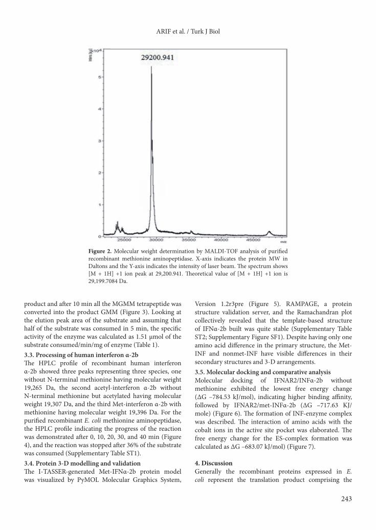
3. Results3.1. Production of recombinant E. coli methionine aminopeptidaseMaximum expression of recombinant enzyme in the soluble and functional form was obtained when the host bacterial cells were induced overnight under 0.4 mM IPTG at 20 °C and 100 rpm. Enzyme was purified by anion exchange chromatography; major enzyme fractions eluted from the Q-Sepharose Column between 0.15 and 0.25 M NaCl were analyzed on SDS-PAGE (Figure 1). The molecular weight determined by MALDI-TOF was 29200.941 Da (Figure 2). The purified enzyme has optimum pH 7.5 and maximum activity was measured at 37 °C in the presence of 2 mM cobalt ions. 3.2. Enzyme activity with different substratesThe specific activity of enzyme with methionyl-p-nitroanilide was 1.64 µmol/min/mg. At time 0, the HPLC profile indicated a single peak for the substrate elution. After 5 min, half of the substrate was converted to the
Figure 1. SDS-PAGE photograph indicating the expressed and purified E. coli methionine aminopeptidase. Lane M - Protein Marker (Thermo Fisher catalogue No. SM 0671), Lane 1- pET21a containing induced cells, Lane 2 - pET21a-MAP containing induced cell lysate. Lane 3 - Purified enzyme.
ARIF et al. / Turk J Biol
243
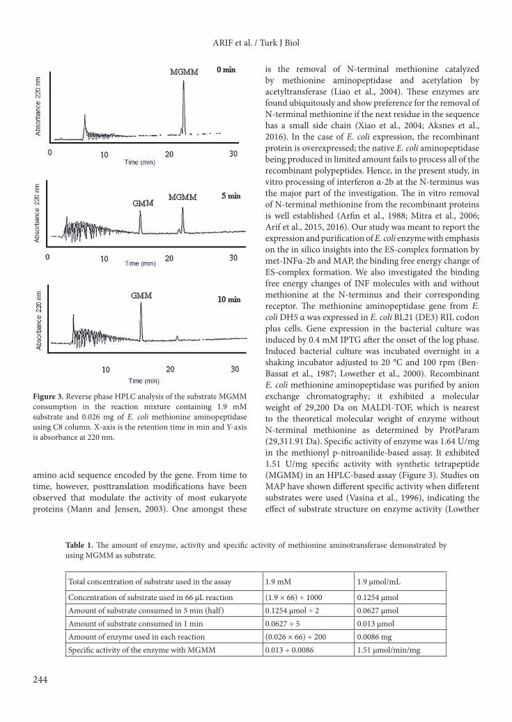
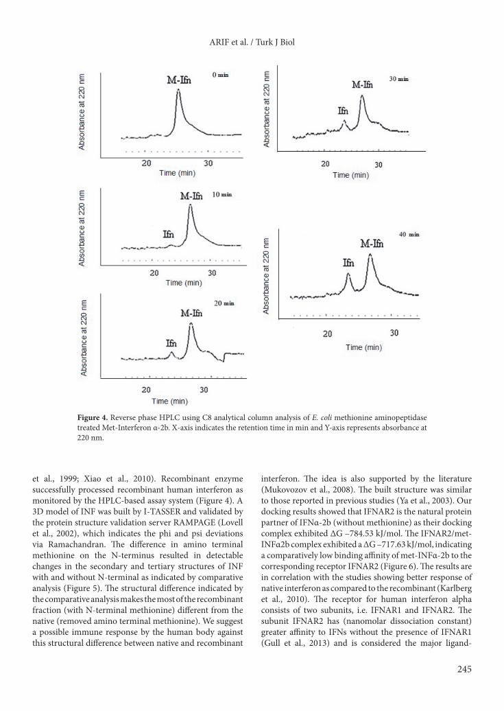
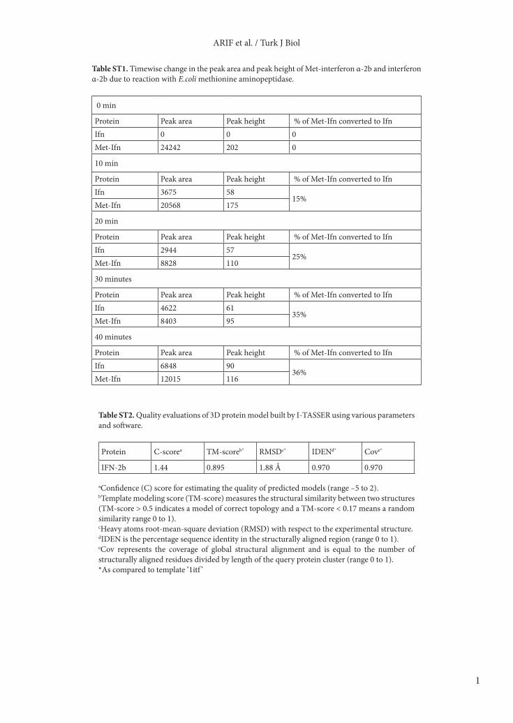
product and after 10 min all the MGMM tetrapeptide was converted into the product GMM (Figure 3). Looking at the elution peak area of the substrate and assuming that half of the substrate was consumed in 5 min, the specific activity of the enzyme was calculated as 1.51 µmol of the substrate consumed/min/mg of enzyme (Table 1).3.3. Processing of human interferon α-2bThe HPLC profile of recombinant human interferon α-2b showed three peaks representing three species, one without N-terminal methionine having molecular weight 19,265 Da, the second acetyl-interferon α-2b without N-terminal methionine but acetylated having molecular weight 19,307 Da, and the third Met-interferon α-2b with methionine having molecular weight 19,396 Da. For the purified recombinant E. coli methionine aminopeptidase, the HPLC profile indicating the progress of the reaction was demonstrated after 0, 10, 20, 30, and 40 min (Figure 4), and the reaction was stopped after 36% of the substrate was consumed (Supplementary Table ST1). 3.4. Protein 3-D modelling and validationThe I-TASSER-generated Met-IFNα-2b protein model was visualized by PyMOL Molecular Graphics System,
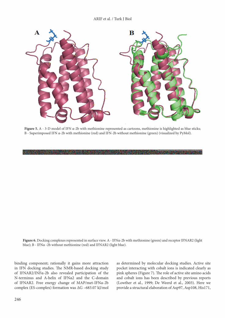
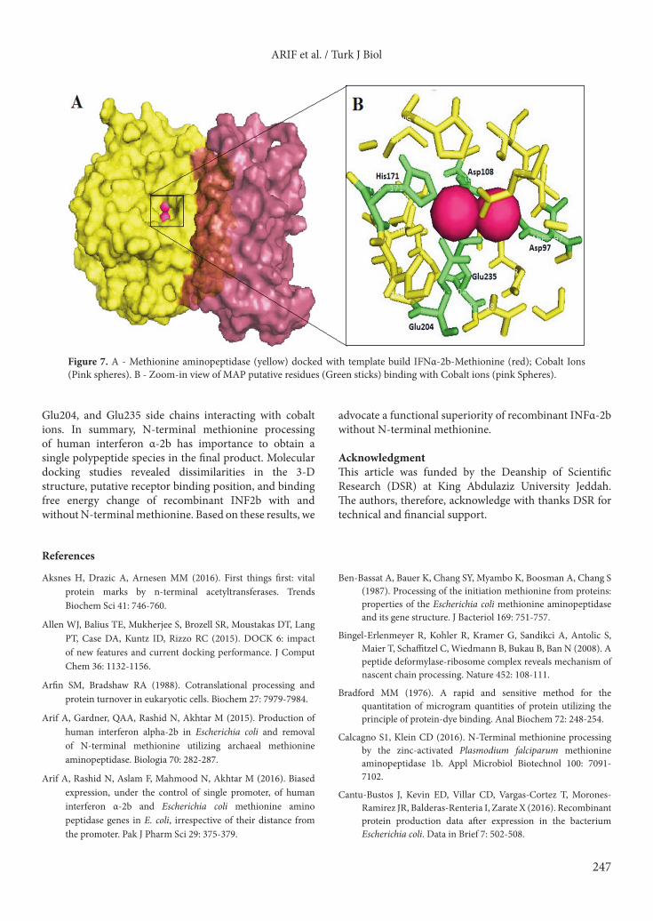
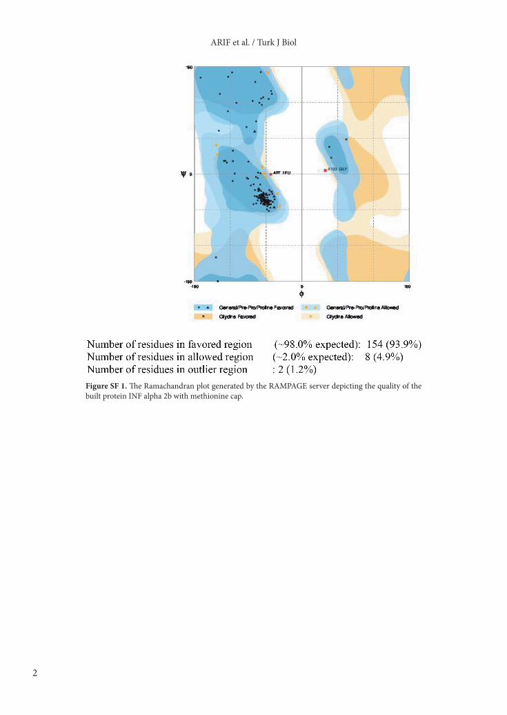
Version 1.2r3pre (Figure 5). RAMPAGE, a protein structure validation server, and the Ramachandran plot collectively revealed that the template-based structure of IFNα-2b built was quite stable (Supplementary Table ST2; Supplementary Figure SF1). Despite having only one amino acid difference in the primary structure, the Met-INF and nonmet-INF have visible differences in their secondary structures and 3-D arrangements. 3.5. Molecular docking and comparative analysisMolecular docking of IFNAR2/INFα-2b without methionine exhibited the lowest free energy change (ΔG –784.53 kJ/mol), indicating higher binding affinity, followed by IFNAR2/met-INFα-2b (ΔG –717.63 KJ/mole) (Figure 6). The formation of INF-enzyme complex was described. The interaction of amino acids with the cobalt ions in the active site pocket was elaborated. The free energy change for the ES-complex formation was calculated as ΔG –683.07 kJ/mol) (Figure 7).
4. DiscussionGenerally the recombinant proteins expressed in E. coli represent the translation product comprising the
Figure 2. Molecular weight determination by MALDI-TOF analysis of purified recombinant methionine aminopeptidase. X-axis indicates the protein MW in Daltons and the Y-axis indicates the intensity of laser beam. The spectrum shows [M + 1H] +1 ion peak at 29,200.941. Theoretical value of [M + 1H] +1 ion is 29,199.7084 Da.
ARIF et al. / Turk J Biol
244
amino acid sequence encoded by the gene. From time to time, however, posttranslation modifications have been observed that modulate the activity of most eukaryote proteins (Mann and Jensen, 2003). One amongst these
is the removal of N-terminal methionine catalyzed by methionine aminopeptidase and acetylation by acetyltransferase (Liao et al., 2004). These enzymes are found ubiquitously and show preference for the removal of N-terminal methionine if the next residue in the sequence has a small side chain (Xiao et al., 2004; Aksnes et al., 2016). In the case of E. coli expression, the recombinant protein is overexpressed; the native E. coli aminopeptidase being produced in limited amount fails to process all of the recombinant polypeptides. Hence, in the present study, in vitro processing of interferon α-2b at the N-terminus was the major part of the investigation. The in vitro removal of N-terminal methionine from the recombinant proteins is well established (Arfin et al., 1988; Mitra et al., 2006; Arif et al., 2015, 2016). Our study was meant to report the expression and purification of E. coli enzyme with emphasis on the in silico insights into the ES-complex formation by met-INFα-2b and MAP, the binding free energy change of ES-complex formation. We also investigated the binding free energy changes of INF molecules with and without methionine at the N-terminus and their corresponding receptor. The methionine aminopeptidase gene from E. coli DH5 α was expressed in E. coli BL21 (DE3) RIL codon plus cells. Gene expression in the bacterial culture was induced by 0.4 mM IPTG after the onset of the log phase. Induced bacterial culture was incubated overnight in a shaking incubator adjusted to 20 °C and 100 rpm (Ben-Bassat et al., 1987; Lowether et al., 2000). Recombinant E. coli methionine aminopeptidase was purified by anion exchange chromatography; it exhibited a molecular weight of 29,200 Da on MALDI-TOF, which is nearest to the theoretical molecular weight of enzyme without N-terminal methionine as determined by ProtParam (29,311.91 Da). Specific activity of enzyme was 1.64 U/mg in the methionyl p-nitroanilide-based assay. It exhibited 1.51 U/mg specific activity with synthetic tetrapeptide (MGMM) in an HPLC-based assay (Figure 3). Studies on MAP have shown different specific activity when different substrates were used (Vasina et al., 1996), indicating the effect of substrate structure on enzyme activity (Lowther
Figure 3. Reverse phase HPLC analysis of the substrate MGMM consumption in the reaction mixture containing 1.9 mM substrate and 0.026 mg of E. coli methionine aminopeptidase using C8 column. X-axis is the retention time in min and Y-axis is absorbance at 220 nm.
Table 1. The amount of enzyme, activity and specific activity of methionine aminotransferase demonstrated by using MGMM as substrate.
Total concentration of substrate used in the assay 1.9 mM 1.9 µmol/mL
Concentration of substrate used in 66 µL reaction (1.9 × 66) ÷ 1000 0.1254 µmolAmount of substrate consumed in 5 min (half) 0.1254 µmol ÷ 2 0.0627 µmolAmount of substrate consumed in 1 min 0.0627 ÷ 5 0.013 µmolAmount of enzyme used in each reaction (0.026 × 66) ÷ 200 0.0086 mgSpecific activity of the enzyme with MGMM 0.013 ÷ 0.0086 1.51 µmol/min/mg
ARIF et al. / Turk J Biol
245
et al., 1999; Xiao et al., 2010). Recombinant enzyme successfully processed recombinant human interferon as monitored by the HPLC-based assay system (Figure 4). A 3D model of INF was built by I-TASSER and validated by the protein structure validation server RAMPAGE (Lovell et al., 2002), which indicates the phi and psi deviations via Ramachandran. The difference in amino terminal methionine on the N-terminus resulted in detectable changes in the secondary and tertiary structures of INF with and without N-terminal as indicated by comparative analysis (Figure 5). The structural difference indicated by the comparative analysis makes the most of the recombinant fraction (with N-terminal methionine) different from the native (removed amino terminal methionine). We suggest a possible immune response by the human body against this structural difference between native and recombinant
interferon. The idea is also supported by the literature (Mukovozov et al., 2008). The built structure was similar to those reported in previous studies (Ya et al., 2003). Our docking results showed that IFNAR2 is the natural protein partner of IFNα-2b (without methionine) as their docking complex exhibited ΔG –784.53 kJ/mol. The IFNAR2/met-INFα2b complex exhibited a ΔG –717.63 kJ/mol, indicating a comparatively low binding affinity of met-INFα-2b to the corresponding receptor IFNAR2 (Figure 6). The results are in correlation with the studies showing better response of native interferon as compared to the recombinant (Karlberg et al., 2010). The receptor for human interferon alpha consists of two subunits, i.e. IFNAR1 and IFNAR2. The subunit IFNAR2 has (nanomolar dissociation constant) greater affinity to IFNs without the presence of IFNAR1 (Gull et al., 2013) and is considered the major ligand-
Figure 4. Reverse phase HPLC using C8 analytical column analysis of E. coli methionine aminopeptidase treated Met-Interferon α-2b. X-axis indicates the retention time in min and Y-axis represents absorbance at 220 nm.
ARIF et al. / Turk J Biol
246
binding component; rationally it gains more attraction in IFN docking studies. The NMR-based docking study of IFNAR2/INFα-2b also revealed participation of the N-terminus and A-helix of IFNα2 and the C-domain of IFNAR2. Free energy change of MAP/met-IFNα-2b complex (ES-complex) formation was ΔG –683.07 kJ/mol
as determined by molecular docking studies. Active site pocket interacting with cobalt ions is indicated clearly as pink spheres (Figure 7). The role of active site amino acids and cobalt ions has been described by previous reports (Lowther et al., 1999; De Weerd et al., 2003). Here we provide a structural elaboration of Asp97, Asp108, His171,
Figure 5. A - 3-D model of IFN α-2b with methionine represented as cartoons, methionine is highlighted as blue sticks; B - Superimposed IFN α-2b with methionine (red) and IFN-2b without methionine (green) (visualized by PyMol).
Figure 6. Docking complexes represented in surface view. A - IFNα-2b with methionine (green) and receptor IFNΑR2 (light blue); B - IFNα -2b without methionine (red) and IFNΑR2 (light blue).
ARIF et al. / Turk J Biol
247
Glu204, and Glu235 side chains interacting with cobalt ions. In summary, N-terminal methionine processing of human interferon α-2b has importance to obtain a single polypeptide species in the final product. Molecular docking studies revealed dissimilarities in the 3-D structure, putative receptor binding position, and binding free energy change of recombinant INF2b with and without N-terminal methionine. Based on these results, we
advocate a functional superiority of recombinant INFα-2b without N-terminal methionine.
AcknowledgmentThis article was funded by the Deanship of Scientific Research (DSR) at King Abdulaziz University Jeddah.The authors, therefore, acknowledge with thanks DSR for technical and financial support.
Figure 7. A - Methionine aminopeptidase (yellow) docked with template build IFNα-2b-Methionine (red); Cobalt Ions (Pink spheres). B - Zoom-in view of MAP putative residues (Green sticks) binding with Cobalt ions (pink Spheres).
References
Aksnes H, Drazic A, Arnesen MM (2016). First things first: vital protein marks by n-terminal acetyltransferases. Trends Biochem Sci 41: 746-760.
Allen WJ, Balius TE, Mukherjee S, Brozell SR, Moustakas DT, Lang PT, Case DA, Kuntz ID, Rizzo RC (2015). DOCK 6: impact of new features and current docking performance. J Comput Chem 36: 1132-1156.
Arfin SM, Bradshaw RA (1988). Cotranslational processing and protein turnover in eukaryotic cells. Biochem 27: 7979-7984.
Arif A, Gardner, QAA, Rashid N, Akhtar M (2015). Production of human interferon alpha-2b in Escherichia coli and removal of N-terminal methionine utilizing archaeal methionine aminopeptidase. Biologia 70: 282-287.
Arif A, Rashid N, Aslam F, Mahmood N, Akhtar M (2016). Biased expression, under the control of single promoter, of human interferon α-2b and Escherichia coli methionine amino peptidase genes in E. coli, irrespective of their distance from the promoter. Pak J Pharm Sci 29: 375-379.
Ben-Bassat A, Bauer K, Chang SY, Myambo K, Boosman A, Chang S (1987). Processing of the initiation methionine from proteins: properties of the Escherichia coli methionine aminopeptidase and its gene structure. J Bacteriol 169: 751-757.
Bingel-Erlenmeyer R, Kohler R, Kramer G, Sandikci A, Antolic S, Maier T, Schaffitzel C, Wiedmann B, Bukau B, Ban N (2008). A peptide deformylase-ribosome complex reveals mechanism of nascent chain processing. Nature 452: 108-111.
Bradford MM (1976). A rapid and sensitive method for the quantitation of microgram quantities of protein utilizing the principle of protein-dye binding. Anal Biochem 72: 248-254.
Calcagno S1, Klein CD (2016). N-Terminal methionine processing by the zinc-activated Plasmodium falciparum methionine aminopeptidase 1b. Appl Microbiol Biotechnol 100: 7091-7102.
Cantu-Bustos J, Kevin ED, Villar CD, Vargas-Cortez T, Morones-Ramirez JR, Balderas-Renteria I, Zarate X (2016). Recombinant protein production data after expression in the bacterium Escherichia coli. Data in Brief 7: 502-508.
ARIF et al. / Turk J Biol
248
Chen A, Sun Y, Zhang W, Peng F, Zhan C, Liu M, Yang Y, Bai Z (2016). Downsizing a pullulanase to a small molecule with improved soluble expression and secretion efficiency in Escherichia coli. Microb Cell Fact 15: 9.
Chill JH, Quadt SR, Levy R, Schreiber G, Anglister J (2003). The human type I interferon receptor: NMR structure reveals the molecular basis of ligand binding. Structure 11: 791-802.
De Weerd NA, Samarajiwa SA, Hertzog PJ (2007). Type I interferon receptors: biochemistry and biological functions. J Biol Chem 282: 20053-20057.
Dolores DNM, Ortiz A (2016). Enzyme replacement therapy for Fabry disease. J Inborn Errors Metabol Screen 4: 1-7.
Elleuche S, Schäfers C, Blank S, Schröder C, Antranikian G (2015). Exploration of extremophiles for high temperature biotechnological processes. Curr Opin Microbiol 25: 113-119.
Giglione C, Boularot A, Meinnel T (2004). Protein N-terminal methionine excision. Cell Mol Life Sci 61: 1455-1474.
Goh HC, Sobota RM, Ghadessy FJ, Nirantar S (2017). Going native: complete removal of protein purification affinity tags by simple modification of existing tags and proteases. Prot Exp Purification 129: 18-24.
Gull I, Samra ZQ, Aslam MS (2013). Heterologous expression, immunochemical and computational analysis of recombinant human interferon alpha 2b. SpringerPlus 2: 264.
Helgren TR, Wangtrakuldee P, Staker BL, Hagen TJ (2016). Advances in bacterial methionine aminopeptidase inhibition. Curr Top Med Chem 16: 397-414.
Jia B, Jeon CO (2016). High-throughput recombinant protein expression in Escherichia coli: status and future perspectives. Open Biol 6: 160196.
Kanodia JS, Kim Y, Tomer R, Khan Z, Chung K, Storey JD, Lu H, Keller PJ, Shvartsman SY (2011). A computational statistics approach for estimating the spatial range of morphogen gradients. Development 138: 4867-4874.
Karlberg H, Lindegren G, Mirazimi A (2010). Comparison of antiviral activity of recombinant and natural interferons against Crimean-Congo hemorrhagic fever virus. Open Virol J 4: 38.
Laemmli UK (1970) Cleavage of structural proteins during the assembly of the head of bacteriophage T4. Nature 227: 680-685.
Lagasse HA, Levin DD, Hengel H, Golding B, Sauna ZE (2017). Fc-fusion drugs have C1q/FcγR binding and signaling properties that can affect their immunogenicity. J Immunol 198: 129.13.
Liao YD, Jeng DJ, Wang CF, Wang SC, Chang ST (2004). Removal of N-terminal methionine from recombinant proteins by engineered E. coli methionine aminopeptidase. Protein Sci 13: 1802-1810.
Lovell SC, Davis IW, Arendall WB, de Bakker PIW, Word JM, Prisant MG, Richardson JS, Richardson GC (2002). Structure validation by Calpha geometry: phi, psi and Cbeta deviation. Proteins: Structure, Function & Genetics 50: 437-450.
Lowether WT, Matthews BW (2000). Structure and function of the methionine aminopeptidases. Biochim Biophys Acta 1477: 157-167.
Lowther WT, Zhang Y, Sampson PB, Honek JF, Matthews BW (1999). Insights into the mechanism of Escherichia coli methionine aminopeptidase from the structural analysis of reaction products and phosphorus-based transition-state analogues. Biochem 38: 14810-14819.
Macindoe G, Mavridis L, Venkatraman V, Devignes MD, Ritchie DW (2010). HexServer: an FFT-based protein-docking server powered by graphics processors. Nucleic Acid Res 38: W445-W449.
Mann M, Jensen ON (2003). Proteomic analysis of post-translational modifications. Nat Biotechnol 2: 255-261.
Miljic D, Miljic P, Doknic M, Pekic S, Stojanovic M, Cvijovic G, Micic D, Popovic V (2013). Growth hormone replacement normalizes impaired fibrinolysis: new insights into endothelial dysfunction in patients with hypopituitarism and growth hormone deficiency, Growth Horm IGF Res 23: 243-248.
Mitra S, Dygas-Holz AM, Jiracek J, Zertova M, Zakova L, Holz RC (2006). A new colorimetric assay for methionyl aminopeptidases: examination of the binding of a new class of pseudo peptide analogue inhibitors. Anal Biochem 357: 43-49.
Mukovozov I, Sabljic T, Hortelano G, Ofosu FA (2008). Factors that contribute to the immmunogenicity of therapeutic recombinant human proteins. Thromb Haemost 100: 874-882.
Nudelman I, Akabayov SR, Schnur E, Biron Z, Levy R, Xu Y, Anglister J (2010). Intermolecular interactions in a 44 kDa interferon - receptor complex detected by asymmetric reverse-protonation and two-dimensional NOESY. Biochem 49: 5117-5133.
Rider P, Carmi Y, Cohen I (2016). Biologics for targeting inflammatory cytokines, clinical uses, and limitations. Int J Cell Biol Article ID 9259646: 11.
Seal BS (2013). Characterization of bacteriophages virulent for Clostridium perfringens and identification of phage lytic enzymes as alternatives to antibiotics for potential control of the bacterium. Poult Sci 92: 526-533.
Shepelkova G, Evstifeev V, Majorov K, Bocharova I, Apt A (2016). Therapeutic effect of recombinant mutated interleukin 11 in the mouse model of tuberculosis. J Infect Dis 214: 496-501.
Tripathi NK (2016). Production and purification of recombinant proteins from Escherichia coli. Chem BioEng Rev 3: 116-133.
Urbano F, Acquafredda A, Aceto G, Penza R, Cavallo L (2012). Unusual pediatric co-morbility: autoimmune thyroiditis and cortico-resistant nephrotic syndrome in a 6-month-old Italian patient. Ital J Pediatr 38: 57.
Vairo F, Netto C, Dorneles A, Mittelstadt S, Wilke M, Doneda D, Michelin K, Ribeiro CB, Quevedo A, Vieira T et al. (2013). Enzyme replacement therapy in a patient with Gaucher disease type III: a paradigmatic case showing severe adverse reactions started a long time after the beginning of treatment. JIMD Rep 11: 1-6.
Vasina JA, Baneyx F (1996). Recombinant protein expression at low temperatures under the transcriptional control of the major Escherichia coli cold shock promoter cspA. Appl Environ Microbiol 62: 1444-1447.
ARIF et al. / Turk J Biol
249
Venkatachalam CM, Jiang X, Oldfield T, Waldman M (2003). LigandFit: a novel method for the shape-directed rapid docking of ligands to protein active sites. J Mol Graph Model 21: 289-307.
Wingfield PT (2017). N-terminal methionine processing. Curr Protoc Protein Sci 88: 6141-6143.
Xiao Q, Zhang F, Nacev BA, Liu JO, Pei D (2010). Protein N-terminal processing substrate specificity of Escherichia coli and human methionine aminopeptidases. Biochem 49: 5588-5599.
Ya LJ, Cui YM, Chen LL, Gu M, Li J, Nan FJ, Ye QZ (2003). Mutations at the S1 sites of methionine aminopeptidases from Escherichia coli and Homo sapiens reveal the residues critical for substrate specificity. Biochem Biophys Res Comm 307: 172.
Yang J, Zhang Y (2015). I-TASSER server: new development for protein structure and function predictions. Nucleic Acid Res 43: W174-W181.
Zhou Y, Guo XC, Yi T, Yoshimoto T, Pei D (2000). Two continuous spectrophotometric assays for methionine aminopeptidase. Analytic Biochem 280: 159-65.
ARIF et al. / Turk J Biol
1
Table ST1. Timewise change in the peak area and peak height of Met-interferon α-2b and interferon α-2b due to reaction with E.coli methionine aminopeptidase.
0 min
Protein Peak area Peak height % of Met-Ifn converted to IfnIfn 0 0 0Met-Ifn 24242 202 0
10 min
Protein Peak area Peak height % of Met-Ifn converted to IfnIfn 3675 58
15%Met-Ifn 20568 175
20 min
Protein Peak area Peak height % of Met-Ifn converted to IfnIfn 2944 57
25%Met-Ifn 8828 110
30 minutes
Protein Peak area Peak height % of Met-Ifn converted to IfnIfn 4622 61
35%Met-Ifn 8403 95
40 minutes
Protein Peak area Peak height % of Met-Ifn converted to IfnIfn 6848 90
36%Met-Ifn 12015 116
Table ST2. Quality evaluations of 3D protein model built by I-TASSER using various parameters and software.
Protein C-scorea TM-scoreb* RMSDc* IDENd* Cove*
IFN-2b 1.44 0.895 1.88 Å 0.970 0.970
aConfidence (C) score for estimating the quality of predicted models (range –5 to 2).bTemplate modeling score (TM-score) measures the structural similarity between two structures (TM-score > 0.5 indicates a model of correct topology and a TM-score < 0.17 means a random similarity range 0 to 1).cHeavy atoms root-mean-square deviation (RMSD) with respect to the experimental structure.dIDEN is the percentage sequence identity in the structurally aligned region (range 0 to 1).eCov represents the coverage of global structural alignment and is equal to the number of structurally aligned residues divided by length of the query protein cluster (range 0 to 1).*As compared to template ‘1itf ’
ARIF et al. / Turk J Biol
2
Figure SF 1. The Ramachandran plot generated by the RAMPAGE server depicting the quality of the built protein INF alpha 2b with methionine cap.