B-SIDER: Computational Algorithm for the Design of Complementary β...
8
B‑SIDER: Computational Algorithm for the Design of Complementary β‑Sheet Sequences Tae-Geun Yu, Hak-Sung Kim,* ,† and Yoonjoo Choi* ,† Department of Biological Sciences, Korea Advanced Institute of Science and Technology (KAIST), Daejeon 34141, Republic of Korea * S Supporting Information ABSTRACT: The β-sheet is an element of protein secondary structure, and intra-/intermolecular β-sheet interactions play pivotal roles in biological regulatory processes including scaffolding, transporting, and oligomerization. In nature, a β- sheet formation is tightly regulated because dysregulated β- stacking often leads to severe diseases such as Alzheimer’s, Parkinson’s, systemic amyloidosis, or diabetes. Thus, the identification of intrinsic β-sheet-forming propensities can provide valuable insight into protein designs for the development of novel therapeutics. However, structure-based design methods may not be generally applicable to such amyloidogenic peptides mainly owing to high structural plasticity and complexity. Therefore, an alternative design strategy based on complementary sequence information is of significant importance. Herein, we developed a database search method called β-Stacking Interaction DEsign for Reciprocity (B-SIDER) for the design of complementary β-strands. This method makes use of the structural database information and generates target-specific score matrices. The discriminatory power of the B-SIDER score function was tested on representative amyloidogenic peptide substructures against a sequence-based score matrix (PASTA 2.0) and two popular ab initio protein design score functions (Rosetta and FoldX). B-SIDER is able to distinguish wild-type amyloidogenic β-strands as favored interactions in a more consistent manner than other methods. B- SIDER was prospectively applied to the design of complementary β-strands for a splitGFP scaffold. Three variants were identified to have stronger interactions than the original sequence selected through a directed evolution, emitting higher fluorescence intensities. Our results indicate that B-SIDER can be applicable to the design of other β-strands, assisting in the development of therapeutics against disease-related amyloidogenic peptides. ■ INTRODUCTION The β-sheet is one of the major units of protein structure, 1 and has a variety of functions in transportation, recognition, scaffolding, and enzymatic processes. 2 The mechanism of a β- sheet formation has recently received significant attention owing to its close relations with several critical diseases such as Alzheimer’s, Parkinson’s, type 2 diabetes, and systemic amyloidosis. 3,4 Such diseases are known to be linked to the precipitation of dysregulated β-stacking between neighboring β-strands. 5-7 In this regard, information on the amino acid propensity of intrinsic β-sheet forming motifs and its use in the design of their complementary sequences are crucial for understanding the mechanism of β-sheet formation and developing potential therapeutics specifically targeting aggre- gation-prone regions. 8 Whereas structure-based protein design approaches have shown notable successes in several cases, 9 their application to de novo β-sheet designs still remains a challenge. 2,10 Although structure-based design approaches require a well-defined protein structure, amyloidogenic peptides usually have highly disordered structures. 11 The structural identification of such peptides has long been hindered by high degrees of structural plasticity, transiency, and complexity owing to a self- oligomerization. 12,13 It is thus necessary to exploit the complementarity across neighboring β-strand pairs using sequence information. Interestingly, significant conservation and covariations of residue pairs between neighboring β-strands have been identified in many different protein families. 14 For instance, pairs of β-branched residues and cysteines are preferred at nonhydrogen-bonded positions. Aromatic residues tend to be paired with valine or glycine. 15 Several computational algorithms have been developed to predict aggregation-prone regions based on the internal β-sheet forming patterns. While differing in detail, they make use of either statistical potentials such as Tango, 16 PASTA, 17 SALSA, 18 BETASCAN, 19 and Waltz 20 or physicochemical properties of amino acids. 21 In addition, consensus methods and machine-learning approaches have also been developed, 22,23 showing fine agreement with the experiment results. It has been reported that β-strand interactions can be stabilized by introducing β-sheet-favored pairs 24-28 and charge pairing between neighboring β-strands. 29-31 Recent studies Received: July 5, 2019 Published: September 12, 2019 Article pubs.acs.org/jcim Cite This: J. Chem. Inf. Model. 2019, 59, 4504-4511 © 2019 American Chemical Society 4504 DOI: 10.1021/acs.jcim.9b00548 J. Chem. Inf. Model. 2019, 59, 4504-4511 Downloaded via KOREA ADVANCED INST SCI AND TECHLGY on November 15, 2019 at 01:31:01 (UTC). See https://pubs.acs.org/sharingguidelines for options on how to legitimately share published articles.
Transcript of B-SIDER: Computational Algorithm for the Design of Complementary β...
B‑SIDER: Computational Algorithm for the Design ofComplementary β‑Sheet SequencesTae-Geun Yu, Hak-Sung Kim,*,† and Yoonjoo Choi*,†
Department of Biological Sciences, Korea Advanced Institute of Science and Technology (KAIST), Daejeon 34141, Republic ofKorea
*S Supporting Information
ABSTRACT: The β-sheet is an element of protein secondarystructure, and intra-/intermolecular β-sheet interactions playpivotal roles in biological regulatory processes includingscaffolding, transporting, and oligomerization. In nature, a β-sheet formation is tightly regulated because dysregulated β-stacking often leads to severe diseases such as Alzheimer’s,Parkinson’s, systemic amyloidosis, or diabetes. Thus, theidentification of intrinsic β-sheet-forming propensities canprovide valuable insight into protein designs for thedevelopment of novel therapeutics. However, structure-based design methods may not be generally applicable to suchamyloidogenic peptides mainly owing to high structural plasticity and complexity. Therefore, an alternative design strategybased on complementary sequence information is of significant importance. Herein, we developed a database search methodcalled β-Stacking Interaction DEsign for Reciprocity (B-SIDER) for the design of complementary β-strands. This methodmakes use of the structural database information and generates target-specific score matrices. The discriminatory power of theB-SIDER score function was tested on representative amyloidogenic peptide substructures against a sequence-based scorematrix (PASTA 2.0) and two popular ab initio protein design score functions (Rosetta and FoldX). B-SIDER is able todistinguish wild-type amyloidogenic β-strands as favored interactions in a more consistent manner than other methods. B-SIDER was prospectively applied to the design of complementary β-strands for a splitGFP scaffold. Three variants wereidentified to have stronger interactions than the original sequence selected through a directed evolution, emitting higherfluorescence intensities. Our results indicate that B-SIDER can be applicable to the design of other β-strands, assisting in thedevelopment of therapeutics against disease-related amyloidogenic peptides.
■ INTRODUCTION
The β-sheet is one of the major units of protein structure,1 andhas a variety of functions in transportation, recognition,scaffolding, and enzymatic processes.2 The mechanism of a β-sheet formation has recently received significant attentionowing to its close relations with several critical diseases such asAlzheimer’s, Parkinson’s, type 2 diabetes, and systemicamyloidosis.3,4 Such diseases are known to be linked to theprecipitation of dysregulated β-stacking between neighboringβ-strands.5−7 In this regard, information on the amino acidpropensity of intrinsic β-sheet forming motifs and its use in thedesign of their complementary sequences are crucial forunderstanding the mechanism of β-sheet formation anddeveloping potential therapeutics specifically targeting aggre-gation-prone regions.8
Whereas structure-based protein design approaches haveshown notable successes in several cases,9 their application tode novo β-sheet designs still remains a challenge.2,10 Althoughstructure-based design approaches require a well-definedprotein structure, amyloidogenic peptides usually have highlydisordered structures.11 The structural identification of suchpeptides has long been hindered by high degrees of structuralplasticity, transiency, and complexity owing to a self-
oligomerization.12,13 It is thus necessary to exploit thecomplementarity across neighboring β-strand pairs usingsequence information.Interestingly, significant conservation and covariations of
residue pairs between neighboring β-strands have beenidentified in many different protein families.14 For instance,pairs of β-branched residues and cysteines are preferred atnonhydrogen-bonded positions. Aromatic residues tend to bepaired with valine or glycine.15 Several computationalalgorithms have been developed to predict aggregation-proneregions based on the internal β-sheet forming patterns. Whilediffering in detail, they make use of either statistical potentialssuch as Tango,16 PASTA,17 SALSA,18 BETASCAN,19 andWaltz20 or physicochemical properties of amino acids.21 Inaddition, consensus methods and machine-learning approacheshave also been developed,22,23 showing fine agreement with theexperiment results.It has been reported that β-strand interactions can be
stabilized by introducing β-sheet-favored pairs24−28 and chargepairing between neighboring β-strands.29−31 Recent studies
Received: July 5, 2019Published: September 12, 2019
Article
pubs.acs.org/jcimCite This: J. Chem. Inf. Model. 2019, 59, 4504−4511
© 2019 American Chemical Society 4504 DOI: 10.1021/acs.jcim.9b00548J. Chem. Inf. Model. 2019, 59, 4504−4511
Dow
nloa
ded
via
KO
RE
A A
DV
AN
CE
D I
NST
SC
I A
ND
TE
CH
LG
Y o
n N
ovem
ber
15, 2
019
at 0
1:31
:01
(UT
C).
See
http
s://p
ubs.
acs.
org/
shar
ingg
uide
lines
for
opt
ions
on
how
to le
gitim
atel
y sh
are
publ
ishe
d ar
ticle
s.
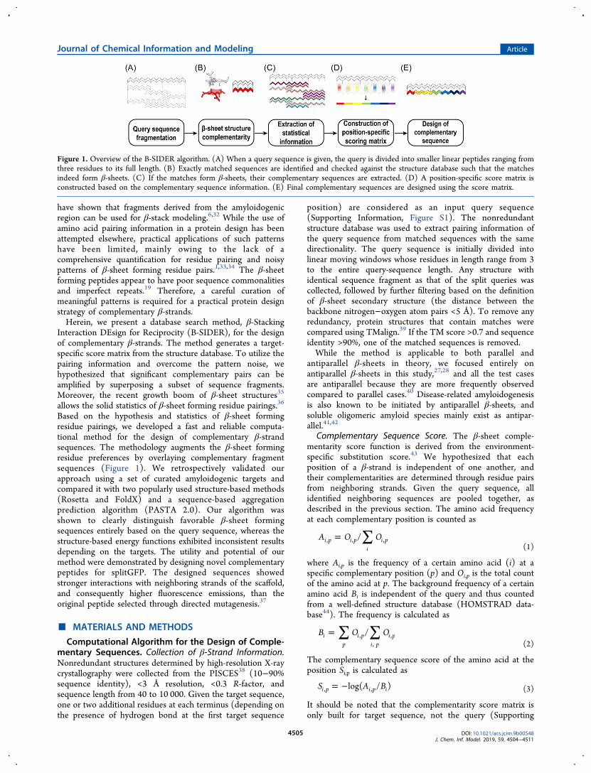
have shown that fragments derived from the amyloidogenicregion can be used for β-stack modeling.6,32 While the use ofamino acid pairing information in a protein design has beenattempted elsewhere, practical applications of such patternshave been limited, mainly owing to the lack of acomprehensive quantification for residue pairing and noisypatterns of β-sheet forming residue pairs.1,33,34 The β-sheetforming peptides appear to have poor sequence commonalitiesand imperfect repeats.19 Therefore, a careful curation ofmeaningful patterns is required for a practical protein designstrategy of complementary β-strands.Herein, we present a database search method, β-Stacking
Interaction DEsign for Reciprocity (B-SIDER), for the designof complementary β-strands. The method generates a target-specific score matrix from the structure database. To utilize thepairing information and overcome the pattern noise, wehypothesized that significant complementary pairs can beamplified by superposing a subset of sequence fragments.Moreover, the recent growth boom of β-sheet structures35
allows the solid statistics of β-sheet forming residue pairings.36
Based on the hypothesis and statistics of β-sheet formingresidue pairings, we developed a fast and reliable computa-tional method for the design of complementary β-strandsequences. The methodology augments the β-sheet formingresidue preferences by overlaying complementary fragmentsequences (Figure 1). We retrospectively validated ourapproach using a set of curated amyloidogenic targets andcompared it with two popularly used structure-based methods(Rosetta and FoldX) and a sequence-based aggregationprediction algorithm (PASTA 2.0). Our algorithm wasshown to clearly distinguish favorable β-sheet formingsequences entirely based on the query sequence, whereas thestructure-based energy functions exhibited inconsistent resultsdepending on the targets. The utility and potential of ourmethod were demonstrated by designing novel complementarypeptides for splitGFP. The designed sequences showedstronger interactions with neighboring strands of the scaffold,and consequently higher fluorescence emissions, than theoriginal peptide selected through directed mutagenesis.37
■ MATERIALS AND METHODS
Computational Algorithm for the Design of Comple-mentary Sequences. Collection of β-Strand Information.Nonredundant structures determined by high-resolution X-raycrystallography were collected from the PISCES38 (10−90%sequence identity), <3 Å resolution, <0.3 R-factor, andsequence length from 40 to 10 000. Given the target sequence,one or two additional residues at each terminus (depending onthe presence of hydrogen bond at the first target sequence
position) are considered as an input query sequence(Supporting Information, Figure S1). The nonredundantstructure database was used to extract pairing information ofthe query sequence from matched sequences with the samedirectionality. The query sequence is initially divided intolinear moving windows whose residues in length range from 3to the entire query-sequence length. Any structure withidentical sequence fragment as that of the split queries wascollected, followed by further filtering based on the definitionof β-sheet secondary structure (the distance between thebackbone nitrogen−oxygen atom pairs <5 Å). To remove anyredundancy, protein structures that contain matches werecompared using TMalign.39 If the TM score >0.7 and sequenceidentity >90%, one of the matched sequences is removed.While the method is applicable to both parallel and
antiparallel β-sheets in theory, we focused entirely onantiparallel β-sheets in this study,27,28 and all the test casesare antiparallel because they are more frequently observedcompared to parallel cases.40 Disease-related amyloidogenesisis also known to be initiated by antiparallel β-sheets, andsoluble oligomeric amyloid species mainly exist as antipar-allel.41,42
Complementary Sequence Score. The β-sheet comple-mentarity score function is derived from the environment-specific substitution score.43 We hypothesized that eachposition of a β-strand is independent of one another, andtheir complementarities are determined through residue pairsfrom neighboring strands. Given the query sequence, allidentified neighboring sequences are pooled together, asdescribed in the previous section. The amino acid frequencyat each complementary position is counted as
A O O/i p i pi
i p, , ,∑=(1)
where Ai,p is the frequency of a certain amino acid (i) at aspecific complementary position (p) and Oi,p is the total countof the amino acid at p. The background frequency of a certainamino acid Bi is independent of the query and thus countedfrom a well-defined structure database (HOMSTRAD data-base44). The frequency is calculated as
B O O/ip
i pi p
i p,,
,∑ ∑=(2)
The complementary sequence score of the amino acid at theposition Si,p is calculated as
S A Blog( / )i p i p i, ,= − (3)
It should be noted that the complementarity score matrix isonly built for target sequence, not the query (Supporting
Figure 1. Overview of the B-SIDER algorithm. (A) When a query sequence is given, the query is divided into smaller linear peptides ranging fromthree residues to its full length. (B) Exactly matched sequences are identified and checked against the structure database such that the matchesindeed form β-sheets. (C) If the matches form β-sheets, their complementary sequences are extracted. (D) A position-specific score matrix isconstructed based on the complementary sequence information. (E) Final complementary sequences are designed using the score matrix.
Journal of Chemical Information and Modeling Article
DOI: 10.1021/acs.jcim.9b00548J. Chem. Inf. Model. 2019, 59, 4504−4511
4505
Information, Figure S1). The score is also completely data-driven, i.e., if an amino acid never appears at a certain position,a high penalty score is imposed. We only considercomplementary amino acids that are found at least once inthe entire identified sequences. The final score is the sum ofthe scores at all positions.Protein Expression and Complementation Assay.
Gene Construction. The gene coding for splitGFP37 consistsof the first through 10th (GFP1−10) and 11th strand(GFP11) templates (Supporting Information, Table S1).They were cloned into pET-28a (Novagen) vector betweenthe Nde-I and Xho-I restriction sites. We introduced additionalmutations to GFP1−10 to inhibit aggregation and for aconvenient expression.45 The GFP11 strand was fused with aP22 virus-like particle scaffolding protein46 for a soluble andstable expression. Mutations on GFP11 were introducedthrough polymerase chain reaction using mutagenic primers(Supporting Information, Table S2), and the resulting geneswere cloned into the pET-28a vector. Six histidine residueswere fused to the N-terminal of the GFP1−10 and GFP11genes as an affinity purification tag.Protein Expression and Purification. All constructs were
transformed into BL21 (DE3) Escherichia coli strains. Thetransformed cells were grown overnight and inoculated into aLuria−Bertani media containing 50 μg/mL of kanamycin at 37°C. The cells were then grown until the optical density of thecells reached 0.6−0.8 at 600 nm, followed by the addition of0.7 mM of isopropyl β-D-1-thiogalactopyranoside for in-duction. After incubation for 16−18 h at 18 °C, the cellswere harvested and suspended in a lysis buffer containing 50mM Tris (pH 8.0), 150 mM NaCl, and 5 mM imidazole. Thesuspended cells were disrupted through sonication, andinsoluble fractions were removed using centrifugation at18 000g for 1 h. The supernatants were filtered with 0.22 μmsyringe filters and purified through affinity chromatographyusing Ni-NTA agarose Superflow (Qiagen). The solutionswere applied to the resin-packed columns and washed with abuffer containing 50 mM Tris (pH 8.0), 150 mM NaCl, and 10mM imidazole, until no proteins were detected through aBradford assay. An elution buffer (50 mM Tris (pH 7.4), 150mM NaCl, 300 mM imidazole) was then applied to thecolumn. The buffer exchange was performed with phosphate-buffered saline (PBS, pH 7.4) using a PD-10 column (GEHealthcare). The concentrations of the proteins weredetermined by measuring the absorbance at 280 nm. Allpurification processes were conducted at 4 °C. The purities ofthe proteins were then evaluated using sodium dodecyl sulfate-polyacrylamide gel electrophoresis (SDS-PAGE).Complementation Assay. The assembly of splitGFP
variants was monitored and measured using a fluorescencecomplementation assay. An excessive amount of GFP1−10 (50pmol) in 180 μL, and 20 μL of equal molar concentration foreach GFP11 strand (3 pmol) were co-incubated in a PBSbuffer (pH 7.4). The fluorescence kinetics (λex = 488 nm/λem= 530 nm) were monitored for 12 h at 25 °C using a TECANinfinite M200 microplate reader at 5 min intervals37 withshaking for 2 s between intervals. Each experiment wasconducted in triplicate using a Nunc F 96 microwell blackplate, blocked with a solution of PBS containing 0.5% ofbovine serum albumin for 30 min before the assay.
■ RESULTS AND DISCUSSIONOverview of the Design Process. We hypothesized that
the observed repetitive amino acid pairing patterns indicate astrong preference toward the β-sheet. It was also assumed thatthe sequence with the most frequent patterns will directly forma β-sheet without considering other environmental contribu-tions.There are two major steps applied in the algorithm: (1) the
extraction of complementarity information of the β-sheet and(2) the construction of a scoring matrix (Figure 1). When atarget sequence is given, one or two additional amino acids ateach terminus are considered as a query sequence. The query isthen fragmented into several pieces of short peptides longerthan three residues in length, and the matching neighboringstrands are collected. All corresponding submatches of longerpeptides (>3 residues in length) are also included. Thesefragmentation and overlaying processes naturally imposeweights on complementary-prone positions and amplify thepattern signals. It should be noted that the additional residuesof the query is considered to avoid underweighting terminalregions and, if additional residue information is not available,the target sequence may be used as a query (SupportingInformation, Table S3 and Figure S1). After the collection ofmatched sequences, a position-specific complementarityscoring matrix is constructed. The scoring matrix obtained isused to evaluate and design the complementarity of β-strandinteractions.
Validation of the Score Function on RetrospectiveCases. In an effort to validate the complementarity score, wemanually curated a test set of naturally occurring β-strand pairswhose environmental effects are minimal. It is known that theβ-strand pairing is in general significantly hindered by localenvironments,47 whereas amyloidogenic peptide segments areknown to form natural β-sheets themselves.17 We thus selecteda set of widely known amyloidogenic structures whoseaggregation-prone regions have been identified (Tables 1 and
S3). When multiple β-strands are present, we assumed that thefirst strands may be the primary amyloid building unit and soonly the first two strands as a pair were extracted for thestructure-based energy calculation.To assess the complementarity of the native sequences, we
compared their scores with those of random sequences thatwere generated using all 20 amino acids in a position-independent manner. Natural amyloidogenic segments areknown to be highly prone to aggregation, and thus they areexpected to be highly preferred, i.e., having fairly low scores inthe random sequence score distributions. Figure 2 shows thatall native sequence scores are ranked extremely low in alldistributions. On average, the native sequences are within 5.2%of the distributions (Figure 2). The results indicate that the
Table 1. Retrospective Test Set
source PDB ID
amyloid-β 2lnq, 2y2a, 2y3l, 3pzzinsulin 2omqIAPP (amylin) 3fth, 5e61prion 3md4, 3nvetau 4e0mβ-2 microglobulin 3loz, 4e0k, 4e0limmunoglobulin 6dj0, 6dixSOD1 5wor
Journal of Chemical Information and Modeling Article
DOI: 10.1021/acs.jcim.9b00548J. Chem. Inf. Model. 2019, 59, 4504−4511
4506
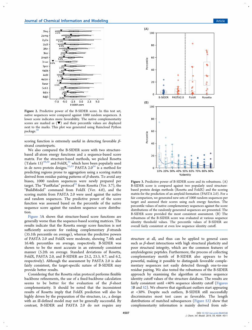
scoring function is extremely useful in detecting favorable β-strand counterparts.We also compared the B-SIDER score with two structure-
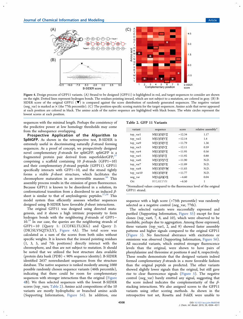
based all-atom energy functions and a sequence-based scorematrix. For the structure-based methods, we picked Rosetta(Talaris 13)49,50 and FoldX,51 which have been popularly usedin de novo protein designs.52,53 PASTA 2.054 is a method forpredicting regions prone to aggregation using a scoring matrixderived from residue pairing patterns of β-sheets. To avoid anybiases, 1000 random sequences were newly prepared pertarget. The “FastRelax” protocol55 from Rosetta (Ver. 3.7), the“BuildModel” command from FoldX (Ver. 4.0), and thescoring matrix from PASTA 2.0 were used against the nativeand random sequences. The predictive power of the scorefunction was assessed based on the percentile of the nativesequence score against the random sequence score distribu-tion.Figure 3A shows that structure-based score functions are
generally worse than the sequence-based scoring matrices. Theresults indicate that the Rosetta energy score function is notsufficiently accurate for ranking complementary β-strands(35.1th percentile on average), whereas the predictive powersof PASTA 2.0 and FoldX were moderate, showing 7.4th and16.4th percentiles on average, respectively. B-SIDER wasshown to be the most accurate in an extremely consistentmanner (5.2th on average. Standard deviations of Rosetta,FoldX, PASTA 2.0, and B-SIDER are 25.2, 23.3, 8.7, and 6.2,respectively). Although the assessment by PASTA 2.0 is alsofairly consistent, the target-specific nature of B-SIDER mayprovide better results.Considering that the Rosetta relax protocol performs flexible
backbone refinements, the use of a fixed-backbone calculationseems to be better for the evaluation of the β-sheetcomplementarity. It should be noted that the inconsistentresults of Rosetta imply that FoldX prediction will also behighly driven by the preparation of the structure, i.e., a designwith an ill-defined model may not be generally successful. Bycontrast, B-SIDER and PASTA 2.0 do not require any
structure at all, and thus can be applied to general casessuch as β-sheet interactions with high structural plasticity andpoor structural integrity, which are the common features ofamyloidogenic peptides. Furthermore, the process of collectingcomplementary motifs of B-SIDER also appears to bepowerful, making it possible to distinguish favorable comple-mentary sequences not easily detected through one-to-oneresidue pairing. We also tested the robustness of the B-SIDERapproach by examining the algorithm at various sequenceidentity cutoff values of the structure database. The results arefairly consistent until <40% sequence identity cutoff (Figures3B and S2). We observe that significant outliers start appearingat <30%. Despite such outliers, B-SIDER still successfullydiscriminates most test cases as favorable. The lengthdistributions of matched subsequences (Figure S3) show thatcomplementarity information is mainly derived from sub-
Figure 2. Predictive power of the B-SIDER score. In this test set,native sequences were compared against 1000 random sequences. Alower score indicates more favorability. The native complementarityscores are marked as (▼) and their percentile values are displayednext to the marks. This plot was generated using Raincloud Pythonpackage.48
Figure 3. Predictive power of B-SIDER score and its robustness. (A)B-SIDER score is compared against two popularly used structure-based protein design methods (Rosetta and FoldX) and the scoringmatrix for the prediction of an amyloid formation (PASTA 2.0). For afair comparison, we generated new sets of 1000 random sequences pertarget and assessed their scores using each energy function. Thepercentile values of native complementary sequences against the scoredistributions of the randomly generated sequences are presented. TheB-SIDER score provided the most consistent assessment. (B) Therobustness of the B-SIDER score was evaluated at various sequenceidentity threshold values. The percentile values of B-SIDER areoverall fairly consistent at even low sequence identity cutoff.
Journal of Chemical Information and Modeling Article
DOI: 10.1021/acs.jcim.9b00548J. Chem. Inf. Model. 2019, 59, 4504−4511
4507
sequences with the minimal length. Perhaps the consistency ofthe predictive power at low homology thresholds may comefrom the subsequence overlapping.Prospective Application of the Algorithm to
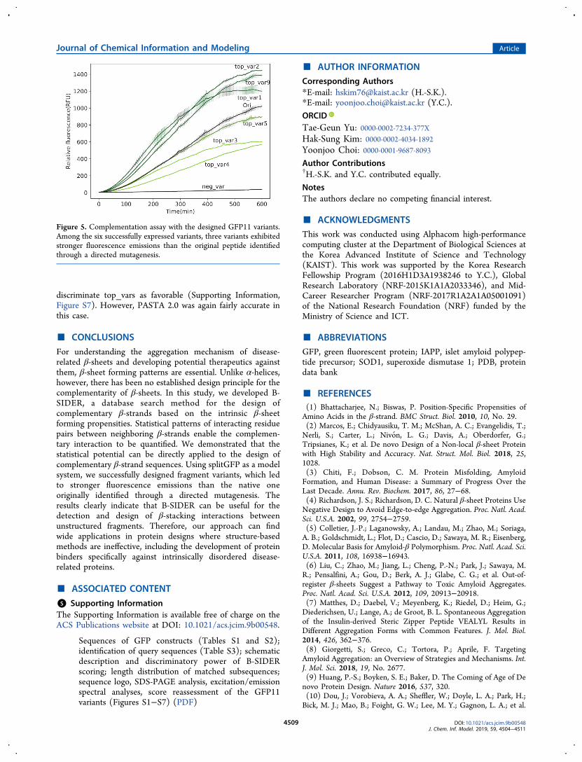
SplitGFP. As shown in the retrospective test, B-SIDER isextremely useful in discriminating naturally β-strand formingsequences. As a proof of concept, we prospectively designednovel complementary β-strands for splitGFP. splitGFP is afragmented protein pair derived from superfolderGFP,37
comprising a scaffold containing 10 β-strands (GFP1−10)and their complementary β-strand peptide (GFP11). GFP11specifically interacts with GFP1−10, and the strand tightlyforms a stable β-sheet structure, which facilitates thechromophore maturation in an irreversible manner.56 Thisassembly process results in the emission of green fluorescence.Because GFP11 is known to be disordered in a solution, itsconformational transition from a disordered to an induced β-sheet is similar to that of amyloidogenic peptides.57,58 Thismodel system thus efficiently assesses whether sequencesdesigned using B-SIDER have favorable β-sheet interactions.The original GFP11 was designed using directed muta-
genesis, and it shows a high intrinsic propensity to formhydrogen bonds with the neighboring β-strands of GFP1−10.59 In our case, the queries are the neighboring strands ofGFP1−10 (Query 1: (CI)FKLTL(KG) and Query 2:(DK)SLVSQTS(LY), Figure 4A). The total score wascalculated as a sum of the scores from both sides withoutspecific weights. It is known that the inward pointing residues(1, 3, 5, and 7th positions) directly interact with thechromophore, and thus are not subject to mutation. It shouldbe noted that we utilized the best structure data available(protein data bank (PDB) < 90% sequence identity). B-SIDERidentified 2637 nonredundant sequences from the structuredatabase. The native sequence is ranked modestly among 1000possible randomly chosen sequence variants (46th percentile),indicating that there could be room for complementarysequences with stronger interactions than the original (Figure4B). We then selected sequences with the lowest B-SIDERscores (top_vars; Table 2). Amino acid compositions of the 10variants are mostly hydrophobic or branched amino acids(Supporting Information, Figure S4). In addition, one
sequence with a high score (>75th percentile) was randomlyselected as a negative control (neg_var, 77th).The selected variants were successfully expressed and
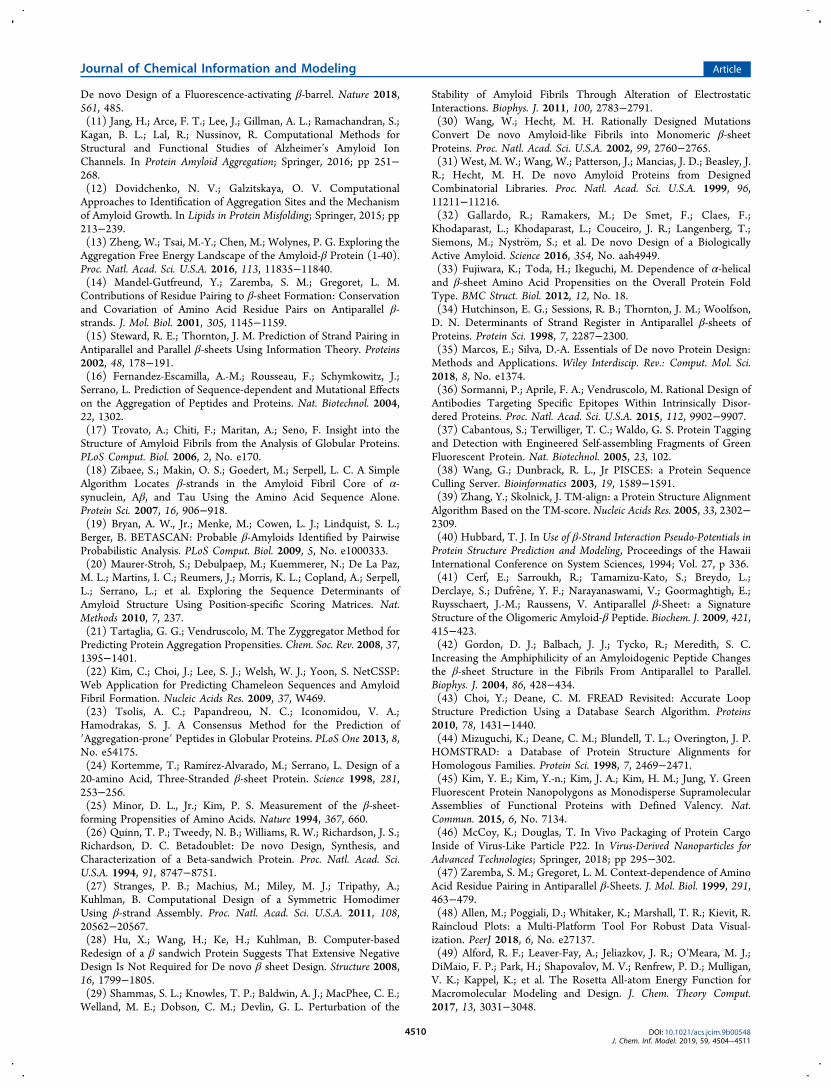
purified (Supporting Information, Figure S5) except for fourclones (top_var6, 7, 8, and 10), which were observed to beinsoluble, perhaps due to aggregation. Among those expressed,three variants (top_var1, 2, and 9) showed faster assemblypatterns and higher signals compared to the original GFP11(Figure 5). No functional aberrance with excitations oremissions was observed (Supporting Information, Figure S6).All successful variants, which emitted stronger fluorescencelevels than the original, were shown to have pairs ofphenylalanine and threonine at positions 6 and 8, respectively.These results demonstrate that the designed variants indeedformed complementary β-strands in a more favorable fashionthan the original peptide as predicted. The other variantsshowed slightly lower signals than the original, but still gaverise to clear fluorescence signals (Figure 5). The negativecontrol (neg_var) barely emitted any signal, suggesting thatthe score indeed indicates the complementarity of the β-stacking interactions. We also assigned scores to the GFP11variants using other scoring methods. As shown in theretrospective test set, Rosetta and FoldX were unable to
Figure 4. Design process of GFP11 variants. (A) Strand to be designed (GFP11) is highlighted in red, and target sequences to consider are shownon the right. Dotted lines represent hydrogen bonds. The residues pointing inward, which are not subject to a mutation, are colored in gray. (B) B-SIDER score of the original GFP11 (▼) is compared against the score distribution of randomly generated sequences. The negative variant(neg_var) is marked as × (the 77th percentile). (C) The position-specific scoring matrix for the target sequences. Amino acids that never appearedat each position are colored in black. The amino acids of the native sequence are highlighted with black boxes. The white circles represent thelowest scores at each position.
Table 2. GFP 11 Variants
variant sequence score relative assemblya
top_var1 MVLVEFVT −12.34 1.17top_var2 MYLVEFVT −12.14 1.4top_var9 MTLVEFVT −11.79 1.36top_var3 MVLVEIVT −12.11 0.59top_var4 MVLVEFVY −11.95 0.56top_var5 MYLVEIVT −11.92 0.88top_var6 MVLVEVVT −11.90 N.D.top_var7 MVLVEFVV −11.89 N.D.top_var8 MVLVEFVW −11.83 N.D.top_var10 MVLVEFVF −11.77 N.D.neg_var MVLGEKVE −4.60 0.04Ori MVLHEYVN −6.50 1
aNormalized values compared to the fluorescence level of the originalGFP11 strand.
Journal of Chemical Information and Modeling Article
DOI: 10.1021/acs.jcim.9b00548J. Chem. Inf. Model. 2019, 59, 4504−4511
4508
discriminate top_vars as favorable (Supporting Information,Figure S7). However, PASTA 2.0 was again fairly accurate inthis case.
■ CONCLUSIONS
For understanding the aggregation mechanism of disease-related β-sheets and developing potential therapeutics againstthem, β-sheet forming patterns are essential. Unlike α-helices,however, there has been no established design principle for thecomplementarity of β-sheets. In this study, we developed B-SIDER, a database search method for the design ofcomplementary β-strands based on the intrinsic β-sheetforming propensities. Statistical patterns of interacting residuepairs between neighboring β-strands enable the complemen-tary interaction to be quantified. We demonstrated that thestatistical potential can be directly applied to the design ofcomplementary β-strand sequences. Using splitGFP as a modelsystem, we successfully designed fragment variants, which ledto stronger fluorescence emissions than the native oneoriginally identified through a directed mutagenesis. Theresults clearly indicate that B-SIDER can be useful for thedetection and design of β-stacking interactions betweenunstructured fragments. Therefore, our approach can findwide applications in protein designs where structure-basedmethods are ineffective, including the development of proteinbinders specifically against intrinsically disordered disease-related proteins.
■ ASSOCIATED CONTENT
*S Supporting InformationThe Supporting Information is available free of charge on theACS Publications website at DOI: 10.1021/acs.jcim.9b00548.
Sequences of GFP constructs (Tables S1 and S2);identification of query sequences (Table S3); schematicdescription and discriminatory power of B-SIDERscoring; length distribution of matched subsequences;sequence logo, SDS-PAGE analysis, excitation/emissionspectral analyses, score reassessment of the GFP11variants (Figures S1−S7) (PDF)
■ AUTHOR INFORMATION
Corresponding Authors*E-mail: [email protected] (H.-S.K.).*E-mail: [email protected] (Y.C.).
ORCIDTae-Geun Yu: 0000-0002-7234-377XHak-Sung Kim: 0000-0002-4034-1892Yoonjoo Choi: 0000-0001-9687-8093Author Contributions†H.-S.K. and Y.C. contributed equally.
NotesThe authors declare no competing financial interest.
■ ACKNOWLEDGMENTSThis work was conducted using Alphacom high-performancecomputing cluster at the Department of Biological Sciences atthe Korea Advanced Institute of Science and Technology(KAIST). This work was supported by the Korea ResearchFellowship Program (2016H1D3A1938246 to Y.C.), GlobalResearch Laboratory (NRF-2015K1A1A2033346), and Mid-Career Researcher Program (NRF-2017R1A2A1A05001091)of the National Research Foundation (NRF) funded by theMinistry of Science and ICT.
■ ABBREVIATIONSGFP, green fluorescent protein; IAPP, islet amyloid polypep-tide precursor; SOD1, superoxide dismutase 1; PDB, proteindata bank
■ REFERENCES(1) Bhattacharjee, N.; Biswas, P. Position-Specific Propensities ofAmino Acids in the β-strand. BMC Struct. Biol. 2010, 10, No. 29.(2) Marcos, E.; Chidyausiku, T. M.; McShan, A. C.; Evangelidis, T.;Nerli, S.; Carter, L.; Nivon, L. G.; Davis, A.; Oberdorfer, G.;Tripsianes, K.; et al. De novo Design of a Non-local β-sheet Proteinwith High Stability and Accuracy. Nat. Struct. Mol. Biol. 2018, 25,1028.(3) Chiti, F.; Dobson, C. M. Protein Misfolding, AmyloidFormation, and Human Disease: a Summary of Progress Over theLast Decade. Annu. Rev. Biochem. 2017, 86, 27−68.(4) Richardson, J. S.; Richardson, D. C. Natural β-sheet Proteins UseNegative Design to Avoid Edge-to-edge Aggregation. Proc. Natl. Acad.Sci. U.S.A. 2002, 99, 2754−2759.(5) Colletier, J.-P.; Laganowsky, A.; Landau, M.; Zhao, M.; Soriaga,A. B.; Goldschmidt, L.; Flot, D.; Cascio, D.; Sawaya, M. R.; Eisenberg,D. Molecular Basis for Amyloid-β Polymorphism. Proc. Natl. Acad. Sci.U.S.A. 2011, 108, 16938−16943.(6) Liu, C.; Zhao, M.; Jiang, L.; Cheng, P.-N.; Park, J.; Sawaya, M.R.; Pensalfini, A.; Gou, D.; Berk, A. J.; Glabe, C. G.; et al. Out-of-register β-sheets Suggest a Pathway to Toxic Amyloid Aggregates.Proc. Natl. Acad. Sci. U.S.A. 2012, 109, 20913−20918.(7) Matthes, D.; Daebel, V.; Meyenberg, K.; Riedel, D.; Heim, G.;Diederichsen, U.; Lange, A.; de Groot, B. L. Spontaneous Aggregationof the Insulin-derived Steric Zipper Peptide VEALYL Results inDifferent Aggregation Forms with Common Features. J. Mol. Biol.2014, 426, 362−376.(8) Giorgetti, S.; Greco, C.; Tortora, P.; Aprile, F. TargetingAmyloid Aggregation: an Overview of Strategies and Mechanisms. Int.J. Mol. Sci. 2018, 19, No. 2677.(9) Huang, P.-S.; Boyken, S. E.; Baker, D. The Coming of Age of Denovo Protein Design. Nature 2016, 537, 320.(10) Dou, J.; Vorobieva, A. A.; Sheffler, W.; Doyle, L. A.; Park, H.;Bick, M. J.; Mao, B.; Foight, G. W.; Lee, M. Y.; Gagnon, L. A.; et al.
Figure 5. Complementation assay with the designed GFP11 variants.Among the six successfully expressed variants, three variants exhibitedstronger fluorescence emissions than the original peptide identifiedthrough a directed mutagenesis.
Journal of Chemical Information and Modeling Article
DOI: 10.1021/acs.jcim.9b00548J. Chem. Inf. Model. 2019, 59, 4504−4511
4509
De novo Design of a Fluorescence-activating β-barrel. Nature 2018,561, 485.(11) Jang, H.; Arce, F. T.; Lee, J.; Gillman, A. L.; Ramachandran, S.;Kagan, B. L.; Lal, R.; Nussinov, R. Computational Methods forStructural and Functional Studies of Alzheimer’s Amyloid IonChannels. In Protein Amyloid Aggregation; Springer, 2016; pp 251−268.(12) Dovidchenko, N. V.; Galzitskaya, O. V. ComputationalApproaches to Identification of Aggregation Sites and the Mechanismof Amyloid Growth. In Lipids in Protein Misfolding; Springer, 2015; pp213−239.(13) Zheng, W.; Tsai, M.-Y.; Chen, M.; Wolynes, P. G. Exploring theAggregation Free Energy Landscape of the Amyloid-β Protein (1-40).Proc. Natl. Acad. Sci. U.S.A. 2016, 113, 11835−11840.(14) Mandel-Gutfreund, Y.; Zaremba, S. M.; Gregoret, L. M.Contributions of Residue Pairing to β-sheet Formation: Conservationand Covariation of Amino Acid Residue Pairs on Antiparallel β-strands. J. Mol. Biol. 2001, 305, 1145−1159.(15) Steward, R. E.; Thornton, J. M. Prediction of Strand Pairing inAntiparallel and Parallel β-sheets Using Information Theory. Proteins2002, 48, 178−191.(16) Fernandez-Escamilla, A.-M.; Rousseau, F.; Schymkowitz, J.;Serrano, L. Prediction of Sequence-dependent and Mutational Effectson the Aggregation of Peptides and Proteins. Nat. Biotechnol. 2004,22, 1302.(17) Trovato, A.; Chiti, F.; Maritan, A.; Seno, F. Insight into theStructure of Amyloid Fibrils from the Analysis of Globular Proteins.PLoS Comput. Biol. 2006, 2, No. e170.(18) Zibaee, S.; Makin, O. S.; Goedert, M.; Serpell, L. C. A SimpleAlgorithm Locates β-strands in the Amyloid Fibril Core of α-synuclein, Aβ, and Tau Using the Amino Acid Sequence Alone.Protein Sci. 2007, 16, 906−918.(19) Bryan, A. W., Jr.; Menke, M.; Cowen, L. J.; Lindquist, S. L.;Berger, B. BETASCAN: Probable β-Amyloids Identified by PairwiseProbabilistic Analysis. PLoS Comput. Biol. 2009, 5, No. e1000333.(20) Maurer-Stroh, S.; Debulpaep, M.; Kuemmerer, N.; De La Paz,M. L.; Martins, I. C.; Reumers, J.; Morris, K. L.; Copland, A.; Serpell,L.; Serrano, L.; et al. Exploring the Sequence Determinants ofAmyloid Structure Using Position-specific Scoring Matrices. Nat.Methods 2010, 7, 237.(21) Tartaglia, G. G.; Vendruscolo, M. The Zyggregator Method forPredicting Protein Aggregation Propensities. Chem. Soc. Rev. 2008, 37,1395−1401.(22) Kim, C.; Choi, J.; Lee, S. J.; Welsh, W. J.; Yoon, S. NetCSSP:Web Application for Predicting Chameleon Sequences and AmyloidFibril Formation. Nucleic Acids Res. 2009, 37, W469.(23) Tsolis, A. C.; Papandreou, N. C.; Iconomidou, V. A.;Hamodrakas, S. J. A Consensus Method for the Prediction of′Aggregation-prone′ Peptides in Globular Proteins. PLoS One 2013, 8,No. e54175.(24) Kortemme, T.; Ramírez-Alvarado, M.; Serrano, L. Design of a20-amino Acid, Three-Stranded β-sheet Protein. Science 1998, 281,253−256.(25) Minor, D. L., Jr.; Kim, P. S. Measurement of the β-sheet-forming Propensities of Amino Acids. Nature 1994, 367, 660.(26) Quinn, T. P.; Tweedy, N. B.; Williams, R. W.; Richardson, J. S.;Richardson, D. C. Betadoublet: De novo Design, Synthesis, andCharacterization of a Beta-sandwich Protein. Proc. Natl. Acad. Sci.U.S.A. 1994, 91, 8747−8751.(27) Stranges, P. B.; Machius, M.; Miley, M. J.; Tripathy, A.;Kuhlman, B. Computational Design of a Symmetric HomodimerUsing β-strand Assembly. Proc. Natl. Acad. Sci. U.S.A. 2011, 108,20562−20567.(28) Hu, X.; Wang, H.; Ke, H.; Kuhlman, B. Computer-basedRedesign of a β sandwich Protein Suggests That Extensive NegativeDesign Is Not Required for De novo β sheet Design. Structure 2008,16, 1799−1805.(29) Shammas, S. L.; Knowles, T. P.; Baldwin, A. J.; MacPhee, C. E.;Welland, M. E.; Dobson, C. M.; Devlin, G. L. Perturbation of the
Stability of Amyloid Fibrils Through Alteration of ElectrostaticInteractions. Biophys. J. 2011, 100, 2783−2791.(30) Wang, W.; Hecht, M. H. Rationally Designed MutationsConvert De novo Amyloid-like Fibrils into Monomeric β-sheetProteins. Proc. Natl. Acad. Sci. U.S.A. 2002, 99, 2760−2765.(31) West, M. W.; Wang, W.; Patterson, J.; Mancias, J. D.; Beasley, J.R.; Hecht, M. H. De novo Amyloid Proteins from DesignedCombinatorial Libraries. Proc. Natl. Acad. Sci. U.S.A. 1999, 96,11211−11216.(32) Gallardo, R.; Ramakers, M.; De Smet, F.; Claes, F.;Khodaparast, L.; Khodaparast, L.; Couceiro, J. R.; Langenberg, T.;Siemons, M.; Nystrom, S.; et al. De novo Design of a BiologicallyActive Amyloid. Science 2016, 354, No. aah4949.(33) Fujiwara, K.; Toda, H.; Ikeguchi, M. Dependence of α-helicaland β-sheet Amino Acid Propensities on the Overall Protein FoldType. BMC Struct. Biol. 2012, 12, No. 18.(34) Hutchinson, E. G.; Sessions, R. B.; Thornton, J. M.; Woolfson,D. N. Determinants of Strand Register in Antiparallel β-sheets ofProteins. Protein Sci. 1998, 7, 2287−2300.(35) Marcos, E.; Silva, D.-A. Essentials of De novo Protein Design:Methods and Applications. Wiley Interdiscip. Rev.: Comput. Mol. Sci.2018, 8, No. e1374.(36) Sormanni, P.; Aprile, F. A.; Vendruscolo, M. Rational Design ofAntibodies Targeting Specific Epitopes Within Intrinsically Disor-dered Proteins. Proc. Natl. Acad. Sci. U.S.A. 2015, 112, 9902−9907.(37) Cabantous, S.; Terwilliger, T. C.; Waldo, G. S. Protein Taggingand Detection with Engineered Self-assembling Fragments of GreenFluorescent Protein. Nat. Biotechnol. 2005, 23, 102.(38) Wang, G.; Dunbrack, R. L., Jr PISCES: a Protein SequenceCulling Server. Bioinformatics 2003, 19, 1589−1591.(39) Zhang, Y.; Skolnick, J. TM-align: a Protein Structure AlignmentAlgorithm Based on the TM-score. Nucleic Acids Res. 2005, 33, 2302−2309.(40) Hubbard, T. J. In Use of β-Strand Interaction Pseudo-Potentials inProtein Structure Prediction and Modeling, Proceedings of the HawaiiInternational Conference on System Sciences, 1994; Vol. 27, p 336.(41) Cerf, E.; Sarroukh, R.; Tamamizu-Kato, S.; Breydo, L.;Derclaye, S.; Dufrene, Y. F.; Narayanaswami, V.; Goormaghtigh, E.;Ruysschaert, J.-M.; Raussens, V. Antiparallel β-Sheet: a SignatureStructure of the Oligomeric Amyloid-β Peptide. Biochem. J. 2009, 421,415−423.(42) Gordon, D. J.; Balbach, J. J.; Tycko, R.; Meredith, S. C.Increasing the Amphiphilicity of an Amyloidogenic Peptide Changesthe β-sheet Structure in the Fibrils From Antiparallel to Parallel.Biophys. J. 2004, 86, 428−434.(43) Choi, Y.; Deane, C. M. FREAD Revisited: Accurate LoopStructure Prediction Using a Database Search Algorithm. Proteins2010, 78, 1431−1440.(44) Mizuguchi, K.; Deane, C. M.; Blundell, T. L.; Overington, J. P.HOMSTRAD: a Database of Protein Structure Alignments forHomologous Families. Protein Sci. 1998, 7, 2469−2471.(45) Kim, Y. E.; Kim, Y.-n.; Kim, J. A.; Kim, H. M.; Jung, Y. GreenFluorescent Protein Nanopolygons as Monodisperse SupramolecularAssemblies of Functional Proteins with Defined Valency. Nat.Commun. 2015, 6, No. 7134.(46) McCoy, K.; Douglas, T. In Vivo Packaging of Protein CargoInside of Virus-Like Particle P22. In Virus-Derived Nanoparticles forAdvanced Technologies; Springer, 2018; pp 295−302.(47) Zaremba, S. M.; Gregoret, L. M. Context-dependence of AminoAcid Residue Pairing in Antiparallel β-Sheets. J. Mol. Biol. 1999, 291,463−479.(48) Allen, M.; Poggiali, D.; Whitaker, K.; Marshall, T. R.; Kievit, R.Raincloud Plots: a Multi-Platform Tool For Robust Data Visual-ization. PeerJ 2018, 6, No. e27137.(49) Alford, R. F.; Leaver-Fay, A.; Jeliazkov, J. R.; O’Meara, M. J.;DiMaio, F. P.; Park, H.; Shapovalov, M. V.; Renfrew, P. D.; Mulligan,V. K.; Kappel, K.; et al. The Rosetta All-atom Energy Function forMacromolecular Modeling and Design. J. Chem. Theory Comput.2017, 13, 3031−3048.
Journal of Chemical Information and Modeling Article
DOI: 10.1021/acs.jcim.9b00548J. Chem. Inf. Model. 2019, 59, 4504−4511
4510
(50) Kuhlman, B.; Dantas, G.; Ireton, G. C.; Varani, G.; Stoddard, B.L.; Baker, D. Design of a Novel Globular Protein Fold with Atomic-level Accuracy. Science 2003, 302, 1364−1368.(51) Schymkowitz, J. W.; Rousseau, F.; Martins, I. C.; Ferkinghoff-Borg, J.; Stricher, F.; Serrano, L. Prediction of Water and MetalBinding Sites and Their Affinities by Using the Fold-X Force Field.Proc. Natl. Acad. Sci. U.S.A. 2005, 102, 10147−10152.(52) Fleishman, S. J.; Whitehead, T. A.; Ekiert, D. C.; Dreyfus, C.;Corn, J. E.; Strauch, E.-M.; Wilson, I. A.; Baker, D. ComputationalDesign of Proteins Targeting the Conserved Stem Region of InfluenzaHemagglutinin. Science 2011, 332, 816−821.(53) Rocklin, G. J.; Chidyausiku, T. M.; Goreshnik, I.; Ford, A.;Houliston, S.; Lemak, A.; Carter, L.; Ravichandran, R.; Mulligan, V.K.; Chevalier, A.; et al. Global Analysis of Protein Folding UsingMassively Parallel Design, Synthesis, and Testing. Science 2017, 357,168−175.(54) Walsh, I.; Seno, F.; Tosatto, S. C.; Trovato, A. PASTA 2.0: anImproved Server for Protein Aggregation Prediction. Nucleic Acids Res.2014, 42, W301.(55) Tyka, M. D.; Keedy, D. A.; Andre, I.; DiMaio, F.; Song, Y.;Richardson, D. C.; Richardson, J. S.; Baker, D. Alternate States ofProteins Revealed by Detailed Energy Landscape Mapping. J. Mol.Biol. 2011, 405, 607−618.(56) Koker, T.; Fernandez, A.; Pinaud, F. Characterization of SplitFluorescent Protein Variants and Quantitative Analyses of Their Self-Assembly Process. Sci. Rep. 2018, 8, No. 5344.(57) Ito, M.; Ozawa, T.; Takada, S. Folding Coupled with Assemblyin Split Green Fluorescent Proteins Studied by Structure-basedMolecular Simulations. J. Phys. Chem. B 2013, 117, 13212−13218.(58) Xu, Y.; Shen, J.; Luo, X.; Zhu, W.; Chen, K.; Ma, J.; Jiang, H.Conformational Transition of Amyloid β-peptide. Proc. Natl. Acad.Sci. U.S.A. 2005, 102, 5403−5407.(59) Miller, K. E.; Kim, Y.; Huh, W.-K.; Park, H.-O. BimolecularFluorescence Complementation (BiFC) Analysis: Advances andRecent Applications for Genome-wide Interaction Studies. J. Mol.Biol. 2015, 427, 2039−2055.
Journal of Chemical Information and Modeling Article
DOI: 10.1021/acs.jcim.9b00548J. Chem. Inf. Model. 2019, 59, 4504−4511
4511