Asymmetric Localization of CK2α During Xenopus … · Xenopus oogenesis 0 D VI) Xenopus 1 A + + +...
9
Open Access Research Article Human Genetics & Embryology Imbrie et al., Human Genet Embryol 2012, S4 http://dx.doi.org/10.4172/2161-0436.S4-001 Human Genet Embryol ISSN: 2161-0436 HGE, an open access journal Molecular Mechanisms of Embryogenesis and Tumorigenesis Asymmetric Localization of CK2α During Xenopus Oogenesis Gregory A. Imbrie, Hao Wu, David C. Seldin and Isabel Dominguez* HematologyOncology Section, Department of Medicine, Boston University Medical School, 650 Albany Street, Boston, MA, USA *Corresponding author: Isabel Dominguez, HematologyOncology Section, Department of Medicine, Boston University Medical School, 650 Albany Street, Boston, MA, USA, Tel. 6174141829 Fax: 617 6387530 Email: isdoming@ bu.edu Received February 22, 2011 Accepted May 02, 2011 Published May 05, 2012 Citation: Imbrie GA, Wu H, Seldin DC, Dominguez I (2012) Asymmetric Nqecnk|cvkqp qh EM4I Fwtkpi Xenopus Oogenesis . Human Genet Embryol S4:001. doi:10.4172/21610436.S4001 Copyright: © 2012 Sofocleous C, et al. This is an openaccess article distributed under the terms of the Creative Commons Attribution License, which permits unrestricted use, distribution, and reproduction in any medium, provided the original author and source are credited. Abstract The establishment of the dorsoventral axis is a fundamental process that occurs after fertilization. Dorsal axis urgek⒉ecvkqp kp htqiu uvctvu koogfkcvgn{ chvgt hgtvknk|cvkqp. cpf fgrgpfu wrqp cevkxcvkqp qh Ypv1Ι/ecvgpkp ukipcnkpi0 Vjg rtqvgkp mkpcug EM4I ecp oqfwncvg Ypv1Ι/ecvgpkp ukipcnkpi cpf ku pgeguuct{ hqt fqtucn czku urgek⒉ecvkqp in Xenopus laevis0 Qwt rtgxkqwu gzrgtkogpvu ujqy vjcv EM4I vtcpuetkrvu cpf rtqvgkp ctg cpkocnn{ nqecnk|gf kp godt{qu. qxgtncrrkpi vjg tgikqp yjgtg Ypv1Ι/ecvgpkp ukipcnkpi ku cevkxcvgf0 Jgtg yg fgvgtokpgf yjgvjgt vjg cpkocn nqecnk|cvkqp qh EM4I kp vjg godt{q ku rtgegfgf d{ kvu nqecnk|cvkqp kp vjg qqe{vg0 Yg hqwpf vjcv EM4I vtcpuetkrvu were detected from stage I, their levels increased during oogenesis, and were animally localized as early as stage KKK0 EM4I vtcpuetkrvu ygtg vtcpuncvgf fwtkpi qqigpguku cpf EM4I rtqvgkp ycu nqecnk|gf vq vjg cpkocn jgokurjgtg qh uvcig XK qqe{vgu0 Yg enqpgf vjg EM4I 5△WVT cpf ujqygf vjcv vjg 40: md EM4I vtcpuetkrv eqpvckpkpi vjg 5△WVT ycu gptkejgf fwtkpi qqigpguku0 D{ kplgevkpi gevqrke oTPCu. yg fgoqpuvtcvgf vjcv dqvj vjg eqfkpi cpf 5△WVT tgikqpu ygtg pgeguuct{ hqt rtqrgt EM4I vtcpuetkrv nqecnk|cvkqp0 Vjku ku vjg ⒉tuv tgrqtv ujqykpi vjg kpxqnxgogpv qh eqfkpi cpf 5△WVT tgikqpu kp cpkocn vtcpuetkrv nqecnk|cvkqp0 Qwt ⒉pfkpiu fgoqpuvtcvg vjg rtg/nqecnk|cvkqp qh EM4I vtcpuetkrv cpf vjwu. EM4I rtqvgkp. kp vjg qqe{vg0 Vjku oc{ jgnr tguvtkev EM4I gzrtguukqp kp rtgrctcvkqp hqt fqtucn czku urgek⒉ecvkqp0 Keywords: CK2α; Animal; Xenopus oocyte; RNA localization; Protein localization; 3'UTR; Coding; Asymmetry Introduction e establishment of the embryonic axes is a fundamental process that dictates the subsequent development of the body plan. In a number of species, the dorsoventral axis is set before the rst cleavage through the repositioning of molecules that are asymmetrically distributed during oogenesis [1,2]. In Xenopus laevis, the molecules essential for the development of dorsal structures are called dorsal determinants. e dorsal determinants are found vegetally in oocytes [3-5], and their composition is beginning to be understood [6-8]. Shortly aer fertilization, the rotation of the egg cortex relative to the underlying cytoplasm moves the dorsal determinants to the prospective dorsal side of the embryo [5]. is cortical rotation is essential for embryonic development because if inhibited, the embryo does not develop any dierentiated structures or embryonic axes [5]. At the end of cortical rotation, the dorsal determinants lie in contact with the equatorial cytoplasm. It is thought that the interaction between the dorsal determinants and the equatorial (marginal) cytoplasm activates the dorsal program [9]. is interaction results in the activation of Wnt/β- catenin signaling [10,11], a pathway required for dorsal axis formation in Xenopus laevis [12-14]. A number of components of Wnt/β-catenin signaling have been identied to play a role in dorsal axis formation in Xenopus laevis [12]. We recently identied CK2 as a bona de component of canonical Wnt signaling that is necessary and sucient for Xenopus laevis dorsal axis formation [15]. CK2 is required for the stabilization of the key component of Wnt/β-catenin signaling, β-catenin [15-18]. Intriguingly, transcript and protein for the catalytic (α) and regulatory (β) subunits of CK2 are enriched in animal blastomeres of the Xenopus morula [19] and are present in animal and equatorial blastomeres in the Xenopus blastula [15,19]. is precise CK2 localization may be required for proper regulation of β-catenin, as the distribution of endogenous CK2 [15] overlaps the equatorial region where β-catenin upregulation occurs in the embryo [11,20]. Asymmetric transcript and protein localization along the animal- vegetal axis of the Xenopus embryo is typically maternally derived from the oocyte [21-25]. During Xenopus laevis oogenesis, a number of transcripts are found localized to the vegetal or animal hemispheres. Vegetally enriched transcripts code for proteins that are essential for dorsal axis formation, e.g. dorsal determinants such as XWnt11 [26], germ layer specication, e.g. VegT [27-29], germ cell lineage specication, e.g. Xdazl [30] and cell movements, e.g. Vg1 [31,32]. Two mechanisms have been proposed for vegetal localization of transcripts during oogenesis, an early and a late pathway (reviewed in [33-35]). e early pathway starts at stage I and is responsible for the localization of transcripts involved in germ cell determination. e late pathway starts around stage II and is responsible for the localization of dorsal determinants and mesendodermal determinants. Both pathways require transcript-specic sequences in the 3’ untranslated regions (UTR) for proper transcript localization [33-35]. Animally located transcripts in Xenopus laevis are important for embryonic germ layer specication, e.g. POU60/Oct- 60 [36,37], cell signaling, e.g. XWnt5a, [38], and cell polarity, e.g. par-1, [39]. A number of studies suggest a complex regulation of animal localization during Xenopus laevis oogenesis, however no mechanism for animal transcript localization has been described. Animally enriched transcripts may localize gradually throughout oogenesis, e.g. Xbub3 [40] and Xoom [41] or late in oogenesis, e.g. XGβ1 [42]. Furthermore, animally enriched transcripts are found localized to the animal subcortical layer, e.g. Ets-1, Ets-2 [43], XGβ1, PAPBP [44], to the perinuclear region, e.g. Xbub3, PABP [44], or throughout the animal hemisphere, e.g.
Transcript of Asymmetric Localization of CK2α During Xenopus … · Xenopus oogenesis 0 D VI) Xenopus 1 A + + +...
Open AccessResearch Article
Human Genetics & Embryology
Imbrie et al., Human Genet Embryol 2012, S4
http://dx.doi.org/10.4172/2161-0436.S4-001
Human Genet Embryol ISSN: 2161-0436 HGE, an open access journal Molecular Mechanisms of Embryogenesis and Tumorigenesis
Asymmetric Localization of CK2! During Xenopus Oogenesis Gregory A. Imbrie, Hao Wu, David C. Seldin and Isabel Dominguez*Hematology-Oncology Section, Department of Medicine, Boston University Medical School, 650 Albany Street, Boston, MA, USA
*Corresponding author: Isabel Dominguez, Hematology-Oncology Section, Department of Medicine, Boston University Medical School, 650 Albany Street, Boston, MA, USA, Tel. 617-414-1829;; Fax: 617- 638-7530;; E-mail: [email protected]
Received February 22, 2011;; Accepted May 02, 2011;; Published May 05, 2012
Citation: Imbrie GA, Wu H, Seldin DC, Dominguez I (2012) Asymmetric Xenopus Oogenesis . Human Genet Embryol S4:001.
doi:10.4172/2161-0436.S4-001
Copyright: © 2012 Sofocleous C, et al. This is an open-access article distributed under the terms of the Creative Commons Attribution License, which permits unrestricted use, distribution, and reproduction in any medium, provided the original author and source are credited.
AbstractThe establishment of the dorso-ventral axis is a fundamental process that occurs after fertilization. Dorsal axis
in Xenopus laevis
were detected from stage I, their levels increased during oogenesis, and were animally localized as early as stage
Keywords: CK2!; Animal; Xenopus oocyte; RNA localization; Protein localization; 3'UTR; Coding; Asymmetry
Introduction"e establishment of the embryonic axes is a fundamental process
that dictates the subsequent development of the body plan. In a number of species, the dorsoventral axis is set before the #rst cleavage through the repositioning of molecules that are asymmetrically distributed during oogenesis [1,2]. In Xenopus laevis, the molecules essential for the development of dorsal structures are called dorsal determinants. "e dorsal determinants are found vegetally in oocytes [3-5], and their composition is beginning to be understood [6-8]. Shortly a$er fertilization, the rotation of the egg cortex relative to the underlying cytoplasm moves the dorsal determinants to the prospective dorsal side of the embryo [5]. "is cortical rotation is essential for embryonic development because if inhibited, the embryo does not develop any di%erentiated structures or embryonic axes [5]. At the end of cortical rotation, the dorsal determinants lie in contact with the equatorial cytoplasm. It is thought that the interaction between the dorsal determinants and the equatorial (marginal) cytoplasm activates the dorsal program [9]. "is interaction results in the activation of Wnt/&-catenin signaling [10,11], a pathway required for dorsal axis formation in Xenopus laevis [12-14].
A number of components of Wnt/&-catenin signaling have been identi#ed to play a role in dorsal axis formation in Xenopus laevis [12]. We recently identi#ed CK2 as a bona #de component of canonical Wnt signaling that is necessary and su'cient for Xenopus laevis dorsal axis formation [15]. CK2 is required for the stabilization of the key component of Wnt/&-catenin signaling, &-catenin [15-18]. Intriguingly, transcript and protein for the catalytic (!) and regulatory (&) subunits of CK2 are enriched in animal blastomeres of the Xenopus morula [19] and are present in animal and equatorial blastomeres in the Xenopus blastula [15,19]. "is precise CK2 localization may be required for proper regulation of &-catenin, as the distribution of endogenous CK2 [15] overlaps the equatorial region where &-catenin upregulation occurs in the embryo [11,20].
Asymmetric transcript and protein localization along the animal-vegetal axis of the Xenopus embryo is typically maternally derived
from the oocyte [21-25]. During Xenopus laevis oogenesis, a number of transcripts are found localized to the vegetal or animal hemispheres. Vegetally enriched transcripts code for proteins that are essential for dorsal axis formation, e.g. dorsal determinants such as XWnt11 [26], germ layer speci#cation, e.g. VegT [27-29], germ cell lineage speci#cation, e.g. Xdazl [30] and cell movements, e.g. Vg1 [31,32]. Two mechanisms have been proposed for vegetal localization of transcripts during oogenesis, an early and a late pathway (reviewed in [33-35]). "e early pathway starts at stage I and is responsible for the localization of transcripts involved in germ cell determination. "e late pathway starts around stage II and is responsible for the localization of dorsal determinants and mesendodermal determinants. Both pathways require transcript-speci#c sequences in the 3’ untranslated regions (UTR) for proper transcript localization [33-35].
Animally located transcripts in Xenopus laevis are important for embryonic germ layer speci#cation, e.g. POU60/Oct- 60 [36,37], cell signaling, e.g. XWnt5a, [38], and cell polarity, e.g. par-1, [39]. A number of studies suggest a complex regulation of animal localization during Xenopus laevis oogenesis, however no mechanism for animal transcript localization has been described. Animally enriched transcripts may localize gradually throughout oogenesis, e.g. Xbub3 [40] and Xoom [41] or late in oogenesis, e.g. XG!1 [42]. Furthermore, animally enriched transcripts are found localized to the animal subcortical layer, e.g. Ets-1, Ets-2 [43], XG!1, PAPBP [44], to the perinuclear region, e.g. Xbub3, PABP [44], or throughout the animal hemisphere, e.g.
Citation: Imbrie GA, Wu H, Seldin DC, Dominguez I (2012) Xenopus Oogenesis . Human Genet Embryol S4:001. doi:10.4172/2161-0436.S4-001
Page 2 of 9
Human Genet Embryol ISSN: 2161-0436 HGE, an open access journal Molecular Mechanisms of Embryogenesis and Tumorigenesis
An3 [31]. In addition, in some cases both protein and transcripts are animally restricted, e.g. Xoom [41,45], in other cases, the protein is animally restricted while the transcripts are ubiquitous, e.g. IP3R [46]. In this study, our goal was to ascertain the dynamics of localization of the catalytic subunit of CK2, CK2!, during Xenopus laevis oogenesis.
Materials and MethodsOocyte collection, culture, treatment and injection
Frogs were housed and used in accordance with relevant guidelines and regulations and according to a protocol approved by the Boston University Institutional Animal Care and Use Committee.
For oocyte isolation, ovaries surgically obtained from tricaine-anesthetized females were subjected to digestion with Liberase Blendzyme (Roche, 1.4 mg/ 10 ml) in OR2 (1X OR2 is 80 (M NaCl, 2.5 (M KCl2, 1 (M Na2HPO4, 3.8 (M NaOH, 5 (M HEPES, pH 7.8, sterile #ltered) for 40 minutes. "e oocytes were then washed 10 times with 50 ml 1X Modi#ed Barth’s Saline (MBS) without Ca2+ and placed in Petri dishes (50-60 oocytes /per 60 mm dish) in 1X MBS (1X MBS is 88 (M NaCl, 1 (M KCl, 10 (M HEPES, 0.8 (M Mg2SO4, 0.3 (M Ca(NO3)2, 0.4 (M CaCl2, pH 7.6 sterile #ltered). Where indicated in the text, stage V and VI oocytes were obtained by manual defolliculation. Oocytes were le$ to rest overnight before manipulation and were stored and maintained during experiments at 18ºC in 1X MBS and the media was changed twice a day.
Oocytes were sorted into stages I to VI according to Dumont [47] based on features including pigmentation and diameter, and were visualized and photographed with a Leica MZ6 dissecting microscope. Oocytes were injected in 1X MBS with 5-10 nl of indicated solutions. "e site of injection varied depending upon the experiment as indicated in the results section. To obtain mature oocytes, stage VI oocytes were treated in vitro with 1µM progesterone for 5-7 hours until GVBD (Germinal Vesicle Breakdown) was morphologically visible as a white spot in the animal pole of the oocyte. GVBD was con#rmed by the absence of nuclei a$er TCA #xation [48].
De-folliculated pigmented oocytes were #xed in fresh MEMFA (MOPS 0.1 M pH 7.4, EGTA 2 mM, 1 mM MgSO4, 1.4% formaldehyde) for 1 hour. A$er #xation, oocytes were dehydrated with MeOH in graded steps to 100% and stored at -20ºC. In situ hybridization was performed as described in [15]. Hybridized probe appears as purple staining. For photography, oocytes were partially bleached and cleared.
Oocyte bisection for RNA and protein analysisBefore bisection for RNA isolation, oocytes were lightly #xed to
avoid loss of cytoplasmic content. Oocytes at stages III and IV were incubated for 10 minutes in 1X MBS/10% methanol. Oocytes at stages V, VI, and matured oocytes were incubated for 5 minutes in 1X MBS/10% methanol. Ooocytes were stripped of their vitelline envelope with forceps. In the case of the oocytes, this eliminated the potential contamination of oocytes with any remaining follicle cells. Oocytes were then laid on their side and cut into animal and vegetal halves with a steel knife. During cutting, the oocyte halves seal shut and the cytoplasmic contents are preserved. Halves were snap frozen on dry-ice and stored at -80°C.
For protein analysis, oocytes were #xed in MEMFA for 18 minutes based on the study of [11]. A$er #xation, oocytes were washed three times in 1X MBS. Oocytes were stripped of their vitelline envelope
and cut into animal and vegetal halves and frozen as described above. During cutting, the halves remain open however there is no cytoplasmic loss. Halves were snap frozen on dry-ice and stored at -80°C.
RNA isolation and analysis of transcript copy numberFor RNA isolation, pools of #ve (oocytes at stages V or VI) or ten
(oocytes at stage III and IV) halves were processed. RNA was extracted with Trizol® (Invitrogen), DNAse I treated, and cDNA was prepared from 1 µg total RNA using the BioRad iScript cDNA Synthesis Kit according to manufacturer’s instructions. Priming was via random and oligo-dT priming. Control “-RT” samples were made with no reverse transcriptase in the reaction. Quantitative PCR (qPCR) was carried out in a 25 µl iTaq Sybr Green reaction (BioRad), in the presence of 400 nM of each primer in a Stratagene mx3000P real-time PCR machine. Samples were analyzed in duplicate. It was determined for each sample and copy number was determined using a standard formula: 10(Ct-40)/-
3.32). Transcript copy number was normalized to the copy number for ornithine decarboxylase (Odc).
Sequences of primers for qPCR were chosen using the Primer Express primer analysis program and each primer set was veri#ed by performing qPCR ampli#cation with the primers on cellular cDNA and plasmid DNA, if applicable. Primers with distinct dissociation curve peaks and linear quantitation of cDNA (over 10 two-fold dilu-tions) and plasmid DNA (over 10 two-fold dilutions) were used for experimental qPCR analysis. When possible, primers were chosen that spanned an intron. "e following sequences were used for qPCR analysis. CK2!: (forward: 5’ AAAGATCCTGGAGAACCTGCG 3’; reverse: 5’ TGTTCGAAGACAAGTGCTGGC 3’); 3’UTR of CK2!: (forward: 5’ ATGAGCCTGATGCCCCATATC 3’; reverse: 5’ ACA-CATTCCATCAGTGCACCC 3’); XWnt5a: (forward: 5’ GGTTTGC-CAAGGAGTTTGTCG 3’; reverse: 5’ TCCGGCCTCATTATTGTG-GAG 3’); XWnt11: (forward: 5’ AGGACAGGCTGTGCAACAAGA 3’; reverse: 5’ TCCACGATGGTTTCGGTGTAG 3’); and GFP: (forward: 5’ TAAACGGCCACAAGTTCAGCG 3’; reverse: 5’ CGGTGGTGCA-GATGAACTTCA 3’).
Protein isolation and immunoblot analysis For protein analysis, pools of 10 halves of stage VI oocytes were
homogenized in 1X Laemmli bu%er. Proteins were electrophoresed and electroblotted onto polyvinylidene di)uoride (PVDF) membranes (Millipore) and quantitative immunoblotting analysis using Fluor-S MultiImager (BioRad) was performed as described in [15]. "e volume equivalent of 0.2 stage VI oocytes (stage VI volume =1µl) was loaded for immunoblotting, as it was within the linear range of detection for all proteins studied (I. Dominguez, unpublished data).
Xenopus CK2! 3'UTR Cloning From a discontinuous megablast (NCBI database) to the mouse
3’UTR (G.A. Imbrie, unpublished data), an EST clone containing CK2! was identi#ed: IMAGE ID: 4971090 (Genbank Accession #: CF289385/CF289386). "is clone was sequenced. By homology alignment, it was found to contain the full-length CK2! gene including a 960bp 3’UTR, extending from base 1395 to 2356. "e 3’UTR contains a polyadenyl-ation signal. "is clone was termed pCMV-Sport6-CK2!-3’UTR. "e 960bp 3’ UTR fragment was isolated by PCR with Elongase, using a forward primer that introduced an XbaI site (GCATTCTAGATAG-GAGCCATCACAGTTGACC) and a reverse primer that introduced a NotI site (GCATGCGGCCGCGATAAATAAAACCATGTTTATT-TACACC).
Whole mount in situ hybridization (WISH)
Citation: Imbrie GA, Wu H, Seldin DC, Dominguez I (2012) Xenopus Oogenesis . Human Genet Embryol S4:001. doi:10.4172/2161-0436.S4-001
Page 3 of 9
Human Genet Embryol ISSN: 2161-0436 HGE, an open access journal Molecular Mechanisms of Embryogenesis and Tumorigenesis
Northern blot analysis "e Xenopus CK2! probe was generated by MluI and NdeI restriction
digestion of the pCMV-Sport6-CK2!-3’UTR plasmid which contained the CK2!-UTR clone, producing a 2060bp fragment containing the CK2! coding sequence, and 684bp of the 3’UTR. "e digestion also produced a 296bp fragment of the most 3’ region of the 3’UTR; these fragments were isolated by gel extraction. Probes were labeled with !32P dCTP using Klenow, puri#ed using a ProbeQuant G-50 Micro Column (Amersham Biosciences) and the percent incorporation was measured in a scintillation counter. Northern blots were hybridized with radioactive labeled probe using Stratagene QuikHyb Solution (catalog #201220) following the manufacturer’s recommended protocol. "e transcripts detected on the Northern membranes were quanti#ed in a phosphoimager (Bio-Rad GS505 Exposure platform, Molecular Imaging Screen-BI and GS-525 Molecular Imager System reader and analyzed with Multi-Analyst so$ware).
Generation of eGFP constructsTo generate pCS2-EGFP and pCS2-EGFP-CK2!, the GFP coding
sequence was ampli#ed by PCR from plasmid pEGFP-C1 with a forward primer containing a ClaI site (GGA TTC ATC GAT ATG GTG AGC AAG GG) and a reverse primer containing StuI and EcoRI sites (AGGCCT GAA TTC CTT GTA CAG CTC GTC). "e product was ligated into the pCS2-CK2! plasmid [15] or pCS2 alone to form pCS2-EGFP-CK2! and pCS2-EGFP, respectively.
To generate pCS2-EGFP-CK2-3’UTR and pCS2-EGFP-3’UTR, the 3’UTR was ampli#ed by PCR and the plasmids pCS2-EGFP and pCS2-EGFP-CK2! were digested with XbaI and NotI. "is digestion removed the SV40 polyadenylation signal present in the pCS2 vector. "e 3’UTR fragment was ligated into the linearized pCS2-EGFP and pCS2-EGFP-CK2! plasmids with Quick Ligase to form pCS2-EGFP-CK2-3’UTR and pCS2-EGFP-3’UTR.
ResultsCK2! transcript levels and dynamic localization during oogenesis
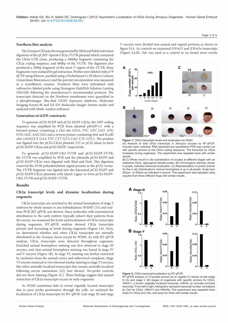
CK2! transcripts are enriched in the animal hemisphere of stage 3 embryos by whole mount in situ hybridization (WISH) [15] and real-time PCR (RT-qPCR, not shown). Since molecules with asymmetrical distribution in the early embryo typically inherit their patterns from the oocyte, we examined the levels and localization of CK2! transcripts during oogenesis. RT-qPCR analysis showed CK2! transcripts present and increasing in levels during oogenesis (Figure 1A). Next, we determined whether and when CK2! transcripts are animally distributed in the Xenopus laevis oocyte by WISH. As with RT-qPCR analysis, CK2! transcripts were detected throughout oogenesis. Enriched animal hemisphere staining was #rst observed in stage III oocytes, and clear animal hemisphere staining was found in stage IV and V oocytes (Figure 1B). In stage VI, staining was further restricted by exclusion from the animal cortex and subcortical cytoplasm. Stage VI oocytes matured in vitro showed similar staining to stage VI oocytes, like other animally localized transcripts that remain animally localized following oocyte maturation [22] (not shown). No-probe controls did not show staining (Figure 1C). "ese #ndings suggest that animal restriction of CK2! transcripts occurs in early oogenesis.
As WISH sometimes fails to reveal vegetally located transcripts due to poor probe penetration through the yolk, we analyzed the localization of CK2! transcripts by RT-qPCR. Late stage III and stage
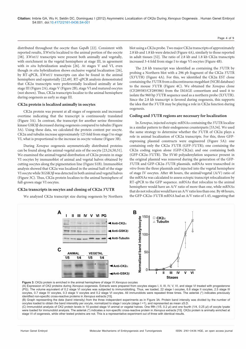
V oocytes were divided into animal and vegetal portions as shown in #gure S1A. As controls we examined XWnt11 and XWnt5a transcripts (Figure 2A,B). Odc was used as a control as we found more evenly
30
20
10
0
CK2
Oocyte StagesI II III
III
IV
IV V
IV
VI
V
V
V VI
II
A
B
250 m 250 m 500 m 500 m 500 m
Copy number (X10-7 )
per oocyte
500 m
250 m
C
Figure 1: CK2 transcripts levels and localization by WISH(A) Analysis of total CK2 transcripts in Xenopus
increases during oogenesis. This experiment was repeated twice with similar results.(B,C) Whole mount in situ hybridization of oocytes at different stages with an antisense CK2 -digoxigenin-labeled probe. (B) Chromogenic staining, shown
for the in situ hybridizations. Animal hemisphere is up in all panels. Scale bars 250µm or 500µm as indicated in picture. This experiment was repeated using oocytes from three different frogs with similar results.
1.5
1
0.5
0
0.09
0.06
0.03
0
0.12
0.08
0.04
0
A
A A A
IIIStage
Ratio
Ratio
Ratio
Ratio CK2
Ratio
Ratio
V V V
StageB V1.5
1
0.5
0
0.09
0.06
0.03
0
0.12
0.08
0.04
0 A V A V A V
Figure 2: CK2 transcript localization by RT-qPCR. RT-qPCR analysis of 10 pooled animal (A) or vegetal (V) halves at late stage
;; XWnt11, a known vegetally localized transcript;; XWnt5, an animally enriched transcript. From left to right, histograms represent transcript number normalized by Odc for CK2 , XWnt11 and XWnt5a. This experiment was repeated three times for CK2 and Odc, and twice for Wnts with similar results.
Citation: Imbrie GA, Wu H, Seldin DC, Dominguez I (2012) Xenopus Oogenesis . Human Genet Embryol S4:001. doi:10.4172/2161-0436.S4-001
Page 4 of 9
Human Genet Embryol ISSN: 2161-0436 HGE, an open access journal Molecular Mechanisms of Embryogenesis and Tumorigenesis
distributed throughout the oocyte than Gapdh [22]. Consistent with reported results, XWnt5a localized to the animal portion of the oocyte [38]. XWnt11 transcripts were present both animally and vegetally, with enrichment in the vegetal hemisphere at stage III, in agreement with in situ hybridization analysis [26]. At stages V and VI, even though in situ hybridization shows exclusive vegetal localization [26], by RT-qPCR, XWnt11 transcripts can also be found in the animal hemisphere and equatorially [22,49]. RT-qPCR analysis demonstrated that CK2! transcripts were preferentially localized animally at late stage III (Figure 2A), stage V (Figure 2B), stage VI and matured oocytes (not shown). "us, CK2! transcripts localize to the animal hemisphere during oogenesis as early as stage III.
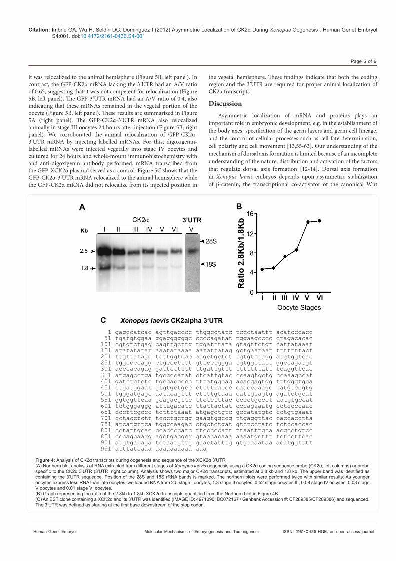
CK2! protein is localized animally in oocytesCK2! protein was present at all stages of oogenesis and increased
overtime indicating that the transcript is continuously translated (Figure 3A). In contrast, the transcript for another serine threonine kinase GSK3& decreased during oogenesis compared to tubulin (Figure 3A). Using these data, we calculated the protein content per oocyte. CK2! and tubulin increase approximately 125 fold from stage I to stage VI, what is proportional to the increase in oocyte volume (Figure 3B).
During Xenopus oogenesis asymmetrically distributed proteins can be found along the animal-vegetal axis of the oocyte [23,24,50,51]. We examined the animal/vegetal distribution of CK2! protein in stage VI oocytes by immunoblot of animal and vegetal halves obtained by cutting oocytes along the pigmentation line (Figure S1B). Immunoblot analysis showed that CK2! was localized in the animal half of the stage VI oocyte while XGSK3& was detected in both animal and vegetal halves (Figure 3C). "us, CK2! protein localizes to the animal hemisphere of full-grown stage VI oocytes.
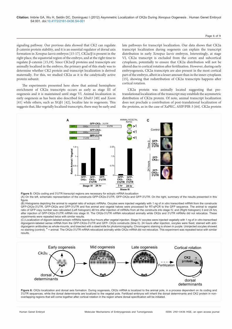
CK2! transcripts in oocytes and cloning of CK2! 3’UTRWe analyzed CK2! transcript size during oogenesis by Northern
blot using a CK2! probe. Two major CK2! transcripts of approximately 2.8 kb and 1.8 kb were detected (Figure 4A), similarly to those reported in adult tissues [52]. "e ratio of 2.8 kb and 1.8 kb CK2! transcripts increased 3-4 fold from stage I to stage VI oocytes (Figure 4B).
"e 2.8 kb transcript was identi#ed as containing the 3’UTR by probing a Northern blot with a 296 pb fragment of the CK2! 3’UTR (X3’UTR) (Figure 4A). For this, we identi#ed the CK2! EST clone containing the 3’UTR from a discontinuous megablast (NCBI database) to the mouse 3’UTR (Figure 4C). We obtained the Xenopus clone (CF289385/CF289386) from the IMAGE consortium and used it to isolate the 960 bp 3’UTR sequence used as a northern probe (X3’UTR). Since the 2.8 kb transcript is favored during oogenesis, this supports the idea that the 3’UTR may be playing a role in CK2! function during oogenesis.
Coding and 3’UTR regions are necessary for localizationIn Xenopus, injected ectopic mRNAs containing the 3’UTR localize
in a similar pattern to their endogenous counterparts [53,54]. We used the same strategy to determine whether the 3’UTR of CK2! plays a role in animal localization of CK2! transcripts. For this, three GFP-expressing plasmid constructs were engineered (Figure 5A): one containing only the CK2! 3’UTR (GFP-3’UTR); one containing the CK2! coding region alone (GFP-CK2!); and one containing both (GFP-CK2!-3’UTR). "e SV40 polyadenylation sequence present in the original plasmid was removed during the generation of the GFP-3’UTR and GFP-CK2!-3’UTR plasmids. mRNAs were transcribed in vitro from the three plasmids and injected into the vegetal hemisphere of stage IV oocytes. A$er 48 hours, the animal/vegetal (A/V) ratio of the mRNAs was calculated to assess ectopic transcript relocalization by RT-qPCR to the GFP sequence. mRNAs that relocalize to the animal hemisphere would have an A/V ratio of more than one, while mRNAs that do not relocalize would have an A/V ratio less than one. By 48 hours, the GFP-CK2!-3’UTR mRNA had an A/V ratio of 1.45, suggesting that
A B
C
I II III IV V VI PG
ANIMAL VEGETAL1/5 1/4 1/5 1/4
Anti-CK2
Anti-GSK3
Anti-Tubulin
Anti-CK2
Anti-GSK3
CK2Tubulin
150
100
50
0Normalized band intensity/oocyte
I II III IV V VI Stages
Figure 3: Xenopus oocytes (A) Expression of CK2 proteins during Xenopus oogenesis. Extracts were prepared from oocytes stages I, II, III, IV, V, VI, and stage VI treated with progesterone
oocytes, 0.7 stage IV oocytes, 0.3 stage V oocytes and 0.2 stage VI oocytes. All immunoblots were repeated three times. The asterisk (*) indicates previously Xenopus extracts [15].
oocytes loaded to obtain the band intensitity per oocyte, normalized to stage I oocyte (stage I =1), and represented as mean ±S.D.
Xenopus stage VI of oogenesis, while other tested proteins are not. This is a representative experiment out of three with identical results.
Citation: Imbrie GA, Wu H, Seldin DC, Dominguez I (2012) Xenopus Oogenesis . Human Genet Embryol S4:001. doi:10.4172/2161-0436.S4-001
Page 5 of 9
Human Genet Embryol ISSN: 2161-0436 HGE, an open access journal Molecular Mechanisms of Embryogenesis and Tumorigenesis
it was relocalized to the animal hemisphere (Figure 5B, le$ panel). In contrast, the GFP-CK2! mRNA lacking the 3’UTR had an A/V ratio of 0.65, suggesting that it was not competent for relocalization (Figure 5B, le$ panel). "e GFP-3’UTR mRNA had an A/V ratio of 0.4, also indicating that these mRNAs remained in the vegetal portion of the oocyte (Figure 5B, le$ panel). "ese results are summarized in Figure 5A (right panel). "e GFP-CK2!-3’UTR mRNA also relocalized animally in stage III oocytes 24 hours a$er injection (Figure 5B, right panel). We corroborated the animal relocalization of GFP-CK2!-3’UTR mRNA by injecting labelled mRNAs. For this, digoxigenin-labelled mRNAs were injected vegetally into stage IV oocytes and cultured for 24 hours and whole-mount immunohistochemistry with and anti-digoxigenin antibody performed. mRNA transcribed from the GFP-XCK2! plasmid served as a control. Figure 5C shows that the GFP-CK2!-3’UTR mRNA relocalized to the animal hemisphere while the GFP-CK2! mRNA did not relocalize from its injected position in
the vegetal hemisphere. "ese #ndings indicate that both the coding region and the 3’UTR are required for proper animal localization of CK2! transcripts.
Discussion Asymmetric localization of mRNA and proteins plays an
important role in embryonic development; e.g. in the establishment of the body axes, speci#cation of the germ layers and germ cell lineage, and the control of cellular processes such as cell fate determination, cell polarity and cell movement [13,55-63]. Our understanding of the mechanism of dorsal axis formation is limited because of an incomplete understanding of the nature, distribution and activation of the factors that regulate dorsal axis formation [12-14]. Dorsal axis formation in Xenopus laevis embryos depends upon asymmetric stabilization of &-catenin, the transcriptional co-activator of the canonical Wnt
I II III IV V VI V
I II III IV V VI
2.8
1.8
Kb
ACK2 3’UTR
28S
18S
16
12
8
4
0
Oocyte Stages
Ratio 2.8Kb/1.8Kb
B
C Xenopus laevis CK2alpha 3‘UTR1 gagccatcac agttgacccc ttggcctatc tccctaattt acatcccacc
51 tgatgtggaa ggaggggggc ccccagatat tggaagcccc ctagacacac 101 cgtgtctgag cagttgcttg tggatttata gtagttctgt cattataaat 151 atatatatat aaatataaaa aatattatag gctgaataat tttttttact 201 ttgttatagc tcttggtcac aagctgctct tgtgtctagg atgtggtcac 251 tggccccagg ctgccctttt gttcctggga tgtggctact ggccagatgt 301 acccacagag gattcttttt ttgattgttt tttttttatt tcaggttcac 351 atgagcctga tgccccatat ctcattgtac ccaagtgctg ccaaagccat 401 gatctctctc tgccaccccc tttatggcag acacgagtgg tttgggtgca 451 ctgatggaat gtgtgctgcc ctttttaccc caaccaaagc catgtccgtg 501 tgggatgagc aatacagttt cttttgtaaa cattgcagtg agatctgcat 551 ggtggttcaa gcagacgttc ttctctttac cccctgccct aatgtgccat 601 tctgggaggg attagacatc ttattactat cccagaaatg cctccccaac 651 cccttcgccc tcttttaaat atgagctgtc gccatatgtc cctgtgaaat 701 cctacctctt tccctgctgg gaagtggccg ttgaggttac caccacctta 751 atcatgttca tgggcaagac ctgctctgat gtctcctatc tctccaccac 801 cctattgcac ccaccccatc ttcccccatt ttaatttgca acgcctgtcc 851 cccagcaagg agctgacgcg gtaacacaaa aaaatgcttt tctccttcac 901 atgtgacaga tctaatgttg gaactatttg gtgtaaataa acatggtttt 951 atttatcaaa aaaaaaaaaa aaa
Figure 4: Xenopus laevis
V oocytes and 0.01 stage VI oocytes.
Citation: Imbrie GA, Wu H, Seldin DC, Dominguez I (2012) Xenopus Oogenesis . Human Genet Embryol S4:001. doi:10.4172/2161-0436.S4-001
Page 6 of 9
Human Genet Embryol ISSN: 2161-0436 HGE, an open access journal Molecular Mechanisms of Embryogenesis and Tumorigenesis
signaling pathway. Our previous data showed that CK2 can regulate &-catenin protein stability, and it is an essential regulator of dorsal axis formation in Xenopus laevis embryos [15-17]. CK2!/& is present in the right place, the equatorial region of the embryo, and at the right time to regulate &-catenin [15,19]. Since CK2!/& proteins and transcripts are animally localized in the embryo, the primary goal of this study was to determine whether CK2 protein and transcript localization is derived maternally. For this, we studied CK2! as it is the catalytically active protein subunit.
"e experiments presented here show that animal hemisphere enrichment of CK2! transcripts occurs as early as stage III of oogenesis and it is maintained until stage VI. Animal localization in early oogenesis as has been also described for Xbub3 [40] and Xoom [41] while others, such as XG&1 [42], localize late in oogenesis. "is suggests that, like vegetally localized transcripts, there may be early and
late pathways for transcript localization. Our data shows that CK2! transcript localization during oogenesis can explain the transcript distribution in early Xenopus laevis embryos. Interestingly, at stage VI, CK2! transcript is excluded from the cortex and subcortical cytoplasm, potentially to ensure that CK2! distribution will not be altered due to cortical rotation a$er fertilization. However, during early embryogenesis, CK2! transcripts are also present in the most cortical part of the embryo, albeit in a lesser amount than in the inner cytoplasm [15], showing that redistribution of CK2! transcripts happens a$er cortical rotation.
CK2! protein was animally located suggesting that pre-translational localization of the transcript may establish the asymmetric distribution of CK2! protein. Of note, animal transcript localization does not preclude a contribution of post-translational localization of the proteins, as in the case of XaPKC, ASIP/PIR-3 [64]. CK2! protein
A
B
CGFP
GFP-CK2
GFP-CK2
GFP-3’UTR
CK2
CK2
3’ UTR
GFP
GFP
3’UTR3’UTR
1.5
1
0.5
0
2.25
1.5
0.75
0
ANIMAL/VEGETALRATIO
CK2 CK2 CK2CK2GFP- GFP- GFP-GFP- GFP-
3’UTR 3’UTR 3’UTR3’UTR
Stage IV 24 hr 3 hr 24 hrStage III
Ratio Animal / Vegetal
><<
111
Control
GFP-CK2 -
CK2 -
CK2 -
3’UTR
3’UTR
GFP-
GFP-
Time 0
24 hours
Figure 5:
in vitro
experiments were repeated twice with similar results.in vitro transcribed
digoxigenin antibodies as whole-mounts, and bisected with a steel knife for photomicrography. Chromogenic staining is shown in purple. Uninjected oocytes showed no staining (control). *results.
Early oogenesis Mid oogenesis Late oogenesis Cortical rotation
Fertilization
dorsaldeterminants dorsal
determinants
Ck2CK2
Figure 6:
Citation: Imbrie GA, Wu H, Seldin DC, Dominguez I (2012) Xenopus Oogenesis . Human Genet Embryol S4:001. doi:10.4172/2161-0436.S4-001
Page 7 of 9
Human Genet Embryol ISSN: 2161-0436 HGE, an open access journal Molecular Mechanisms of Embryogenesis and Tumorigenesis
levels increase during oogenesis corresponding to the reported increase in total CK2 activity during oogenesis [52]. "e magnitude of change of CK2! protein and transcript levels during oogenesis di%er, suggesting that CK2! may be regulated through translational or posttranslational mechanisms that could include phosphorylation by CK2! [65].
Additionally, we showed an enrichment of the 2.8 kb CK2! transcript containing the 3'UTR during oogenesis, correlating with increased animal localization of the transcript. A similar shi$ in CK2! transcripts ratio was observed in quiescent versus proliferating mouse cells (G. A. Imbrie, unpublished data) suggesting that the ratio between the transcripts may play a role in the regulation of CK2! in diverse systems, including other animal models.
Our data shows that CK2! transcript localization requires the coding and 3’UTR regions, which is a novel mechanism for RNA localization in Xenopus oocytes, as vegetal transcript localization depends exclusively on 3'UTR sequences [66]. Although this mechanism is novel for Xenopus, recent work identi#es additional regions involved in RNA localization in other models. For example, the 5'UTR for gurken localization in Drosophila oocytes and Glutelin in Oryza sativa; and the coding region for ASH1 localization at the budding tip in Saccharomyces cerevisiae and the 10-kDa "-zein RNA in Oryza sativa [67-69]. Since transcripts and/or proteins for other regulators of dorsal axis speci#cation, such as CK2!, XGSK3!, dishevelled, Apc, axin, Tcf-3 and !-catenin are, like CK2!, animally restricted [15,22,49], it will be interesting to test whether their localization also depends upon coding and 3'UTR sequences. Given the diversity in the timing and distribution patterns of animally localized transcripts and proteins, it is possible that diverse mechanisms may be used for their localization in the Xenopus oocyte. For example, Ets-1, Ets-2 localize subcortically [43], Xbub3 localizes perinuclearly [40], and some mRNAs and proteins localize dynamically during oogenesis between cellular regions, e.g. PABP [44]. Based on the importance of animally located transcripts in embryonic development, it will be important to determine the molecular and biochemical mechanism(s) of animal localization (e.g. the RNA sequences required for localization, the role of RNA-binding proteins, the dependence on active transport or anchoring to cellular structures and the contribution of the proposed transcript degradation in the vegetal hemisphere [70]).
In summary, we found that CK2! transcript and protein are localized to the animal hemisphere during Xenopus laevis oogenesis. "ese data are consistent with a model (Figure 6) in which maternal localization ensures that CK2! is localized animally while the dorsal determinants are located vegetally. Cortical rotation a$er fertilization relocalizes the dorsal determinants to the dorsal equatorial region of the embryo, overlapping the region where CK2 proteins are enriched. CK2 will act on the dorsal determinants to upregulate &-catenin and initiate dorsal axis formation. Although many questions remain to be answered, our results on CK2! demonstrates the importance for both the coding and 3’UTR sequences for localization, and therefore may serve as a model for the study of other animally distributed transcripts in Xenopus laevis and other model organisms. Acknowledgements
Developmental Interest Group for helpful discussions. Supported by AHA Grant in
References
1. expression of embryonic axes: comparisons between different model organisms. Med Sci 20: 526-538.
2. Eyal-Giladi H (1997) Establishment of the axis in chordates: facts and speculations. Development 124: 2285-2296.
3. axis formation in Xenopus, is inactivated by UV irradiation of the oocyte. Development 119: 277-285.
4. Fujisue M, Kobayakawa Y, Yamana K (1993) Ocurrence of dorsal axis-inducing activity around the vegetal pole of an uncleaved Xenopus egg and displacement
5. years since Spemann and Mangold. Curr Top Dev Biol 30: 253-285.
6. Establishment of the dorsal-ventral axis in Xenopus embryos coincides with the dorsal enrichment of dishevelled that is dependent on cortical rotation. J Cell Biol 146: 427-437.
7.
Xenopus embryos. Cell 120: 857-871.
8. kinesin light chain and translocates during cortical rotation in Xenopus eggs. Development 130: 5425-5436.
9. Kageura H (1997) Activation of dorsal development by contact between the
Xenopus laevis. Development 124: 1543-1551.
10. Xenopus egg
Wnt pathway. Dev Biol 191: 69-79.
11. Establishment of the dorso-ventral axis in Xenopus embryos is presaged by early asymmetries in beta-catenin that are modulated by the Wnt signaling pathway. J Cell Biol 136: 1123-1136.
12. Xenopus. Development 131: 3491-3499.
13. polarity. Semin Cell Dev Biol 17: 168-174.
14. in the Xenopus egg with the Wnt/beta-catenin signaling pathway. Mech Dev 89: 93-102.
15. Xenopus embryos. Dev Biol 274:
110-124.
16. Song DH, Sussman DJ, Seldin DC (2000) Endogenous protein kinase CK2 participates in Wnt Signaling in mammary epithelial cells. J Biol Chem 275: 23790-23797.
17. Song DH, Dominguez I, Mizuno J, Kaut M, Mohr SC, et al. (2003) CK2 phosphorylation of the armadillo repeat region of beta-catenin potentiates Wnt signaling. J Biol Chem 278: 24018-24025.
18. Wu H, Symes K, Seldin DC, Dominguez I (2009) Threonine 393 of beta-catenin regulates interaction with Axin. J Cell Biochem 108: 52-63.
19. Dominguez I, Mizuno J, Wu H, Imbrie GA, Symes K, et al. (2005) A role for CK2alpha/beta in Xenopus early embryonic development. Mol Cell Biochem 274: 125-131.
20.
embryos. Mech Dev 57: 191-198.
21. animal-vegetal axis during early Xenopus development. J Embryol Exp Morphol 95: 15-35.
Citation: Imbrie GA, Wu H, Seldin DC, Dominguez I (2012) Xenopus Oogenesis . Human Genet Embryol S4:001. doi:10.4172/2161-0436.S4-001
Page 8 of 9
Human Genet Embryol ISSN: 2161-0436 HGE, an open access journal Molecular Mechanisms of Embryogenesis and Tumorigenesis
22. Xenopus
laevis
23. animal-vegetal axis of frog eggs. Dev Biol 58: 1-10.
24. Xenopus laevis. An atlas of the histology. 1991: Springer-Verlag.
25. Xenopus revealed by subtractive immunization. Dev Biol 192: 446-454.
26. Xenopus wnt gene. Development 119: 1161-1173.
27. Lustig KD, Kroll KL, Sun EE, Kirschner MW (1996) Expression cloning of a Xenopusblastopore lip formation. Development 122: 4001-4012.
28. Stennard F, Carnac G, Gurdon JB (1996) The Xenopus T-box gene,
mesoderm formation. Development 122: 4179-4188.
29. Zhang J, King ML (1996) Xenopusduring oogenesis and encodes a novel T-box transcription factor involved in mesodermal patterning. Development 122: 4119-4129.
30. Houston DW, Zhang J, Maines JZ, Wasserman SA, King ML (1998) A Xenopus
homologue of Drosophila boule. Development 125: 171-180.
31. Xenopus eggs. Cell 42: 769-777.
32. hemisphere in Xenopus eggs codes for a growth factor related to TGF-beta. Cell 51: 861-867.
33. Xenopus oocytes. Semin Cell Dev Biol 8: 529-540.
34.
35. in Xenopus oocytes. Methods 51: 146-151.
36. Xenopus
37. antagonize maternal VegT activity and beta-Catenin signaling in Xenopus embryos. Embo J 26: 2942-2954.
38. Xenopus Wnt-1/int-1-related proteins and
characterization of their transient expression during embryonic development. Dev Biol 143: 230-234.
39. Xenopus
laevis. Mech Dev 1: S143-S148.
40. Goto T, Kinoshita T (1999) Maternal transcripts of mitotic checkpoint gene, Xenopus early embryo.
41. protein maternally expressed and involved in the gastrulation movement of Xenopus embryos. Int J Dev Biol 43: 479-485.
42.
localized to the animal pole of Xenopus laevis oocyte and embryos. Mech Dev 59: 141-151.
43. Meyer D, Durliat M, Senan F, Wolff M, Andre M, et al. (1997) Ets-1 and Ets-2 proto-oncogenes exhibit differential and restricted expression patterns during Xenopus laevis oogenesis and embryogenesis. Int J Dev Biol 41: 607-620.
44. Schroeder KE, Yost HJ (1996) Xenopusis localized during oogenesis and associated with large complexes in blastula. Dev Genet 19: 268-276.
45. functions as a transmembrane protein for gastrulation movement in Xenopus embryos. Dev Growth Differ 43: 25-31.
46. XenopusCell 73: 555-570.
47. Xenopus laevis (Daudin). I. Stages of oocyte development in laboratory maintained animals. J Morphol 136: 153-179.
48. Dominguez I, Marshall MS, Gibbs JB, Garcia de Herreros A, Cornet ME, et
phosphatidylcholine-hydrolysing phospholipase C. Embo J 10: 3215-3220.
49. the Xenopus laevis87-91.
50. deployment of oogenetically derived Xenopus yolk/nonyolk proteins. J Embryol Exp Morphol 97 Suppl: 45-64.
51. Xenopus revealed by subtractive immunization. Dev Biol 192: 446-454.
52. the subunits of protein kinase CK2 during oogenesis in Xenopus laevis. Eur J Biochem 232: 671-676.
53. plasm component, to the mitochondrial cloud in Xenopus stage I oocytes. Development 122: 2947-2953.
54. Kloc M, Larabell C, Etkin LD (1996) Elaboration of the messenger transport Xenopus
oocytes. Dev Biol 180: 119-130.
55. embryos: some common principles. Med Sci 20: 414-423.
56. Heasman J (2006) Maternal determinants of embryonic cell fate. Semin Cell Dev Biol 17: 93-98.
57.
58.
59. Sci 119: 979-987.
60.
61. cortex of ascidian oocytes, zygotes and embryos. Biol Cell 97: 35-49.
62. White JA, Heasman J (2008) Maternal control of pattern formation in Xenopus laevis. J Exp Zoolog B Mol Dev Evol 310: 73-84.
63. cell stage: cytoplasmic and cortical reorganizations that pattern the ascidian embryo. Dev Dyn 236: 1716-1731.
64.
Xenopus oocytes. Development 127: 5021-5031.
65. the stability of the regulatory CK2beta subunit. Oncogene 21: 3754-3764.
66. Cell Mol Life Sci 69: 535-552.
67.
transcript. Dev Biol 221: 435-446.
68.
reporter particle in vivo. Curr Biol 9: 333-336.
Citation: Imbrie GA, Wu H, Seldin DC, Dominguez I (2012) Xenopus Oogenesis . Human Genet Embryol S4:001. doi:10.4172/2161-0436.S4-001
Page 9 of 9
Human Genet Embryol ISSN: 2161-0436 HGE, an open access journal Molecular Mechanisms of Embryogenesis and Tumorigenesis
Submit your next manuscript and get advantages of OMICS Group submissionsUnique features:
Special features:
Molecular Mechanisms of Embryogenesis and Tumorigenesis
69. 70. St Johnston D (2005) Moving messages: the intracellular localization of