Mitochondrial localization of two brain proteins,Mitochondrial localization of two brain proteins,...
124
Mitochondrial localization of two brain proteins, p42 IP4 /centaurin-α1/ADAP1 and CNP, and their involvement in regulation of mitochondrial Ca 2+ Dissertation zur Erlangung des akademischen Grades doctor rerum naturalium (Dr. rer. nat.) genehmigt durch die Fakultät für Naturwissenschaften der Otto-von-Guericke-Universität Magdeburg von M. Sc. Anastasia Galvita geb. am 11.05.1973 in Novosibirsk, Russia Gutachter: Prof. Dr. Georg Reiser Prof. Dr. Carsten Culmsee eingereicht am: 15 Januar, 2010 verteidigt am: 28 Juni 2010
Transcript of Mitochondrial localization of two brain proteins,Mitochondrial localization of two brain proteins,...
Mitochondrial localization of two brain proteins,
p42IP4 /centaurin-α1/ADAP1 and CNP, and their
involvement in regulation of mitochondrial Ca2+
Dissertation
zur Erlangung des akademischen Grades
doctor rerum naturalium
(Dr. rer. nat.)
genehmigt durch die Fakultät für Naturwissenschaften der Otto-von-Guericke-Universität Magdeburg
von M. Sc. Anastasia Galvita
geb. am 11.05.1973 in Novosibirsk, Russia
Gutachter: Prof. Dr. Georg Reiser
Prof. Dr. Carsten Culmsee
eingereicht am: 15 Januar, 2010
verteidigt am: 28 Juni 2010
ACKNOWLEGEMENTS
I am very grateful to those who gave me all kinds of help and support during this work
at the Institute of Neurobiochemistry, Otto-von-Guericke University Magdegurg.
First of all, I would like to sincerely thank my supervisor, Prof. Dr. Georg Reiser, for
providing me an opportunity to join his lab to complete my Ph.D. work. His invaluable
knowledge, constructive discussion, professional guidance and constant support enabled me
to achieve my goals efficiently and easily, and to be of great benefit to my scientific career
throughout my life. I am happy to be a member of the Graduate School directed by Prof.
Reiser, and the wonderful lectures, seminars and practical courses he organized opened my
scientific ken and stimulated my idea. Of course, many thanks also to our Graduate School
secretary Frau Manuela Dullin-Viehweg, who assisted the nice organization of the scientific
and social activities.
I am greatly indebted to the group of Dr. Tamara Azarashvili from Institute of
Theoretical and Experimental Biophysics RAS, Pushchino, Russia: Dr. Dmitry Grachev, Dr.
Olga Krestinina and Yulia Baburina. During their visits in Magdeburg they were introducing
and helping me with mitochondrial techniques. My special appreciation goes to Dr. Dmitry
Grachev who performed the measurement of mitochondrial parameters in chamber in the
frame of our cooperative project. The data of these measurements are presented in the
following Figures: 3.3.1; 3.3.2; 3.3.3; 3.3.4; 3.3.6; 3.3.7; 3.6.3; 3.6.4; 3.6.5; 3.6.6.
I am thankful to Dr. Roland Hartig from Institute of Immunology, Otto-von-Guericke
University Magdeburg. He performed flow cytometry analysis.
I am grateful to Dr. Rolf Stricker for valuable scientific suggestions and discussions.
His long-standing research experience and in-depth knowledge in the field of protein
chemistry was important for channelling the direction of my study. He helped me in the
critical analysis of the data obtained, which was crucial to the completion of my work.
I am thankful to Dr. Fariba Sedehizade and Dr. Theodor Hanck who helped me in my
work involving the molecular biology aspect, in the establishment of stable cell lines and in
the introduction of the GST pull-down assays.
I am also thankful to Dr. Mohan E. Tulapurkar for helping me with the techniques of
confocal laser scanning microscopy
I also appreciate Dr. Abidat Schneider, Frau Evelyn Busse, Frau Ilka Kramer, Frau
Petra Grüneberg, for their excellent technical assistance, as well as other colleagues in the lab,
Dr. Andrea Haase, Frau Anke Imrich, Frau Annette Jürgen, Frau Claudia Borrmann, Dr.
Denise Ecke, Frau Dorothe Terhardt, Dr. Elena Sokolova, Dr. Ewa Ostrowska, Dr. Gregor
2

Zündorf, Dr. Marina Sergeeva, Dr. Mikhail L. Strokin, Dr. Rainer Schäfer, Herr Rongyu Li,
Frau Sabine Hein, Dr. Stefan Kahlert, Dr. Stepan Aleshin, Dr. Tanuja Rohatgi, Dr. Victoria
Bunik, Dr. Weibo Luo and Dr. Ying Fei Wang for their cooperation and support, and Frau
Ines Klaes, Herr Peter Ehrbarth for their kind support, during the course of this work.
Finally, I would express my special gratitude to my husband Dr. Vladimir Galvita for
his advises, encouragement and love, and to my parents for their support and understanding.
3
Contents
1. Introduction .......................................................................................................................... 7
1.1 Mitochondria in cellular Ca2+ signalling and cell death. ............................................. 7 1.1.1 Permeability transition pore (PTP) ............................................................................. 7 1.1.2 Re-evaluation of the classical model of PTP.............................................................. 9 1.1.3 Role of PTP in cell death.......................................................................................... 11
1.2 p42IP4 /centaurin-α1/ADAP1 ........................................................................................ 13 1.2.1 p42IP4 /centaurin-α1 domain structure ...................................................................... 13 1.2.2 Localization of p42IP4 in brain.................................................................................. 14 1.2.3 Interaction partners of p42IP4 .................................................................................... 15 1.2.4 Known functions of p42IP4........................................................................................ 16 1.2.5 The ligands of p42IP4 ................................................................................................ 18
1.3 2,3'-Cyclic nucleotide-3'-phosphodiesterase (CNP).................................................. 20 1.3.1 Two isoforms of CNP............................................................................................... 20 1.3.2 Motifs in primary structure and modifications of CNP............................................ 20 1.3.3 Mitochondrial localization of CNP........................................................................... 22 1.3.4 Enzymatic characteristics of CNP ............................................................................ 22 1.3.5 Three-dimensional structural features of CNP and link to RNA metabolism.......... 23 1.3.6 Interaction partners of CNP...................................................................................... 24 1.3.7 Functions of CNP ..................................................................................................... 25
1.4 Aims of this study.......................................................................................................... 28 1.4.1 Motivation ................................................................................................................ 28 1.4.2 Main aims and goals of study ................................................................................... 28 1.4.3 Strategy..................................................................................................................... 29
2. Materials and Methods ...................................................................................................... 30
2.1 Materials ........................................................................................................................ 30 2.1.1 Chemicals and Reagents ........................................................................................... 30 2.1.2 Antibodies................................................................................................................. 31 2.1.3 Cells, medium and related reagents .......................................................................... 32 2.1.4 Animals..................................................................................................................... 33 2.1.5 Plasmid vectors:........................................................................................................ 33 2.1.6 Small interfering RNAs (siRNAs)............................................................................ 34 2.1.7 Enzymes.................................................................................................................... 34 2.1.8. Molecular weight markers ....................................................................................... 34 2.1.9 Kits............................................................................................................................ 35 2.1.10 Laboratory instruments........................................................................................... 35 2.1.11 Buffers and solvents ............................................................................................... 37 2.1.12. Oligonuleotides for PCR and sequencing.............................................................. 38
2.2 Methods.......................................................................................................................... 39 2.2.1 RT-PCR .................................................................................................................... 39 2.2.2 Plasmid constructs .................................................................................................... 39 2.2.3 Agarose gel electrophoresis of DNA........................................................................ 40
4
2.2.4 Transformation of E. coli with plasmid DNA by heat-shock method (CaCl2 based)........................................................................................................................................... 40 2.2.5 Expression and purification of glutathione S-transferase (GST) and GST-p42IP4 constructs from E. coli....................................................................................................... 41 2.2.6 Cell culture and transfection..................................................................................... 42 2.2.7 Confocal microscopy................................................................................................ 42 2.2.8 Subcellular fractionation by differential centrifugation ........................................... 43 2.2.9 Preparation of mitochondrial lysate and cytosolic fraction from rat brain............... 43 2.2.10 Isolation of mitochondria from cultured cells ........................................................ 44 2.2.11 Subfractionation of mitochondria........................................................................... 44 2.2.12 Electrophoresis and immunoblotting...................................................................... 45 2.2.13 GST-pull-down assay ............................................................................................. 45 2.2.14 Immunoprecipitation and co-immunoprecipitation ................................................ 46 2.2.15 Isolation of rat brain mitochondria for functional study......................................... 46 2.2.16 Evaluation of mitochondrial functions ................................................................... 47 2.2.17 Detection of CNP activity in isolated mitochondria............................................... 47 2.2.18 Small interfering RNA (siRNA)............................................................................. 48 2.2.19 Apoptosis detection ................................................................................................ 48
2.3 Statistical analysis ......................................................................................................... 49
3. Results ................................................................................................................................. 50
3.1. Possible role of p42IP4 in cell protection or cell death ............................................... 50 3.1.1 Influence of p42IP4 overexpression on apoptosis induced in N2a cells. ................... 50 3.1.2 Influence of p42IP4 overexpression on cell cycle in N2a cells. ................................. 52
3.2. Novel localization of p42IP4: mitochondrial localization........................................... 54 3.2.1 p42IP4 is localized in mitochondria, isolated from transfected CHO cells ............... 54 3.2.2 p42IP4 is localized in inner mitochondrial compartments of transfected CHO cells. 55 3.2.3 p42IP4 is localized in mitochondria, isolated from transfected N2a cells ................. 57
3.3 p42IP4 is involved in regulation of mitochondrial Ca2+ .............................................. 60 3.3.1 p42IP4 accelerates Ca 2+-induced PTP opening in mitochondria from N2a cells...... 60 3.3.2 Effects of p42IP4 ligands, phosphatidylinositol(3,4,5)trisphosphate (PIP3) and inositol(1,3,4,5)tetrakisphosphate (IP4), on mitochondrial functions................................ 69
3.4. Interaction of p42IP4 with CNP and α-tubulin in rat brain mitochondria.............. 70 3.4.1 Localization of p42IP4, CNP and α-tubulin in rat brain mitochondria ...................... 71 3.4.2 p42IP4 interacts with CNP and α-tubulin in vitro....................................................... 71 3.4.3 p42IP4 interacts with CNP and α-tubulin in vivo........................................................ 72 3.4.4 CNP interacts with α-tubulin in vivo......................................................................... 73
3.5. Mitochondrial localization of CNP............................................................................. 74 3.5.1 CNP localization in sub-mitochondrial fractions of RBM ....................................... 74 3.5.2 CNP interaction with ANT and VDAC.................................................................... 75
3.6. CNP is involved in regulation of mitochondrial Ca2+ ............................................... 76 3.6.1 Influence of Ca 2+-induced PTP on CNP activity in RBM....................................... 76 3.6.2 Effect of CNP knock-down on Ca2+-induced PTP ................................................... 77 3.6.3 Effects of CNP substrates, 2’,3’-cAMP, 2’,3’-cNADP, on mitochondrial functions........................................................................................................................................... 82
4. Discussion............................................................................................................................ 89
5
4.1 The mechanism of mitochondrial Ca2+-induced PTP opening remains unknown . 89
4.2 Known functions of p42IP4............................................................................................ 89
4.3 Novel localization and function of p42IP4. Involvement of p42IP4 and its ligands in regulation of mitochondrial Ca2+-induced PTP opening................................................. 90
4.4 Known functions of CNP.............................................................................................. 92
4.5 Novel function of CNP. Involvement of CNP and its substrates in regulation of mitochondrial Ca2+-induced PTP opening........................................................................ 93
4.6 Hypothesis for functional importance of interaction between p42IP4, CNP and α-tubulin in mitochondria...................................................................................................... 95 4.7 Conclusions. Novel regulators of Ca2+-induced PTP opening. ............................... 100
5. Abstract ............................................................................................................................. 101
6. Zusammenfassung............................................................................................................ 102
7. References ......................................................................................................................... 105
8. Abbreviations.................................................................................................................... 119
9. Appendix ........................................................................................................................... 121
Curriculum vitae............................................................................................................... 121
Publications and presentations ........................................................................................ 122
Conferences and symposiums .......................................................................................... 123
6
1. Introduction
1.1 Mitochondria in cellular Ca2+ signalling and cell death. A basic notion of cell biology is that calcium, in its ionic form (Ca2+), is a ubiquitous
second messenger of eukaryotic cells, participating in numerous signal transduction pathways.
Cellular Ca2+ signals are crucial in the control of most physiological processes, cell injury and
programmed cell death. Mitochondria, in addition to their function as cellular power plants,
have been recognized to play a central role in Ca2+ homeostasis and cellular Ca2+ signalling
(Duszynski et al., 2006).
It has been known for several decades that sequestration of vast amounts of Ca2+ in
the mitochondria occurs under various pathophysiological conditions and contributes to the
demise of the cells (Krieger and Duchen, 2002). In these paradigms, the loss of the balance
between plasma membrane Ca2+ influx and Ca2+ export leads to a sustained elevation in
cytosolic Ca2+ from 100 nM to ≥ 1 μM, inducing a progressive increase in mitochondrial Ca2+
uptake. Mitochondria dampen changes in cytosolic Ca2+ loads and sustain cellular Ca2+
homeostasis that is required for normal function of cells. However, mitochondria take up a
limited amount of calcium up to a certain threshold. Accumulation of Ca2+ over-load leads to
increased permeability of the inner mitochondrial membrane due to formation of an
unselective pore at the contact site between outer and inner membranes (Smaili et al., 2000;
Sullivan et al., 2005). This permeability transition pore (PTP) forms the major Ca2+ efflux
pathway from mitochondria. Since mitochondria both accumulate and release Ca2+, these
organelles play various roles within cells (Giacomello et al., 2007).
Opening of the PTP by Ca2+ is effectively modulated by several factors, including
reactive oxygen species (ROS), pH, mitochondrial membrane potential (Δψm), the level of
which is affected by a variety of metabolic intermediates, signalling molecules and drugs
(Bernardi, 1999). Although Ca2+-dependent mitochondrial membrane permeabilization has
been implicated in a broad range of cell death and tissue injury, the exact mechanism of the
PTP opening remains elusive (Hajnoczky et al., 2006). Therefore study and search for new
proteins and molecules involved in control of Ca2+-induced PTP opening will provide
additional insight into pathways that are critical for cell survival and cell death.
1.1.1 Permeability transition pore (PTP)
The mitochondrial permeability transition is defined as the sudden nonselective
increase in the permeability of the inner mitochondrial membrane (IMM) to solutes of
molecular mass less than ~1500 Da, and results in loss of mitochondrial membrane potential,
7
mitochondrial swelling, and rupture of the outer mitochondrial membrane (OMM). More than
30 years ago, it was first reported that the mitochondrial permeability transition is a
consequence of Ca2+-induced increased permeability of the IMM that is characterized by
simultaneous stimulation of ATPase, uncoupling of oxidative phosphorylation, and loss of
respiratory control (Hunter et al., 1976).
Later it was suggested that the mitochondrial permeability transition is not a
consequence of non-specific mitochondrial membrane damage; it is, instead, the result of the
opening of an authentic pore or megachannel. The last 25 years of study dealing with the
mitochondrial permeability transition have been characterized by an intensive search for the
identity of the components of the mitochondrial permeability transition megachannel.
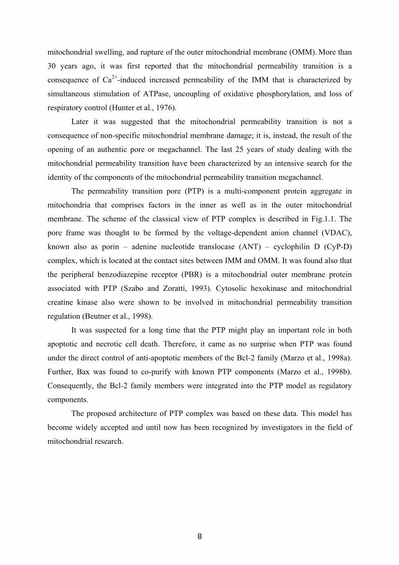
The permeability transition pore (PTP) is a multi-component protein aggregate in
mitochondria that comprises factors in the inner as well as in the outer mitochondrial
membrane. The scheme of the classical view of PTP complex is described in Fig.1.1. The
pore frame was thought to be formed by the voltage-dependent anion channel (VDAC),
known also as porin – adenine nucleotide translocase (ANT) – cyclophilin D (CyP-D)
complex, which is located at the contact sites between IMM and OMM. It was found also that
the peripheral benzodiazepine receptor (PBR) is a mitochondrial outer membrane protein
associated with PTP (Szabo and Zoratti, 1993). Cytosolic hexokinase and mitochondrial
creatine kinase also were shown to be involved in mitochondrial permeability transition
regulation (Beutner et al., 1998).
It was suspected for a long time that the PTP might play an important role in both
apoptotic and necrotic cell death. Therefore, it came as no surprise when PTP was found
under the direct control of anti-apoptotic members of the Bcl-2 family (Marzo et al., 1998a).
Further, Bax was found to co-purify with known PTP components (Marzo et al., 1998b).
Consequently, the Bcl-2 family members were integrated into the PTP model as regulatory
components.
The proposed architecture of PTP complex was based on these data. This model has
become widely accepted and until now has been recognized by investigators in the field of
mitochondrial research.
8
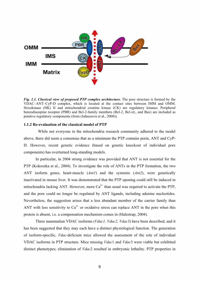
Fig. 1.1. Classical view of proposed PTP complex architecture. The pore structure is formed by the VDAC–ANT–CyP-D complex, which is located at the contact sites between IMM and OMM. Hexokinase (HK) II and mitochondrial creatine kinase (CK) are regulatory kinases. Peripheral benzodiazepine receptor (PBR) and Bcl-2-family members (Bcl-2, Bcl-xL, and Bax) are included as putative regulatory components (from (Juhaszova et al., 2008)). 1.1.2 Re-evaluation of the classical model of PTP
While not everyone in the mitochondria research community adhered to the model
above, there did seem a consensus that as a minimum the PTP contains porin, ANT and CyP-
D. However, recent genetic evidence (based on genetic knockout of individual pore
components) has overturned long-standing models.
In particular, in 2004 strong evidence was provided that ANT is not essential for the
PTP (Kokoszka et al., 2004). To investigate the role of ANTs in the PTP formation, the two
ANT isoform genes, heart-muscle (Ant1) and the systemic (Ant2), were genetically
inactivated in mouse liver. It was demonstrated that the PTP opening could still be induced in
mitochondria lacking ANT. However, more Ca2+ than usual was required to activate the PTP,
and the pore could no longer be regulated by ANT ligands, including adenine nucleotides.
Nevertheless, the suggestion arises that a less abundant member of the carrier family than
ANT with less sensitivity to Ca2+ or oxidative stress can replace ANT in the pore when this
protein is absent, i.e. a compensation mechanism comes in (Halestrap, 2004).
Three mammalian VDAC isoforms (Vdac1, Vdac2, Vdac3) have been described, and it
has been suggested that they may each have a distinct physiological function. The generation
of isoform-specific, Vdac-deficient mice allowed the assessment of the role of individual
VDAC isoforms in PTP structure. Mice missing Vdac1 and Vdac3 were viable but exhibited
distinct phenotypes; elimination of Vdac2 resulted in embryonic lethality. PTP properties in
9
mitochondria from Vdac1-null mice were indistinguishable from those of wild type mice
(Krauskopf et al., 2006).
In subsequent work it was found that mitochondria from Vdac 1, Vdac 3-, and Vdac 1/
Vdac 3-null mice exhibited Ca2+ and oxidative stress–induced opening of PTPs that were
indistinguishable from wild type mitochondria (Baines et al., 2007). Furthermore, mouse
embryonic fibroblasts lacking VDAC (Vdac1, Vdac2, Vdac3, various combinations of two, or
all three VDAC isoforms), showed similar patterns of PTP induction compared to control
cells. In addition, the existence of a VDAC-independent model of Bcl-2 family member–
mediated cell killing was confirmed. Wild type and Vdac-deficient mitochondria and cells
responded to activation or over expression of Bax and Bid by equivalent cytochrome c
release, caspase cleavage, and cell death. These results make a strong argument against any
indispensable role of VDACs in both PTP-mediated and Bax–Bak-mediated cell death. The
experiments with Vdac-deficient cells and mitochondria strongly cast doubt on the validity of
the classical model of PTP formation, which assumes that the pore contains VDAC and is
formed at the contact sites between mitochondrial membranes.
While VDAC and ANT had been excluded as essential pore components, it is
possible, that these proteins serve some regulatory function. The function of hexokinase II as
a regulator of the pore, however, is yet unresolved. Only the role of the CyP-D has been
clearly established. Recently, genetic studies from four independent groups on CyP-D
knockout mice (Ppif−/− mice) confirmed the critical role of CyP-D in regulation of the PTP
machinery and provided new insights into the role of PTP in cell death (Baines et al., 2005;
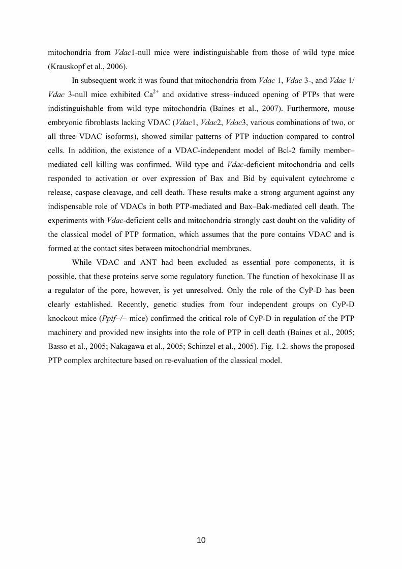
Basso et al., 2005; Nakagawa et al., 2005; Schinzel et al., 2005). Fig. 1.2. shows the proposed
PTP complex architecture based on re-evaluation of the classical model.
10
Fig. 1.2 Current view of proposed PTP complex architecture, based on reappraisal of the classical model. The elements comprising the pore itself (denoted “PTP” for permeability transition pore) are presently unidentified, but are probably regulated by the adjacent elements as indicated. Note that VDAC, portrayed as a “shadow,” is no longer seen as an essential pore component based on recent genetic evidence. Question mark symbols signify, where open questions remain (from (Juhaszova et al., 2008)).
Even now, despite major efforts, the molecular nature of the PTP remains enigmatic
(Juhaszova et al., 2008). Very recently, it was suggested that the protein fulfilling the role of
the transmembrane pore component is the mitochondrial phosphate carrier (PiC) (Leung and
Halestrap, 2008). Moreover, these studies implicate the PiC in PTP formation and are
consistent with a calcium-triggered conformational change of the PiC, facilitated by CyP-D,
inducing pore opening (Leung et al., 2008). It was proposed that this is enhanced by an
association of the PiC with the "c" conformation of the ANT, as agents that modulate pore
opening may act on either or both of the two proteins, PiC and ANT (Halestrap, 2009).
1.1.3 Role of PTP in cell death
It has been known, and accepted by most investigators for the last 10 years, that cells
can undergo a sudden increase in mitochondrial inner membrane permeability to ions and
solutes. This disequilibrium causes a dissipation of the membrane potential, loss of ion
homeostasis, and impairment of ATP synthesis, which results in passive swelling of the
organelle, release of cytochrome c and cellular apoptosis, or necrotic cell death.
Recent genetic experiments provided critical new insights into fundamental
mechanisms of cell death (Baines et al., 2005; Basso et al., 2005; Nakagawa et al., 2005;
Schinzel et al., 2005). In particular, CyP-D-deficient cells also responded to various apoptotic
stimuli in a similar manner as the wild type cells but showed considerable resistance to
necrotic cell death, suggesting that CyP-D is not a central component of the apoptotic death
pathway. These conclusions lead to a revision of the current dogma and support the idea that
11
the PTP is not a major participant in apoptotic cell death, but rather plays an important role in
necrotic cell death. This seems to be the case at least in cardiac myocytes, neurons, and
fibroblasts. These ideas represent a significant contribution to the understanding of the role,
which mitochondria play in cell death.
Recently, a model for the activation of the PTP for cell death was proposed (Grimm
and Brdiczka, 2007). It was suggested that the PTP alters its composition during apoptosis,
that anti-apoptotic factors are released and pro-apoptotic subunits recruited for apoptosis
induction (Grimm and Brdiczka, 2007). The best evidence for this is that hexokinase inhibits
cytochrome c (Cyt c) release. The structural change of VDAC achieved by interaction with
ANT is recognized at the mitochondrial surface and leads to higher affinity of hexokinase.
Recent observations suggest that this contact site structure is the preferred target of Bax
molecules. It was found that hexokinase and Bax compete for the same binding site of VDAC
(Pastorino et al., 2002). Moreover, VDAC and ANT were immuno-precipitated with Bax
from extracts of cardiomyocytes (Capano and Crompton, 2002). The binding of hexokinase to
the contact sites is induced by protein kinase B (Akt) (Gottlob et al., 2001). Activated protein
kinase B can suppress apoptosis. The activity of mitochondrial bound hexokinase was found
to be important for protein kinase B-linked suppression of Cyt c release and apoptosis
(Majewski et al., 2004).
Hexokinase was furthermore used as a tool to isolate the contact site forming complex of
outer membrane VDAC and inner membrane ANT. Cyt c remained attached to the
hexokinase VDAC-ANT complexes that were reconstituted in phospholipid vesicles. The
vesicles were loaded with malate and treated with Bax in increasing concentrations. Bax
liberated the endogenous Cyt c but did not release the internal malate. The Bax dependent
liberation of endogenous Cyt c was abolished when the VDAC-ANT complex was dissociated
by bongkrekate or by stabilizing the hexokinase binding through addition of glucose. The
internal malate could be released through opening the PTP by addition of Ca2+. This
suggested that Bax did not form unspecific pores for malate but released the Cyt c
dependently on the actual structure of the VDAC-ANT complex (Vyssokikh et al., 2002;
Vyssokikh et al., 2004). Thus, the permeability transition in cell death is brought about by
dynamic protein complexes that have the physiological function to regulate energy
metabolism. These protein complexes change according to the needs of the cell and respond
to different external and internal stimuli. During cell death the associations are altered in a
way that causes the permeability transition in mitochondria (Grimm and Brdiczka, 2007).
12
It was also shown that conventionally defined “pro-apoptotic” and “anti-apoptotic”
members of the Bcl-2 family are critical mediators of protection signalling downstream of
GSK-3β, where they regulate the susceptibility of the PTP to oxidant stress. Moreover, in the
context of limiting PTP induction, the actions of the Bcl-2 family are effecting changes in cell
survival and death via necrosis rather than apoptosis-related pathways (Juhaszova et al.,
2008).
1.2 p42IP4 /centaurin-α1/ADAP1 1.2.1 p42IP4 /centaurin-α1 domain structure
p42IP4 is a brain-specific protein that is also called centaurin-α1 and
phosphatidylinositoltrisphosphate binding protein, PIP3 BP (Hammonds-Odie et al., 1996;
Stricker et al., 1997). This protein specifically recognizes two second messengers, the
membrane lipid phosphatidylinositol(3,4,5)trisphosphate (PIP3) and the soluble
inositol(1,3,4,5)tetrakisphosphate (IP4) (Hanck et al., 1999).
Previously, a receptor protein with high affinity for IP4 was solubilized from pig
cerebellar membranes and purified (Donie and Reiser, 1991). The protein was identified in
SDS-PAGE after photoaffinity labelling as a 42 kDa protein band, p42IP4 (Reiser et al., 1991).
Later, cloning of pig p42IP4 (Stricker et al., 1997) had revealed that this protein is
homologous to centaurin-α, the PIP3 binding protein cloned from a rat brain (Hammonds-Odie
et al., 1996). The protein PIP3 BP, virtually identical to pig p42IP4, was cloned from bovine
brain and characterized as a PIP3 binding protein (Tanaka et al., 1997). In addition, the
existence of rat p42IP4 was established (Aggensteiner et al., 1998).
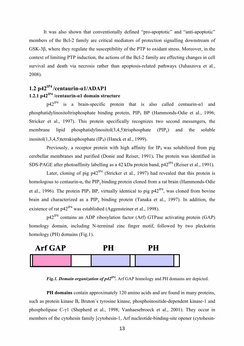
p42IP4 contains an ADP ribosylation factor (Arf) GTPase activating protein (GAP)
homology domain, including N-terminal zinc finger motif, followed by two pleckstrin
homology (PH) domains (Fig.1).
Arf GAP PH PHArf GAP PH PH
Fig.1. Domain organization of p42IP4. Arf GAP homology and PH domains are depicted.
PH domains contain approximately 120 amino acids and are found in many proteins,
such as protein kinase B, Bruton´s tyrosine kinase, phosphoinositide-dependent kinase-1 and
phospholipase C-γ1 (Shepherd et al., 1998; Vanhaesebroeck et al., 2001). They occur in
members of the cytohesin family [cytohesin-1, Arf nucleotide-binding-site opener (cytohesin-
13
2), general receptor for phosphoinositides-1 (cytohesin-3)] (Jackson et al., 2000), RasGTPase
activating proteins (GAP1m and GAP1IP4BP) (Cozier et al., 2000) and members of the
centaurin-α family (p42IP4/centaurin-α1; PIP3BP, and centaurin-α2) (Tanaka et al., 1997;
Stricker et al., 1999; Venkateswarlu et al., 1999; Hanck et al., 2004). For these proteins
specific in vitro binding to PIP3 and IP4 has been reported (Jackson et al., 2000; Lemmon and
Ferguson, 2000). Most of the PH domain-containing proteins have a single PH domain, which
is required for ligand binding. In contrast, the centaurin family proteins contain two PH
domains in tandem orientation. Both PH domains of p42IP4 within the protein are required for
full in vitro PIP3 binding activity (Tanaka et al., 1997). PH domains are also required for
phosphoinositide 3-kinase-dependent translocation to the plasma membrane (Hammonds-
Odie et al., 1996; Tanaka et al., 1997; Tanaka et al., 1999; Venkateswarlu et al., 1999;
Sedehizade et al., 2002).
The ARF GAP domain was originally identified in members of a distinct family of
ARF1 GAPs (Cukierman et al., 1995; Poon et al., 1996). It contains a characteristic C4 - type
zinc finger motif and a conserved arginine that is required for activity, within a particular
spacing (CX2CX16CX2CX4R) (Kahn et al., 2008). It was demonstrated that p42IP4 is
functionally homologous to Gcs1, a yeast ARF GAP, but it was not possible to detect any
ARF GAP activity for p42IP4 in this time period (Venkateswarlu et al., 1999; Jackson et al.,
2000). Only recently the in vivo Arf GAP activity of p42IP4 towards Arf6 has been described
(Venkateswarlu et al., 2004). Therefore, p42IP4 was renamed systematically ADAP1 (A Dual
PH domain ARF GAP) to stress the relationships within the ARF GAP superfamily (Kahn et
al., 2008).
1.2.2 Localization of p42IP4 in brain
p42IP4 is rather exclusively expressed in brain (Stricker et al., 2003). The respective
cDNA has been cloned from pig (Stricker et al., 1997), rat (Hammonds-Odie et al., 1996);
(Aggensteiner et al., 1998), bovine (Tanaka et al., 1997) and human brain (Venkateswarlu et
al., 1999; Sedehizade et al., 2002). Expression of p42IP4 was described to be neurone-specific
(Kreutz et al., 1997; Tanaka et al., 1999; Sedehizade et al., 2002). p42IP4 protein is localized in
a majority of neuronal cells of human brain sections. In the hypothalamus a subpopulation of
paraventricular and infundibular nucleus neurons were strongly immunoreactive for p42IP4. In
cortical areas, the protein was predominantly found in large pyramidal cells. Some
immunoreactivity for p42IP4 was also observed in the pyramidal cells of the hippocampal
formation. Thus, p42IP4 is expressed mainly in the neurons of hippocampus, cortex,
cerebellum and hypothalamus (Sedehizade et al., 2002).
14
1.2.3 Interaction partners of p42IP4
In an attempt to delineate the functions of this protein, several protein interaction
partners of p42IP4 have been determined. The first protein partner identified for p42IP4 was
casein kinases I (CKI) α (Dubois et al., 2001). Mammalian CKI belong to a family of
serine/threonine protein kinases involved in diverse cellular processes including cell cycle
progression, membrane trafficking, circadian rhythms, and wingless int (Wnt) signalling. It
was shown that CKI α co-purifies with p42IP4 in brain and that both proteins interact in vitro
and form a complex in cells. In addition, it was shown that p42IP4 associates in vitro with all
mammalian CKI isoforms. However, p42IP4 is not a substrate for CKI α and has no effect on
CKI α activity.
Later, interaction of p42IP4 with protein kinase C (PKC) was demonstrated
(Zemlickova et al., 2003). PKCs comprise a family of serine/threonine kinases classified into
different groups according to activation parameters: conventional PKCs (PKCα, βI, βII, and
γ), novel PKCs (PKCδ, ε, η, and θ), atypical PKCs (PKCζ and ι/λ), and PKD/PKCμ. Among
many other functions, PKC isoforms are involved in nuclear functions, such as cell
proliferation, differentiation, and apoptosis. PKC isoforms are also involved in the regulation
of cytoskeletal organisation. Members of the PKC family are regulated by several lipid
second messengers, such as PIP3. Binding of p42IP4 to all members of the PKC family and its
phosphorylation by isoforms from all PKC classes was shown. The sites of phosphorylation
by PKCα on p42IP4 were identified as S87 (peptide ARFESK) and T276 (peptide
WFTMDDRR) (Zemlickova et al., 2003).
Nucleolin has been identified as p42IP4 interacting protein (Dubois et al., 2003).
Nucleolin, the major protein expressed in the nucleolus, is involved in ribosome biogenesis
(Ginisty et al., 1999). It was shown that nucleolin and p42IP4 associated both in vitro and in
vivo, and that the interaction is abolished by treatment with RNAse, indicating the presence of
a complex composed of nucleolin, p42IP4, and RNA.
It was demonstrated that in vitro, p42IP4 binds F-actin directly, with actin binding
activity localized to the PIP3-binding PH domain (Thacker et al., 2004). It was shown that
p42IP4 binds Arfs in vitro and colocalizes with Arf6 and Arf5 in vivo (Thacker et al., 2004).
Arfs are a family of GTPases that function in vesicular trafficking and cytoskeletal
organization. In addition to its characterized roles in trafficking, Arf6 has been implicated in
neuronal differentiation. Arfs are regulated by GTP-exchange factors, which facilitate GTP
binding, and by Arf GAPs, which stimulate GTP hydrolysis. It was reported that p42IP4
15
expression diminishes cortical actin and decreases Arf6GTP levels consistent with it
functioning as an Arf6 GAP in vivo (Venkateswarlu et al., 2004).
More recently, the kinesin motor protein KIF13B/ guanylate kinase-associated
kinesin GAKIN was identified as a p42IP4 binding partner (Venkateswarlu et al., 2005). The
KIFs are a superfamily of microtubule-associated motor proteins that mediate intracellular
vesicle and organelle transport, and cell division. These motor proteins utilize the energy
generated from ATP hydrolysis to transport intracellular vesicles and organelles along
microtubules (MT). It was demonstrated that direct binding between p42IP4 and KIF13B could
concentrate p42IP4 at the leading edges of the cell periphery and suppress the ARF6 GAP
activity of p42IP4 in intact cells. Identification of interaction between p42IP4 and KIF13B
suggests that KIF13B may transport ARF6 and/or PIP3 using p42IP4 as its receptor
(Venkateswarlu, 2005).
The peptidase nardilysin, a member of the M16 family of zinc
metalloendopeptidases, was found to bind specifically to p42IP4 (Stricker et al., 2006).
Furthermore, this interaction is controlled by the cognate cellular ligands of p42IP4 (Stricker et
al., 2006). Nardilysin is a metalloendopeptidase that was identified based on its ability to
cleave peptides at the N-terminus of arginine and lysine residues in dibasic moieties
(Chesneau et al., 1994). Recently it was shown that nardilysin is involved in the metabolism
of amyloid precursor protein. Nardilysin enhances the α-secretase activity of a disintegrin and
metalloprotease, which results in a decrease in the amount of amyloid-β peptide generated
(Hiraoka et al., 2007).
Our laboratory has also established that a protein that is called Ran binding protein in
microtubule-organizing center (RanBPM) interacts with p42IP4 (Haase et al., 2008). RanBPM
is a scaffold protein found in the nervous and immune system. IP4, a specific ligand for p42IP4,
is a concentration-dependent and stereoselective inhibitor of this interaction. It was
hypothesized that RanBPM could act as a modulator together with p42IP4 in synaptic
plasticity. More recently, RanBPM was implicated also as a novel and potent regulator of
amyloid precursor protein processing. It was found that RanBPM strongly increased β-
secretase cleavage of amyloid precursor protein and amyloid-β generation (Lakshmana et al.,
2009).
1.2.4 Known functions of p42IP4
Various functions have been proposed for p42IP4. This protein contributes to the
activation of extracellular regulated kinase in growth factor signalling, linking the pathway of
phosphoinositide 3-kinase (PI 3-K) to the pathway of extracellular regulated kinase mitogen-
16
activated protein kinase. This occurs through its ability to interact with PIP3 (Hayashi et al.,
2006). There are reports, which showed the involvement of this protein in the activation
cascade of the transcription factor activator protein-1 (Chanda et al., 2003). Thrombin-
dependent trafficking of p42IP4 between cytosol and plasma membrane was demonstrated in
vivo in cells. All three protein domains of p42IP4 are important for the epidermal growth
factor-mediated intracellular trafficking (Sedehizade et al., 2005).
p42IP4, containing an ARF GAP domain, specifically inhibits in vivo GTP loading of
ARF6 and redistribution of ARF6 from the endosomal compartment to the plasma membrane,
which is indicative of its activation. p42IP4 also inhibits cortical actin formation in a PIP3-
dependent manner. Therefore, p42IP4 negatively regulates ARF6 activity by functioning as an
in vivo PIP3-dependent ARF6 GAP (Venkateswarlu et al., 2004). Later, it was shown that by
acting as an ARF6 GAP, p42IP4 is able to switch off ARF6 and so inhibit its ability to mediate
β 2-adrenoceptor internalization (Lawrence et al., 2005).
p42IP4 expression produces dramatic effects on the actin cytoskeleton, decreasing
stress fibers, diminishing cortical actin, and enhancing membrane ruffles and filopodia. Thus,
it was suggested that p42IP4 may be a component of the neuronal PI 3-K cascade that leads to
regulation of the neuronal actin cytoskeleton (Thacker et al., 2004). Moreover, p42IP4
regulates the actin cytoskeleton via both ARF GAP-dependent and ARF GAP-independent
mechanisms (Thacker et al., 2004).
It was shown that KIF13B directly interacts with p42IP4, and mediates the transport
of PIP3-containing vesicles (Horiguchi et al., 2006). Recombinant KIF13B and p42IP4 form a
complex on synthetic liposomes containing PIP3 and support the motility of the liposomes
along MT in vitro. Finally it was suggest that, in neurons, the KIF13B - p42IP4 complex
transports PIP3 to the neurite ends and regulates neuronal polarity formation (Horiguchi et al.,
2006).
In cultured dissociated hippocampal neurons, p42IP4 localizes to dendrites, dendritic
spines and the postsynaptic region (Moore et al., 2007). Both filopodia and lamellipodia have
been implicated in dendritic branching and spine formation. Following synaptogenesis in
cultured neurons, wild-type p42IP4 expression increases dendritic filopodia and spine-like
protrusions. Moreover, p42IP4 was found to function through GAP-dependent Arf regulation
of dendritic branching and spines. Thus, p42IP4 was demonstrated as developmentally
expressed Arf GAP, which is required for dendritic differentiation in developing neurons.
(Moore et al., 2007).
17
p42IP4 has already been linked to short-term regulation of behavioural responses in
rat brain (Reiser and Bernstein, 2004). Down-regulation of the mRNA and protein levels
within 2 h in amygdala, hypothalamus and cingulate/retrospenial cortex following acoustic
and electric stimulation have been shown. This is the first indication that p42IP4 may play an
important role in the signal transduction pathways regulating plasticity in neuronal cells
(Reiser and Bernstein, 2004).
Very interestingly, p42IP4 seems to be involved also in neuronal diseases, since p42IP4
was found to be up-regulated in neurons in the brain of Alzheimer’s disease (AD) patients and
is detected in AD plaques (Reiser and Bernstein, 2002, , 2004). It was hypothesized that
p42IP4 might be a pivotal signalling protein in determining neuronal survival in the
pathogenesis of AD, by interacting with CK I and nucleolin–RNA complexes (Reiser and
Bernstein, 2004).
1.2.5 The ligands of p42IP4
Phosphoinositides and inositol polyphosphates are central players in intracellular
signal transduction (Prestwich, 2004). Receptor-regulated PI 3-K phosphorylates
phosphatidylinositol(4,5)bisphosphate on position 3, yielding PIP3, which is involved in many
cellular responses including membrane vesicle trafficking, protein sorting, cytoskeletal
rearrangement, apoptosis and chemotaxis (Toker and Cantley, 1997).
Receptor-activated phospholipase C isoforms hydrolyse
phosphatidylinositol(4,5)bisphosphate, yielding inositol (1,4,5) trisphosphate (IP3) and
diacylglycerol (Berridge, 1993). The signal of IP3 is terminated either by specific enzymatic
dephosphorylation in position 5 or phosphorylation in position 3 by the IP3 3-kinase to IP4
(Berridge, 1993; Stephens et al., 1993). IP4 has attracted particular attention as a putative
second messenger. Its physiological role, however, is not yet understood. For instance, IP4 has
been reported to regulate the frequency of Ca2+ oscillations (Zhu et al., 2000), and to facilitate
store-operated calcium influx by inhibition of IP3 5-phosphatase (Hermosura et al., 2000).
PIP3 mediates its effects by recruiting proteins to the membrane that are capable of
binding tightly and, at least in some cases, specifically to the lipid through phosphoinositide
binding domains, most commonly PH domains (Ferguson et al., 2000; Lemmon and
Ferguson, 2000). Thus, an artificially plasma membrane-targeted p42IP4 bypasses the
requirement of PIP3 for its involvement in ARF6 inactivation, suggesting that PIP3 is required
for recruitment of p42IP4 to the plasma membrane but not for its activity (Venkateswarlu et al.,
2004).
18
However, it was shown that phosphorylation of the second messenger IP3 to inositol
IP4 establishes another mode of PH domain regulation through a soluble ligand. At
physiological concentrations, IP4 promoted PH domain binding to PIP3. In primary mouse
thymocytes, this was required for full activation of a protein tyrosine kinase called Itk after T
cell receptor engagement. Thus, IP4 acts as an important “third messenger” in vivo (Huang et
al., 2007).
In neutrophils, chemoattractant stimulation triggered rapid elevation in IP4
concentration. Depletion of IP4 enhanced membrane translocation of the PIP3-specific PH
domain. This led to enhanced sensitivity to chemoattractant stimulation, elevated superoxide
production, and enhanced neutrophil recruitment to inflamed peritoneal cavity. On the
contrary, increase of intracellular IP4 concentration blocked PH domain-mediated membrane
translocation of target proteins and dramatically decreased the sensitivity of neutrophils to
chemoattractant stimulation (Jia et al., 2007). Futhermore, IP4 was shown to play a role in
maintaining neutrophil survival. Depletion of IP4 leads to accelerated neutrophil spontaneous
death. Finally, it was established that IP4 is essential modulators of neutrophil function and
innate immunity (Jia et al., 2008).
In neurons, as a consequence of the elongation of one neurite, the axon is specified
and the cell acquires its polarity. In developing hippocampal neurons, PIP3 was found that
accumulates in the tip of the growing processes. Moreover, it was suggest that PIP3 is
involved in axon specification, possibly by stimulating neurite outgrowth (Menager et al.,
2004). Later, it was proposed that the KIF13B - p42IP4 complex transports PIP3-containing
vesicles from the cell body to the distal end of the neurites (Horiguchi et al., 2006).
It was shown that PI 3-K activation and subsequent generation of PIP3 are
responsible for membrane translocation of p42IP4. Moreover, by activation of the
phospholipase C-coupled thrombin receptor and subsequent activation of PI 3-K via
epidermal growth factor in HEK 293 cells it was demonstrated that in vivo cellular trafficking
of the human p42IP4 is dependent on products of both PI 3-K and IP3 3-kinase, PIP3 and IP4
(Sedehizade et al., 2005).
Interestingly, cognate cellular ligands of p42IP4, PIP3 and IP4, control the interaction of
p42IP4 with the peptidase nardilysin. Moreover, it was suggested that PIP3 and IP4 can
modulate the recruitment of proteins, which are docked to p42IP4, to specific cellular
compartments (Stricker et al., 2006). IP4 is also a concentration-dependent and stereoselective
inhibitor for the interaction of p42IP4 with RanBPM (Haase et al., 2008).
19
1.3 2,3'-Cyclic nucleotide-3'-phosphodiesterase (CNP) CNP is a myelin-associated protein, an enzyme abundantly present in the central
nervous system of mammals and some vertebrates. In vitro, CNP specifically catalyzes the
hydrolysis of 2',3'-cyclic nucleotides to produce 2'-nucleotides, but the physiologically
relevant in vivo substrate is still unknown. The purified protein from brain is highly basic,
with an isolectric point close to 9.
1.3.1 Two isoforms of CNP
Oligodendrocytes, Schwann cells, and myelin from all species examined manifest
two isoforms, designated CNP1 (~ 46 kDa) and CNP2 (~ 48 kDa). These have identical
amino acid sequences except for a 20-residue extension at the N-terminus that is exclusive to
CNP2 (Kurihara et al., 1990; Kurihara et al., 1992; Gravel et al., 1994). One gene (Douglas et
al., 1992; Monoh et al., 1993) consisting of four exons and two promoters gives rise to two
RNA transcripts, one of which can generate both proteins. Both polypeptides are synthesized
on free ribosomes (Gillespie et al., 1990), both are enzymatically active, and both are post-
translationally modified by isoprenylation at the C-terminus (De Angelis and Braun, 1994;
O'Neill and Braun, 2000).
1.3.2 Motifs in primary structure and modifications of CNP
The sequence identity or homology is relatively strong among amino acid sequences
of CNP from different species, as deduced from cDNAs for bullfrog, chicken, mouse, rat,
bovine, and human. All CNP species have strongly conserved consensus motifs for the
potential binding of nucleotide phosphoryl groups in both the N-terminal and C-terminal
domains.
ATPase motifs, along with the adenine recognition motif (YFGKRPPG), are
common to many nucleotide binding proteins and enzymes. Interestingly, since they fall
outside the essential catalytic domain of CNP (Lee et al., 2001), they must serve another
nucleotide-related function. ATPase activity has not been detected in purified preparations of
CNP or in the recombinant protein. Although some preliminary experiments suggest that ATP
(and other nucleoside triphosphates) may bind to CNP, definitive experiments have not yet
been done.
By alignments of nucleotide binding motifs with other enzymes, it was noted that
CNP shares modest sequence similarities with polynucleotide kinase (Koonin and
Gorbalenya, 1990; Gravel et al., 1994), and it was suggested that CNP might also possess this
20
activity. This possibility was subsequently tested with catalytically active recombinant CNP
in a polynucleotide kinase assay, with no such activity being evident (Prinos et al., 1995).
The C-terminal isoprenylation motifs, present in CNP of all species, have been the
focus of some attention. Prenylated proteins are posttranslationally modified by formation of
cysteine thioethers with the isoprenoid lipids farnesyl (C-15) or geranylgeranyl (C-20) at or
near the carboxy terminus. Most of these proteins are members of signal-transducing
pathways. CNP is typical of a large category of proteins bearing a cys-A1, A2-X motif in
which A1 and A2 are aliphatic amino acids and X is any amino acid; these are removed once
the cys has become isoprenylated. The terminal carboxyl end then may become modified by
methylation (Sinensky, 2000). It was shown that isoprenylation mediates the binding of CNP
to membranes, explaining the avid association with myelin in the absence of a protein
transmembrane domain (Braun et al., 1991; De Angelis and Braun, 1994). It was determined
that the isoprenylated cysteine in CNP undergoes the additional carboxymethylation step (Cox
et al., 1994). The consequences of this for CNP biology were not established, but a body of
evidence suggests that there is biological importance of this modification for some proteins,
and that methylation greatly enhances the association of farnesylated peptides with lipid
bilayers (Sinensky, 2000).
More recently, the prenylation was determined to be a process necessary to
permanently anchor CNP to the plasma membrane (Esposito et al., 2008). A 13 residue, C-
terminal CNP fragment, C13, was demonstrated to be directly responsible for CNP membrane
anchoring. Furthermore, a general model was proposed, in which the post-translational
lipidation is an important biomolecular trick to enlarge the hydrophobic surface and to enable
the contact of the protein with the membrane (Esposito et al., 2008).
Modification of CNP by fatty acylation (palmitoylation) has also been
demonstrated, although the site for palmitoylation (a cysteine residue) has yet to be identified
(Agrawal et al., 1990b). Further, acylation occurs independently of isoprenylation, since the
C397S mutant CNP is just as strongly palmitoylated as the wild-type protein (De Angelis and
Braun, 1994). The addition of these hydrophobic groups has significance for the targeting of
proteins to specific membrane microdomains (‘‘lipid rafts’’). Lipid rafts are rich in
glycosphingolipids and cholesterol and are believed to have important roles in the
organization of signalling complexes (Kim and Pfeiffer, 1999).
The 20 amino acid domain at the N-terminus of CNP2 (referred to as N2 for N-
terminal domain of CNP2) is highly conserved among all species studied. Its structure meets
the basic requirements for a mitochondrial targeting signal in that it has a net positive charge,
21
some hydroxylated and hydrophobic residues, lacks acidic residues, and is predicted to form
an α-helix (von Heijne et al., 1989; Neupert, 1997). The N2 domain is capable of being
phosphorylated on serines 9 and 22 (O'Neill and Braun, 2000). In oligodendrocytes
phosphorylation of serine 9 occurs after treatment with phorbol ester, a known activator of
PKC, whereas serine 22 (putative protein kinase A phosphorylation site) is constitutively
phosphorylated. CNP2 may either be phosphorylated by PKC and/or protein kinase A
(Bradbury et al., 1984; Bradbury and Thompson, 1984; Vartanian et al., 1988; Agrawal et al.,
1990b; Vartanian et al., 1992; Agrawal et al., 1994).
1.3.3 Mitochondrial localization of CNP
Previously CNP had been observed associated with mitochondria in adrenal cells
(McFerran and Burgoyne, 1997). Recently, it was demonstrated that CNP2 is translocated to
mitochondria by virtue of a mitochondrial targeting signal at the N-terminus (Lee et al.,
2006). It was established that the N2 domain of CNP2 was sufficient for mitochondrial
localization. This was determined by finding that a fusion protein of N2 with green
fluorescent protein (GFP) was efficiently targeted to mitochondria. PKC-mediated
phosphorylation of the targeting signal at Ser22, and, to a smaller extent, Ser9, inhibits CNP2
translocation to mitochondria, thus retaining CNP in the cytoplasm. CNP2 is imported into
mitochondria and the targeting signal cleaved, yielding a mature, truncated form similar in
size to CNP1. CNP2 is entirely processed in adult liver and embryonic brain, indicating that it
is localized specifically to mitochondria in non-myelinating cells. Therefore, a broader
biological role for CNP2 in mitochondria during myelination was suggested. The biochemical
analysis and electron microscopy of immunogold-labeled CNP demonstrate that CNP2 is
peripherally and tightly associated with the mitochondrial inner membrane on the side facing
the intermembrane space (Lee et al., 2006). Furthermore, in amoeboid microglial cells in
developing rat brain, CNP was associated primarily with the plasma membrane, filopodial
projections and mitochondria (Wu et al., 2006).
All these data indicate that a mitochondrial role for CNP is likely to be different from
its specific role in the cytoplasm. However, there were no data on the function of CNP in
mitochondria so far.
1.3.4 Enzymatic characteristics of CNP
All 2’,3’-cyclic mono-nucleotides can be hydrolyzed by CNP to yield 2’-nucleotides
exclusively. CNP does not act upon 3’, 5’-cyclic mononucleotides (cAMP, cGMP). This
enzyme does not cleave other cyclophosphate rings. Although physiologically relevant
substrates with 2’,3’-cyclic termini have not yet been reported, numerous cyclic phosphate-
22
containing RNAs that are generated as intermediate products of splicing reactions are present
in most cells.
Although a range of kinetic parameters have been reported, the most rigorous study
of CNP to date shows that purified recombinant CNP1 hydrolyzes cyclic NADP (in the assay
of (Sogin, 1976)) with a Km of 0.26 mM and a kcat of 836 s-1 at 25 0C (Lee et al., 2001). The
catalytic domain resides in the C-terminal region comprising two-thirds of the protein.
Mapping of the catalytic domain by deletion mutant analysis, by chemical modification
studies of specific amino acids and by site-directed mutagenesis have provided new insights.
Contrary to earlier reports, cysteine residues have nonessential roles for enzymatic activity.
On the other hand, two specific histidines (positions 230 and 309 in CNP1) are essential for
catalytic activity (Lee et al., 2001); they are part of two tetrapeptide motifs, H-X-T/S-X
(where X is often a hydrophobic residue), that are essential for the enzymatic activity of the
other three classes of related enzymes (Hofmann et al., 2002).
1.3.5 Three-dimensional structural features of CNP and link to RNA metabolism
The solution structure of the catalytic core fragment of CNP (residues 163 to 378 of
rat CNP1) has been determined by NMR (Kozlov et al., 2003). Interestingly, the folded
structure is remarkably similar to known structures of two other proteins. The first protein is
cyclic nucleotide phosphodiesterase from the plant Arabidopsis thaliana. This is an enzyme
involved in the tRNA splicing pathway, known to hydrolyze ADP-ribose 1’,2’-cyclic
phosphate (Hofmann et al., 2000). The second protein is bacterial 2’-5’ RNA ligase from
Thermus thermophilus, which is an enzyme that ligates tRNA half-molecules containing 2’,
3’-cyclic phosphate and 5’-hydroxy termini (Kato et al., 2003).
Collectively, this group of enzymes appears to constitute a super-family of proteins,
with enzymatic activities not yet well understood in a physiological context, but with an
apparent link (inferred for CNP) to RNA metabolism. This inference for CNP is further
strengthened by the observation that the β-sheet surface in the active site has an abundance of
positively charged and aromatic residues, a common feature of RNA-binding proteins.
Coupled with a large binding cavity, this suggests that RNA could be a substrate for CNP
(Kozlov et al., 2003).
Indeed, very recently, it was shown that CNP is an RNA-binding protein (Gravel et
al., 2009). Furthermore, by using precipitation analyses, it was demonstrated that CNP
associates with poly(A)(+) mRNAs in vivo and suppresses translation in vitro in a dose-
dependent manner. Isolated RNA aptamers can suppress the inhibitory effect of CNP on
translation. It was also demonstrated that CNP1 could bridge an association between tubulin
23

and RNA. It was suggested that CNP1 may regulate the expression of mRNAs in
oligodendrocytes of the central nervous system (Gravel et al., 2009).
1.3.6 Interaction partners of CNP
In earlier studies of cultured oligodendrocytes, CNP appears to co-localize with both
actin and tubulin-based cytoskeletal networks (Dyer and Benjamins, 1988, , 1989; Dyer and
Matthieu, 1994; Dyer et al., 1995). Later, CNP was shown to bind to the actin-based
cytoskeleton (De Angelis and Braun, 1996).
Initial work showed detailed confocal images of CNP and tubulin colocalized in
primary cultures of differentiated oligodendrocytes (Dyer and Benjamins, 1989). The
colocalization was particularly along the microtubular veins in the membraneous sheets and in
numerous discrete large ‘‘cuff-like’’ punctate structures that dot the microtubule network.
Further biochemical evidence for CNP-tubulin interaction was derived from
observations that MT in cultured rat thyroid cells became dissociated from the plasma
membrane after treatment with lovastatin, a drug that blocks isoprenylation. Since tubulin
lacks the motif for isoprenylation, it was reasoned that an isoprenylated linker protein was
responsible for membrane attachment of MT. A 48kDa isoprenylated protein was later
identified to be CNP (Laezza et al., 1997).
Subsequently, CNP was found not only to be associated with MT in cultured rat
thyroid cells and brain tissue, but was also co-purified with MT by successive cycles of
polymerization and depolymerization. Thus, CNP was identified as a microtubule-associated
protein, which furthermore possesses microtubule polymerization activity in vitro (Bifulco et
al., 2002). The domain responsible for this activity, but not for tubulin binding, was identified
as the last 13 amino acids at the C terminus of CNP. This peptide containing C-terminal 13
residues caused MT to polymerize in vitro. Moreover, preliminary in vitro data suggested that
CNP phosphorylation by PKC negatively regulates microtubule polymerization.
Submembranous colocalization of the proteins and CNP-dependent microtubule organization
suggest that CNP is a membrane-bound microtubule-associated protein that can link tubulin to
membranes and may regulate cytoplasmic microtubule distribution (Bifulco et al., 2002).
Oligodendrocytes extend arborized processes that are supported by MT and
microfilaments. CNP was shown to mediate process formation in oligodendrocytes (Lee et al.,
2005). It was reported that tubulin is a major CNP-interacting protein. In vitro, CNP binds
preferentially to tubulin heterodimers compared with MT and induces MT assembly by
copolymerizing with tubulin. Moreover, CNP overexpression induces dramatic morphology
changes in both glial and non-glial cells, resulting in MT and F-actin reorganization and
24
formation of branched processes. These morphological effects are attributed to CNP-MT
assembly activity. Accordingly, cultured oligodendrocytes from CNP-deficient mice extend
smaller outgrowths with less arborized processes. Therefore, it was proposed that CNP is an
important component of the cytoskeletal machinery that directs process outgrowth in
oligodendrocytes (Lee et al., 2005).
Recently, substantial colocalization of CNP with Juxtanodin in the myelin sheath
was shown. Juxtanodin is an oligodendroglial protein featuring a putative C-terminal actin-
binding domain. Furthermore, expression of Juxtanodin promoted arborization of cultured
OLN-93 cells and CNP trafficking to the process arbors of cultured primary oligodendrocyte
precursors (Zhang et al., 2005).
1.3.7 Functions of CNP
Numerous studies were performed to link CNP to any inherited diseases of myelin and
neurological mutations in mice, which can cause an assortment of demyelinating and
dysmyelinating pathologies. In studies of encephalitogenic proteins that could function as
self-antigens in inflammatory demyelinating diseases, CNP has been considered as a potential
autoantigen. However, CNP was found not to be encephalitogenic in rodents, although a heat-
shock protein related peptide domain of CNP was shown to alter the course of experimental
autoimmune encephalomyelitis (Birnbaum et al., 1996). Later it was confirmed that CNP was
non-encephalitogenic in several mouse strains, even though several CNP epitopes could
induce T-cell responses in these mice (Maatta et al., 1998; Morris-Downes et al., 2002).
Several studies implicated CNP as an autoantigen in multiple sclerosis. It was reported
that antibodies to CNP were detected in sera of 74% of patients with multiple sclerosis
(Walsh and Murray, 1998). These antibodies were present as IgM in high titer also in
cerebrospinal fluid. Further, CNP-containing immune complexes were found in brain tissue of
some patients. Curiously, the antibody response was against CNP1 and not CNP2. However,
both isoforms were reported to bind the C3 complement, fueling speculation about a role for
CNP in the pathogenesis of multiple sclerosis (Walsh and Murray, 1998).
CNP has been considered as a potential antigenic target for a T-cell response in the
pathogenesis of demyelinating diseases. A recombinant human CNP was used to isolate
several specific T-cell lines from a patient with multiple sclerosis, as well as from a healthy
control (Rosener et al., 1997). Further, human CNP from brain as well as specific CNP
peptides were used to screen for human T-cell responses. Primary proliferative responses
were detected in some patients with multiple sclerosis as well in healthy controls. The T-cell
responses were directed mainly to the CNP1 polypeptide region 343–373. Despite a detailed
25
analysis of their findings, an unambiguous involvement of CNP-specific T cells in multiple
sclerosis could not be established (Muraro et al., 2002).
Expression of CNP in amoeboid microglial cells in developing rat brain from
prenatal day 18 to postnatal day 10 was demonstrated in vivo and in vitro, respectively. The
functional role of CNP in amoeboid microglial cells remains speculative. Given its expression
in amoeboid microglial cells transiently occurring in the perinatal brain and that it is markedly
elevated in activated microglia, it is suggested that the enzyme may be linked to the major
functions of the cell type such as release of chemokines and cytokines. In relation to this,
CNP may play a key role associated with transportation of cytoplasmic materials (Wu et al.,
2006).
Converging evidence from imaging, microarray, genetic, and other studies suggests
that abnormalities in myelin may play a role in schizophrenia. The expression of CNP has
been reported to be reduced in the schizophrenic brain (Peirce et al., 2006). Further
confirmation implicating myelin abnormalities in the etiology of schizophrenia has been
revealed. A genetic variation in the Cnp gene, rs2070106, has recently been shown to be
associated with schizophrenia in Caucasians (Voineskos et al., 2008). However, no
association between genetic variations in the Cnp gene and schizophrenia in the Han Chinese
population was found (Tang et al., 2007).
In a molecular genetic approach to CNP function, the expression of CNP in
oligodendrocytes was increased by introduction of the human gene into mice (Gravel et al.,
1996). A transgenic line expressing a six-fold increase of CNP protein developed
abnormalities suggestive of a gain of function due to the extra CNP. In adult mice, large
vacuoles were observed, surrounded by myelin membranes that extended from myelin
internodes. Further, an absence of the major dense lines from compact myelin was seen in
about half of the myelin sheaths. Oligodendrocytes cultured from these adult animals
manifested a more robust and aggressive regrowth of cellular processes. This was consistent
with other studies, which connected CNP to the cytoskeleton and to process extensions.
Further observations on these CNP overexpressing mice revealed aberrant oligodendrocyte
and myelin membrane formation during early stages of oligodendrocytes differentiation (Yin
et al., 1997).
Generation of Cnp1-null mutant mice (Lappe-Siefke et al., 2003) has thus far
provided little insight into CNP function in myelinogenesis. In these animals that lack CNP
protein, myelination appears to follow a normal course and is morphologically normal.
However, the adult mice manifest a severe neurodegenerative disorder, with axonal swellings
26
and Wallerian degeneration causing a large microglial response, hydrocephalus and premature
death. Therefore, it was suggested that CNP is essential for axonal survival.
Later it was reported that Cnp1-null mice have disrupted axoglial interactions, which
underlie the clustering of ion channels and of cell adhesion molecules, regulate gene
expression, and control cell survival in the central nervous system (Rasband et al., 2005).
Nodal sodium channels and paranodal adhesion proteins are initially clustered normally, but
become progressively disorganized with age. This disorganization is prominent in older
Cnp1-null mice and occurs before axonal degeneration and microglial invasion. Disrupted
axoglial signalling underlies progressive axonal degeneration, observed later in the central
nervous system of Cnp1-null mice. Thus, it was suggested that CNP is a glial protein required
for maintaining the integrity of paranodes (Rasband et al., 2005).
Changes in brain white matter are prominent features of the aging brain. These
include glial cell activation, disruption of myelin membranes with resultant reorganization of
the molecular components of the node of Ranvier, and loss of myelinated fibers associated
with inflammation and oxidative stress. Overexpression of CNP was already implicated in
age-related changes in myelin and axons (Gravel et al., 1996; Yin et al., 1997; Lappe-Siefke
et al., 2003).
Very recently it was shown that with age, excess CNP which is found in myelin and
throughout brain white matter accompanied by proteolytic fragments of CNP (Hinman et al.,
2008). It was suggested that incomplete degradation of CNP due to failure of the proteasomal
system and aberrant degradation by calpain-1 leads to age-related CNP accumulation and
proteolysis. Moreover, it was proposed that these phenomena result in age-related dysfunction
of CNP in the lipid raft, which may lead to myelin and axonal pathology (Hinman et al.,
2008).
27
1.4 Aims of this study 1.4.1 Motivation
Previously, expression of the p42IP4 protein in chinese hamster ovary (CHO) cells
stably transfected with pcDNA-p42IP4 was shown in cytosol, membranes and nucleus
(Sedehizade et al., 2002). The membrane fraction in that study also contained mitochondria.
Moreover, the yeast protein Gcs1p, which is structurally and functionally related to p42IP4, is
localized in mitochondria and is involved in maintenance of mitochondrial morphology
(Huang et al., 2002). Finally, a high probability for mitochondrial localization of p42IP4 had
been predicted by the program PSORT II (http://psort.nibb.ac.jp/; Nakai and Kanehisa, 1992).
Therefore, we hypothesized localization of p42IP4 in mitochondria.
Previously, CNP was found in mitochondria of adrenal medullary chromaffin cell
cultures (McFerran and Burgoyne, 1997), amoeboid microglial cells (Wu et al., 2006) and
oligodendrocyte cell line (OLN93 cells) (Lee et al., 2006), but the role of CNP in
mitochondria is still obscure. We found localization of both, p42IP4 and CNP, in the inner
membrane fraction of mitochondria. This indicates some unknown functions for these
proteins in mitochondrial physiology.
1.4.2 Main aims and goals of study
The main aim of this investigation was to study as possible mitochondrial function the
participation of p42IP4 and CNP in Ca2+-induced PTP. Cellular Ca2+ signals are crucial in the
control of cell death. Mitochondria dampen cytosolic Ca2+ loads and support cellular Ca2+
homeostasis. However, mitochondria take up a limited amount of calcium up to a certain
threshold. Accumulation of Ca2+ above this threshold leads to increased permeability of the
inner mitochondrial membrane due to formation of an unselective pore, which is called the
PTP.
Since Ca2+-induced PTP opening is important in mitochondrial events leading to
programmed cell death, we investigated whether p42IP4 and CNP are involved in Ca2+-
induced PTP functioning in mitochondria and therefore control pore-dependent apoptosis. The
results of our investigation might be helpful for understanding the death response of
mitochondria leading to neurodegeneration, based on the action of the PTP complex.
To improve the understanding of p42IP4 and CNP functions in mitochondria, we
planned in the current study to establish the possible p42IP4 /CNP interaction and to identify
further interacting proteins.
28
1.4.3 Strategy
To establish exact mitochondrial localization of p42IP4 , CHO cells and N2a (mouse
neuroblastoma) cells stably transfected with pcDNA-p42IP4 were analysed by subcellular
fractionation, confocal microscopy and submitochondrial fractionation.
To find out protein partners of p42IP4 and CNP in RBM, GST pull-down binding assay
and immunoprecipitation were used.
To perform functional studies of mitochondri, we established the method of isolation
of functionally active mitochondria from cultured cells. The effect of the Ca2+-induced PTP
opening on functional state of isolated mitochondria from different types of cells and tissues
in different conditions was examined.
The influence of overexpression of p42IP4 in N2a cells and the influence of CNP
knock-down in OLN-93 cells on mitochondrial functions during Ca2+-induced PTP opening
were investigated.
29
2. Materials and Methods
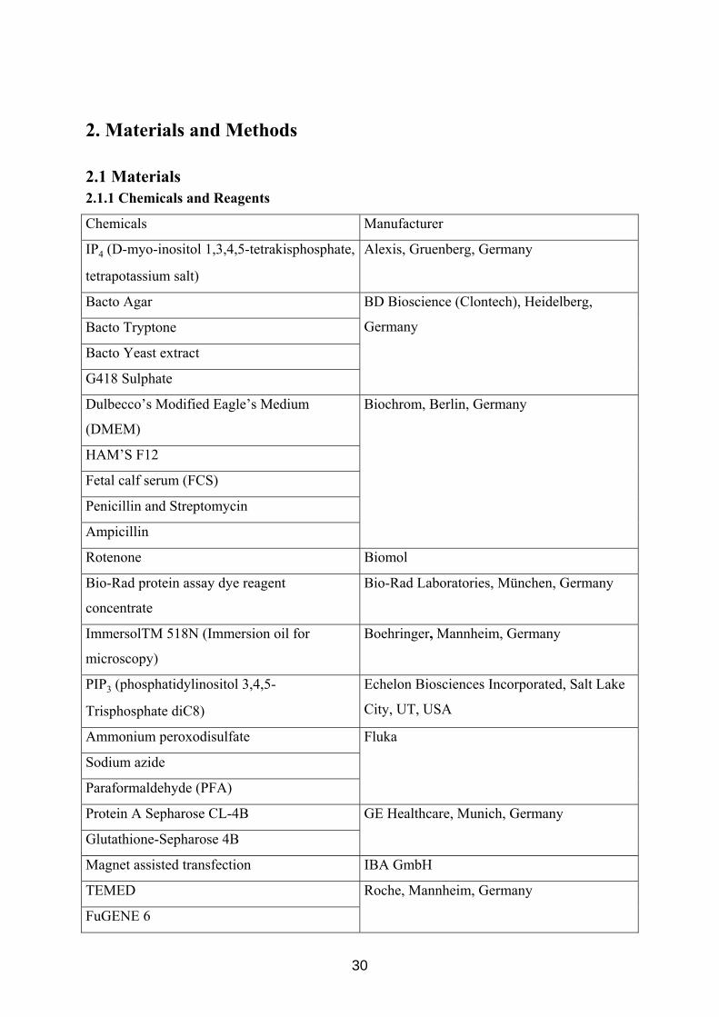
2.1 Materials 2.1.1 Chemicals and Reagents
Chemicals Manufacturer
IP4 (D-myo-inositol 1,3,4,5-tetrakisphosphate,
tetrapotassium salt)
Alexis, Gruenberg, Germany
Bacto Agar
Bacto Tryptone
Bacto Yeast extract
G418 Sulphate
BD Bioscience (Clontech), Heidelberg,
Germany
Dulbecco’s Modified Eagle’s Medium
(DMEM)
HAM’S F12
Fetal calf serum (FCS)
Penicillin and Streptomycin
Ampicillin
Biochrom, Berlin, Germany
Rotenone Biomol
Bio-Rad protein assay dye reagent
concentrate
Bio-Rad Laboratories, München, Germany
ImmersolTM 518N (Immersion oil for
microscopy)
Boehringer, Mannheim, Germany
PIP3 (phosphatidylinositol 3,4,5-
Trisphosphate diC8)
Echelon Biosciences Incorporated, Salt Lake
City, UT, USA
Ammonium peroxodisulfate
Sodium azide
Paraformaldehyde (PFA)
Fluka
Protein A Sepharose CL-4B
Glutathione-Sepharose 4B
GE Healthcare, Munich, Germany
Magnet assisted transfection IBA GmbH
TEMED
FuGENE 6
Roche, Mannheim, Germany
30
protease inhibitor cocktail tablets
trypsin
ponceau S solution (0.2% in 3% acetic acid)
Acrylamide
N, N’-Methylenbisacrylamide
SERVA, Heidelberg, Germany
Protein G agarose
Dimethyl sulfoxide (DMSO)
TEMED
β-mercaptoethanol
Igepal CA630
Bromphenol blue
PAP pen for immunostaining
poly-L-lysine
Isopropyl b-D-thiogalactoside
2’,3’cyclic AMP
2’,3’cyclic GMP
2’,3’cyclic NADP
Sigma, Taufkirchen, Germany
All other chemical reagents from Carl Roth
2.1.2 Antibodies
2.1.2.1 Primary antibodies
Antibody Manufacturer
Rabbit anti-CNP (antibody 27695)
Rabbit anti-SOD 2 (Mn-superoxide dismutase
2, antibody 13533)
Abcam, Cambridge, UK
Mouse monoclonal anti-Nucleoporin p62
Mouse monoclonal anti-Cyt c (cytochrome c,
clone 6H2.B4)
BD Biosciences, Heidelberg, Germany
Mouse monoclonal anti-plasma membrane
calcium ATPase (clone 5F10)
Mouse monoclonal anti- Cyt c (cytochrome c,
clone 7H8.2C12)
Biozol, Eching, Germany
Rabbit anti-VDAC (voltage-dependent anion Calbiochem, Schwalbach, Germany
31
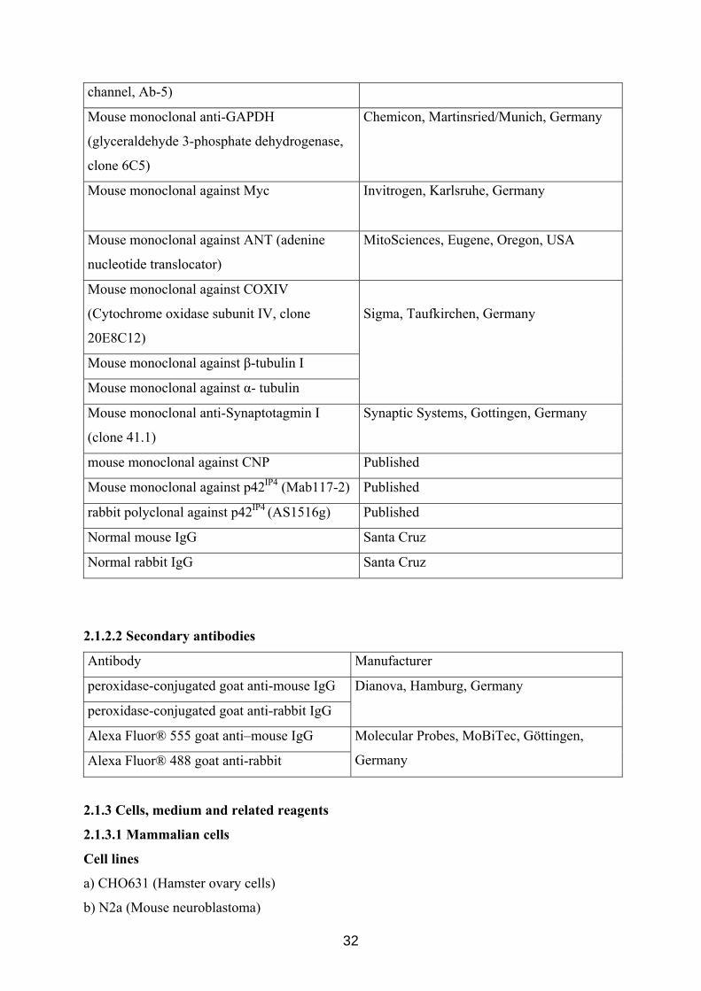
channel, Ab-5)
Mouse monoclonal anti-GAPDH
(glyceraldehyde 3-phosphate dehydrogenase,
clone 6C5)
Chemicon, Martinsried/Munich, Germany
Mouse monoclonal against Myc Invitrogen, Karlsruhe, Germany
Mouse monoclonal against ANT (adenine
nucleotide translocator)
MitoSciences, Eugene, Oregon, USA
Mouse monoclonal against COXIV
(Cytochrome oxidase subunit IV, clone
20E8C12)
Mouse monoclonal against β-tubulin I
Mouse monoclonal against α- tubulin
Sigma, Taufkirchen, Germany
Mouse monoclonal anti-Synaptotagmin I
(clone 41.1)
Synaptic Systems, Gottingen, Germany
mouse monoclonal against CNP Published
Mouse monoclonal against p42IP4 (Mab117-2) Published
rabbit polyclonal against p42IP4 (AS1516g) Published
Normal mouse IgG Santa Cruz
Normal rabbit IgG Santa Cruz
2.1.2.2 Secondary antibodies
Antibody Manufacturer
peroxidase-conjugated goat anti-mouse IgG
peroxidase-conjugated goat anti-rabbit IgG
Dianova, Hamburg, Germany
Alexa Fluor® 555 goat anti–mouse IgG
Alexa Fluor® 488 goat anti-rabbit
Molecular Probes, MoBiTec, Göttingen,
Germany
2.1.3 Cells, medium and related reagents
2.1.3.1 Mammalian cells
Cell lines
a) CHO631 (Hamster ovary cells)
b) N2a (Mouse neuroblastoma)
32
c) OLN93 (Rat oligodendrocytes)
d) HEK293 (Human embryonic kidney)
Cell culture medium and solvents
CHO631 cells - HAM’S F-12: 1.17 g/L NaHCO3, 1.802 g/L D-glucose, 0.146 g/L L-
glutamine, 100U/ml Penicillin, 100 µg/ml Streptomycin
N2a cells - DMEM: 3.7 g/L NaHCO3, 4.5 g/L D-glucose, 1.028 g/L N-Acetyl L-alanyl-L-
glutamine, 10% FCS, 100U/ml Penicillin, 100 µg/ml Streptomycin
OLN93 cells - DMEM: 3.7 g/L NaHCO3, 4.5 g/L D-glucose, 1.028 g/L N-Acetyl L-alanyl-L-
glutamine, 10% FCS, 100U/ml Penicillin, 100 µg/ml Streptomycin
HEK-293 cells- DMEM/HAM’S F12 (1:1) with 2mM Glutamine, 10% FCS, 100U/ml
Penicillin, 100 µg/ml Streptomycin
HBSS (Hanck’s balanced salt solution) without Ca2+ and Mg2+ from PAA.
G418 sulphate was from Calbiochem. stock: 500 mg/ml, working concentration: 500 µg/ ml
2.1.3.2.Bacterial cells:
Bacteria strains: DH5a , XL1-Blue, BL21
LB medium
10 g/l Bacto-tryptone (BD Bioscience), 5 g/l Bacto-yeast extract (BD Bioscience),
10 g/l NaCl, pH 7.0
LB agar plates
LB medium, 18 g/l Bacto-agar (BD Bioscience), appropriate antibiotic (100 μg/ml ampicillin)
2.1.4 Animals
Male Sprague Dawley rats from Harlan-Winkelmann GmbH, Borchen, Germany
2.1.5 Plasmid vectors:
pcDNA3.1+ from Invitrogen
pcDNA3.1/Myc-His (B) from Invitrogen
pGEX 5X-2 from GE Healthcare
pEGFP-C2 from BD Bioscience (Clontech)
33
2.1.6 Small interfering RNAs (siRNAs)
ON-TARGETplus SMARTpool of 4 siRNAs targeting rat CNP1 from Dharmacon, Chicago,
IL, USA
siRNA name siRNA sequence
Sense Sequence
GCACAAGCUUUGCCCGAAAUU
siGENOME ON-TARGETplus SMARTpool
duplex (9), J-080116-09, CNP1
Antisense Sequence
5’-PUUUCGGGCAAAGCUUGUGCUU
Sense Sequence
CCGAGGAGUACAAGCGUCUUU
siGENOME ON-TARGETplus SMARTpool
duplex (10), J-080116-10, CNP1
Antisense Sequence
5’-PAGACGCUUGUACUCCUCGGUU
Sense Sequence
GUGAGGAGAUCUUACGGCAUU
siGENOME ON-TARGETplus SMARTpool
duplex (11), J-080116-11, CNP1
Antisense Sequence
5’-PUGCCGUAAGAUCUCCUCACUU
Sense Sequence
GUUCCGAGACCCUCCGAAAUU
siGENOME ON-TARGETplus SMARTpool
duplex (12), J-080116-12, CNP1
Antiense Sequence
5’-PUUUCGGAGGGUCUCGGAACUU
Non-silencing siRNA from Qiagen.
2.1.7 Enzymes
Enzymes and buffer Manufacturer
T4 DNA Ligase Gibco BRL (Life Technologies), Karlsruhe, Germany
Eco RI
NotI
Tango+ (yellow) (10X) buffer
MBI Fermentas, St. Leon-Rot, Germany
2.1.8. Molecular weight markers
Nucleic acid standard markers
GeneRuler™
1 kb and 100 bp DNA ladders from MBI Fermentas.
34
Protein standard marker
Precision Plus Protein All Blue Standard (250-10 kDa) from Bio-Rad
2.1.9 Kits
Type of kit Usage Manufacturer
BigDye Terminator Cycle
Sequencing
Ready Reaction kit
DNA sequencing Applied Biosystems,
Warrington, UK
ECL+Plus detection of western blot Amersham Pharmacia
Biotech, Buckinghamshire,
UK
ApoAlert Caspase-3
Colorimetric assay Kit
Detection of apoptosis BD Bioscience (Clontech),
Heidelberg; Germany
Concert Rapid PCR
Purification system
Cleaning of PCR, restriction
digestion product
Concert Rapid Gel Extraction
system
Cleaning of PCR product
from gel
Gibco BRL (Life
Technologies), Karlsruhe,
Germany
Invisorb Spin Plasmid Mini
kit
Plasmid isolation Invitek, Berlin, Germany
HiSpeed Plasmid Midi kit Plasmid isolation
Hotstar Taq Master Mix kit PCR
Omniscript Reverse
Transcription kit
Making of cDNA
Flexi Gene DNA kit Isolation of Genomic DNA
from cell culture
RNase-Free DNase Set Removal of genomic DNA
during RNA isolation
RNeasy Maxi, Midi and Mini
kit (Isolation of RNA)
Isolation of RNA
Qiagen, Hilden, Germany
Supersignal West Pico kit Detection of western blot Pierce, Rockford, IL, USA
2.1.10 Laboratory instruments
Instrument Manufacturer
35
ABI PRISMTM 310 Genetic Analyzer Applied Biosystems Division, Foster City,
CA, USA
Mighty Small II (for western blotting
electrophoresis)
Amersham Pharmacia Biotech,
Buckinghamshire, UK
Ultrasonic homogenizer Bandelin electronic, Berlin, Germany.
T3 Thermocycler Biometra, Göttingen, Germany
Electrophoresis power supply
Gel electrophoresis system
Semi-dry Transfer Cell
Mini PROTEAN II (Tankblot)
GS-800 Calibrated Densitometer
Bio-Rad Laboratories, Munich, Germany
LSM510 laser scanning confocal microscope Carl Zeiss, Jena, Germany
Thermomixer comfort Eppendorf, Hamburg, Germany
Biofuge pico and 13 R centrifuges
Megafuge 1.0 R centrifuge
Sorvall®
RC-5B Refrigerated Superspeed
Centrifuge
Sorvall®
discoveryTM
90 ultraspeed centrifuge
Heraeus cell culture incubator
Heraeus refrigerator (-80oC)
LaminAir®
(clean bench)
Kendro, Hanau, Germany
Millipore purification system
Ultra-pure water system
Millipore, Schwalbach, Germany
Rotator Labinco BA, Netherlands
Microplate reader
Microplate reader Spectramax M5
Molecular Devices
Innova 4230 Refrigerated incubator shaker New Brunswick Scientific, Nürtingen,
Germany
TPP+-selective electrode
Ca2+-sensitive electrode
Nico, Moscow, Russia
UV/visible Spectrophotometer Pharmacia Biotech
Balance (analytical and preparative) Sartorius, Göttingen, Germany
36
Gel-blotting-papers
Protran BA83 Cellulosenitrat (E) (0.2 µm)
Protran BA79 nitrocellulose transfer
membrane (0.1 µm)
Schleicher & Schuell, Dassel, Germany
Eagle Eye Still video system Stratagene, Heidelberg, Germany
Vivaspin 6 filter concentrator Vivascience AG, Hannover, Germany
pH Meter (pH526) WTW
2.1.11 Buffers and solvents
All buffers were prepared with distilled water.
2.1.11.1 Buffers and solvents
1x HBSS : 145 mM NaCl, 5.4 mM KCl, 1mM MgCl2, 1.8 mM CaCl
2, 25 mM glucose, 20
mM HEPES, pH 7.4 adjusted with 1.67 M Tris (hydroxymethyl)-aminomethane
1x PBS: 137 mM NaCl, 2.6 mM KCl, 8.1 mM Na2PHO4, 1.4 mM KH2PO4, pH 7.4
E. coli-lysis buffer (PBS+ 1% Triton X- 100):137 mM NaCl, 2.6 mM KCl, 8.1 mM Na2PHO4,
1.4 mM KH2PO4, pH 7.4, 1% Triton X- 100
Storage buffer: 50 mM Tris/HCl pH 8,0, 100 mM NaCl, 1 mM mercaptoethanol, 20%
glycerol
1xTBE: 89 mM Tris, 89 mM Boric acid, 2 mM EDTA, pH 8.0
1xTE: 10 mM Tris/HCl, pH 7.4, 1 mM EDTA, pH 8.0
Ethidium bromide solution: 10 mg/ml
TCM: 10 mM Tris/HCl, pH 7.5, 10 mM CaCl2, 10 mM MgCl2
4% PFA (Paraformaldehyde) solution: 4% PFA, 120 mM sodium phosphate, pH 7.4, 4%
Saccharose
FTP: Blocking and Washing buffer (immunostaining):
17% (v/v) fetal calf serum, 20 mM Na2HPO4, 450 mM NaCl, 0.3% Triton X- 100
Lysis buffer: 50 mM Tris/HCl pH 7.4, 150 mM NaCl, 1% Igepal CA630, 0.25% Na-
deoxycholate, 1 mM EDTA and 1 mM NaF
60% Acrylamid/Bis: 58.4% Acrylamide, 1.6% N,N’-Methylen-bisacrylamide
Resolving buffer: 750 mM Tris/HCl, pH 8.8
Stacking buffer: 250 mM Tris/HCl, pH 6.8
SDS solution: 10% (w/v) SDS in H2O
37
PER solution: 10% (w/v) Ammoniumperoxodisulfat in H2O
4x Laemmli Sample buffer: 500 mM Tris/HCl, pH6.8, 8% SDS, 40% Glycerol
5x Laemmli Running buffer: 125 mM Tris, 960 mM Glycine, 0.5% SDS, pH 8.5
1x Transfer buffer: 25 mM Tris, 192 mM Glycine, 20% (v/v) Methanol
Membrane Stripping buffer: 62.5 mM Tris, pH 6.8, 100 mM β-Mercaptoethanol, 2% SDS
PBST: 137 mM NaCl, 2.6 mM KCl, 8.1 mM Na2PHO4, 1.4 mM KH2PO4, pH 7.4
0.1% Tween 20
50% Protein G agarose slurry or 50% Protein A sepharose slurry: 1 ml 1x PBS and 4 µl 10%
sodium azide
2.1.11.2 Buffers for mitochondria studies
All buffers for mitochondria studies were prepared with bidistilled water
Buffer A: 0.32 M sucrose, 0.5 mM EDTA, 0.5 mM EGTA, and 10 mM Tris-HCl, pH 7.4
Buffer for mitochondria isolation:
0.32 M sucrose, 0.5 mM EDTA, 0.5 mM EGTA, 0.5% bovine serum albumin (BSA; fraction
V) and 10 mM Tris-HCl, pH 7.4
Buffer for washing of mitochondria:
0.32 M sucrose, and 10 mM Tris-HCl, pH 7.4
Incubation medium:
100 mM KCl, 100 mM sucrose, 10 mM Tris-HCl, 0.4 mM K2HPO4, 5 mM potassium
succinate, 1.5 µM oligomycin and 2 µM rotenone, pH 7.4
2.1.12. Oligonuleotides for PCR and sequencing.
(Synthesized by MWG, Biotech, Ebersberg, Germany)
cDNA Primer name Primer sequence Tm
(°C)
application
THGAPDHFW 5’-
ATGGTGAAGGTCGGTGTGAACGGATTT
GG-3’
mice
GAPDH
THGAPDHRV 5’-
TTACTCCTTGGAGGCCATGTAGGCCAT
GAG-3’
58° PCR
mice THMMP42F1 5’-TTCTCCGACTGCCAACTCCTAC-3’ 58° PCR
38
p42IP4 THMMP42R6 5’-GACAATGGTTATGCCATGAGG -3’
pGEX 5’ Seq.
Fw
5’-GGGCTGGCAAGCCACGTTTGGTG-3’ 55° Sequencing
pGEX 3’ Seq.
Rev
5’-CCGGGAGCTGCATGTGTCAGAGG-3’ 55° Sequencing
T7 promoter 5’-GTAATACGACTCACTATAGGGC-3’ 55° Sequencing
BGH seq 5’-CTAGAAGGCACAGTCGAGG-3’ 55° Sequencing
2.2 Methods 2.2.1 RT-PCR
Total RNA was extracted from cultured cells using RNeasy Mini kit (Qiagen). One
microgram of RNA was reverse-transcribed using Omniscript™ Reverse Transcription kit
(Qiagen), and the resulting cDNA was amplified in the presence of appropriate primers for 30
cycles by PCR using HotStarTaq™ Master Mix kit (Qiagen) for 15 min at 95ºC, followed by
repeated cycles of 30 s at 94ºC, 90 s at 58º C, 60 s at 72ºC, then a final 10 min extension at
72ºC. The reaction products were analyzed by electrophoresis with 1% agarose gel containing
ethidium bromide, and visualized by Bio-Rad gel document system (Bio-Rad).
2.2.2 Plasmid constructs
A NotI DNA fragment coding the full length p42IP4 was excised from pBluescript+-
p42IP4 (Sedehizade et al., 2002) and sub-cloned into pGEX-5X-2 vector (GE Healthcare,
Munich, Germany). An EcoRI DNA fragment with the full length p42IP4 cDNA was excised
from pBluescript+-p42IP4 and sub-cloned into the EcoRI site of pcDNA3.1Myc vector
(Invitrogen, Karlsruhe, Germany). At first, the original pBluescript+-p42IP4 construct was
digested with the NotI or EcoRI to obtain DNA fragments encoding for the p42IP4. Then the
fragments obtained were ligated into the linearized pGEX-5X-2 and pcDNA3.1Myc vectors
respectively. The ligated product was purified using the DNA purification kit from
Gibco/BRL. The purified DNA was checked for the presence of the inset by digestion of the
ligated DNA using NotI or EcoRI, respectively. The successfully ligated DNA constructs
were transformed into E.coli using the heat-shock method (CaCl2 based). The E.coli were
plated and allowed to grow for 24 hours at 37ºC on LB-agar plates containing 100 mg/ml
ampicilin, as a selection marker. The positive clones were picked, and cultivated in 5 ml of
LB medium containing ampicillin for 24 h. DNA was prepared from the above cultures using
the DNA-Mini-prep kit (Qiagen). The DNA obtained from the mini prep was digested with
39
NotI or EcoRI to recheck for the presence of the inset of the DNA encoding p42IP4 in the
appropriate construct. The DNA from the positive clones was sequenced using the DNA
sequencing kit from ABI PRISM, using the appropriate set of primers.
The clones that expressed the complete p42IP4 without any point mutations and frame
shift, were retransformed in E.coli using heat-shock method (CaCl2 based), and the DNA was
isolated using the DNA Midi-Prep kit (Qiagen).
The construct p42IP4-pcDNA3 and CHO cells, stably transfected with pcDNA3-p42IP4
were described before (Sedehizade et al., 2002). The sequence of all constructs used was
verified by sequencing on automated sequencer (ABI PRISMTM 310 Genetic Analyzer) with
the dye terminator cycle sequencing kit (ABI-Prism, Applied Biosystems) according to the
manufacturer’s instructions in both directions prior to use.
2.2.3 Agarose gel electrophoresis of DNA
To check the quality of DNA (PCR product, recombinant plasmid DNA, restriction
analysis) 1% agarose (Sigma, Taufkirchen, Germany) gel and 1.5% gel for DNA laddering
assay in 0.5x TBE buffer were made. Gel was pre-stained in ethidium bromide (10 mg/ml)
(Sigma). DNA samples were prepared in 6x loading buffer (containing bromophenol blue
dye) (MBI Fermentas). Gel was run in 0.5x TBE for 60 min at 80 V for PCR samples, at 100
V for plasmid DNA and 3 h at 30V for genomic DNA. Depending on the fragment size either
GeneRuler 100bp DNA Ladder (1 kb) or GeneRuler DNA Ladder Mix (10 kb) (MBI
Fermentas) used. DNA bands were visualised under UV-Transilluminator in an Eagle Eye II
video system (Stratagene, Heidelberg, Germany).
2.2.4 Transformation of E. coli with plasmid DNA by heat-shock method (CaCl2 based)
To make competent cells, single colony of E.coli was grown in 5 ml LB media
overnight at 37°C with shaking at 250 rpm. 1 ml of overnight bacterial culture was then
transferred to 100 ml of fresh LB media and continued to grow at 37°C with shaking till the
cells reached their logarithmic phase i.e, OD 600= 0.3-0.4. Then the bacterial suspension was
centrifuged at 3,000 g for 5 min at 4°C. Supernatant was discarded and cell pellet was
resuspended in 10 ml of ice-cold 100 mM CaCl2 solution. The cell suspension was then
incubated on ice in cold room (4°C) for 1 h and centrifuged for 5 min at 3,000 g at 4 °C.
Supernatant was discarded carefully and pellet was resuspended in 1 ml of ice-cold 100 mM
CaCl2 containing 30% glycerine (1 ml solution= 700 µl 100 mM CaCl2+ 300µl of 100%
glycerine). From this competent cell suspension aliquots of 200 µl were made, frozen in liquid
nitrogen and stored at -80°C.
40
To transform bacteria, 200 µl of CaCl2 competent cells for each reaction were thawed
on ice. To the cells we added 100 µl of cold TCM buffer and either 10-20 ng of super-coiled
plasmid DNA or 20 µl of ligation mix. Cells were incubated on ice for 20 min and then heat –
shock given for 90 s at 42°C. Transformation reaction mix was immediately incubated on ice
for 1 min, then 700 µl of LB media (pre-warmed at 37°C and without any antibiotic) was
added to transformation mix and incubated for 30-60 min with shaking at 37°C. 100 µl of the
transformation mixture in case of super-coiled DNA was plated on the LB-agar plate
containing suitable antibiotic. For ligation transformation, reaction mix was centrifuged
briefly, supernatant was discarded and pellet was resuspended in the residual volume and
plated on LB-agar containing suitable antibiotic. LB-agar plates were incubated up-side down
at 37°C to facilitate growth of tranformants under appropriate antibiotic selection pressure.
2.2.5 Expression and purification of glutathione S-transferase (GST) and GST-p42IP4
constructs from E. coli.
For expression in E. coli, the pGEX-5X-2 constructs were transformed in BL21 (GE
Healthcare). The expression and purification of the fusion protein was performed in the
presence Triton-X 100 (Roth, Karlsruhe, Germany) according to a published procedure
(Stricker et al., 2006) with modifications. In brief, E. coli transformed with the pGEX-5X-2-
p42IP4 construct were grown in LB medium containing ampicillin to absorbance at 600 nm,
A600 = 0.6-0.8 and expression of the fusion protein was induced by the addition of 0.1 mM
isopropyl b-D-thiogalactoside (Sigma). The cells were grown for an additional 3 h at 25 °C,
collected by centrifugation, and stored at 80°C until use. Frozen E. coli pellets were
solubilized in E. coli-lysis buffer (137 mM NaCl, 2.6 mM KCl, 8.1 mM Na2PHO4, 1.4 mM
KH2PO4, pH 7.4, 1% Triton X-100) containing a cocktail of protease inhibitors (Roche,
Mannheim, Germany) by sonication (5x10 s). After centrifugation (10,000 g for 10 min at
4°C), glutathione-sepharose slurry was added to the supernatant and incubated on a rotator
(60 min at 4°C). The beads were sedimented by centrifugation (500 g for 5 min at 4°C) and
washed three times with E. coli-lysis buffer. To remove non-specifically bound proteins, the
beads were incubated in lysis buffer supplemented with 1 M NaCl (15 min at 4°C) and after
centrifugation the beads were resuspended in storage buffer (50 mM Tris/HCl pH 8,0, 100
mM NaCl, 1 mM mercaptoethanol and 20% glycerol).
The beads and lysates were analyzed by SDS-PAG electrophoresis. The purified fusion
proteins were stored as a 50% (v/v) beads suspension and used for GST pull-down assay.
41
2.2.6 Cell culture and transfection
CHO cells were maintained in Hams’ F12 medium supplemented with 10% fetal calf
serum (Biochrom, Berlin, Germany) and 100 µg/ml penicillin, and 100 µg/ml streptomycin in
a humidified 5% CO2 atmosphere at 37 °C. N2a cells were maintained under the same
conditions with the exception that Dulbecco's Modified Eagle's Medium was used.
OLN93 cells, an oligodendrocyte cell line (Richter-Landsberg and Heinrich, 1996),
kindly provided by Prof. C. Richter-Landsberg (University of Oldenburg, Germany), were
grown at 37°C in a 5% CO2 atmosphere in Dulbecco’s modified Eagle’s medium (Biochrom
KG) supplemented with 10% fetal calf serum (Biochrom KG, Berlin,Germany) containing 2
mM glutamine, 100 µg/ml penicillin, and 100 µg/ml streptomycin.
Transfections were carried out using FuGENE 6 (Roche) according to manufacturer's
instructions. The cells were stably transfected with the respective plasmid DNA (2 μg).
Before transfection the cells were grown in 10-cm dishes to 60% confluency. The transfected
cells were kept for 8–10 h in serum- and antibiotic-free medium. After 24-48 h, the
transfected cells were cultivated in selection medium containing 400 μg/ml G418
(Calbiochem, Schwalbach, Germany). Single cell colonies were isolated after 3 weeks by
cloning rings, verified at the transcription and translation level by RT-PCR and western blot
and used for further experiments.
2.2.7 Confocal microscopy
For immunofluorescence, N2a cells stably transfected with p42IP4-pcDNA3.1Myc and
CHO cells, stably transfected with pcDNA-p42IP4 were grown on poly-L-lysine-coated
coverslips. Cells were fixed in 4% paraformaldehyde, blocked and permeabilized in blocking
buffer (17% (v/v) fetal calf serum, 20 mM Na2HPO4, 450 mM NaCl, 0.3% Triton X- 100) and
incubated with rabbit polyclonal anti-Myc antibody (Invitrogen, Karlsruhe, Germany; 0.8
μg/μl) or rabbit polyclonal anti-p42IP4 antibody (AS1516g) and mouse monoclonal anti-Cyt c
antibody (clone 6H2.B4; BD Bioscience; Heidelberg; Germany) overnight at 4°C, as
described in the respective figure legends. After washing, the secondary antibody was applied
(Alexa Fluor® 555 goat anti–mouse IgG antibody at 1:100; Alexa Fluor® 488 goat anti-rabbit
at 1:200 Molecular Probes, MoBiTec, Göttingen, Germany) for 2 h at 25°C. Mounted slides
were observed with a confocal laser scanning microscope LSM510 (Carl Zeiss, Jena,
Germany).
42
2.2.8 Subcellular fractionation by differential centrifugation
Subcellular fractionation and organelle isolation techniques were done by modification
of previously reported procedures (Sharer et al., 2002; Leissring et al., 2004). All steps were
carried out at 4°C. N2a cells were washed two times in phosphate-buffered saline, collected in
ice-cold buffer A (0.32 M sucrose, 0.5 mM EDTA, 0.5 mM EGTA, and 10 mM Tris-HCl, pH
7.4) containing cocktail of protease inhibitors (Roche) and gently disrupted using a Potter-
Elvehjem homogenizer. The cell homogenate was then subjected to centrifugation at 500 g for
10 min, yielding a nuclear pellet and supernatant. The mitochondrial fraction was obtained
from this supernatant after centrifugation at 12,000 g for 10 min. Finally, the 12,000 g
supernatant was further fractionated at 49,000 g for 30 min, yielding the soluble cytosolic
fraction and membrane pellet. After each centrifugation step, the resulting pellets were
washed by resuspension in an excess of buffer A, followed by centrifugation at the
appropriate relative centrifugal force.
The protein concentration was determined by the Bradford method (Bio-Rad Protein
assay, Bio-Rad, Munich, Germany) using bovine serum albumin as standard.
2.2.9 Preparation of mitochondrial lysate and cytosolic fraction from rat brain
Rat brains were rapidly removed and placed within 30 s in ice-cold buffer A
containing 0.02% bovine serum albumin (fatty acid free, fraction V). All manipulations were
carried out at 4οC. The tissue was homogenized in a Potter-Elvehjem homogenizer; the ratio
of brain tissue to buffer used for isolation was 1:10 (w/v). The homogenate was centrifuged at
2,000 g for 3 min. The pellet of mitochondria was obtained by centrifugation of the 2,000 g
supernatant at 12,000 g for 10 min. RBM were suspended in ice-cold solution, containing
0.32 M sucrose and 10 mM Tris-HCl (pH 7.4) and were additionally washed by centrifugation
at 12,000 g for 10 min.
For preparation of mitochondrial lysate, RBM aliquots (4 mg of protein) were
suspended in lysis buffer (50 mM Tris/HCl pH 7.4, 150 mM NaCl, 1% Igepal CA630, 0.25%
Na-deoxycholate, 1 mM EDTA and 1 mM NaF) containing a cocktail of protease inhibitors
(Roche). Then suspensions were rotated on a rotation wheel for 30 min at 4°C, solubilized by
ultrasonic treatment and centrifuged at 14,000 g for 15 min. The 14,000 g supernatant gives
the mitochondrial lysate.
For preparation of brain cytosolic fraction, the brain tissue was homogenized using an
Ultraturrax three times for 20 s at 4°C in buffer A containing a cocktail of protease inhibitors
(Roche). The homogenates were then centrifuged at 400 g for 5 min at 4°C. The supernatant
43
obtained was centrifuged at 100,000 g for 30 min at 4°C. The 100,000 g supernatant gives the
cytosolic fraction.
2.2.10 Isolation of mitochondria from cultured cells
Isolation of mitochondria from cells was carried out at 4οC according to a published
procedure (Leissring et al., 2004) with modifications. Confluent monolayers of N2a or CHO
cells, cultured in 15 cm dishes, or confluent monolayers of OLN93 cells, cultured in T-75
flasks, were washed with phosphate-buffered saline, collected by scraping in buffer A,
containing 0.02% bovine serum albumin (fatty acid free, fraction V), and pelleted at 500 g for
5 min. Cells were re-suspended in the same ice-cold buffer and disrupted with a Potter-
Elvehjem homogenizer. The cell homogenate was centrifuged twice at 500 g for 5 min to
remove unbroken cells and nuclei, and twice at 3,000 g for 4 min to remove trapped
peroxisomes and other organelles. The supernatant obtained was centrifuged at 12,000 g for
10 min. The mitochondrial pellet was suspended in ice-cold medium containing 0.32 M
sucrose and 10 mM Tris–HCl (pH 7.4) and centrifuged at 12,000 g for 10 min. The
mitochondria isolated were re-suspended in the same ice-cold medium. Mitochondria from
N2a and OLN93cells were used for functional studies.
2.2.11 Subfractionation of mitochondria
Mitochondria isolated from CHO cells were subfractionated by using a phosphate
swelling-shrinking method described previously (Bijur and Jope, 2003), with minor
modifications. Briefly, mitochondria were subjected to swelling by the addition of swelling
buffer (10 mM KH2PO4, pH 7.4) containing a cocktail of protease inhibitors, and incubated
for 20 min at 4οC with gentle mixing. An equal volume of shrinking buffer (10 mM KH2PO4,
pH 7.4, 32% sucrose, 30% glycerol, 10 mM MgCl2, and protease inhibitors) was added and
samples were incubated for an additional 20 min at 4οC. The suspension was centrifuged at
10,000 g for 10 min, yielding a supernatant containing the outer membrane and the
intermembrane space fractions, which was saved as mixture 1, and a pellet comprised of
mitoplasts. The mitoplasts were washed three times in the mixture of swelling and shrinking
buffer (1 : 1), then resuspended in 500 μl of swelling buffer and sonicated 10 s x 3 with a 1-
min interval on ice by 40% of maximal power. This suspension containing the inner
membrane and matrix fractions was saved as mixture 2. Mixtures 1 and 2 were centrifuged at
150,000 g for 1 h. The supernatant from mixture 1 was saved as the intermembrane space
fraction and the supernatant from mixture 2 was saved as the matrix fraction. Proteins from
the intermembrane space fraction were concentrated using a Vivaspin 6 filter concentrator
(Vivascience AG, Hannover, Germany). The outer membrane pellet in mixture 1 and the
44
inner membrane pellet in mixture 2 were washed once in swelling buffer and centrifuged at
150,000 g for 1 h. The outer and inner membrane pellets were solubilized in lysis buffer
containing protease inhibitors, as described above.
Mitoplast and outer membranes of rat brain mitochondria were obtained according to
previously reported procedures (Rice and Lindsay, 1997).
2.2.12 Electrophoresis and immunoblotting
For immunoblotting, proteins solubilized in Laemmli buffer were separated under
denaturing conditions on sodium dodecyl sulphate (SDS)-polyacrylamide gel electrophoresis
(PAGE) using 10% or 12.5% gels and transferred to nitrocellulose membranes (Protran BA83
or BA79, Whatman, Dassel, Germany). Precision Plus Pre-stained Standards (Bio-Rad) were
used as markers. Equal transfer was controlled by Ponceau red staining of the membrane.
After overnight blocking, the membrane was incubated with the appropriate primary antibody.
The monoclonal anti-CNP antibody was described before (Reiser et al., 1994). A polyclonal
rabbit serum raised against p42IP4 (AS1516g) and monoclonal antibody, Mab117-2, were
described before (Haase et al., 2008). Anti-Cyt c (cytochrome c, clone 7H8.2C12, Biozol,
Eching, Germany), pan anti-plasma membrane calcium ATPase (clone 5F10, Biozol), anti-
Cytochrome oxidase subunit IV (clone 20E8C12, MitoSciences, Eugene, Oregon), anti-ANT
(adenine nucleotide translocator, MitoSciences), anti-SOD 2 (Mn-superoxide dismutase 2,
antibody 13533, Abcam Cambridge, UK), anti-CNP (antibody 27695, Abcam), anti-VDAC
(voltage-dependent anion channel, Ab-5, Calbiochem), anti Synaptotagmin I (clone 41.1,
Synaptic Systems, Gottingen, Germany), anti-Nucleoporin p62 (BD Biosciences, Heidelberg,
Germany), anti-GAPDH (glyceraldehyde 3-phosphate dehydrogenase, clone 6C5, Chemicon,
Martinsried/Munich, Germany), anti-β-tubulin I (Sigma) and anti-α-tubulin (Clone B-5-1-2,
Sigma) antibodies were used for immunoblotting. Immunoreactivity was detected using the
appropriate secondary antibody conjugated to horseradish peroxidase (Dianova; Hamburg,
Germany). Peroxidase activity was detected with enhanced chemiluminescence reagents
(Super-Signal West Pico; Pierce, Rockford IL).
Quantification of the band densities from western blots was carried out using a GS800
calibrated densitometer and QUANTITY ONE software (Bio-Rad, Munich, Germany). Films
were scanned and the intensities of the bands were quantified.
2.2.13 GST-pull-down assay
For the GST pull-down, mitochondrial lysate was precleared by rotation 4 - 8 h with
glutathione-Sepharose beads at 4°C. After centrifugation (500 g 5 min at 4°C) glutathione-
45
Sepharose beads were discarded and precleared lysate was used for binding. To determine
protein-protein interaction in vitro, equal amounts of GST and GST-p42IP4 fusion proteins
immobilized on glutathione-Sepharose beads were incubated with brain cytosolic fractions or
precleared mitochondrial lysates (4 mg of protein) overnight on a rotating wheel at 4°C. After
centrifugation (1000 g for 5 min at 4°C) the beads were washed five times with 1 ml of lysis
buffer described above. The proteins were eluted from the beads with 10 μl of 2× Laemmli
buffer, separated by SDS–PAGE, transferred to nitrocellulose membranes and immunoblotted
as described above.
2.2.14 Immunoprecipitation and co-immunoprecipitation
For immunoprecipitation, RBM lysates were precleared by protein G-agarose (Sigma)
or A-sepharose beads (GE Healthcare), depending on the primary antibody used, for 2 h at
4°C. Cytosolic fractions and precleared RBM lysates were incubated with primary antibody,
i.e. monoclonal anti-CNP antibody (described before in (Reiser et al., 1994), monoclonal anti-
α-tubulin antibody (Clone B-5-1-2, Sigma), or polyclonal rabbit antiserum against p42IP4
(AS1516g, described before in (Haase et al., 2008) for 2 h on ice. Then it was mixed with 20
µl of suspension of protein G-agarose or A-sepharose beads, respectively, for overnight
incubation at 4°C under rotation with a following centrifugation at 500 g for 5 min. The
immunoprecipitated complexes were washed with lysis buffer (described above) five times.
Samples were eluted in 10 μl of 2× Laemmli buffer and used for electrophoresis and
immunoblotting as described above.
2.2.15 Isolation of rat brain mitochondria for functional study
Rat brains were rapidly removed (within 30 s) and placed in ice-cold buffer A,
containing 0.02% bovine serum albumin (fatty acid free, fraction V). All solutions used were
ice-cold; all manipulations were carried out at +4οC. The tissue was homogenized in a a
Potter-Elvehjem homogenizer; the ratio of brain tissue to isolation medium was 1:10 (w/v).
The homogenate was centrifuged at 2000 g for 3 min. The pellet of mitochondria was
obtained by centrifugation of the 2000 g supernatant at 12,500 g for 10 min. At the next step,
in representative experiments, mitochondria were purified on Percoll gradient (15%-23%-
40%) according to published procedures (Sims, 1990). Rat brain mitochondria were
suspended in ice-cold solution, containing 0.32 M sucrose and 10 mM Tris-HCl (pH 7.4) and
were additionally washed by centrifugation at 11,500 g for 10 min. The protein concentrations
in the stock mitochondrial suspensions were 25-30 mg/ml.
46
2.2.16 Evaluation of mitochondrial functions
Mitochondrial functions were measured, as described earlier (Azarashvili et al., 2007).
RBM (1.0 mg protein/ml) or mitochondria from cultured cells (0.7 mg of protein/ml) were
incubated in respiration buffer containing 100 mM KCl, 100 mM sucrose, 10 mM Tris-HCl,
0.4 mM K2HPO4, in the presence of 2 μM rotenone and 1.5 μM oligomycin, pH 7.4 at 25°C,
in an open chamber. Succinate (5 mM potassium succinate) was used as mitochondrial
respiratory substrate. PTP opening was induced by threshold Ca2+ load. For Ca2+ loading,
additions of Ca2+ per mg of protein: 70 or 200 nanomoles for mitochondria isolated from N2a,
80-120 nanomoles for RBM and 70 nanomoles for mitochondria isolated from OLN93 cells
were used. All experiments were performed in an open chamber.
Extramitochondrial Ca2+ was determined with a Ca2+-sensitive electrode (Nico,
Russia). The mitochondrial membrane potential was determined by measuring
tetraphenylphosphonium (TPP+) in the incubation medium with a TPP+-sensitive electrode.
Oxygen consumption rate was detected with a Clark-type O2 electrode.
Changes in Ca2+ and TPP+ concentration, as well as in oxygen consumption were
simultaneously monitored in the incubation medium. Parameters of Ca2+ transport, rate of
Ca2+ influx, Ca2+ capacity (amount of Ca2+ taken up by mitochondria before PTP opening),
lag time for Ca2+ efflux after Ca2+ addition under PTP opening were evaluated, as described
earlier (Azarashvili et al., 2007).
In some experiments mitochondria were pre-incubated with IP4 (D-myo-inositol 1,3,4,5-
tetrakisphosphate, tetrapotassium salt, Alexis, Gruenberg, Germany) or PIP3
(phosphatidylinositol 3,4,5-Trisphosphate diC8, Echelon Biosciences Incorporated, Salt Lake
City, UT, USA) for 20 min before loading with Ca2+.
Swelling of RBM was measured as a change in scattering of the mitochondrial
suspension absorbance at 540 nm (A540) using Microplate reader Spectramax M5 (Molecular
Devices) at 30°C. Standard incubation medium for swelling assay contained 125 mM KCl, 10
mM Tris, 2 mM KH2PO4, 10 mM succinate, 0.5 µM oligomycine, 0.5 µM rotenone. The
concentration of protein in the cuvette was 0.45 mg protein/ml. The swelling was initiated by
addition of 450 µM Ca2+.
2.2.17 Detection of CNP activity in isolated mitochondria
CNP activity was detected according to published procedure (Bradbury and
Thompson, 1984). Aliquots of the mitochondrial suspension (rat brain mitochondria or
OLN93 cells) containing 40 µg of protein were taken from the chamber under control
47
conditions and after PT opening and were then solubilized in Laemmli buffer. Solubilized
mitochondria were separated by electrophoresis in a 12.5% polyacrylamide gel and were
transferred to nitrocellulose membranes. CNP activity was detected on membrane after
blotting. The membrane was pre-incubated for 1 h in 50 mM MES (4-morpholine-
ethanesulfonic acid) buffer, pH 6.1, containing 30 mM MgCl2, 0.1% Triton X-100, 3 M
guanidinium chloride, 1 mM EDTA, 1 mM dithiothreitol and 5% glycerol, then for 1 h in the
same buffer without guanidinium chloride. The pre-incubation stage is essential for recovery
of enzyme activity after SDS- electrophoresis. The blot was then stained for activity by using
Nitro Blue Tetrazolium [3.3’(3,3’-dimetoxy-4.4’-diphenylene)2,2’-di-p-nitrophenyl-5,5’-
diphenylditetrazolium] chloride and phenazine methosulphate to detect NADPH produced in
enzyme-linked reaction for CNP. The coloured reaction product was immobilized with diluted
agarose. Equal volumes of 1% agarose in water at 55°C and reaction medium (200 mM MES
buffer pH 6.1, containing 60 mM MgCl2, 0.2 % Triton X-100, 0.1 mM 2’,3’-cyclic NADP
and 4 mg/ml glucose-6-phosphate, 0.4 mg /ml Nitro Blue Tetrazolium, 0.04 mg/ml phenazine
methosulphate and 0.7 units of glucose-6-phosphate dehydrogenase) were rapidly mixed and
poured on the nitrocellulose membrane. Coloured reaction was visible within 15 min and was
stopped with 10% acetic acid. CNP activity in the bands (intensity of coloured band) on the
blot was evaluated by determining the optical density of the coloured product.
2.2.18 Small interfering RNA (siRNA)
Chemically synthesized pool of siRNAs targeting CNP was obtained from
Dharmacon, Chicago, IL. To knockdown the endogenous CNP, OLN93 cells were transiently
transfected with rat CNP siRNAs by using magnet assisted transfection, according to the
manufacturer’s protocol (IBA GmbH, Germany). Briefly, one day before transfection cells
were plated on a T-75 flasks. On the following day, the cells (50% confluent) were incubated
for 15 min with CNP siRNAs or scrambled siRNA and magnet assisted transfection reagents
mixture on the magnet plate. Afterwards, transfected cells were cultured for 48 h under
normal conditions. Non-silencing siRNA served as a scrambled siRNA control (Qiagen). CNP
knockdown was assessed by Western blot at 48 h after transfection.
2.2.19 Apoptosis detection
For inducing apoptosis N2a cells were exposed to 300 nM wortmannin, 10 nM and 100
nM staurosporine or/and 100 μM LY 294002. Detection of apoptosis was checked using
DNA- laddering and caspase-3 assays.
For DNA laddering analysis, genomic DNA from 5 × 10⁶ cells was isolated using Flexi
Gene DNA kit (Qiagen) after 6 and 24 h incubation with the apoptotic-inducing drugs in
48
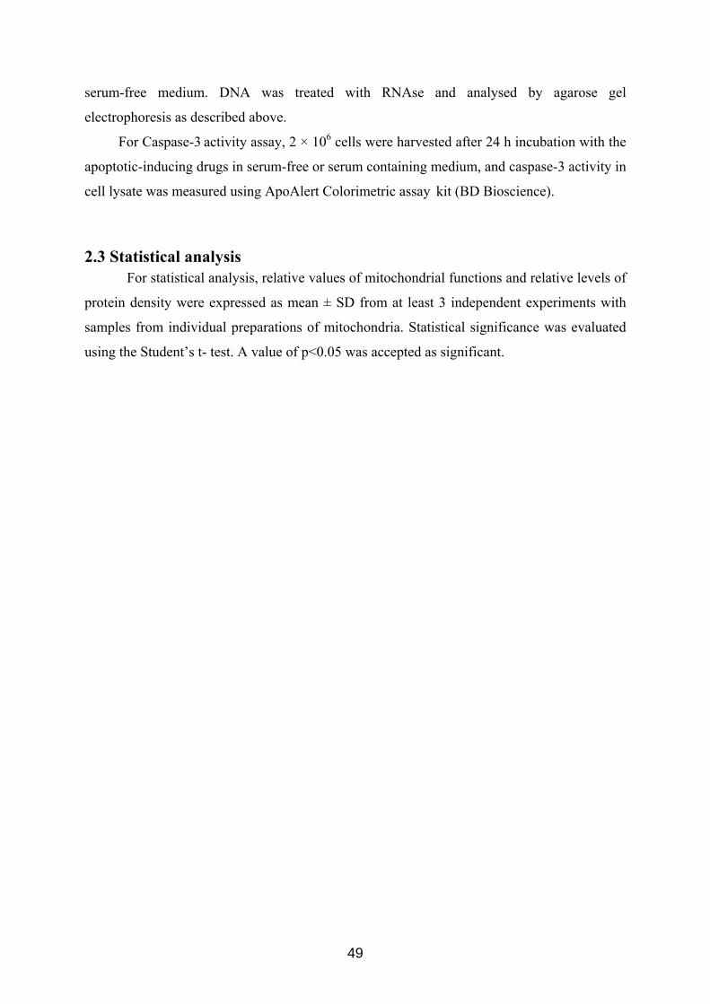
serum-free medium. DNA was treated with RNAse and analysed by agarose gel
electrophoresis as described above.
For Caspase-3 activity assay, 2 × 10⁶ cells were harvested after 24 h incubation with the
apoptotic-inducing drugs in serum-free or serum containing medium, and caspase-3 activity in
cell lysate was measured using ApoAlert Colorimetric assay kit (BD Bioscience).
2.3 Statistical analysis For statistical analysis, relative values of mitochondrial functions and relative levels of
protein density were expressed as mean ± SD from at least 3 independent experiments with
samples from individual preparations of mitochondria. Statistical significance was evaluated
using the Student’s t- test. A value of p<0.05 was accepted as significant.
49
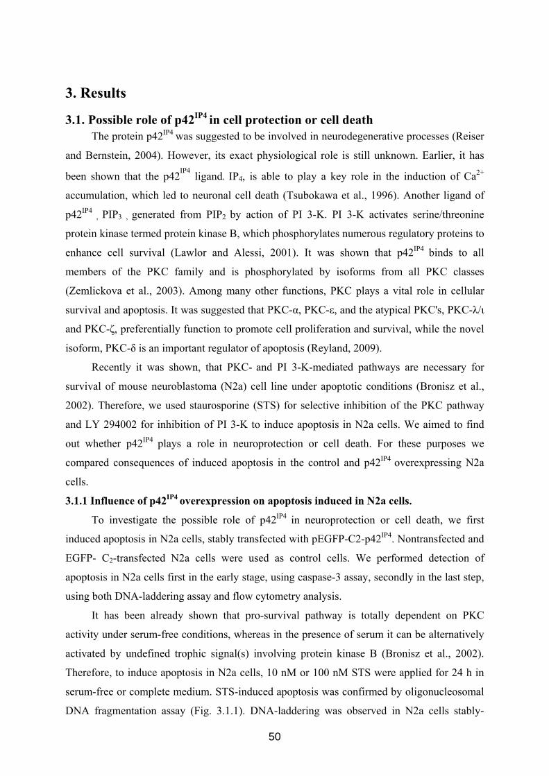
3. Results
3.1. Possible role of p42IP4 in cell protection or cell death The protein p42IP4 was suggested to be involved in neurodegenerative processes (Reiser
and Bernstein, 2004). However, its exact physiological role is still unknown. Earlier, it has
been shown that the p42IP4 ligand, IP4, is able to play a key role in the induction of Ca2+
accumulation, which led to neuronal cell death (Tsubokawa et al., 1996). Another ligand of
p42IP4 , PIP3 , generated from PIP2 by action of PI 3-K. PI 3-K activates serine/threonine
protein kinase termed protein kinase B, which phosphorylates numerous regulatory proteins to
enhance cell survival (Lawlor and Alessi, 2001). It was shown that p42IP4 binds to all
members of the PKC family and is phosphorylated by isoforms from all PKC classes
(Zemlickova et al., 2003). Among many other functions, PKC plays a vital role in cellular
survival and apoptosis. It was suggested that PKC-α, PKC-ε, and the atypical PKC's, PKC-λ/ι
and PKC-ζ, preferentially function to promote cell proliferation and survival, while the novel
isoform, PKC-δ is an important regulator of apoptosis (Reyland, 2009).
Recently it was shown, that PKC- and PI 3-K-mediated pathways are necessary for
survival of mouse neuroblastoma (N2a) cell line under apoptotic conditions (Bronisz et al.,
2002). Therefore, we used staurosporine (STS) for selective inhibition of the PKC pathway
and LY 294002 for inhibition of PI 3-K to induce apoptosis in N2a cells. We aimed to find
out whether p42IP4 plays a role in neuroprotection or cell death. For these purposes we
compared consequences of induced apoptosis in the control and p42IP4 overexpressing N2a
cells.
3.1.1 Influence of p42IP4 overexpression on apoptosis induced in N2a cells.
To investigate the possible role of p42IP4 in neuroprotection or cell death, we first
induced apoptosis in N2a cells, stably transfected with pEGFP-C2-p42IP4. Nontransfected and
EGFP- C2-transfected N2a cells were used as control cells. We performed detection of
apoptosis in N2a cells first in the early stage, using caspase-3 assay, secondly in the last step,
using both DNA-laddering assay and flow cytometry analysis.
It has been already shown that pro-survival pathway is totally dependent on PKC
activity under serum-free conditions, whereas in the presence of serum it can be alternatively
activated by undefined trophic signal(s) involving protein kinase B (Bronisz et al., 2002).
Therefore, to induce apoptosis in N2a cells, 10 nM or 100 nM STS were applied for 24 h in
serum-free or complete medium. STS-induced apoptosis was confirmed by oligonucleosomal
DNA fragmentation assay (Fig. 3.1.1). DNA-laddering was observed in N2a cells stably-
50
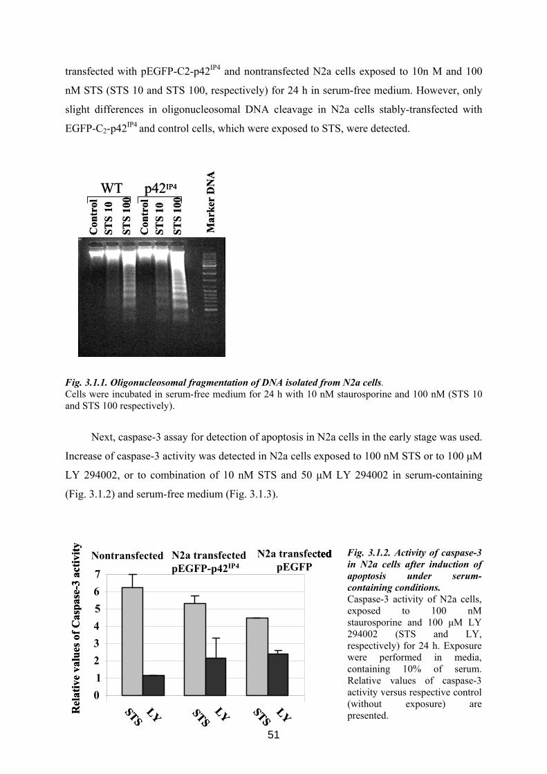
transfected with pEGFP-C2-p42IP4 and nontransfected N2a cells exposed to 10n M and 100
nM STS (STS 10 and STS 100, respectively) for 24 h in serum-free medium. However, only
slight differences in oligonucleosomal DNA cleavage in N2a cells stably-transfected with
EGFP-C2-p42IP4 and control cells, which were exposed to STS, were detected.
Con
trol
Con
trol
Mar
ker
DN
A
STS
10
STS
100
STS
10
STS
100
WT p42IP4
Con
trol
Con
trol
Mar
ker
DN
A
STS
10
STS
100
STS
10
STS
100
WT p42IP4
Fig. 3.1.1. Oligonucleosomal fragmentation of DNA isolated from N2a cells. Cells were incubated in serum-free medium for 24 h with 10 nM staurosporine and 100 nM (STS 10 and STS 100 respectively).
Next, caspase-3 assay for detection of apoptosis in N2a cells in the early stage was used.
Increase of caspase-3 activity was detected in N2a cells exposed to 100 nM STS or to 100 μM
LY 294002, or to combination of 10 nM STS and 50 μM LY 294002 in serum-containing
(Fig. 3.1.2) and serum-free medium (Fig. 3.1.3).
51
Rel
ativ
e va
lues
of C
aspa
se-3
activ
ity
01234567
Nontransfected N2a transfectedpEGFP-p42IP4
N2a transfected pEGFP
STSLY STSLY STSLYR
elat
ive
valu
esof
Cas
pase
-3ac
tivity
01234567
Nontransfected N2a transfectedpEGFP-p42IP4
N2a transfected pEGFP
STSLY STSLY STSLY
01234567
Nontransfected N2a transfectedpEGFP-p42IP4
N2a transfected pEGFP
STSLY STSLY STSLYSTSLY STSLY STSLY
Fig. 3.1.2. Activity of caspase-3 in N2a cells after induction of apoptosis under serum-containing conditions. Caspase-3 activity of N2a cells, exposed to 100 nM staurosporine and 100 μM LY 294002 (STS and LY, respectively) for 24 h. Exposure were performed in media, containing 10% of serum. Relative values of caspase-3 activity versus respective control (without exposure) are presented.
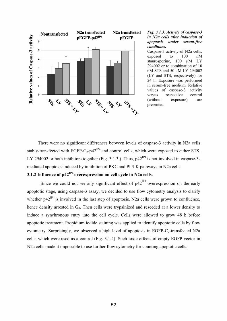
Rel
ativ
e va
lues
of C
aspa
se-3
activ
ity
0
1
2
3
4
5
6
Nontransfected N2a transfectedpEGFP-p42IP4
N2a transfected pEGFP
STSLY STS + LYSTSLY STS + LYSTSLY STS + LY
Rel
ativ
e va
lues
of C
aspa
se-3
activ
ity
0
1
2
3
4
5
6
Nontransfected N2a transfectedpEGFP-p42IP4
N2a transfected pEGFP
STSLY STS + LYSTSLY STS + LYSTSLY STS + LY
Fig. 3.1.3. Activity of caspase-3 in N2a cells after induction of apoptosis under serum-free conditions. Caspase-3 activity of N2a cells, exposed to 100 nM staurosporine, 100 μM LY 294002 or to combination of 10 nM STS and 50 μM LY 294002 (LY and STS, respectively) for 24 h. Exposure was performed in serum-free medium. Relative values of caspase-3 activity versus respective control (without exposure) are presented.
There were no significant differences between levels of caspase-3 activity in N2a cells
stably-transfected with EGFP-C2-p42IP4 and control cells, which were exposed to either STS,
LY 294002 or both inhibitors together (Fig. 3.1.3.). Thus, p42IP4 is not involved in caspase-3-
mediated apoptosis induced by inhibition of PKC and PI 3-K pathways in N2a cells.
3.1.2 Influence of p42IP4 overexpression on cell cycle in N2a cells.
Since we could not see any significant effect of p42IP4
overexpression on the early
apoptotic stage, using caspase-3 assay, we decided to use flow cytometry analysis to clarify
whether p42IP4 is involved in the last step of apoptosis. N2a cells were grown to confluence,
hence density arrested in G0. Then cells were trypsinized and reseeded at a lower density to
induce a synchronous entry into the cell cycle. Cells were allowed to grow 48 h before
apoptotic treatment. Propidium iodide staining was applied to identify apoptotic cells by flow
cytometry. Surprisingly, we observed a high level of apoptosis in EGFP-C2-transfected N2a
cells, which were used as a control (Fig. 3.1.4). Such toxic effects of empty EGFP vector in
N2a cells made it impossible to use further flow cytometry for counting apoptotic cells.
52
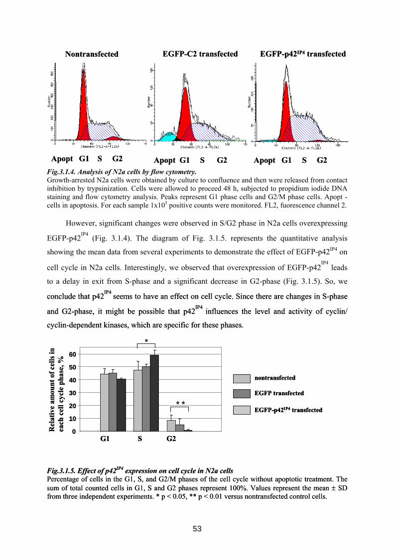
Apopt G1 S G2 Apopt G1 S G2 Apopt G1 S G2
Nontransfected EGFP-C2 transfected EGFP-p42IP4 transfected
Apopt G1 S G2 Apopt G1 S G2 Apopt G1 S G2
Nontransfected EGFP-C2 transfected EGFP-p42IP4 transfected
Fig.3.1.4. Analysis of N2a cells by flow cytometry. Growth-arrested N2a cells were obtained by culture to confluence and then were released from contact inhibition by trypsinization. Cells were allowed to proceed 48 h, subjected to propidium iodide DNA staining and flow cytometry analysis. Peaks represent G1 phase cells and G2/M phase cells. Apopt - cells in apoptosis. For each sample 1x10⁵ positive counts were monitored. FL2, fuorescence channel 2.
However, significant changes were observed in S/G2 phase in N2a cells overexpressing
EGFP-p42IP4 (Fig. 3.1.4). The diagram of Fig. 3.1.5. represents the quantitative analysis
showing the mean data from several experiments to demonstrate the effect of EGFP-p42IP4 on
cell cycle in N2a cells. Interestingly, we observed that overexpression of EGFP-p42IP4
leads
to a delay in exit from S-phase and a significant decrease in G2-phase (Fig. 3.1.5). So, we
conclude that p42IP4 seems to have an effect on cell cycle. Since there are changes in S-phase
and G2-phase, it might be possible that p42IP4 influences the level and activity of cyclin/
cyclin-dependent kinases, which are specific for these phases.
conclude that p42IP4 seems to have an effect on cell cycle. Since there are changes in S-phase
and G2-phase, it might be possible that p42IP4 influences the level and activity of cyclin/
cyclin-dependent kinases, which are specific for these phases.
0
10
20
30
40
50
60
Rel
ativ
e am
ount
of c
ells
in
each
cell
cycl
eph
ase,
%
nontransfected
EGFP transfected
EGFP-p42IP4 transfected
G1 S G2
*
* *
0
10
20
30
40
50
60
Rel
ativ
e am
ount
of c
ells
in
each
cell
cycl
eph
ase,
%
nontransfected
EGFP transfected
EGFP-p42IP4 transfected
G1 S G2
nontransfected
EGFP transfected
EGFP-p42IP4 transfected
G1 S G2
*
* *
Fig.3.1.5. Effect of p42IP4 expression on cell cycle in N2a cells Fig.3.1.5. Effect of p42Percentage of cells in the G1, S, and G2/M phases of the cell cycle without apoptotic treatment. The sum of total counted cells in G1, S and G2 phases represent 100%. Values represent the mean ± SD from three independent experiments. * p < 0.05, ** p < 0.01 versus nontransfected control cells.
Percentage of cells in the G1, S, and G2/M phases of the cell cycle without apoptotic treatment. The sum of total counted cells in G1, S and G2 phases represent 100%. Values represent the mean ± SD from three independent experiments. * p < 0.05, ** p < 0.01 versus nontransfected control cells.
IP4 expression on cell cycle in N2a cells
53
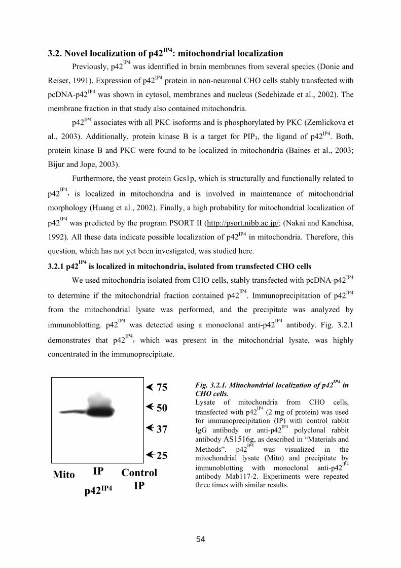
3.2. Novel localization of p42IP4: mitochondrial localization Previously, p42IP4 was identified in brain membranes from several species (Donie and
Reiser, 1991). Expression of p42IP4 protein in non-neuronal CHO cells stably transfected with
pcDNA-p42IP4 was shown in cytosol, membranes and nucleus (Sedehizade et al., 2002). The
membrane fraction in that study also contained mitochondria.
p42IP4 associates with all PKC isoforms and is phosphorylated by PKC (Zemlickova et
al., 2003). Additionally, protein kinase B is a target for PIP3, the ligand of p42IP4. Both,
protein kinase B and PKC were found to be localized in mitochondria (Baines et al., 2003;
Bijur and Jope, 2003).
Furthermore, the yeast protein Gcs1p, which is structurally and functionally related to
p42IP4, is localized in mitochondria and is involved in maintenance of mitochondrial
morphology (Huang et al., 2002). Finally, a high probability for mitochondrial localization of
p42IP4 was predicted by the program PSORT II (http://psort.nibb.ac.jp/; (Nakai and Kanehisa,
1992). All these data indicate possible localization of p42IP4 in mitochondria. Therefore, this
question, which has not yet been investigated, was studied here.
3.2.1 p42IP4 is localized in mitochondria, isolated from transfected CHO cells
We used mitochondria isolated from CHO cells, stably transfected with pcDNA-p42IP4
to determine if the mitochondrial fraction contained p42IP4. Immunoprecipitation of p42IP4
from the mitochondrial lysate was performed, and the precipitate was analyzed by
immunoblotting. p42IP4 was detected using a monoclonal anti-p42IP4 antibody. Fig. 3.2.1
demonstrates that p42IP4, which was present in the mitochondrial lysate, was highly
concentrated in the immunoprecipitate.
Fig. 3.2.1. Mitochondrial localization of p42IP4 in CHO cells. Lysate of mitochondria from CHO cells, transfected with p42IP4 (2 mg of protein) was used for immunoprecipitation (IP) with control rabbit IgG antibody or anti-p42IP4 polyclonal rabbit antibody AS1516g, as described in “Materials and Methods”. p42IP4 was visualized in the mitochondrial lysate (Mito) and precipitate by immunoblotting with monoclonal anti-p42IP4
antibody Mab117-2. Experiments were repeated three times with similar results.
50
37
25
75
Mito IP
p42IP4
ControlIP
54
3.2.2 p42IP4 is localized in inner mitochondrial compartments of transfected CHO cells
To investigate the compartmentalization of p42IP4 within mitochondria, mitochondria
from CHO cells expressing p42IP4 were subfractionated into outer membrane, intermembrane
space, inner membrane and matrix fractions. p42IP4 was found in all mitochondrial fractions
of CHO cells (Fig. 3.2.2. A), but seems to be enriched in the intermembrane space. Separation
of each mitochondrial compartment was verified by immunoblotting of the subfractions with
mitochondrial compartment-specific proteins, VDAC (for outer membrane), Cyt c (for
intermembrane space), cytochrome oxidase subunit IV (for inner membrane), and the Mn-
superoxide dismutase (SOD2) for matrix, as shown in Fig. 3.2.2. A. VDAC, an outer
membrane protein, is enriched at contact sites between the outer and inner membranes and is,
therefore, typically detected in both outer membrane and inner membrane fractions (Bijur and
Jope, 2003). SOD2 was stained not only in the matrix fraction. Probably due to the fragility of
the inner membrane of mitochondria isolated from CHO cells SOD2 was also detected in the
intermembrane space.
Mitochondrial compartments contain different amounts of protein. Therefore, to
calculate the total p42IP4 content in each fraction, we had to take into account the whole
protein amount in each subfraction of mitochondria. The relative distribution of p42IP4 was
obtained by calculating the ratio between the total p42IP4 protein content in each
submitochondrial fraction over the sum of p42IP4 contents of all fractions. The result shown in
Fig. 3.2.2. B reveals that p42IP4 is localized predominantly at the inner membrane and in the
intermembrane space of mitochondria, which is similar to the localization of Cyt c. p42IP4 is
detectable only to a small degree in matrix and outer membrane fractions.
55
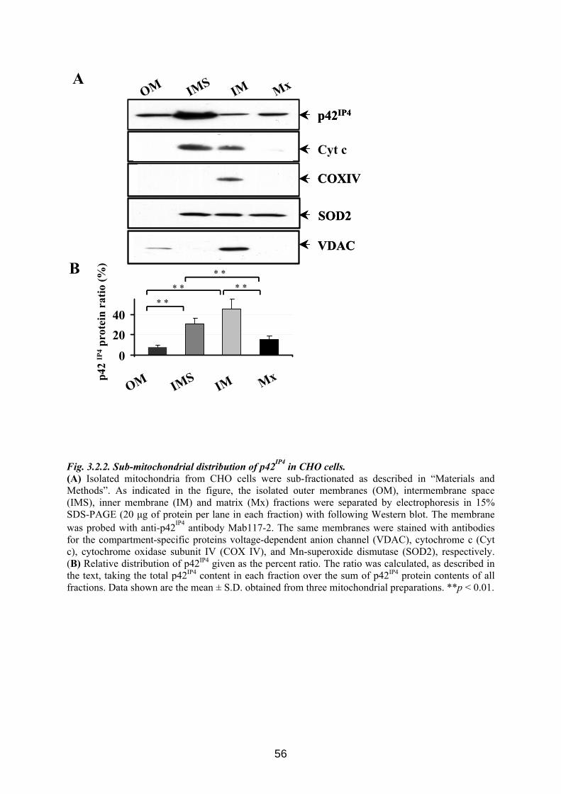
Cyt c
OM IMS IM Mx
02040
OM IMS IM Mx
* ** * * *
* *
Ap4
2 IP
4pr
otei
nra
tio (%
)BVDAC
SOD2
p42IP4
COXIV
VDAC
SOD2
p42IP4
COXIV
Fig. 3.2.2. Sub-mitochondrial distribution of p42IP4 in CHO cells. (A) Isolated mitochondria from CHO cells were sub-fractionated as described in “Materials and Methods”. As indicated in the figure, the isolated outer membranes (OM), intermembrane space (IMS), inner membrane (IM) and matrix (Mx) fractions were separated by electrophoresis in 15% SDS-PAGE (20 μg of protein per lane in each fraction) with following Western blot. The membrane was probed with anti-p42IP4 antibody Mab117-2. The same membranes were stained with antibodies for the compartment-specific proteins voltage-dependent anion channel (VDAC), cytochrome c (Cyt c), cytochrome oxidase subunit IV (COX IV), and Mn-superoxide dismutase (SOD2), respectively. (B) Relative distribution of p42IP4 given as the percent ratio. The ratio was calculated, as described in the text, taking the total p42IP4 content in each fraction over the sum of p42IP4 protein contents of all fractions. Data shown are the mean ± S.D. obtained from three mitochondrial preparations. **p < 0.01.
56
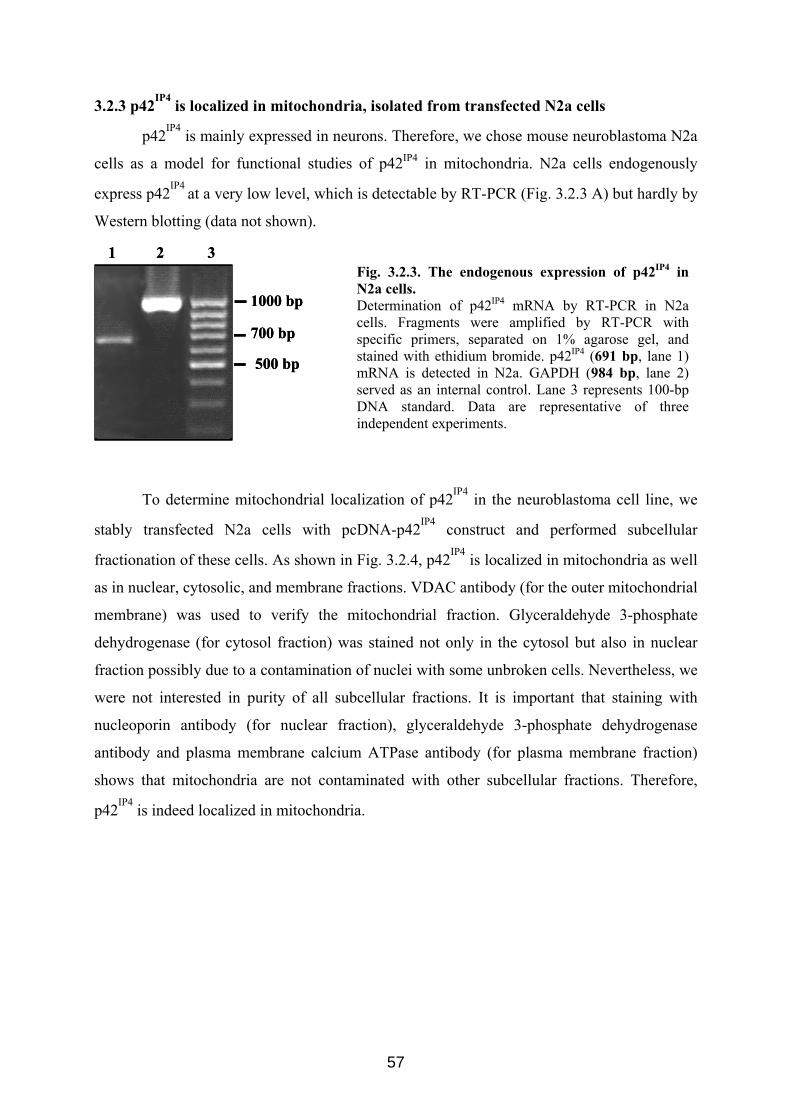
3.2.3 p42IP4 is localized in mitochondria, isolated from transfected N2a cells
p42IP4 is mainly expressed in neurons. Therefore, we chose mouse neuroblastoma N2a
cells as a model for functional studies of p42IP4 in mitochondria. N2a cells endogenously
express p42IP4 at a very low level, which is detectable by RT-PCR (Fig. 3.2.3 A) but hardly by
Western blotting (data not shown).
1000 bp
700 bp
500 bp
1 2 3
1000 bp
700 bp
500 bp
1 2 3Fig. 3.2.3. The endogenous expression of p42IP4 in N2a cells. Determination of p42IP4 mRNA by RT-PCR in N2a cells. Fragments were amplified by RT-PCR with specific primers, separated on 1% agarose gel, and stained with ethidium bromide. p42IP4 (691 bp, lane 1) mRNA is detected in N2a. GAPDH (984 bp, lane 2) served as an internal control. Lane 3 represents 100-bp DNA standard. Data are representative of three independent experiments.
To determine mitochondrial localization of p42IP4 in the neuroblastoma cell line, we
stably transfected N2a cells with pcDNA-p42IP4 construct and performed subcellular
fractionation of these cells. As shown in Fig. 3.2.4, p42IP4 is localized in mitochondria as well
as in nuclear, cytosolic, and membrane fractions. VDAC antibody (for the outer mitochondrial
membrane) was used to verify the mitochondrial fraction. Glyceraldehyde 3-phosphate
dehydrogenase (for cytosol fraction) was stained not only in the cytosol but also in nuclear
fraction possibly due to a contamination of nuclei with some unbroken cells. Nevertheless, we
were not interested in purity of all subcellular fractions. It is important that staining with
nucleoporin antibody (for nuclear fraction), glyceraldehyde 3-phosphate dehydrogenase
antibody and plasma membrane calcium ATPase antibody (for plasma membrane fraction)
shows that mitochondria are not contaminated with other subcellular fractions. Therefore,
p42IP4 is indeed localized in mitochondria.
57
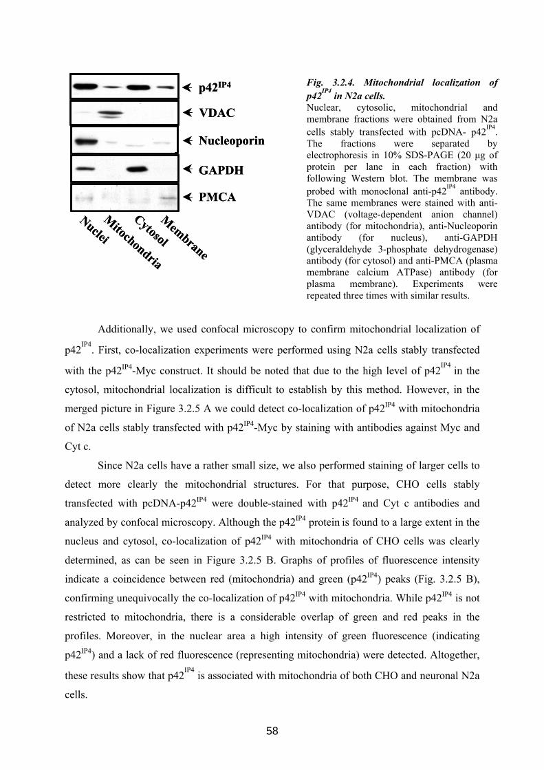
Additionally, we used confocal microscopy to confirm mitochondrial localization of
p42IP4. First, co-localization experiments were performed using N2a cells stably transfected
with the p42IP4-Myc construct. It should be noted that due to the high level of p42IP4 in the
cytosol, mitochondrial localization is difficult to establish by this method. However, in the
merged picture in Figure 3.2.5 A we could detect co-localization of p42IP4 with mitochondria
of N2a cells stably transfected with p42IP4-Myc by staining with antibodies against Myc and
Cyt c.
Since N2a cells have a rather small size, we also performed staining of larger cells to
detect more clearly the mitochondrial structures. For that purpose, CHO cells stably
transfected with pcDNA-p42IP4 were double-stained with p42IP4 and Cyt c antibodies and
analyzed by confocal microscopy. Although the p42IP4 protein is found to a large extent in the
nucleus and cytosol, co-localization of p42IP4 with mitochondria of CHO cells was clearly
determined, as can be seen in Figure 3.2.5 B. Graphs of profiles of fluorescence intensity
indicate a coincidence between red (mitochondria) and green (p42IP4) peaks (Fig. 3.2.5 B),
confirming unequivocally the co-localization of p42IP4 with mitochondria. While p42IP4 is not
restricted to mitochondria, there is a considerable overlap of green and red peaks in the
profiles. Moreover, in the nuclear area a high intensity of green fluorescence (indicating
p42IP4) and a lack of red fluorescence (representing mitochondria) were detected. Altogether,
these results show that p42IP4 is associated with mitochondria of both CHO and neuronal N2a
cells.
Fig. 3.2.4. Mitochondrial localization of p42IP4 in N2a cells. Nuclear, cytosolic, mitochondrial and membrane fractions were obtained from N2a cells stably transfected with pcDNA- p42IP4. The fractions were separated by electrophoresis in 10% SDS-PAGE (20 μg of protein per lane in each fraction) with following Western blot. The membrane was probed with monoclonal anti-p42IP4 antibody. The same membranes were stained with anti-VDAC (voltage-dependent anion channel) antibody (for mitochondria), anti-Nucleoporin antibody (for nucleus), anti-GAPDH (glyceraldehyde 3-phosphate dehydrogenase) antibody (for cytosol) and anti-PMCA (plasma membrane calcium ATPase) antibody (for plasma membrane). Experiments were repeated three times with similar results.
Nuclei
Mitochondria
Cytosol
p42IP4
VDAC
Nucleoporin
Membrane
GAPDH
PMCA
Nuclei
Mitochondria
Cytosol
p42IP4
VDAC
Nucleoporin
Membrane
GAPDH
PMCA
58
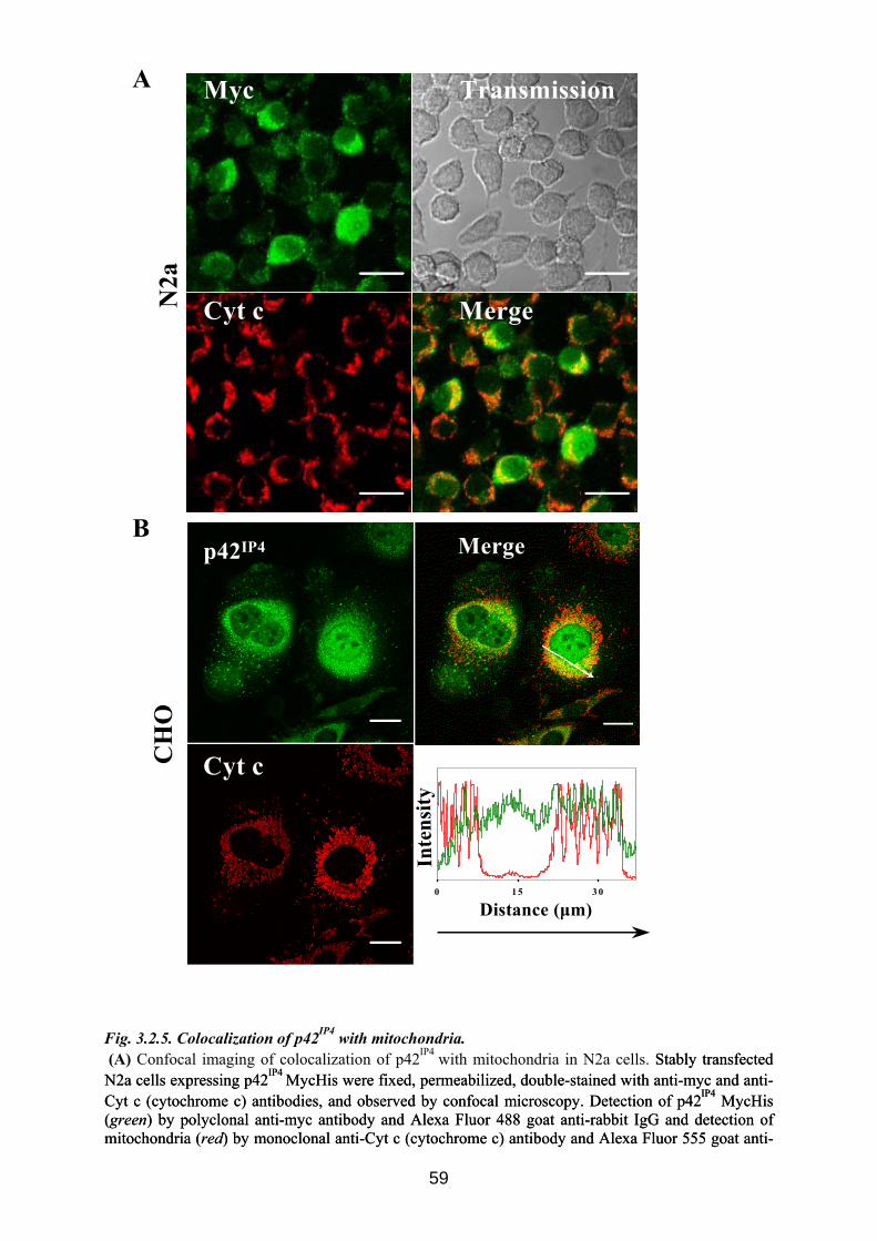
0 1 5 3 0
Cyt c
p42IP4 Merge
Distance (μm)
Inte
nsity
CH
O
Myc
Cyt c Merge
Transmission
N2a
B
A
Fig. 3.2.5. Colocalization of p42IP4 with mitochondria. (A) Confocal imaging of colocalization of p42IP4 with mitochondria in N2a cells. Stably transfected N2a cells expressing p42IP4 MycHis were fixed, permeabilized, double-stained with anti-myc and anti-Cyt c (cytochrome c) antibodies, and observed by confocal microscopy. Detection of p42IP4 MycHis (green) by polyclonal anti-myc antibody and Alexa Fluor 488 goat anti-rabbit IgG and detection of mitochondria (red) by monoclonal anti-Cyt c (cytochrome c) antibody and Alexa Fluor 555 goat anti-
Stably transfected N2a cells expressing p42IP4 MycHis were fixed, permeabilized, double-stained with anti-myc and anti-Cyt c (cytochrome c) antibodies, and observed by confocal microscopy. Detection of p42IP4 MycHis (green) by polyclonal anti-myc antibody and Alexa Fluor 488 goat anti-rabbit IgG and detection of mitochondria (red) by monoclonal anti-Cyt c (cytochrome c) antibody and Alexa Fluor 555 goat anti-
59
mouse IgG was carried out. The merged picture in the lower right panel shows co-localization of p42IP4 with mitochondria (yellow). The right upper panel shows a transmission light picture. Scale bar indicates 20 μm. Pictures shown are representative of at least three independent experiments. (B) Confocal imaging of colocalization of p42IP4 with mitochondria in CHO cells. Stably transfected CHO cells expressing p42IP4 were fixed, permeabilized, double-stained with polyclonal rabbit serum raised against p42IP4 (AS1516g) and anti-Cyt c (cytochrome c) antibodies, and observed by confocal microscopy. Left upper panel shows detection of p42IP4 (green) by polyclonal rabbit serum and Alexa Fluor 488 goat anti-rabbit IgG, and the left lower panel shows detection of mitochondria (red) by monoclonal anti-Cyt c (cytochrome c) antibody and Alexa Fluor 555 goat anti-mouse IgG. The merged picture in the right upper panel shows co-localization of p42IP4 with mitochondria (yellow). Scale bar indicates 20 μm. The curves (right lower panel) represent the fluorescence intensity distribution determined for a cross-section of the cell, as indicated by the white arrow in the merged image. Arrow indicates the orientation of the section. Pictures shown are representative of at least three independent experiments.
3.3 p42IP4 is involved in regulation of mitochondrial Ca2+ The findings that p42IP4 is localized at the mitochondrial inner membrane and in the
intermembrane space, raised the question of possible functions of this protein in
mitochondria. To address this issue, we used a recently developed method of isolation of
functionally active mitochondria from cultured cells and studied mitochondrial functions
during Ca2+-induced PTP opening (Azarashvili et al., 2005; Azarashvili et al., 2007).
We investigated whether p42IP4 might be involved in the regulation of mitochondrial
Ca2+ transport mechanisms. The PTP forms a Ca2+ efflux pathway from mitochondria through
a large pore (Smaili et al., 2000). To study the influence of p42IP4 on mitochondrial functions
during Ca2+-induced PTP opening in mitochondria, we compared the functional parameters of
mitochondria, which were isolated from N2a wild type cells, or from cells transfected with
p42IP4, or transfected with empty vector. The effect of the over-expression of p42IP4 on Ca2+
transport, mitochondrial membrane potential (ΔΨm), and oxygen consumption rate were
examined in isolated mitochondria upon Ca2+-induced PTP opening.
3.3.1 p42IP4 accelerates Ca 2+-induced PTP opening in mitochondria from N2a cells
We used the open recording chamber with installed electrodes selective for O2, Ca2+
and TPP+, as described in previous reports (Azarashvili et al., 2005; Azarashvili et al., 2007).
The ability of mitochondria isolated from N2a wild type cells, or from cells transfected with
p42IP4, or transfected with empty vector to accumulate Ca2+ was investigated using two
approaches.
First, Ca2+ was added in small bolus additions (70 nanomoles of Ca2+ per mg of
protein per addition) to estimate the Ca2+ threshold (Ca2+ capacity) for each kind of
mitochondria. Figure 3.3.1 A shows an experiment with mitochondria isolated from wild type
N2a cells. After addition of the mitochondrial suspension into the chamber, the mitochondria
60
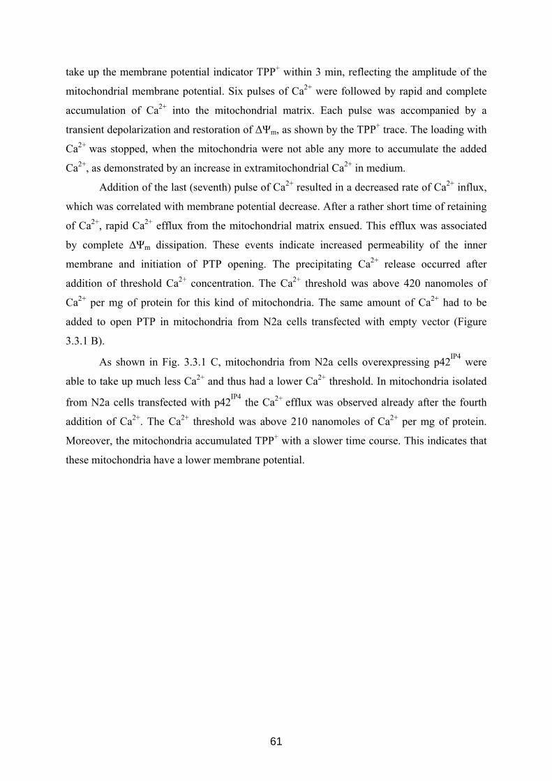
take up the membrane potential indicator TPP+ within 3 min, reflecting the amplitude of the
mitochondrial membrane potential. Six pulses of Ca2+ were followed by rapid and complete
accumulation of Ca2+ into the mitochondrial matrix. Each pulse was accompanied by a
transient depolarization and restoration of ΔΨm, as shown by the TPP+ trace. The loading with
Ca2+ was stopped, when the mitochondria were not able any more to accumulate the added
Ca2+, as demonstrated by an increase in extramitochondrial Ca2+ in medium.
Addition of the last (seventh) pulse of Ca2+ resulted in a decreased rate of Ca2+ influx,
which was correlated with membrane potential decrease. After a rather short time of retaining
of Ca2+, rapid Ca2+ efflux from the mitochondrial matrix ensued. This efflux was associated
by complete ΔΨm dissipation. These events indicate increased permeability of the inner
membrane and initiation of PTP opening. The precipitating Ca2+ release occurred after
addition of threshold Ca2+ concentration. The Ca2+ threshold was above 420 nanomoles of
Ca2+ per mg of protein for this kind of mitochondria. The same amount of Ca2+ had to be
added to open PTP in mitochondria from N2a cells transfected with empty vector (Figure
3.3.1 B).
As shown in Fig. 3.3.1 C, mitochondria from N2a cells overexpressing p42IP4 were
able to take up much less Ca2+ and thus had a lower Ca2+ threshold. In mitochondria isolated
from N2a cells transfected with p42IP4 the Ca2+ efflux was observed already after the fourth
addition of Ca2+. The Ca2+ threshold was above 210 nanomoles of Ca2+ per mg of protein.
Moreover, the mitochondria accumulated TPP+ with a slower time course. This indicates that
these mitochondria have a lower membrane potential.
61
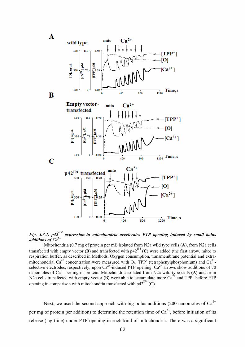
Fig. 3.3.1. p42IP4 expression in mitochondria accelerates PTP opening induced by small bolus additions of Ca2+.
Mitochondria (0.7 mg of protein per ml) isolated from N2a wild type cells (A), from N2a cells transfected with empty vector (B) and transfected with p42IP4 (C) were added (the first arrow, mito) to respiration buffer, as described in Methods. Oxygen consumption, transmembrane potential and extra-mitochondrial Ca2+ concentration were measured with O2, TPP+ (tetraphenylphosphonium) and Ca2+-selective electrodes, respectively, upon Ca2+-induced PTP opening. Ca2+ arrows show additions of 70 nanomoles of Ca2+ per mg of protein. Mitochondria isolated from N2a wild type cells (A) and from N2a cells transfected with empty vector (B) were able to accumulate more Ca2+ and TPP+ before PTP opening in comparison with mitochondria transfected with p42IP4 (C).
Next, we used the second approach with big bolus additions (200 nanomoles of Ca2+
per mg of protein per addition) to determine the retention time of Ca2+, before initiation of its
release (lag time) under PTP opening in each kind of mitochondria. There was a significant
62
decrease in the Ca2+ threshold and acceleration of PTP opening in mitochondria isolated from
N2a cells transfected with p42IP4 in comparison with mitochondria from N2a wild type cells
(Fig. 3.3.1). Therefore, we performed determination of lag time with the amount of Ca2+
which had to be added to open PTP in N2a cells transfected with p42IP4. With this amount of
Ca2+ we expected that in mitochondria from N2a cells transfected with p42IP4 the lag time
would be shorter than in mitochondria from N2a wild type cells.
Figure 3.3.2 clearly shows the difference in lag time for PTP opening between
mitochondria isolated from N2a wild type cells (A), and from N2a cells transfected with
p42IP4 (C). The kinetics of Ca2+ uptake in these kinds of mitochondria were different, namely
the rate of Ca2+ influx was decreased in mitochondria from N2a cells overexpressing p42IP4 in
comparison with mitochondria from wild type. Lag time for PTP opening in mitochondria
from N2a cells transfected with empty vector (Fig. 3.3.2 B) was the same as for mitochondria
isolated from N2a wild type cells (Fig. 3.3.2 A).
63
Fig. 3.3.2. p42IP4 expression in mitochondria accelerates PTP opening induced by big bolus additions of Ca2+. Mitochondria (0.7 mg of protein per ml) isolated from N2a wild type cells (A), from N2a cells transfected with empty vector (B) and from N2a cells transfected with p42IP4 (C) were added (the first arrow, mito) to respiration buffer, as described in Methods. Oxygen consumption, transmembrane potential and extra-mitochondrial Ca2+ concentration were measured with O2, TPP+ (tetraphenylphosphonium) and Ca2+-selective electrodes, respectively, upon Ca2+-induced PTP opening. Ca2+ arrows show additions of 200 nanomoles of Ca2+ per mg of protein. The difference in time before Ca
2+ release (lag time) for PTP opening is illustrated between mitochondria isolated from
N2a wild type cells (A), and from N2a cells transfected with p42IP4 (C). Lag time for PTP opening in mitochondria from N2a cells transfected with empty vector (B) was the same as for mitochondria isolated from N2a wild type cells (A). Results shown are representative of at least three independent experiments.
64
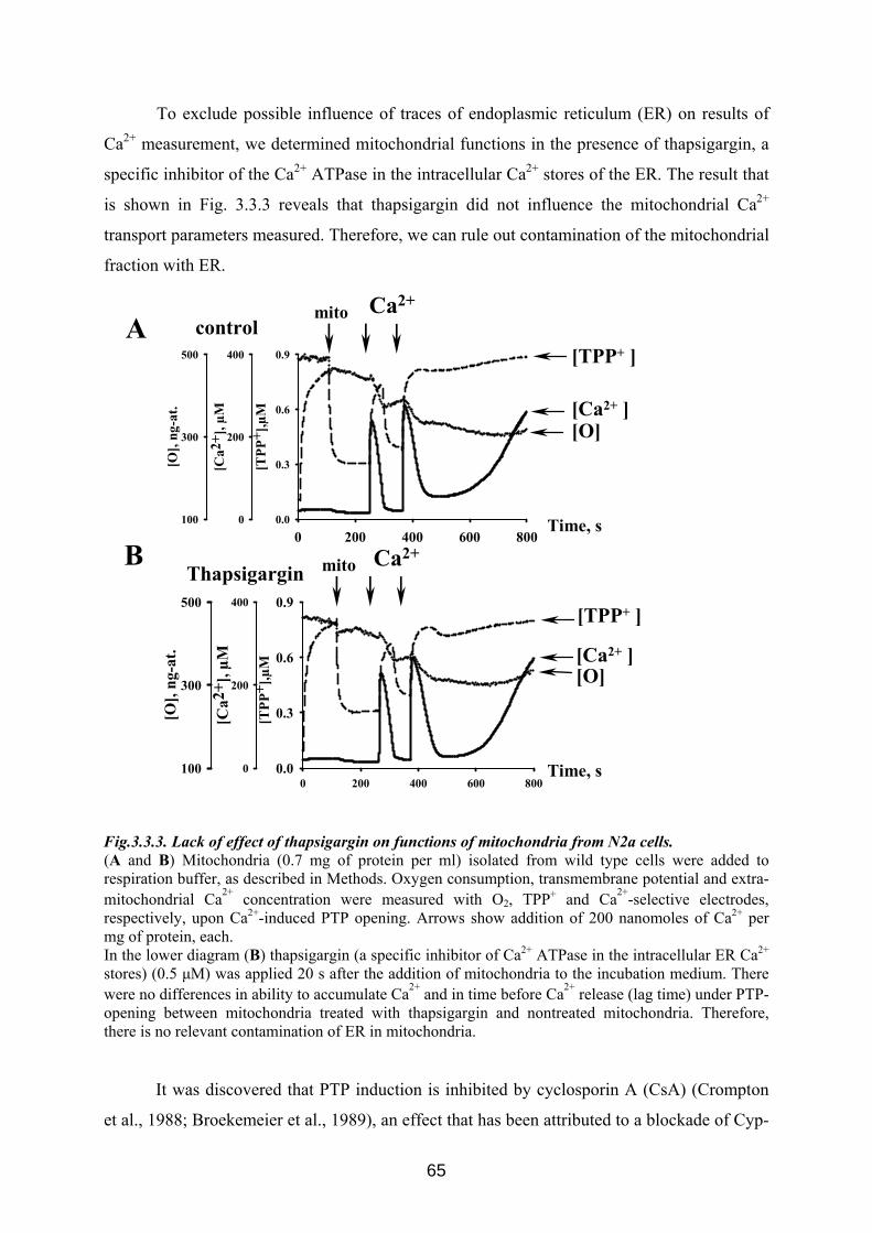
To exclude possible influence of traces of endoplasmic reticulum (ER) on results of
Ca2+ measurement, we determined mitochondrial functions in the presence of thapsigargin, a
specific inhibitor of the Ca2+ ATPase in the intracellular Ca2+ stores of the ER. The result that
is shown in Fig. 3.3.3 reveals that thapsigargin did not influence the mitochondrial Ca2+
transport parameters measured. Therefore, we can rule out contamination of the mitochondrial
fraction with ER.
0 200 400 600 800
[TPP
+ ],µ
M
0.0
0.3
0.6
0.9
[Ca2
+ ], µ
M
0
200
400
[O],
ng-a
t.
100
300
500
0 200 400 600 800
[TPP
+ ],µ
M
0.0
0.3
0.6
0.9
[Ca2
+ ], µ
M
0
200
400
[O],
ng-a
t.
100
300
500
control
Thapsigargin
A
B
[O] [Ca2+ ]
[TPP+ ]
Time, s
Ca2+mito
[O] [Ca2+ ]
[TPP+ ]
Ca2+mito
Time, s
Fig.3.3.3. Lack of effect of thapsigargin on functions of mitochondria from N2a cells. (A and B) Mitochondria (0.7 mg of protein per ml) isolated from wild type cells were added to respiration buffer, as described in Methods. Oxygen consumption, transmembrane potential and extra-mitochondrial Ca2+ concentration were measured with O2, TPP P
+ and Ca2+-selective electrodes, respectively, upon Ca2+-induced PTP opening. Arrows show addition of 200 nanomoles of Ca2+ per mg of protein, each. In the lower diagram (B) thapsigargin (a specific inhibitor of Ca2+ ATPase in the intracellular ER Ca2+ stores) (0.5 μM) was applied 20 s after the addition of mitochondria to the incubation medium. There were no differences in ability to accumulate Ca2+ and in time before Ca2+ release (lag time) under PTP-opening between mitochondria treated with thapsigargin and nontreated mitochondria. Therefore, there is no relevant contamination of ER in mitochondria.
It was discovered that PTP induction is inhibited by cyclosporin A (CsA) (Crompton
et al., 1988; Broekemeier et al., 1989), an effect that has been attributed to a blockade of Cyp-
65
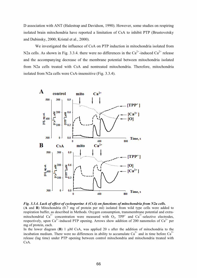
D association with ANT (Halestrap and Davidson, 1990). However, some studies on respiring
isolated brain mitochondria have reported a limitation of CsA to inhibit PTP (Brustovetsky
and Dubinsky, 2000; Kristal et al., 2000).
We investigated the influence of CsA on PTP induction in mitochondria isolated from
N2a cells. As shown in Fig. 3.3.4. there were no differences in the Ca2+-induced Ca2+ release
and the accompanying decrease of the membrane potential between mitochondria isolated
from N2a cells treated with CsA and nontreated mitochondria. Therefore, mitochondria
isolated from N2a cells were CsA-insensitive (Fig. 3.3.4).
Fig. 3.3.4. Lack of effect of cyclosporine A (CsA) on functions of mitochondria from N2a cells. (A and B) Mitochondria (0.7 mg of protein per ml) isolated from wild type cells were added to respiration buffer, as described in Methods. Oxygen consumption, transmembrane potential and extra-mitochondrial Ca2+ concentration were measured with O2, TPP P
+ and Ca2+-selective electrodes, respectively, upon Ca2+-induced PTP opening. Arrows show addition of 200 nanomoles of Ca2+ per mg of protein, each. In the lower diagram (B) 1 μM CsA, was applied 20 s after the addition of mitochondria to the incubation medium. There were no differences in ability to accumulate Ca2+ and in time before Ca2+ release (lag time) under PTP opening between control mitochondria and mitochondria treated with CsA.
66
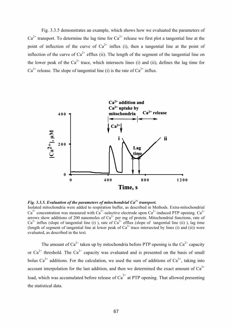
Fig. 3.3.5 demonstrates an example, which shows how we evaluated the parameters of
Ca2+ transport. To determine the lag time for Ca2+ release we first plot a tangential line at the
point of inflection of the curve of Ca2+ influx (i), then a tangential line at the point of
inflection of the curve of Ca2+ efflux (ii). The length of the segment of the tangential line on
the lower peak of the Ca2+ trace, which intersects lines (i) and (ii), defines the lag time for
Ca2+ release. The slope of tangential line (i) is the rate of Ca2+ influx.
0 4 0 0 8 0 0 1 2 0 0
[Ca2
+ ], µ
M
0
2 0 0
4 0 0
Lag time
ii
Ca2+ release
Ca2+
Ca2+ addition and Ca2+ uptake by mitochondria
Time, s
i
Lag time
ii
Ca2+ release
Ca2+
Ca2+ addition and Ca2+ uptake by mitochondria
Time, s
i
Fig. 3.3.5. Evaluation of the parameters of mitochondrial Ca2+ transport. Isolated mitochondria were added to respiration buffer, as described in Methods. Extra-mitochondrial Ca2+ concentration was measured with Ca2+-selective electrode upon Ca2+-induced PTP opening. Ca2+ arrows show additions of 200 nanomoles of Ca2+ per mg of protein. Mitochondrial functions, rate of Ca2+ influx (slope of tangential line (i) ), rate of Ca2+ efflux (slope of tangential line (ii) ), lag time (length of segment of tangential line at lower peak of Ca2+ trace intersected by lines (i) and (ii)) were evaluated, as described in the text.
The amount of Ca2+ taken up by mitochondria before PTP opening is the Ca2+ capacity
or Ca2+ threshold. The Ca2+ capacity was evaluated and is presented on the basis of small
bolus Ca2+ additions. For the calculation, we used the sum of additions of Ca2+, taking into
account interpolation for the last addition, and then we determined the exact amount of Ca2+
load, which was accumulated before release of Ca2+
at PTP opening. That allowed presenting
the statistical data.
67
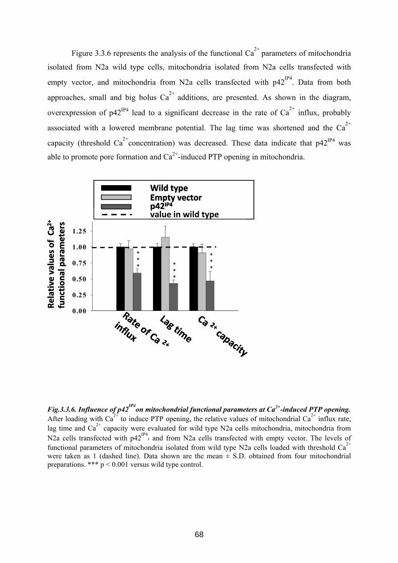
Figure 3.3.6 represents the analysis of the functional Ca2+ parameters of mitochondria
isolated from N2a wild type cells, mitochondria isolated from N2a cells transfected with
empty vector, and mitochondria from N2a cells transfected with p42IP4. Data from both
approaches, small and big bolus Ca2+ additions, are presented. As shown in the diagram,
overexpression of p42IP4 lead to a significant decrease in the rate of Ca2+ influx, probably
associated with a lowered membrane potential. The lag time was shortened and the Ca2+
capacity (threshold Ca2+concentration) was decreased. These data indicate that p42IP4 was
able to promote pore formation and Ca2+-induced PTP opening in mitochondria.
0.00
0.25
0.50
0.75
1.00
1.25
Ca 2+capacity
Rate of Ca 2+
influx
Lag time
Wild type
p42IP4Empty vector
value in wild type
*** *
**
***
Relative
values
of Ca2
+
function
al param
eters
0.00
0.25
0.50
0.75
1.00
1.25
Ca 2+capacity
Rate of Ca 2+
influx
Lag time
Wild type
p42IP4Empty vector
value in wild type
*** *
**
***
Relative
values
of Ca2
+
function
al param
eters
Fig.3.3.6. Influence of p42IP4on mitochondrial functional parameters at Ca2+-induced PTP opening. After loading with Ca2+ to induce PTP opening, the relative values of mitochondrial Ca2+ influx rate, lag time and Ca2+ capacity were evaluated for wild type N2a cells mitochondria, mitochondria from N2a cells transfected with p42IP4, and from N2a cells transfected with empty vector. The levels of functional parameters of mitochondria isolated from wild type N2a cells loaded with threshold Ca2+ were taken as 1 (dashed line). Data shown are the mean ± S.D. obtained from four mitochondrial preparations. *** p < 0.001 versus wild type control.
68
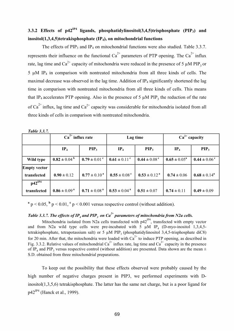
3.3.2 Effects of p42IP4 ligands, phosphatidylinositol(3,4,5)trisphosphate (PIP3) and
inositol(1,3,4,5)tetrakisphosphate (IP4), on mitochondrial functions
The effects of PIP3 and IP4 on mitochondrial functions were also studied. Table 3.3.7.
represents their influence on the functional Ca2+ parameters of PTP opening. The Ca2+ influx
rate, lag time and Ca2+ capacity of mitochondria were reduced in the presence of 5 µM PIP3 or
5 µM IP4 in comparison with nontreated mitochondria from all three kinds of cells. The
maximal decrease was observed in the lag time. Addition of IP4 significantly shortened the lag
time in comparison with nontreated mitochondria from all three kinds of cells. This means
that IP4 accelerates PTP opening. Also in the presence of 5 µM PIP3 the reduction of the rate
of Ca2+ influx, lag time and Ca2+ capacity was considerable for mitochondria isolated from all
three kinds of cells in comparison with nontreated mitochondria.
Table 3.3.7. Ca2+ influx rate Lag time Ca2+ capacity
IP4 PIP3 IP4 PIP3 IP4 PIP3
Wild type 0.82 ± 0.04 b 0.79 ± 0.01 c 0.61 ± 0.11 c 0.44 ± 0.08 c 0.65 ± 0.05a 0.44 ± 0.06 c
Empty vector
transfected 0.90 ± 0.12 0.77 ± 0.10 a 0.55 ± 0.08 c 0.53 ± 0.12 a 0.74 ± 0.06 0.68 ± 0.14a
p42IP4
transfected 0.86 ± 0.09 a 0.71 ± 0.08 a 0.53 ± 0.04 a 0.51 ± 0.07 0.74 ± 0.11 0.49 ± 0.09
a p < 0.05, b p < 0.01, c p < 0.001 versus respective control (without addition). Table 3.3.7. The effects of IP4 and PIP3 on Ca2+ parameters of mitochondria from N2a cells.
Mitochondria isolated from N2a cells transfected with p42IP4, transfected with empty vector and from N2a wild type cells were pre-incubated with 5 µM IP4 (D-myo-inositol 1,3,4,5-tetrakisphosphate, tetrapotassium salt) or 5 µM PIP3 (phosphatidylinositol 3,4,5-trisphosphate diC8) for 20 min. After that, the mitochondria were loaded with Ca2+ to induce PTP opening, as described in Fig. 3.3.2. Relative values of mitochondrial Ca2+ influx rate, lag time and Ca2+ capacity in the presence of IP4 and PIP3 versus respective control (without addition) are presented. Data shown are the mean ± S.D. obtained from three mitochondrial preparations.
To keep out the possibility that these effects observed were probably caused by the
high number of negative charges present in PIP3, we performed experiments with D-
inositol(1,3,5,6) tetrakisphosphate. The latter has the same net charge, but is a poor ligand for
p42IP4 (Hanck et al., 1999).
69
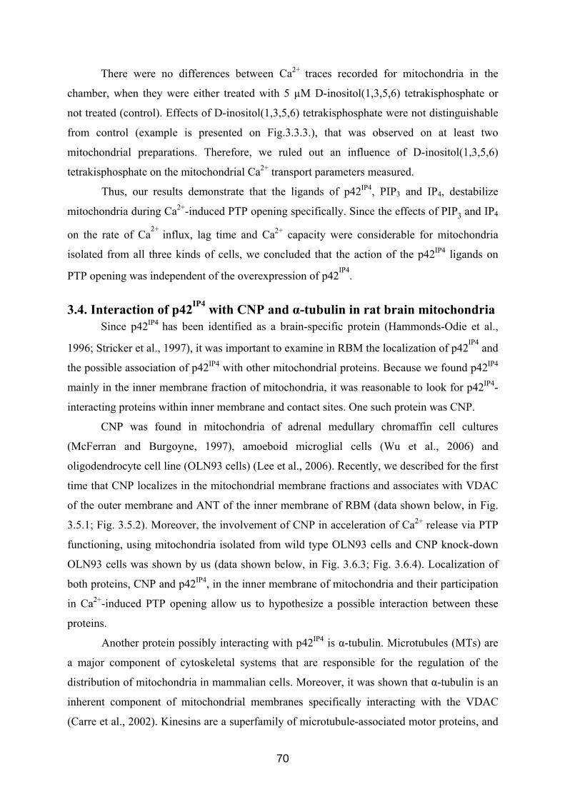
There were no differences between Ca2+ traces recorded for mitochondria in the
chamber, when they were either treated with 5 µM D-inositol(1,3,5,6) tetrakisphosphate or
not treated (control). Effects of D-inositol(1,3,5,6) tetrakisphosphate were not distinguishable
from control (example is presented on Fig.3.3.3.), that was observed on at least two
mitochondrial preparations. Therefore, we ruled out an influence of D-inositol(1,3,5,6)
tetrakisphosphate on the mitochondrial Ca2+ transport parameters measured.
Thus, our results demonstrate that the ligands of p42IP4, PIP3 and IP4, destabilize
mitochondria during Ca2+-induced PTP opening specifically. Since the effects of PIP3 and IP4
on the rate of Ca2+ influx, lag time and Ca2+ capacity were considerable for mitochondria
isolated from all three kinds of cells, we concluded that the action of the p42IP4 ligands on
PTP opening was independent of the overexpression of p42IP4.
3.4. Interaction of p42IP4 with CNP and α-tubulin in rat brain mitochondria Since p42IP4 has been identified as a brain-specific protein (Hammonds-Odie et al.,
1996; Stricker et al., 1997), it was important to examine in RBM the localization of p42IP4 and
the possible association of p42IP4 with other mitochondrial proteins. Because we found p42IP4
mainly in the inner membrane fraction of mitochondria, it was reasonable to look for p42IP4-
interacting proteins within inner membrane and contact sites. One such protein was CNP.
CNP was found in mitochondria of adrenal medullary chromaffin cell cultures
(McFerran and Burgoyne, 1997), amoeboid microglial cells (Wu et al., 2006) and
oligodendrocyte cell line (OLN93 cells) (Lee et al., 2006). Recently, we described for the first
time that CNP localizes in the mitochondrial membrane fractions and associates with VDAC
of the outer membrane and ANT of the inner membrane of RBM (data shown below, in Fig.
3.5.1; Fig. 3.5.2). Moreover, the involvement of CNP in acceleration of Ca2+ release via PTP
functioning, using mitochondria isolated from wild type OLN93 cells and CNP knock-down
OLN93 cells was shown by us (data shown below, in Fig. 3.6.3; Fig. 3.6.4). Localization of
both proteins, CNP and p42IP4, in the inner membrane of mitochondria and their participation
in Ca2+-induced PTP opening allow us to hypothesize a possible interaction between these
proteins.
Another protein possibly interacting with p42IP4 is α-tubulin. Microtubules (MTs) are
a major component of cytoskeletal systems that are responsible for the regulation of the
distribution of mitochondria in mammalian cells. Moreover, it was shown that α-tubulin is an
inherent component of mitochondrial membranes specifically interacting with the VDAC
(Carre et al., 2002). Kinesins are a superfamily of microtubule-associated motor proteins, and
70
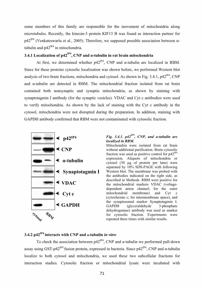
some members of this family are responsible for the movement of mitochondria along
microtubules. Recently, the kinesin-3 protein KIF13 B was found as interaction partner for
p42IP4 (Venkateswarlu et al., 2005). Therefore, we supposed possible association between α-
tubulin and p42IP4 in mitochondria.
3.4.1 Localization of p42IP4, CNP and α-tubulin in rat brain mitochondria
At first, we determined whether p42IP4, CNP and α-tubulin are localized in RBM.
Since for these proteins cytosolic localization was shown before, we performed Western blot
analysis of two brain fractions, mitochondria and cytosol. As shown in Fig. 3.4.1, p42IP4, CNP
and α-tubulin are detected in RBM. The mitochondrial fraction isolated from rat brain
contained both nonsynaptic and synaptic mitochondria, as shown by staining with
synaptotagmin I antibody (for the synaptic vesicles). VDAC and Cyt c antibodies were used
to verify mitochondria. As shown by the lack of staining with the Cyt c antibody in the
cytosol, mitochondria were not disrupted during the preparation. In addition, staining with
GAPDH antibody confirmed that RBM were not contaminated with cytosolic fraction.
CNP
p42IP4
Synaptotagmin I
VDAC
cytosol
RBM
α-tubulin
Cyt c
GAPDH
Fig. 3.4.1. p42IP4, CNP, and α-tubulin are localized in RBM. Mitochondria were isolated from rat brain without additional purification. Brain cytosolic fraction was used as positive control for p42IP4
expression. Aliquots of mitochondria or cytosol (30 µg of protein per lane) were separated by 10% SDS-PAGE with following Western blot. The membrane was probed with the antibodies indicated on the right side, as described in Methods. RBM were positive for the mitochondrial markers VDAC (voltage-dependent anion channel; for the outer mitochondrial membrane) and Cyt c (cytochrome c; for intermembrane space), and the synaptosomal marker Synaptotagmin I. GAPDH (glyceraldehyde 3-phosphate dehydrogenase) antibody was used as marker for cytosolic fraction. Experiments were repeated three times with similar results.
3.4.2 p42IP4 interacts with CNP and α-tubulin in vitro
To check the association between p42IP4, CNP and α-tubulin we performed pull-down
assay using GST-p42IP4 fusion protein, expressed in bacteria. Since p42IP4, CNP and α-tubulin
localize to both cytosol and mitochondria, we used these two subcellular fractions for
interaction studies. Cytosolic fraction or mitochondrial lysate were incubated with
71
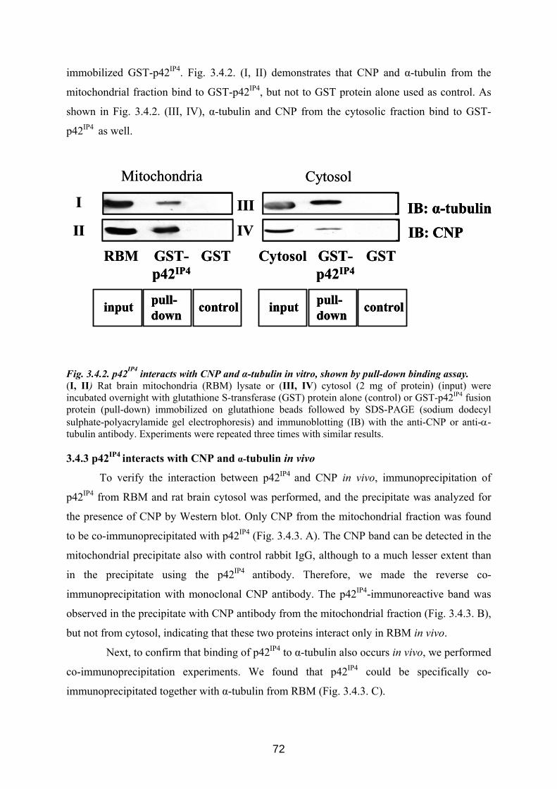
immobilized GST-p42IP4. Fig. 3.4.2. (I, II) demonstrates that CNP and α-tubulin from the
mitochondrial fraction bind to GST-p42IP4, but not to GST protein alone used as control. As
shown in Fig. 3.4.2. (III, IV), α-tubulin and CNP from the cytosolic fraction bind to GST-
p42IP4 as well.
RBM GSTGST-p42IP4
Cytosol GSTGST-p42IP4
IB: α-tubulinIB: CNP
Mitochondria Cytosol
I
II
III
IV
input pull-down control input pull-
down control
RBM GSTGST-p42IP4
Cytosol GSTGST-p42IP4
IB: α-tubulinIB: CNPIB: α-tubulinIB: CNP
Mitochondria Cytosol
I
II
III
IV
input pull-down controlinput pull-down control input pull-
down controlinput pull-down control
Fig. 3.4.2. p42IP4 interacts with CNP and α-tubulin in vitro, shown by pull-down binding assay. (I, II) Rat brain mitochondria (RBM) lysate or (III, IV) cytosol (2 mg of protein) (input) were incubated overnight with glutathione S-transferase (GST) protein alone (control) or GST-p42IP4 fusion protein (pull-down) immobilized on glutathione beads followed by SDS-PAGE (sodium dodecyl sulphate-polyacrylamide gel electrophoresis) and immunoblotting (IB) with the anti-CNP or anti-α-tubulin antibody. Experiments were repeated three times with similar results.
3.4.3 p42IP4 interacts with CNP and α-tubulin in vivo
To verify the interaction between p42IP4 and CNP in vivo, immunoprecipitation of
p42IP4 from RBM and rat brain cytosol was performed, and the precipitate was analyzed for
the presence of CNP by Western blot. Only CNP from the mitochondrial fraction was found
to be co-immunoprecipitated with p42IP4 (Fig. 3.4.3. A). The CNP band can be detected in the
mitochondrial precipitate also with control rabbit IgG, although to a much lesser extent than
in the precipitate using the p42IP4 antibody. Therefore, we made the reverse co-
immunoprecipitation with monoclonal CNP antibody. The p42IP4-immunoreactive band was
observed in the precipitate with CNP antibody from the mitochondrial fraction (Fig. 3.4.3. B),
but not from cytosol, indicating that these two proteins interact only in RBM in vivo.
Next, to confirm that binding of p42IP4 to α-tubulin also occurs in vivo, we performed
co-immunoprecipitation experiments. We found that p42IP4 could be specifically co-
immunoprecipitated together with α-tubulin from RBM (Fig. 3.4.3. C).
72
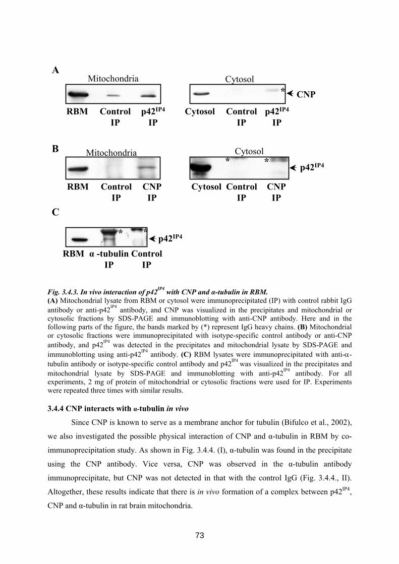
CNP IP
ControlIP
CNP IP
ControlIP
CytosolMitochondria
CNP
Cytosol p42IP4
IPControl
IPRBM p42IP4
IPControl
IP
A
*
Mitochondria
RBM
Cytosol
p42IP4
Cytosol
B**
p42IP4
RBM α -tubulinIP
ControlIP
C
* *
Fig. 3.4.3. In vivo interaction of p42IP4 with CNP and α-tubulin in RBM. (A) Mitochondrial lysate from RBM or cytosol were immunoprecipitated (IP) with control rabbit IgG antibody or anti-p42IP4 antibody, and CNP was visualized in the precipitates and mitochondrial or cytosolic fractions by SDS-PAGE and immunoblotting with anti-CNP antibody. Here and in the following parts of the figure, the bands marked by (*) represent IgG heavy chains. (B) Mitochondrial or cytosolic fractions were immunoprecipitated with isotype-specific control antibody or anti-CNP antibody, and p42IP4 was detected in the precipitates and mitochondrial lysate by SDS-PAGE and immunoblotting using anti-p42IP4 antibody. (C) RBM lysates were immunoprecipitated with anti-α-tubulin antibody or isotype-specific control antibody and p42IP4 was visualized in the precipitates and mitochondrial lysate by SDS-PAGE and immunoblotting with anti-p42IP4 antibody. For all experiments, 2 mg of protein of mitochondrial or cytosolic fractions were used for IP. Experiments were repeated three times with similar results.
3.4.4 CNP interacts with α-tubulin in vivo
Since CNP is known to serve as a membrane anchor for tubulin (Bifulco et al., 2002),
we also investigated the possible physical interaction of CNP and α-tubulin in RBM by co-
immunoprecipitation study. As shown in Fig. 3.4.4. (I), α-tubulin was found in the precipitate
using the CNP antibody. Vice versa, CNP was observed in the α-tubulin antibody
immunoprecipitate, but CNP was not detected in that with the control IgG (Fig. 3.4.4., II).
Altogether, these results indicate that there is in vivo formation of a complex between p42IP4,
CNP and α-tubulin in rat brain mitochondria.
73
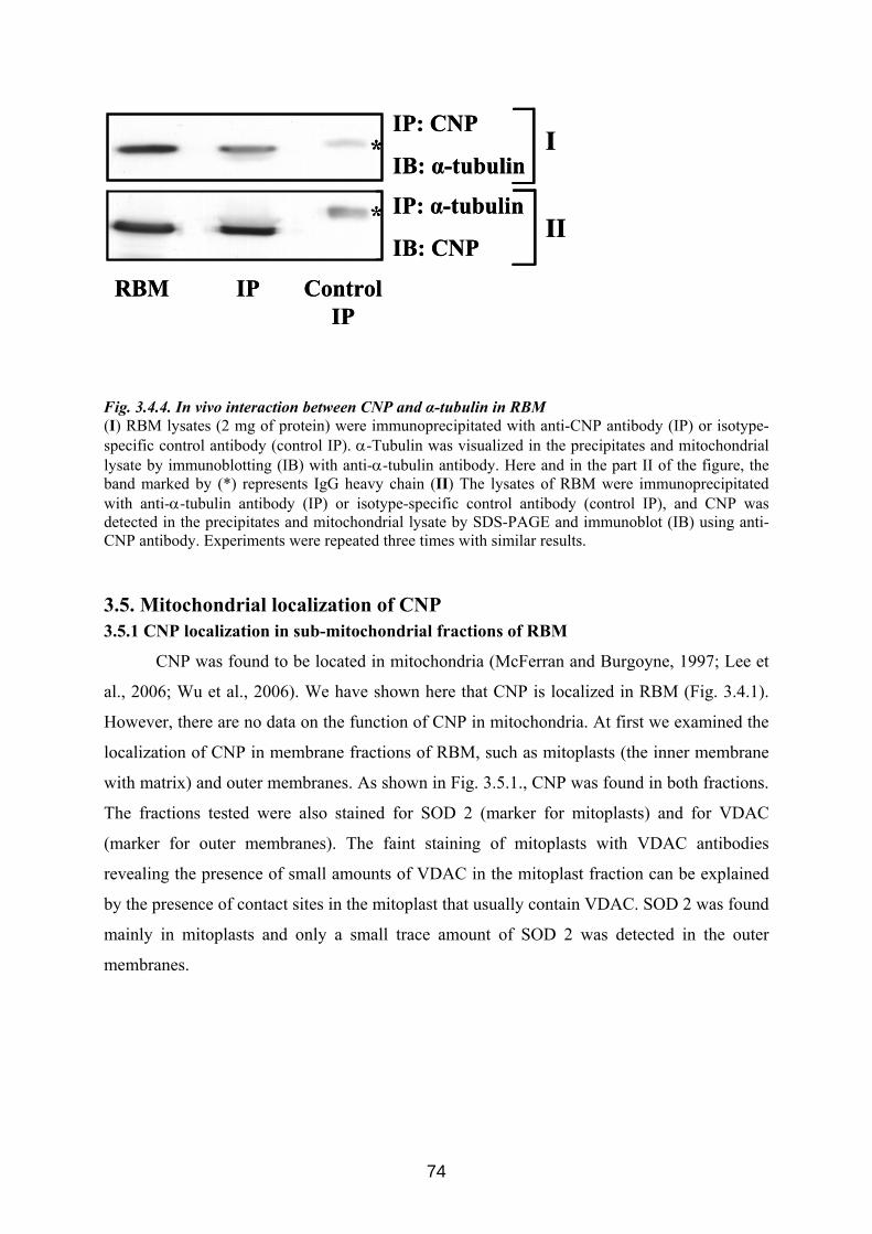
RBM IP Control IP
*
* IP: α-tubulin
IB: CNP
IP: CNP
IB: α-tubulin
II
I
RBM IP Control IP
*
* IP: α-tubulin
IB: CNP
IP: CNP
IB: α-tubulin
II
I
Fig. 3.4.4. In vivo interaction between CNP and α-tubulin in RBM (I) RBM lysates (2 mg of protein) were immunoprecipitated with anti-CNP antibody (IP) or isotype-specific control antibody (control IP). α-Tubulin was visualized in the precipitates and mitochondrial lysate by immunoblotting (IB) with anti-α-tubulin antibody. Here and in the part II of the figure, the band marked by (*) represents IgG heavy chain (II) The lysates of RBM were immunoprecipitated with anti-α-tubulin antibody (IP) or isotype-specific control antibody (control IP), and CNP was detected in the precipitates and mitochondrial lysate by SDS-PAGE and immunoblot (IB) using anti-CNP antibody. Experiments were repeated three times with similar results.
3.5. Mitochondrial localization of CNP 3.5.1 CNP localization in sub-mitochondrial fractions of RBM
CNP was found to be located in mitochondria (McFerran and Burgoyne, 1997; Lee et
al., 2006; Wu et al., 2006). We have shown here that CNP is localized in RBM (Fig. 3.4.1).
However, there are no data on the function of CNP in mitochondria. At first we examined the
localization of CNP in membrane fractions of RBM, such as mitoplasts (the inner membrane
with matrix) and outer membranes. As shown in Fig. 3.5.1., CNP was found in both fractions.
The fractions tested were also stained for SOD 2 (marker for mitoplasts) and for VDAC
(marker for outer membranes). The faint staining of mitoplasts with VDAC antibodies
revealing the presence of small amounts of VDAC in the mitoplast fraction can be explained
by the presence of contact sites in the mitoplast that usually contain VDAC. SOD 2 was found
mainly in mitoplasts and only a small trace amount of SOD 2 was detected in the outer
membranes.
74
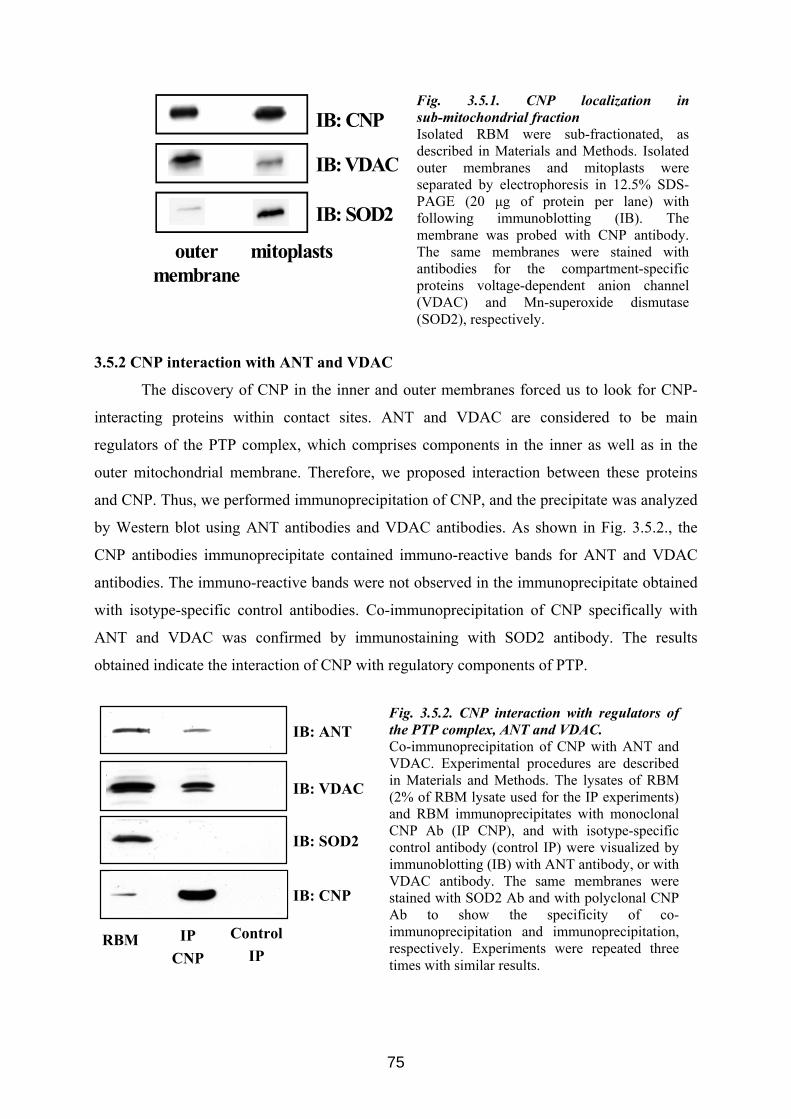
IB: VDAC
IB: SOD2
IB: CNP
outer membrane
mitoplasts
Fig. 3.5.1. CNP localization in sub-mitochondrial fraction Isolated RBM were sub-fractionated, as described in Materials and Methods. Isolated outer membranes and mitoplasts were separated by electrophoresis in 12.5% SDS-PAGE (20 μg of protein per lane) with following immunoblotting (IB). The membrane was probed with CNP antibody. The same membranes were stained with antibodies for the compartment-specific proteins voltage-dependent anion channel (VDAC) and Mn-superoxide dismutase (SOD2), respectively.
3.5.2 CNP interaction with ANT and VDAC
The discovery of CNP in the inner and outer membranes forced us to look for CNP-
interacting proteins within contact sites. ANT and VDAC are considered to be main
regulators of the PTP complex, which comprises components in the inner as well as in the
outer mitochondrial membrane. Therefore, we proposed interaction between these proteins
and CNP. Thus, we performed immunoprecipitation of CNP, and the precipitate was analyzed
by Western blot using ANT antibodies and VDAC antibodies. As shown in Fig. 3.5.2., the
CNP antibodies immunoprecipitate contained immuno-reactive bands for ANT and VDAC
antibodies. The immuno-reactive bands were not observed in the immunoprecipitate obtained
with isotype-specific control antibodies. Co-immunoprecipitation of CNP specifically with
ANT and VDAC was confirmed by immunostaining with SOD2 antibody. The results
obtained indicate the interaction of CNP with regulatory components of PTP.
Fig. 3.5.2. CNP interaction with regulators of the PTP complex, ANT and VDAC. Co-immunoprecipitation of CNP with ANT and VDAC. Experimental procedures are described in Materials and Methods. The lysates of RBM (2% of RBM lysate used for the IP experiments) and RBM immunoprecipitates with monoclonal CNP Ab (IP CNP), and with isotype-specific control antibody (control IP) were visualized by immunoblotting (IB) with ANT antibody, or with VDAC antibody. The same membranes were stained with SOD2 Ab and with polyclonal CNP Ab to show the specificity of co-immunoprecipitation and immunoprecipitation, respectively. Experiments were repeated three times with similar results.
IB: VDAC
IB: ANT
RBM IPCNP
ControlIP
IB: SOD2
IB: CNP
75
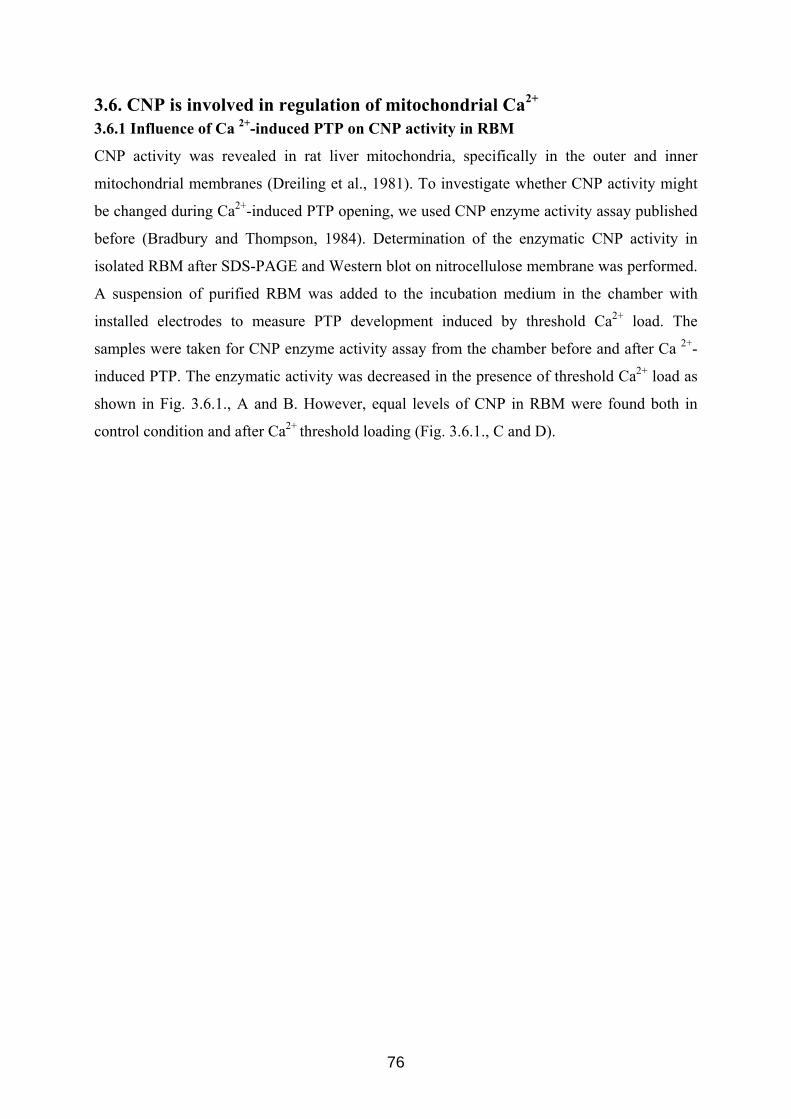
3.6. CNP is involved in regulation of mitochondrial Ca2+
3.6.1 Influence of Ca 2+-induced PTP on CNP activity in RBM
CNP activity was revealed in rat liver mitochondria, specifically in the outer and inner
mitochondrial membranes (Dreiling et al., 1981). To investigate whether CNP activity might
be changed during Ca2+-induced PTP opening, we used CNP enzyme activity assay published
before (Bradbury and Thompson, 1984). Determination of the enzymatic CNP activity in
isolated RBM after SDS-PAGE and Western blot on nitrocellulose membrane was performed.
A suspension of purified RBM was added to the incubation medium in the chamber with
installed electrodes to measure PTP development induced by threshold Ca2+ load. The
samples were taken for CNP enzyme activity assay from the chamber before and after Ca 2+-
induced PTP. The enzymatic activity was decreased in the presence of threshold Ca2+ load as
shown in Fig. 3.6.1., A and B. However, equal levels of CNP in RBM were found both in
control condition and after Ca2+ threshold loading (Fig. 3.6.1., C and D).
76
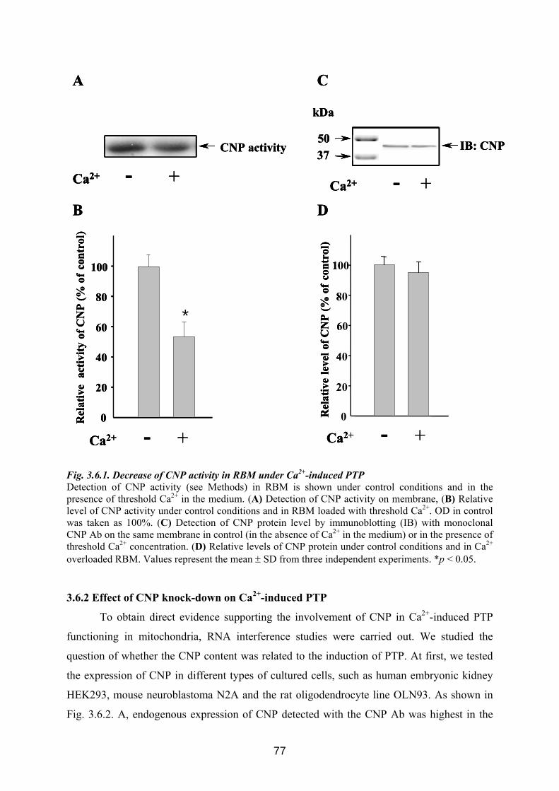
C
IB: CNP
kDa
5037
A
CNP activity
Ca2+ - + Ca2+ - +D
0
20
40
60
80
100
Rel
ativ
e le
velo
f CN
P (%
of
cont
rol)
B
*
Rel
ativ
e a
ctiv
ityof
CN
P (%
of
cont
rol)
100
80
60
40
20
0
*
Ca2+ - + Ca2+ - +
C
IB: CNP
kDa
5037
A
CNP activity
Ca2+ - + Ca2+ - +
C
IB: CNP
kDa
5037
A
CNP activity
Ca2+ - +Ca2+ - + Ca2+ - +D
0
20
40
60
80
100
Rel
ativ
e le
velo
f CN
P (%
of
cont
rol)
B
*
Rel
ativ
e a
ctiv
ityof
CN
P (%
of
cont
rol)
100
80
60
40
20
0
*
Ca2+ - + Ca2+ - +
D
0
20
40
60
80
100
Rel
ativ
e le
velo
f CN
P (%
of
cont
rol)
B
*
Rel
ativ
e a
ctiv
ityof
CN
P (%
of
cont
rol)
100
80
60
40
20
0
*
Ca2+ - + Ca2+ - +
Fig. 3.6.1. Decrease of CNP activity in RBM under Ca2+-induced PTP Detection of CNP activity (see Methods) in RBM is shown under control conditions and in the presence of threshold Ca2+ in the medium. (A) Detection of CNP activity on membrane, (B) Relative level of CNP activity under control conditions and in RBM loaded with threshold Ca2+. OD in control was taken as 100%. (C) Detection of CNP protein level by immunoblotting (IB) with monoclonal CNP Ab on the same membrane in control (in the absence of Ca2+ in the medium) or in the presence of threshold Ca2+ concentration. (D) Relative levels of CNP protein under control conditions and in Ca2+ overloaded RBM. Values represent the mean ± SD from three independent experiments. *p < 0.05. 3.6.2 Effect of CNP knock-down on Ca2+-induced PTP
To obtain direct evidence supporting the involvement of CNP in Ca2+-induced PTP
functioning in mitochondria, RNA interference studies were carried out. We studied the
question of whether the CNP content was related to the induction of PTP. At first, we tested
the expression of CNP in different types of cultured cells, such as human embryonic kidney
HEK293, mouse neuroblastoma N2A and the rat oligodendrocyte line OLN93. As shown in
Fig. 3.6.2. A, endogenous expression of CNP detected with the CNP Ab was highest in the
77
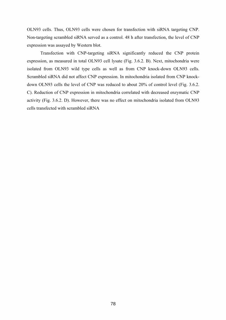
OLN93 cells. Thus, OLN93 cells were chosen for transfection with siRNA targeting CNP.
Non-targeting scrambled siRNA served as a control. 48 h after transfection, the level of CNP
expression was assayed by Western blot.
Transfection with CNP-targeting siRNA significantly reduced the CNP protein
expression, as measured in total OLN93 cell lysate (Fig. 3.6.2. B). Next, mitochondria were
isolated from OLN93 wild type cells as well as from CNP knock-down OLN93 cells.
Scrambled siRNA did not affect CNP expression. In mitochondria isolated from CNP knock-
down OLN93 cells the level of CNP was reduced to about 20% of control level (Fig. 3.6.2.
C). Reduction of CNP expression in mitochondria correlated with decreased enzymatic CNP
activity (Fig. 3.6.2. D). However, there was no effect on mitochondria isolated from OLN93
cells transfected with scrambled siRNA
78
IB: CNPA
HEK 293N2a
OLN 93 C
0
20
40
60
80
100
120
140*
Rel
ativ
eex
pres
sion
of C
NP
(% o
fnon
tran
sfec
ted
cells
)
SOD2
CNP
–+
+–
––
Scrambled siRNACNP siRNA
CNP
β-tubulin
B
–+
+–
––
Scrambled siRNACNP siRNA
CNPactivity
D
0
20
40
60
80
100
120
–+
+–
––
Scrambled siRNACNP siRNA
Rel
ativ
eac
tivity
of C
NP
(% o
fnon
tran
sfec
ted
cells
)
*
Total cell lysate
Mitochondria Mitochondria
Total cell lysates:
* *
IB: CNPA
HEK 293N2a
OLN 93 C
0
20
40
60
80
100
120
140*
Rel
ativ
eex
pres
sion
of C
NP
(% o
fnon
tran
sfec
ted
cells
)
SOD2
CNP
–+
+–
––
Scrambled siRNACNP siRNA
CNP
β-tubulin
B
–+
+–
––
Scrambled siRNACNP siRNA
CNPactivity
D
0
20
40
60
80
100
120
0
20
40
60
80
100
120
–+
+–
––
Scrambled siRNACNP siRNA
Rel
ativ
eac
tivity
of C
NP
(% o
fnon
tran
sfec
ted
cells
)
*
Total cell lysate
Mitochondria Mitochondria
Total cell lysates:
* *
Fig. 3.6.2. CNP protein level in different types of cultured cells. CNP protein level and enzymatic activity in OLN93 mitochondria and OLN93 cells downregulated by siRNA. (A) Endogeneous expression of CNP protein in HEK 293, N2a and OLN 93 was evaluated by Western blotting, as described in Materials and methods. Experiments were repeated three times with similar results. Twenty-five µg of total cell lysates were loaded. (B) OLN93 cells were transfected with either 50 nM scrambled siRNA (control siRNA) or CNP siRNAs. At 48 h after transfection, CNP protein level in total cell lysates was determined by Western blot analysis. Representative data from three independent experiments are given, and β-tubulin I served as control for specificity of knock-down. (C) OLN93 cells were transfected with either CNP siRNAs (50 nM) or scrambled siRNA (50 nM), after 48 h mitochondria were isolated. The reduction of CNP expression in mitochondria was evaluated by Western blotting. CNP and SOD2 protein bands were quantified by densitometry. The histogram shows relative units as ratio of CNP to SOD2. Mitochondria isolated from non-transfected OLN93 cells were used as reference and assigned the value of 100% . Values represent the mean ± SD from three independent experiments. *** p < 0.001 (D) CNP activity in mitochondria isolated from OLN93 cells, transfected with siRNA or non-transfected, was detected, as described in Materials and methods. The activity of CNP in mitochondria isolated from non-transfected OLN93 cells was taken as 100%. Values represent the mean ± SD from three independent experiments. * p<0.05.
79
To investigate whether the reduction in CNP level in mitochondria could influence the
Ca2+-induced PTP opening, we measured the parameters of mitochondria isolated from wild
type OLN93 cells, scrambled siRNA-treated OLN93 cells as well as cells, transfected with
siRNA targeting CNP, as described above.
Fig. 3.6.3. A shows that threshold Ca2+ concentration in mitochondria isolated from
wild type OLN93 cells was achieved after four additions of Ca2+ (70 nanomoles of Ca2+ per
mg of protein each), giving a threshold Ca2+ for OLN93 mitochondria of 280 nanomoles per
mg of protein in the given experiment. A similar threshold was found in mitochondria isolated
from scrambled siRNA-treated OLN93 cells (Fig.3.6.3. B). Mitochondria isolated from CNP
knock-down OLN93 cells released Ca2+ after the third addition (also 70 nanomoles of Ca2+
per mg of protein each), yielding a lower threshold calcium concentration which was equal to
210 nanomoles Ca2+ per mg of protein (Fig. 3.6.3. C).
80
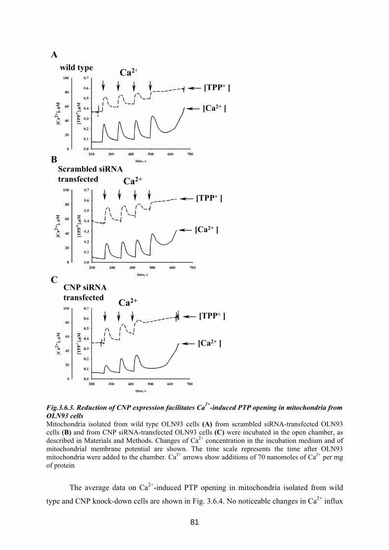
A
B
C
Ca2+
Ca2+
Ca2+
[Ca2+ ]
[TPP+ ]
[Ca2+ ]
[TPP+ ]
[Ca2+ ]
[TPP+ ]
wild type
Scrambled siRNAtransfected
CNP siRNAtransfected
Fig.3.6.3. Reduction of CNP expression facilitates Ca2+-induced PTP opening in mitochondria from OLN93 cells Mitochondria isolated from wild type OLN93 cells (A) from scrambled siRNA-transfected OLN93 cells (B) and from CNP siRNA-transfected OLN93 cells (C) were incubated in the open chamber, as described in Materials and Methods. Changes of Ca2+ concentration in the incubation medium and of mitochondrial membrane potential are shown. The time scale represents the time after OLN93 mitochondria were added to the chamber. Ca2+ arrows show additions of 70 nanomoles of Ca2+ per mg of protein
The average data on Ca2+-induced PTP opening in mitochondria isolated from wild
type and CNP knock-down cells are shown in Fig. 3.6.4. No noticeable changes in Ca2+ influx
81
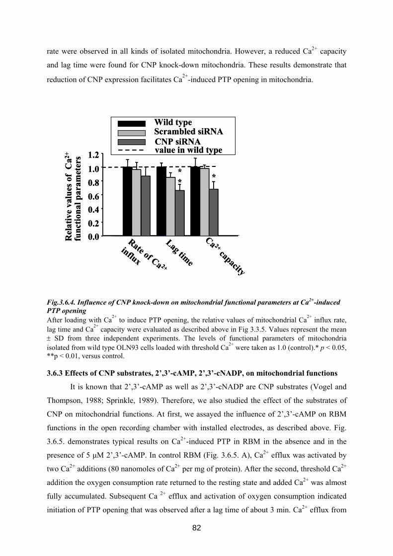
rate were observed in all kinds of isolated mitochondria. However, a reduced Ca2+ capacity
and lag time were found for CNP knock-down mitochondria. These results demonstrate that
reduction of CNP expression facilitates Ca2+-induced PTP opening in mitochondria.
Rate of Ca 2+
influx
1.2
0.00.20.40.60.81.0
Wild type Scrambled siRNACNP siRNAvalue in wild type
Rel
ativ
eva
lues
of C
a2+
func
tiona
l par
amet
ers
Lag time
Ca 2+capacity
***
Rate of Ca 2+
influx
1.2
0.00.20.40.60.81.0
Wild type Scrambled siRNACNP siRNAvalue in wild type
Rel
ativ
eva
lues
of C
a2+
func
tiona
l par
amet
ers
Lag time
Ca 2+capacity
***
Fig.3.6.4. Influence of CNP knock-down on mitochondrial functional parameters at Ca2+-induced PTP opening After loading with Ca2+ to induce PTP opening, the relative values of mitochondrial Ca2+ influx rate, lag time and Ca2+ capacity were evaluated as described above in Fig 3.3.5. Values represent the mean ± SD from three independent experiments. The levels of functional parameters of mitochondria isolated from wild type OLN93 cells loaded with threshold Ca2+ were taken as 1.0 (control).* p < 0.05, **p < 0.01, versus control. 3.6.3 Effects of CNP substrates, 2’,3’-cAMP, 2’,3’-cNADP, on mitochondrial functions
It is known that 2’,3’-cAMP as well as 2’,3’-cNADP are CNP substrates (Vogel and
Thompson, 1988; Sprinkle, 1989). Therefore, we also studied the effect of the substrates of
CNP on mitochondrial functions. At first, we assayed the influence of 2’,3’-cAMP on RBM
functions in the open recording chamber with installed electrodes, as described above. Fig.
3.6.5. demonstrates typical results on Ca2+-induced PTP in RBM in the absence and in the
presence of 5 μM 2’,3’-cAMP. In control RBM (Fig. 3.6.5. A), Ca2+ efflux was activated by
two Ca2+ additions (80 nanomoles of Ca2+ per mg of protein). After the second, threshold Ca2+
addition the oxygen consumption rate returned to the resting state and added Ca2+ was almost
fully accumulated. Subsequent Ca 2+ efflux and activation of oxygen consumption indicated
initiation of PTP opening that was observed after a lag time of about 3 min. Ca2+ efflux from
82
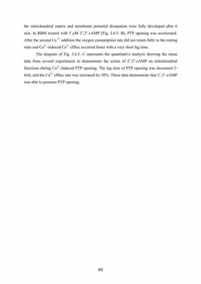
the mitochondrial matrix and membrane potential dissipation were fully developed after 6
min. In RBM treated with 5 μM 2’,3’-cAMP (Fig. 3.6.5. B), PTP opening was accelerated.
After the second Ca 2+ addition the oxygen consumption rate did not return fully to the resting
state and Ca2+-induced Ca2+ efflux occurred faster with a very short lag time.
The diagram of Fig. 3.6.5. C represents the quantitative analysis showing the mean
data from several experiments to demonstrate the action of 2’,3’-cAMP on mitochondrial
functions during Ca2+-induced PTP opening. The lag time of PTP opening was decreased 2-
fold, and the Ca2+ efflux rate was increased by 30%. These data demonstrate that 2’,3’-cAMP
was able to promote PTP opening.
83
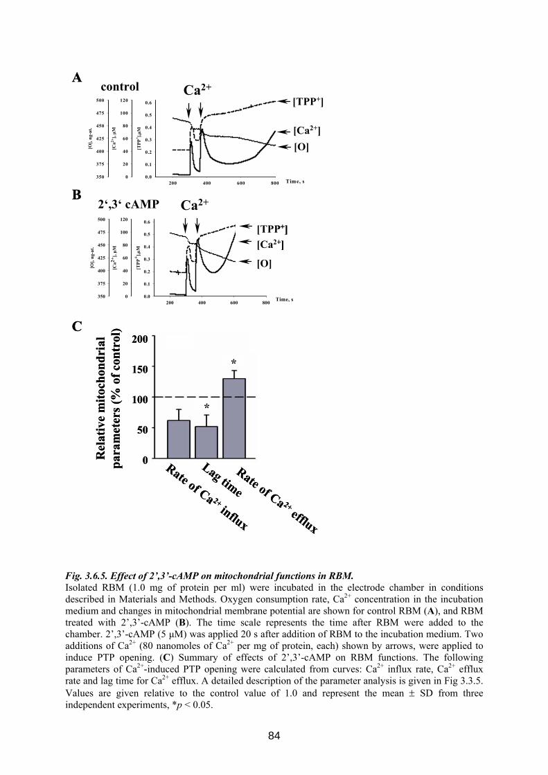
A
B
C
Time, s200 400 600 800
[TPP
+ ],µM
0.0
0.1
0.2
0.3
0.4
0.5
0.6
[Ca2+
], µM
0
20
40
60
80
100
120
[O],
ng-a
t.
350
375
400
425
450
475
500
[O][Ca2+]
[TPP+]control
200 400 600 800
[TPP
+ ],µM
0.0
0.1
0.2
0.3
0.4
0.5
0.6
[Ca2+
], µM
0
20
40
60
80
100
120
[O],
ng-a
t.
350
375
400
425
450
475
500
Time, s
[O]
[Ca2+][TPP+]
2‘,3‘ cAMP Ca2+
Ca2+R
elat
ive
mito
chon
dria
lpa
ram
eter
s (%
of c
ontr
ol)
200
150
100
50
0 Rate of Ca 2+influx
Lag time
Rate of Ca 2+efflux
*
*
A
B
C
Time, s200 400 600 800
[TPP
+ ],µM
0.0
0.1
0.2
0.3
0.4
0.5
0.6
[Ca2+
], µM
0
20
40
60
80
100
120
[O],
ng-a
t.
350
375
400
425
450
475
500
[O][Ca2+]
[TPP+]control
200 400 600 800
[TPP
+ ],µM
0.0
0.1
0.2
0.3
0.4
0.5
0.6
[Ca2+
], µM
0
20
40
60
80
100
120
[O],
ng-a
t.
350
375
400
425
450
475
500
Time, s
[O]
[Ca2+][TPP+]
200 400 600 800
[TPP
+ ],µM
0.0
0.1
0.2
0.3
0.4
0.5
0.6
[Ca2+
], µM
0
20
40
60
80
100
120
[O],
ng-a
t.
350
375
400
425
450
475
500
Time, s
[O]
[Ca2+][TPP+]
[O]
[Ca2+][TPP+]
2‘,3‘ cAMP Ca2+
Ca2+R
elat
ive
mito
chon
dria
lpa
ram
eter
s (%
of c
ontr
ol)
200
150
100
50
0 Rate of Ca 2+influx
Lag time
Rate of Ca 2+efflux
*
*
Fig. 3.6.5. Effect of 2’,3’-сAMP on mitochondrial functions in RBM. Isolated RBM (1.0 mg of protein per ml) were incubated in the electrode chamber in conditions described in Materials and Methods. Oxygen consumption rate, Ca2+ concentration in the incubation medium and changes in mitochondrial membrane potential are shown for control RBM (A), and RBM treated with 2’,3’-сAMP (B). The time scale represents the time after RBM were added to the chamber. 2’,3’-сAMP (5 μM) was applied 20 s after addition of RBM to the incubation medium. Two additions of Ca2+ (80 nanomoles of Ca2+ per mg of protein, each) shown by arrows, were applied to induce PTP opening. (C) Summary of effects of 2’,3’-сAMP on RBM functions. The following parameters of Ca2+-induced PTP opening were calculated from curves: Ca2+ influx rate, Ca2+ efflux rate and lag time for Ca2+ efflux. A detailed description of the parameter analysis is given in Fig 3.3.5. Values are given relative to the control value of 1.0 and represent the mean ± SD from three independent experiments, *p < 0.05.
84
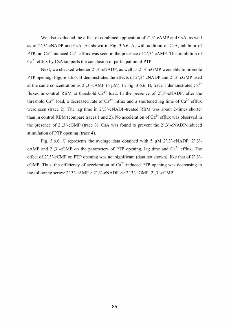
We also evaluated the effect of combined application of 2’,3’-cAMP and CsA, as well
as of 2’,3’-cNADP and CsA. As shown in Fig. 3.6.6. A, with addition of CsA, inhibitor of
PTP, no Ca2+-induced Ca2+ efflux was seen in the presence of 2’,3’-cAMP. This inhibition of
Ca2+ efflux by CsA supports the conclusion of participation of PTP.
Next, we checked whether 2’,3’-cNADP, as well as 2’,3’-cGMP were able to promote
PTP opening. Figure 3.6.6. B demonstrates the effects of 2’,3’-cNADP and 2’,3’-cGMP used
at the same concentration as 2’,3’-cAMP (5 μM). In Fig. 3.6.6. B, trace 1 demonstrates Ca2+
fluxes in control RBM at threshold Ca2+ load. In the presence of 2’,3’-cNADP, after the
threshold Ca2+ load, a decreased rate of Ca2+ influx and a shortened lag time of Ca2+ efflux
were seen (trace 2). The lag time in 2’,3’-cNADP-treated RBM was about 2-times shorter
than in control RBM (compare traces 1 and 2). No acceleration of Ca2+ efflux was observed in
the presence of 2’,3’-cGMP (trace 3). CsA was found to prevent the 2’,3’-cNADP-induced
stimulation of PTP opening (trace 4).
Fig. 3.6.6. C represents the average data obtained with 5 µM 2’,3’-cNADP, 2’,3’-
cAMP and 2’,3’-cGMP on the parameters of PTP opening, lag time and Ca2+ efflux. The
effect of 2’,3’-cCMP on PTP opening was not significant (data not shown), like that of 2’,3’-
cGMP. Thus, the efficiency of acceleration of Ca2+-induced PTP opening was decreasing in
the following series: 2’,3’-cAMP > 2’,3’-cNADP >> 2’,3’-cGMP, 2’,3’-cCMP.
85
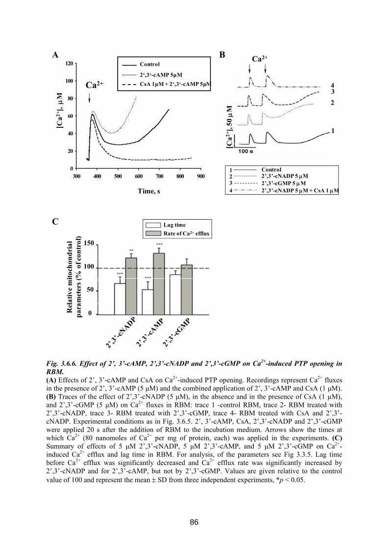
300 400 500 600 700 800 9000
20
40
60
80
100
120 Control2':3'-cAMP 5 µMCsA 1µM + 2':3'-cAMP 5 µM
Time, s 43
12
Control
2’,3’-cNADP 5 μM + CsA 1 μM
2’,3’-cNADP 5 μM2’,3’-cGMP 5 μM
Ca2+
1
34
2
[Ca2+
], 50
μM
150
100
50
0Rel
ativ
e m
itoch
ondr
ial
para
met
ers (
% o
f con
trol
)
C
BA
Ca2+
[Ca2+
], μ
M
Lag timeRate of Ca2+ efflux
Control
2‘,3‘-cAMP 5μMCsA 1μM + 2‘,3‘-cAMP 5μM
Fig. 3.6.6. Effect of 2’, 3’-сAMP, 2’,3’-сNADP and 2’,3’-сGMP on Ca2+-induced PTP opening in RBM. (A) Effects of 2’, 3’-сAMP and CsA on Ca2+-induced PTP opening. Recordings represent Ca2+ fluxes in the presence of 2’, 3’-сAMP (5 μM) and the combined application of 2’, 3’-сAMP and CsA (1 μM). (B) Traces of the effect of 2’,3’-сNADP (5 μM), in the absence and in the presence of CsA (1 µM), and 2’,3’-сGMP (5 μM) on Ca2+ fluxes in RBM: trace 1 -control RBM, trace 2- RBM treated with 2’,3’-сNADP, trace 3- RBM treated with 2’,3’-сGMP, trace 4- RBM treated with CsA and 2’,3’-сNADP. Experimental conditions as in Fig. 3.6.5. 2’, 3’-сAMP, CsA, 2’,3’-сNADP and 2’,3’-сGMP were applied 20 s after the addition of RBM to the incubation medium. Arrows show the times at which Ca2+ (80 nanomoles of Ca2+ per mg of protein, each) was applied in the experiments. (C) Summary of effects of 5 μM 2’,3’-сNADP, 5 μM 2’,3’-сAMP, and 5 μM 2’,3’-сGMP on Ca2+- induced Ca2+ efflux and lag time in RBM. For analysis, of the parameters see Fig 3.3.5. Lag time before Ca2+ efflux was significantly decreased and Ca2+ efflux rate was significantly increased by 2’,3’-сNADP and for 2’,3’-сAMP, but not by 2’,3’-сGMP. Values are given relative to the control value of 100 and represent the mean ± SD from three independent experiments, *p < 0.05.
86
Induction of mitochondrial swelling is a prominent feature of PTP. Therefore, we
examined the influence of 2’,3’-cAMP and 2’,3’-cNADP on Ca2+-induced swelling of isolated
RBM to corroborate our hypothesis that the CNP substrates are involved in PTP regulation.
Swelling was initiated by addition of Ca2+ to mitochondria incubated in standard medium (see
Materials and Methods). Typical curves in Fig. 3.6.7. A demonstrate the decrease of light
scattering at 540 nm, which indicates swelling. RBM swelling was accelerated in the presence
of 5 μM 2’,3’-cAMP or 5 μM 2’,3’-cNADP.
We used the half time for reaching the maximum of the RBM swelling as characteristic
parameter for quantification. The data in Fig. 3.6.7. B show that the swelling of RBM is
significantly enhanced by 2’,3’-cAMP and 2’,3’-cNADP.
87
0
40
80
120
0
40
80
120
**
Contro
l2’
,3’-cA
MP
2’,3’
-cNADP
T1/
2, s
BTime, s
0 100 200 300 400 500
OD
540n
m
0.60
0.62
0.64
0.66
0.68
0.70
0.72
A/2
T1/2
A21
3
Control
2’,3’-cAMP 2’,3’-cNADP
Fig. 3.6.7. Effect of 2’,3’-сNADP, 2’,3’-сAMP on RBM swelling (A) Effect of 2’,3’-сNADP, 2’,3’-сAMP on RBM swelling. Swelling was initiated by addition of 100 µM Ca2+ to RBM incubated in standard medium (see Materials and Methods). Light scattering at 540 nm in RBM was measured. Trace 1- control- Ca2+-induced swelling of RBM, trace 2 - effect of 5 μM 2’,3’-сNADP, trace 3 - effect of 2’,3’-сAMP. (B) Average results of half time for reaching the maximal swelling of RBM in the presence of 2’,3’-сNADP and 2’,3’-сAMP. Values are given relative to the control value of 100 and represent the mean ± SD from three independent experiments, * p < 0.05, versus control
88
4. Discussion
4.1 The mechanism of mitochondrial Ca2+-induced PTP opening remains unknown
Mitochondria are important regulators of cellular Ca2+ homeostasis, producers of ATP
via oxidative phosphorylation, and regulators of cell death pathways (Sullivan et al., 2005).
Mitochondria assist in maintaining Ca2+ homeostasis by sequestering and releasing Ca2+.
Isolated mitochondria in the presence of phosphate take up Ca2+ to a fixed capacity, in a
membrane potential (∆Ψm)-dependent fashion (Chalmers and Nicholls, 2003). When the
mitochondria become overloaded with Ca2+, they undergo the cataclysmic mitochondrial
permeability transition. Mitochondrial permeability transition is manifested as a sudden
opening of a non-selective megachannel, PTP, in the inner mitochondrial membrane. The PTP
opening increases the permeability to solutes with molecular mass up to 1500 Da in response
to mitochondrial Ca2+ overload and/or oxidative stress (Bernardi, 1999; Halestrap, 2006). The
Ca2+-induced PTP opening is a complex process with many known inducers, modulators, and
inhibitors. The PTP is a multi-protein complex formed in the contact sites between outer and
inner membranes of mitochondria to increase the permeability of the inner membrane. Main
events of PTP opening are collapse of the membrane potential, Ca2+-induced Ca2+ efflux,
induction of mitochondrial swelling and release of apoptotic factors (Bernardi, 1999;
Halestrap et al., 2002; Zoratti et al., 2005). The quest for the pore identity has been ongoing
for several decades. However, the PTP structure remains unknown (Juhaszova et al., 2008).
Evidence has been accumulating that numerous signalling molecules and drugs affect the
Ca2+-induced PTP opening. Thus, it is important to explore fully the mechanism of
mitochondrial Ca2+-induced PTP opening and to search for new proteins involved in control
of this complex process.
4.2 Known functions of p42IP4
Different functions have been already proposed for p42IP4. There were reports which
showed the involvement of this protein in the activation of the mitogen-activated protein
kinase cascade of the extracellular regulated kinase (Hayashi et al., 2006), in the ARF6
signalling pathway (Lawrence et al., 2005) or in the transcription factor activator protein-1
activation cascade (Chanda et al., 2003). p42IP4 was found to be involved in dendritic
differentiation/synaptic plasticity and/or transport of PIP3-containing vesicles along axons to
regulate neuronal cell polarity (Horiguchi et al., 2006; Moore et al., 2007). In neuronal cells,
89
p42IP4 regulates the actin cytoskeleton via ARF GAP-dependent and ARF GAP-independent
mechanisms (Thacker et al., 2004; Venkateswarlu et al., 2004). p42IP4 has already been linked
to short-term regulation of behavioural responses in rat brain (Reiser et al., 2004). Very
interestingly, p42IP4 seems to be involved in neuronal diseases, since we found that p42IP4 is
up-regulated in neurons in the brain of AD patients and is detected in AD plaques (Reiser and
Bernstein, 2002, 2004). Although various functions of p42IP4 have been proposed, there were
no data concerning a role of this protein related to mitochondria.
4.3 Novel localization and function of p42IP4. Involvement of p42IP4 and its ligands in regulation of mitochondrial Ca2+-induced PTP opening
Previously, analysis of subcellular localization of the p42IP4 protein in non-neuronal
CHO cells stably transfected with pcDNA-p42IP4 was performed. p42IP4 was found in
membranes, cytosol, and nucleus (Sedehizade et al., 2002). In this study, we detected that
p42IP4 is localized both in RBM and in mitochondria of the transfected cell lines CHO and
N2a. Moreover, in mitochondria of CHO cells, p42IP4 was found predominantly at the inner
membrane and in the intermembrane space of mitochondria, and to a lesser degree in the
matrix and outer membrane. Such mitochondrial localization of the p42IP4 allowed us to
suppose that p42IP4 might be involved in the regulation of mitochondrial Ca2+ transport
mechanisms.
We used the mouse neuroblastoma N2a cells as an expression system as we have
demonstrated that the N2a cells do endogenously express the p42IP4. The neuronal nature of
N2a cells and the endogenous expression of the p42IP4 make these cells an ideal system for
studying the mitochondrial functions. Two approaches, where Ca2+ was added either in small
or big bolus additions, were used to compare the ability of mitochondria to accumulate and
retain Ca2+ before PTP opening. Study of the functional parameters of mitochondria isolated
from N2a cells transfected with p42IP4 revealed that overexpression of p42IP4 accelerates Ca2+-
induced PTP opening in mitochondria from these cells. In comparison with the mitochondria
isolated from N2a cells transfected with p42IP4, mitochondria from N2a wild type cells can
accumulate significantly more Ca2+ before they undergo Ca2+-induced PTP. Thus, an
important function of mitochondrial p42IP4 in neurons seems to be regulation of mitochondrial
Ca2+ through PTP opening.
90
Moreover, in comparison with mitochondria from N2a wild type cells, mitochondria
isolated from N2a cells transfected with p42IP4 have a lower membrane potential. This could
cause p42IP4-containing mitochondria to take up less Ca2+ and thus lead to destabilization.
It was found that CsA did not prevent PTP opening in mitochondria isolated from N2a
cells. Tissue-specific differences in the nature of PTP induction are already well known. In
liver mitochondria, CsA nearly completely inhibits PTP induction, whereas in
nonsynaptosomal brain mitochondria CsA has very modest effects (Kristal et al., 2000). It
was also reported that only in the presence of a low concentration of exogenous ADP, CsA
fully inhibits PTP in brain mitochondria exposed to Ca2+ (Hansson et al., 2004). Thus, the
inhibition of the PTP by CsA depends on the model, which is used.
Mitochondria isolated from brain represent a heterogeneous mixture from different cell
types (Bambrick et al., 2006). Therefore, they have differing sensitivities to Ca2+ as inducer of
PTP and to CsA as inhibitor of the PTP. It was shown that CsA increased the calcium uptake
capacity of mitochondria isolated from astrocytes, while it had no effect on PTP in neuronal
mitochondria. It was also observed that the astrocytes had a maximal mitochondrial calcium
uptake capacity, which was twice as high as that of the neurons (Bambrick et al., 2006).
Moreover, a difference in Ca2+ accumulation between synaptic and nonsynaptic mitochondria
was shown (Brown et al., 2006). The reduced Ca2+ buffering capacity of synaptic
mitochondria and their increased susceptibility to undergo PTP was proposed to contribute to
synapse and neurite degeneration (Brown et al., 2006). Thus, the acceleration of Ca2+-induced
PTP opening by overexpression of p42IP4, which we describe here, may have pathological
implications for neurons in the brain.
The function of “higher order” inositol phosphates, such as the ligand of p42IP4, IP4, in
biological processes, has remained greatly a mystery (Miller et al., 2008). Early studies
addressing the function of IP4 examined Ca2+ responses, as it was initially thought that IP3 3-
kinase functioned to regulate strictly the intracellular concentration of IP3 by simply
converting it to IP4. Studies examining the function of IP4 in endothelial and neuroblastoma
cells demonstrated that IP4 could directly activate Ca2+ channels in the plasma membrane
(Luckhoff and Clapham, 1992; Tsubokawa et al., 1996; Szinyei et al., 1999). It was also
described that IP4 inhibited IP3-activated Ca2+ permeable ion channels, which were purified
from liver plasma membrane (Mayrleitner et al., 1995).
Other functions for IP4 include the direct competitive inhibition of inositol phosphate
5-phosphatase in a mast cell line, thus preventing the conversion of IP3 to IP2 and facilitating
store-operated Ca2+ entry (Hermosura et al., 2000). However, it was demonstrated in the same
91
report that at high levels, IP4 inhibits IP3 receptors. The net effect was an inhibition of the
Ca2+ signal. The discrepancies between some of these reports may be explained by the
differences in the role for IP4 in specific cell types as well as the strength of IP3 receptor
stimulation (Miller et al., 2008).
Very recently a new function of IP4 as a negative regulator of Ca2+ was described in
immune B cells (Miller et al., 2008). While IP3 was known to regulate positively ER-mediated
Ca2+ release upon binding to the IP3 receptor on the ER membrane, IP4 possessed inhibitory
functions. IP4 inhibited store-operated calcium channels. IP4 also negatively regulated the
localization of signalling molecules in immune cells. Interestingly, it was proposed that IP4
mediated its inhibitory function by competing with PIP3 for PH domain-containing proteins,
and p42IP4 is one of these proteins (Miller et al., 2008).
Moreover, the mechanism of feedback inhibition for regulating Ca2+ uptake was
suggested to be a general for lymphocytes, as IP4 was also capable of inhibiting store-operated
Ca2+ channels in thymocytes, and peripheral T cells (Miller et al., 2007).
In this study, we have demonstrated the influence of p42IP4 ligands, IP4 and PIP3, on
functions of mitochondria isolated from N2a cells under Ca2+-induced PTP opening. To check
the possible influence of binding of the ligands to p42IP4 on mitochondrial functions, we
compared the functional parameters of mitochondria, which were isolated from N2a wild type
cells, or from cells transfected with p42IP4, or transfected with empty vector. The Ca2+ influx
rate, lag time, and Ca2+ capacity of mitochondria were reduced in the presence of PIP3 or IP4
in comparison with nontreated mitochondria from all three kinds of cells. The maximal
decrease was observed in the lag time. Addition of IP4 significantly shortened the lag time in
comparison with nontreated mitochondria from all three kinds of cells. Also in the presence of
PIP3, the reduction of the rate of Ca2+ influx, lag time and Ca2+ capacity was considerable for
mitochondria isolated from all three kinds of cells in comparison with nontreated
mitochondria
Therefore, for the first time we have shown that IP4 as well as PIP3 destabilize
mitochondria during Ca2+-induced PTP opening. However, the action of the p42IP4 ligands on
PTP opening was found to be independent of the overexpression of p42IP4.
4.4 Known functions of CNP The majority of studies investigating the role of CNP were focused exclusively on the
expression of CNP in oligodendrocytes and Schwann cells and the involvement of CNP in
myelinogenesis. However, there is increasing evidence showing that this enzyme has possible
92
functions in mitochondria. Thus, CNP activity was revealed in rat liver mitochondria,
specifically in the outer and inner mitochondrial membranes (Dreiling et al., 1981). CNP was
found to be localized in mitochondria of adrenal medullary chromaffin cell cultures and in
mitochondria of amoeboid microglial cells (McFerran and Burgoyne, 1997; Wu et al., 2006).
The in vivo biological roles of CNP are still largely unknown, and the possible
function of CNP in mitochondria is even more enigmatic. Two CNP isoforms, CNP1 (46
kDa) and CNP2 (48 kDa), were found which are encoded by a single gene (Kurihara et al.,
1992; Gravel et al., 1994). The two isoforms are due to the presence of two alternative
translation start sites (O'Neill et al., 1997). Several posttranslational modifications of CNP,
such as phosphorylation, isoprenylation and acylation, with unclear functional consequences
are known (Vogel and Thompson, 1988; Sprinkle, 1989; Agrawal et al., 1990a, 1990b; De
Angelis and Braun, 1994; Stricker et al., 1994). A recent study of transfected oligodendrocyte
cells (OLN93), overexpressing CNP1 and CNP2 showed that CNP2 can be translocated to
mitochondria due to the presence of a mitochondrial targeting signal at the N-terminus which
is cleaved upon import into mitochondria (Lee et al., 2006). The translocation of CNP2 is
regulated via phosphorylation of the targeting signal by PKC (Lee et al., 2006). Nevertheless,
the definite functions of CNP in mitochondria were not understood.
4.5 Novel function of CNP. Involvement of CNP and its substrates in regulation of mitochondrial Ca2+-induced PTP opening
We have found that CNP localized in the inner and outer membranes of rat brain
mitochondria. Such CNP localization in mitochondria indicates its possible concentration in
contact sites, where the PTP complex is located. The PTP was previously considered as a
complex formed by the association of ANT of the inner membrane and CyP-D of the matrix,
the VDAC and PBR localized in the outer membrane and additional proteins such, as proteins
of the Bcl-2 family, creatine kinase and hexokinase.
The two Ant genes in mice were knocked out in hepatocytes (Kokoszka et al., 2004).
This led to a number of secondary responses such as the upregulation of Cyt c, the down-
regulation of the uncoupling protein 2, the stimulation of the respiratory rate and an increased
mitochondrial membrane potential. Experiments on the PTP revealed that its sensitivity for
Ca2+ was reduced. Also, ANT ligands such as atractyloside and ADP had no modifying effect
on the PTP anymore. Since PTP could still be induced, the authors of this study suggested that
ANT is not essential for the PTP (Kokoszka et al., 2004). Nevertheless, the fact that the
93
deletion of the two mouse ANT forms led to only a minor effect on the PTP was surprising
and caused several different interpretations. The first is a doubt that Ant-1/2-deficient tissues
are indeed devoid of ANT activity. This possibility is supported by the recent discovery of
ANT4 (Rodic et al., 2005). An alternative explanation is that other carriers of this gene family
might substitute for ANT (Halestrap, 2004). Although recent experiments with mitochondria
from Ant- and Vdac-deficient mice demonstrated that both proteins are not indispensable
structural elements of the unselective pore, ANT and VDAC could still be considered as
regulators or modulators of PTP (Baines et al., 2007; Juhaszova et al., 2008). We have shown
the interaction of CNP with ANT and VDAC, which was the first indication of the
involvement of CNP in regulation of mitochondrial PTP.
Furthermore, we revealed that the enzymatic activity of CNP in RBM was reduced
under PTP opening, whereas the levels of CNP protein detected before and after PTP opening
were unchanged. Earlier, CNP activity was discovered in rat liver mitochondria (Dreiling et
al., 1981) and later, in mitochondria in cultured adrenal cells (McFerran and Burgoyne, 1997).
The finding that CNP activity in RBM decreased under Ca2+-induced PTP opening provided
us additional evidence for CNP functioning in mitochondria.
The functional importance of CNP in mitochondria was unequivocally confirmed by
RNA interference experiments using OLN93 cells. These cells contain a high endogenous
level of CNP. OLN93 cells were transfected with siRNA targeting CNP. As a result, the
endogenous CNP protein expression level was reduced. Mitochondria isolated from CNP
knock-down OLN93 cells possessed reduced level of CNP and decreased enzymatic CNP
activity in comparison with mitochondria isolated from wild type OLN93 cells. Mitochondria
isolated from scrambled non-targeting siRNA OLN93 cells served as a control. The most
important finding in this context is that lowered level of CNP and reduced CNP activity
correlated with facilitation of Ca2+-induced PTP opening. These results allow us to suppose
that the CNP level and activity in mitochondria are important for the regulation of PTP
development.
CNP hydrolyzes 2’,3’-cyclic nucleotides and oligonucleotides containing a 2’,3’-
cyclic terminus to produce 2’-nucleotides. The influence of the CNP substrates, 2’,3’-cyclic
nucleotides, on the mitochondrial function has not yet been investigated. In our experiments,
we for the first time studied the effect of the CNP substrates on mitochondrial functions. We
found that 2’,3’-cAMP and 2’,3’-cNADP did not affect Ca2+ transport and other functions in
mitochondria having the PTP closed. However, 2’,3’-cAMP and 2’,3’-cNADP enhanced PTP
opening after Ca2+ loading. This effect was seen on Ca2+ transport, membrane potential
94
dissipation and swelling of rat brain mitochondria. Both CNP substrates were able to shorten
the lag time and to increase the rate of Ca2+ efflux from rat brain mitochondria during Ca2+-
induced PTP opening. 2’,3’-cGMP and 2’,3’-cCMP were not effective in induction of Ca2+-
induced PTP opening. The potency sequence of 2’,3’-cyclic nucleotides to stimulate Ca2+-
induced PTP opening in RBM was determined as: 2’,3’-cAMP >2’,3’-cNADP>> 2’,3’-
cGMP, 2’,3’-cCMP. Interestingly, the weak capacity of 2’,3’-cCMP to activate Ca2+-induced
PTP opening is in parallel with the earlier reported lower capacity of CNP to hydrolyze 2’,3’-
cCMP compared to the cAMP analog (Drummond et al., 1962).
The enzymatic CNP activity was decreased under Ca2+-induced PTP opening. Hence,
hydrolysis of 2’,3’-cyclic nucleotides was prevented. Consequently, this increases the
efficiency of the action of 2’,3’-cAMP and 2’,3’-cNADP on stimulation of the Ca2+-induced
PTP opening in a feedback cycle. Therefore, we propose that in living cells inhibition of CNP
activity under Ca2+-induced PTP opening in mitochondria contributes to elevating the 2’,3’-
cyclic nucleotides level. An important question is still, what are the sources of the 2’,3’-
cyclic nucleotides in mitochondria. Oligonucleotides containing a 2’,3’-cyclic terminus could
be generated as 2’,3’-cyclic intermediate in the enzymatic degradation of RNA as well as in
processing and splicing reactions for mammalian RNA. On the other hand, we can suppose
that at the conditions of our experiments, under threshold Ca2+ load the mitochondrial soluble
adenylyl cyclase was activated by Ca2+ and 3’,5’-cAMP was produced. 2’,3’-cAMP could be
created by transformation of the 3’,5’-cyclic terminus to the 2’,3’-cyclic one. This can occur
in alkaline conditions, which are prevalent in mitochondria under threshold Ca2+ load.
Thus, CNP substrates, 2’,3’-cAMP and 2’,3’-cNADP, in turn seem to work as second
messengers by promoting the mitochondrial PTP opening.
4.6 Hypothesis for functional importance of interaction between p42IP4, CNP and α-tubulin in mitochondria.
Several protein interaction partners of p42IP4 were determined. Among these are
protein kinases, such as casein kinase I (Dubois et al., 2001), and PKC (Zemlickova et al.,
2003), several ARFs (Thacker et al., 2004; Venkateswarlu et al., 2004), F-actin (Thacker et
al., 2004) and nucleolin (Dubois et al., 2003). More recently, further interesting protein
interactions were reported for p42IP4. Among these proteins were (i) the kinesin motor protein
KIF13B/ GAKIN (Venkateswarlu et al., 2005; Horiguchi et al., 2006), (ii) the peptidase
nardilysin, a N-arginine dibasic convertase (Stricker et al., 2006), and (iii) the Ran binding
protein in microtubule-organizing centre, RanBPM (Haase et al., 2008). While a variety of
95
protein interaction partners of p42IP4 in different cellular compartments were determined, no
proteins interacting with p42IP4 in mitochondria have been found so far.
In the present study, we demonstrated that p42IP4 is specifically associated with CNP
and α-tubulin in mitochondrial and cytosolic fractions of rat brain by pull-down assay.
Interestingly, the co-immunoprecipitation of p42IP4 and CNP was observed only in
mitochondria but not in the cytosol. The co-immunoprecipitation can be taken as evidence for
in vivo interaction between the respective proteins. Since technically it was not possible to
show interaction all three proteins at the same time, therefore we demonstrated co-
immunoprecipitation of p42IP4 with CNP, p42IP4 with α-tubulin, and CNP with α-tubulin in
mitochondrial fraction. These co-immunoprecipitations led us to suggestion that in vivo
complex formed between p42IP4, CNP and α-tubulin in rat brain mitochondria.
CNP is one of the major proteins of myelin. Myelin-producing oligodendrocytes play
an essential role in supporting normal neuronal functions of the mammalian central nervous
system. Cnp1-null mice appeared to myelinate normally but suffered severe
neurodegeneration from axonal loss with increasing age (Lappe-Siefke et al., 2003). Further
analysis revealed that CNP deficiency caused major abnormalities to the structure of the
paranodal loops of the optic nerves of mice as early as 3 months of age, which is before any
visible onset of axonal degeneration (Rasband et al., 2005). Paranodal loops contact the
axolemma for axon–glial signalling, which is critical for axonal integrity and organization
(Salzer, 2003). Therefore, it was suggested that the loss of CNP from myelin disrupts axon–
glial interactions which in turn causes many axons to degenerate (Rasband et al., 2005). Thus,
in myelinating glia the function of CNP is crucial in maintaining axonal integrity. Moreover,
expression of CNP in developing neuronal cells was revealed (Cho et al., 2003). These
findings suggest significant contribution of CNP to the functioning of neurons.
Another protein that binds to p42IP4 is α-tubulin, protein belonging to MT. We
hypothesize that an association between p42IP4
, CNP and α-tubulin in mitochondria can be
implicated in the regulation of Ca2+ signalling by different cellular organelles. Fig. 4.1 shows
our hypothesis based on present findings and data from the current literature.
96
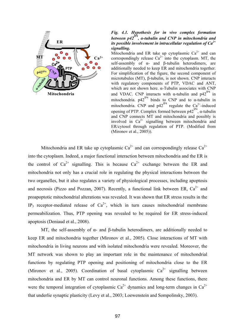
ER
Ca2+MT
Mitochondria
CNP
α-tu
bulin
p42IP4CNP
α-tu
bulin
α-tu
bulin
p42IP4
Fig. 4.1. Hypothesis for in vivo complex formation between p42IP4, α-tubulin and CNP in mitochondria and its possible involvement in intracellular regulation of Ca2+ signalling. Mitochondria and ER take up cytoplasmic Ca2+ and can correspondingly release Ca2+ into the cytoplasm. MT, the self-assembly of α- and β-tubulin heterodimers, are additionally needed to keep ER and mitochondria together. For simplification of the figure, the second component of microtubules (MT), β-tubulin, is not shown. CNP interacts with regulatory components of PTP, VDAC and ANT, which are not shown here. α-Tubulin associates with CNP and VDAC. CNP interacts with α-tubulin and p42IP4 in mitochondria. p42IP4 binds to CNP and to α-tubulin in mitochondria. CNP and p42IP4 regulate the Ca2+-induced opening of PTP. Complex formed between p42IP4, α-tubulin and CNP connects MT and mitochondria and possibly is involved in Ca2+ signalling between mitochondria and ER/cytosol through regulation of PTP. (Modified from (Mironov et al., 2005)).
Mitochondria and ER take up cytoplasmic Ca2+ and can correspondingly release Ca2+
into the cytoplasm. Indeed, a major functional interaction between mitochondria and the ER is
the control of Ca2+ signalling. This is because Ca2+ exchange between the ER and
mitochondria not only has a crucial role in regulating the physical interactions between the
two organelles, but it also regulates a variety of physiological processes, including apoptosis
and necrosis (Pizzo and Pozzan, 2007). Recently, a functional link between ER, Ca2+ and
proapoptotic mitochondrial alterations was revealed. It was shown that ER stress results in the
IP3 receptor-mediated release of Ca2+, which in turn causes mitochondrial membrane
permeabilization. Thus, PTP opening was revealed to be required for ER stress-induced
apoptosis (Deniaud et al., 2008).
MT, the self-assembly of α- and β-tubulin heterodimers, are additionally needed to
keep ER and mitochondria together (Mironov et al., 2005). Close interactions of MT with
mitochondria in living neurons and with isolated mitochondria were revealed. Moreover, the
MT network was shown to play an important role in the maintenance of mitochondrial
functions by regulating PTP opening and positioning of mitochondria close to the ER
(Mironov et al., 2005). Coordination of basal cytoplasmic Ca2+ signalling between
mitochondria and ER by MT can control neuronal functions. Among these functions, there
were the temporal integration of cytoplasmic Ca2+ dynamics and long-term changes in Ca2+
that underlie synaptic plasticity (Levy et al., 2003; Loewenstein and Sompolinsky, 2003).
97
α-Tubulin was shown to be a component of mitochondrial membranes specifically
interacting with the VDAC (Carre et al., 2002). VDAC mediates the complex interactions
between mitochondria and other parts of the cell by transporting anions, cations, ATP, Ca2+
and metabolites. Thus, VDAC plays an important role in coordinating the communication
between mitochondria and cytosol (Grimm and Brdiczka, 2007). Moreover, there were some
indications that VDAC may be part of mitochondrial microdomains linking Ca2+ efflux from
the ER to mitochondrial matrix Ca2+ influx, in itself a potent trigger of PTP (Szabadkai et al.,
2006).
CNP interacts with regulatory components of PTP, VDAC and ANT. Thus, CNP and
α-tubulin binding to each other also associate with PTP. p42IP4 binds to CNP and to α-tubulin
in RBM. Therefore, the complex, which is formed by p42IP4, CNP and α-tubulin, connects
MT and mitochondria. Moreover, p42IP4
and CNP are involved in control of Ca2+-induced PTP
opening. Therefore, one of the important functions of this connection between MT and
mitochondria could be the regulation of Ca2+ signalling between cellular organelles through
regulation of PTP.
Thus, we hypothesize that in the brain p42IP4 possibly acts together with CNP and α–
tubulin in order to regulate mitochondrial Ca2+ transport mechanisms. The reduction of
mitochondrial Ca2+ buffering capacity induced by p42IP4 may increase the vulnerability of
neurons to PTP and thereby contribute to processes leading to neurodegeneration.
This hypothesis could also be applied to AD development. Indeed, it was described
that in neocortical areas of AD patients the neuronal expression of the protein p42IP4 is
markedly increased, and the protein is associated with neuritic plaques (Reiser and Bernstein,
2002). Moreover, p42IP4 interacts with the protein kinase CK Iα (Dubois et al., 2001), which
hyperphosphorylates β-amyloid precursor protein in the brains of AD patients (Walter et al.,
2000). Specific interaction of p42IP4 with nucleolin, another player in AD pathology, has also
been revealed (Dubois et al., 2003). However, the significance of the increase in p42IP4 protein
expression in terms of AD pathology is still unknown (Reiser and Bernstein, 2004). Our
recent finding that nardilysin is an interacting partner for p42IP4 (Stricker et al., 2006) might
also be of interest regarding the possible involvement of p42IP4 in AD pathology. Nardilysin
enhances the cleavage of amyloid precursor protein by α-secretase through the activation of a
disintegrin and metalloprotease proteases, which results in a decreased amount of amyloid-β
peptide, the principal component of senile plaques in the brains of patients with AD (Hiraoka
et al., 2007).
98
Involvement of CNP in AD was claimed in several studies. However, its role is still
completely unclear. In AD patients, the activity of CNP was shown to be decreased in the
hippocampus and putamen (Reinikainen et al., 1989). Another study showed a significant
decrease in CNP protein in AD brain (Vlkolinsky et al., 2001), in contrast to a recent report,
which demonstrated that the protein level of CNP in AD hippocampus was increased (Sultana
et al., 2007).
Neurodegeneration in AD has been linked to dysregulation of intracellular Ca2+
(Giacomello et al., 2007). In an adult triple transgenic mouse model of AD that exhibits
intraneuronal accumulation of amyloid-β proteins, the resting free Ca2+ concentration was
shown to be greatly elevated in cortical neurons (Lopez et al., 2008). This Ca2+ deregulation in
the AD brain may be associated with defective mitochondrial functions. Moreover, in neurons
from AD brain, morphological alterations of mitochondria were revealed (Baloyannis, 2006).
99
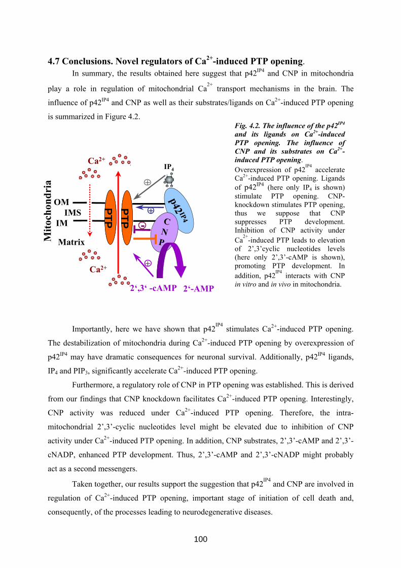
4.7 Conclusions. Novel regulators of Ca2+-induced PTP opening. In summary, the results obtained here suggest that p42IP4 and CNP in mitochondria
play a role in regulation of mitochondrial Ca2+ transport mechanisms in the brain. The
influence of p42IP4 and CNP as well as their substrates/ligands on Ca2+-induced PTP opening
is summarized in Figure 4.2. Fig. 4.2. The influence of the p42IP4 and its ligands on Ca2+-induced PTP opening. The influence of CNP and its substrates on Ca2+-induced PTP opening. Overexpression of p42IP4 accelerate Ca2+-induced PTP opening. Ligands of p42IP4 (here only IP4 is shown) stimulate PTP opening. CNP-knockdown stimulates PTP opening, thus we suppose that CNP suppresses PTP development. Inhibition of CNP activity under Ca2+-induced PTP leads to elevation of 2’,3’cyclic nucleotides levels (here only 2’,3’-cAMP is shown), promoting PTP development. In addition, p42IP4 interacts with CNP in vitro and in vivo in mitochondria.
Ca2+
OM
IMIMS
p42 IP4
Importantly, here we have shown that p42IP4 stimulates Ca2+-induced PTP opening.
The destabilization of mitochondria during Ca2+-induced PTP opening by overexpression of
p42IP4 may have dramatic consequences for neuronal survival. Additionally, p42IP4 ligands,
IP4 and PIP3, significantly accelerate Ca2+-induced PTP opening.
Furthermore, a regulatory role of CNP in PTP opening was established. This is derived
from our findings that CNP knockdown facilitates Ca2+-induced PTP opening. Interestingly,
CNP activity was reduced under Ca2+-induced PTP opening. Therefore, the intra-
mitochondrial 2’,3’-cyclic nucleotides level might be elevated due to inhibition of CNP
activity under Ca2+-induced PTP opening. In addition, CNP substrates, 2’,3’-cAMP and 2’,3’-
cNADP, enhanced PTP development. Thus, 2’,3’-cAMP and 2’,3’-cNADP might probably
act as a second messengers.
Taken together, our results support the suggestion that p42IP4 and CNP are involved in
regulation of Ca2+-induced PTP opening, important stage of initiation of cell death and,
consequently, of the processes leading to neurodegenerative diseases.
Mito
chon
dria
Matrix
IP4
+PTP
+
PTPPTPPTP C
NP
--
+Ca2+
2‘,3‘ -cAMP 2‘-AMP
100
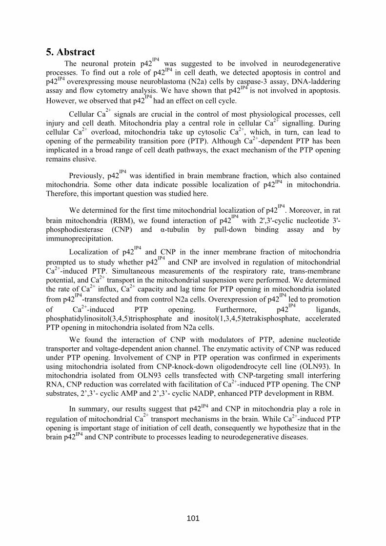
5. Abstract The neuronal protein p42IP4 was suggested to be involved in neurodegenerative
processes. To find out a role of p42IP4 in cell death, we detected apoptosis in control and p42IP4 overexpressing mouse neuroblastoma (N2a) cells by caspase-3 assay, DNA-laddering assay and flow cytometry analysis. We have shown that p42IP4 is not involved in apoptosis. However, we observed that p42IP4 had an effect on cell cycle.
Cellular Ca2+ signals are crucial in the control of most physiological processes, cell injury and cell death. Mitochondria play a central role in cellular Ca2+ signalling. During cellular Ca2+ overload, mitochondria take up cytosolic Ca2+, which, in turn, can lead to opening of the permeability transition pore (PTP). Although Ca2+-dependent PTP has been implicated in a broad range of cell death pathways, the exact mechanism of the PTP opening remains elusive.
Previously, p42IP4 was identified in brain membrane fraction, which also contained mitochondria. Some other data indicate possible localization of p42IP4 in mitochondria. Therefore, this important question was studied here.
We determined for the first time mitochondrial localization of p42IP4. Moreover, in rat brain mitochondria (RBM), we found interaction of p42IP4 with 2',3'-cyclic nucleotide 3'-phosphodiesterase (CNP) and α-tubulin by pull-down binding assay and by immunoprecipitation.
Localization of p42IP4 and CNP in the inner membrane fraction of mitochondria prompted us to study whether p42IP4 and CNP are involved in regulation of mitochondrial Ca2+-induced PTP. Simultaneous measurements of the respiratory rate, trans-membrane potential, and Ca2+ transport in the mitochondrial suspension were performed. We determined the rate of Ca2+ influx, Ca2+ capacity and lag time for PTP opening in mitochondria isolated from p42IP4-transfected and from control N2a cells. Overexpression of p42IP4 led to promotion of Ca2+-induced PTP opening. Furthermore, p42IP4 ligands, phosphatidylinositol(3,4,5)trisphosphate and inositol(1,3,4,5)tetrakisphosphate, accelerated PTP opening in mitochondria isolated from N2a cells.
We found the interaction of CNP with modulators of PTP, adenine nucleotide transporter and voltage-dependent anion channel. The enzymatic activity of CNP was reduced under PTP opening. Involvement of CNP in PTP operation was confirmed in experiments using mitochondria isolated from CNP-knock-down oligodendrocyte cell line (OLN93). In mitochondria isolated from OLN93 cells transfected with CNP-targeting small interfering RNA, CNP reduction was correlated with facilitation of Ca2+-induced PTP opening. The CNP substrates, 2’,3’- cyclic AMP and 2’,3’- cyclic NADP, enhanced PTP development in RBM.
In summary, our results suggest that p42IP4 and CNP in mitochondria play a role in regulation of mitochondrial Ca2+ transport mechanisms in the brain. While Ca2+-induced PTP opening is important stage of initiation of cell death, consequently we hypothesize that in the brain p42IP4 and CNP contribute to processes leading to neurodegenerative diseases.
101
6. Zusammenfassung
Verschiedene in der Literatur beschriebene Befunde deuten auf die Bedeutung des
gehirnspezifischen Proteins p42IP4 bei neurodegenerativen/neuroprotektiven Prozessen hin.
p42IP4 wird auch als Centaurin α-1, und nach einem neueren Vorschlag als ADAP1
bezeichnet. Ein Ziel der vorliegenden Arbeit war aufzuklären, ob p42IP4 eine Rolle bei
Neuroprotektion oder Zelltod spielt.
In einem ersten Ansatz wurden die Auswirkungen der Überexpression von p42IP4 auf
die induzierte Apoptose untersucht. Dazu wurde die Mäuse-Neuroblastoma-Zelllinie N2a
eingesetzt. Der Nachweis der Apoptose in den N2a-Zellen erfolgte mittels Caspase-3-Assay,
DNA-Fragmentierung und Durchflusszytometrie. Hierbei ergaben sich keine Hinweise auf
eine direkte Beteiligung von p42IP4 an der Apoptose. Wir konnten aber feststellen, dass die
Überexpression von p42IP4 einen Effekt auf die Verteilung der Zellen im Zellzyklus hat. In
den p42IP4 überexprimierenden Zellen kam es zu einer Verschiebung des Anteils der Zellen
von der G2-Phase zur S-Phase.
Mitochondrien spielen eine zentrale Rolle in der zellulären Ca2+-Homöostase und
beim Ca2+-Signalling. Bei der Überladung von Zellen mit Ca2+ können Mitochondrien große
Menge an Ca2+ aufnehmen, was in der Folge zur Öffnung der Permeabilitätstransitionspore
(PTP) führen kann. Die Beteiligung einer Ca2+-abhängigen PTP an vielen zu Zelltod und
Gewebsschädigungen führenden Vorgängen konnte gezeigt werden. Aber der genaue
Mechanismus der PTP ist noch weitgehend unbekannt. Daher ist die Suche nach neuen
Proteinen und Molekülen, die an der Kontrolle der Ca2+-induzierten PTP beteiligt sind äußerst
wichtig.
p42IP4 wurde ursprünglich in Membranfraktionen aus Gehirn identifiziert, die auch
Mitochondrien enthielten. Das Hefeprotein Gsc1p, das strukturell und funktionell mit p42IP4
eng verwandt ist, ist an der Aufrechterhaltung der mitochondrialen Morphologie beteiligt.
p42IP4 interagiert mit Proteinkinase C Isoformen, für die ebenfalls eine mitochondriale
Lokalisation beschrieben wurde. Eine hohe Wahrscheinlichkeit für eine mitochondriale
Lokalisation von p42IP4 wurde durch das Programm PSORTII vorhergesagt. Diese Befunde
deuteten auf eine mögliche mitochondriale Lokalisation und Funktion von p42IP4 hin. Daher
wurde diese wichtige Fragestellung in der vorliegenden Arbeit untersucht.
Wir konnten mit verschiedenen Methoden erstmals eine mitochondriale Lokalisation
von p42IP4 in Mitochondrien aus Rattenhirn, sowie aus N2A- und CHO-Zellen zeigen.
Darüberhinaus fanden wir mittels Glutathion-S-Transferase Pull-Down-Versuchen und
102
Koimmunopräzipitation eine Interaktion von p42IP4 mit den Proteinen 2',3'-cyklische
Nukleotid 3'-Phosphodiesterase (CNP) und α-Tubulin.
Wir untersuchten die Verteilung von p42IP4 und CNP in den mitochondrialen
Kompartimenten durch Subfraktionierung von Mitochondrien. Die Lokalisation von p42IP4
und CNP in der inneren Membranfraktion und in Kontaktstellen zwischen innerer und äußerer
Membran deuteten auf eine funktionelle Rolle beider Proteine in Mitochondrien hin. Daher
wurden im Folgenden Studien zur möglichen Beteiligung von p42IP4 und CNP an der
mitochondrialen Ca2+- induzierten PTP durchgeführt. Wir etablierten eine Methode zur
Isolierung von funktionell aktiven Mitochondrien aus Zellkulturen und untersuchten
mitochondriale Funktionen während der Ca2+- induzierten PTP. Eine simultane Messung der
Atmungsrate, des Ca2+-Transports und des mitochondrialen Membranpotenzials in
Mitochondriensuspensionen erfolgte in einer offenen Messkammer. Dabei wurden selektive
Elektroden für O2, Ca2+ und Tetraphenylphosphonium (TPP+) eingesetzt. Letztere diente zur
Membranpotentialmessung. Damit wurden die Geschwindigkeit des Ca2+-Einstroms, die Ca2+-
Kapazität und die Verzögerungszeit für die Ca2+-induzierte PTP bestimmt. Es wurden
Messungen für Mitochondrien aus p42IP4-transfizierten und nichttransfizierten N2a-Zellen
durchgeführt. Die Überexpression von p42IP4 führte zu einer signifikanten Forcierung der
Ca2+-induzierten Öffnung der PTP. Phosphatidylinositol(3,4,5)trisphosphat und
Inositol(1,3,4,5)tetraphosphat, beides spezifische Liganden für p42IP4, beschleunigten die
Öffnung der PTP in Mitochondrien aus N2a-Zellen. Erstaunlicherweise war dieser Einfluss
der p42IP4-Liganden auf die PTP-Öffnung unabhängig von der Überexpression von p42IP4.
In Rattenhirnmitochondrien fanden wir mittels Koimmunopräzipitation eine
Proteinwechselwirkung von CNP mit Modulatoren der PTP, dem Adeninnukleotid-
Transporter (ANT) und dem spannungsabhängigen Anionen-Kanal (VDAC). Darüberhinaus
konnten wir zeigen, dass die Enzymaktivität von CNP bei der PTP-Öffnung reduziert war,
während der CNP-Proteingehalt gleich blieb. Eine Beteiligung von CNP an der PTP-
Regulation wurde in weiteren Experimenten bestätigt, bei denen Mitochondrien aus OLN93-
Zellen, einer Oligodendrozyten-Zelllinie, verwendet wurden, bei denen CNP durch
Transfektion mit siRNA herunterreguliert wurde. In den Mitochondrien aus diesen Zellen
korrelierte die Reduktion der CNP-Expression mit einer Erniedrigung der Schwelle für eine
Ca2+-induzierte PTP Bildung. Des Weiteren erhöhten 2',3'-cAMP und 2',3'-cyklisches NADP,
beides Substrate der CNP, die PTP Bildung in Mitochondrien aus Rattenhirn.
Zusammenfassend zeigen die hier beschriebenen Ergebnisse, dass p42IP4 und CNP in
Mitochondrien eine wichtige Funktion bei der Regulation mitochondrialer Ca2+-
103

Transportvorgänge im Gehirn ausüben. Da die Ca2+-induzierte PTP-Bildung einen wichtigen
Schritt bei der Einleitung des Zelltods darstellt, könnten p42IP4 und CNP an Prozessen
beteiligt sein, die zu neurodegenerativen Krankheiten führen.
104
7. References
Aggensteiner M, Stricker R, Reiser G (1998) Identification of rat brain p42(IP4), a high-affinity inositol(1,3,4,5)tetrakisphosphate/phosphatidylinositol(3,4,5)trisphosphate binding protein. Biochim Biophys Acta 1387:117-128.
Agrawal HC, Sprinkle TJ, Agrawal D (1990a) 2',3'cyclic nucleotide-3'-phosphodiesterase in peripheral nerve myelin is phosphorylated by a phorbol ester-sensitive protein kinase. Biochem Biophys Res Commun 170:817-823.
Agrawal HC, Sprinkle TJ, Agrawal D (1990b) 2',3'-cyclic nucleotide-3'-phosphodiesterase in the central nervous system is fatty-acylated by thioester linkage. J Biol Chem 265:11849-11853.
Agrawal HC, Sprinkle TJ, Agrawal D (1994) In vivo phosphorylation of 2',3'-cyclic nucleotide 3'-phosphohydrolase (CNP): CNP in brain myelin is phosphorylated by forskolin- and phorbol ester-sensitive protein kinases. Neurochem Res 19:721-728.
Azarashvili T, Krestinina O, Yurkov I, Evtodienko Y, Reiser G (2005) High-affinity peripheral benzodiazepine receptor ligand, PK11195, regulates protein phosphorylation in rat brain mitochondria under control of Ca(2+). J Neurochem 94:1054-1062.
Azarashvili T, Grachev D, Krestinina O, Evtodienko Y, Yurkov I, Papadopoulos V, Reiser G (2007) The peripheral-type benzodiazepine receptor is involved in control of Ca2+-induced permeability transition pore opening in rat brain mitochondria. Cell Calcium 42:27-39.
Baines CP, Kaiser RA, Sheiko T, Craigen WJ, Molkentin JD (2007) Voltage-dependent anion channels are dispensable for mitochondrial-dependent cell death. Nat Cell Biol 9:550-555.
Baines CP, Song CX, Zheng YT, Wang GW, Zhang J, Wang OL, Guo Y, Bolli R, Cardwell EM, Ping P (2003) Protein kinase Cepsilon interacts with and inhibits the permeability transition pore in cardiac mitochondria. Circ Res 92:873-880.
Baines CP, Kaiser RA, Purcell NH, Blair NS, Osinska H, Hambleton MA, Brunskill EW, Sayen MR, Gottlieb RA, Dorn GW, Robbins J, Molkentin JD (2005) Loss of cyclophilin D reveals a critical role for mitochondrial permeability transition in cell death. Nature 434:658-662.
Baloyannis SJ (2006) Mitochondrial alterations in Alzheimer's disease. J Alzheimers Dis 9:119-126.
Bambrick LL, Chandrasekaran K, Mehrabian Z, Wright C, Krueger BK, Fiskum G (2006) Cyclosporin A increases mitochondrial calcium uptake capacity in cortical astrocytes but not cerebellar granule neurons. J Bioenerg Biomembr 38:43-47.
Basso E, Fante L, Fowlkes J, Petronilli V, Forte MA, Bernardi P (2005) Properties of the permeability transition pore in mitochondria devoid of Cyclophilin D. J Biol Chem 280:18558-18561.
Bernardi P (1999) Mitochondrial transport of cations: channels, exchangers, and permeability transition. Physiol Rev 79:1127-1155.
Berridge MJ (1993) Inositol trisphosphate and calcium signalling. Nature 361:315-325.
105
Beutner G, Ruck A, Riede B, Brdiczka D (1998) Complexes between porin, hexokinase, mitochondrial creatine kinase and adenylate translocator display properties of the permeability transition pore. Implication for regulation of permeability transition by the kinases. Biochim Biophys Acta 1368:7-18.
Bifulco M, Laezza C, Stingo S, Wolff J (2002) 2',3'-Cyclic nucleotide 3'-phosphodiesterase: a membrane-bound, microtubule-associated protein and membrane anchor for tubulin. Proc Natl Acad Sci U S A 99:1807-1812.
Bijur GN, Jope RS (2003) Rapid accumulation of Akt in mitochondria following phosphatidylinositol 3-kinase activation. J Neurochem 87:1427-1435.
Birnbaum G, Kotilinek L, Schlievert P, Clark HB, Trotter J, Horvath E, Gao E, Cox M, Braun PE (1996) Heat shock proteins and experimental autoimmune encephalomyelitis (EAE): I. Immunization with a peptide of the myelin protein 2',3' cyclic nucleotide 3' phosphodiesterase that is cross-reactive with a heat shock protein alters the course of EAE. J Neurosci Res 44:381-396.
Bradbury JM, Thompson RJ (1984) Photoaffinity labelling of central-nervous-system myelin. Evidence for an endogenous type I cyclic AMP-dependent kinase phosphorylating the larger subunit of 2',3'-cyclic nucleotide 3'-phosphodiesterase. Biochem J 221:361-368.
Bradbury JM, Campbell RS, Thompson RJ (1984) Endogenous cyclic AMP-stimulated phosphorylation of a Wolfgram protein component in rabbit central-nervous-system myelin. Biochem J 221:351-359.
Braun PE, De Angelis D, Shtybel WW, Bernier L (1991) Isoprenoid modification permits 2',3'-cyclic nucleotide 3'-phosphodiesterase to bind to membranes. J Neurosci Res 30:540-544.
Broekemeier KM, Dempsey ME, Pfeiffer DR (1989) Cyclosporin A is a potent inhibitor of the inner membrane permeability transition in liver mitochondria. J Biol Chem 264:7826-7830.
Bronisz A, Gajkowska B, Domanska-Janik K (2002) PKC and Raf-1 inhibition-related apoptotic signalling in N2a cells. J Neurochem 81:1176-1184.
Brown MR, Sullivan PG, Geddes JW (2006) Synaptic mitochondria are more susceptible to Ca2+overload than nonsynaptic mitochondria. J Biol Chem 281:11658-11668.
Brustovetsky N, Dubinsky JM (2000) Limitations of cyclosporin A inhibition of the permeability transition in CNS mitochondria. J Neurosci 20:8229-8237.
Capano M, Crompton M (2002) Biphasic translocation of Bax to mitochondria. Biochem J 367:169-178.
Carre M, Andre N, Carles G, Borghi H, Brichese L, Briand C, Braguer D (2002) Tubulin is an inherent component of mitochondrial membranes that interacts with the voltage-dependent anion channel. J Biol Chem 277:33664-33669.
Chalmers S, Nicholls DG (2003) The relationship between free and total calcium concentrations in the matrix of liver and brain mitochondria. J Biol Chem 278:19062-19070.
Chanda SK, White S, Orth AP, Reisdorph R, Miraglia L, Thomas RS, DeJesus P, Mason DE, Huang Q, Vega R, Yu DH, Nelson CG, Smith BM, Terry R, Linford AS, Yu Y, Chirn GW, Song C, Labow MA, Cohen D, King FJ, Peters EC, Schultz PG, Vogt PK, Hogenesch JB, Caldwell JS
106
(2003) Genome-scale functional profiling of the mammalian AP-1 signaling pathway. Proc Natl Acad Sci U S A 100:12153-12158.
Chesneau V, Pierotti AR, Barre N, Creminon C, Tougard C, Cohen P (1994) Isolation and characterization of a dibasic selective metalloendopeptidase from rat testes that cleaves at the amino terminus of arginine residues. J Biol Chem 269:2056-2061.
Cho SJ, Jung JS, Shin SC, Jin I, Ko BH, Kim Kwon Y, Suh-Kim H, Moon IS (2003) Nonspecific association of 2',3'-cyclic nucleotide 3'-phosphodiesterase with the rat forebrain postsynaptic density fraction. Exp Mol Med 35:486-493.
Cox ME, Gao EN, Braun PE (1994) C-terminal CTII motif of 2',3'-cyclic nucleotide 3'-phosphodiesterase undergoes carboxylmethylation. J Neurosci Res 39:513-518.
Cozier GE, Lockyer PJ, Reynolds JS, Kupzig S, Bottomley JR, Millard TH, Banting G, Cullen PJ (2000) GAP1IP4BP contains a novel group I pleckstrin homology domain that directs constitutive plasma membrane association. J Biol Chem 275:28261-28268.
Crompton M, Ellinger H, Costi A (1988) Inhibition by cyclosporin A of a Ca2+-dependent pore in heart mitochondria activated by inorganic phosphate and oxidative stress. Biochem J 255:357-360.
Cukierman E, Huber I, Rotman M, Cassel D (1995) The ARF1 GTPase-activating protein: zinc finger motif and Golgi complex localization. Science 270:1999-2002.
De Angelis DA, Braun PE (1994) Isoprenylation of brain 2',3'-cyclic nucleotide 3'-phosphodiesterase modulates cell morphology. J Neurosci Res 39:386-397.
De Angelis DA, Braun PE (1996) 2',3'-Cyclic nucleotide 3'-phosphodiesterase binds to actin-based cytoskeletal elements in an isoprenylation-independent manner. J Neurochem 67:943-951.
Deniaud A, Sharaf el dein O, Maillier E, Poncet D, Kroemer G, Lemaire C, Brenner C (2008) Endoplasmic reticulum stress induces calcium-dependent permeability transition, mitochondrial outer membrane permeabilization and apoptosis. Oncogene 27:285-299.
Donie F, Reiser G (1991) Purification of a high-affinity inositol 1,3,4,5-tetrakisphosphate receptor from brain. Biochem J 275 ( Pt 2):453-457.
Douglas AJ, Fox MF, Abbott CM, Hinks LJ, Sharpe G, Povey S, Thompson RJ (1992) Structure and chromosomal localization of the human 2',3'-cyclic nucleotide 3'-phosphodiesterase gene. Ann Hum Genet 56:243-254.
Dreiling CE, Schilling RJ, Reitz RC (1981) 2',3'-cyclic nucleotide 3'-phosphohydrolase in rat liver mitochondrial membranes. Biochim Biophys Acta 640:114-120.
Drummond GI, Iyer NT, Keith J (1962) Hydrolysis of ribonucleoside 2',3'-cyclic phosphates by a diesterase from brain. J Biol Chem 237:3535–3539.
Dubois T, Zemlickova E, Howell S, Aitken A (2003) Centaurin-alpha 1 associates in vitro and in vivo with nucleolin. Biochem Biophys Res Commun 301:502-508.
Dubois T, Kerai P, Zemlickova E, Howell S, Jackson TR, Venkateswarlu K, Cullen PJ, Theibert AB, Larose L, Roach PJ, Aitken A (2001) Casein kinase I associates with members of the centaurin-alpha family of phosphatidylinositol 3,4,5-trisphosphate-binding proteins. J Biol Chem 276:18757-18764.
107
Duszynski J, Koziel R, Brutkowski W, Szczepanowska J, Zablocki K (2006) The regulatory role of mitochondria in capacitative calcium entry. Biochim Biophys Acta 1757:380-387.
Dyer CA, Benjamins JA (1988) Antibody to galactocerebroside alters organization of oligodendroglial membrane sheets in culture. J Neurosci 8:4307-4318.
Dyer CA, Benjamins JA (1989) Organization of oligodendroglial membrane sheets. I: Association of myelin basic protein and 2',3'-cyclic nucleotide 3'-phosphohydrolase with cytoskeleton. J Neurosci Res 24:201-211.
Dyer CA, Matthieu JM (1994) Antibodies to myelin/oligodendrocyte-specific protein and myelin/oligodendrocyte glycoprotein signal distinct changes in the organization of cultured oligodendroglial membrane sheets. J Neurochem 62:777-787.
Dyer CA, Philibotte TM, Billings-Gagliardi S, Wolf MK (1995) Cytoskeleton in myelin-basic-protein-deficient shiverer oligodendrocytes. Dev Neurosci 17:53-62.
Esposito C, Scrima M, Carotenuto A, Tedeschi A, Rovero P, D'Errico G, Malfitano AM, Bifulco M, D'Ursi AM (2008) Structures and micelle locations of the nonlipidated and lipidated C-terminal membrane anchor of 2',3'-cyclic nucleotide-3'-phosphodiesterase. Biochemistry 47:308-319.
Ferguson KM, Kavran JM, Sankaran VG, Fournier E, Isakoff SJ, Skolnik EY, Lemmon MA (2000) Structural basis for discrimination of 3-phosphoinositides by pleckstrin homology domains. Mol Cell 6:373-384.
Giacomello M, Drago I, Pizzo P, Pozzan T (2007) Mitochondrial Ca2+ as a key regulator of cell life and death. Cell Death Differ 14:1267-1274.
Gillespie CS, Bernier L, Brophy PJ, Colman DR (1990) Biosynthesis of the myelin 2',3'-cyclic nucleotide 3'-phosphodiesterases. J Neurochem 54:656-661.
Ginisty H, Sicard H, Roger B, Bouvet P (1999) Structure and functions of nucleolin. J Cell Sci 112 ( Pt 6):761-772.
Gottlob K, Majewski N, Kennedy S, Kandel E, Robey RB, Hay N (2001) Inhibition of early apoptotic events by Akt/PKB is dependent on the first committed step of glycolysis and mitochondrial hexokinase. Genes Dev 15:1406-1418.
Gravel M, DeAngelis D, Braun PE (1994) Molecular cloning and characterization of rat brain 2',3'-cyclic nucleotide 3'-phosphodiesterase isoform 2. J Neurosci Res 38:243-247.
Gravel M, Peterson J, Yong VW, Kottis V, Trapp B, Braun PE (1996) Overexpression of 2',3'-cyclic nucleotide 3'-phosphodiesterase in transgenic mice alters oligodendrocyte development and produces aberrant myelination. Mol Cell Neurosci 7:453-466.
Gravel M, Robert F, Kottis V, Gallouzi IE, Pelletier J, Braun PE (2009) 2',3'-Cyclic nucleotide 3'-phosphodiesterase: a novel RNA-binding protein that inhibits protein synthesis. J Neurosci Res 87:1069-1079.
Grimm S, Brdiczka D (2007) The permeability transition pore in cell death. Apoptosis 12:841-855.
Haase A, Nordmann C, Sedehizade F, Borrmann C, Reiser G (2008) RanBPM, a novel interaction partner of the brain-specific protein p42(IP4)/centaurin alpha-1. J Neurochem.
Hajnoczky G, Csordas G, Das S, Garcia-Perez C, Saotome M, Sinha Roy S, Yi M (2006) Mitochondrial calcium signalling and cell death: approaches for
108
assessing the role of mitochondrial Ca2+ uptake in apoptosis. Cell Calcium 40:553-560.
Halestrap AP (2004) Mitochondrial permeability: dual role for the ADP/ATP translocator? Nature 430:1 p following 983.
Halestrap AP (2006) Calcium, mitochondria and reperfusion injury: a pore way to die. Biochem Soc Trans 34:232-237.
Halestrap AP (2009) What is the mitochondrial permeability transition pore? J Mol Cell Cardiol 46:821-831.
Halestrap AP, Davidson AM (1990) Inhibition of Ca2(+)-induced large-amplitude swelling of liver and heart mitochondria by cyclosporin is probably caused by the inhibitor binding to mitochondrial-matrix peptidyl-prolyl cis-trans isomerase and preventing it interacting with the adenine nucleotide translocase. Biochem J 268:153-160.
Halestrap AP, McStay GP, Clarke SJ (2002) The permeability transition pore complex: another view. Biochimie 84:153-166.
Hammonds-Odie LP, Jackson TR, Profit AA, Blader IJ, Turck CW, Prestwich GD, Theibert AB (1996) Identification and cloning of centaurin-alpha. A novel phosphatidylinositol 3,4,5-trisphosphate-binding protein from rat brain. J Biol Chem 271:18859-18868.
Hanck T, Stricker R, Sedehizade F, Reiser G (2004) Identification of gene structure and subcellular localization of human centaurin alpha 2, and p42IP4, a family of two highly homologous, Ins 1,3,4,5-P4-/PtdIns 3,4,5-P3-binding, adapter proteins. J Neurochem 88:326-336.
Hanck T, Stricker R, Krishna UM, Falck JR, Chang YT, Chung SK, Reiser G (1999) Recombinant p42IP4, a brain-specific 42-kDa high-affinity Ins(1,3,4,5)P4 receptor protein, specifically interacts with lipid membranes containing Ptd-Ins(3,4,5)P3. Eur J Biochem 261:577-584.
Hansson MJ, Mansson R, Mattiasson G, Ohlsson J, Karlsson J, Keep MF, Elmer E (2004) Brain-derived respiring mitochondria exhibit homogeneous, complete and cyclosporin-sensitive permeability transition. J Neurochem 89:715-729.
Hayashi H, Matsuzaki O, Muramatsu S, Tsuchiya Y, Harada T, Suzuki Y, Sugano S, Matsuda A, Nishida E (2006) Centaurin-alpha1 is a phosphatidylinositol 3-kinase-dependent activator of ERK1/2 mitogen-activated protein kinases. In: J Biol Chem, pp 1332-1337.
Hermosura MC, Takeuchi H, Fleig A, Riley AM, Potter BV, Hirata M, Penner R (2000) InsP4 facilitates store-operated calcium influx by inhibition of InsP3 5-phosphatase. Nature 408:735-740.
Hinman JD, Chen CD, Oh SY, Hollander W, Abraham CR (2008) Age-dependent accumulation of ubiquitinated 2',3'-cyclic nucleotide 3'-phosphodiesterase in myelin lipid rafts. Glia 56:118-133.
Hiraoka Y, Ohno M, Yoshida K, Okawa K, Tomimoto H, Kita T, Nishi E (2007) Enhancement of alpha-secretase cleavage of amyloid precursor protein by a metalloendopeptidase nardilysin. J Neurochem 102:1595-1605.
Hofmann A, Grella M, Botos I, Filipowicz W, Wlodawer A (2002) Crystal structures of the semireduced and inhibitor-bound forms of cyclic nucleotide phosphodiesterase from Arabidopsis thaliana. J Biol Chem 277:1419-1425.
Hofmann A, Zdanov A, Genschik P, Ruvinov S, Filipowicz W, Wlodawer A (2000) Structure and mechanism of activity of the cyclic phosphodiesterase of
109
Appr>p, a product of the tRNA splicing reaction. Embo J 19:6207-6217.
Horiguchi K, Hanada T, Fukui Y, Chishti AH (2006) Transport of PIP3 by GAKIN, a kinesin-3 family protein, regulates neuronal cell polarity. J Cell Biol 174:425-436.
Huang CF, Chen CC, Tung L, Buu LM, Lee FJ (2002) The yeast ADP-ribosylation factor GAP, Gcs1p, is involved in maintenance of mitochondrial morphology. J Cell Sci 115:275-282.
Huang YH, Grasis JA, Miller AT, Xu R, Soonthornvacharin S, Andreotti AH, Tsoukas CD, Cooke MP, Sauer K (2007) Positive regulation of Itk PH domain function by soluble IP4. Science 316:886-889.
Hunter DR, Haworth RA, Southard JH (1976) Relationship between configuration, function, and permeability in calcium-treated mitochondria. J Biol Chem 251:5069-5077.
Jackson TR, Kearns BG, Theibert AB (2000) Cytohesins and centaurins: mediators of PI 3-kinase-regulated Arf signaling. Trends Biochem Sci 25:489-495.
Jia Y, Schurmans S, Luo HR (2008) Regulation of innate immunity by inositol 1,3,4,5-tetrakisphosphate. Cell Cycle 7:2803-2808.
Jia Y, Subramanian KK, Erneux C, Pouillon V, Hattori H, Jo H, You J, Zhu D, Schurmans S, Luo HR (2007) Inositol 1,3,4,5-tetrakisphosphate negatively regulates phosphatidylinositol-3,4,5- trisphosphate signaling in neutrophils. Immunity 27:453-467.
Juhaszova M, Wang S, Zorov DB, Nuss HB, Gleichmann M, Mattson MP, Sollott SJ (2008) The identity and regulation of the mitochondrial permeability transition pore: where the known meets the unknown. Ann N Y Acad Sci 1123:197-212.
Kahn RA, Bruford E, Inoue H, Logsdon JM, Jr., Nie Z, Premont RT, Randazzo PA, Satake M, Theibert AB, Zapp ML, Cassel D (2008) Consensus nomenclature for the human ArfGAP domain-containing proteins. J Cell Biol 182:1039-1044.
Kato M, Shirouzu M, Terada T, Yamaguchi H, Murayama K, Sakai H, Kuramitsu S, Yokoyama S (2003) Crystal structure of the 2'-5' RNA ligase from Thermus thermophilus HB8. J Mol Biol 329:903-911.
Kim T, Pfeiffer SE (1999) Myelin glycosphingolipid/cholesterol-enriched microdomains selectively sequester the non-compact myelin proteins CNP and MOG. J Neurocytol 28:281-293.
Kokoszka JE, Waymire KG, Levy SE, Sligh JE, Cai J, Jones DP, MacGregor GR, Wallace DC (2004) The ADP/ATP translocator is not essential for the mitochondrial permeability transition pore. Nature 427:461-465.
Koonin EV, Gorbalenya AE (1990) Related domains in yeast tRNA ligase, bacteriophage T4 polynucleotide kinase and RNA ligase, and mammalian myelin 2',3'-cyclic nucleotide phosphohydrolase revealed by amino acid sequence comparison. FEBS Lett 268:231-234.
Kozlov G, Lee J, Elias D, Gravel M, Gutierrez P, Ekiel I, Braun PE, Gehring K (2003) Structural evidence that brain cyclic nucleotide phosphodiesterase is a member of the 2H phosphodiesterase superfamily. J Biol Chem 278:46021-46028.
Krauskopf A, Eriksson O, Craigen WJ, Forte MA, Bernardi P (2006) Properties of the permeability transition in VDAC1(-/-) mitochondria. Biochim Biophys Acta 1757:590-595.
110
Kreutz MR, Bockers TM, Sabel BA, Hulser E, Stricker R, Reiser G (1997) Expression and subcellular localization of p42IP4/centaurin-alpha, a brain-specific, high-affinity receptor for inositol 1,3,4,5-tetrakisphosphate and phosphatidylinositol 3,4,5-trisphosphate in rat brain. Eur J Neurosci 9:2110-2124.
Krieger C, Duchen MR (2002) Mitochondria, Ca2+ and neurodegenerative disease. Eur J Pharmacol 447:177-188.
Kristal BS, Staats PN, Shestopalov AI (2000) Biochemical characterization of the mitochondrial permeability transition in isolated forebrain mitochondria. Dev Neurosci 22:376-383.
Kurihara T, Monoh K, Sakimura K, Takahashi Y (1990) Alternative splicing of mouse brain 2',3'-cyclic-nucleotide 3'-phosphodiesterase mRNA. Biochem Biophys Res Commun 170:1074-1081.
Kurihara T, Tohyama Y, Yamamoto J, Kanamatsu T, Watanabe R, Kitajima S (1992) Origin of brain 2',3'-cyclic-nucleotide 3'-phosphodiesterase doublet. Neurosci Lett 138:49-52.
Laezza C, Wolff J, Bifulco M (1997) Identification of a 48-kDa prenylated protein that associates with microtubules as 2',3'-cyclic nucleotide 3'-phosphodiesterase in FRTL-5 cells. FEBS Lett 413:260-264.
Lakshmana MK, Yoon IS, Chen E, Bianchi E, Koo EH, Kang DE (2009) Novel role of RanBP9 in BACE1 processing of amyloid precursor protein and amyloid beta peptide generation. J Biol Chem 284:11863-11872.
Lappe-Siefke C, Goebbels S, Gravel M, Nicksch E, Lee J, Braun PE, Griffiths IR, Nave KA (2003) Disruption of Cnp1 uncouples oligodendroglial functions in axonal support and myelination. Nat Genet 33:366-374.
Lawlor MA, Alessi DR (2001) PKB/Akt: a key mediator of cell proliferation, survival and insulin responses? J Cell Sci 114:2903-2910.
Lawrence J, Mundell SJ, Yun H, Kelly E, Venkateswarlu K (2005) Centaurin-alpha 1, an ADP-ribosylation factor 6 GTPase activating protein, inhibits beta 2-adrenoceptor internalization. Mol Pharmacol 67:1822-1828.
Lee J, Gravel M, Gao E, O'Neill RC, Braun PE (2001) Identification of essential residues in 2',3'-cyclic nucleotide 3'-phosphodiesterase. Chemical modification and site-directed mutagenesis to investigate the role of cysteine and histidine residues in enzymatic activity. J Biol Chem 276:14804-14813.
Lee J, Gravel M, Zhang R, Thibault P, Braun PE (2005) Process outgrowth in oligodendrocytes is mediated by CNP, a novel microtubule assembly myelin protein. J Cell Biol 170:661-673.
Lee J, O'Neill RC, Park MW, Gravel M, Braun PE (2006) Mitochondrial localization of CNP2 is regulated by phosphorylation of the N-terminal targeting signal by PKC: implications of a mitochondrial function for CNP2 in glial and non-glial cells. Mol Cell Neurosci 31:446-462.
Leissring MA, Farris W, Wu X, Christodoulou DC, Haigis MC, Guarente L, Selkoe DJ (2004) Alternative translation initiation generates a novel isoform of insulin-degrading enzyme targeted to mitochondria. Biochem J 383:439-446.
Lemmon MA, Ferguson KM (2000) Signal-dependent membrane targeting by pleckstrin homology (PH) domains. Biochem J 350 Pt 1:1-18.
Leung AW, Halestrap AP (2008) Recent progress in elucidating the molecular mechanism of the mitochondrial permeability transition pore. Biochim Biophys Acta 1777:946-952.
111
Leung AW, Varanyuwatana P, Halestrap AP (2008) The mitochondrial phosphate carrier interacts with cyclophilin D and may play a key role in the permeability transition. J Biol Chem 283:26312-26323.
Levy M, Faas GC, Saggau P, Craigen WJ, Sweatt JD (2003) Mitochondrial regulation of synaptic plasticity in the hippocampus. J Biol Chem 278:17727-17734.
Loewenstein Y, Sompolinsky H (2003) Temporal integration by calcium dynamics in a model neuron. Nat Neurosci 6:961-967.
Lopez JR, Lyckman A, Oddo S, Laferla FM, Querfurth HW, Shtifman A (2008) Increased intraneuronal resting [Ca2+] in adult Alzheimer's disease mice. J Neurochem 105:262-271.
Luckhoff A, Clapham DE (1992) Inositol 1,3,4,5-tetrakisphosphate activates an endothelial Ca(2+)-permeable channel. Nature 355:356-358.
Maatta JA, Kaldman MS, Sakoda S, Salmi AA, Hinkkanen AE (1998) Encephalitogenicity of myelin-associated oligodendrocytic basic protein and 2',3'-cyclic nucleotide 3'-phosphodiesterase for BALB/c and SJL mice. Immunology 95:383-388.
Majewski N, Nogueira V, Bhaskar P, Coy PE, Skeen JE, Gottlob K, Chandel NS, Thompson CB, Robey RB, Hay N (2004) Hexokinase-mitochondria interaction mediated by Akt is required to inhibit apoptosis in the presence or absence of Bax and Bak. Mol Cell 16:819-830.
Marzo I, Brenner C, Zamzami N, Susin SA, Beutner G, Brdiczka D, Remy R, Xie ZH, Reed JC, Kroemer G (1998a) The permeability transition pore complex: a target for apoptosis regulation by caspases and bcl-2-related proteins. J Exp Med 187:1261-1271.
Marzo I, Brenner C, Zamzami N, Jurgensmeier JM, Susin SA, Vieira HL, Prevost MC, Xie Z, Matsuyama S, Reed JC, Kroemer G (1998b) Bax and adenine nucleotide translocator cooperate in the mitochondrial control of apoptosis. Science 281:2027-2031.
Mayrleitner M, Schafer R, Fleischer S (1995) IP3 receptor purified from liver plasma membrane is an (1,4,5)IP3 activated and (1,3,4,5)IP4 inhibited calcium permeable ion channel. Cell Calcium 17:141-153.
McFerran B, Burgoyne R (1997) 2',3'-Cyclic nucleotide 3'-phosphodiesterase is associated with mitochondria in diverse adrenal cell types. J Cell Sci 110 ( Pt 23):2979-2985.
Menager C, Arimura N, Fukata Y, Kaibuchi K (2004) PIP3 is involved in neuronal polarization and axon formation. J Neurochem 89:109-118.
Miller AT, Chamberlain PP, Cooke MP (2008) Beyond IP3: roles for higher order inositol phosphates in immune cell signaling. Cell Cycle 7:463-467.
Miller AT, Sandberg M, Huang YH, Young M, Sutton S, Sauer K, Cooke MP (2007) Production of Ins(1,3,4,5)P4 mediated by the kinase Itpkb inhibits store-operated calcium channels and regulates B cell selection and activation. Nat Immunol 8:514-521.
Mironov SL, Ivannikov MV, Johansson M (2005) [Ca2+]i signaling between mitochondria and endoplasmic reticulum in neurons is regulated by microtubules. From mitochondrial permeability transition pore to Ca2+-induced Ca2+ release. J Biol Chem 280:715-721.
Monoh K, Kurihara T, Takahashi Y, Ichikawa T, Kumanishi T, Hayashi S, Minoshima S, Shimizu N (1993) Structure, expression and chromosomal localization of the gene encoding human 2',3'-cyclic-nucleotide 3'-phosphodiesterase. Gene 129:297-301.
112
Moore CD, Thacker EE, Larimore J, Gaston D, Underwood A, Kearns B, Patterson SI, Jackson T, Chapleau C, Pozzo-Miller L, Theibert A (2007) The neuronal Arf GAP centaurin alpha1 modulates dendritic differentiation. J Cell Sci 120:2683-2693.
Morris-Downes MM, McCormack K, Baker D, Sivaprasad D, Natkunarajah J, Amor S (2002) Encephalitogenic and immunogenic potential of myelin-associated glycoprotein (MAG), oligodendrocyte-specific glycoprotein (OSP) and 2',3'-cyclic nucleotide 3'-phosphodiesterase (CNPase) in ABH and SJL mice. J Neuroimmunol 122:20-33.
Muraro PA, Kalbus M, Afshar G, McFarland HF, Martin R (2002) T cell response to 2',3'-cyclic nucleotide 3'-phosphodiesterase (CNPase) in multiple sclerosis patients. J Neuroimmunol 130:233-242.
Nakagawa T, Shimizu S, Watanabe T, Yamaguchi O, Otsu K, Yamagata H, Inohara H, Kubo T, Tsujimoto Y (2005) Cyclophilin D-dependent mitochondrial permeability transition regulates some necrotic but not apoptotic cell death. Nature 434:652-658.
Nakai K, Kanehisa M (1992) A knowledge base for predicting protein localization sites in eukaryotic cells. Genomics 14:897-911.
Neupert W (1997) Protein import into mitochondria. Annu Rev Biochem 66:863-917. O'Neill RC, Braun PE (2000) Selective synthesis of 2',3'-cyclic nucleotide 3'-
phosphodiesterase isoform 2 and identification of specifically phosphorylated serine residues. J Neurochem 74:540-546.
O'Neill RC, Minuk J, Cox ME, Braun PE, Gravel M (1997) CNP2 mRNA directs synthesis of both CNP1 and CNP2 polypeptides. J Neurosci Res 50:248-257.
Pastorino JG, Shulga N, Hoek JB (2002) Mitochondrial binding of hexokinase II inhibits Bax-induced cytochrome c release and apoptosis. J Biol Chem 277:7610-7618.
Peirce TR, Bray NJ, Williams NM, Norton N, Moskvina V, Preece A, Haroutunian V, Buxbaum JD, Owen MJ, O'Donovan MC (2006) Convergent evidence for 2',3'-cyclic nucleotide 3'-phosphodiesterase as a possible susceptibility gene for schizophrenia. Arch Gen Psychiatry 63:18-24.
Pizzo P, Pozzan T (2007) Mitochondria-endoplasmic reticulum choreography: structure and signaling dynamics. Trends Cell Biol 17:511-517.
Poon PP, Wang X, Rotman M, Huber I, Cukierman E, Cassel D, Singer RA, Johnston GC (1996) Saccharomyces cerevisiae Gcs1 is an ADP-ribosylation factor GTPase-activating protein. Proc Natl Acad Sci U S A 93:10074-10077.
Prestwich GD (2004) Phosphoinositide signaling; from affinity probes to pharmaceutical targets. Chem Biol 11:619-637.
Prinos P, Slack C, Lasko DD (1995) 5'phosphorylation of DNA in mammalian cells: identification of a polymin P-precipitable polynucleotide kinase. J Cell Biochem 58:115-131.
Rasband MN, Tayler J, Kaga Y, Yang Y, Lappe-Siefke C, Nave KA, Bansal R (2005) CNP is required for maintenance of axon-glia interactions at nodes of Ranvier in the CNS. Glia 50:86-90.
Reinikainen KJ, Pitkanen A, Riekkinen PJ (1989) 2',3'-cyclic nucleotide-3'-phosphodiesterase activity as an index of myelin in the post-mortem brains of patients with Alzheimer's disease. Neurosci Lett 106:229-232.
113
Reiser G, Bernstein HG (2002) Neurons and plaques of Alzheimer's disease patients highly express the neuronal membrane docking protein p42IP4/centaurin alpha. Neuroreport 13:2417-2419.
Reiser G, Bernstein HG (2004) Altered expression of protein p42IP4/centaurin-alpha 1 in Alzheimer's disease brains and possible interaction of p42IP4 with nucleolin. Neuroreport 15:147-148.
Reiser G, Kunzelmann U, Steinhilber G, Binmoller FJ (1994) Generation of a monoclonal antibody against the myelin protein CNP (2',3'-cyclic nucleotide 3'-phosphodiesterase) suitable for biochemical and for immunohistochemical investigations of CNP. Neurochem Res 19:1479-1485.
Reiser G, Striggow F, Hackmann C, Schwegler H, Yilmazer-Hanke DM (2004) Short-term down-regulation of the brain-specific, PtdIns(3,4,5)P3/Ins(1,3,4,5)P4-binding, adapter protein, p42IP4/centaurin-alpha 1 in rat brain after acoustic and electric stimulation. Neurochem Int 45:89-93.
Reiser G, Schafer R, Donie F, Hulser E, Nehls-Sahabandu M, Mayr GW (1991) A high-affinity inositol 1,3,4,5-tetrakisphosphate receptor protein from brain is specifically labelled by a newly synthesized photoaffinity analogue, N-(4-azidosalicyl)aminoethanol(1)-1-phospho-D-myo-inositol 3,4,5-trisphosphate. Biochem J 280 ( Pt 2):533-539.
Reyland ME (2009) Protein kinase C isoforms: Multi-functional regulators of cell life and death. Front Biosci 14:2386-2399.
Rice JE, Lindsay JG (1997) Subcellular fractionation of mitochondria. In: Subcellular fractionation: a practical approach (Graham JM, Rickwood D, eds), pp 107-142. Oxford: IRL Press.
Richter-Landsberg C, Heinrich M (1996) OLN-93: a new permanent oligodendroglia cell line derived from primary rat brain glial cultures. J Neurosci Res 45:161-173.
Rodic N, Oka M, Hamazaki T, Murawski MR, Jorgensen M, Maatouk DM, Resnick JL, Li E, Terada N (2005) DNA methylation is required for silencing of ant4, an adenine nucleotide translocase selectively expressed in mouse embryonic stem cells and germ cells. Stem Cells 23:1314-1323.
Rosener M, Muraro PA, Riethmuller A, Kalbus M, Sappler G, Thompson RJ, Lichtenfels R, Sommer N, McFarland HF, Martin R (1997) 2',3'-cyclic nucleotide 3'-phosphodiesterase: a novel candidate autoantigen in demyelinating diseases. J Neuroimmunol 75:28-34.
Salzer JL (2003) Polarized domains of myelinated axons. Neuron 40:297-318. Schinzel AC, Takeuchi O, Huang Z, Fisher JK, Zhou Z, Rubens J, Hetz C, Danial NN,
Moskowitz MA, Korsmeyer SJ (2005) Cyclophilin D is a component of mitochondrial permeability transition and mediates neuronal cell death after focal cerebral ischemia. Proc Natl Acad Sci U S A 102:12005-12010.
Sedehizade F, von Klot C, Hanck T, Reiser G (2005) p42(IP4)/centaurin alpha1, a brain-specific PtdIns(3,4,5)P3/Ins(1,3,4,5)P4-binding protein: membrane trafficking induced by epidermal growth factor is inhibited by stimulation of phospholipase C-coupled thrombin receptor. Neurochem Res 30:1319-1330.
Sedehizade F, Hanck T, Stricker R, Horstmayer A, Bernstein HG, Reiser G (2002) Cellular expression and subcellular localization of the human
114
Ins(1,3,4,5)P(4)-binding protein, p42(IP4), in human brain and in neuronal cells. Brain Res Mol Brain Res 99:1-11.
Sharer JD, Shern JF, Van Valkenburgh H, Wallace DC, Kahn RA (2002) ARL2 and BART enter mitochondria and bind the adenine nucleotide transporter. Mol Biol Cell 13:71-83.
Shepherd PR, Withers DJ, Siddle K (1998) Phosphoinositide 3-kinase: the key switch mechanism in insulin signalling. Biochem J 333 ( Pt 3):471-490.
Sims NR (1990) Rapid isolation of metabolically active mitochondria from rat brain and subregions using Percoll density gradient centrifugation. J Neurochem 55:698-707.
Sinensky M (2000) Recent advances in the study of prenylated proteins. Biochim Biophys Acta 1484:93-106.
Smaili SS, Hsu YT, Youle RJ, Russell JT (2000) Mitochondria in Ca2+ signaling and apoptosis. J Bioenerg Biomembr 32:35-46.
Sogin DC (1976) 2',3'-Cyclic NADP as a substrate for 2',3'-cyclic nucleotide 3'-phosphohydrolase. J Neurochem 27:1333-1337.
Sprinkle TJ (1989) 2',3'-cyclic nucleotide 3'-phosphodiesterase, an oligodendrocyte-Schwann cell and myelin-associated enzyme of the nervous system. Crit Rev Neurobiol 4:235-301.
Stephens LR, Jackson TR, Hawkins PT (1993) Agonist-stimulated synthesis of phosphatidylinositol(3,4,5)-trisphosphate: a new intracellular signalling system? Biochim Biophys Acta 1179:27-75.
Stricker R, Lottspeich F, Reiser G (1994) The myelin protein CNP (2',3'-cyclic nucleotide 3'-phosphodiesterase): immunoaffinity purification of CNP from pig and rat brain using a monoclonal antibody and phosphorylation of CNP by cyclic nucleotide-dependent protein kinases. Biol Chem Hoppe Seyler 375:205-209.
Stricker R, Adelt S, Vogel G, Reiser G (1999) Translocation between membranes and cytosol of p42IP4, a specific inositol 1,3,4,5-tetrakisphosphate/phosphatidylinositol 3,4, 5-trisphosphate-receptor protein from brain, is induced by inositol 1,3,4,5-tetrakisphosphate and regulated by a membrane-associated 5-phosphatase. Eur J Biochem 265:815-824.
Stricker R, Vandekerckhove J, Krishna MU, Falck JR, Hanck T, Reiser G (2003) Oligomerization controls in tissue-specific manner ligand binding of native, affinity-purified p42(IP4)/centaurin alpha1 and cytohesins-proteins with high affinity for the messengers D-inositol 1,3,4,5-tetrakisphosphate/phosphatidylinositol 3,4,5-trisphosphate. Biochim Biophys Acta 1651:102-115.
Stricker R, Chow KM, Walther D, Hanck T, Hersh LB, Reiser G (2006) Interaction of the brain-specific protein p42IP4/centaurin-alpha1 with the peptidase nardilysin is regulated by the cognate ligands of p42IP4, PtdIns(3,4,5)P3 and Ins(1,3,4,5)P4, with stereospecificity. J Neurochem 98:343-354.
Stricker R, Hulser E, Fischer J, Jarchau T, Walter U, Lottspeich F, Reiser G (1997) cDNA cloning of porcine p42IP4, a membrane-associated and cytosolic 42 kDa inositol(1,3,4,5)tetrakisphosphate receptor from pig brain with similarly high affinity for phosphatidylinositol (3,4,5)P3. FEBS Lett 405:229-236.
115
Sullivan PG, Rabchevsky AG, Waldmeier PC, Springer JE (2005) Mitochondrial permeability transition in CNS trauma: cause or effect of neuronal cell death? J Neurosci Res 79:231-239.
Sultana R, Boyd-Kimball D, Cai J, Pierce WM, Klein JB, Merchant M, Butterfield DA (2007) Proteomics analysis of the Alzheimer's disease hippocampal proteome. J Alzheimers Dis 11:153-164.
Szabadkai G, Bianchi K, Varnai P, De Stefani D, Wieckowski MR, Cavagna D, Nagy AI, Balla T, Rizzuto R (2006) Chaperone-mediated coupling of endoplasmic reticulum and mitochondrial Ca2+ channels. J Cell Biol 175:901-911.
Szabo I, Zoratti M (1993) The mitochondrial permeability transition pore may comprise VDAC molecules. I. Binary structure and voltage dependence of the pore. FEBS Lett 330:201-205.
Szinyei C, Behnisch T, Reiser G, Reymann KG (1999) Inositol 1,3,4,5-tetrakisphosphate enhances long-term potentiation by regulating Ca2+ entry in rat hippocampus. J Physiol 516 ( Pt 3):855-868.
Tanaka K, Horiguchi K, Yoshida T, Takeda M, Fujisawa H, Takeuchi K, Umeda M, Kato S, Ihara S, Nagata S, Fukui Y (1999) Evidence that a phosphatidylinositol 3,4,5-trisphosphate-binding protein can function in nucleus. J Biol Chem 274:3919-3922.
Tanaka K, Imajoh-Ohmi S, Sawada T, Shirai R, Hashimoto Y, Iwasaki S, Kaibuchi K, Kanaho Y, Shirai T, Terada Y, Kimura K, Nagata S, Fukui Y (1997) A target of phosphatidylinositol 3,4,5-trisphosphate with a zinc finger motif similar to that of the ADP-ribosylation-factor GTPase-activating protein and two pleckstrin homology domains. Eur J Biochem 245:512-519.
Tang F, Qu M, Wang L, Ruan Y, Lu T, Zhang H, Liu Z, Yue W, Zhang D (2007) Case-control association study of the 2',3'-cyclic nucleotide 3'-phosphodiesterase (CNP) gene and schizophrenia in the Han Chinese population. Neurosci Lett 416:113-116.
Thacker E, Kearns B, Chapman C, Hammond J, Howell A, Theibert A (2004) The arf6 GAP centaurin alpha-1 is a neuronal actin-binding protein which also functions via GAP-independent activity to regulate the actin cytoskeleton. Eur J Cell Biol 83:541-554.
Toker A, Cantley LC (1997) Signalling through the lipid products of phosphoinositide-3-OH kinase. Nature 387:673-676.
Tsubokawa H, Oguro K, Robinson HP, Masuzawa T, Kawai N (1996) Intracellular inositol 1,3,4,5-tetrakisphosphate enhances the calcium current in hippocampal CA1 neurones of the gerbil after ischaemia. J Physiol 497 ( Pt 1):67-78.
Vanhaesebroeck B, Leevers SJ, Ahmadi K, Timms J, Katso R, Driscoll PC, Woscholski R, Parker PJ, Waterfield MD (2001) Synthesis and function of 3-phosphorylated inositol lipids. Annu Rev Biochem 70:535-602.
Vartanian T, Szuchet S, Dawson G (1992) Oligodendrocyte-substratum adhesion activates the synthesis of specific lipid species involved in cell signaling. J Neurosci Res 32:69-78.
Vartanian T, Sprinkle TJ, Dawson G, Szuchet S (1988) Oligodendrocyte substratum adhesion modulates expression of adenylate cyclase-linked receptors. Proc Natl Acad Sci U S A 85:939-943.
116
Venkateswarlu K (2005) Centaurin-alpha1 and KIF13B kinesin motor protein interaction in ARF6 signalling. Biochem Soc Trans 33:1279-1281.
Venkateswarlu K, Brandom KG, Lawrence JL (2004) Centaurin-alpha1 is an in vivo phosphatidylinositol 3,4,5-trisphosphate-dependent GTPase-activating protein for ARF6 that is involved in actin cytoskeleton organization. J Biol Chem 279:6205-6208.
Venkateswarlu K, Hanada T, Chishti AH (2005) Centaurin-alpha1 interacts directly with kinesin motor protein KIF13B. J Cell Sci 118:2471-2484.
Venkateswarlu K, Oatey PB, Tavare JM, Jackson TR, Cullen PJ (1999) Identification of centaurin-alpha1 as a potential in vivo phosphatidylinositol 3,4,5-trisphosphate-binding protein that is functionally homologous to the yeast ADP-ribosylation factor (ARF) GTPase-activating protein, Gcs1. Biochem J 340 ( Pt 2):359-363.
Vlkolinsky R, Cairns N, Fountoulakis M, Lubec G (2001) Decreased brain levels of 2',3'-cyclic nucleotide-3'-phosphodiesterase in Down syndrome and Alzheimer's disease. Neurobiol Aging 22:547-553.
Vogel US, Thompson RJ (1988) Molecular structure, localization, and possible functions of the myelin-associated enzyme 2',3'-cyclic nucleotide 3'-phosphodiesterase. J Neurochem 50:1667-1677.
Voineskos AN, de Luca V, Bulgin NL, van Adrichem Q, Shaikh S, Lang DJ, Honer WG, Kennedy JL (2008) A family-based association study of the myelin-associated glycoprotein and 2',3'-cyclic nucleotide 3'-phosphodiesterase genes with schizophrenia. Psychiatr Genet 18:143-146.
von Heijne G, Steppuhn J, Herrmann RG (1989) Domain structure of mitochondrial and chloroplast targeting peptides. Eur J Biochem 180:535-545.
Vyssokikh M, Zorova L, Zorov D, Heimlich G, Jurgensmeier J, Schreiner D, Brdiczka D (2004) The intra-mitochondrial cytochrome c distribution varies correlated to the formation of a complex between VDAC and the adenine nucleotide translocase: this affects Bax-dependent cytochrome c release. Biochim Biophys Acta 1644:27-36.
Vyssokikh MY, Zorova L, Zorov D, Heimlich G, Jurgensmeier JJ, Brdiczka D (2002) Bax releases cytochrome c preferentially from a complex between porin and adenine nucleotide translocator. Hexokinase activity suppresses this effect. Mol Biol Rep 29:93-96.
Walsh MJ, Murray JM (1998) Dual implication of 2',3'-cyclic nucleotide 3' phosphodiesterase as major autoantigen and C3 complement-binding protein in the pathogenesis of multiple sclerosis. J Clin Invest 101:1923-1931.
Walter J, Schindzielorz A, Hartung B, Haass C (2000) Phosphorylation of the beta-amyloid precursor protein at the cell surface by ectocasein kinases 1 and 2. J Biol Chem 275:23523-23529.
Wu CY, Lu J, Cao Q, Guo CH, Gao Q, Ling EA (2006) Expression of 2',3'-cyclic nucleotide 3'-phosphodiesterase in the amoeboid microglial cells in the developing rat brain. Neuroscience 142:333-341.
Yin X, Peterson J, Gravel M, Braun PE, Trapp BD (1997) CNP overexpression induces aberrant oligodendrocyte membranes and inhibits MBP accumulation and myelin compaction. J Neurosci Res 50:238-247.
Zemlickova E, Dubois T, Kerai P, Clokie S, Cronshaw AD, Wakefield RI, Johannes FJ, Aitken A (2003) Centaurin-alpha(1) associates with and is
117
phosphorylated by isoforms of protein kinase C. Biochem Biophys Res Commun 307:459-465.
Zhang B, Cao Q, Guo A, Chu H, Chan YG, Buschdorf JP, Low BC, Ling EA, Liang F (2005) Juxtanodin: an oligodendroglial protein that promotes cellular arborization and 2',3'-cyclic nucleotide-3'-phosphodiesterase trafficking. Proc Natl Acad Sci U S A 102:11527-11532.
Zhu DM, Tekle E, Huang CY, Chock PB (2000) Inositol tetrakisphosphate as a frequency regulator in calcium oscillations in HeLa cells. J Biol Chem 275:6063-6066.
Zoratti M, Szabo I, De Marchi U (2005) Mitochondrial permeability transitions: how many doors to the house? Biochim Biophys Acta 1706:40-52.
118
8. Abbreviations 2’,3’-cAMP, 2’,3’cyclic AMP;
2’,3’-cGMP, 2’,3’cyclic GMP;
2’,3’-cNADP, 2’,3’cyclic NADP;
Ab, antibody;
AD, Alzheimer’s disease;
ADAP, a Dual PH domain ARF GAP;
ANT, adenine nucleotide transporter;
ARF GAP, ARF GTPase activating protein;
ARF, ADP ribosylation factor;
ATP, adenosine 5-triphosphate;
BSA, bovine serum albumin;
CHO, Chinese hamster ovary cells;
CK I, casein kinase I;
CNP, 2', 3'-cyclic nucleotide 3'-phosphodiesterase;
COXIV, cytochrome oxidase subunit IV;
CsA, cyclosporin A;
CyP-D, cyclophilin D;
Cyt c, cytochrome c;
DMEM, Dulbecco’s Modified Eagle’s Medium;
DNA, deoxyribonucleic acid;
EDTA, ethylene diamaine tetraacetic acid;
EGF, epidermal growth factor;
EGTA, ethylene glycol tetraacetic acid;
ER, endoplasmic reticulum;
FCS, fetal calf serum;
G418, Geneticin sulphate;
GST, glutathione S-transferase;
HEK293, human embryonic kidney cell line;
IM/IMM, inner mitochondrial membrane;
IMS, intermembrane space;
IP3, inositol (1,4,5) trisphosphate;
IP4, inositol(1,3,4,5)tetrakisphosphate;
119

KIF, kinesin;
MT, microtubules;
Mx, matrix;
N2a, mouse neuroblastoma cells;
OLN-93, oligodendrocyte cell line;
OM/OMM outer mitochondrial membrane;
PAGE, polyacrylamide gel electrophoresis;
PBR, peripheral benzodiazepine receptor;
PBS, phosphate-buffered saline;
PCR, polymerase chain reaction;
pEGFP-C2, enhanced green fluorescencent protein expression vector;
PH, pleckstrin homology;
PI 3-K, phoshoinositide 3-kinase or phosphatidylinositol 3-kinase;
PiC, mitochondrial phosphate carrier;
PIP2, phosphatidylinositol(4,5)bisphosphate;
PIP3 BP, phosphatidylinositoltrisphosphate binding protein;
PIP3, phosphatidylinositol(3,4,5)trisphosphate;
PKC, protein kinase C;
PTP, permeability transition pore;
RanBPM, Ran binding protein in microtubule-organizing centre;
RBM, rat brain mitochondria;
ROS, reactive oxygen species;
SDS, sodium dodecyl sulphate;
siRNA, small interfering RNA;
SOD2, Mn-superoxide dismutase;
STS, staurosporin;
TPP+, tetraphenylphosphonium;
VDAC, voltage-dependent anion channel;
Δψm, mitochondrial membrane potential.
120
9. Appendix
Curriculum vitae Name: Anastasia V. Galvita
Birthday: May 11, 1973
Place of Birth: Novosibirsk, Russia
Nationality: Russian
Education and Experience 1995
Novosibirsk State University, Novosibirsk, Russia
Master of Science in chemistry
Master thesis: “Cloning of Staphylococcus aureus α-toxin gene. Synthesis of Ni-chelate
sorbent for affinity purification of the recombinant α-toxin form
1995-2002 Institute of Bioorganic Chemistry, Russian Academy of Science, Siberian Branch,
Novosibirsk, Russia.
Research project:
Study of dynamic of DNAse activity induction in serum of animals.
Study of catalytic diversity of DNA hydrolyzing IgG antibodies
2003-present
Institute for Neurobiochemistry, Medical Faculty, Otto-von-Guericke University, Magdeburg,
Germany.
PhD-project :
“Mitochondrial localization of p42IP4 (Centaurin-α1) and interaction with 2', 3'-cyclic
nucleotide 3'-phosphodiesterase (CNP) and their involvement in regulation of mitochondrial
Ca2+.”
24. 11. 2009, Magdeburg
Anastasia Galvita
121
Publications and presentations
1. Anastasia Galvita , Dmitry Grachev , Tamara Azarashvili , Yulia Baburina , Olga
Krestinina, Rolf Stricker, and Georg Reiser. The brain-specific protein, p42IP4
(ADAP1) is localized in mitochondria and involved in regulation of mitochondrial
Ca2+. J Neurochem. 2009 Jun;109(6):1701-13.
2. Tamara Azarashvili, Olga Krestinina, Anastasia Galvita, Dmitry Grachev, Yulia
Baburina, Rolf Stricker, Yuri Evtodienko, and Georg Reiser. Ca2+-dependent
permeability transition regulation in rat brain mitochondria by 2’,3’-cyclic nucleotides
and 2’,3’-cyclic nucleotide 3’-phosphodiesterase. Am J Physiol Cell Physiol. 2009
Jun;296(6):C1428-39.
3. Buneva VN, Kudryavtseva AN, Gal'vita AV, Dubrovskaya VV, Khokhlova OV,
Kalinina IA, Galenok VA, Nevinsky GA. Dynamics of antibody nuclease activity in
blood of women during pregnancy and lactation. Biochemistry (Mosc). 2003 Aug;
68(8):890-900.
4. Andrievskaya OA, Buneva VN, Baranovskii AG, Gal'vita AV, Benzo ES, Naumov
VA, Nevinsky GA. Catalytic diversity of polyclonal RNA-hydrolyzing IgG antibodies
from the sera of patients with systemic lupus erythematosus. Immunol Lett. 2002 May
1; 81(3):191-8.
5. Breusov AA, Gal'vita AV, Benzo ES, Baranovskii AG, Prints AV, Naumov VA,
Buneva VN, Nevinsky GA. Comparison of the Level of DNA-Hydrolyzing Polyclonal
IgG Antibodies in Sera of Patients with Hashimoto's Thyroiditis and Nontoxic Nodal
Goiter. Russ J Immunol. 2001 Apr; 6(1):17-28.
6. Kovalenko GA, Gal'vita AV. Dynamic of DNAse activity induction in serum of
normal animals after heterogeneous DNA injection and in hereditary pathology, Vopr
Virusol. 2001 Mar-Apr; 46(2):29-32. Russian.
7. Kovalenko GA, Gal'vita AV. Time course of serum nuclease activity in mice
infected with plasmodium malariae. Bull Exp Biol Med. 2000 Sep; 130(9):877-8.
8. Nevinsky GA, Kanyshkova TG, Semenov DV, Vlassov AV, Gal'vita AV, Buneva
VN. Secretory immunoglobulin A from healthy human mothers' milk catalyzes
nucleic acid hydrolysis. Appl Biochem Biotechnol. 2000 Jan-Mar;83(1-3):115-29;
discussion 129-30, 145-53.
122
Conferences and symposiums
1. Galvita A., Grachev D., Baburina Y, Krestinina O., Azarashvili T., Stricker R., Reiser
G. The neuron-specific protein, p42IP4 (Centaurin-α1) is localized in mitochondria, interacts
with 2’,3’-cyclic nucleotide 3’- phosphodiesterase and is involved in regulation and control of
mitochondrial Ca2+. Proceedings of the 12th Joint Meeting “Signal Transduction- Receptors,
Mediators and Genes”, Weimar, Germany, Oct 29-31, 2008. Poster session (PB6), p.107.
2. Galvita A., Azarashvili T., Krestinina O., Grachev D., Evtodienko Y., Stricker R.,
Reiser G. In brain mitochondria, calcium and cell death-associated permeability transition are
controlled by possibly associated proteins, 2’,3’-CNPase, Centaurin-alpha1 and peripheral
benzodiazepine receptor, and their substrates/ligands. Proceedings of the 12th Joint Meeting
“Signal Transduction- Receptors, Mediators and Genes”, Weimar, Germany, Oct 29-31, 2008.
Workshop E5.
3. Anastasia Galvita, Dmitry Grachev, Yulia Baburina, Olga Krestinina, Tamara
Azarashvili, Rolf Stricker, and Georg Reiser. Mitochondrial localization and interaction of
Centaurin-α1 (p42IP4 ) and CNP (2',3'-cyclic nucleotide 3'-phosphodiesterase) and their
involvement in regulation of mitochondrial Ca2+ and control of permeability transition.
Proceedings of National Conference: Molecular pathways in health and disease of the nervous
system, Homburg/Saar, Germany, Sept 11-13, 2008. Poster session (P 8), p.14.
4. Galvita A., Baburina Y, Grachev D., Krestinina O., Azarashvili T., Stricker R., Reiser
G. Novel subcellular localization of two brain-specific proteins, p42IP4 (Centaurin-α1) and
CNP (2',3'-cyclic nucleotide 3'-phosphodiesterase) in mitochondria. Proceedings of the 5th
International Symposium on Neuroprotection and Neurorepair: Cerebral Ischemia and Stroke,
Magdeburg, Germany, May 17-20, 2008. Poster session (P 33), p.18
5. Galvita A., Azarashvili T., Stricker R., Schild L., Krestinina O., Grachev D., Baburina
Y., Reiser G. Brain IP4 and PtdInsP3-binding protein, p42IP4, is involved in the regulation of
Ca2+ transport in mitochondria. Proceedings of the International Conference
“Neurodegenerative Diseases: Molecular Mechanisms in a Functional Genomics Framework”
MDC, Berlin, Germany, September 6-9, 2006. Poster session (P 20), p.61
6. Baranovskii A.G., Gorbunov D.A., Matyushin V.G., Gal'vita A.V., Benzo E.S.,
Ershova N.A., Kudryavtseva A.N. Possible induction pathways of natural catalytic antibodies.
Proceedings of the International Conference “RNA as Therapeutic and Genomics Target”,
Novosibirsk, August 30-September 2, 2001, NIBC Poster Session (19), P. 115.
123
7. Gal’vita A.V., Kanyshkova T.G., Isaeva N.V., Polosukhina D.I., Nevinsky G.A.
Effect of DNA-hydrolyzing antibodies on cell division and immunoresponse of experimental
mice. Proceedings of the Second International Conference on Bioinformatics of Genome
Regulation and Structure, BGRS’2000, Novosibirsk, August 7-11, 2000, 3, 92-94.
8. Nevinsky G., Baranovskii A., Matushin V., Galvita A., Vlassov A., Ershova N.,
Buneva V. Variability of Substrate Specificity of Antibodies from the Blood of Patients with
Different Autoimmune and Viral Diseases in Hydrolysis of DNA and RNA. Proceedings of
18th International Congress of Biochemistry and Molecular Biology, Birmingham, UK, 16-20
July, 2000, 245 (P 858).
124