Astrocyte response to IFN-γ limits IL-6-mediated microglia ...
14
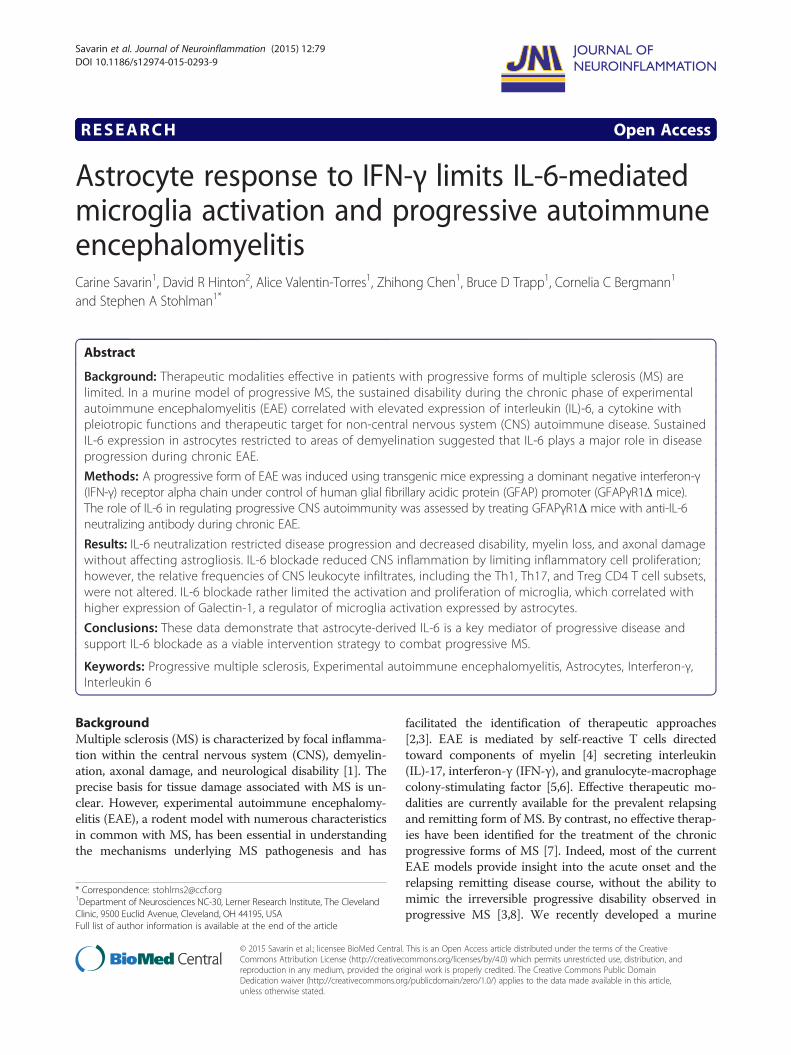
RESEARCH Open Access Astrocyte response to IFN-γ limits IL-6-mediated microglia activation and progressive autoimmune encephalomyelitis Carine Savarin 1 , David R Hinton 2 , Alice Valentin-Torres 1 , Zhihong Chen 1 , Bruce D Trapp 1 , Cornelia C Bergmann 1 and Stephen A Stohlman 1* Abstract Background: Therapeutic modalities effective in patients with progressive forms of multiple sclerosis (MS) are limited. In a murine model of progressive MS, the sustained disability during the chronic phase of experimental autoimmune encephalomyelitis (EAE) correlated with elevated expression of interleukin (IL)-6, a cytokine with pleiotropic functions and therapeutic target for non-central nervous system (CNS) autoimmune disease. Sustained IL-6 expression in astrocytes restricted to areas of demyelination suggested that IL-6 plays a major role in disease progression during chronic EAE. Methods: A progressive form of EAE was induced using transgenic mice expressing a dominant negative interferon-γ (IFN-γ) receptor alpha chain under control of human glial fibrillary acidic protein (GFAP) promoter (GFAPγR1Δ mice). The role of IL-6 in regulating progressive CNS autoimmunity was assessed by treating GFAPγR1Δ mice with anti-IL-6 neutralizing antibody during chronic EAE. Results: IL-6 neutralization restricted disease progression and decreased disability, myelin loss, and axonal damage without affecting astrogliosis. IL-6 blockade reduced CNS inflammation by limiting inflammatory cell proliferation; however, the relative frequencies of CNS leukocyte infiltrates, including the Th1, Th17, and Treg CD4 T cell subsets, were not altered. IL-6 blockade rather limited the activation and proliferation of microglia, which correlated with higher expression of Galectin-1, a regulator of microglia activation expressed by astrocytes. Conclusions: These data demonstrate that astrocyte-derived IL-6 is a key mediator of progressive disease and support IL-6 blockade as a viable intervention strategy to combat progressive MS. Keywords: Progressive multiple sclerosis, Experimental autoimmune encephalomyelitis, Astrocytes, Interferon-γ, Interleukin 6 Background Multiple sclerosis (MS) is characterized by focal inflamma- tion within the central nervous system (CNS), demyelin- ation, axonal damage, and neurological disability [1]. The precise basis for tissue damage associated with MS is un- clear. However, experimental autoimmune encephalomy- elitis (EAE), a rodent model with numerous characteristics in common with MS, has been essential in understanding the mechanisms underlying MS pathogenesis and has facilitated the identification of therapeutic approaches [2,3]. EAE is mediated by self-reactive T cells directed toward components of myelin [4] secreting interleukin (IL)-17, interferon-γ (IFN-γ), and granulocyte-macrophage colony-stimulating factor [5,6]. Effective therapeutic mo- dalities are currently available for the prevalent relapsing and remitting form of MS. By contrast, no effective therap- ies have been identified for the treatment of the chronic progressive forms of MS [7]. Indeed, most of the current EAE models provide insight into the acute onset and the relapsing remitting disease course, without the ability to mimic the irreversible progressive disability observed in progressive MS [3,8]. We recently developed a murine * Correspondence: [email protected] 1 Department of Neurosciences NC-30, Lerner Research Institute, The Cleveland Clinic, 9500 Euclid Avenue, Cleveland, OH 44195, USA Full list of author information is available at the end of the article JOURNAL OF NEUROINFLAMMATION © 2015 Savarin et al.; licensee BioMed Central. This is an Open Access article distributed under the terms of the Creative Commons Attribution License (http://creativecommons.org/licenses/by/4.0) which permits unrestricted use, distribution, and reproduction in any medium, provided the original work is properly credited. The Creative Commons Public Domain Dedication waiver (http://creativecommons.org/publicdomain/zero/1.0/) applies to the data made available in this article, unless otherwise stated. Savarin et al. Journal of Neuroinflammation (2015) 12:79 DOI 10.1186/s12974-015-0293-9
Transcript of Astrocyte response to IFN-γ limits IL-6-mediated microglia ...
JOURNAL OF NEUROINFLAMMATION
Savarin et al. Journal of Neuroinflammation (2015) 12:79 DOI 10.1186/s12974-015-0293-9
RESEARCH Open Access
Astrocyte response to IFN-γ limits IL-6-mediatedmicroglia activation and progressive autoimmuneencephalomyelitisCarine Savarin1, David R Hinton2, Alice Valentin-Torres1, Zhihong Chen1, Bruce D Trapp1, Cornelia C Bergmann1
and Stephen A Stohlman1*
Abstract
Background: Therapeutic modalities effective in patients with progressive forms of multiple sclerosis (MS) arelimited. In a murine model of progressive MS, the sustained disability during the chronic phase of experimentalautoimmune encephalomyelitis (EAE) correlated with elevated expression of interleukin (IL)-6, a cytokine withpleiotropic functions and therapeutic target for non-central nervous system (CNS) autoimmune disease. SustainedIL-6 expression in astrocytes restricted to areas of demyelination suggested that IL-6 plays a major role in diseaseprogression during chronic EAE.
Methods: A progressive form of EAE was induced using transgenic mice expressing a dominant negative interferon-γ(IFN-γ) receptor alpha chain under control of human glial fibrillary acidic protein (GFAP) promoter (GFAPγR1Δ mice).The role of IL-6 in regulating progressive CNS autoimmunity was assessed by treating GFAPγR1Δ mice with anti-IL-6neutralizing antibody during chronic EAE.
Results: IL-6 neutralization restricted disease progression and decreased disability, myelin loss, and axonal damagewithout affecting astrogliosis. IL-6 blockade reduced CNS inflammation by limiting inflammatory cell proliferation;however, the relative frequencies of CNS leukocyte infiltrates, including the Th1, Th17, and Treg CD4 T cell subsets,were not altered. IL-6 blockade rather limited the activation and proliferation of microglia, which correlated withhigher expression of Galectin-1, a regulator of microglia activation expressed by astrocytes.
Conclusions: These data demonstrate that astrocyte-derived IL-6 is a key mediator of progressive disease andsupport IL-6 blockade as a viable intervention strategy to combat progressive MS.
Keywords: Progressive multiple sclerosis, Experimental autoimmune encephalomyelitis, Astrocytes, Interferon-γ,Interleukin 6
BackgroundMultiple sclerosis (MS) is characterized by focal inflamma-tion within the central nervous system (CNS), demyelin-ation, axonal damage, and neurological disability [1]. Theprecise basis for tissue damage associated with MS is un-clear. However, experimental autoimmune encephalomy-elitis (EAE), a rodent model with numerous characteristicsin common with MS, has been essential in understandingthe mechanisms underlying MS pathogenesis and has
* Correspondence: [email protected] of Neurosciences NC-30, Lerner Research Institute, The ClevelandClinic, 9500 Euclid Avenue, Cleveland, OH 44195, USAFull list of author information is available at the end of the article
© 2015 Savarin et al.; licensee BioMed CentralCommons Attribution License (http://creativecreproduction in any medium, provided the orDedication waiver (http://creativecommons.orunless otherwise stated.
facilitated the identification of therapeutic approaches[2,3]. EAE is mediated by self-reactive T cells directedtoward components of myelin [4] secreting interleukin(IL)-17, interferon-γ (IFN-γ), and granulocyte-macrophagecolony-stimulating factor [5,6]. Effective therapeutic mo-dalities are currently available for the prevalent relapsingand remitting form of MS. By contrast, no effective therap-ies have been identified for the treatment of the chronicprogressive forms of MS [7]. Indeed, most of the currentEAE models provide insight into the acute onset and therelapsing remitting disease course, without the ability tomimic the irreversible progressive disability observed inprogressive MS [3,8]. We recently developed a murine
. This is an Open Access article distributed under the terms of the Creativeommons.org/licenses/by/4.0) which permits unrestricted use, distribution, andiginal work is properly credited. The Creative Commons Public Domaing/publicdomain/zero/1.0/) applies to the data made available in this article,
Savarin et al. Journal of Neuroinflammation (2015) 12:79 Page 2 of 14
model of progressiveMS using GFAPγR1Δ transgenic micein which astrocytes are deficient in IFN-γ signaling charac-terized by prolonged disability and increased mortality [9].Several pathological features of progressive MS, includingincreased demyelination, axonal loss, and astrogliosis, wereobserved in GFAPγR1Δ mice with chronic EAE [9]. More-over, the progressive disability and sustained demyelinationcorrelated with elevated expression of IL-6, a cytokine withpleiotropic functions and a therapeutic target for non-CNSautoimmune diseases, such as rheumatoid arthritis [10].Inhibition of IL-6 signaling is also effective in several mur-ine models of inflammatory diseases, including colitis,asthma, and cancer [11-13].IL-6, an activator of acute phase responses, is expressed
during all forms of inflammation [14], including withinthe CNS of MS patients [15] and animals with acute EAE[16]. IL-6 is crucial in the induction of CNS autoimmuneattack by promoting peripheral induction of self-reactiveT cells, facilitating their recruitment into the CNS andlimiting the regulatory T cell (Treg) response. Mice lack-ing IL-6 are resistant to EAE [17-19], consistent with theabsence of inflammatory cells within the CNS paren-chyma [18,19] and its essential role in T cell activation[20]. IL-6 also interacts with CNS endothelial cells result-ing in increased vascular cell adhesion molecule-1(VCAM-1) expression, a prerequisite for CNS entry ofintegrin α4β1+ effector T cells [21]. In addition to facili-tating CNS entry of autoimmune T cells, IL-6 is requiredfor activation of encephalitogenic Th17 cells [22] andenhances acute inflammation by suppressing expressionof the Foxp3 transcription factor [23], thereby limitingthe suppressive activity of Treg. In addition to a pro-inflammatory role in the context of acute inflammation,IL-6 also exhibits a variety of functions within the CNSeither totally or partially independent of its role in in-flammation. For example, it is associated with protectiveCNS functions by promoting neuronal survival and re-generation [24,25], as well as inducing oligodendrocytedifferentiation, thereby facilitating myelin repair [26].Astrocytes are the primary cells secreting IL-6 in the
CNS of both patients with MS and in rodents with acuteEAE [27,28]. Targeted inhibition of NF-κB in astrocytesis associated with EAE recovery by reducing inflamma-tion and increasing both IL-6 expression [29] andremyelination [30]. These data suggest a protective rolefor IL-6 during CNS autoimmunity. Nevertheless, con-stitutive IL-6 expression by astrocytes results in neur-onal damage and astrogliosis [31]. Similarly, inhibitionof IFN-γ signaling to astrocytes results in a progressiveform of EAE and increased IL-6 [9], supporting a dele-terious function of IL-6 during progressive disease.Therefore, the role of IL-6 during the remission stagesof CNS autoimmunity or during progressive disease re-mains unclear.
Astrocytes, in addition to secreting IL-6 during bothMS and acute EAE, also actively regulate microglia atmultiple levels during both pro-inflammatory injury andrepair. Astrocytes attenuate microglial activation, reducemicroglial secretion of both pro- and anti-inflammatorycytokines, as well as limit microglial-supported T cellproliferation in vitro [32]. Specific gene deletions inastrocytes confirm they both attenuate and activatemicroglia in vivo during EAE. For example, Galectin-1secretion by astrocytes limits microglial activation andclinical symptoms [33]. Similarly, expression of a domin-ant negative NF-κB repressor in astrocytes diminishesclinical EAE associated with attenuated tumor necrosisfactor (TNF) secretion by microglia [29]. By contrast,inhibiting astrocyte IFN-γ signaling sustains disabilityand correlates with increased TNF and IL-6 [9]. Thus,astrocytes, which play critical roles in both acute andchronic CNS injury, respond to changes by actively shapingthe environment and influence repair by altering the de-structive or protective state of microglia.The role of IL-6 in regulating progressive CNS auto-
immunity was examined in the transgenic GFAPγR1Δmouse model of progressive EAE [34]. Similar to acuteEAE, neither the inability of the astrocytes to respond toIFN-γ nor the progressive clinical phenotype altered thepredominant expression of IL-6 by astrocytes. BlockingIL-6 activity limited progression of clinical symptoms,promoted clinical recovery, and restrained the extent ofdemyelination and axonal damage. However, neither thecomposition of leukocyte population nor their predom-inant cytokine secretion patterns were altered. IL-6neutralization reduced the frequency of both activatedmacrophages and microglia, but selectively decreasedthe sustained proliferation of microglia associated withprogressive disease, suggesting a differential effect onmicroglia versus macrophages. These data indicate thatsustained IL-6 secretion by astrocytes during progressivedisease is deleterious to both clinical disability and tissuedamage and that these affects may be mediated indirectlyvia microglia. IL-6 blockade may thus provide a viableintervention approach in secondary progressive or pri-mary progressive MS, for which few therapeutic modal-ities are currently available.
Materials and methodsMiceHomozygous H-2b GFAP/IFN-γR1ΔIC (GFAPγR1Δ) trans-genic mice [34], expressing a dominant negative IFN-γ re-ceptor alpha chain under control of human glial fibrillaryacidic protein (GFAP) promoter, were bred locally. C57BL/6(H-2b) wild-type (WT) mice were purchased from theNational Cancer Institute (Frederick, MD, USA). All proce-dures were performed in compliance with protocol number
Savarin et al. Journal of Neuroinflammation (2015) 12:79 Page 3 of 14
1165 approved by the Cleveland Clinic Institutional AnimalCare and Use Committee.
Experimental autoimmune encephalomyelitisEAE was induced by subcutaneous injection of 300 μgmyelin oligodendrocyte glycoprotein (MOG)35–55 peptideemulsified in phosphate-buffered saline (PBS) and incom-plete Freund’s adjuvant (IFA; Sigma-Aldrich, St. Louis,MO, USA) supplemented with 5 mg/ml Mycobacteriumtuberculosis, strain H37Ra (Difco, Detroit, MI, USA), aspreviously described [9]. Mice were immunized with200 μl emulsion distributed subcutaneously over two flanksites. One hundred nanograms of pertussis toxin (Sigma-Aldrich) was injected intraperitoneally (i.p.) on the day ofinitial immunization and 48 h later. Animals were scoreddaily in a blinded fashion for clinical symptoms as follows:0 = no signs of disease; 1 = flaccid tail or hind limb weak-ness; 2 = flaccid tail and hind limb weakness, loss of right-ing reflex; 3 = partial hind limb paralysis; 4 = completehind limb paralysis; 5 =moribund or dead.
Anti-IL-6 treatmentThe MP5-20 F3 hybridoma secreting a rat IgG2a IL-6neutralizing monoclonal antibody (mAb) and the con-trol GL-113 hybridoma secreting a rat IgG1 mAb spe-cific for β-galactosidase (β-gal) were originally obtainedfrom Dr. Robert Coffman (DNAX Corp, Palo Alto, CA,USA). Hybridomas were adapted to BD Cell Serum-Free Medium (BD, Bedford MA, USA) and the serum-free mAb collected after propagation of the hybridomain a BD CELLine device. Ig concentrations were deter-mined by optical density at 480 nM, diluted to 1 mg/mlin endotoxin-free PBS, and stored at −20°C until use.Mice were divided into two groups of equivalent averageclinical scores and injected i.p. with 500 μg mAb on thespecified days post MOG35–55 peptide immunization.
Isolation of CNS-derived cells and flow cytometryMice were perfused with ice-cold PBS before the brainand spinal cord were harvested and homogenized inDulbecco’s PBS using Tenbroeck tissue grinders (KimbleChase, Vineland, NJ, USA). Mononuclear cells were con-centrated from the homogenates by centrifugation at450 × g for 7 min at 4°C. Cell pellets were resuspended inRPMI 1640 medium supplemented with 25 mM HEPES(pH 7.2) and adjusted to 30% Percoll (Pharmacia, Uppsala,Sweden). A 70% Percoll underlay was added prior tocentrifugation at 800 × g for 30 min at 4°C. Cells were re-covered from the 30%/70% interface, washed with RPMI,and then incubated for 10 min on ice in fluorescence acti-vated cell sorting (FACS) buffer with mouse serum andanti-CD16/CD32 mAb (clone 2.4G2, BD Biosciences, SanDiego, CA, USA) to limit unspecific binding. FITC-, PE-,PerCP-, and APC-conjugated surface markers (all from
BD Biosciences unless specified), including CD45 (30-F11), CD4 (GK1.5), CD11b (clone m1/70), I-A/I-E (clone2G9), and F4/80 (Serotec, Raleigh, NC, USA), were thenadded and cells incubated for 30 min on ice. Cells werewashed with FACS buffer prior to analysis. For intracellu-lar staining, CNS-derived cells were stimulated for 6 hwith phorbol 12-myristate 13-acetate (PMA) (10 ng/ml)(Acros Organics, Geel, Belgium) and ionomycin (1 μM)(Calbiochem, Spring Valley, CA, USA), with Monensin(2 μM) (Calbiochem) added for the last 2 h. Followingstimulation, surface molecules were detected as describedabove. Cells were permeabilized using Cytofix/Cytopermsolution (BD Biosciences) and incubated for 30 min on icewith fluorescent mAb specific for IFN-γ (XMG1.2; BDBiosciences), IL-17 (TC11-18H10; BD Biosciences), orFoxp3 (FJK-16 s; eBiosciences). Cells were then washedusing Perm/Wash buffer according to the manufacturer’sinstructions. For proliferation, 1 mg of bromodeoxyuridine(BrdU) (BD Biosciences) in PBS was administrated i.p.24 h prior to sacrifice. Mononuclear cells were preparedfrom the CNS as described above, stained for surface mol-ecules, and subsequently stained for intranuclear BrdU ac-cording to the manufacturer’s instructions using the FITCBrdU flow kit (BD Biosciences). Data were acquired on aFACSCalibur flow cytometer (BD Biosciences) and ana-lyzed using FlowJo software (TreeStar Inc., Ashland, OR,USA).
Real-time PCRFollowing PBS perfusion, snap-frozen brains were placedinto TRIzol (Invitrogen, Grand Island, NY, USA) andhomogenized using a TissueLyser with stainless beads(Qiagen, Valencia, CA, USA). RNA extraction was per-formed according to the manufacturer’s instructionsfollowed by DNase I (Ambion, Austin, TX, USA) treat-ment for 30 min at 37°C. cDNA was then synthetizedusing M-MLV Reverse Transcriptase (Invitrogen), oligo-dT primers (20 μM) (Promega, Madison, WI, USA), andrandom primers (20 μM) (Promega). Gene expressionanalysis was performed by quantitative real-time PCR usinga 7500 Fast real-time PCR system (Applied Biosystems,Foster City, CA, USA), SYBR Green Master Mix (AppliedBiosystems), and the following primers: GAPDH: F: 5′-TGCACCACCAACTGCTTAG-3′, R: 5′-GGATGCAGGGATGATGTTC-3′; GFAP: F: 5′- CAGTGTGTCAGCCCCACTGA-3′, R: 5′- CAGTGTGTCAGCCCCACTGA-3′.Galectin-1 and heme oxygenase-1 mRNA levels were ana-lyzed using TaqMan primers and 2X Universal TaqManFast Master Mix (Applied Biosystems). Transcript levelswere normalized to the housekeeping gene GAPDH andconverted to a linearized value using the following formula:
2 CTGAPDH− CT
Geneð Þ � 1; 000, where CT represents the thresh-old cycle value.
Savarin et al. Journal of Neuroinflammation (2015) 12:79 Page 4 of 14
HistopathologyMice were perfused with ice-cold PBS and spinal cordsfixed in Zn Formalin. Spinal cords were separated intosix sections, two of each corresponding to the cervical,thoracic, and lumbar regions and embedded in paraffin.Cross sections (6 μm) were stained with hematoxylinand eosin (H&E) or luxol fast blue (LFB). For identifi-cation of activated astrocytes and axonal damage, sec-tions were incubated with mouse anti-GFAP (AbCam,Cambridge, MA, USA), SMI-31/SMI-32 (StenbergerMonoclonals Inc., Lutherville, MD, USA), or anti-amyloidprecursor protein (APP) mAb (Millipore, Billerica, MA,USA) overnight at 4°C. Biotinylated goat anti-mouse Abwas added for 1 h at room temperature (RT). Sectionswere then incubated with Vectastain ABC kit (VectorLaboratories, Burlingame, CA, USA) and 3,3′-diamino-benzidine (Sigma-Aldrich). For IL-6 detection, heat-mediated antigen retrieval was performed in 0.1 Mcitrate buffer, pH 6.0. Sections were then incubated in0.3% H2O2 for 20 min followed by rabbit anti-IL-6 Ab(AbCam) overnight at RT. Biotinylated anti-rabbit Abwas added for 30 min at RT. Staining was revealed withVectastain ABC kit and 3,3′-diaminobenzidine. Stainedtissue sections of all six levels on individual glass slideswere scanned with an Aperio ScanScope (Aperio, Vista,CA, USA) at 40× and digitally imaged at high resolution.Aperio software was used to quantify areas of demyelin-ation. Axonal damage was quantitated by counting thenumber of APP+ axons in lesions at all six levels and nor-malized to 100 μm2 of demyelinated lesion area.
Confocal microscopyMice were anesthetized and perfused with 4% parafor-maldehyde (PFA) in PBS. Spinal cords were removed,separated into portions as described above, and post-fixed in 4% PFA for additional 24 h. Tissues were cryo-protected in 20% glycerol for 24 h before 30-μm free-floating sections were prepared with a sliding microtome(Leica Microsystems, Wetzlar, Germany). Double immu-nolabeling was performed as previously described [35].Briefly, sections were microwaved in 0.01 M citratebuffer (pH 6.0) followed by pretreatment with 1% TritonX in PBS. Sections were blocked with PBS containing3% normal goat serum and 0.01% Triton X for 30 minand incubated with primary Ab diluted in blocking reagentovernight at 4°C (rabbit anti-GFAP, Dako, Carpenteria, CA,USA, 1:1,000; mouse anti-Iba-1, CCF Hybridoma Core,Cleveland, OH, USA, 1:250; goat anti-IL-6, R&D Systems,Minneapolis, MN, USA, 1:20). To confirm specificity, pri-mary Abs were omitted on adjacent sections. The sectionswere washed and incubated with species-specific secondaryAbs conjugated to FITC or Cy5 (Jackson ImmunoResearch,West Grove, PA, USA) for 2 h at RT. Sections were rinsed,mounted with VECTASHIELD (Vector Labs, Burlingame,
CA, USA) and examined on a Leica TCS confocal micro-scope (Leica Microsystems). Images were analyzed off-line with Volocity software version 6.1.2 (PerkinElmer,Waltham, MA, USA).
Statistical analysisData represent the mean ± SEM and significance was de-termined by two-tailed Student’s t test or ANOVA withBonferroni post-test. A value of P < 0.05 was consideredstatistically significant. Graphs were plotted using Graph-Pad Prism 4.0c software.
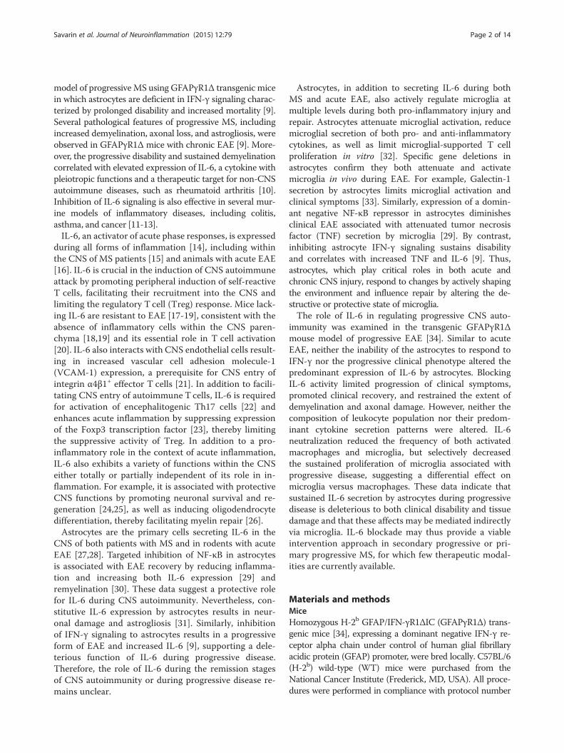
ResultsAstrocytes secrete IL-6 during progressive EAEAstrocytes are the predominant source of IL-6 in theCNS of patients with MS and mice with acute EAE [27],and during acute EAE, its expression is independent ofthe ability of astrocytes to respond to IFN-γ [9]. By con-trast, progressive EAE in GFAPγR1Δ mice, in which theastrocytes are unable to respond to IFN-γ, correlatedwith sustained IL-6 mRNA expression [9]. Its adverserole within the CNS [36] suggested that IL-6 may con-tribute to progressive disease. As numerous cell types in-cluding myeloid cells and microglia can secrete IL-6, wedetermined if the inability of astrocytes to respond toIFN-γ signaling altered the cellular source or distribu-tion of IL-6 during acute and chronic/progressive dis-ease. The majority of cells secreting IL-6 during acuteEAE in both WT and GFAPγR1Δ mice co-express GFAP,confirming their identity as astrocytes (Figure 1A). Bycontrast, during acute EAE in both groups, IL-6 expres-sion was undetectable in Iba-1+ cells (Figure 1B). Thesedata demonstrate that the predominant source of IL-6during acute EAE is not altered by the inability of astro-cytes to respond to IFN-γ. Neither the cell type(s) se-creting IL-6 nor their anatomical location relative tothe white matter lesions has been established during ei-ther chronic or progressive forms of EAE. In contrastto WT mice, where astrogliosis is diminished duringchronic EAE and limited to white matter areas, astro-cyte activation is sustained during progressive diseasein GFAPγR1Δ mice, including in gray matter distalfrom the demyelinating lesions [9]. Although the num-ber of cells secreting IL-6 in the absence of IFN-γ sig-naling to astrocytes was increased relative to WT mice,IL-6+ cells in mice with progressive EAE were locatedwithin white matter areas (Figure 1C), similar to thedistribution in WT mice. Moreover, IL-6 remained un-detectable in Iba-1+ cells (data not shown) and co-localized mainly with GFAP+ cells located within or ad-jacent to areas of myelin loss (Figure 1D), supportingastrocytes as the primary source of IL-6 during bothacute and chronic EAE. Although IFN-γ signaling limitsastrocyte activation to areas of demyelination, IL-6
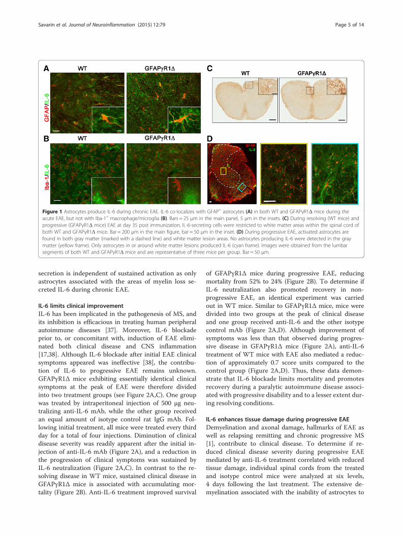
Figure 1 Astrocytes produce IL-6 during chronic EAE. IL-6 co-localizes with GFAP+ astrocytes (A) in both WT and GFAPγR1Δ mice during theacute EAE, but not with Iba-1+ macrophage/microglia (B). Bars = 25 μm in the main panel, 5 μm in the insets. (C) During resolving (WT mice) andprogressive (GFAPγR1Δ mice) EAE at day 35 post immunization, IL-6-secreting cells were restricted to white matter areas within the spinal cord ofboth WT and GFAPγR1Δ mice. Bar = 200 μm in the main figure, bar = 50 μm in the inset. (D) During progressive EAE, activated astrocytes arefound in both gray matter (marked with a dashed line) and white matter lesion areas. No astrocytes producing IL-6 were detected in the graymatter (yellow frame). Only astrocytes in or around white matter lesions produced IL-6 (cyan frame). Images were obtained from the lumbarsegments of both WT and GFAPγR1Δ mice and are representative of three mice per group. Bar = 50 μm.
Savarin et al. Journal of Neuroinflammation (2015) 12:79 Page 5 of 14
secretion is independent of sustained activation as onlyastrocytes associated with the areas of myelin loss se-creted IL-6 during chronic EAE.
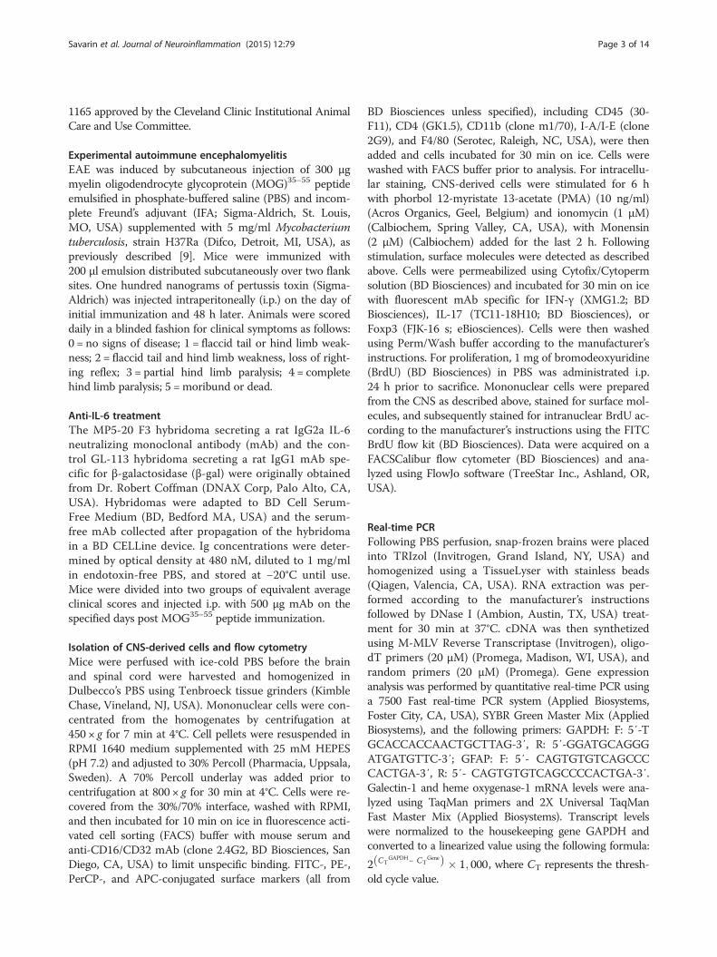
IL-6 limits clinical improvementIL-6 has been implicated in the pathogenesis of MS, andits inhibition is efficacious in treating human peripheralautoimmune diseases [37]. Moreover, IL-6 blockadeprior to, or concomitant with, induction of EAE elimi-nated both clinical disease and CNS inflammation[17,38]. Although IL-6 blockade after initial EAE clinicalsymptoms appeared was ineffective [38], the contribu-tion of IL-6 to progressive EAE remains unknown.GFAPγR1Δ mice exhibiting essentially identical clinicalsymptoms at the peak of EAE were therefore dividedinto two treatment groups (see Figure 2A,C). One groupwas treated by intraperitoneal injection of 500 μg neu-tralizing anti-IL-6 mAb, while the other group receivedan equal amount of isotype control rat IgG mAb. Fol-lowing initial treatment, all mice were treated every thirdday for a total of four injections. Diminution of clinicaldisease severity was readily apparent after the initial in-jection of anti-IL-6 mAb (Figure 2A), and a reduction inthe progression of clinical symptoms was sustained byIL-6 neutralization (Figure 2A,C). In contrast to the re-solving disease in WT mice, sustained clinical disease inGFAPγR1Δ mice is associated with accumulating mor-tality (Figure 2B). Anti-IL-6 treatment improved survival
of GFAPγR1Δ mice during progressive EAE, reducingmortality from 52% to 24% (Figure 2B). To determine ifIL-6 neutralization also promoted recovery in non-progressive EAE, an identical experiment was carriedout in WT mice. Similar to GFAPγR1Δ mice, mice weredivided into two groups at the peak of clinical diseaseand one group received anti-IL-6 and the other isotypecontrol mAb (Figure 2A,D). Although improvement ofsymptoms was less than that observed during progres-sive disease in GFAPγR1Δ mice (Figure 2A), anti-IL-6treatment of WT mice with EAE also mediated a reduc-tion of approximately 0.7 score units compared to thecontrol group (Figure 2A,D). Thus, these data demon-strate that IL-6 blockade limits mortality and promotesrecovery during a paralytic autoimmune disease associ-ated with progressive disability and to a lesser extent dur-ing resolving conditions.
IL-6 enhances tissue damage during progressive EAEDemyelination and axonal damage, hallmarks of EAE aswell as relapsing remitting and chronic progressive MS[1], contribute to clinical disease. To determine if re-duced clinical disease severity during progressive EAEmediated by anti-IL-6 treatment correlated with reducedtissue damage, individual spinal cords from the treatedand isotype control mice were analyzed at six levels,4 days following the last treatment. The extensive de-myelination associated with the inability of astrocytes to
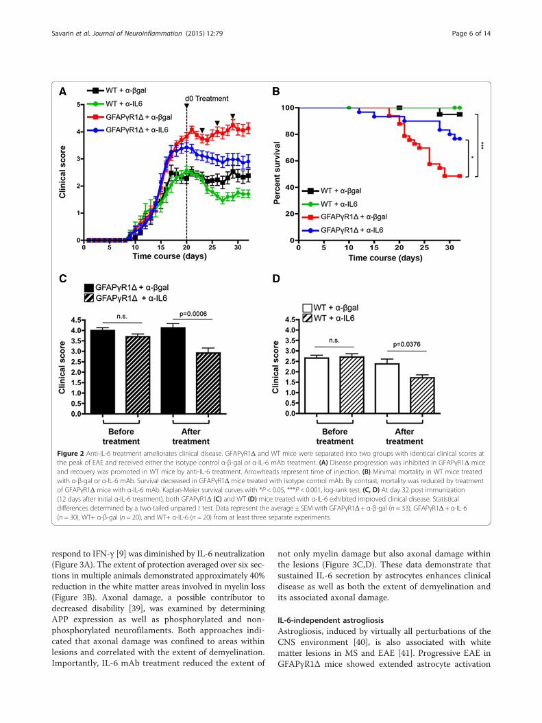
Figure 2 Anti-IL-6 treatment ameliorates clinical disease. GFAPγR1Δ and WT mice were separated into two groups with identical clinical scores atthe peak of EAE and received either the isotype control α-β-gal or α-IL-6 mAb treatment. (A) Disease progression was inhibited in GFAPγR1Δ miceand recovery was promoted in WT mice by anti-IL-6 treatment. Arrowheads represent time of injection. (B) Minimal mortality in WT mice treatedwith α-β-gal or α-IL-6 mAb. Survival decreased in GFAPγR1Δ mice treated with isotype control mAb. By contrast, mortality was reduced by treatmentof GFAPγR1Δ mice with α-IL-6 mAb. Kaplan-Meier survival curves with *P < 0.05, ***P < 0.001, log-rank test. (C, D) At day 32 post immunization(12 days after initial α-IL-6 treatment), both GFAPγR1Δ (C) and WT (D) mice treated with α-IL-6 exhibited improved clinical disease. Statisticaldifferences determined by a two-tailed unpaired t test. Data represent the average ± SEM with GFAPγR1Δ + α-β-gal (n = 33), GFAPγR1Δ + α-IL-6(n = 30), WT+ α-β-gal (n = 20), and WT+ α-IL-6 (n = 20) from at least three separate experiments.
Savarin et al. Journal of Neuroinflammation (2015) 12:79 Page 6 of 14
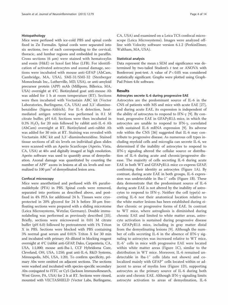
respond to IFN-γ [9] was diminished by IL-6 neutralization(Figure 3A). The extent of protection averaged over six sec-tions in multiple animals demonstrated approximately 40%reduction in the white matter areas involved in myelin loss(Figure 3B). Axonal damage, a possible contributor todecreased disability [39], was examined by determiningAPP expression as well as phosphorylated and non-phosphorylated neurofilaments. Both approaches indi-cated that axonal damage was confined to areas withinlesions and correlated with the extent of demyelination.Importantly, IL-6 mAb treatment reduced the extent of
not only myelin damage but also axonal damage withinthe lesions (Figure 3C,D). These data demonstrate thatsustained IL-6 secretion by astrocytes enhances clinicaldisease as well as both the extent of demyelination andits associated axonal damage.
IL-6-independent astrogliosisAstrogliosis, induced by virtually all perturbations of theCNS environment [40], is also associated with whitematter lesions in MS and EAE [41]. Progressive EAE inGFAPγR1Δ mice showed extended astrocyte activation
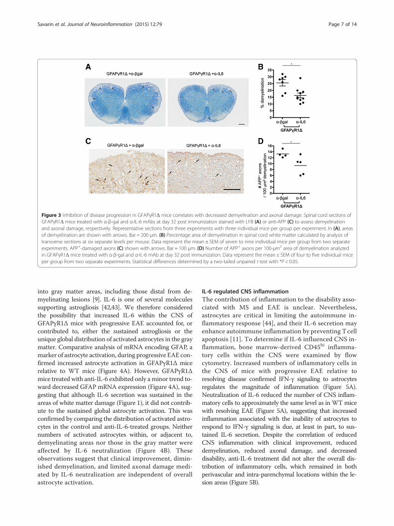
Figure 3 Inhibition of disease progression in GFAPγR1Δ mice correlates with decreased demyelination and axonal damage. Spinal cord sections ofGFAPγR1Δ mice treated with α-β-gal and α-IL-6 mAbs at day 32 post immunization stained with LFB (A) or anti-APP (C) to assess demyelinationand axonal damage, respectively. Representative sections from three experiments with three individual mice per group per experiment. In (A), areasof demyelination are shown with arrows. Bar = 200 μm. (B) Percentage area of demyelination in spinal cord white matter calculated by analysis oftransverse sections at six separate levels per mouse. Data represent the mean ± SEM of seven to nine individual mice per group from two separateexperiments. APP+-damaged axons (C) shown with arrows. Bar = 100 μm. (D) Number of APP+ axons per 100-μm2 area of demyelination analyzedin GFAPγR1Δ mice treated with α-β-gal and α-IL-6 mAb at day 32 post immunization. Data represent the mean ± SEM of four to five individual miceper group from two separate experiments. Statistical differences determined by a two-tailed unpaired t test with *P < 0.05.
Savarin et al. Journal of Neuroinflammation (2015) 12:79 Page 7 of 14
into gray matter areas, including those distal from de-myelinating lesions [9]. IL-6 is one of several moleculessupporting astrogliosis [42,43]. We therefore consideredthe possibility that increased IL-6 within the CNS ofGFAPγR1Δ mice with progressive EAE accounted for, orcontributed to, either the sustained astrogliosis or theunique global distribution of activated astrocytes in the graymatter. Comparative analysis of mRNA encoding GFAP, amarker of astrocyte activation, during progressive EAE con-firmed increased astrocyte activation in GFAPγR1Δ micerelative to WT mice (Figure 4A). However, GFAPγR1Δmice treated with anti-IL-6 exhibited only a minor trend to-ward decreased GFAP mRNA expression (Figure 4A), sug-gesting that although IL-6 secretion was sustained in theareas of white matter damage (Figure 1), it did not contrib-ute to the sustained global astrocyte activation. This wasconfirmed by comparing the distribution of activated astro-cytes in the control and anti-IL-6-treated groups. Neithernumbers of activated astrocytes within, or adjacent to,demyelinating areas nor those in the gray matter wereaffected by IL-6 neutralization (Figure 4B). Theseobservations suggest that clinical improvement, dimin-ished demyelination, and limited axonal damage medi-ated by IL-6 neutralization are independent of overallastrocyte activation.
IL-6 regulated CNS inflammationThe contribution of inflammation to the disability asso-ciated with MS and EAE is unclear. Nevertheless,astrocytes are critical in limiting the autoimmune in-flammatory response [44], and their IL-6 secretion mayenhance autoimmune inflammation by preventing Tcellapoptosis [11]. To determine if IL-6 influenced CNS in-flammation, bone marrow-derived CD45hi inflamma-tory cells within the CNS were examined by flowcytometry. Increased numbers of inflammatory cells inthe CNS of mice with progressive EAE relative toresolving disease confirmed IFN-γ signaling to astrocytesregulates the magnitude of inflammation (Figure 5A).Neutralization of IL-6 reduced the number of CNS inflam-matory cells to approximately the same level as in WT micewith resolving EAE (Figure 5A), suggesting that increasedinflammation associated with the inability of astrocytes torespond to IFN-γ signaling is due, at least in part, to sus-tained IL-6 secretion. Despite the correlation of reducedCNS inflammation with clinical improvement, reduceddemyelination, reduced axonal damage, and decreaseddisability, anti-IL-6 treatment did not alter the overall dis-tribution of inflammatory cells, which remained in bothperivascular and intra-parenchymal locations within the le-sion areas (Figure 5B).
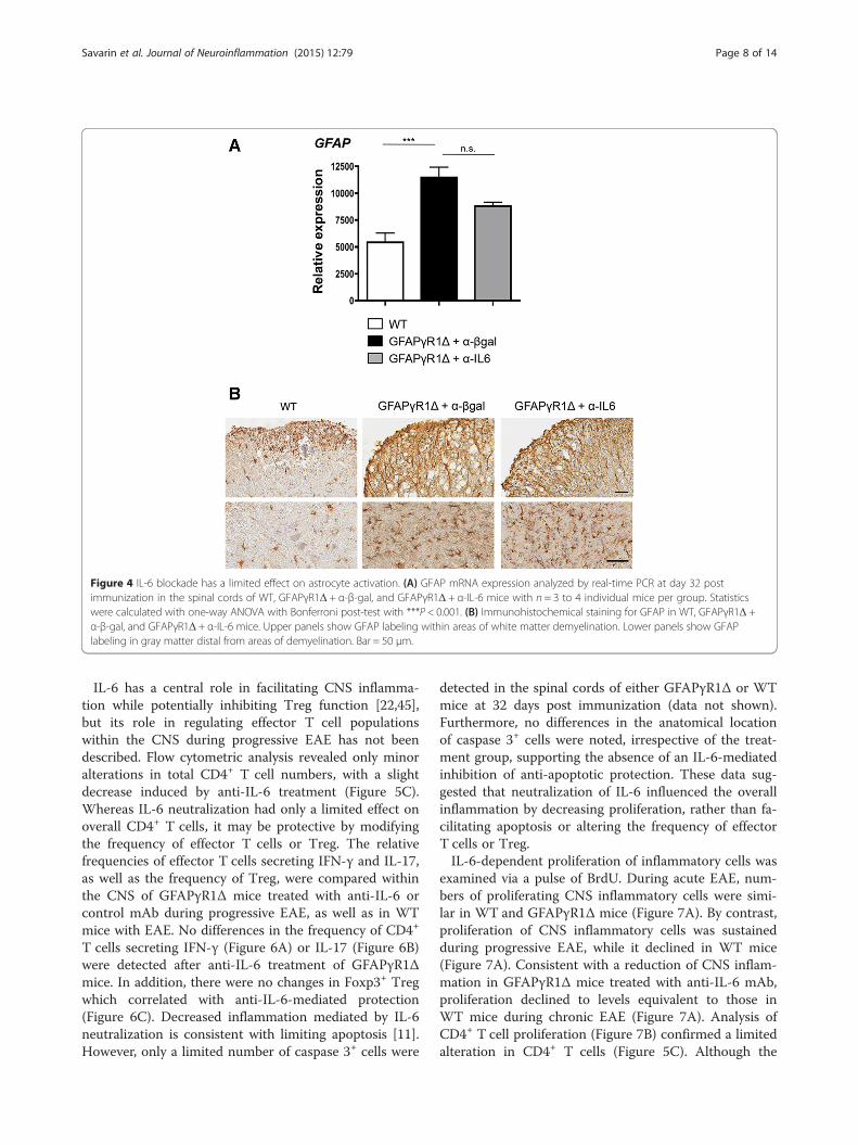
Figure 4 IL-6 blockade has a limited effect on astrocyte activation. (A) GFAP mRNA expression analyzed by real-time PCR at day 32 postimmunization in the spinal cords of WT, GFAPγR1Δ + α-β-gal, and GFAPγR1Δ + α-IL-6 mice with n = 3 to 4 individual mice per group. Statisticswere calculated with one-way ANOVA with Bonferroni post-test with ***P < 0.001. (B) Immunohistochemical staining for GFAP in WT, GFAPγR1Δ +α-β-gal, and GFAPγR1Δ + α-IL-6 mice. Upper panels show GFAP labeling within areas of white matter demyelination. Lower panels show GFAPlabeling in gray matter distal from areas of demyelination. Bar = 50 μm.
Savarin et al. Journal of Neuroinflammation (2015) 12:79 Page 8 of 14
IL-6 has a central role in facilitating CNS inflamma-tion while potentially inhibiting Treg function [22,45],but its role in regulating effector T cell populationswithin the CNS during progressive EAE has not beendescribed. Flow cytometric analysis revealed only minoralterations in total CD4+ T cell numbers, with a slightdecrease induced by anti-IL-6 treatment (Figure 5C).Whereas IL-6 neutralization had only a limited effect onoverall CD4+ T cells, it may be protective by modifyingthe frequency of effector T cells or Treg. The relativefrequencies of effector T cells secreting IFN-γ and IL-17,as well as the frequency of Treg, were compared withinthe CNS of GFAPγR1Δ mice treated with anti-IL-6 orcontrol mAb during progressive EAE, as well as in WTmice with EAE. No differences in the frequency of CD4+
T cells secreting IFN-γ (Figure 6A) or IL-17 (Figure 6B)were detected after anti-IL-6 treatment of GFAPγR1Δmice. In addition, there were no changes in Foxp3+ Tregwhich correlated with anti-IL-6-mediated protection(Figure 6C). Decreased inflammation mediated by IL-6neutralization is consistent with limiting apoptosis [11].However, only a limited number of caspase 3+ cells were
detected in the spinal cords of either GFAPγR1Δ or WTmice at 32 days post immunization (data not shown).Furthermore, no differences in the anatomical locationof caspase 3+ cells were noted, irrespective of the treat-ment group, supporting the absence of an IL-6-mediatedinhibition of anti-apoptotic protection. These data sug-gested that neutralization of IL-6 influenced the overallinflammation by decreasing proliferation, rather than fa-cilitating apoptosis or altering the frequency of effectorT cells or Treg.IL-6-dependent proliferation of inflammatory cells was
examined via a pulse of BrdU. During acute EAE, num-bers of proliferating CNS inflammatory cells were simi-lar in WT and GFAPγR1Δ mice (Figure 7A). By contrast,proliferation of CNS inflammatory cells was sustainedduring progressive EAE, while it declined in WT mice(Figure 7A). Consistent with a reduction of CNS inflam-mation in GFAPγR1Δ mice treated with anti-IL-6 mAb,proliferation declined to levels equivalent to those inWT mice during chronic EAE (Figure 7A). Analysis ofCD4+ T cell proliferation (Figure 7B) confirmed a limitedalteration in CD4+ T cells (Figure 5C). Although the
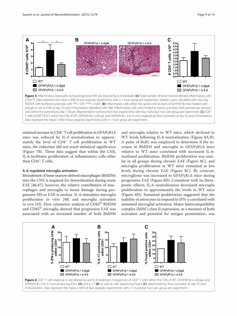
Figure 5 Inflammatory leukocytes during progressive EAE are reduced by IL-6 blockade. (A) Total number of bone marrow-derived inflammatory cells(CD45hi). Data represent the mean ± SEM of two separate experiments with n = 4 per group per experiment. Statistics were calculated with one-wayANOVA with Bonferroni post-test with **P < 0.01; ***P < 0.001. (B) Inflammatory cells within the spinal cord sections of GFAPγR1Δ mice treated withα-β-gal or α-IL-6 mAb at day 32 post immunization identified with H&E. Inflammatory cells were limited to lesions and were both perivascular (arrows)and within the parenchyma. Bar = 50 μm. Representative sections from two experiments with four individual mice per group per experiment. (C) CD4+
T cells (CD45hiCD4+) within the CNS of WT, GFAPγR1Δ + α-β-gal, and GFAPγR1Δ + α-IL-6 mice analyzed by flow cytometry at day 32 post immunization.Data represent the mean ± SEM of two separate experiments with n= 4 per group per experiment.
Savarin et al. Journal of Neuroinflammation (2015) 12:79 Page 9 of 14
minimal increase in CD4+ Tcell proliferation in GFAPγR1Δmice was reduced by IL-6 neutralization to approxi-mately the level of CD4+ T cell proliferation in WTmice, the reduction did not reach statistical significance(Figure 7B). These data suggest that within the CNS,IL-6 facilitates proliferation of inflammatory cells otherthan CD4+ T cells.
IL-6 regulated microglia activationRecruitment of bone marrow-derived macrophages (BMDM)into the CNS is required for demyelination during acuteEAE [46,47]; however, the relative contribution of mac-rophages and microglia to tissue damage during pro-gressive MS or EAE is unclear. IL-6 stimulates microgliaproliferation in vitro [48] and microglia activationin vivo [43]. Flow cytometric analysis of CD45hi BMDMand CD45lo microglia showed that progressive EAE wasassociated with an increased number of both BMDM
Figure 6 CD4+ T cell response is not altered by anti-IL-6 treatment. FrequeGFAPγR1Δ + α-IL-6 mice producing IFN-γ (A) and IL-17 (B) as well as cells eimmunization. Data represent the mean ± SEM of two separate experiment
and microglia relative to WT mice, which declined toWT levels following IL-6 neutralization (Figure 8A,B).A pulse of BrdU was employed to determine if the in-crease in BMDM and microglia in GFAPγR1Δ micerelative to WT mice correlated with increased IL-6-mediated proliferation. BMDM proliferation was simi-lar in all groups during chronic EAE (Figure 8C), andmicroglia proliferation in WT mice remained at lowlevels during chronic EAE (Figure 8C). By contrast,microgliosis was increased in GFAPγR1Δ mice duringprogressive EAE (Figure 8D). Consistent with its thera-peutic effects, IL-6 neutralization decreased microgliaproliferation to approximately the levels in WT mice(Figure 8D). Sustained proliferation suggested that theinability of astrocytes to respond to IFN-γ correlated withsustained microglial activation. Major histocompatibilitycomplex (MHC) class II expression, as a measure of bothactivation and potential for antigen presentation, was
ncies of CD4+ T cells within the CNS of WT, GFAPγR1Δ + α-β-gal, andxpressing Foxp3 (C) determined by flow cytometry at day 32 posts with n = 4 pooled mice per group per experiment.
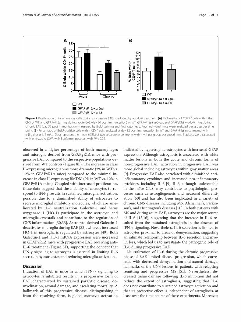
Figure 7 Proliferation of inflammatory cells during progressive EAE is reduced by anti-IL-6 treatment. (A) Proliferation of CD45hi cells within theCNS of WT and GFAPγR1Δ mice during acute EAE (day 20 post immunization) or WT, GFAPγR1Δ + α-β-gal, and GFAPγR1Δ + α-IL-6 mice duringchronic EAE (day 32 post immunization) measured by BrdU staining and flow cytometry. Four individual mice were analyzed per group per timepoint. (B) Percentage of BrdU-positive cells within CD4+ cells analyzed at day 32 post immunization in WT and GFAPγR1Δ mice treated withα-β-gal or α-IL-6 mAb. Data represent the mean ± SEM of two separate experiments with n = 4 per group per experiment. Statistics were calculatedwith one-way ANOVA with Bonferroni post-test with *P < 0.05.
Savarin et al. Journal of Neuroinflammation (2015) 12:79 Page 10 of 14
observed in a higher percentage of both macrophagesand microglia derived from GFAPγR1Δ mice with pro-gressive EAE compared to the respective populations de-rived fromWTcontrols (Figure 8E). The increase in classII-expressing microglia was more dramatic (2% in WTvs.12% in GFAPγR1Δ mice) compared to the minimal in-crease in class II-expressing BMDM (9% inWTvs. 12% inGFAPγR1Δ mice). Coupled with increased proliferation,these data suggest that the inability of astrocytes to re-spond to IFN-γ results in sustained microglial activation,possibly due to a diminished ability of astrocytes tosecrete microglial inhibitory molecules, which are ame-liorated by IL-6 neutralization. Galectin-1 and hemeoxygenase 1 (HO-1) participate in the astrocyte andmicroglia crosstalk and contribute to the regulation ofCNS inflammation [30,33]. Astrocyte-derived Galectin-1deactivates microglia during EAE [33], whereas increasedHO-1 in microglia is regulated by astrocytes [49]. BothGalectin-1 and HO-1 mRNA expression were increasedin GFAPγR1Δ mice with progressive EAE receiving anti-IL-6 treatment (Figure 8F), supporting the concept thatIFN-γ signaling to astrocytes is essential in limiting IL-6secretion by astrocytes and reducing microglia activation.
DiscussionInduction of EAE in mice in which IFN-γ signaling toastrocytes is inhibited results in a progressive form ofEAE characterized by sustained paralytic disease, de-myelination, axonal damage, and escalating mortality. Ahallmark of this progressive disease, distinguishing itfrom the resolving form, is global astrocyte activation
indicated by hypertrophic astrocytes with increased GFAPexpression. Although astrogliosis is associated with whitematter lesions in both the acute and chronic forms ofnon-progressive EAE, activation in progressive EAE wasmore global including astrocytes within gray matter areas[9]. Progressive EAE also correlated with diminished anti-inflammatory cytokines and increased pro-inflammatorycytokines, including IL-6 [9]. IL-6, although undetectablein the naïve CNS, may contribute to physiological pro-cesses such as astrogliogenesis and neuronal differenti-ation [50] and has also been implicated in a variety ofchronic CNS diseases including MS, Alzheimer’s, Parkin-son’s, and Huntington’s diseases [50]. In both patients withMS and during acute EAE, astrocytes are the major sourceof IL-6 [15,16], suggesting that the increase in IL-6 re-sulted from the sustained astrogliosis in the absence ofIFN-γ signaling. Nevertheless, IL-6 secretion is limited toastrocytes proximal to areas of demyelination, suggestingan intimate relationship between IL-6 secretion and mye-lin loss, which led us to investigate the pathogenic role ofIL-6 during progressive EAE.Neutralization of IL-6 during the chronic progressive
phase of EAE limited disease progression, which corre-lated with decreased demyelination and axonal damage,hallmarks of the CNS lesions in patients with relapsingremitting and progressive MS [51]. Nevertheless, de-creased tissue damage following IL-6 inhibition did notreduce the extent of astrogliosis, suggesting that IL-6does not contribute to sustained astrocyte activation andthat its protective effect is independent of astrogliosis, atleast over the time course of these experiments. Moreover,
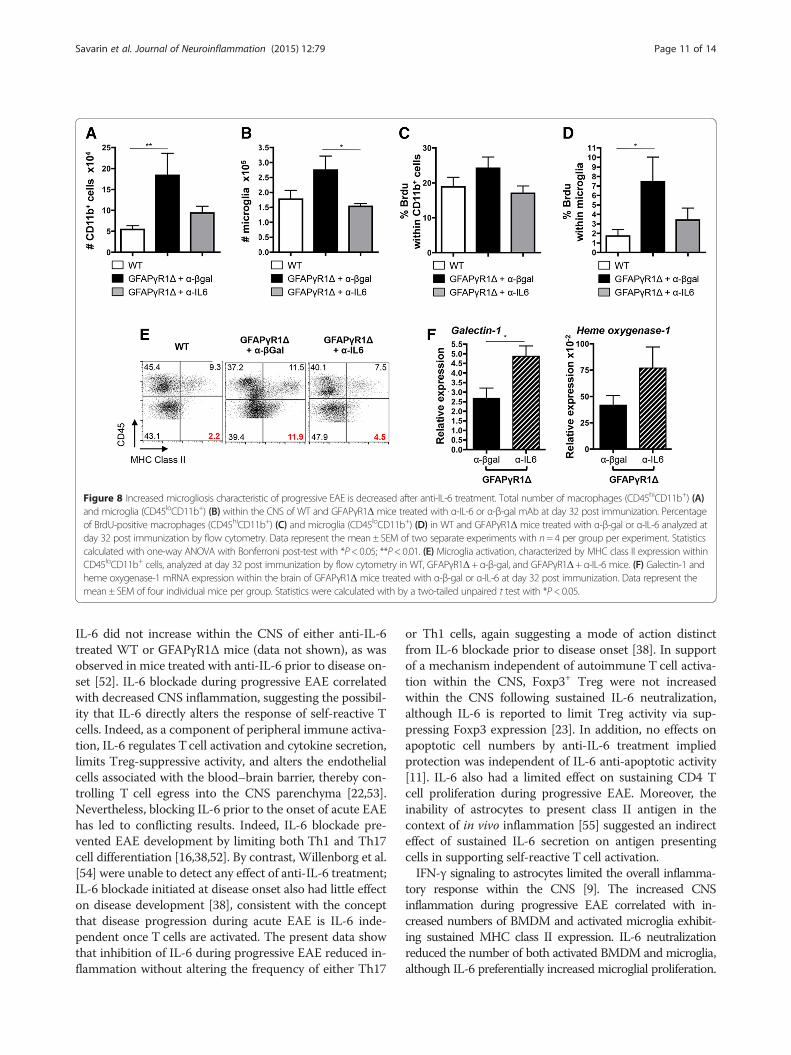
Figure 8 Increased microgliosis characteristic of progressive EAE is decreased after anti-IL-6 treatment. Total number of macrophages (CD45hiCD11b+) (A)and microglia (CD45loCD11b+) (B) within the CNS of WT and GFAPγR1Δ mice treated with α-IL-6 or α-β-gal mAb at day 32 post immunization. Percentageof BrdU-positive macrophages (CD45hiCD11b+) (C) and microglia (CD45loCD11b+) (D) in WT and GFAPγR1Δ mice treated with α-β-gal or α-IL-6 analyzed atday 32 post immunization by flow cytometry. Data represent the mean ± SEM of two separate experiments with n= 4 per group per experiment. Statisticscalculated with one-way ANOVA with Bonferroni post-test with *P< 0.05; **P< 0.01. (E) Microglia activation, characterized by MHC class II expression withinCD45loCD11b+ cells, analyzed at day 32 post immunization by flow cytometry in WT, GFAPγR1Δ+ α-β-gal, and GFAPγR1Δ+ α-IL-6 mice. (F) Galectin-1 andheme oxygenase-1 mRNA expression within the brain of GFAPγR1Δ mice treated with α-β-gal or α-IL-6 at day 32 post immunization. Data represent themean ± SEM of four individual mice per group. Statistics were calculated with by a two-tailed unpaired t test with *P< 0.05.
Savarin et al. Journal of Neuroinflammation (2015) 12:79 Page 11 of 14
IL-6 did not increase within the CNS of either anti-IL-6treated WT or GFAPγR1Δ mice (data not shown), as wasobserved in mice treated with anti-IL-6 prior to disease on-set [52]. IL-6 blockade during progressive EAE correlatedwith decreased CNS inflammation, suggesting the possibil-ity that IL-6 directly alters the response of self-reactive Tcells. Indeed, as a component of peripheral immune activa-tion, IL-6 regulates Tcell activation and cytokine secretion,limits Treg-suppressive activity, and alters the endothelialcells associated with the blood–brain barrier, thereby con-trolling T cell egress into the CNS parenchyma [22,53].Nevertheless, blocking IL-6 prior to the onset of acute EAEhas led to conflicting results. Indeed, IL-6 blockade pre-vented EAE development by limiting both Th1 and Th17cell differentiation [16,38,52]. By contrast, Willenborg et al.[54] were unable to detect any effect of anti-IL-6 treatment;IL-6 blockade initiated at disease onset also had little effecton disease development [38], consistent with the conceptthat disease progression during acute EAE is IL-6 inde-pendent once T cells are activated. The present data showthat inhibition of IL-6 during progressive EAE reduced in-flammation without altering the frequency of either Th17
or Th1 cells, again suggesting a mode of action distinctfrom IL-6 blockade prior to disease onset [38]. In supportof a mechanism independent of autoimmune T cell activa-tion within the CNS, Foxp3+ Treg were not increasedwithin the CNS following sustained IL-6 neutralization,although IL-6 is reported to limit Treg activity via sup-pressing Foxp3 expression [23]. In addition, no effects onapoptotic cell numbers by anti-IL-6 treatment impliedprotection was independent of IL-6 anti-apoptotic activity[11]. IL-6 also had a limited effect on sustaining CD4 Tcell proliferation during progressive EAE. Moreover, theinability of astrocytes to present class II antigen in thecontext of in vivo inflammation [55] suggested an indirecteffect of sustained IL-6 secretion on antigen presentingcells in supporting self-reactive T cell activation.IFN-γ signaling to astrocytes limited the overall inflamma-
tory response within the CNS [9]. The increased CNSinflammation during progressive EAE correlated with in-creased numbers of BMDM and activated microglia exhibit-ing sustained MHC class II expression. IL-6 neutralizationreduced the number of both activated BMDM and microglia,although IL-6 preferentially increased microglial proliferation.
Savarin et al. Journal of Neuroinflammation (2015) 12:79 Page 12 of 14
Similarly, MHC class II expression was down regulated byIL-6 blockade more efficiently on microglia. This observationmay explain the limited effect of anti-IL-6 treatment onCD4+ T cell proliferation, as microglia exhibit limited antigenpresentation functions in vivo [56,57]. These data suggestthat increased tissue damage may correlate with the inabilityof the astrocyte to down regulate microglial activity in theabsence of IFN-γ signaling. Recent in vivo data support theconcept that astrocytes regulate microglial activity during in-flammation. Inhibition of NF-κB in astrocytes diminishedclinical disease during EAE by attenuating microglia activa-tion [29,30]. Similarly, Galectin-1 secretion by astrocyteslimits both microglial activation and EAE [33], whereasastrocyte-mediated induction of HO-1 in microglia limitsneuroinflammation [49]. Increased expression of bothGalectin-1 and HO-1 after anti-IL-6 treatment, associatedwith decreased microglia proliferation and MHC class II ex-pression, thus supports the concept that the crosstalk be-tween astrocytes and microglia during the chronic phase ofEAE is essential for disease resolution. However, it remainsunclear whether astrocyte-derived IL-6 acts directly onmicroglia or indirectly via an autocrine-induced alteration inastrocyte function [58]. It is also possible that IL-6 itself altersastrocyte functions, which also contributes to tissue dam-age during progressive EAE. Astrocytes regulate diverseCNS functions from neurogenesis to defending from bothinternal and external insults [40]. Their critical contribu-tions to regulating chronic CNS autoimmune disease aresupported by the expanding number of alterations inastrocyte function that result in progressive types of EAE[9,59,60]. Furthermore, detrimental effects of IL-6 secre-tion by astrocytes on CNS function and pathobiology areexemplified by analysis of mice in which IL-6 is constitu-tively expressed by astrocytes [61]. Sustained IL-6 resultsin localized neuroinflammation, neurodegeneration, lossof blood–brain barrier integrity, and impaired learning[31,62,63]. Finally, reduced disease severity after elimin-ation of B cells secreting IL-6 in two EAE models as wellas in patients with relapsing remitting MS [64,65] supportsa destructive role for IL-6 during CNS autoimmunity.
ConclusionsThese data demonstrate that one anti-inflammatory activityof IFN-γ is to prevent sustained IL-6 expression by astro-cytes, which plays an essential role in disease progressionduring progressive EAE. IL-6 is critical in mediating de-myelination, axonal damage, and sustaining both CNS in-flammation and disability during progressive EAE. Thesedata demonstrated the therapeutic potential of IL-6 block-ade in limiting both the disability and tissue damage in amodel of progressive EAE. This approach, currently in useto treat patients with peripheral autoimmune disease, maythus be efficacious in the treatment of the progressiveforms of MS.
AbbreviationsAPP: amyloid precursor protein; BMDM: bone marrow-derived macrophage;BrdU: bromodeoxyuridine; CNS: central nervous system; EAE: experimentalautoimmune encephalomyelitis; β-gal: β-galactosidase; GFAP: glial fibrillaryacidic protein; H&E: hematoxylin and eosin; HO-1: heme oxygenase-1;IL: interleukin; IFN-γ: interferon-γ; i.p.: intraperitoneally; LFB: luxol fast blue;mAb: monoclonal antibody; MHC: major histocompatibility complex;MOG: myelin oligodendrocyte glycoprotein; MS: multiple sclerosis;PFA: paraformaldehyde; PMA: phorbol 12-myristate 13-acetate; TNF: tumornecrosis factor; Treg: regulatory T cell; WT: wild type.
Competing interestsThe authors declare that they have no competing interests.
Authors’ contributionsCS performed the experiments, collected and analyzed the data, and wrotethe manuscript; DRH analyzed all histopathology and edited the manuscript;AVT assisted with the experiments and edited the manuscript; ZC carried outthe confocal microscopy analysis; BDT interpreted the data and edited themanuscript; CCB interpreted the data and wrote the manuscript; SASdesigned the research, provided the materials, interpreted the data, andwrote the manuscript. All authors read and approved the final manuscript.
AcknowledgementsThe authors would like to thank Natasha Towne, Kate Stenson, Wen Wei, andErnesto Barron for their exceptional technical assistance. This work wassupported by the Multiple Sclerosis Society research grant RG4007B5 andCancer Center Support grant P30CA014089.
Author details1Department of Neurosciences NC-30, Lerner Research Institute, The ClevelandClinic, 9500 Euclid Avenue, Cleveland, OH 44195, USA. 2Department ofPathology, Keck School of Medicine, University of Southern California, LosAngeles, CA 90033, USA.
Received: 1 December 2014 Accepted: 2 April 2015
References1. Lassmann H, van Horssen J, Mahad D. Progressive multiple sclerosis:
pathology and pathogenesis. Nat Rev Neurol. 2012;8:647–56.2. Gold R, Linington C, Lassmann H. Understanding pathogenesis and
therapy of multiple sclerosis via animal models: 70 years of merits andculprits in experimental autoimmune encephalomyelitis research. Brain.2006;129:1953–71.
3. Rangachari M, Kuchroo VK. Using EAE to better understand principles ofimmune function and autoimmune pathology. J Autoimmun. 2013;45:31–9.
4. Fletcher JM, Lalor SJ, Sweeney CM, Tubridy N, Mills KH. T cells in multiplesclerosis and experimental autoimmune encephalomyelitis. Clin ExpImmunol. 2010;162:1–11.
5. Codarri L, Gyulveszi G, Tosevski V, Hesske L, Fontana A, Magnenat L, et al.RORgammat drives production of the cytokine GM-CSF in helper T cells,which is essential for the effector phase of autoimmune neuroinflammation.Nat Immunol. 2011;12:560–7.
6. Ponomarev ED, Shriver LP, Maresz K, Pedras-Vasconcelos J, Verthelyi D, DittelBN. GM-CSF production by autoreactive T cells is required for the activationof microglial cells and the onset of experimental autoimmune encephalomyelitis.J Immunol. 2007;178:39–48.
7. Comi G. Disease-modifying treatments for progressive multiple sclerosis.Mult Scler. 2013;19:1428–36.
8. Simmons SB, Pierson ER, Lee SY, Goverman JM. Modeling the heterogeneityof multiple sclerosis in animals. Trends Immunol. 2013;34:410–22.
9. Hindinger C, Bergmann CC, Hinton DR, Phares TW, Parra GI, Hussain S, et al.IFN-gamma signaling to astrocytes protects from autoimmune mediatedneurological disability. PLoS One. 2012;7, e42088.
10. Smolen JS, Beaulieu A, Rubbert-Roth A, Ramos-Remus C, Rovensky J, Alecock E,et al. Effect of interleukin-6 receptor inhibition with tocilizumab in patients withrheumatoid arthritis (OPTION study): a double-blind, placebo-controlled,randomised trial. Lancet. 2008;371:987–97.
11. Atreya R, Mudter J, Finotto S, Mullberg J, Jostock T, Wirtz S, et al. Blockadeof interleukin 6 trans signaling suppresses T-cell resistance against apoptosis
Savarin et al. Journal of Neuroinflammation (2015) 12:79 Page 13 of 14
in chronic intestinal inflammation: evidence in Crohn’s disease and experimentalcolitis in vivo. Nat Med. 2000;6:583–8.
12. Becker C, Fantini MC, Schramm C, Lehr HA, Wirtz S, Nikolaev A, et al. TGF-beta suppresses tumor progression in colon cancer by inhibition of IL-6trans-signaling. Immunity. 2004;21:491–501.
13. Rincon M, Irvin CG. Role of IL-6 in asthma and other inflammatorypulmonary diseases. Int J Biol Sci. 2012;8:1281–90.
14. Kishimoto T, Akira S, Narazaki M, Taga T. Interleukin-6 family of cytokinesand gp130. Blood. 1995;86:1243–54.
15. Maimone D, Guazzi GC, Annunziata P. IL-6 detection in multiple sclerosisbrain. J Neurol Sci. 1997;146:59–65.
16. Gijbels K, Van Damme J, Proost P, Put W, Carton H, Billiau A. Interleukin 6production in the central nervous system during experimentalautoimmune encephalomyelitis. Eur J Immunol. 1990;20:233–5.
17. Samoilova EB, Horton JL, Hilliard B, Liu TS, Chen Y. IL-6-deficient mice areresistant to experimental autoimmune encephalomyelitis: roles of IL-6 inthe activation and differentiation of autoreactive T cells. J Immunol.1998;161:6480–6.
18. Okuda Y, Sakoda S, Bernard CC, Fujimura H, Saeki Y, Kishimoto T, et al. IL-6-deficient mice are resistant to the induction of experimental autoimmuneencephalomyelitis provoked by myelin oligodendrocyte glycoprotein. IntImmunol. 1998;10:703–8.
19. Mendel I, Katz A, Kozak N, Ben-Nun A, Revel M. Interleukin-6 functions inautoimmune encephalomyelitis: a study in gene-targeted mice. Eur JImmunol. 1998;28:1727–37.
20. Dienz O, Rincon M. The effects of IL-6 on CD4 T cell responses. Clin Immunol.2009;130:27–33.
21. Eugster HP, Frei K, Kopf M, Lassmann H, Fontana A. IL-6-deficient miceresist myelin oligodendrocyte glycoprotein-induced autoimmuneencephalomyelitis. Eur J Immunol. 1998;28:2178–87.
22. Bettelli E, Carrier Y, Gao W, Korn T, Strom TB, Oukka M, et al. Reciprocaldevelopmental pathways for the generation of pathogenic effector TH17and regulatory T cells. Nature. 2006;441:235–8.
23. Korn T, Mitsdoerffer M, Croxford AL, Awasthi A, Dardalhon VA, Galileos G,et al. IL-6 controls Th17 immunity in vivo by inhibiting the conversion ofconventional T cells into Foxp3+ regulatory T cells. Proc Natl Acad Sci U S A.2008;105:18460–5.
24. Hirota H, Kiyama H, Kishimoto T, Taga T. Accelerated nerve regenerationin mice by upregulated expression of interleukin (IL) 6 and IL-6 receptorafter trauma. J Exp Med. 1996;183:2627–34.
25. Loddick SA, Turnbull AV, Rothwell NJ. Cerebral interleukin-6 is neuroprotectiveduring permanent focal cerebral ischemia in the rat. J Cereb Blood FlowMetab. 1998;18:176–9.
26. Zhang PL, Izrael M, Ainbinder E, Ben-Simchon L, Chebath J, Revel M.Increased myelinating capacity of embryonic stem cell derivedoligodendrocyte precursors after treatment by interleukin-6/solubleinterleukin-6 receptor fusion protein. Mol Cell Neurosci. 2006;31:387–98.
27. Gruol DL, Nelson TE. Physiological and pathological roles of interleukin-6in the central nervous system. Mol Neurobiol. 1997;15:307–39.
28. Erta M, Quintana A, Hidalgo J. Interleukin-6, a major cytokine in the centralnervous system. Int J Biol Sci. 2012;8:1254–66.
29. Brambilla R, Persaud T, Hu X, Karmally S, Shestopalov VI, Dvoriantchikova G,et al. Transgenic inhibition of astroglial NF-kappa B improves functionaloutcome in experimental autoimmune encephalomyelitis by suppressingchronic central nervous system inflammation. J Immunol. 2009;182:2628–40.
30. Brambilla R, Morton PD, Ashbaugh JJ, Karmally S, Lambertsen KL, Bethea JR.Astrocytes play a key role in EAE pathophysiology by orchestrating in theCNS the inflammatory response of resident and peripheral immune cellsand by suppressing remyelination. Glia. 2014;62:452–67.
31. Campbell IL, Abraham CR, Masliah E, Kemper P, Inglis JD, Oldstone MB, et al.Neurologic disease induced in transgenic mice by cerebral overexpressionof interleukin 6. Proc Natl Acad Sci U S A. 1993;90:10061–5.
32. Shih AY, Fernandes HB, Choi FY, Kozoriz MG, Liu Y, Li P, et al. Policing thepolice: astrocytes modulate microglial activation. J Neurosci.2006;26:3887–8.
33. Starossom SC, Mascanfroni ID, Imitola J, Cao L, Raddassi K, Hernandez SF,et al. Galectin-1 deactivates classically activated microglia and protects frominflammation-induced neurodegeneration. Immunity. 2012;37:249–63.
34. Hindinger C, Gonzalez JM, Bergmann CC, Fuss B, Hinton DR, Atkinson RD,et al. Astrocyte expression of a dominant-negative interferon-gamma receptor.J Neurosci Res. 2005;82:20–31.
35. Chen Z, Jalabi W, Shpargel KB, Farabaugh KT, Dutta R, Yin X, et al.Lipopolysaccharide-induced microglial activation and neuroprotectionagainst experimental brain injury is independent of hematogenous TLR4.J Neurosci. 2012;32:11706–15.
36. Wang J, Asensio VC, Campbell IL. Cytokines and chemokines as mediatorsof protection and injury in the central nervous system assessed intransgenic mice. Curr Top Microbiol Immunol. 2002;265:23–48.
37. Md Yusof MY, Emery P. Targeting interleukin-6 in rheumatoid arthritis.Drugs. 2013;73:341–56.
38. Serada S, Fujimoto M, Mihara M, Koike N, Ohsugi Y, Nomura S, et al. IL-6blockade inhibits the induction of myelin antigen-specific Th17 cells andTh1 cells in experimental autoimmune encephalomyelitis. Proc Natl AcadSci U S A. 2008;105:9041–6.
39. Basso AS, Frenkel D, Quintana FJ, Costa-Pinto FA, Petrovic-Stojkovic S,Puckett L, et al. Reversal of axonal loss and disability in a mouse modelof progressive multiple sclerosis. J Clin Invest. 2008;118:1532–43.
40. Sofroniew MV, Vinters HV. Astrocytes: biology and pathology. ActaNeuropathol. 2010;119:7–35.
41. Black JA, Newcombe J, Waxman SG. Astrocytes within multiple sclerosislesions upregulate sodium channel Nav1.5. Brain. 2010;133:835–46.
42. Okada S, Nakamura M, Mikami Y, Shimazaki T, Mihara M, Ohsugi Y, et al.Blockade of interleukin-6 receptor suppresses reactive astrogliosis andameliorates functional recovery in experimental spinal cord injury.J Neurosci Res. 2004;76:265–76.
43. Campbell IL, Erta M, Lim SL, Frausto R, May U, Rose-John S, et al.Trans-signaling is a dominant mechanism for the pathogenic actionsof interleukin-6 in the brain. J Neurosci. 2014;34:2503–13.
44. Gimsa U, Mitchison NA, Brunner-Weinzierl MC. Immune privilege as anintrinsic CNS property: astrocytes protect the CNS against T-cell-mediatedneuroinflammation. Mediators Inflamm. 2013;2013:320519.
45. Kimura A, Kishimoto T. IL-6: regulator of Treg/Th17 balance. Eur J Immunol.2010;40:1830–5.
46. Ajami B, Bennett JL, Krieger C, McNagny KM, Rossi FM. Infiltratingmonocytes trigger EAE progression, but do not contribute to the residentmicroglia pool. Nat Neurosci. 2011;14:1142–9.
47. Moreno M, Bannerman P, Ma J, Guo F, Miers L, Soulika AM, et al.Conditional ablation of astroglial CCL2 suppresses CNS accumulation ofM1 macrophages and preserves axons in mice with MOG peptide EAE.J Neurosci. 2014;34:8175–85.
48. Streit WJ, Hurley SD, McGraw TS, Semple-Rowland SL. Comparativeevaluation of cytokine profiles and reactive gliosis supports a critical rolefor interleukin-6 in neuron-glia signaling during regeneration. J NeurosciRes. 2000;61:10–20.
49. Min KJ, Yang MS, Kim SU, Jou I, Joe EH. Astrocytes induce hemeoxygenase-1 expression in microglia: a feasible mechanism for preventing excessivebrain inflammation. J Neurosci. 2006;26:1880–7.
50. Spooren A, Kolmus K, Laureys G, Clinckers R, De Keyser J, Haegeman G,et al. Interleukin-6, a mental cytokine. Brain Res Rev.2011;67:157–83.
51. Bruck W. The pathology of multiple sclerosis is the result of focalinflammatory demyelination with axonal damage. J Neurol.2005;252:V3–9.
52. Gijbels K, Brocke S, Abrams JS, Steinman L. Administration of neutralizingantibodies to interleukin-6 (IL-6) reduces experimental autoimmuneencephalomyelitis and is associated with elevated levels of IL-6 bioactivityin central nervous system and circulation. Mol Med. 1995;1:795–805.
53. de Vries HE, Blom-Roosemalen MC, van Oosten M, de Boer AG, van BerkelTJ, Breimer DD, et al. The influence of cytokines on the integrity of theblood–brain barrier in vitro. J Neuroimmunol. 1996;64:37–43.
54. Willenborg DO, Fordham SA, Cowden WB, Ramshaw IA. Cytokines andmurine autoimmune encephalomyelitis: inhibition or enhancement ofdisease with antibodies to select cytokines, or by delivery of exogenouscytokines using a recombinant vaccinia virus system. Scand J Immunol.1995;41:31–41.
55. Horwitz MS, Evans CF, Klier FG, Oldstone MB. Detailed in vivo analysis ofinterferon-gamma induced major histocompatibility complex expression in thecentral nervous system: astrocytes fail to express major histocompatibilitycomplex class I and II molecules. Lab Invest. 1999;79:235–42.
56. Greter M, Heppner FL, Lemos MP, Odermatt BM, Goebels N, Laufer T, et al.Dendritic cells permit immune invasion of the CNS in an animal model ofmultiple sclerosis. Nat Med. 2005;11:328–34.
Savarin et al. Journal of Neuroinflammation (2015) 12:79 Page 14 of 14
57. McMahon EJ, Bailey SL, Castenada CV, Waldner H, Miller SD. Epitopespreading initiates in the CNS in two mouse models of multiple sclerosis.Nat Med. 2005;11:335–9.
58. Quintana A, Erta M, Ferrer B, Comes G, Giralt M, Hidalgo J. Astrocyte-specificdeficiency of interleukin-6 and its receptor reveal specific roles in survival,body weight and behavior. Brain Behav Immun. 2013;27:162–73.
59. Haroon F, Drogemuller K, Handel U, Brunn A, Reinhold D, Nishanth G, et al.Gp130-dependent astrocytic survival is critical for the control of autoimmunecentral nervous system inflammation. J Immunol. 2011;186:6521–31.
60. Mishra PK, Hsuchou H, Ouyang S, Kastin AJ, Wu X, Pan W. Loss of astrocyticleptin signaling worsens experimental autoimmune encephalomyelitis. BrainBehav Immun. 2013;34:98–107.
61. Chiang CS, Stalder A, Samimi A, Campbell IL. Reactive gliosis as a consequenceof interleukin-6 expression in the brain: studies in transgenic mice. DevNeurosci. 1994;16:212–21.
62. Barnum SR, Jones JL, Muller-Ladner U, Samimi A, Campbell IL. Chroniccomplement C3 gene expression in the CNS of transgenic mice withastrocyte-targeted interleukin-6 expression. Glia. 1996;18:107–17.
63. Heyser CJ, Masliah E, Samimi A, Campbell IL, Gold LH. Progressive decline inavoidance learning paralleled by inflammatory neurodegeneration intransgenic mice expressing interleukin 6 in the brain. Proc Natl Acad Sci U S A.1997;94:1500–5.
64. Molnarfi N, Schulze-Topphoff U, Weber MS, Patarroyo JC, Prod’homme T,Varrin-Doyer M, et al. MHC class II-dependent B cell APC function is requiredfor induction of CNS autoimmunity independent of myelin-specific antibodies.J Exp Med. 2013;210:2921–37.
65. Barr TA, Shen P, Brown S, Lampropoulou V, Roch T, Lawrie S, et al. B celldepletion therapy ameliorates autoimmune disease through ablation ofIL-6-producing B cells. J Exp Med. 2012;209:1001–10.
Submit your next manuscript to BioMed Centraland take full advantage of:
• Convenient online submission
• Thorough peer review
• No space constraints or color figure charges
• Immediate publication on acceptance
• Inclusion in PubMed, CAS, Scopus and Google Scholar
• Research which is freely available for redistribution
Submit your manuscript at www.biomedcentral.com/submit