APNEIC OXYGENATION; PULMONARY AND CARDI- …oaji.net/articles/2015/1592-1421516184.pdf · APNEIC...
14
The Greek E-Journal of Perioperative Medicine 2010; 8:70-83 (ISSN 1109-6888) www.anesthesia.gr/ejournal Ελληνικό Περιοδικό Περιεγχειρητικής Ιατρικής 2010; 8:70-83 (ISSN 1109-6888) www.anesthesia.gr/ejournal ©2010 Society of Anesthesiology and Intensive Medicine of Northern Greece ©2010 Δηαιρεία Αναιζθηζιολογίας και Δνηαηικής Ιαηρικής Βορείοσ Δλλάδος 70 APNEIC OXYGENATION; PULMONARY AND CARDI- OVASCULAR EFFECTS 1 Kolettas A, 2 Grosomanidis V, 2 Fyntanidou B, 3 Kotzampassi K, 5 Kolettas V, 4 Theodosiadis P, 2 Karakoulas K, 2 Mitos G, 2 Aidoni Z, 2 Vasilakos D ABSTRACT Apneic oxygenation; pulmonary and cardiovascular effects Kolettas A, Grosomanidis V, Fyntanidou B, Kotzampassi K, Kolettas V, Theodo- siadis P, Karakoulas K, Mitos G, Aidoni Z, Vasilakos D Apneic oxygenation is an adjunct „ventilation‟ technique that involves insufflation of oxygen at varying flows through a catheter that is inserted through the endotra- cheal tube and is positioned above the carina. Apneic oxygenation improves gas exchange efficiency and preserves the arterial oxygenation at an acceptable level. The understanding of the mechanism that is responsible for the sustained high al- veolar and blood oxygenation levels requires the substantial knowledge of the physiology regarding the transport and exchange of alveolar gases. Application of apneic oxygenation may be necessary in some clinical situations, in which no movement of the chest or the lungs may be desirable, such as during cardiothorac- ic surgery or during some radiological procedures, in order to eliminate respirato- ry motion artifacts. Moreover, it is used during the apnea test for the diagnosis of brain death, to ensure an adequate arterial oxygenation. Under ideal conditions, apneic oxygenation could theoretically be sufficient to provide enough oxygen for survival for a longer time period. However, accumulation of carbon dioxide would remain the limiting factor. Hypercapnia and subsequent acidosis are re- sponsible for most of the respiratory and cardiovascular effects of apneic oxyge- nation. Apneic oxygenation is an alternative technique, which contributes to the maintenance of oxyge- nation during apnea by intratracheal insufflation of oxygen at different flow rates. Oxygen is in- sufflated through a catheter that is inserted through the endotracheal tube and is positioned just above the trachea bifurcation at a flow rang- ing between 2L/min and 10L/min. A modified apneic oxygenation technique is represented by the endobrochial apneic oxygenation. In this case oxygen is insufflated through a catheter that is inserted through a double lumen endotra- cheal tube and is placed in one of the two major bronchus. Besides oxygenation maintenance, carbon dioxide accumulation cannot be avoided. However, if high flow rates are used, carbon di- oxide removal can be partly enhanced. Historic reference The first report on apneic oxygenation was made in 1947 by Draper, Whitehead and Spenc- er, who studied the pulmonary gas exchange 1: Thessaloniki Heart Institute, St. Luke‟s Hospital, Greece 2: Anesthesia and Intensive Care Clinic of Aristotle Universi- ty of Thessaloniki, Greece 3: Surgical Clinic of Aristotle University of Thessaloniki, Greece 4: "Euromedica Central Clinic", Thessaloniki, Greece 5: Cardiological Clinic, General Hospital of Chalkidiki, Poli- giros, Greece
Transcript of APNEIC OXYGENATION; PULMONARY AND CARDI- …oaji.net/articles/2015/1592-1421516184.pdf · APNEIC...
The Greek E-Journal of Perioperative Medicine 2010; 8:70-83 (ISSN 1109-6888) www.anesthesia.gr/ejournal
Ελληνικό Περιοδικό Περιεγχειρητικής Ιατρικής 2010; 8:70-83 (ISSN 1109-6888) www.anesthesia.gr/ejournal
©2010 Society of Anesthesiology and Intensive Medicine of Northern Greece
©2010 Δηαιρεία Αναιζθηζιολογίας και Δνηαηικής Ιαηρικής Βορείοσ Δλλάδος
70
APNEIC OXYGENATION; PULMONARY AND CARDI-
OVASCULAR EFFECTS
1Kolettas A,
2Grosomanidis V,
2Fyntanidou B,
3Kotzampassi K,
5Kolettas V,
4Theodosiadis P,
2Karakoulas K,
2Mitos G,
2Aidoni Z,
2Vasilakos D
ABSTRACT
Apneic oxygenation; pulmonary and cardiovascular effects
Kolettas A, Grosomanidis V, Fyntanidou B, Kotzampassi K, Kolettas V, Theodo-
siadis P, Karakoulas K, Mitos G, Aidoni Z, Vasilakos D
Apneic oxygenation is an adjunct „ventilation‟ technique that involves insufflation
of oxygen at varying flows through a catheter that is inserted through the endotra-
cheal tube and is positioned above the carina. Apneic oxygenation improves gas
exchange efficiency and preserves the arterial oxygenation at an acceptable level.
The understanding of the mechanism that is responsible for the sustained high al-
veolar and blood oxygenation levels requires the substantial knowledge of the
physiology regarding the transport and exchange of alveolar gases. Application of
apneic oxygenation may be necessary in some clinical situations, in which no
movement of the chest or the lungs may be desirable, such as during cardiothorac-
ic surgery or during some radiological procedures, in order to eliminate respirato-
ry motion artifacts. Moreover, it is used during the apnea test for the diagnosis of
brain death, to ensure an adequate arterial oxygenation. Under ideal conditions,
apneic oxygenation could theoretically be sufficient to provide enough oxygen for
survival for a longer time period. However, accumulation of carbon dioxide
would remain the limiting factor. Hypercapnia and subsequent acidosis are re-
sponsible for most of the respiratory and cardiovascular effects of apneic oxyge-
nation.
Apneic oxygenation is an alternative technique,
which contributes to the maintenance of oxyge-
nation during apnea by intratracheal insufflation
of oxygen at different flow rates. Oxygen is in-
sufflated through a catheter that is inserted
through the endotracheal tube and is positioned
just above the trachea bifurcation at a flow rang-
ing between 2L/min and 10L/min. A modified
apneic oxygenation technique is represented by
the endobrochial apneic oxygenation. In this
case oxygen is insufflated through a catheter
that is inserted through a double lumen endotra-
cheal tube and is placed in one of the two major
bronchus. Besides oxygenation maintenance,
carbon dioxide accumulation cannot be avoided.
However, if high flow rates are used, carbon di-
oxide removal can be partly enhanced.
Historic reference
The first report on apneic oxygenation was
made in 1947 by Draper, Whitehead and Spenc-
er, who studied the pulmonary gas exchange
1: Thessaloniki Heart Institute, St. Luke‟s Hospital, Greece
2: Anesthesia and Intensive Care Clinic of Aristotle Universi-
ty of Thessaloniki, Greece
3: Surgical Clinic of Aristotle University of Thessaloniki,
Greece
4: "Euromedica Central Clinic", Thessaloniki, Greece
5: Cardiological Clinic, General Hospital of Chalkidiki, Poli-
giros, Greece
The Greek E-Journal of Perioperative Medicine 2010; 8:70-83 (ISSN 1109-6888) www.anesthesia.gr/ejournal
Ελληνικό Περιοδικό Περιεγχειρητικής Ιατρικής 2010; 8:70-83 (ISSN 1109-6888) www.anesthesia.gr/ejournal
©2010 Society of Anesthesiology and Intensive Medicine of Northern Greece
©2010 Δηαιρεία Αναιζθηζιολογίας και Δνηαηικής Ιαηρικής Βορείοσ Δλλάδος
71
and the ultimate survival of dogs subjected to a
45min period of diffusion respiration[1]. The
head of the animals was placed in a chamber at
which a low pressure valve was attached.
Throughout the experiment 6-8L/min of oxygen
was admitted into the chamber. Apnea was in-
duced and maintained for a standard time period
of 45min by infusion of 1% thiopental sodium
and in order to ensure airway patency of the
dogs, an artificial airway device was used. The
data of this study show that during 45min diffu-
sion respiration there is a marked accumulation
of carbon dioxide in the pulmonary alveoli and a
consistent decrease in the venous blood pH.
However, all of the animals subjected to this
ordeal, survived.
Frumin, Epstein and Cohen used in 1959 the
term “apneic oxygenation” - which was first
employed by Nahas in 1956 - instead of the old-
er “diffusion oxygenation” to avoid the miscon-
ception regarding the mechanism that is respon-
sible for the sustained high alveolar and blood
oxygenation levels[2]. After induction of anes-
thesia, a cuffed endotracheal tube was inserted
in eight healthy patients, who were scheduled
for a variety of minor operations. Following de-
nitrogenation, mechanical ventilation was
stopped, whereas the patient remained con-
nected to the circle apparatus, which was filled
with oxygen 100% at a flow rate of 6-8L/min.
SpO2, pH, PaCO2 and the epinephrine and nore-
pinephrine concentrations in arterial plasma
were determined at standard time intervals. Dur-
ing the whole study period, arterial pressure and
the electrocardiogram were determined either
continuously or at intervals no greater than
5min. They reported a linear rise in PaCO2 and
a decrease in arterial pH to about 6.72, while
hemoglobin was fully saturated with oxygen
throughout the apneic period (more than
30min). The arterial plasma epinephrine and
norepinephrine concentrations increased as the
apnea progressed. A limitation of this study
might be that apnea was interrupted, since the
criterion for administration of neuromuscular
blocking agent was the detection of spontaneous
respiration.
Two years later, in 1961, Millar and Morris stu-
died the effects of 60min of apneic oxygenation
in adrenalectomized dogs[3]. They attempted to
determine the extent to which release of norepi-
nephrine and epinephrine from areas outside the
adrenal medulla contributes to the increased
plasma catecholamine concentrations during
apneic oxygenation, namely the extent of the
compensatory sympatho-adrenal stimulation.
From the results of this study it is concluded,
that increases in plasma norepinephrine levels
consistently accompany the rise in PaCO2 and
therefore it is reasonable to believe that release
of norepinephrine during apneic oxygenation
from extra-adrenal areas or organs is an early
response to respiratory acidosis. In this study
apneic oxygenation was induced by the connec-
tion of the endotracheal tube to a T-piece - with
a loose expiratory valve - through which oxy-
gen was delivered at a low flow rate. This tech-
nique resulted in larger carbon dioxide accumu-
lation compared to that during endotracheal
oxygen insufflation through a catheter that is
positioned at the carina level.
Fraiolo, Sceffer and Steffenson studied apneic
oxygenation in 13 patients undergoing microla-
ryngoscopy with a pharyngeal catheter for oxy-
gen administration at a flow rate of 6L/min and
18 patients having minor surgical procedures
with a cuffed endotracheal tube connected to an
oxygen filled Collins spirometer for oxygen
administration[4]. Any alteration of PaO2, Pa-
CO2, PAN2, pH, FRC (Functional Residual Ca-
pacity) and the oxygen uptake was recorded,
while ECG and the arterial pressure were conti-
nuously monitored. Fraiolo et al reported that
PaO2 decrease was faster and more profound in
patients with a FRC/weight ratio <50ml/kg.
Therefore, they suggested that the FRC/weight
ratio could provide a useful guide for selecting
patients that might be suitable for apneic oxy-
genation. The results of this study were attri-
buted to the fact that patients with larger body
mass index would have a greater amount of to-
tal body nitrogen as well as increased cardiac
output and, therefore, a higher rate of nitrogen
return to the lung. Nitrogen returning to the
lung will replace alveolar oxygen causing a
lower PAO2 and PaO2. However, the oxygena-
tion was preserved at an acceptable level for a
time period of 15min and the worst PaO2 value
recorded during the study was 100mmHg.
The Greek E-Journal of Perioperative Medicine 2010; 8:70-83 (ISSN 1109-6888) www.anesthesia.gr/ejournal
Ελληνικό Περιοδικό Περιεγχειρητικής Ιατρικής 2010; 8:70-83 (ISSN 1109-6888) www.anesthesia.gr/ejournal
©2010 Society of Anesthesiology and Intensive Medicine of Northern Greece
©2010 Δηαιρεία Αναιζθηζιολογίας και Δνηαηικής Ιαηρικής Βορείοσ Δλλάδος
72
In 1982 Pesenti et al studied a combination of
apneic oxygenation and extracorporeal removal
of carbon dioxide (ECCO2R) in an experimental
model of preterm lambs[5]. The aim of this
study was to explore the possibility of hyaline
membrane disease prevention. According to
this study, apneic oxygenation in combination
with extracorporeal removal of the carbon dio-
xide resulted in fewer ventilation related com-
plications and better outcome, since there was
time for the respiratory mechanics to stabilize
before the onset of mechanical ventilation. The
results of this study would be more impressive
if oxygenation was restored only by apneic oxy-
genation.
In a similar study, Nielsen et al, combined ap-
neic oxygenation with extracorproreal removal
of the carbon dioxide in an experimental swine
model, in which acute lung injury was con-
ducted through recurring wash out of the surfac-
tant[6]. Normal PaO2 levels were restored by the
combination of apneic oxygenation and the
extracorporeal device membrane.
Finally, Cook et al studied in 1998 apneic oxy-
genation in infants and young children[7]. Their
aim was to determine the time period, during
which the application of apneic oxygenation
could be considered as a safe technique. They
reported that PaO2 levels were acceptable during
the whole time of the study (5min). Moreover,
they concluded that, opposed to adults, in in-
fants the most restrictive factor of the applica-
tion of apneic oxygenation was the linear de-
crease in PaO2 and not the increase in PaCO2.
Transport of gases under apneic oxygenation
conditions.
The movement of gases from one area to anoth-
er depends on the pressure gradient between
these two spaces. This pressure gradient may be
a result of hydrostatic pressure gradient - and in
this case the movement is more active (trans-
port) - or a result of partial pressure gradient
(the pressure that one component of a mixture
of gases would exert if it were alone in a con-
tainer) - and in this case the movement is less
effective macroscopically (diffusion). The gas
velocity depends on the airway resistance in the
case of transport or on the resistance properties
of the membrane across which the gas mole-
cules are transferred in the case of diffusion.
Oxygen
In the alveoli, oxygen is continuously diffusing
down its concentration gradient into the blood,
and is replaced by diffusion of carbon dioxide
in the opposite direction across the alveolar ca-
pillary membrane.
Oxygen passes into the pulmonary capillaries
by passive diffusion across the alveolar-
capillary membrane (oxygen diffusion from the
air to the liquid state). The continuous oxygen
diffusion is explained by the preservable alveo-
lar-capillary partial pressure gradient due to the
oxygen bound to hemoglobin[8,9].
The oxygen diffusing capacity depends on par-
tial pressure gradient across the alveolar-
capillary membrane, on the temperature, on the
alveolar-capillary membrane surface area, on
the distance until hemoglobin is reached and on
the oxygen molecular weight[10,11]. The more
soluble the gas, the bigger is the number of the
molecules that are available for diffusion, under
a given pressure gradient. Moreover, diffusion
capacity is proportionally better when the alveo-
lar membrane surface area is bigger. On the
other hand, the diffusion capacity is decreased
when the alveolar-capillary membrane is thick-
ened. In addition to this, the gas diffusion ca-
pacity is proportional to the gas molecule veloc-
ity, which is inverse proportional to the square
root of its molecular weight. Finally, the diffu-
sion capacity will be increased when the tem-
perature is high, since this will cause a greater
molecular kinetic energy. However, the temper-
ature level is not taken into consideration, since
it is thought to remain constant. All of the
above is included in the following equation:
MWd
SAPD (1-1)
where D is the diffusion velocity, ΓP is the
pressure gradient between the alveoli and the
capillary, A is the diffusion alveolar-capillary
membrane surface area, S is the water solubility
of oxygen, d is the diffusion distance (mem-
The Greek E-Journal of Perioperative Medicine 2010; 8:70-83 (ISSN 1109-6888) www.anesthesia.gr/ejournal
Ελληνικό Περιοδικό Περιεγχειρητικής Ιατρικής 2010; 8:70-83 (ISSN 1109-6888) www.anesthesia.gr/ejournal
©2010 Society of Anesthesiology and Intensive Medicine of Northern Greece
©2010 Δηαιρεία Αναιζθηζιολογίας και Δνηαηικής Ιαηρικής Βορείοσ Δλλάδος
73
brane width) and MW is the oxygen molecular
weight.
Two parameters that describe the physicochem-
ical properties of the gas are included in this eq-
uation (1-1): the solubility (S) and the molecular
weight (MB). The ratio S/ MW is proportional
to the gas diffusion coefficient. Therefore, the
gas diffusion velocity is proportional to the gas
diffusion coefficient for a given partial pressure
gradient and a specific diffusion membrane. If
oxygen diffusion coefficient is presumed to be
1, then the relative diffusion coefficients for the
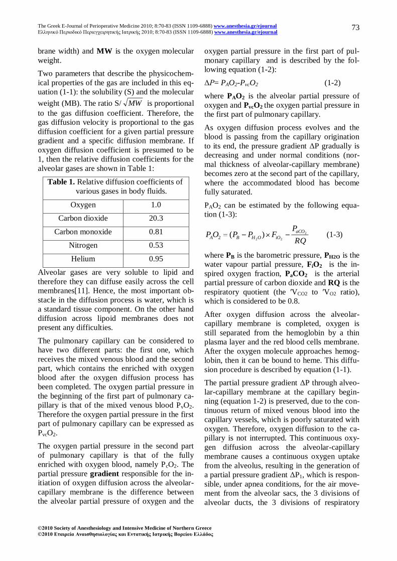
alveolar gases are shown in Table 1:
Table 1. Relative diffusion coefficients of
various gases in body fluids.
Oxygen 1.0
Carbon dioxide 20.3
Carbon monoxide 0.81
Nitrogen 0.53
Helium 0.95
Alveolar gases are very soluble to lipid and
therefore they can diffuse easily across the cell
membranes[11]. Hence, the most important ob-
stacle in the diffusion process is water, which is
a standard tissue component. On the other hand
diffusion across lipoid membranes does not
present any difficulties.
The pulmonary capillary can be considered to
have two different parts: the first one, which
receives the mixed venous blood and the second
part, which contains the enriched with oxygen
blood after the oxygen diffusion process has
been completed. The oxygen partial pressure in
the beginning of the first part of pulmonary ca-
pillary is that of the mixed venous blood PvO2.
Therefore the oxygen partial pressure in the first
part of pulmonary capillary can be expressed as
PvcO2.
The oxygen partial pressure in the second part
of pulmonary capillary is that of the fully
enriched with oxygen blood, namely PcO2. The
partial pressure gradient responsible for the in-
itiation of oxygen diffusion across the alveolar-
capillary membrane is the difference between
the alveolar partial pressure of oxygen and the
oxygen partial pressure in the first part of pul-
monary capillary and is described by the fol-
lowing equation (1-2):
ΔΡ= PAO2-PvcO2 (1-2)
where PAO2 is the alveolar partial pressure of
oxygen and PvcO2 the oxygen partial pressure in
the first part of pulmonary capillary.
As oxygen diffusion process evolves and the
blood is passing from the capillary origination
to its end, the pressure gradient ΔP gradually is
decreasing and under normal conditions (nor-
mal thickness of alveolar-capillary membrane)
becomes zero at the second part of the capillary,
where the accommodated blood has become
fully saturated.
PAO2 can be estimated by the following equa-
tion (1-3):
RQ
PFPPOP
aCO
iOOHBA2
22)(2
(1-3)
where PB is the barometric pressure, PH2O is the
water vapour partial pressure, FiO2 is the in-
spired oxygen fraction, PaCO2 is the arterial
partial pressure of carbon dioxide and RQ is the
respiratory quotient (the ′VCO2 to ′VO2 ratio),
which is considered to be 0.8.
After oxygen diffusion across the alveolar-
capillary membrane is completed, oxygen is
still separated from the hemoglobin by a thin
plasma layer and the red blood cells membrane.
After the oxygen molecule approaches hemog-
lobin, then it can be bound to heme. This diffu-
sion procedure is described by equation (1-1).
The partial pressure gradient ΔP through alveo-
lar-capillary membrane at the capillary begin-
ning (equation 1-2) is preserved, due to the con-
tinuous return of mixed venous blood into the
capillary vessels, which is poorly saturated with
oxygen. Therefore, oxygen diffusion to the ca-
pillary is not interrupted. This continuous oxy-
gen diffusion across the alveolar-capillary
membrane causes a continuous oxygen uptake
from the alveolus, resulting in the generation of
a partial pressure gradient ΔP1, which is respon-
sible, under apnea conditions, for the air move-
ment from the alveolar sacs, the 3 divisions of
alveolar ducts, the 3 divisions of respiratory
The Greek E-Journal of Perioperative Medicine 2010; 8:70-83 (ISSN 1109-6888) www.anesthesia.gr/ejournal
Ελληνικό Περιοδικό Περιεγχειρητικής Ιατρικής 2010; 8:70-83 (ISSN 1109-6888) www.anesthesia.gr/ejournal
©2010 Society of Anesthesiology and Intensive Medicine of Northern Greece
©2010 Δηαιρεία Αναιζθηζιολογίας και Δνηαηικής Ιαηρικής Βορείοσ Δλλάδος
74
bronchioles, the 13 divisions of terminal bron-
chioles, the 2 divisions of bronchioles, the main
bronchus, the trachea and the pharyngeal cavity
to the alveoli. In a setting of oxygen insuffla-
tion at a standard flow somewhere in the tra-
cheobronchial tree (in case of a single insuffla-
tion catheter, the best place would be just above
the carina), a second partial pressure gradient
ΔP2 is generated.
The pressure gradient ΔP2 can be expressed
generally by the following equation:
airwaysRVP 2
(1-4)
The magnitude of ΔP2 is not constant. ΔP2 at the
level of the tip of insufflation catheter is greater
than at lower levels, since a part of ΔP2 is al-
ways consumed to allow the oxygen to over-
come the airway resistance during its downward
motion to alveoli with the given flow ′V. ΔP2
has the same direction as ΔP1 and therefore is
added to it. Thus, the total pressure gradient
ΔPtotal from the tip of insufflation catheter to the
alveolar side of the alveolar-capillary membrane
is the sum of ΔP1 and ΔP2 as described by the
equation (1-5):
21 PPPtotal (1-5)
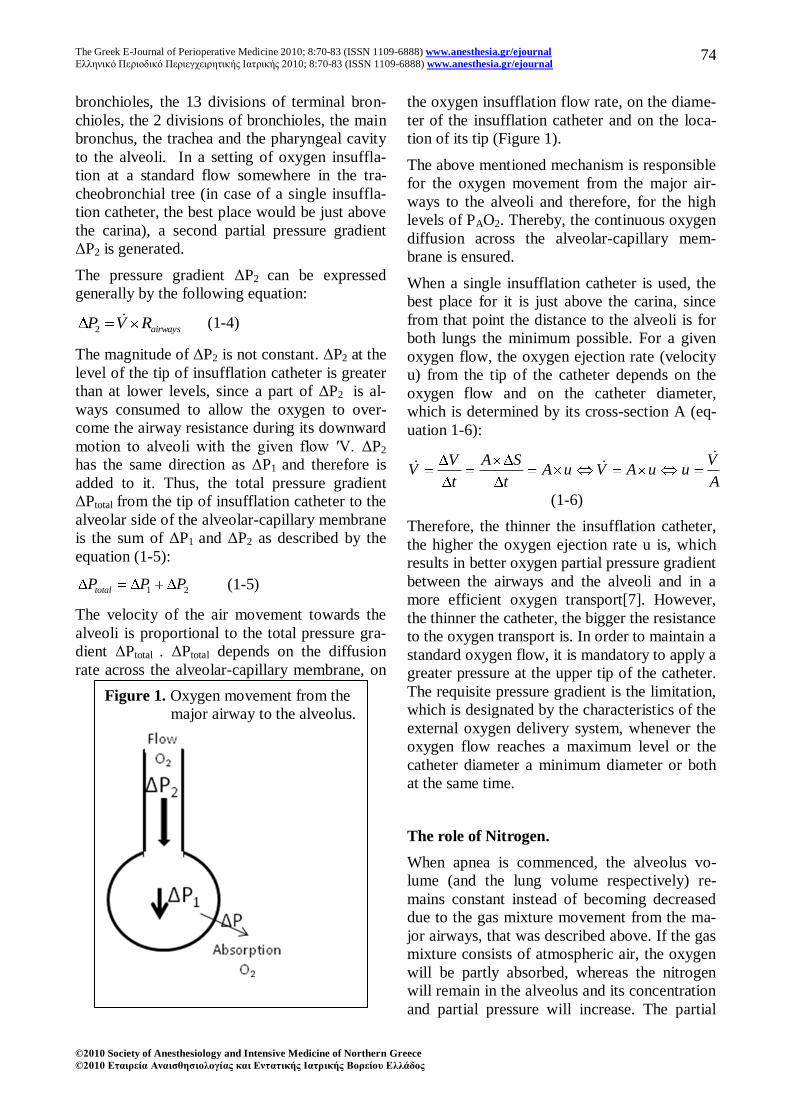
The velocity of the air movement towards the
alveoli is proportional to the total pressure gra-
dient ΔPtotal . ΔPtotal depends on the diffusion
rate across the alveolar-capillary membrane, on
the oxygen insufflation flow rate, on the diame-
ter of the insufflation catheter and on the loca-
tion of its tip (Figure 1).
The above mentioned mechanism is responsible
for the oxygen movement from the major air-
ways to the alveoli and therefore, for the high
levels of PAO2. Thereby, the continuous oxygen
diffusion across the alveolar-capillary mem-
brane is ensured.
When a single insufflation catheter is used, the
best place for it is just above the carina, since
from that point the distance to the alveoli is for
both lungs the minimum possible. For a given
oxygen flow, the oxygen ejection rate (velocity
u) from the tip of the catheter depends on the
oxygen flow and on the catheter diameter,
which is determined by its cross-section A (eq-
uation 1-6):
A
VuuAVuA
t
SA
t
VV
(1-6)
Therefore, the thinner the insufflation catheter,
the higher the oxygen ejection rate u is, which
results in better oxygen partial pressure gradient
between the airways and the alveoli and in a
more efficient oxygen transport[7]. However,
the thinner the catheter, the bigger the resistance
to the oxygen transport is. In order to maintain a
standard oxygen flow, it is mandatory to apply a
greater pressure at the upper tip of the catheter.
The requisite pressure gradient is the limitation,
which is designated by the characteristics of the
external oxygen delivery system, whenever the
oxygen flow reaches a maximum level or the
catheter diameter a minimum diameter or both
at the same time.
The role of Nitrogen.
When apnea is commenced, the alveolus vo-
lume (and the lung volume respectively) re-
mains constant instead of becoming decreased
due to the gas mixture movement from the ma-
jor airways, that was described above. If the gas
mixture consists of atmospheric air, the oxygen
will be partly absorbed, whereas the nitrogen
will remain in the alveolus and its concentration
and partial pressure will increase. The partial
Figure 1. Oxygen movement from the
major airway to the alveolus.
The Greek E-Journal of Perioperative Medicine 2010; 8:70-83 (ISSN 1109-6888) www.anesthesia.gr/ejournal
Ελληνικό Περιοδικό Περιεγχειρητικής Ιατρικής 2010; 8:70-83 (ISSN 1109-6888) www.anesthesia.gr/ejournal
©2010 Society of Anesthesiology and Intensive Medicine of Northern Greece
©2010 Δηαιρεία Αναιζθηζιολογίας και Δνηαηικής Ιαηρικής Βορείοσ Δλλάδος
75
pressure of nitrogen depends on the total pres-
sure in the alveolus (which depends on the at-
mospheric pressure) and on the nitrogen con-
centration in the gas mixture (equation 1-7).
22)(2 iNOHBA FPPNP (1-7)
This procedure results almost immediate (in
2min in humans) in a decrease of the alveolar
partial pressure of oxygen (PAO2) and hypox-
ia[12].
If the gas mixture consists of pure oxygen, the
oxygen that is absorbed is replaced by the same
amount of oxygen and not by nitrogen. There-
fore, PAO2 will decrease proportionally to the
increase of PACO2 (3-6mmHg/min after the first
minutes). In this setting, hypoxia may occur af-
ter a longer time period.
In a setting, where before the apnea initiation,
ventilation (mechanical or spontaneous) with
pure oxygen is performed, there will be no ni-
trogen in the alveolus gas mixture and the initial
PAO2 will be 660mmHg. Provided that oxygen
is insufflated into the trachea and the patients
have patent airway, hypoxia will occur in this
case even later (theoretically after 100min).
Fraioli, Sheffer et al report another mechanism,
which explains how nitrogen contributes to the
PAO2 decrease[4]. Even when nitrogen has been
entirely washed out of the alveoli by hyperventi-
lation with pure oxygen, the nitrogen that is dis-
solved in blood diffuses across the alveolar-
capillary membrane due to the partial pressure
gradient between blood and alveolus. The nitro-
gen diffusion results in a PAO2 decline, which
depends on the duration of the pure oxygen ven-
tilation, on the total body nitrogen reservoir, on
the cardiac output, on the FRC and on the body
temperature.
The role of carbon dioxide.
Carbon dioxide (CO2) is much more soluble
than oxygen in water and undergoes a much
more rapid (20 times more) tissue diffusion, de-
spite the fact that its vapor density is bigger (ta-
ble 1-1)[10]. Carbon dioxide is generated in the
mitochondria and is then transported in the cy-
toplasm, in the interstitium, in the venous blood
and in the pulmonary capillaries. During this
transport its partial pressure is decreased. Then,
carbon dioxide diffuses across the alveolar-
capillary membrane and is then washed out of
the alveolus by ventilation.
Carbon dioxide diffuses across the alveolar-
capillary membrane and is converted from the
liquid to the gas phase. Carbon dioxide diffu-
sion depends on the parameters that are de-
scribed in the equation 1-1. Due to the greater
diffusion capacity of carbon dioxide, the requi-
site partial pressure gradient is much less
(6mmHg) compared to the one required for the
oxygen diffusion (60mmHg)[10]. In the pulmo-
nary capillaries carbonic acid is dissociated to
carbon dioxide and water under the action of
carbonic anhydrase (equation 1-8).
H2CO3 H2O + CO2 (1-8)
Then, carbon dioxide diffuses across the alveo-
lar-capillary membrane and is washed out to the
atmospheric air. Carbon dioxide elimination
depends on its production rate and on the venti-
lation pattern. For a given carbon dioxide pro-
duction rate, PACO2 is inverse proportional to
the lung ventilation[13]. During apnea there are
no active respirations and therefore no active
gas movement. This results in carbon dioxide
accumulation in the alveolus, since carbon dio-
xide is not washed out. After the first minutes
of apnea, the increase rate of PACO2 is 3-
6mmHg/min, whereas PAO2 decreases[12].
Oxygen insufflation during apnea at a flow big-
ger from the oxygen absorption rate is consi-
dered to contribute to a progressive alveolar gas
mixture replacement. The replacement rate de-
pends on all parameters that influence the oxy-
gen ejection rate through the tip of the catheter.
The partial alveolar gas mixture replacement
contributes to the carbon dioxide wash out and
therefore to a limited reduction of PACO2 and
PaCO2 increase rate.
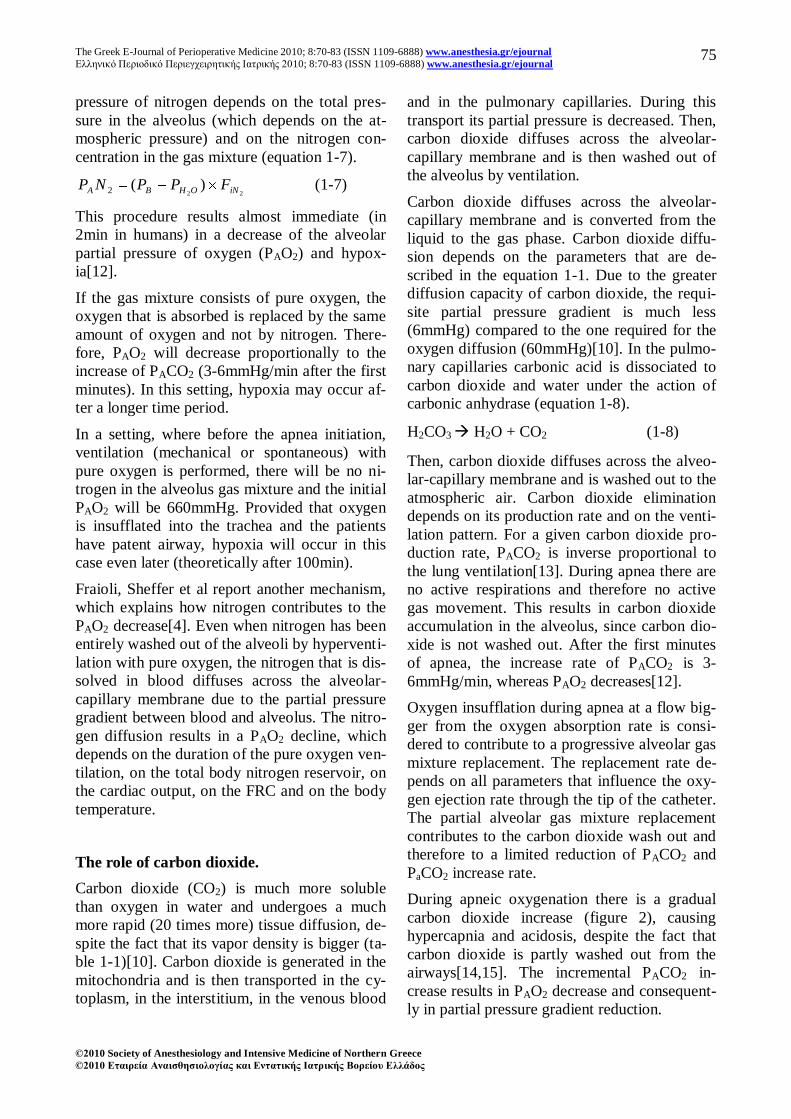
During apneic oxygenation there is a gradual
carbon dioxide increase (figure 2), causing
hypercapnia and acidosis, despite the fact that
carbon dioxide is partly washed out from the
airways[14,15]. The incremental PACO2 in-
crease results in PAO2 decrease and consequent-
ly in partial pressure gradient reduction.
The Greek E-Journal of Perioperative Medicine 2010; 8:70-83 (ISSN 1109-6888) www.anesthesia.gr/ejournal
Ελληνικό Περιοδικό Περιεγχειρητικής Ιατρικής 2010; 8:70-83 (ISSN 1109-6888) www.anesthesia.gr/ejournal
©2010 Society of Anesthesiology and Intensive Medicine of Northern Greece
©2010 Δηαιρεία Αναιζθηζιολογίας και Δνηαηικής Ιαηρικής Βορείοσ Δλλάδος
76
PATHOPHYSIOLOGIC CHANGES DUR-
ING APNEIC OXYGENATION
Hypercapnia results in hydrogen ions [H+] blood
concentration increase and consequently in aci-
dosis. Buffers are substances that minimize the
acid-base changes. One of the most important
extracellular buffers in blood is the bicarbonate
buffer system (H2CO3/HCO3-). Its action is de-
scribed by the following equation (1-8):
H2O+CO2 H2CO3 H++HCO3
- (1-8)
The initial step is catalyzed by the enzyme car-
bonic anhydrase. Carbonic acid dissociates then
immediately into [H+] and [HCO3
-]. This reac-
tion is accelerated by [CO2] rise. The relation-
ship between pH and buffer pKa may be calcu-
lated by the Henderson-Hasselbalch equation
(equation 1-9), where 0.03 stands for the solu-
bility coefficient of carbon dioxide in plasma:
2*03.0
log 3'
aCOP
HCOpKpH (1-9)
The Henderson-Hasselbalch equation can be
transformed into the Henderson equation, which
has no logarithms (equation 1-10):
3
2*24HCO
PH
aCO
(1-10)
where [H+] is expressed as nEq/L, PaCO2 as
mmHg and [HCO3-] as mEq/L.
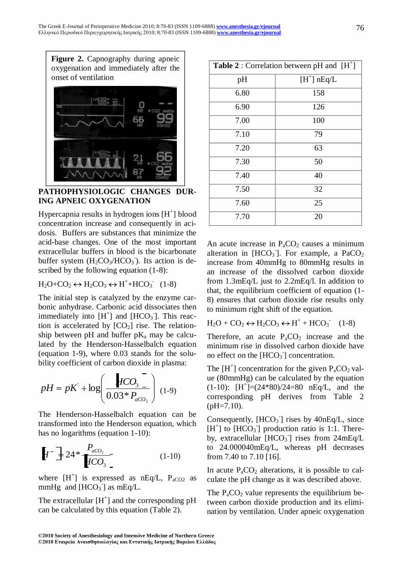
The extracellular [H+] and the corresponding pH
can be calculated by this equation (Table 2).
Table 2 : Correlation between pH and [Η+]
pH [H+] nEq/L
6.80 158
6.90 126
7.00 100
7.10 79
7.20 63
7.30 50
7.40 40
7.50 32
7.60 25
7.70 20
An acute increase in PaCO2 causes a minimum
alteration in [HCO3-]. For example, a PaCO2
increase from 40mmHg to 80mmHg results in
an increase of the dissolved carbon dioxide
from 1.3mEq/L just to 2.2mEq/l. In addition to
that, the equilibrium coefficient of equation (1-
8) ensures that carbon dioxide rise results only
to minimum right shift of the equation.
H2O + CO2 H2CO3 H+ + HCO3
- (1-8)
Therefore, an acute PaCO2 increase and the
minimum rise in dissolved carbon dioxide have
no effect on the [HCO3-] concentration.
The [H+] concentration for the given PaCO2 val-
ue (80mmHg) can be calculated by the equation
(1-10): [Η+]=(24*80)/24=80 nEq/L, and the
corresponding pH derives from Table 2
(pH=7.10).
Consequently, [HCO3-] rises by 40nEq/L, since
[H+] to [HCO3
-] production ratio is 1:1. There-
by, extracellular [HCO3-] rises from 24mEq/L
to 24.000040mEq/L, whereas pH decreases
from 7.40 to 7.10 [16].
In acute PaCO2 alterations, it is possible to cal-
culate the pH change as it was described above.
The PaCO2 value represents the equilibrium be-
tween carbon dioxide production and its elimi-
nation by ventilation. Under apneic oxygenation
Figure 2. Capnography during apneic
oxygenation and immediately after the
onset of ventilation
The Greek E-Journal of Perioperative Medicine 2010; 8:70-83 (ISSN 1109-6888) www.anesthesia.gr/ejournal
Ελληνικό Περιοδικό Περιεγχειρητικής Ιατρικής 2010; 8:70-83 (ISSN 1109-6888) www.anesthesia.gr/ejournal
©2010 Society of Anesthesiology and Intensive Medicine of Northern Greece
©2010 Δηαιρεία Αναιζθηζιολογίας και Δνηαηικής Ιαηρικής Βορείοσ Δλλάδος
77
carbon dioxide is no longer washed out of the
lungs, since there is no ventilation, and this re-
sults in annihilation of the partial pressure gra-
dient, which is the reason for the incremental
PaCO2 increase. The PaCO2 increase rate de-
pends on the body temperature, on the alertness
or sedation of the patient, on the underlying pa-
thology (fever, sepsis, trauma, operative stress,
tremor), on the mechanical ventilation, on the
use of muscle relaxants etc. The PaCO2 increase
results in acidosis, which is the main mechan-
ism through which apnea oxygenation affects
the different systems.
The principal effects of hypercapnia and subse-
quent acidosis include the decreased myocardial
contractility and the suppression of the smooth
muscle fibers[17], which are compensated by
the increased secretion of catecholamine, caused
by hypercapnia. According to experimental stu-
dies in rats, hypercapnia induces an increase in
coronary circulation. This seems to be the result
of the release of nitric oxid (NO) and the simul-
taneous activation of ATP channels[18]. More-
over, hypercapnia and acidosis are responsible
for an increased tendency to fibrillation and oth-
er arrhythmias. Increased carbon dioxide levels
have an indirect effect on the vasomotor center
in brain, which induces a huge sympathetic ac-
tion, causing vasoconstriction of blood vessels
and increase of systemic vascular resistance and
blood pressure[19]. However, as acidosis be-
comes more excessive and concentration of
[H+] and [K+] increase, the main effect is dila-
tation of blood vessels and decrease of systemic
vascular resistance [19]. When apneic oxygena-
tion is applied for a prolonged time period
(>30min), there is a big possibility that it will be
necessary to support the circulation with vaso-
constrictors (such as phenylephrine or norepi-
nephrine).
In an unpublished experimental study in pigs
(doctorate thesis of Kolettas A with the title
“Actions of Apneic Oxygenation on the Circula-
tory and Respiratory Systems under General
Anesthesia”), the systemic vascular resistance
(SVR) increased during the first 20min after the
initiation of apneic oxygenation. Then the SVR
decreased and reached lower levels than before
apneic oxygenation was commenced. SVR re-
mained low even after the piglets were recon-
nected to the ventilator (after 40min). After
10min of mechanical ventilation and restoration
of the acid-base homeostasis, SVR returned to
the initial values.
Hypoxic pulmonary vasoconstriction (HPV) is a
physiologic response of resistance pulmonary
arteries to alveolar hypoxia. HPV regulates ven-
tilation-perfusion matching, aiming to the re-
duction of shunt fraction and the optimization
of systemic PaO2. The core mechanism of HPV
is the smooth muscle cell of the resistance pul-
monary arteries. The mechanism of HPV is very
complex. The Redox theory proposes the coor-
dinated action of a redox sensor (the proximal
mitochondrial electron transport chain) that ge-
nerates a diffusible mediator (a reactive oxygen
species (ROS)) that regulates an effector protein
(voltage gated potassium (Kv) and calcium
channels). Inhibition of oxygen-sensitive Kv
channels depolarizes pulmonary artery smooth
muscle cells, resulting in activation of Ca+2
channels and in Ca+2
influx and vasoconstric-
tion [23]. Hypoxia and hypercapnia induced
acidosis triggers the above cascade causing an
increase of pulmonary vascular resistance
(PVR).
The above mentioned experimental study of un-
published doctorate thesis of Kolettas A dem-
onstrated a statistical significant increase of
PVR after the initiation of apneic oxygenation,
which returned to normal values after the pig-
lets were reconnected to the ventilator.
In the study of Lynch et al, hypercapnia caused
constriction of the isolated pulmonary arteries
of rats. However, this response was not present
when the endothelium of the vessels was re-
moved. Furthermore, the non specific inhibition
of the nitric oxide synthase (NOS) by 10-3
M L-
NAME (L-nitro-arginine mehthyl ester) also
diminished this response. In conclusion, the da-
ta of this study suggest that hypercapnia causes
constriction of the pulmonary vessels, which is
related to the endothelium and the reduced pro-
duction of NO [24].
Hypercapnia causes alterations in the cerebral
blood flow and in the consciousness level. Pa-
The Greek E-Journal of Perioperative Medicine 2010; 8:70-83 (ISSN 1109-6888) www.anesthesia.gr/ejournal
Ελληνικό Περιοδικό Περιεγχειρητικής Ιατρικής 2010; 8:70-83 (ISSN 1109-6888) www.anesthesia.gr/ejournal
©2010 Society of Anesthesiology and Intensive Medicine of Northern Greece
©2010 Δηαιρεία Αναιζθηζιολογίας και Δνηαηικής Ιαηρικής Βορείοσ Δλλάδος
78
rallel to the PaCO2 rise, the cerebral blood flow
increases in a rate of 1-2ml/100g/min for every
1mmHg [17]. Hypercapnic induced acidosis ac-
tivates the endothelial NOS, via prostaglandin
PGE2. This process depends on K+
and Ca2+
channels. This results in vasodilatation of the
cerebral circulation and cerebral blood flow in-
crease [25].
When PaCO2 reaches 60-75mmHg, the non-
anaesthetized patient starts to experience dysp-
nea. This results in stimulation of the respiratory
center and in increase of the frequency and the
depth of the respiratory efforts.
PaCO2 values that exceed 90-120mmHg, induce
carbon dioxide anesthesia[26]. Moreover, the
inhalation of a gas mixture with more than 30%
carbon dioxide induces anesthesia, which is
complicated by convulsions[27]. At carbon dio-
xide levels greater than 150mmHg, the respira-
tory center is suppressed and a vicious cycle is
evoked, which may lead to death[28].
In the literature there is a report of one patient
who recovered after hypercapnia. He underwent
a cosmetic surgery of 4-6 hours duration under
manual ventilation with a face mask. His oxy-
genation was preserved at acceptable levels
(SaO2>90%). The initial blood gases analysis
revealed a severe respiratory acidosis (pH=6.60
and PaCO2=375 mmHg). The patient was then
intubated and after a period of mechanical ven-
tilation and a return of plasma pH towards nor-
mal, he woke up without any neurological defi-
cit[20].
CLINICAL APPLICATION OF APNEIC
OXYGENATION
Since apneic oxygenation can maintain PaO2
above normal levels for a short but important
time period, this technique can be applied in a
cannot intubate- cannot ventilate (CICV) sit-
uation (jet ventilation is an alternative technique
of apneic oxygenation). In fact, this method
provides us with time, until a more effective
and permanent way of ventilation is ensured or
until muscle relaxants action is reversed and
spontaneous ventilation starts again.
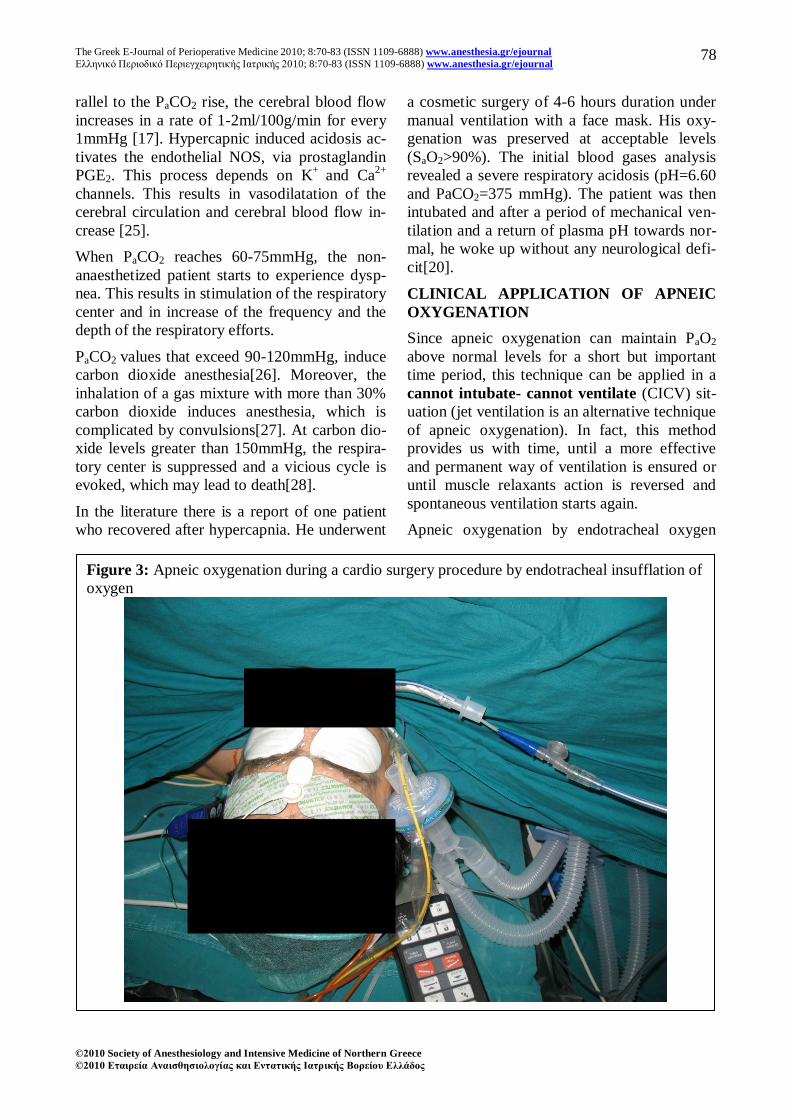
Apneic oxygenation by endotracheal oxygen
Figure 3: Apneic oxygenation during a cardio surgery procedure by endotracheal insufflation of
oxygen
The Greek E-Journal of Perioperative Medicine 2010; 8:70-83 (ISSN 1109-6888) www.anesthesia.gr/ejournal
Ελληνικό Περιοδικό Περιεγχειρητικής Ιατρικής 2010; 8:70-83 (ISSN 1109-6888) www.anesthesia.gr/ejournal
©2010 Society of Anesthesiology and Intensive Medicine of Northern Greece
©2010 Δηαιρεία Αναιζθηζιολογίας και Δνηαηικής Ιαηρικής Βορείοσ Δλλάδος
79
insufflation is applied during apnea test for the
diagnosis of brain death. Thereby, a sufficient
oxygenation is ensured for an acceptable time
period (for at least 10min). In the mean while
we observe for any respiratory efforts due to the
carbon dioxide increase. The application of oth-
er ventilation techniques is not recommended,
since they interfere in a more intense way in the
carbon dioxide removal and they cause some
kind of respiratory efforts, which deteriorate the
value of the test.
Apneic oxygenation can be applied during some
surgical or radiological procedures, where it is
mandatory to have absolutely no movement of
the lungs, the thoracic cage, the diaphragm and
the abdomen for a short time period. Such pro-
cedures are conducted in cardio and thoracic
surgery and in surgery of the trachea, bronchus
and larynx.
In coronary surgery the use of the internal
mammary artery graft has been proven to be su-
perior to the venous graft in regard to the long
term patency. Moreover, there is no need for
central anastomosis, when the mammary artery
graft is used, which results in minimizing the
risk of an atheromatic plug to be detached from
the inner wall of the aorta leading to embolism
of the cerebral circulation. However, during the
internal mammary artery harvest, the cognate
lung might be expanded in the surgical field,
causing difficulties in the harvest and increasing
the risk of possible injury of the graft, especial-
ly when PEEP is applied. One strategy that may
help us to deal with this situation is the applica-
tion of ventilation with small tidal volumes,
high respiratory rates and a low level of PEEP
or ZEEP. However, even this ventilation pattern
may induce an increase in the lung volumes in
emphysematic patients, whose lung compliance
is above normal levels. Another alternative
might be the one-lung ventilation technique,
which requires the insertion and positioning of a
double lumen endotracheal tube, a method that
is associated with some risks. A third alterna-
tive would be the application of apneic oxyge-
nation, which provides the best condition for
the internal mammary harvest (figure 3).
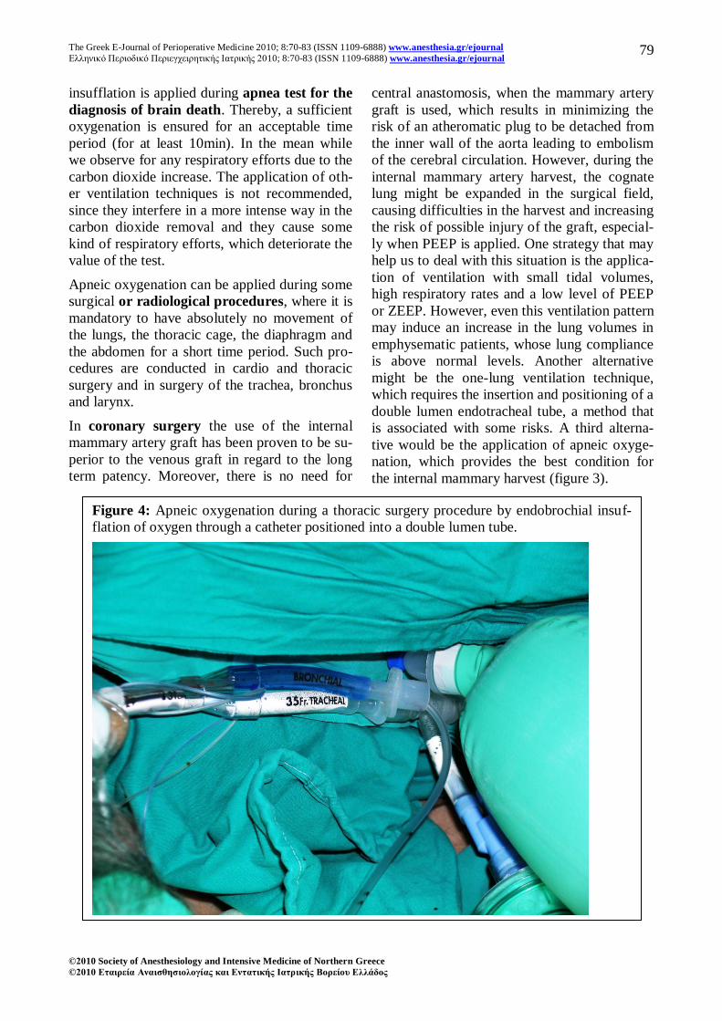
Figure 4: Apneic oxygenation during a thoracic surgery procedure by endobrochial insuf-
flation of oxygen through a catheter positioned into a double lumen tube.
The Greek E-Journal of Perioperative Medicine 2010; 8:70-83 (ISSN 1109-6888) www.anesthesia.gr/ejournal
Ελληνικό Περιοδικό Περιεγχειρητικής Ιατρικής 2010; 8:70-83 (ISSN 1109-6888) www.anesthesia.gr/ejournal
©2010 Society of Anesthesiology and Intensive Medicine of Northern Greece
©2010 Δηαιρεία Αναιζθηζιολογίας και Δνηαηικής Ιαηρικής Βορείοσ Δλλάδος
80
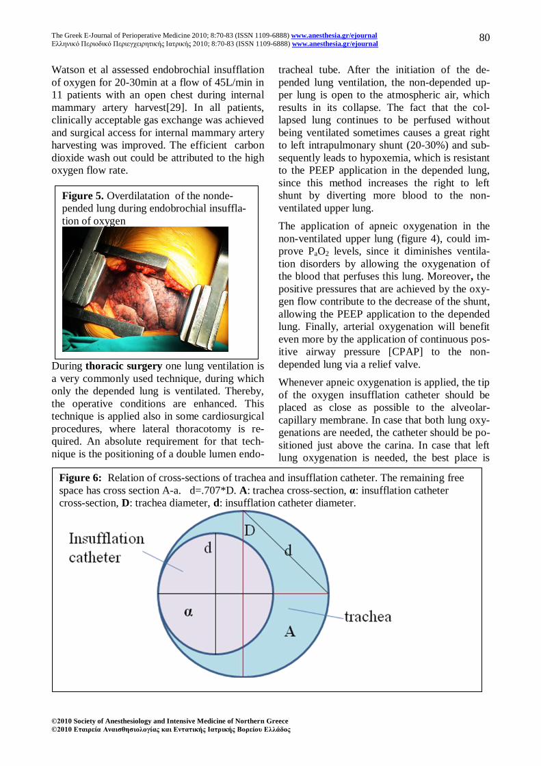
Watson et al assessed endobrochial insufflation
of oxygen for 20-30min at a flow of 45L/min in
11 patients with an open chest during internal
mammary artery harvest[29]. In all patients,
clinically acceptable gas exchange was achieved
and surgical access for internal mammary artery
harvesting was improved. The efficient carbon
dioxide wash out could be attributed to the high
oxygen flow rate.
During thoracic surgery one lung ventilation is
a very commonly used technique, during which
only the depended lung is ventilated. Thereby,
the operative conditions are enhanced. This
technique is applied also in some cardiosurgical
procedures, where lateral thoracotomy is re-
quired. An absolute requirement for that tech-
nique is the positioning of a double lumen endo-
tracheal tube. After the initiation of the de-
pended lung ventilation, the non-depended up-
per lung is open to the atmospheric air, which
results in its collapse. The fact that the col-
lapsed lung continues to be perfused without
being ventilated sometimes causes a great right
to left intrapulmonary shunt (20-30%) and sub-
sequently leads to hypoxemia, which is resistant
to the PEEP application in the depended lung,
since this method increases the right to left
shunt by diverting more blood to the non-
ventilated upper lung.
The application of apneic oxygenation in the
non-ventilated upper lung (figure 4), could im-
prove PaO2 levels, since it diminishes ventila-
tion disorders by allowing the oxygenation of
the blood that perfuses this lung. Moreover, the
positive pressures that are achieved by the oxy-
gen flow contribute to the decrease of the shunt,
allowing the PEEP application to the depended
lung. Finally, arterial oxygenation will benefit
even more by the application of continuous pos-
itive airway pressure [CPAP] to the non-
depended lung via a relief valve.
Whenever apneic oxygenation is applied, the tip
of the oxygen insufflation catheter should be
placed as close as possible to the alveolar-
capillary membrane. In case that both lung oxy-
genations are needed, the catheter should be po-
sitioned just above the carina. In case that left
lung oxygenation is needed, the best place is
Figure 5. Overdilatation of the nonde-
pended lung during endobrochial insuffla-
tion of oxygen
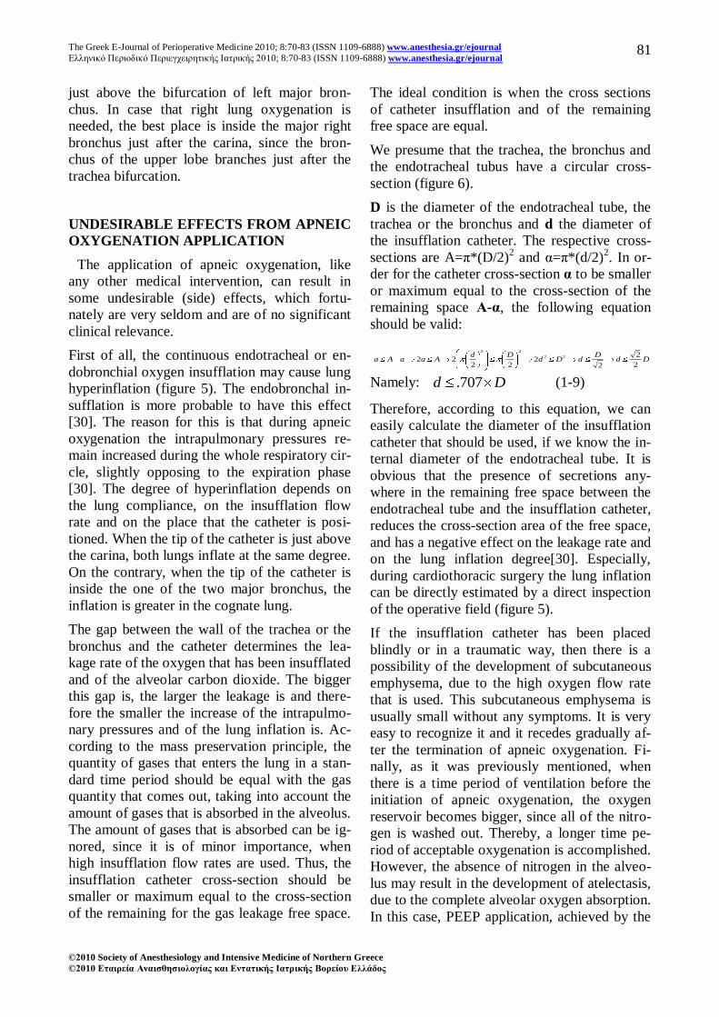
Figure 6: Relation of cross-sections of trachea and insufflation catheter. The remaining free
space has cross section A-a. d=.707*D. A: trachea cross-section, α: insufflation catheter
cross-section, D: trachea diameter, d: insufflation catheter diameter.
The Greek E-Journal of Perioperative Medicine 2010; 8:70-83 (ISSN 1109-6888) www.anesthesia.gr/ejournal
Ελληνικό Περιοδικό Περιεγχειρητικής Ιατρικής 2010; 8:70-83 (ISSN 1109-6888) www.anesthesia.gr/ejournal
©2010 Society of Anesthesiology and Intensive Medicine of Northern Greece
©2010 Δηαιρεία Αναιζθηζιολογίας και Δνηαηικής Ιαηρικής Βορείοσ Δλλάδος
81
just above the bifurcation of left major bron-
chus. In case that right lung oxygenation is
needed, the best place is inside the major right
bronchus just after the carina, since the bron-
chus of the upper lobe branches just after the
trachea bifurcation.
UNDESIRABLE EFFECTS FROM APNEIC
OXYGENATION APPLICATION
The application of apneic oxygenation, like
any other medical intervention, can result in
some undesirable (side) effects, which fortu-
nately are very seldom and are of no significant
clinical relevance.
First of all, the continuous endotracheal or en-
dobronchial oxygen insufflation may cause lung
hyperinflation (figure 5). The endobronchal in-
sufflation is more probable to have this effect
[30]. The reason for this is that during apneic
oxygenation the intrapulmonary pressures re-
main increased during the whole respiratory cir-
cle, slightly opposing to the expiration phase
[30]. The degree of hyperinflation depends on
the lung compliance, on the insufflation flow
rate and on the place that the catheter is posi-
tioned. When the tip of the catheter is just above
the carina, both lungs inflate at the same degree.
On the contrary, when the tip of the catheter is
inside the one of the two major bronchus, the
inflation is greater in the cognate lung.
The gap between the wall of the trachea or the
bronchus and the catheter determines the lea-
kage rate of the oxygen that has been insufflated
and of the alveolar carbon dioxide. The bigger
this gap is, the larger the leakage is and there-
fore the smaller the increase of the intrapulmo-
nary pressures and of the lung inflation is. Ac-
cording to the mass preservation principle, the
quantity of gases that enters the lung in a stan-
dard time period should be equal with the gas
quantity that comes out, taking into account the
amount of gases that is absorbed in the alveolus.
The amount of gases that is absorbed can be ig-
nored, since it is of minor importance, when
high insufflation flow rates are used. Thus, the
insufflation catheter cross-section should be
smaller or maximum equal to the cross-section
of the remaining for the gas leakage free space.
The ideal condition is when the cross sections
of catheter insufflation and of the remaining
free space are equal.
We presume that the trachea, the bronchus and
the endotracheal tubus have a circular cross-
section (figure 6).
D is the diameter of the endotracheal tube, the
trachea or the bronchus and d the diameter of
the insufflation catheter. The respective cross-
sections are A=π*(D/2)2 and α=π*(d/2)
2. In or-
der for the catheter cross-section α to be smaller
or maximum equal to the cross-section of the
remaining space Α-α, the following equation
should be valid:
Namely: Dd 707. (1-9)
Therefore, according to this equation, we can
easily calculate the diameter of the insufflation
catheter that should be used, if we know the in-
ternal diameter of the endotracheal tube. It is
obvious that the presence of secretions any-
where in the remaining free space between the
endotracheal tube and the insufflation catheter,
reduces the cross-section area of the free space,
and has a negative effect on the leakage rate and
on the lung inflation degree[30]. Especially,
during cardiothoracic surgery the lung inflation
can be directly estimated by a direct inspection
of the operative field (figure 5).
If the insufflation catheter has been placed
blindly or in a traumatic way, then there is a
possibility of the development of subcutaneous
emphysema, due to the high oxygen flow rate
that is used. This subcutaneous emphysema is
usually small without any symptoms. It is very
easy to recognize it and it recedes gradually af-
ter the termination of apneic oxygenation. Fi-
nally, as it was previously mentioned, when
there is a time period of ventilation before the
initiation of apneic oxygenation, the oxygen
reservoir becomes bigger, since all of the nitro-
gen is washed out. Thereby, a longer time pe-
riod of acceptable oxygenation is accomplished.
However, the absence of nitrogen in the alveo-
lus may result in the development of atelectasis,
due to the complete alveolar oxygen absorption.
In this case, PEEP application, achieved by the
DdD
dDdDd
AaaAa2
2
22
2222 22
22
The Greek E-Journal of Perioperative Medicine 2010; 8:70-83 (ISSN 1109-6888) www.anesthesia.gr/ejournal
Ελληνικό Περιοδικό Περιεγχειρητικής Ιατρικής 2010; 8:70-83 (ISSN 1109-6888) www.anesthesia.gr/ejournal
©2010 Society of Anesthesiology and Intensive Medicine of Northern Greece
©2010 Δηαιρεία Αναιζθηζιολογίας και Δνηαηικής Ιαηρικής Βορείοσ Δλλάδος
82
use of a specific release valve, can partially in-
hibit the development of atelectatic lung re-
gions, due to denitrogenation.
REFERENCES
1. Draper WB, Whitehead RW, Spencer JN.
Studies on diffusion respiration. Anesthesi-
ology 1947; 8:524-33.
2. Frumin JM, Epstein RM, Cohen G. Apneic
oxygenation in man. Anesthesiology 1959;
20:789-98.
3. Millar RA, Morris ME. Apneic oxygenation
in adrenalectomized dogs. Anesthesiology
1961; 22:433-9
4. Fraioli RL, Sheffer LA, Steffenson JL. Pul-
monary and cardiovascular effects of apneic
oxygenation in man. Anesthesiology 1973;
39:588-96.
5. Pesenti A I , Kolobow T, Buckhold DK,
Pierce JE, Huang HI, Chen V. Prevention of
Hyaline Membrane Disease in Premature
Lambs by Apneic Oxygenation and Extra-
corporeal Carbon Dioxide Removal. Inten-
sive Care Med 1982; 8:11-7.
6. Nielsen ND, Kjærgaard B, Koefoed-Nielsen
J, Steensen CO, Larsson A. Apneic Oxyge-
nation Combined With Extracorporeal Arte-
riovenous Carbon Dioxide Removal Pro-
vides Sufficient Gas Exchange in Experi-
mental Lung Injury. ASAIO Journal 2008;
54:401-5.
7. Cook TM, Wolf AR, Henderson AJW.
Changes in blood-gas tensions during apneic
oxygenation in pediatric patients. Br J
Anaesth 1998; 81:338-42.
8. Smyth E, Egan DT. Apneic Oxygenation
Associated with Patient-Controlled Analge-
sia. J Clin Anesth 1998; 10:499-501.
9. Homdahl MH. Apneic diffusion oxygena-
tion. Acta Chir Scand 1956; 212S: 1-128.
10. Andrew B Lumb; Nunn‟s Applied Respira-
tory Physiology, 6th
edition. Philadelphia:
Elsevier 2005, pp139-40.
11. Diffusion of Oxygen and Carbon Dioxide
through the Respiratory Membrane. In: Ar-
thur C Guyton, John E Hall; Textbook of
Medical Physiology 9th
edition. Philadel-
phia: WB Saunders 1996, p503.
12. Andrew B Lumb; Nunn‟s Applied Respira-
tory Physiology, 6th
edition. Philadelphia:
Elsevier 2005, pp159-60.
13. Andrew B Lumb; Nunn‟s Applied Respira-
tory Physiology, 6th
edition. Philadelphia:
Elsevier 2005, p157.
14. Benditt J, Pollock M, Roa J, Celli B. Trans-
tracheal delivery of gas decreases the oxy-
gen cost of breathing. Am Rev Respir Dis
1993; 147:1207-10.
15. Nahum A, Burke WC, Ravenscraft SA, et
al. Lung mechanics and gas exchange dur-
ing pressure-control ventilation in dogs:
augmentation of CO2 elimination by an in-
tratracheal catheter. Am Rev Respir Dis
1992; 146:965-73.
16. Acid-Base Balance. In: Morgan GE, Mik-
hail MS, Murray MJ, eds. Clinical Anesthe-
siology. New York: Lange 4th
ed, 2006,
pp711-12.
17. Andrew B Lumb; Nunn‟s Applied Respira-
tory Physiology, 6th
edition. Philadelphia:
Elsevier 2005, pp329-31.
18. Heintz A, Damm M, Brand M, Koch T,
Deussen A. Coronary flow regulation in
mouse heart during hypercapnic acidosis:
role of NO and its compensation during
eNOS impairment. Cardiovasc Res 2008;
77:188-96.
19. Local Control of Blood Flow in Response to
Tissue Needs. In: Arthur C Guyton, John E
Hall; Textbook of Medical Physiology 11th
edition. Philadelphia: Elsevier 2006, p203.
20. Potkin RT, Swenson ER. Resuscitation from
severe acute hypercapnia. Determinants of
tolerance and survival. Chest 1992;
102:1742-5.
21. Goldstein B, Shannon DC, Todres ID. Su-
percarbia in children: clinical course and
outcome. Crit Care Med 1990; 18:166-8.
The Greek E-Journal of Perioperative Medicine 2010; 8:70-83 (ISSN 1109-6888) www.anesthesia.gr/ejournal
Ελληνικό Περιοδικό Περιεγχειρητικής Ιατρικής 2010; 8:70-83 (ISSN 1109-6888) www.anesthesia.gr/ejournal
©2010 Society of Anesthesiology and Intensive Medicine of Northern Greece
©2010 Δηαιρεία Αναιζθηζιολογίας και Δνηαηικής Ιαηρικής Βορείοσ Δλλάδος
83
22. Slinger P, Blundell PE, Metcalf IR. Man-
agement of massive grain aspiration. Anes-
thesiology 1997; 87:993-5.
23. Moudgil R, Michelakis E, Archer S. Hypox-
ic pulmonary vasoconstriction. J Appl Phy-
siol 2005; 98:390-403.
24. Lynch F, Sweeney M, O'Regan RG,
McLoughlin P. Hypercapnia-induced con-
traction in isolated pulmonary arteries is en-
dothelium-dependent. Respir Physiol 2000;
121:65-74.
25. Najarian T, Marrache AM, Dumont I, Hardy
P, Beauchamp MH, Hou X, Peri K, Gobeil F
Jr, Varma DR, Chemtob S. Prolonged
hypercapnia-evoked cerebral hyperemia via
K(+) channel- and prostaglandin E2-
dependent endothelial nitric oxide synthase
induction. Circ Res 2000; 87:1149-56.
26. Refsum HE. Relationship between state of
consiousness and arterial hypoxemia and
hypercapnia in patients with pulmonary in-
sufficiency. Clin Sci 1963; 25:361-7.
27. Leake CD, Waters RM. The anesthetic
properties of carbon dioxide. J Pharmacol
Exp Ther 1928; 33:280-1.
28. Respiratory insufficiency-Pathophysiology,
Diagnosis, Oxygen Therapy. In: Arthur C
Guyton, John E Hall; Textbook of Medical
Physiology 9th
edition. Philadelphia: WB
Saunders 1996, p543.
29. Watson J N R, Szarko R, Mackenzie F C,
Sequeira J A, Barnas M G. Continuous En-
dobronchial Insufflation During Internal
Mammary Artery Harvest. Anesth Analg
1992; 75:219-25.
30. Pinsky MR, Delgado E, Hete B. The Effect
of Tracheal Gas insufflation on Gas Ex-
change Efficiency; Anesth Analg 2006;
103:1213-9.
CORRESPONDENCE:
Alexandros Kolettas: Anaesthesiologist, Thessaloniki Heart Institute, St. Luke‟s Hospital
Address: Kardia, 57500 Thessaloniki, p.o.box 087, Greece
tel: (0030) 2310430197
e-mail: [email protected]
Keywords: Apneic oxygenation, Tracheal Gas insufflation, pulmonary gas exchange