Affinity Labeling of Equine Anti-β-lactoside Antibodies *
4
BIOCHEMISTRY (1963), Biochem. J. 88,220. (1958), Biochim. Biophys. Acta 29,587. 651. 761, Habeeb, A. F. S. A., Cassidy, H. G., and Singer, S. J. Howard, A. N., and Wild, F. (1957), Biochem. J. 65, Johnson, L. N., and Phillips, D. C. (1965), Nature 206, Karush, F. (1957), J. Am. Chem. Soc. 79,3380. Koshland, M. E., Englberger, F. M., and Shapanka, Latham, H. G., May, E. F., and Mossetig, E. (1950), Metzger, H., and Mannik, M. (1964), J. Exptl. Med. Metzger, H., Wofsy, L., and Singer, S. J. (1963), Metzger, H., Wofsy, L., and Singer, S. J. (1964), R. (1966), Biochemistry 5,641. J. Org. Chem. 15,884. 120,765. Biochemistry 2, 979. Proc. Natl. Acad. Sci. U. S. 51,612. Seidman, M., and Link, K. P. (1950), J. Am. Chem. SOC. 72,4325. Singer, S. J., and Doolittle, R. F. (1966), Science 153: 13. Singer, S. J., Wofsy, L., and Good, A. H. (1964), in Molecular and Cellular Basis of Antibody Forma- tion, Sterzl, J., Ed., Prague, Czechoslovakian Academy of Sciences, p 135. Tabachnick, M., and Sobotka, H. (1959), J. Bid. Chem. 234,1726. Thompson, A., Wolfrom, M. L., and Pascu, E. (1963), Methods Carbohydrate Chem. 2,215. Utsumi, S., and Karush, F. (1964), Biochemistry 3, 1329. Wofsy, L., Metzger, H., and Singer, S. J. (1962), Bio- chemistry I, 1031. Affinity Labeling of Equine Anti-,8-lactoside Antibodies* Leon Wofsy, Norman R. Klinman, and Fred Karusht ABSTRACT: Equine anti-Lac antibodies of two varieties, yG and yG(T)* isolated from a single horse, have been labeled specifically with OD-Lac as affinity labeling re- agent. The reaction conditions were the same as those employed in affinity labeling of pooled rabbit anti-Lac antibodies with the same reagent. Azo spectra of H and L chains from the labeled equine antibodies of both A ffinity labeling studies with rabbit anti-Lac' and anti-Gal antibodies point to particular chemical features shared by antibodies of similar (saccharide) specificity which distinguish them from antibodies of grossly dif- ferent (benzenoid) specificity (Wofsy et ai., 1967). The results produced in affinity labeling of antibenzenoid hapten antibodies (namely, the selective modification at * From the Department of Bacteriology and Immunology, University of California, Berkeley, California (L. W,), and the Department of Microbiology, School of Medicine, University of Pennsylvania, Philadelphia, Pa. (N. R. I<. and F. I<.). Receiued March 13, 1967. This work was aided by grants from the Na- tional Institutes of Health (AI-06610 and HE-06293) and the National Science Foundation (GB-3070). t Recipient of a Public Health research career award from the National Institute of Allergy and Infectious Diseases. Abbreviations used: anti-Lac and anti-Gal, antibodies specific for the respective haptenic groups p-azophenyl 6- lactoside and p-azophenyl p-galactoside; OD-Lac, the reagent o-diazoniumphenyl 6-lactoside; SDS, sodium dodecyl sulfate; PPO, 2,5-diphenyloxazole; POPOP, 1,4-bis[2-(5-phenyloxazolyl)]- 1988 benzene. varieties are qualitatively indistinguishable. However, these spectra show that the residues selectively modified by affinity labeling of the equine anti-Lac antibodies differ distinctly from those labeled on both H and L chains of the rabbit antibodies. The possible genetic or evolutionary significance of these characteristic distinc- tions is not yet established. active sites of tyrosine residues on both heavy (H) and light (L) chains) have been shown in the case of anti- dinitrophenyl antibodies to hold for all of four species thus far studied (A. H. Good, Z. Ovary, and S. J. Singer, unpublished data). In the present study equine 7s anti-Lac antibodies of two structural varieties, yG and YG(T)~ (Rockey et a/., 1964; Klinman et ul., 1964, 1965), have been labeled with the same reagent, OD-Lac, and under the same conditions as were employed in af- finity labeling of rabbit anti-Lac antibodies (Wofsy et ul., 1967). This was formerly designated yA on the basis of its carbo- hydrate content and behavior in immunoelectrophoresis. Recent observations have indicated its identity with the T component of equine antiserum as judged by its antigenic identity with similarly prepared diphtheria antitoxin and tetanus antitoxin (R. Genes, N. R. Klinman, R. Hirsh, and F. Karush, unpub- lished data). In view of the antigenic similarity betheen rG- immunoglobulin and the T component (Weir and Porter, 1966) and the extensive homology of the C-terminal sequences o their heavy chains (Weir et a/., 1966), the designation rG(T). suggested by Weir et al. (1966) will be used instead of yA. WOFSY, KLINMAN, AND KARUSH
Transcript of Affinity Labeling of Equine Anti-β-lactoside Antibodies *
B I O C H E M I S T R Y
(1963), Biochem. J . 88,220.
(1958), Biochim. Biophys. Acta 29,587.
651.
761,
Habeeb, A. F. S. A., Cassidy, H. G., and Singer, S. J.
Howard, A. N., and Wild, F. (1957), Biochem. J . 65,
Johnson, L. N., and Phillips, D. C. (1965), Nature 206,
Karush, F. (1957), J. Am. Chem. Soc. 79,3380. Koshland, M. E., Englberger, F. M., and Shapanka,
Latham, H. G., May, E. F., and Mossetig, E. (1950),
Metzger, H., and Mannik, M. (1964), J . Exptl. Med.
Metzger, H., Wofsy, L., and Singer, S. J. (1963),
Metzger, H., Wofsy, L., and Singer, S. J. (1964),
R. (1966), Biochemistry 5,641.
J. Org. Chem. 15,884.
120,765.
Biochemistry 2, 979.
Proc. Natl. Acad. Sci. U. S. 51,612. Seidman, M., and Link, K. P. (1950), J . Am. Chem.
SOC. 72,4325. Singer, S. J., and Doolittle, R. F. (1966), Science 153:
13. Singer, S. J., Wofsy, L., and Good, A. H. (1964), in
Molecular and Cellular Basis of Antibody Forma- tion, Sterzl, J., Ed., Prague, Czechoslovakian Academy of Sciences, p 135.
Tabachnick, M., and Sobotka, H. (1959), J . Bid. Chem. 234,1726.
Thompson, A., Wolfrom, M. L., and Pascu, E. (1963), Methods Carbohydrate Chem. 2,215.
Utsumi, S., and Karush, F. (1964), Biochemistry 3, 1329.
Wofsy, L., Metzger, H., and Singer, S. J. (1962), Bio- chemistry I , 1031.
Affinity Labeling of Equine Anti-,8-lactoside Antibodies*
Leon Wofsy, Norman R. Klinman, and Fred Karusht
ABSTRACT: Equine anti-Lac antibodies of two varieties, yG and yG(T)* isolated from a single horse, have been labeled specifically with OD-Lac as affinity labeling re- agent. The reaction conditions were the same as those employed in affinity labeling of pooled rabbit anti-Lac antibodies with the same reagent. Azo spectra of H and L chains from the labeled equine antibodies of both
A ffinity labeling studies with rabbit anti-Lac' and anti-Gal antibodies point to particular chemical features shared by antibodies of similar (saccharide) specificity which distinguish them from antibodies of grossly dif- ferent (benzenoid) specificity (Wofsy et ai., 1967). The results produced in affinity labeling of antibenzenoid hapten antibodies (namely, the selective modification at
* From the Department of Bacteriology and Immunology, University of California, Berkeley, California (L. W,), and the Department of Microbiology, School of Medicine, University of Pennsylvania, Philadelphia, Pa. (N. R. I<. and F. I<.). Receiued March 13, 1967. This work was aided by grants from the Na- tional Institutes of Health (AI-06610 and HE-06293) and the National Science Foundation (GB-3070).
t Recipient of a Public Health research career award from the National Institute of Allergy and Infectious Diseases.
Abbreviations used: anti-Lac and anti-Gal, antibodies specific for the respective haptenic groups p-azophenyl 6- lactoside and p-azophenyl p-galactoside; OD-Lac, the reagent o-diazoniumphenyl 6-lactoside; SDS, sodium dodecyl sulfate; PPO, 2,5-diphenyloxazole; POPOP, 1,4-bis[2-(5-phenyloxazolyl)]-
1988 benzene.
varieties are qualitatively indistinguishable. However, these spectra show that the residues selectively modified by affinity labeling of the equine anti-Lac antibodies differ distinctly from those labeled on both H and L chains of the rabbit antibodies. The possible genetic or evolutionary significance of these characteristic distinc- tions is not yet established.
active sites of tyrosine residues on both heavy (H) and light (L) chains) have been shown in the case of anti- dinitrophenyl antibodies to hold for all of four species thus far studied (A. H. Good, Z . Ovary, and S. J. Singer, unpublished data). In the present study equine 7s anti-Lac antibodies of two structural varieties, yG and YG(T)~ (Rockey et a/., 1964; Klinman et ul., 1964, 1965), have been labeled with the same reagent, OD-Lac, and under the same conditions as were employed in af- finity labeling of rabbit anti-Lac antibodies (Wofsy et ul., 1967).
This was formerly designated yA on the basis of its carbo- hydrate content and behavior in immunoelectrophoresis. Recent observations have indicated its identity with the T component of equine antiserum as judged by its antigenic identity with similarly prepared diphtheria antitoxin and tetanus antitoxin (R. Genes, N. R. Klinman, R. Hirsh, and F. Karush, unpub- lished data). In view of the antigenic similarity betheen rG- immunoglobulin and the T component (Weir and Porter, 1966) and the extensive homology of the C-terminal sequences o their heavy chains (Weir et a/ . , 1966), the designation rG(T). suggested by Weir et al. (1966) will be used instead of yA.
W O F S Y , K L I N M A N , A N D K A R U S H
V O L . 6, N O . 7, J U L Y 1 9 6 7
Materials and Methods
Preparation of Antigens and Antibodies. The prepara- tion of antigens has been previously described (Karush, 1957). The antibodies used in this study, as well as the immunization and purification procedures by which they were obtained, have been presented in detail elsewhere (Klinman et al., 1965, 1966). The particular antibody preparations employed were isolated from a pool of serum taken from the same horse during the 12th to 42nd week of a second course of immunization. Injec- tions at biweekly or monthly intervals consisted of 40- 80 mg of antigen dispersed in incomplete Freund's ad- juvant or alum precipitated. The immunizing antigen during this period was the azoconjugate of the Lac haptenic group and hemocyanin, Limulus polyphemus.
After the multiple types of anti-Lac antibodies were coprecipitated from the serum and freed from antigen as before, y G and yG(T) immunoglobulins were sep- arated by placing purified antibody (10 mg/ml) on a DEAE-cellulose column (Whatman DE 52 microgranu- lar 1 mequiv/g) equilibrated to 0.01 M sodium phosphate buffer (pH 7.85) and increasing the ionic strength by stepwise addition of sodium chloride first to a concen- tration of 0.04 M and then to 0.3 M. The purity of the y G and yG(T) preparations was established as before on the basis of their characteristic immunoelectrophoretic patterns developed with goat antisera to heavy chains of equine immunoglobulins.
The binding of tritium-labeled p-(p '-dimethylamino- benzene)azophenyl P-lactoside by y G and rG(T) anti- bodies was measured by equilibrium dialysis by a pre- viously described technique (Karush, 1957). Free-dye concentrations were obtained by scintillation counting in Bray's (1960) phosphor solution. The r G antibody preparation bound hapten at 25" with a KO of 8.2 X lo6 M-1; the yG(T) showed a KO of 1.8 X lo7 M-I. More extensive analysis of the binding properties of these anti- bodies is presented elsewhere (Karush and Sela, 1967).
Reagents and Afinity Labeling Procedures. The re- agents, buffers, labeling procedures, work-up of labeled proteins, and spectral assay methods were the same as those employed in affinity labeling of rabbit anti-Lac antibodies with OD-Lac (Wofsy et af . , 1967). Where the H and L chains of labeled antibodies were examined, the antibodies were mildly reduced and alkylated and then, following labeling, the chains were separated on Sephadex G-100 in 1 N propionic acid exactly as in the rabbit studies.
Spectral determinations were made with a Zeiss PMQ I1 spectrophotometer. Protein concentrations were ob- tained from values of ODzsomr in 0.5% SDS-0.02 M
sodium phosphate buffer (pH 6.2) based on the following extinction coefficients for 1 % protein solutions: for y G and yG(T) antibodies, E:Z mp 14.7; for both types of H chains, e:grnr 15.4; and for L chains, e ; z r n p 14.0 (Rockey, 1967).
Tritiated o-aminophenyl P-lactoside was synthesized by the general methods described in Wofsy et al. (1967) starting with the condensation of acetobromolactose with tritiated o-nitrophenol (New England Nuclear).
WAVELENGTH, MILLIMICRONS
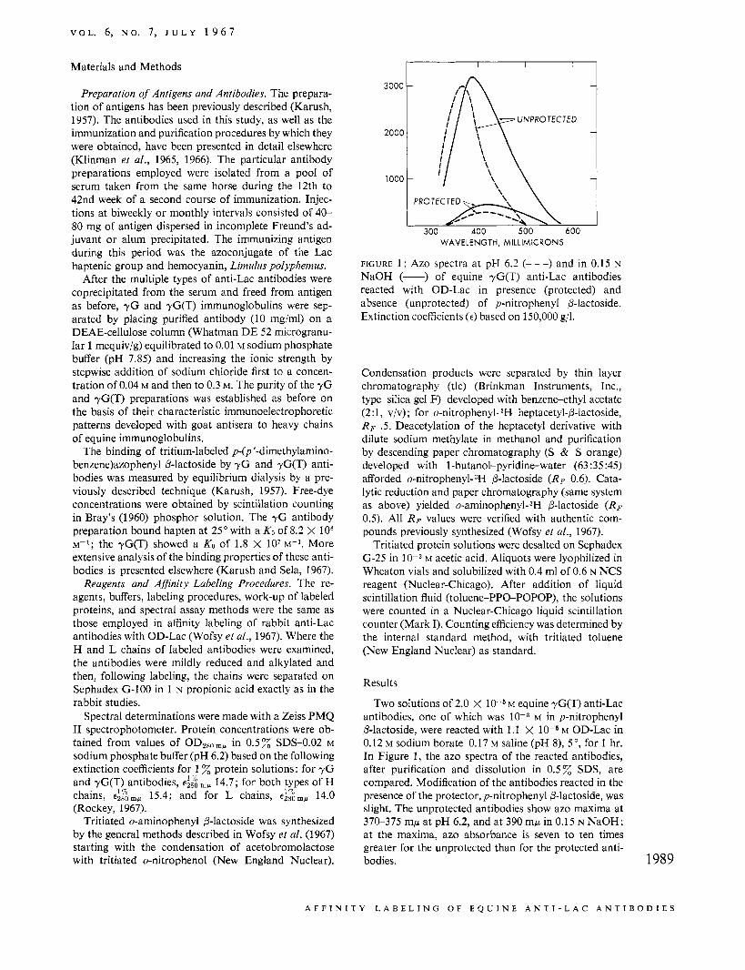
FIGURE 1: Azo spectra at pH 6.2 (---) and in 0.15 N NaOH (-) of equine yG(T) anti-Lac antibodies reacted with OD-Lac in presence (protected) and absence (unprotected) of p-nitrophenyl P-lactoside. Extinction coefficients (E) based on 150,000 g/l.
Condensation products were separated by thin layer chromatography (tlc) (Brinkman Instruments, Inc., type silica gel F) developed with benzene-ethyl acetate (2 : 1, V/V) ; for o-nitrophenyl- 3H heptacetyl-@-lactoside, R F .5. Deacetylation of the heptacetyl derivative with dilute sodium methylate in methanol and purification by descending paper chromatography (S & S orange) developed with 1-butanol-pyridine-water (63 :35:45) afforded o-nitrophenyL3H 6-lactoside (RF 0.6). Cata- lytic reduction and paper chromatography (same system as above) yielded o-aminophenyl-3H P-lactoside (RF 0.5). All RF values were verified with authentic com- pounds previously synthesized (Wofsy et a[., 1967).
Tritiated protein solutions were desalted on Sephadex G-25 in 10-3 M acetic acid. Aliquots were lyophilized in Wheaton vials and solubilized with 0.4 ml of 0.6 N NCS reagent (Nuclear-Chicago). After addition of liquid scintillation fluid (toluene-PPGPOPOP), the solutions were counted in a Nuclear-Chicago liquid scintillation counter (Mark I). Counting efficiency was determined by the internal standard method, with tritiated toluene (New England Nuclear) as standard.
Results
Two solutions of 2.0 X l o - S M equine yG(T) anti-Lac antibodies, one of which was 10-3 M in p-nitrophenyl P-lactoside, were reacted with 1.1 X M OD-Lac in 0.12 M sodium borate-0.17 M saline (pH 8), 5" , for 1 hr. In Figure 1, the azo spectra of the reacted antibodies, after purification and dissolution in 0.5% SDS, are compared. Modification of the antibodies reacted in the presence of the protector, p-nitrophenyl P-lactoside, was slight. The unprotected antibodies show azo maxima at 370-375 mp at pH 6.2, and at 390 mp in 0.15 N NaOH; at the maxima, azo absorbance is seven to ten times greater for the unprotected than for the protected anti- bodies. 1989
A F F I N I T Y L A B E L I N G O F E Q U I N E A N T I - L A C A N T I B O D I E S
U l O C H E M l S T K Y
1 2 x 1 0 ~
e 6 ' 4 \
l y q f $ - j \ 0 400 600 400 600 400 600
\ 2 \ \ -. \ \\ '. \
WAVELENGTH, MILLIMICRONS
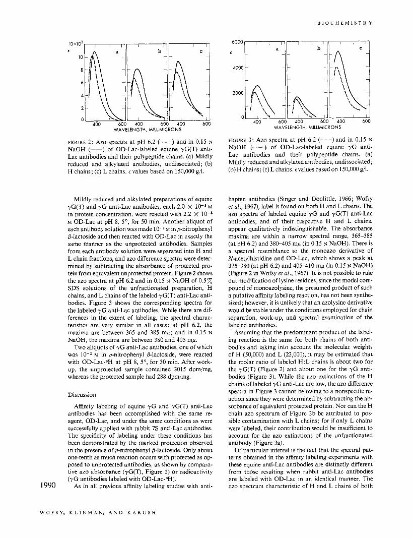
FIGURE 2: Azo spectra at pH 6.2 (- - -) and in 0.15 N NaOH (-) of OD-Lac-labeled equine yG(T) anti- Lac antibodies and their polypeptide chains. (a) Mildly reduced and alkylated antibodies, undissociated ; (b) H chains; (c) L chains. E values based on 150,000 g/l.
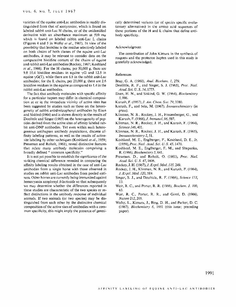
Mildly reduced and alkylated preparations of equine rG(T) and y G anti-Lac antibodies, each 2.0 X low6 M in protein concentration, were reacted with 2.2 X M OD-Lac at pH 8, 5" , for 50 min. Another aliquot of each antibody solution was made 1 0 - 3 ~ inp-nitrophenyl P-lactoside and then reacted with OD-Lac in exactly the same manner as the unprotected antibodies. Samples from each antibody solution were separated into H and L chain fractions, and azo difference spectra were deter- mined by subtracting the absorbance of protected pro- tein from equivalent unprotected protein. Figure 2 shows the azo spectra at pH 6.2 and in 0.15 N NaOH of 0.5% SDS solutions of the unfractionated preparation, H chains, and L chains of the labeled yG(T) anti-Lac anti- bodies. Figure 3 shows the corresponding spectra for the labeled y G anti-Lac antibodies. While there are dif- ferences in the extent of labeling, the spectral charac- teristics are very similar in all cases: at pH 6.2, the maxima are between 365 and 385 mp; and in 0.15 N
NaOH, the maxima are between 380 and 405 mp. Two aliquots of y G anti-Lac antibodies, one of which
was 10-3 M in p-nitrophenyl P-lactoside, were reacted with O D - L ~ C - ~ H at pH 8, 5", for 30 min. After work- up, the unprotected sample contained 3015 dpm/mg, whereas the protected sample had 288 dpm/mg.
Discussion
Affinity labeling of equine y G and yG(T) anti-Lac antibodies has been accomplished with the same re- agent, OD-Lac, and under the same conditions as were successfully applied with rabbit 7 s anti-Lac antibodies. The specificity of labeling under these conditions has been demonstrated by the marked protection observed in the presence of p-nitrophenyl P-lactoside. Only about one-tenth as much reaction occurs with protected as op- posed to unprotected antibodies, as shown by compara- tive azo absorbance (yG(T), Figure 1) or radioactivity (yG antibodies labeled with OD-Lac-3H).
As in all previous affinity labeling studies with anti- 1990
FIGURE 3 : Azo spectra at pH 6.2 (- - -) and in 0.15 N
NaOH (-) of OD-Lac-labeled equine y G anti- Lac antibodies and their polypeptide chains. (a) Mildly reduced and alkylated antibodies, undissociated ; (b) H chains; (c) L chains. E values based on 150,000 g/l.
hapten antibodies (Singer and Doolittle, 1966; Wofsy et al., 1967), label is found on both H and L chains. The azo spectra of labeled equine y G and yG(T) anti-Lac antibodies, and of their respective H and L chains, appear qualitatively indistinguishable. The absorbance maxima are within a narrow spectral range, 365-385 (at pH 6.2) and 380-405 mp (in 0.15 N NaOH). There is a spectral resemblance to the monoazo derivative of N-acetylhistidine and OD-Lac, which shows a peak at 375-380 (at pH 6.2) and 405-410 mp (in 0.15 N NaOH) (Figure 2 in Wofsy et al., 1967). It is not possible to rule out modification of lysine residues, since the model com- pound of monoazolysine, the presumed product of such a putative affinity labeling reaction, has not been synthe- sized; however, it is unlikely that an azolysine derivative would be stable under the conditions employed for chain separation, work-up, and spectral examination of the labeled antibodies.
Assuming that the predominant product of the label- ing reaction is the same for both chains of both anti- bodies and taking into account the molecular weights of H (50,000) and L (23,000), it may be estimated that the molar ratio of labeled H:L chains is about two for the yG(T) (Figure 2) and about one for the y G anti- bodies (Figure 3). While the azo extinctions of the H chains of labeled y G anti-Lac are low, the azo difference spectra in Figure 3 cannot be owing to a nonspecific re- action since they were determined by subtracting the ab- sorbance of equivalent protected protein. Nor can the H chain azo spectrum of Figure 3b be attributed to pos- sible contamination with L chains; for if only L chains were labeled, their contribution would be insufficient to account for the azo extinctions of the unfractionated antibody (Figure 3a).
Of particular interest is the fact that the spectral pat- terns obtained in the affinity labeling experiments with these equine anti-Lac antibodies are distinctly different from those resulting when rabbit anti-Lac antibodies are labeled with OD-Lac in an identical manner. The azo spectrum characteristic of H and L chains of both
W O F S Y , K L I N M A N ; A N D K A R U S H
V O L . 6, N O . 7, J U L Y 1 9 6 7
varieties of the equine anti-Lac antibodies is readily dis- tinguished from that of azotyrosine, which is found on labeled rabbit anti-Lac H chains, or of the unidentified derivative with an absorbance maximum at 510 mp which is found on labeled rabbit anti-Lac L chains (Figures 4 and 5 in Wofsy et ai., 1967). In view of the possibility that histidine is the residue selectively labeled on both chains of both classes of the equine anti-Lac antibodies, it may be relevant to consider data on the comparative histidine content of the chains of equine and rabbit anti-Lac antibodies (Rockey, 1967; Koshland et ai., 1966). For the H chains, per 50,000 g, there are 9.8-10.4 histidine residues in equine yG and 12.5 in equine yG(T), while there are 6.8 in the rabbit anit-Lac antibodies; for the L chains, per 23,000 g, there are 2.6 histidine residues in the equine as compared to 1.4 in the rabbit anti-Lac antibodies.
The fact that antibody molecules with specific affinity for a particular hapten may differ in chemical composi- tion at or in the immediate vicinity of active sites has been suggested by studies such as those on the hetero- geneity of rabbit antidinitrophenyl antibodies by Eisen and Siskind (1964) and is shown directly in the results of Doolittle and Singer (1965) on the heterogeneity of pep- tides derived from the active sites of affinity labeled rab- bit anti-DNP antibodies. Yet even within such hetero- geneous antihapten antibody populations, discrete af- finity labeling patterns, as well as the results of active- site labeling by other techniques (Koshland et al., 1959; Pressman and Roholt, 1961), reveal distinctive features that relate many antibody molecules comprising a broadly defined “ common specificity.”
It is not yet possible to establish the significance of the striking chemical difference revealed in comparing the affinity labeling results obtained in the case of anti-Lac antibodies from a single horse with those observed in studies on rabbit anti-Lac antibodies from pooled anti- sera. Other horses are currently being immunized against hemocyanin azophenyl P-lactoside so that subsequently we may determine whether the differences reported in these studies are characteristic of the two species or re- flect distinctions in the antibody response of individual animals. If two animals (or two species) may be dis- tinguished from each other by the distinctive chemical composition of the active sites of antibodies with a com- mon specificity, this might imply the presence of geneti-
cally determined variants (or of species specific evolu- tionary alternatives) in the amino acid sequences of those portions of the H and L chains that define anti- body specificity.
Acknowledgment
The contribution of John Kimura in the synthesis of reagents and the protector hapten used in this study is gratefully acknowledged.
References
Bray, G. A. (1960), Anal. Biochem. I , 279. Doolittle, R. F., and Singer, S. J. (1965), Proc. Natl.
Eisen, H. N., and Siskind, G. W. (1964), Biochemistry
Karush, F. (1957), J . Am. Chern. Soc. 79, 3380. Karush, F., and Sela, M. (1967), Immunochemistry (in
Klinman, N. R., Rockey, J. H., Frauenberger, G., and
Klinman, N. R., Rockey, J. H., and Karush, F. (1964))
Klinman, N. R., Rockey, J. H., and Karush, F. (1965),
Koshland, M. E., Englberger, F., Koshland, D. E., Jr.
Koshland, M. E., Englberger, F. M., and Shapanka,
Pressman, D., and Roholt, 0. (1961), Proc. Natl.
Rockey, J. H. (1967), J . Exptl. Med. 125, 249. Rockey, J. H., Klinman, N. R., and Karush, F. (1964),
J . Exptl. Med. 120, 589. Singer, S. J., and Doolittle, R. F. (1966)) Science 153,
13. Weir, R. C., and Porter, R. R. (1966), Biochem. J . 100,
63. Weir, R. C. , Porter, R. R., and Givol, D. (1966),
Nature 212,205. Wofsy, L., Kimura, J., Bing, D. H., and Parker, D. C.
(1967), Biochemistry 6, 1981 (this issue; preceding
Acad. Sci. U. S. 54,1773.
3,996.
press).
Karush, F. (1966), .I. Zmmunol. 96,587.
Science 146,401.
Immunochemistry 2,51.
(1959), Proc. Natl. Acad. Sci. U. S. 45,1470.
R. (1966), Biochemisrry 5,641.
Acad. Sci. U. S . 47,1606.
paper).
1991
A I F I N I T Y L A U t L I N L O F L Q U I N E A N T I - L A C A N T I B O D I E S