Antibodies for Phospho-Protein Analysis
40
Antibodies for Phospho-Protein Analysis
Transcript of Antibodies for Phospho-Protein Analysis

Antibodies for Phospho-Protein Analysis
ひょすし 06.6.21 2:19 PM ページ 1
Antibodies for Phospho-Protein Analysis
細胞内シグナル伝達に関わっているタンパク質の活性化にはそのタンパク質に含まれているセリン/スレオニン、チロシン残基のリン酸化が大きく関わっており、様々なキナーゼやホスファターゼによって制御されています。リン酸化タンパク質は、T細胞/B細胞シグナリング、アポトーシス、細胞成
長コントロール、細胞周期、細胞骨格の再構成、転写などの重要な細胞プロセスで中心的な役割を果たしています。
リン酸化タンパク質の解析に広く汎用されているウエスタンブロッティング法、その他免疫沈降法、免疫組織染色法などに有用なPhospho-Specific Antibody(リン酸化部位特異的認
識抗体)シリーズ製品をご紹介いたします。
ひょすし 06.6.21 2:19 PM ページ 2
目 次
1 製品一覧⋯⋯⋯⋯⋯⋯⋯⋯⋯⋯⋯⋯⋯⋯⋯⋯⋯⋯⋯⋯⋯⋯⋯⋯⋯1-4
Tyrosine, Serine, and Theronine Phosphorylation Detection
Phospho-Specific Antibodies
Antibody Sampler Kit
2 製品概略 ⋯⋯⋯⋯⋯⋯⋯⋯⋯⋯⋯⋯⋯⋯⋯⋯⋯⋯⋯⋯⋯⋯⋯⋯5-31
各製品の簡単なBackgroundとウエスタンブロッティング法
による解析データ例を掲載しています。
3 製品一覧(CAT. NO.順)⋯⋯⋯⋯⋯⋯⋯⋯⋯⋯⋯⋯⋯⋯⋯⋯32-35
REACTHu HumanMs MouseRat RatD DogC ChickenF Frog
略号
APPS (Applications)WB Western blottingIF ImmunofluorescenceIHC ImmunohistochemistryFCM Flow cytometryIP Immunoprecipitation
商標Alexa Fluor® はMolecular Probes社の登録商標です。
【お願いおよびご注意事項】
本カタログに掲載の価格は2006年07月現在の希望小売価格です。
価格は予告なく変更される場合がありますのであらかじめご了承下さい。
また、記載の価格に消費税は含まれていません。
ご注文は必ずカタログ番号でお願い致します。
商品の交換および返品は、品質管理上ご容赦願います。
製品一覧
Tyrosine, Serine, and Theronine Phosphorylation DetectionDESCRIPTION CLONE REACT APPS FORMAT SIZE CAT. NO. 希望小売価格 PAGE
Phosphotyrosine PY20 Hu, Ms, Rat, D, C, F WB, IF, IHC, IP Purified 1 mg 610000 ¥19,000 5
WB, IP Biotin 50μg 610007 ¥43,000 5
WB, IP Biotin 150μg 610008 ¥78,000 5
WB HRPO 50μg 610011 ¥35,000 5
WB HRPO 150μg 610012 ¥66,000 5
Phosphotyrosine Polyclonal Hu, Ms, Rat, D, C, F WB, IF, IHC, FCM, IP Purified 50μg 610009 ¥31,000 5
WB, IF, IHC, FCM, IP Purified 150μg 610010 ¥53,000 5
Phosphotyrosine RC20 Hu, Ms, Rat, D, C, F WB AKP 50μg 610019 ¥43,000 5
WB AKP 150μg 610020 ¥78,000 5
WB, IP Biotin 50μg 610021 ¥43,000 5
WB, IP Biotin 150μg 610022 ¥78,000 5
WB HRPO 150μg 610024 ¥66,000 5
Phosphotyrosine PY69 Hu, Ms, Rat, D, C, F WB, IF, IHC, FCM, IP Purified 1 mg 610430 ¥19,000 5
IP Agarose 500μl 610015 ¥58,000 5
Phosphoserine 19 Hu, Rat WB, IF Purified 50μg 612546 ¥31,000 5
WB, IF Purified 150μg 612547 ¥53,000 5
Phosphoserine / Threonine 22a Hu, Rat WB, IF Purified 50μg 612548 ¥31,000 5
WB, IF Purified 150μg 612549 ¥53,000 5
Phospho-Specific AntibodiesDESCRIPTION CLONE REACT APPS FORMAT SIZE CAT. NO. 希望小売価格 PAGE
Actopaxin (pS8) J160-366 Hu WB Purified 0.1 mg 558374 ¥68,000 5
Akt (pS472/pS473) 104A282 Hu, Ms, Rat WB, IP Purified 50μg 550747 ¥38,000 6
Akt (pS473) J177-204.20 Hu WB Purified 0.1 mg 558368 ¥68,000 6
Akt (pT308) J1-223.371 Hu, Ms WB Purified 0.1 mg 558316 ¥68,000 6
Akt (pY326) K7-642 Ms WB Purified 0.1 mg 558384 ¥68,000 6
Bcr (pY177) J52-309 Hu WB Purified 0.1 mg 558248 ¥68,000 6
BLNK (pY84) J117-1278 Hu WB Purified 0.1 mg 558366 ¥68,000 6
Btk (pY551) & Itk (pY511) 24a/BTK (Y551) Hu WB Purified 0.1 mg 558034 ¥68,000 7
Caveolin (pY14) 56 Hu, Ms, Rat WB, IF, IHC, FCM Purified 50μg 611338 ¥31,000 7
WB, IF, IHC, FCM Purified 150μg 611339 ¥53,000 7
Caveolin 2 (pY27) 40/Caveolin 2 Hu WB Purified 0.1 mg 558364 ¥68,000 7
c-Cbl (pY700) 47 Hu WB, IF Purified 50μg 612304 ¥31,000 8
WB, IF Purified 150μg 612305 ¥53,000 8
c-Cbl (pY774) 29/c-Cbl (Y774) Hu WB Purified 0.1 mg 558035 ¥68,000 8
CD3ζ(CD247) (pY142) K25-407.69 Hu WB Purified 0.1 mg 558402 ¥68,000 8
CD22 (BL-CAM) (pY828) 46 Hu WB Purified 0.1 mg 558029 ¥68,000 8
CD22 (BL-CAM) (pY843) 12a Hu WB Purified 0.1 mg 558030 ¥68,000 8
CD45 (pS999) J143-1270 Hu WB Purified 0.1 mg 558376 ¥68,000 9
CD117 (c-kit) (pY568/pY570) K39-686 Hu WB Purified 0.1 mg 558390 ¥68,000 9
CD221 (IGF-1 Receptor) (pY950) J95-626 Hu WB Purified 0.1 mg 558373 ¥68,000 9
1
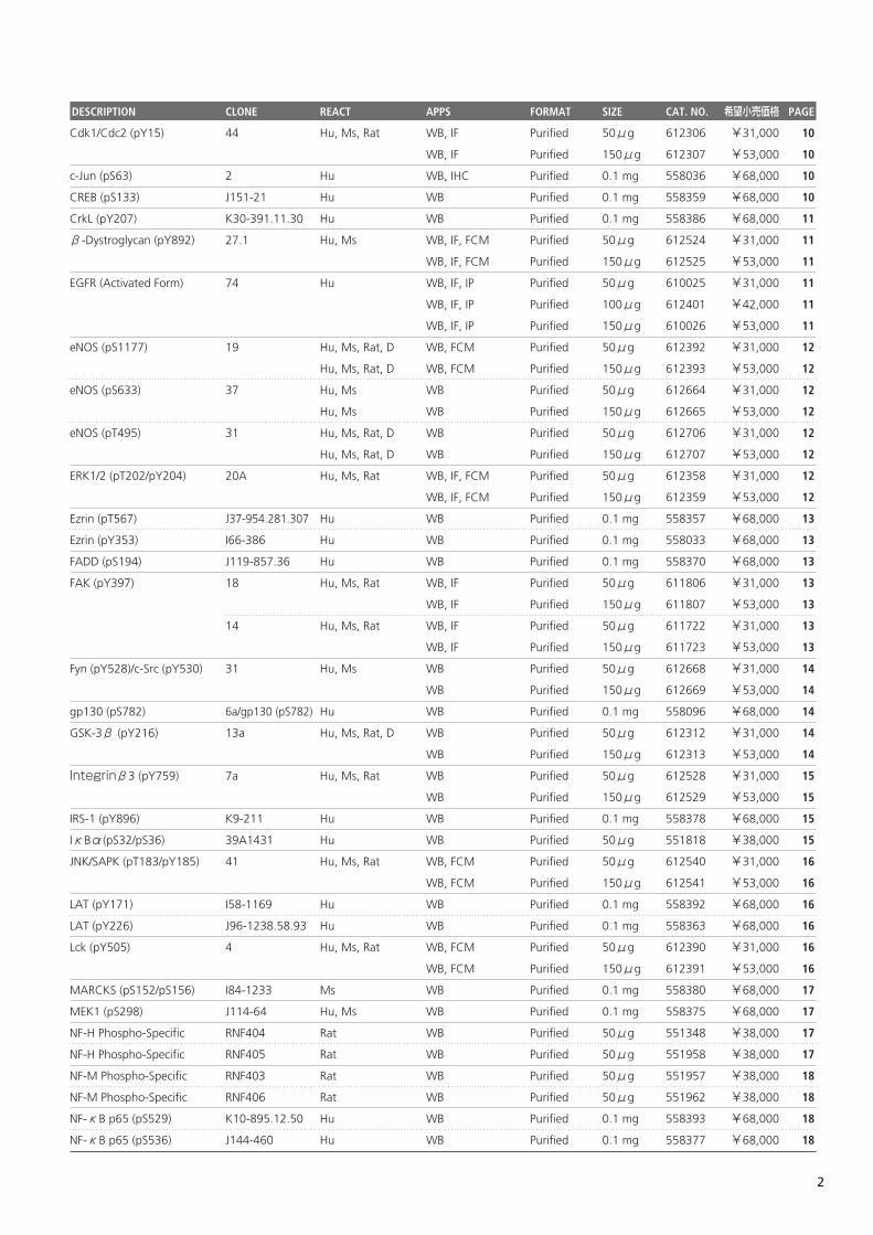
Cdk1/Cdc2 (pY15) 44 Hu, Ms, Rat WB, IF Purified 50μg 612306 ¥31,000 10
WB, IF Purified 150μg 612307 ¥53,000 10
c-Jun (pS63) 2 Hu WB, IHC Purified 0.1 mg 558036 ¥68,000 10
CREB (pS133) J151-21 Hu WB Purified 0.1 mg 558359 ¥68,000 10
CrkL (pY207) K30-391.11.30 Hu WB Purified 0.1 mg 558386 ¥68,000 11
β-Dystroglycan (pY892) 27.1 Hu, Ms WB, IF, FCM Purified 50μg 612524 ¥31,000 11
WB, IF, FCM Purified 150μg 612525 ¥53,000 11
EGFR (Activated Form) 74 Hu WB, IF, IP Purified 50μg 610025 ¥31,000 11
WB, IF, IP Purified 100μg 612401 ¥42,000 11
WB, IF, IP Purified 150μg 610026 ¥53,000 11
eNOS (pS1177) 19 Hu, Ms, Rat, D WB, FCM Purified 50μg 612392 ¥31,000 12
Hu, Ms, Rat, D WB, FCM Purified 150μg 612393 ¥53,000 12
eNOS (pS633) 37 Hu, Ms WB Purified 50μg 612664 ¥31,000 12
Hu, Ms WB Purified 150μg 612665 ¥53,000 12
eNOS (pT495) 31 Hu, Ms, Rat, D WB Purified 50μg 612706 ¥31,000 12
Hu, Ms, Rat, D WB Purified 150μg 612707 ¥53,000 12
ERK1/2 (pT202/pY204) 20A Hu, Ms, Rat WB, IF, FCM Purified 50μg 612358 ¥31,000 12
WB, IF, FCM Purified 150μg 612359 ¥53,000 12
Ezrin (pT567) J37-954.281.307 Hu WB Purified 0.1 mg 558357 ¥68,000 13
Ezrin (pY353) I66-386 Hu WB Purified 0.1 mg 558033 ¥68,000 13
FADD (pS194) J119-857.36 Hu WB Purified 0.1 mg 558370 ¥68,000 13
FAK (pY397) 18 Hu, Ms, Rat WB, IF Purified 50μg 611806 ¥31,000 13
WB, IF Purified 150μg 611807 ¥53,000 13
14 Hu, Ms, Rat WB, IF Purified 50μg 611722 ¥31,000 13
WB, IF Purified 150μg 611723 ¥53,000 13
Fyn (pY528)/c-Src (pY530) 31 Hu, Ms WB Purified 50μg 612668 ¥31,000 14
WB Purified 150μg 612669 ¥53,000 14
gp130 (pS782) 6a/gp130 (pS782) Hu WB Purified 0.1 mg 558096 ¥68,000 14
GSK-3β (pY216) 13a Hu, Ms, Rat, D WB Purified 50μg 612312 ¥31,000 14
WB Purified 150μg 612313 ¥53,000 14
Integrinβ3 (pY759) 7a Hu, Ms, Rat WB Purified 50μg 612528 ¥31,000 15
WB Purified 150μg 612529 ¥53,000 15
IRS-1 (pY896) K9-211 Hu WB Purified 0.1 mg 558378 ¥68,000 15
IκBα(pS32/pS36) 39A1431 Hu WB Purified 50μg 551818 ¥38,000 15
JNK/SAPK (pT183/pY185) 41 Hu, Ms, Rat WB, FCM Purified 50μg 612540 ¥31,000 16
WB, FCM Purified 150μg 612541 ¥53,000 16
LAT (pY171) I58-1169 Hu WB Purified 0.1 mg 558392 ¥68,000 16
LAT (pY226) J96-1238.58.93 Hu WB Purified 0.1 mg 558363 ¥68,000 16
Lck (pY505) 4 Hu, Ms, Rat WB, FCM Purified 50μg 612390 ¥31,000 16
WB, FCM Purified 150μg 612391 ¥53,000 16
MARCKS (pS152/pS156) I84-1233 Ms WB Purified 0.1 mg 558380 ¥68,000 17
MEK1 (pS298) J114-64 Hu, Ms WB Purified 0.1 mg 558375 ¥68,000 17
NF-H Phospho-Specific RNF404 Rat WB Purified 50μg 551348 ¥38,000 17
NF-H Phospho-Specific RNF405 Rat WB Purified 50μg 551958 ¥38,000 17
NF-M Phospho-Specific RNF403 Rat WB Purified 50μg 551957 ¥38,000 18
NF-M Phospho-Specific RNF406 Rat WB Purified 50μg 551962 ¥38,000 18
NF-κB p65 (pS529) K10-895.12.50 Hu WB Purified 0.1 mg 558393 ¥68,000 18
NF-κB p65 (pS536) J144-460 Hu WB Purified 0.1 mg 558377 ¥68,000 18
2
DESCRIPTION CLONE REACT APPS FORMAT SIZE CAT. NO. 希望小売価格 PAGE
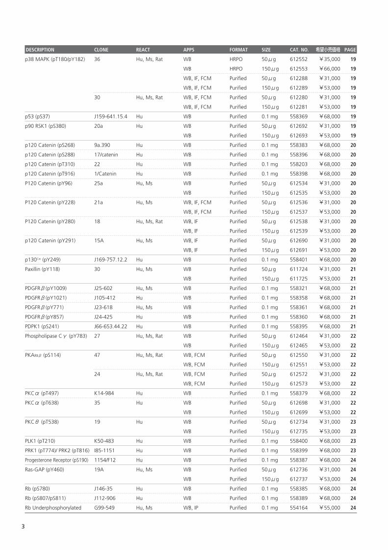
p38 MAPK (pT180/pY182) 36 Hu, Ms, Rat WB HRPO 50μg 612552 ¥35,000 19
WB HRPO 150μg 612553 ¥66,000 19
WB, IF, FCM Purified 50μg 612288 ¥31,000 19
WB, IF, FCM Purified 150μg 612289 ¥53,000 19
30 Hu, Ms, Rat WB, IF, FCM Purified 50μg 612280 ¥31,000 19
WB, IF, FCM Purified 150μg 612281 ¥53,000 19
p53 (pS37) J159-641.15.4 Hu WB Purified 0.1 mg 558369 ¥68,000 19
p90 RSK1 (pS380) 20a Hu WB Purified 50μg 612692 ¥31,000 19
WB Purified 150μg 612693 ¥53,000 19
p120 Catenin (pS268) 9a.390 Hu WB Purified 0.1 mg 558383 ¥68,000 20
p120 Catenin (pS288) 17/catenin Hu WB Purified 0.1 mg 558396 ¥68,000 20
p120 Catenin (pT310) 22 Hu WB Purified 0.1 mg 558203 ¥68,000 20
p120 Catenin (pT916) 1/Catenin Hu WB Purified 0.1 mg 558398 ¥68,000 20
P120 Catenin (pY96) 25a Hu, Ms WB Purified 50μg 612534 ¥31,000 20
WB Purified 150μg 612535 ¥53,000 20
P120 Catenin (pY228) 21a Hu, Ms WB, IF, FCM Purified 50μg 612536 ¥31,000 20
WB, IF, FCM Purified 150μg 612537 ¥53,000 20
P120 Catenin (pY280) 18 Hu, Ms, Rat WB, IF Purified 50μg 612538 ¥31,000 20
WB, IF Purified 150μg 612539 ¥53,000 20
p120 Catenin (pY291) 15A Hu, Ms WB, IF Purified 50μg 612690 ¥31,000 20
WB, IF Purified 150μg 612691 ¥53,000 20
p130Cas (pY249) J169-757.12.2 Hu WB Purified 0.1 mg 558401 ¥68,000 20
Paxillin (pY118) 30 Hu, Ms WB Purified 50μg 611724 ¥31,000 21
WB Purified 150μg 611725 ¥53,000 21
PDGFRβ(pY1009) J25-602 Hu, Ms WB Purified 0.1 mg 558321 ¥68,000 21
PDGFRβ(pY1021) J105-412 Hu WB Purified 0.1 mg 558358 ¥68,000 21
PDGFRβ(pY771) J23-618 Hu, Ms WB Purified 0.1 mg 558361 ¥68,000 21
PDGFRβ(pY857) J24-425 Hu WB Purified 0.1 mg 558360 ¥68,000 21
PDPK1 (pS241) J66-653.44.22 Hu WB Purified 0.1 mg 558395 ¥68,000 21
Phospholipase Cγ (pY783) 27 Hu, Ms, Rat WB Purified 50μg 612464 ¥31,000 22
WB Purified 150μg 612465 ¥53,000 22
PKARIIβ (pS114) 47 Hu, Ms, Rat WB, FCM Purified 50μg 612550 ¥31,000 22
WB, FCM Purified 150μg 612551 ¥53,000 22
24 Hu, Ms, Rat WB, FCM Purified 50μg 612572 ¥31,000 22
WB, FCM Purified 150μg 612573 ¥53,000 22
PKCα (pT497) K14-984 Hu WB Purified 0.1 mg 558379 ¥68,000 22
PKCα (pT638) 35 Hu WB Purified 50μg 612698 ¥31,000 22
WB Purified 150μg 612699 ¥53,000 22
PKCθ (pT538) 19 Hu WB Purified 50μg 612734 ¥31,000 23
WB Purified 150μg 612735 ¥53,000 23
PLK1 (pT210) K50-483 Hu WB Purified 0.1 mg 558400 ¥68,000 23
PRK1 (pT774)/ PRK2 (pT816) I85-1151 Hu WB Purified 0.1 mg 558399 ¥68,000 23
Progesterone Receptor (pS190) 1154/F12 Hu WB Purified 0.1 mg 558387 ¥68,000 24
Ras-GAP (pY460) 19A Hu, Ms WB Purified 50μg 612736 ¥31,000 24
WB Purified 150μg 612737 ¥53,000 24
Rb (pS780) J146-35 Hu WB Purified 0.1 mg 558385 ¥68,000 24
Rb (pS807/pS811) J112-906 Hu WB Purified 0.1 mg 558389 ¥68,000 24
Rb Underphosphorylated G99-549 Hu, Ms WB, IP Purified 0.1 mg 554164 ¥55,000 24
3
DESCRIPTION CLONE REACT APPS FORMAT SIZE CAT. NO. 希望小売価格 PAGE
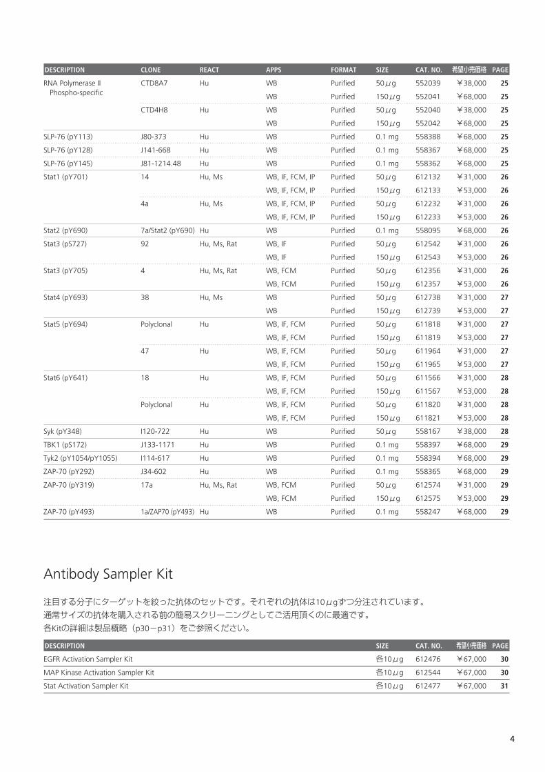
RNA Polymerase II CTD8A7 Hu WB Purified 50μg 552039 ¥38,000 25Phospho-specific WB Purified 150μg 552041 ¥68,000 25
CTD4H8 Hu WB Purified 50μg 552040 ¥38,000 25
WB Purified 150μg 552042 ¥68,000 25
SLP-76 (pY113) J80-373 Hu WB Purified 0.1 mg 558388 ¥68,000 25
SLP-76 (pY128) J141-668 Hu WB Purified 0.1 mg 558367 ¥68,000 25
SLP-76 (pY145) J81-1214.48 Hu WB Purified 0.1 mg 558362 ¥68,000 25
Stat1 (pY701) 14 Hu, Ms WB, IF, FCM, IP Purified 50μg 612132 ¥31,000 26
WB, IF, FCM, IP Purified 150μg 612133 ¥53,000 26
4a Hu, Ms WB, IF, FCM, IP Purified 50μg 612232 ¥31,000 26
WB, IF, FCM, IP Purified 150μg 612233 ¥53,000 26
Stat2 (pY690) 7a/Stat2 (pY690) Hu WB Purified 0.1 mg 558095 ¥68,000 26
Stat3 (pS727) 92 Hu, Ms, Rat WB, IF Purified 50μg 612542 ¥31,000 26
WB, IF Purified 150μg 612543 ¥53,000 26
Stat3 (pY705) 4 Hu, Ms, Rat WB, FCM Purified 50μg 612356 ¥31,000 26
WB, FCM Purified 150μg 612357 ¥53,000 26
Stat4 (pY693) 38 Hu, Ms WB Purified 50μg 612738 ¥31,000 27
WB Purified 150μg 612739 ¥53,000 27
Stat5 (pY694) Polyclonal Hu WB, IF, FCM Purified 50μg 611818 ¥31,000 27
WB, IF, FCM Purified 150μg 611819 ¥53,000 27
47 Hu WB, IF, FCM Purified 50μg 611964 ¥31,000 27
WB, IF, FCM Purified 150μg 611965 ¥53,000 27
Stat6 (pY641) 18 Hu WB, IF, FCM Purified 50μg 611566 ¥31,000 28
WB, IF, FCM Purified 150μg 611567 ¥53,000 28
Polyclonal Hu WB, IF, FCM Purified 50μg 611820 ¥31,000 28
WB, IF, FCM Purified 150μg 611821 ¥53,000 28
Syk (pY348) I120-722 Hu WB Purified 50μg 558167 ¥38,000 28
TBK1 (pS172) J133-1171 Hu WB Purified 0.1 mg 558397 ¥68,000 29
Tyk2 (pY1054/pY1055) I114-617 Hu WB Purified 0.1 mg 558394 ¥68,000 29
ZAP-70 (pY292) J34-602 Hu WB Purified 0.1 mg 558365 ¥68,000 29
ZAP-70 (pY319) 17a Hu, Ms, Rat WB, FCM Purified 50μg 612574 ¥31,000 29
WB, FCM Purified 150μg 612575 ¥53,000 29
ZAP-70 (pY493) 1a/ZAP70 (pY493) Hu WB Purified 0.1 mg 558247 ¥68,000 29
Antibody Sampler Kit
注目する分子にターゲットを絞った抗体のセットです。それぞれの抗体は10μgずつ分注されています。
通常サイズの抗体を購入される前の簡易スクリーニングとしてご活用頂くのに最適です。
各Kitの詳細は製品概略(p30-p31)をご参照ください。
DESCRIPTION CLONE REACT APPS FORMAT SIZE CAT. NO. 希望小売価格 PAGE
EGFR Activation Sampler Kit 各10μg 612476 ¥67,000 30
MAP Kinase Activation Sampler Kit 各10μg 612544 ¥67,000 30
Stat Activation Sampler Kit 各10μg 612477 ¥67,000 31
4
DESCRIPTION CLONE REACT APPS FORMAT SIZE CAT. NO. 希望小売価格 PAGE
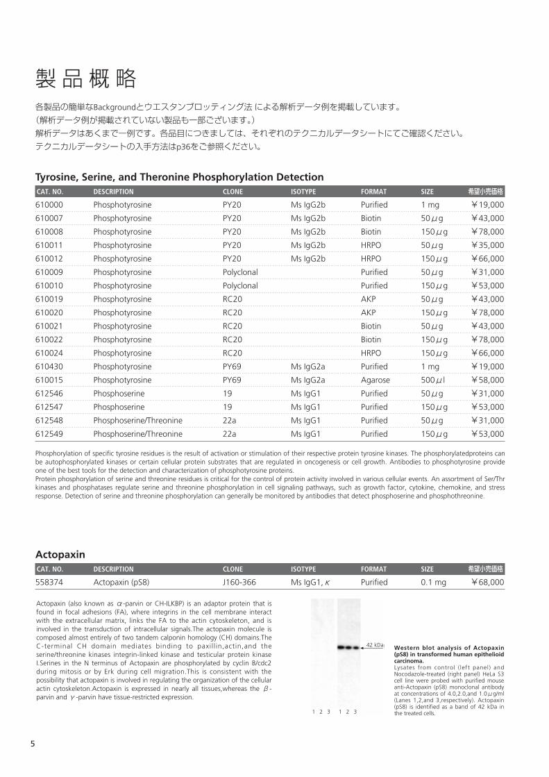
製品概略各製品の簡単なBackgroundとウエスタンブロッティング法による解析データ例を掲載しています。
(解析データ例が掲載されていない製品も一部ございます。)
解析データはあくまで一例です。各品目につきましては、それぞれのテクニカルデータシートにてご確認ください。
テクニカルデータシートの入手方法はp36をご参照ください。
Tyrosine, Serine, and Theronine Phosphorylation DetectionCAT. NO. DESCRIPTION CLONE ISOTYPE FORMAT SIZE 希望小売価格
610000 Phosphotyrosine PY20 Ms IgG2b Purified 1 mg ¥19,000
610007 Phosphotyrosine PY20 Ms IgG2b Biotin 50μg ¥43,000
610008 Phosphotyrosine PY20 Ms IgG2b Biotin 150μg ¥78,000
610011 Phosphotyrosine PY20 Ms IgG2b HRPO 50μg ¥35,000
610012 Phosphotyrosine PY20 Ms IgG2b HRPO 150μg ¥66,000
610009 Phosphotyrosine Polyclonal Purified 50μg ¥31,000
610010 Phosphotyrosine Polyclonal Purified 150μg ¥53,000
610019 Phosphotyrosine RC20 AKP 50μg ¥43,000
610020 Phosphotyrosine RC20 AKP 150μg ¥78,000
610021 Phosphotyrosine RC20 Biotin 50μg ¥43,000
610022 Phosphotyrosine RC20 Biotin 150μg ¥78,000
610024 Phosphotyrosine RC20 HRPO 150μg ¥66,000
610430 Phosphotyrosine PY69 Ms IgG2a Purified 1 mg ¥19,000
610015 Phosphotyrosine PY69 Ms IgG2a Agarose 500μl ¥58,000
612546 Phosphoserine 19 Ms IgG1 Purified 50μg ¥31,000
612547 Phosphoserine 19 Ms IgG1 Purified 150μg ¥53,000
612548 Phosphoserine/Threonine 22a Ms IgG1 Purified 50μg ¥31,000
612549 Phosphoserine/Threonine 22a Ms IgG1 Purified 150μg ¥53,000
Phosphorylation of specific tyrosine residues is the result of activation or stimulation of their respective protein tyrosine kinases. The phosphorylatedproteins canbe autophosphorylated kinases or certain cellular protein substrates that are regulated in oncogenesis or cell growth. Antibodies to phosphotyrosine provideone of the best tools for the detection and characterization of phosphotyrosine proteins.Protein phosphorylation of serine and threonine residues is critical for the control of protein activity involved in various cellular events. An assortment of Ser/Thrkinases and phosphatases regulate serine and threonine phosphorylation in cell signaling pathways, such as growth factor, cytokine, chemokine, and stressresponse. Detection of serine and threonine phosphorylation can generally be monitored by antibodies that detect phosphoserine and phosphothreonine.
ActopaxinCAT. NO. DESCRIPTION CLONE ISOTYPE FORMAT SIZE 希望小売価格
558374 Actopaxin (pS8) J160-366 Ms IgG1,κ Purified 0.1 mg ¥68,000
Western blot analysis of Actopaxin(pS8) in transformed human epithelioidcarcinoma.Lysates from control ( left panel) andNocodazole-treated (right panel) HeLa S3cell line were probed with purified mouseanti-Actopaxin (pS8) monoclonal antibodyat concentrations of 4.0,2.0,and 1.0μg/ml(Lanes 1,2,and 3,respectively). Actopaxin(pS8) is identified as a band of 42 kDa inthe treated cells.
42 kDa
1 2 3 1 2 3
Actopaxin (also known as α-parvin or CH-ILKBP) is an adaptor protein that isfound in focal adhesions (FA), where integrins in the cell membrane interactwith the extracellular matrix, links the FA to the actin cytoskeleton, and isinvolved in the transduction of intracellular signals.The actopaxin molecule iscomposed almost entirely of two tandem calponin homology (CH) domains.TheC-terminal CH domain mediates binding to paxil l in,actin,and theserine/threonine kinases integrin-linked kinase and testicular protein kinaseI.Serines in the N terminus of Actopaxin are phosphorylated by cyclin B/cdc2during mitosis or by Erk during cell migration.This is consistent with thepossibility that actopaxin is involved in regulating the organization of the cellularactin cytoskeleton.Actopaxin is expressed in nearly all tissues,whereas the β-parvin and γ-parvin have tissue-restricted expression.
5
AktCAT. NO. DESCRIPTION CLONE ISOTYPE FORMAT SIZE 希望小売価格
550747 Akt (pS472/pS473) 104A282 Ms IgG1 Purified 50μg ¥38,000
558368 Akt (pS473) J177-204.20 Ms IgG1,κ Purified 0.1 mg ¥68,000
558316 Akt (pT308) J1-223.371 Ms IgG1,κ Purified 0.1 mg ¥68,000
558384 Akt (pY326) K7-642 Ms IgG2a,κ Purified 0.1 mg ¥68,000
BcrCAT. NO. DESCRIPTION CLONE ISOTYPE FORMAT SIZE 希望小売価格
558248 Bcr (pY177) J52-309 Ms IgG2b,κ Purified 0.1 mg ¥68,000
BLNKCAT. NO. DESCRIPTION CLONE ISOTYPE FORMAT SIZE 希望小売価格
558366 BLNK (pY84) J117-1278 Ms IgG2b,κ Purified 0.1 mg ¥68,000
Western blot analysis of BLNK (pY84) inhuman Burkitt's lymphoma. Lysates from control ( left panel) andhydrogen peroxide-activated (right panel)Ramos cells were probed with purifiedmouse anti-BLNK (pY84) monoclonalantibody) at concentrations of 0.125,0.0625, and 0.032μg/ml (Lanes 1, 2, and3, respectively). BLNK (pY84) is identified asa band of about 68 kDa in the treated cells.
68 kDa
1 2 3 1 2 3
B cell activation is initiated by crosslinking the B cell receptor, which leads toactivation of non-receptor protein tyrosine kinases (PTK), including Btk, Syk, andthree Src kinases, Fyn, Lyn, and Blk. Activated PTKs then phosphorylate multiplecellular proteins involved in B lymphocyte signaling. Syk is responsible for thetyrosine phosphorylation of B cell l inker protein (BLNK), a member of the SLP-76family of adapter proteins. Phosphorylation of human BLNK at tyrosines 84,178, and 189 (Y84, Y178, and Y189) creates docking sites for PLCγ2, leadingto the activation of downstream signaling pathways.
Western blot analysis of Bcr (pY177).Lysates from control (left panel) and PDGF-treated (right panel) NIH/3T3 mouseembryo cell line were probed with mAbJ52-309 at 0.25, 0.0125,and 0.0625μg/ml(lanes 1,2,and 3,respectively). Bcr (pY177)is identified as a band of 160 kDa intreated cells.
160 kDa
1 2 3 1 2 3
The BCR (breakpoint clustoer region) gene was first identified by its presence inthe BCR-ABL fusion oncogene of the Philadelphia chromosome associated withchronic myelogenous leukemia. The Bcr protein has serine/threonine kinaseactivity and participates in platelet-derived growth factor (PDGF)-mediatedsignal transduction. The Tyrosine 177 (Y177) of the Bcr portion of Bcr-Abl playsan important role in the induction of myeloproliferative disease.
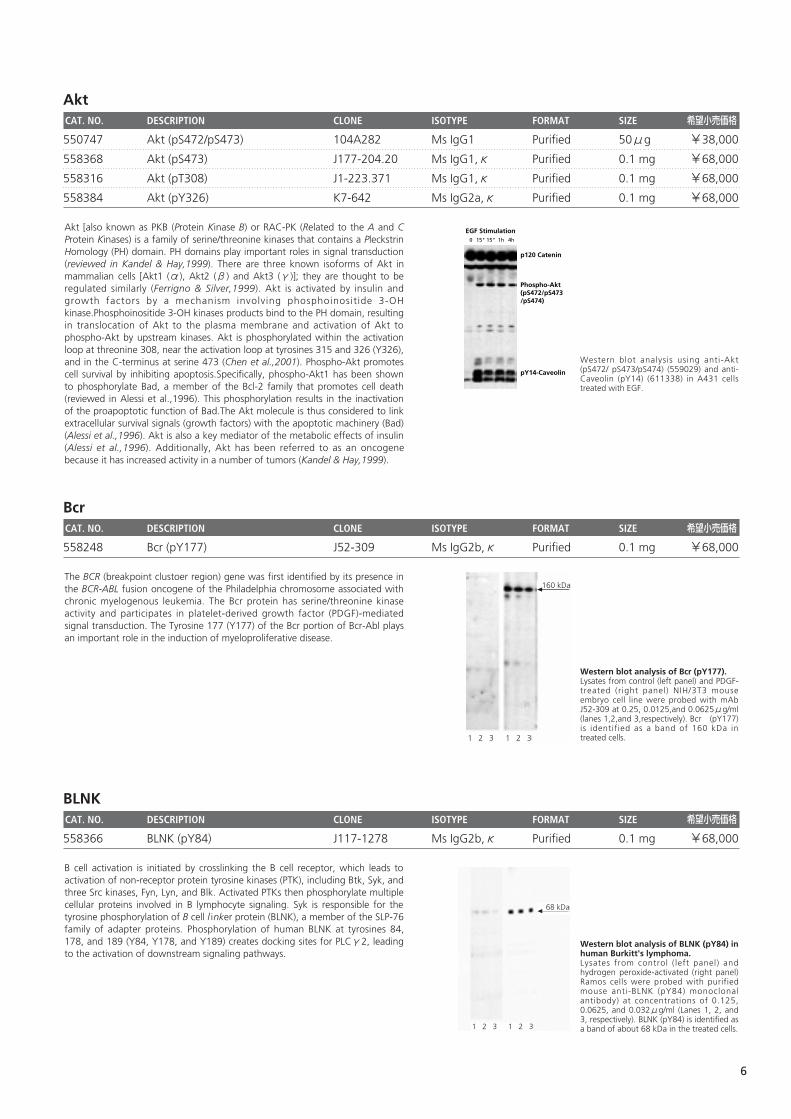
Western blot analysis using anti-Akt(pS472/ pS473/pS474) (559029) and anti-Caveolin (pY14) (611338) in A431 cellstreated with EGF.
EGF Stimulation
pY14-Caveolin
p120 Catenin
Phospho-Akt(pS472/pS473/pS474)
0 15" 15" 1h 4h
Akt [also known as PKB (Protein Kinase B) or RAC-PK (Related to the A and CProtein Kinases) is a family of serine/threonine kinases that contains a PleckstrinHomology (PH) domain. PH domains play important roles in signal transduction(reviewed in Kandel & Hay,1999). There are three known isoforms of Akt inmammalian cells [Akt1 (α), Akt2 (β) and Akt3 (γ)]; they are thought to beregulated similarly (Ferrigno & Silver,1999). Akt is activated by insulin andgrowth factors by a mechanism involving phosphoinositide 3-OHkinase.Phosphoinositide 3-OH kinases products bind to the PH domain, resultingin translocation of Akt to the plasma membrane and activation of Akt tophospho-Akt by upstream kinases. Akt is phosphorylated within the activationloop at threonine 308, near the activation loop at tyrosines 315 and 326 (Y326),and in the C-terminus at serine 473 (Chen et al.,2001). Phospho-Akt promotescell survival by inhibiting apoptosis.Specifically, phospho-Akt1 has been shownto phosphorylate Bad, a member of the Bcl-2 family that promotes cell death(reviewed in Alessi et al.,1996). This phosphorylation results in the inactivationof the proapoptotic function of Bad.The Akt molecule is thus considered to linkextracellular survival signals (growth factors) with the apoptotic machinery (Bad)(Alessi et al.,1996). Akt is also a key mediator of the metabolic effects of insulin(Alessi et al.,1996). Additionally, Akt has been referred to as an oncogenebecause it has increased activity in a number of tumors (Kandel & Hay,1999).
6
Btk CAT. NO. DESCRIPTION CLONE ISOTYPE FORMAT SIZE 希望小売価格
558034 Btk (pY551) & Itk (pY511) 24a/BTK (Y551) Ms IgG1 Purified 0.1 mg ¥68,000
CaveolinCAT. NO. DESCRIPTION CLONE ISOTYPE FORMAT SIZE 希望小売価格
611338 Caveolin (pY14) 56 Ms IgG1 Purified 50μg ¥31,000
611339 Caveolin (pY14) 56 Ms IgG1 Purified 150μg ¥53,000
Caveolin 2CAT. NO. DESCRIPTION CLONE ISOTYPE FORMAT SIZE 希望小売価格
558364 Caveolin 2 (pY27) 40/Caveolin 2 Ms IgG1,κ Purified 0.1 mg ¥68,000
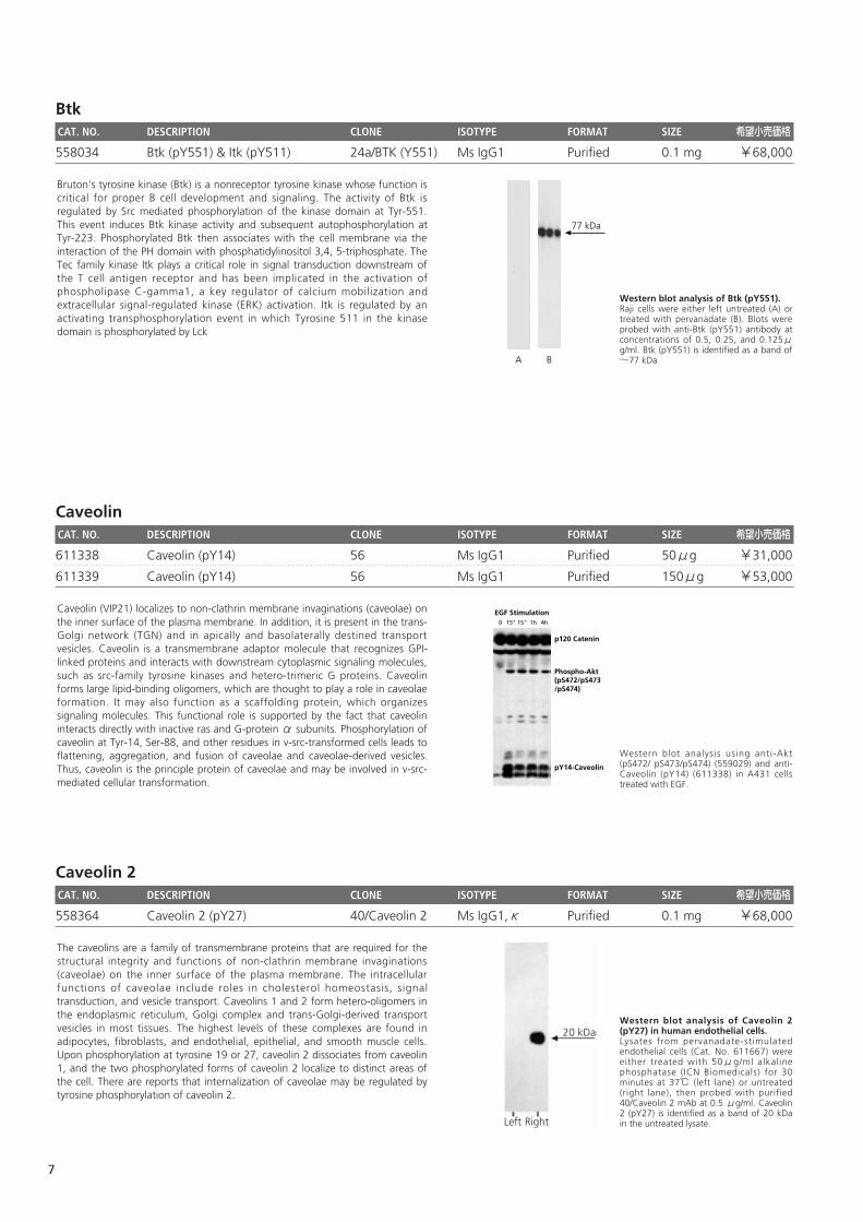
Western blot analysis of Caveolin 2(pY27) in human endothelial cells. Lysates from pervanadate-stimulatedendothelial cells (Cat. No. 611667) wereeither treated with 50μg/ml alkalinephosphatase (ICN Biomedicals) for 30minutes at 37℃ (left lane) or untreated(right lane), then probed with purified40/Caveolin 2 mAb at 0.5 μg/ml. Caveolin2 (pY27) is identified as a band of 20 kDain the untreated lysate.Left Right
20 kDa
The caveolins are a family of transmembrane proteins that are required for thestructural integrity and functions of non-clathrin membrane invaginations(caveolae) on the inner surface of the plasma membrane. The intracellularfunctions of caveolae include roles in cholesterol homeostasis, signaltransduction, and vesicle transport. Caveolins 1 and 2 form hetero-oligomers inthe endoplasmic reticulum, Golgi complex and trans-Golgi-derived transportvesicles in most tissues. The highest levels of these complexes are found inadipocytes, fibroblasts, and endothelial, epithelial, and smooth muscle cells.Upon phosphorylation at tyrosine 19 or 27, caveolin 2 dissociates from caveolin1, and the two phosphorylated forms of caveolin 2 localize to distinct areas ofthe cell. There are reports that internalization of caveolae may be regulated bytyrosine phosphorylation of caveolin 2.
Western blot analysis using anti-Akt(pS472/ pS473/pS474) (559029) and anti-Caveolin (pY14) (611338) in A431 cellstreated with EGF.
EGF Stimulation
pY14-Caveolin
p120 Catenin
Phospho-Akt(pS472/pS473/pS474)
0 15" 15" 1h 4h
Caveolin (VIP21) localizes to non-clathrin membrane invaginations (caveolae) onthe inner surface of the plasma membrane. In addition, it is present in the trans-Golgi network (TGN) and in apically and basolaterally destined transportvesicles. Caveolin is a transmembrane adaptor molecule that recognizes GPI-linked proteins and interacts with downstream cytoplasmic signaling molecules,such as src-family tyrosine kinases and hetero-trimeric G proteins. Caveolinforms large lipid-binding oligomers, which are thought to play a role in caveolaeformation. It may also function as a scaffolding protein, which organizessignaling molecules. This functional role is supported by the fact that caveolininteracts directly with inactive ras and G-protein α subunits. Phosphorylation ofcaveolin at Tyr-14, Ser-88, and other residues in v-src-transformed cells leads toflattening, aggregation, and fusion of caveolae and caveolae-derived vesicles.Thus, caveolin is the principle protein of caveolae and may be involved in v-src-mediated cellular transformation.
Western blot analysis of Btk (pY551). Raji cells were either left untreated (A) ortreated with pervanadate (B). Blots wereprobed with anti-Btk (pY551) antibody atconcentrations of 0.5, 0.25, and 0.125μg/ml. Btk (pY551) is identified as a band of~77 kDa
77 kDa
A B
Bruton's tyrosine kinase (Btk) is a nonreceptor tyrosine kinase whose function iscritical for proper B cell development and signaling. The activity of Btk isregulated by Src mediated phosphorylation of the kinase domain at Tyr-551.This event induces Btk kinase activity and subsequent autophosphorylation atTyr-223. Phosphorylated Btk then associates with the cell membrane via theinteraction of the PH domain with phosphatidylinositol 3,4, 5-triphosphate. TheTec family kinase Itk plays a critical role in signal transduction downstream ofthe T cell antigen receptor and has been implicated in the activation ofphospholipase C-gamma1, a key regulator of calcium mobilization andextracellular signal-regulated kinase (ERK) activation. Itk is regulated by anactivating transphosphorylation event in which Tyrosine 511 in the kinasedomain is phosphorylated by Lck
7
c-CblCAT. NO. DESCRIPTION CLONE ISOTYPE FORMAT SIZE 希望小売価格
612304 c-Cbl (pY700) 47 Ms IgG1 Purified 50μg ¥31,000
612305 c-Cbl (pY700) 47 Ms IgG1 Purified 150μg ¥53,000
558035 c-Cbl (pY774) 29/c-Cbl (Y774) Ms IgG1 Purified 0.1 mg ¥68,000
CD3ζ(CD247)CAT. NO. DESCRIPTION CLONE ISOTYPE FORMAT SIZE 希望小売価格
558402 CD3ζ(CD247) (pY142) K25-407.69 Ms IgG1,κ Purified 0.1 mg ¥68,000
CD22(BL-CAM) CAT. NO. DESCRIPTION CLONE ISOTYPE FORMAT SIZE 希望小売価格
558029 CD22 (pY828) 46 Ms IgG1 Purified 0.1 mg ¥68,000
558030 CD22 (pY843) 12a Ms IgG1 Purified 0.1 mg ¥68,000
Western blot analysis of CD22 (pY843). Daudi cells were either left untreated (A) ortreated with pervanadate (B). Blots wereprobed with anti-CD22(pY843) antibody at concentrationsof 0.0625, 0.03125, and 0.0156μg/ml.CD22 (pY843) is identified as a band of ~150 kDa
150 kDa
A B
CD22 is a glycosylated type I integral membrane protein expressed in B cells.Upon cross linking of the B cell antigen receptor, CD22 is phosphorylatedleading to the recruitment of PLCγ, PI3K, Syk and Grb2 thus causing signaltransduction. Tyrosine phosphorylation of residue 843 is required for efficientSHP-1 recruitment to the cytoplasmic tail of CD22.
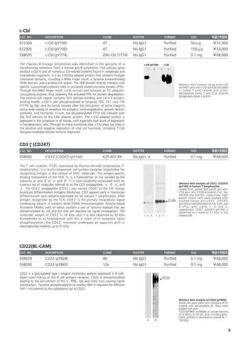
Western blot analysis of CD3ζ (CD247)(pY142) in human T lymphocytes.Lysates from control (left panel) and anti-CD3-plus anti-CD28-activated (Cat. No.555329 and 555725, respectively; rightpanel) Jurkat cells were probed withpurified mouse anti-CD3ζ (CD247)(pY142) at concentrations of 0.5, 0.25, and0.125μ g/ml (Lanes 1, 2, and 3,respectively). CD3ζ (CD247) (pY142) isidentified as a band of 21 kDa in thetreated cells.
21 kDa
1 2 3 1 2 3
The T cell receptor (TCR), expressed by thymus-derived lymphocytes (Tlymphocytes), is a multi-component cell-surface complex responsible forrecognizing antigen in the context of MHC molecules. The antigen-specificbinding component of the TCR, Ti, is a heterodimer of the variable Ig-likesubunits α and β or γ and δ. Ti is non-covalently associated with aninvariant set of molecules referred to as the CD3 polypeptides, γ, δ, ε, andζ. The CD3ζ polypeptide (CD3ζ) was named CD247 at the 7th HumanLeukocyte Differentiation Antigens Workshop. CD3 appears early in thymocytedifferentiation and remains expressed on all mature T lymphocytes. Afterantigen recognition by the TCR, CD3ζ is the primary intracellular signaltransducing subunit. It contains three ITAMs (Immunoreceptor Tyrosine-basedActivation Motifs), each of which contains a pair of tyrosine residues that arephosphorylated by Lck and Fyn and are required for signal propagation. Themolecular weight of CD3ζ is 16 kDa, and it is also observed as 32-kDahomodimers or as heterodimers with the γ chain of Fc receptors. Uponphosphorylation, the CD3ζ monomer undergoes an apparent shift inelectrophoretic mobility up to 21 kDa.
Western blot analysis using antic-Cbl(610441) and anti-c-Cbl (pY700) (612304)in Jurkat T cells treated with eitherpervanadate (lanes 1 and 3) or alkalinephosphatase (lanes 2 and 4).
c-Cbl (pY700) c-Cbl
1 2 3 4
Cbl (Casitas B-lineage lymphoma) was identified in the genome of atransforming retrovirus from a mouse pre-B lymphoma. The cellular geneproduct c-Cbl is one of numerous Cbl-related proteins found in vertebrate andinvertebrate organisms. It is an 120-kDa adapter protein that contains multiplefunctional domains, including a RING finger motif, a tyrosine kinase-binding(TKB) domain, and a proline-rich region. The TKB domain directly interacts withspecific auto-phosphorylation sites in activated protein-tyrosine kinases (PTK).Through the RING finger motif, c-Cbl recruits and activates an E2 ubiquitin-conjugating enzyme, thus targeting the activated PTK for protein degradation.The proline-rich region contains SH3 domain-binding and 14-3-3 protein-binding motifs. c-Cbl is also phosphorylated at tyrosines 700, 731, and 774(Y774) by Syk- and Src-family kinases after the stimulation of some integrinsand a wide variety of receptors for antigens, immunoglobulins, growth factors,cytokines, and hormones. In turn, the phosphorylated Y774 site interacts withthe SH2 domain of the CRK adapter protein. The c-Cbl adapter protein isexpressed in the cytoplasm in all tissues, with especially high levels of expressionin hematopoietic cells. Through its many functional sites, c-Cbl plays key roles inthe positive and negative regulation of vital cell functions, including T CellReceptor-mediated cellular immune responses.
8
CD45CAT. NO. DESCRIPTION CLONE ISOTYPE FORMAT SIZE 希望小売価格
558376 CD45 (pS999) J143-1270 Ms IgG1,κ Purified 0.1 mg ¥68,000
CD117 (c-kit)CAT. NO. DESCRIPTION CLONE ISOTYPE FORMAT SIZE 希望小売価格
558390 CD117 (c-kit)(pY568/pY570) K39-686 Ms IgG1,κ Purified 0.1 mg ¥68,000
CD221 (IGF-1 Receptor)CAT. NO. DESCRIPTION CLONE ISOTYPE FORMAT SIZE 希望小売価格
558373 CD221 (IGF-1 Receptor)(pY950) J95-626 Ms IgG2b,κ Purified 0.1 mg ¥68,000
Western blot analysis of IGF-1 receptor(pY950) in transformed humanepithelial cells. Lysates from control (left panel) and IGF-I-treated (Cat. No. 354037, right panel) 293fetal kidney cell line were probed withpurified mouse anti-IGF-1 receptor (pY950)monoclonal antibody at concentrations of0.125, 0.0625, and 0.03125μg/ml (Lanes1, 2, and 3, respectively). IGF-1 receptor(pY950) is identified as a band of 90 kDa inthe treated cells.1 2 3 1 2 3
90 kDa
Insulin-like growth factor-1 (IGF-1) receptor, or CD221, is a receptor tyrosinekinase that has high affinity for IGF-1 and low affinity for insulin and IGF-2. Eachtransmembrane protein complex is formed from a pair of 1367 amino-acidprecursor proteins. After the 30 amino-acid signal sequences are removed, thepaired polypeptides are cleaved to derive an α and β chain from each. Thetwo extracellular α chains form the ligand-binding domain, while thetransmembrane-and-intracellular β chains carry the kinase activity. Uponstimulation by its ligand, IGF-1 receptor autophosphorylates multiple tyrosinesites in its β chains and phosphorylates other signaling mediators that regulatecell growth, development, and neoplastic transformation. Phosphorylation ofthe tyrosine 950 (Y950) of IGF-1 receptor β chain signals up-regulation of thetranscriptional activator protein Id2.
Western blot analysis of c-Kit(pY568/pY570) in humanerythroleukemia. Lysates from control (left panel) and SCF-treated (Cat. No. 354105, right panel) HELcells were probed with purified mouse anti-c-Kit (CD117) (pY568/pY570) monoclonalantibody at concentrations of 0.125,0.0625, and 0.032μg/ml (lanes 1, 2, and3, respectively). c-Kit (pY568/ pY570) isidentified as a band of 145 kDa in thetreated cells.
145 kDa
1 2 3 1 2 3
c-Kit (also known as CD117) is a transmembrane tyrosine kinase receptor thatbinds stem cell factor (SCF, also known as kit ligand, mast cell growth factor,and steel factor) and is involved in the regulation of a wide range of tissues atvarious developmental stages. c-Kit plays major roles in the regulation ofhematopoiesis and germ cell proliferation and survival. Mutations of c-kit areassociated with a wide variety of cancers and developmental diseases. Uponactivation by its ligand, c-Kit dimerizes and autophosphorylates at multiplecytoplasmic sites, which bind to downstream signal transduction molecules.Specifically, the phosphorylated tyrosines 568 and 570 (pY568/pY570) in thejuxtamembrane domain of c-Kit bind to several signaling molecules, includingthe adapter proteins SHC and APS; the tyrosine kinases Lyn, Fyn, and CHK; andthe protein tyrosine phosphatase SHP-2, all of which may interact to regulateSCF signaling.
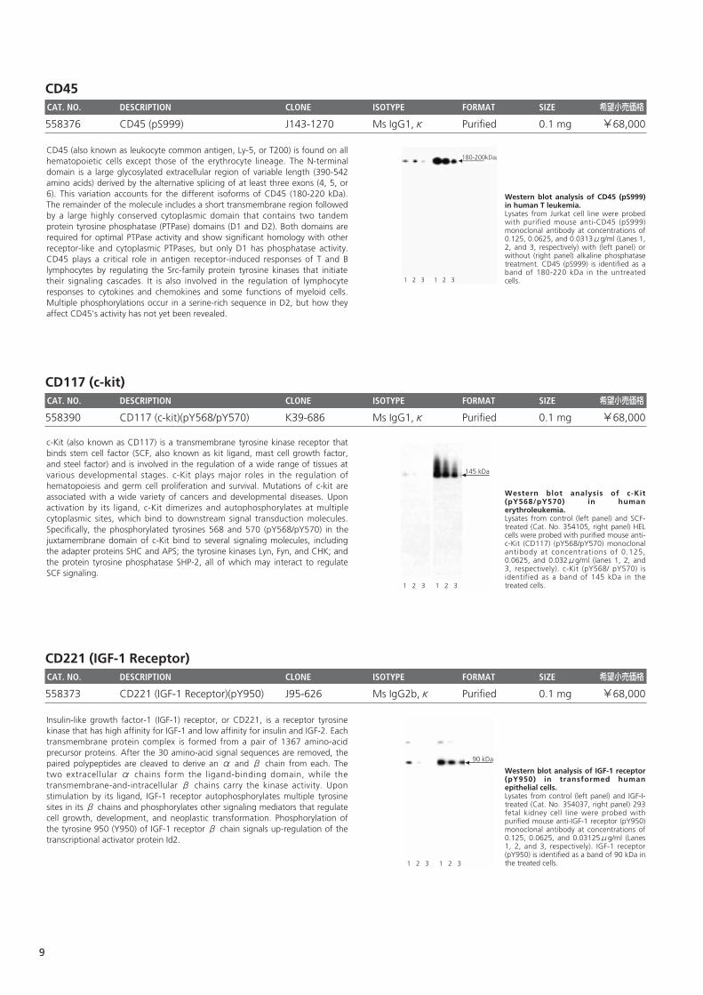
Western blot analysis of CD45 (pS999)in human T leukemia. Lysates from Jurkat cell line were probedwith purified mouse anti-CD45 (pS999)monoclonal antibody at concentrations of0.125, 0.0625, and 0.0313μg/ml (Lanes 1,2, and 3, respectively) with (left panel) orwithout (right panel) alkaline phosphatasetreatment. CD45 (pS999) is identified as aband of 180-220 kDa in the untreatedcells.1 2 3 1 2 3
180-200kDaCD45 (also known as leukocyte common antigen, Ly-5, or T200) is found on allhematopoietic cells except those of the erythrocyte lineage. The N-terminaldomain is a large glycosylated extracellular region of variable length (390-542amino acids) derived by the alternative splicing of at least three exons (4, 5, or6). This variation accounts for the different isoforms of CD45 (180-220 kDa).The remainder of the molecule includes a short transmembrane region followedby a large highly conserved cytoplasmic domain that contains two tandemprotein tyrosine phosphatase (PTPase) domains (D1 and D2). Both domains arerequired for optimal PTPase activity and show significant homology with otherreceptor-like and cytoplasmic PTPases, but only D1 has phosphatase activity.CD45 plays a critical role in antigen receptor-induced responses of T and Blymphocytes by regulating the Src-family protein tyrosine kinases that initiatetheir signaling cascades. It is also involved in the regulation of lymphocyteresponses to cytokines and chemokines and some functions of myeloid cells.Multiple phosphorylations occur in a serine-rich sequence in D2, but how theyaffect CD45's activity has not yet been revealed.
9
Cdk1/Cdc2CAT. NO. DESCRIPTION CLONE ISOTYPE FORMAT SIZE 希望小売価格
612306 Cdk1/Cdc2 (pY15) 44 Ms IgG1 Purified 50μg ¥31,000
612307 Cdk1/Cdc2 (pY15) 44 Ms IgG1 Purified 150μg ¥53,000
c-Jun CAT. NO. DESCRIPTION CLONE ISOTYPE FORMAT SIZE 希望小売価格
558036 c-Jun (pS63) 2 Ms IgG1 Purified 0.1 mg ¥68,000
CREBCAT. NO. DESCRIPTION CLONE ISOTYPE FORMAT SIZE 希望小売価格
558359 CREB (pS133) J151-21 Ms IgG1,κ Purified 0.1 mg ¥68,000
Western blot analysis of CREB (pS133)in human T leukemia. Lysates from control (left panel) and PMA-activated (right panel) Jurkat cells wereprobed with purified mouse anti-CREB(pS133) monoclonal antibody atconcentrations of 0.25, 0.125, and 0.06μg/ml (Lanes 1, 2, and 3, respectively). CREB(pS133) is identified as a band of 43 kDa inthe treated cells. Treatment with lambdaphosphatse removes all bands in blots ofcontrol and PMA-activated lysates (data notshown), demonstrating the antibody'sspecificity for the phosphorylated protein.
43 kDa
1 2 3 1 2 3
Transcription of various genes is regulated by the cyclic AMP (cAMP) signaltransduction pathway through a family of cAMP Responsive Element (CRE)-Binding transcription factors that include CREB, CREM (CRE Modulator), andATF-1 (Activating Transcription Factor 1). The genes for these transcriptionfactors encode multiple isoforms that are created by alternative splicing,alternative initiation codons, and alternative intronic promoters. Homo- andheterodimers of these members of the basic domain-and-leucine zippersuperfamily of proteins bind the palindromic TGACGTCA sequence of CRE.CREB is expressed in all somatic cells, and many different stimuli activate CREBby phosphorylation of its serine 133 (S133). The transcriptional activity andspecificity of CREB are regulated by the signaling pathways initiated by thevarious stimuli. Transcriptional regulation by CREB family members has beenimplicated in many physiological responses to extracellular and environmentalstimuli, such as memory, tissue growth and development, and homeostasis.
Western blot analysis of c-Jun (pS63). Human endothelial cells were either leftuntreated (A) or treated with calyculin A(B). Blots were probed with anti-c-jun(pS63) antibody at concentrations of 0.063,0.032, and 0.016μg/ml. c-Jun (pS63) isidentified as a band of ~39 kDa
39 kDa
A B
The activator protein transcription factor (AP-1) was identified as a protein thatrecognizes specific sequences in the cis-control regions of the SV40 virus andthe human metallothionein IIA gene. AP-1 is composed of protein products oftwo different gene families: jun and fos. The AP-1 transcription factor is either ahomodimer of Jun proteins or a heterodimer of Jun and Fos proteins. Thetranscriptional activity of Jun is enhanced by phosphorylation in its activationdomain at Ser63 and Ser73. Phosphorylation at both sites is necessary forstimulating the activating function of Jun. Jun is phosphorylated by JNK proteinkinases that are activated by the same signals that potentiate Jun activity.
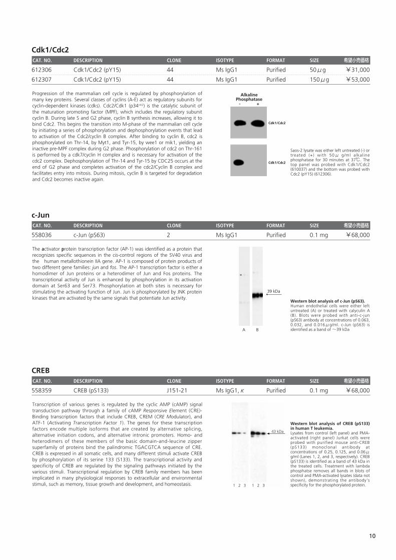
Saos-2 lysate was either left untreated (-) ortreated (+) with 50μ g/ml alkalinephosphatase for 30 minutes at 37℃. Thetop panel was probed with Cdk1/Cdc2(610037) and the bottom was probed withCdc2 (pY15) (612306).
Cdk1/Cdc2
Cdk1/Cdc2
AlkalinePhosphatase
- +
Progression of the mammalian cell cycle is regulated by phosphorylation ofmany key proteins. Several classes of cyclins (A-E) act as regulatory subunits forcyclin-dependent kinases (cdks). Cdc2/Cdk1 (p34cdc2) is the catalytic subunit ofthe maturation promoting factor (MPF), which includes the regulatory subunitcyclin B. During late S and G2 phase, cyclin B synthesis increases, allowing it tobind Cdc2. This begins the transition into M-phase of the mammalian cell cycleby initiating a series of phosphorylation and dephosphorylation events that leadto activation of the Cdc2/cyclin B complex. After binding to cyclin B, cdc2 isphosphorylated on Thr-14, by Myt1, and Tyr-15, by wee1 or mik1, yielding aninactive pre-MPF complex during G2 phase. Phosphorylation of cdc2 on Thr-161is performed by a cdk7/cyclin H complex and is necessary for activation of thecdc2 complex. Dephosphorylation of Thr-14 and Tyr-15 by CDC25 occurs at theend of G2 phase and completes activation of the cdc2/Cyclin B complex andfacilitates entry into mitosis. During mitosis, cyclin B is targeted for degradationand Cdc2 becomes inactive again.
10
CrkL CAT. NO. DESCRIPTION CLONE ISOTYPE FORMAT SIZE 希望小売価格
558386 CrkL (pY207) K30-391.11.30 Ms IgG2a,κ Purified 0.1 mg ¥68,000
β-DystroglycanCAT. NO. DESCRIPTION CLONE ISOTYPE FORMAT SIZE 希望小売価格
612524 β-Dystroglycan (pY892) 27.1 Ms IgG1 Purified 50μg ¥31,000
612525 β-Dystroglycan (pY892) 27.1 Ms IgG1 Purified 150μg ¥53,000
EGFRCAT. NO. DESCRIPTION CLONE ISOTYPE FORMAT SIZE 希望小売価格
610025 EGFR (Activated Form) 74 Ms IgG1 Purified 50μg ¥31,000
612401 EGFR (Activated Form) 74 Ms IgG1 Purified 100μg ¥42,000
610026 EGFR (Activated Form) 74 Ms IgG1 Purified 150μg ¥53,000
Western blot analysis using anti-ActivatedEGFR (610025) and anti-Akt(pS472/pS473/pS474) (559029) in A431cells treated with EGF.
EGF (100 ng/ml)
Akt (pS473/ pS473/pS474)
EGFR (activated)
0 5' 15' 1h
pp120eps8
4h 8h
Epidermal Growth Factor (EGF) elicits a variety of cellular responses that areinitiated by EGF receptor (EGF-R) binding and activation of intrinsic tyrosinekinase activity. Following ligand binding, EGF receptor is autophosphorylatedand, in turn, phosphorylates several other endogenous proteins, includingphospholipase Cγ. These events initiate a number of intracellular responsesthat include increased levels of intracellular Ca 2+ and transient expression of thenuclear oncogene products c-Myc and c-Fos. This antibody is unique in that itreacts only with the tyrosine phophorylated (activated) EGF receptor.
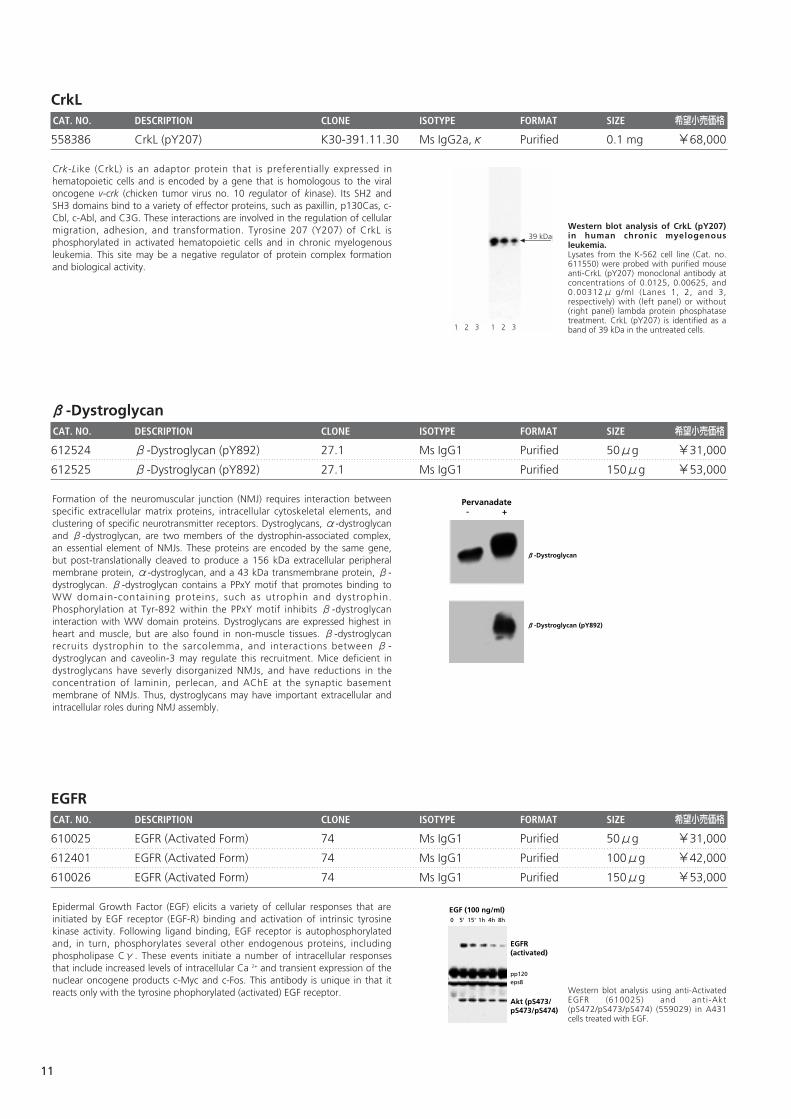
Pervanadate- +
β-Dystroglycan
β-Dystroglycan (pY892)
Formation of the neuromuscular junction (NMJ) requires interaction betweenspecific extracellular matrix proteins, intracellular cytoskeletal elements, andclustering of specific neurotransmitter receptors. Dystroglycans, α-dystroglycanand β-dystroglycan, are two members of the dystrophin-associated complex,an essential element of NMJs. These proteins are encoded by the same gene,but post-translationally cleaved to produce a 156 kDa extracellular peripheralmembrane protein, α-dystroglycan, and a 43 kDa transmembrane protein, β-dystroglycan. β-dystroglycan contains a PPxY motif that promotes binding toWW domain-containing proteins, such as utrophin and dystrophin.Phosphorylation at Tyr-892 within the PPxY motif inhibits β-dystroglycaninteraction with WW domain proteins. Dystroglycans are expressed highest inheart and muscle, but are also found in non-muscle tissues. β-dystroglycanrecruits dystrophin to the sarcolemma, and interactions between β-dystroglycan and caveolin-3 may regulate this recruitment. Mice deficient indystroglycans have severly disorganized NMJs, and have reductions in theconcentration of laminin, perlecan, and AChE at the synaptic basementmembrane of NMJs. Thus, dystroglycans may have important extracellular andintracellular roles during NMJ assembly.
Western blot analysis of CrkL (pY207)in human chronic myelogenousleukemia. Lysates from the K-562 cell line (Cat. no.611550) were probed with purified mouseanti-CrkL (pY207) monoclonal antibody atconcentrations of 0.0125, 0.00625, and0.00312μ g/ml (Lanes 1, 2, and 3,respectively) with (left panel) or without(right panel) lambda protein phosphatasetreatment. CrkL (pY207) is identified as aband of 39 kDa in the untreated cells.1 2 3 1 2 3
39 kDa
Crk-Like (CrkL) is an adaptor protein that is preferentially expressed inhematopoietic cells and is encoded by a gene that is homologous to the viraloncogene v-crk (chicken tumor virus no. 10 regulator of kinase). Its SH2 andSH3 domains bind to a variety of effector proteins, such as paxillin, p130Cas, c-Cbl, c-Abl, and C3G. These interactions are involved in the regulation of cellularmigration, adhesion, and transformation. Tyrosine 207 (Y207) of CrkL isphosphorylated in activated hematopoietic cells and in chronic myelogenousleukemia. This site may be a negative regulator of protein complex formationand biological activity.
11
eNOSCAT. NO. DESCRIPTION CLONE ISOTYPE FORMAT SIZE 希望小売価格
612392 eNOS (pS1177) 19 Ms IgG1 Purified 50μg ¥31,000
612393 eNOS (pS1177) 19 Ms IgG1 Purified 150μg ¥53,000
612664 eNOS (pS633) 37 Ms IgG1 Purified 50μg ¥31,000
612665 eNOS (pS633) 37 Ms IgG1 Purified 150μg ¥53,000
612706 eNOS (pT495) 31 Ms IgG1 Purified 50μg ¥31,000
612707 eNOS (pT495) 31 Ms IgG1 Purified 150μg ¥53,000
ERKCAT. NO. DESCRIPTION CLONE ISOTYPE FORMAT SIZE 希望小売価格
612358 ERK1/2 (pT202/pY204) 20A Ms IgG1 Purified 50μg ¥31,000
612359 ERK1/2 (pT202/pY204) 20A Ms IgG1 Purified 150μg ¥53,000
Western blot analysis using anti-ERK1(610030) and anti-ERK1/2 (pT202/pY204)(612358) in A431 cells either untreated(lanes 1 and 3) or treated with EGF (lanes 2and 4).
ERK1/2 (pT202/pY204) ERK1
1 2 3 4
The family of serine/threonine kinases known as ERKs (extracellular signalregulated kinases) or MAPKs (mitogen-activated protein kinases) are activatedafter cell stimulation by a variety of hormones and growth factors. A myriad ofproteins, such as kinases, phosphatases, transcription factors, and cytoskeletalproteins, are substrates for the active ERK. These proteins implicate ERKfunction in the control of cell proliferation and differentiation, as well as in theregulation of the cytoskeleton. Activation of ERK is normally transient and cellspossess dual specificity phosphatases which are responsible for its down-regulation. ERK1 is a 44kDa member of the ERK family and shares 85%homology with ERK2. In rat, these proteins are phosphorylated at T202/Y204and T183/Y185, respectively. ERK1 and 2 have been implicated in growth factorsignaling, as well as other signal transduction pathways. Growth factorstimulation leads to activation of Ras and Raf, leading to phosphorylation ofMEK1 (MAPK/ERK kinase) which, in turn, activates ERK via dualphosphorylation. Thus, ERK1 and 2 are critical kinases in multiple signaltransduction pathways that regulate cell growth and differentiation.
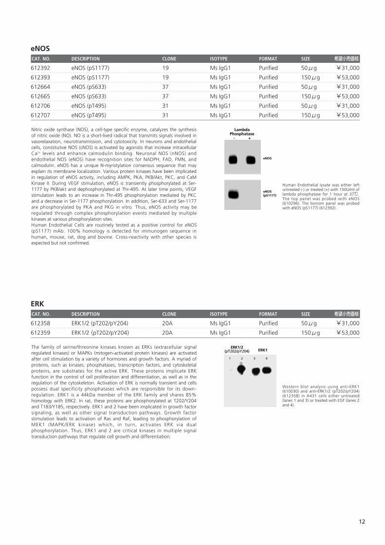
Human Endothelial lysate was either leftuntreated (-) or treated (+) with 150U/ml oflambda phosphatase for 1 hour at 37℃.The top panel was probed with eNOS(610296). The bottom panel was probedwith eNOS (pS1177) (612392).
LambdaPhosphatase
- +
eNOS
eNOS (pS1177)
Nitric oxide synthase (NOS), a cell-type specific enzyme, catalyzes the synthesisof nitric oxide (NO). NO is a short-lived radical that transmits signals involved invasorelaxation, neurotransmission, and cytotoxicity. In neurons and endothelialcells, constitutive NOS (cNOS) is activated by agonists that increase intracellularCa2+ levels and enhance calmodulin binding. Neuronal NOS (nNOS) andendothelial NOS (eNOS) have recognition sites for NADPH, FAD, FMN, andcalmodulin. eNOS has a unique N-myristylation consensus sequence that mayexplain its membrane localization. Various protein kinases have been implicatedin regulation of eNOS activity, including AMPK, PKA, PKB/Akt, PKC, and CaMKinase II. During VEGF stimulation, eNOS is transiently phosphorylated at Ser-1177 by PKB/akt and dephosphorylated at Thr-495. At later time points, VEGFstimulation leads to an increase in Thr-495 phosphorylation mediated by PKCand a decrease in Ser-1177 phosphorylation. In addition, Ser-633 and Ser-1177are phosphorylated by PKA and PKG in vitro. Thus, eNOS activity may beregulated through complex phosphorylation events mediated by multiplekinases at various phosphorylation sites. Human Endothelial Cells are routinely tested as a positive control for eNOS(pS1177) mAb. 100% homology is detected for immunogen sequence inhuman, mouse, rat, dog and bovine. Cross-reactivity with other species isexpected but not confirmed.
12
EzrinCAT. NO. DESCRIPTION CLONE ISOTYPE FORMAT SIZE 希望小売価格
558357 Ezrin (pT567) J37-954.281.307 Ms IgG1,κ Purified 0.1 mg ¥68,000
558033 Ezrin (pY353) I66-386 Ms IgG1 Purified 0.1 mg ¥68,000
FADDCAT. NO. DESCRIPTION CLONE ISOTYPE FORMAT SIZE 希望小売価格
558370 FADD (pS194) J119-857.36 Ms IgG1,κ Purified 0.1 mg ¥68,000
FAKCAT. NO. DESCRIPTION CLONE ISOTYPE FORMAT SIZE 希望小売価格
611806 FAK (pY397) 18 Ms IgG1 Purified 50μg ¥31,000
611807 FAK (pY397) 18 Ms IgG1 Purified 150μg ¥53,000
611722 FAK (pY397) 14 Ms IgG1 Purified 50μg ¥31,000
611723 FAK (pY397) 14 Ms IgG1 Purified 150μg ¥53,000
Western blot analysis using anti-FAK(pY397) (611722) and anti-FAK (610087) inhuman fibroblast cells either untreated(lanes 1 and 3) or treated with pervanadate(lanes 2 and 4).
FAK (pY397)FAK
1 2 3 4
Focal Adhesion Kinase (FAK) is a cytoplasmic tyrosine kinase that colocalizeswith integrins in focal adhesions. This cellular localization is directed by a 125amino acid sequence at the C-terminus called the "Focal Adhesion Targeting"sequence (FAT). The binding of extracellular matrix ligands to integrins triggersautophosphorylation at Tyr-397, and activation of FAK through phosphorylationof Tyr residues (Tyr-576 and Tyr577) in the kinase domain activation loop. Forexample, cell adhesion to a fibronectin substratum involves concurrentactivation of Src and phosphorylation of the FAK activation loop. In addition,phosphorylation of other Tyr residues (Tyr-925, and Tyr-861) creates bindingsites for SH2 domains of intracellular signaling molecules such as Src, PI3 kinase,and Grb2. FAK's ability to bind numerous structural and signaling proteins via avariety of interactions is important for FAK activation level, and for FAKinteraction with a variety of substrates localized to sites of cell adhesion. Thus,FAK activity is regulated by a complex set of phosphorylation sites, and thisphospho-regulation could be important for cell motility, cell growth, cytoskeletalorganization, and adhesion-dependent cell survival.
Western blot analysis of FADD (pS194)in human epidermis. Lysates from control ( left panel) andcalyculin A-plus-okadaic acid-treated (rightpanel) human A-431 epidermoid carcinomawere probed with purified mouse anti-FADD (pS194) monoclonal antibody atconcentrations of 2.0, 1.0, and 0.5μg/ml(lanes 1, 2, and 3, respectively). FADD(pS194) is identified as a band of 27 kDa inthe treated cells.
27 kDa
1 2 3 1 2 3
During apoptosis, cells exhibit morphological signs of the death process: cellshrinkage, membrane blebbing, and chromatin condensation. The role of thecell surface cytokine receptor, Fas (Apo-1, CD95), in apoptosis has been wellcharacterized. The tumor necrosis factor (TNF) receptor type I (TNFRI, CD120a)and TNF-related apoptosis-inducing ligand receptor 2 (TRAILR2, DR5) can triggercell death, as well as various other responses. Fas, TNFRI, and TRAILR2 affect acommon target in the cell death pathway, FADD (Fas-Associated via DeathDomain or FAS-Associating protein with Death Domain, also known as MORT1).FADD is an adaptor protein that specifically binds to Fas and other deathdomain-containing proteins via their homologous death domains. FADD alsocontains an N-terminal Death Effector Domain (DED) that interacts with theDED-containing procaspases-8 and -10 to initiate apoptosis. The role of FADDserine 194 (S194) phosphorylation in the regulation of apoptosis and cell cycleprogression is under investigation.
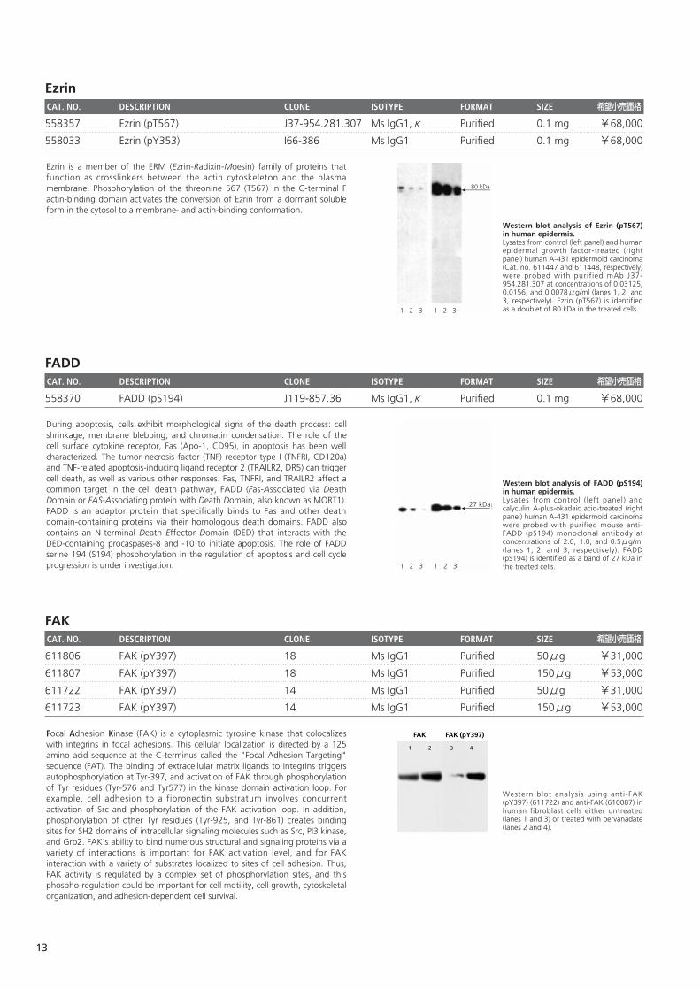
Western blot analysis of Ezrin (pT567)in human epidermis. Lysates from control (left panel) and humanepidermal growth factor-treated (rightpanel) human A-431 epidermoid carcinoma(Cat. no. 611447 and 611448, respectively)were probed with purif ied mAb J37-954.281.307 at concentrations of 0.03125,0.0156, and 0.0078μg/ml (lanes 1, 2, and3, respectively). Ezrin (pT567) is identifiedas a doublet of 80 kDa in the treated cells.1 2 3 1 2 3
80 kDa
Ezrin is a member of the ERM (Ezrin-Radixin-Moesin) family of proteins thatfunction as crosslinkers between the actin cytoskeleton and the plasmamembrane. Phosphorylation of the threonine 567 (T567) in the C-terminal Factin-binding domain activates the conversion of Ezrin from a dormant solubleform in the cytosol to a membrane- and actin-binding conformation.
13
FynCAT. NO. DESCRIPTION CLONE ISOTYPE FORMAT SIZE 希望小売価格
612668 Fyn (pY528)/c-Src (pY530) 31 Ms IgG2b Purified 50μg ¥31,000
612669 Fyn (pY528)/c-Src (pY530) 31 Ms IgG2b Purified 150μg ¥53,000
gp130CAT. NO. DESCRIPTION CLONE ISOTYPE FORMAT SIZE 希望小売価格
558096 gp130 (pS782) 6a/gp130 (pS782) Ms IgG2b,κ Purified 0.1 mg ¥68,000
GSK-3βCAT. NO. DESCRIPTION CLONE ISOTYPE FORMAT SIZE 希望小売価格
612312 GSK-3β (pY216) 13a Ms IgG1 Purified 50μg ¥31,000
612313 GSK-3β (pY216) 13a Ms IgG1 Purified 150μg ¥53,000
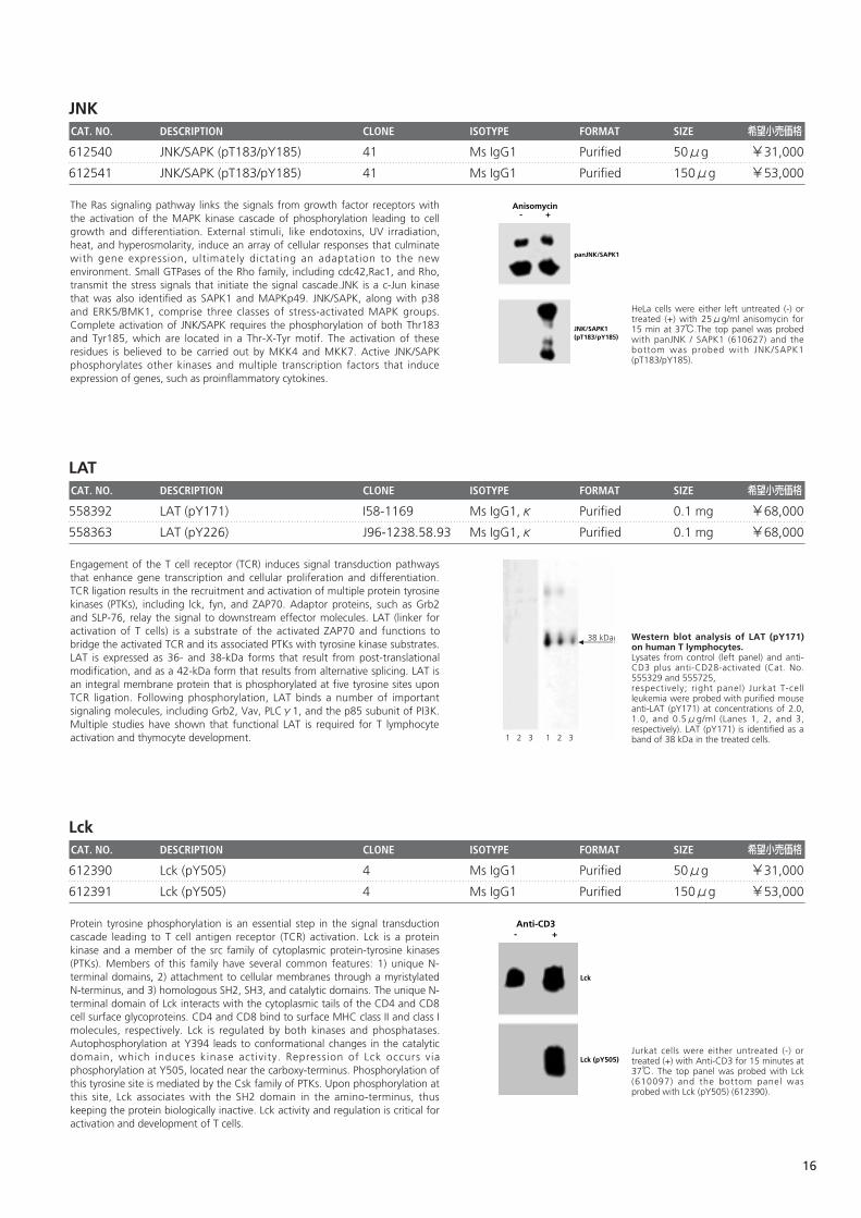
RSV-3T3 lysate was either left untreated (-)or treated (+) with 50μg/ml alkalinephosphatase for 30 minutes at 37℃. Thetop panel was probed with GSK-3β(610201) and the bottom was probed withGSK-3β (pY216) (612312).
AlkalinePhosphatase
- +
GSK-3β (pY216)
GSK-3β
Glycogen Synthase Kinase-3β (GSK-3β) is a serine/threonine kinase thataffects glycogen metabolism by phosphorylating and down-regulating theactivity of muscle glycogen synthase. GSK-3β is identical to the Tau ProteinKinase I (TPK I) that plays a role in the formation of the histopathological brainlesions of Alzheimer's disease (AD). Phosphorylation of the cytoskeletal protein,tau, by GSK-3β converts these proteins into paired helical filaments (PHF)which are found in the neurofibrillary tangles and degenerative neurites of ADpatients. Regulation of GSK-3β activity through both serine and tyrosinephosphorylation is a critical determinant of cell death or survival. Factors thatpromote cell survival, such as growth factors, activate Akt which, in turn,phosphorylates GSK-3β at Ser-9, leading to inactivation of its kinase activity.On the contrary, events that promote cell death, such as growth factor removal,cause increases in phosphorylation within the catalytic domain at Tyr-216 andstimulate kinase activity. Thus, GSK-3β is a tightly regulated death promotingkinase that regulates the activty of various proteins, including cytokeletal andenzymatic proteins.
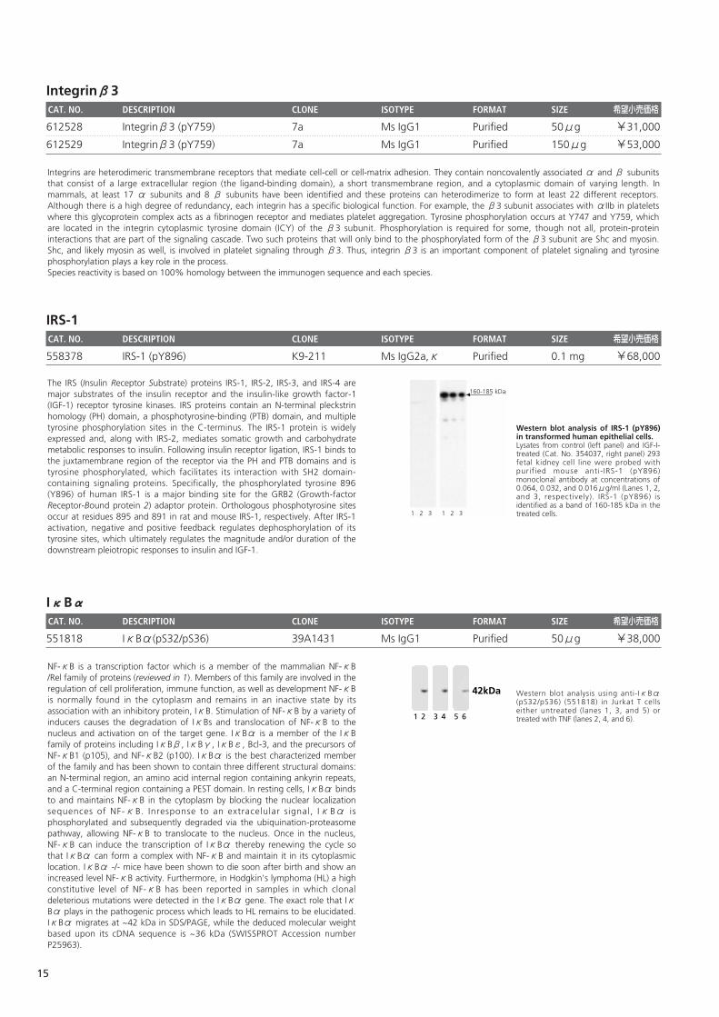
Western blot analysis of gp130 (pS782).NIH3T3 cells were either left untreated (A)or treated with Calyculin/Okadaic acid (B).Blots were probed with anti-gp130 (pS782)antibody at concentrations of 0.0078,0.0039, and 0.0019μg/ml. gp130 (pS782)is identified as a band of ~130kDa.
130kDa
Leukemia Inhibitory Factor (LIF) signals through a heterodimeric receptorcomplex consisting of LIFR and gp130. LIF-stimulated dimerization of LIFR andgp130 results in signal transduction through the Jak/Tyk family of nonreceptorprotein tyrosine kinases. Serine 782 is the major site for gp130 phosphorylationand this phosphorylation is involved in the regulation of the cell surfaceexpression of the receptor polypeptide.
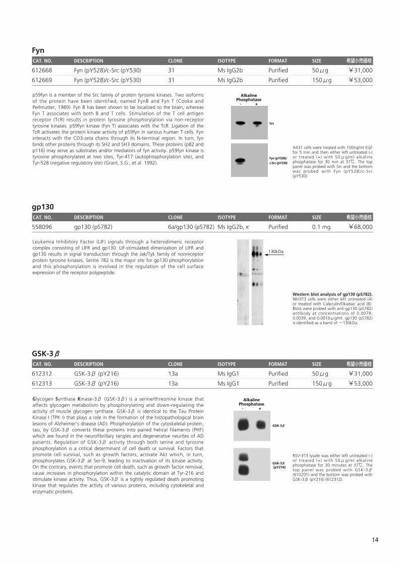
A431 cells were treated with 100ng/ml EGFfor 5 min and then either left untreated (-)or treated (+) with 50μg/ml alkalinephosphatase for 30 min at 37℃. The toppanel was probed with Src and the bottomwas probed with Fyn (pY528)/c-Src(pY530).
AlkalinePhosphatase
- +
Src
Fyn (pY528)/c-Src (pY530)
p59fyn is a member of the Src family of protein tyrosine kinases. Two isoformsof the protein have been identified, named FynB and Fyn T (Cooke andPerlmutter, 1989). Fyn B has been shown to be localized to the brain, whereasFyn T associates with both B and T cells. Stimulation of the T cell antigenreceptor (TcR) results in protein tyrosine phosphorylation via non-receptortyrosine kinases. p59fyn kinase (Fyn T) associates with the TcR. Ligation of theTcR activates the protein kinase activity of p59fyn in various human T cells. Fyninteracts with the CD3-zeta chains through its N-terminal region. In turn, fynbinds other proteins through its SH2 and SH3 domains. These proteins (p82 andp116) may serve as substrates and/or mediators of fyn activity. p59fyn kinase istyrosine phosphorylated at two sites, Tyr-417 (autophosphorylation site), andTyr-528 (negative regulatory site) (Grant, S.G., et al. 1992).
14
Integrinβ3CAT. NO. DESCRIPTION CLONE ISOTYPE FORMAT SIZE 希望小売価格
612528 Integrinβ3 (pY759) 7a Ms IgG1 Purified 50μg ¥31,000
612529 Integrinβ3 (pY759) 7a Ms IgG1 Purified 150μg ¥53,000
IRS-1CAT. NO. DESCRIPTION CLONE ISOTYPE FORMAT SIZE 希望小売価格
558378 IRS-1 (pY896) K9-211 Ms IgG2a,κ Purified 0.1 mg ¥68,000
IκBαCAT. NO. DESCRIPTION CLONE ISOTYPE FORMAT SIZE 希望小売価格
551818 IκBα(pS32/pS36) 39A1431 Ms IgG1 Purified 50μg ¥38,000
Western blot analysis using anti-IκBα(pS32/pS36) (551818) in Jurkat T cellseither untreated (lanes 1, 3, and 5) ortreated with TNF (lanes 2, 4, and 6).
42kDa
1 2 3 4 5 6
NF-κB is a transcription factor which is a member of the mammalian NF-κB/Rel family of proteins (reviewed in 1). Members of this family are involved in theregulation of cell proliferation, immune function, as well as development NF-κBis normally found in the cytoplasm and remains in an inactive state by itsassociation with an inhibitory protein, IκB. Stimulation of NF-κB by a variety ofinducers causes the degradation of IκBs and translocation of NF-κB to thenucleus and activation on of the target gene. IκBα is a member of the IκBfamily of proteins including IκBβ, IκBγ, IκBε, Bcl-3, and the precursors ofNF-κB1 (p105), and NF-κB2 (p100). IκBα is the best characterized memberof the family and has been shown to contain three different structural domains:an N-terminal region, an amino acid internal region containing ankyrin repeats,and a C-terminal region containing a PEST domain. In resting cells, IκBα bindsto and maintains NF-κB in the cytoplasm by blocking the nuclear localizationsequences of NF-κB. Inresponse to an extracelular signal, IκBα isphosphorylated and subsequently degraded via the ubiquination-proteasomepathway, allowing NF-κB to translocate to the nucleus. Once in the nucleus,NF-κB can induce the transcription of IκBα thereby renewing the cycle sothat IκBα can form a complex with NF-κB and maintain it in its cytoplasmiclocation. IκBα -/- mice have been shown to die soon after birth and show anincreased level NF-κB activity. Furthermore, in Hodgkin's lymphoma (HL) a highconstitutive level of NF-κB has been reported in samples in which clonaldeleterious mutations were detected in the IκBα gene. The exact role that IκBα plays in the pathogenic process which leads to HL remains to be elucidated.IκBα migrates at ~42 kDa in SDS/PAGE, while the deduced molecular weightbased upon its cDNA sequence is ~36 kDa (SWISSPROT Accession numberP25963).
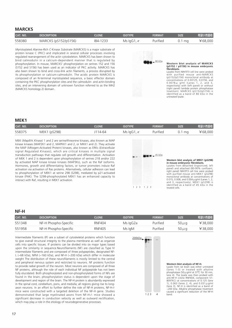
Western blot analysis of IRS-1 (pY896)in transformed human epithelial cells. Lysates from control (left panel) and IGF-I-treated (Cat. No. 354037, right panel) 293fetal kidney cell line were probed withpurified mouse anti- IRS-1 (pY896)monoclonal antibody at concentrations of0.064, 0.032, and 0.016μg/ml (Lanes 1, 2,and 3, respectively). IRS-1 (pY896) isidentified as a band of 160-185 kDa in thetreated cells.1 2 3 1 2 3
160-185 kDaThe IRS (Insulin Receptor Substrate) proteins IRS-1, IRS-2, IRS-3, and IRS-4 aremajor substrates of the insulin receptor and the insulin-like growth factor-1(IGF-1) receptor tyrosine kinases. IRS proteins contain an N-terminal pleckstrinhomology (PH) domain, a phosphotyrosine-binding (PTB) domain, and multipletyrosine phosphorylation sites in the C-terminus. The IRS-1 protein is widelyexpressed and, along with IRS-2, mediates somatic growth and carbohydratemetabolic responses to insulin. Following insulin receptor ligation, IRS-1 binds tothe juxtamembrane region of the receptor via the PH and PTB domains and istyrosine phosphorylated, which facilitates its interaction with SH2 domain-containing signaling proteins. Specifically, the phosphorylated tyrosine 896(Y896) of human IRS-1 is a major binding site for the GRB2 (Growth-factorReceptor-Bound protein 2) adaptor protein. Orthologous phosphotyrosine sitesoccur at residues 895 and 891 in rat and mouse IRS-1, respectively. After IRS-1activation, negative and positive feedback regulates dephosphorylation of itstyrosine sites, which ultimately regulates the magnitude and/or duration of thedownstream pleiotropic responses to insulin and IGF-1.
Integrins are heterodimeric transmembrane receptors that mediate cell-cell or cell-matrix adhesion. They contain noncovalently associated α and β subunitsthat consist of a large extracellular region (the ligand-binding domain), a short transmembrane region, and a cytoplasmic domain of varying length. Inmammals, at least 17 α subunits and 8 β subunits have been identified and these proteins can heterodimerize to form at least 22 different receptors.Although there is a high degree of redundancy, each integrin has a specific biological function. For example, the β3 subunit associates with αIIb in plateletswhere this glycoprotein complex acts as a fibrinogen receptor and mediates platelet aggregation. Tyrosine phosphorylation occurs at Y747 and Y759, whichare located in the integrin cytoplasmic tyrosine domain (ICY) of the β3 subunit. Phosphorylation is required for some, though not all, protein-proteininteractions that are part of the signaling cascade. Two such proteins that will only bind to the phosphorylated form of the β3 subunit are Shc and myosin.Shc, and likely myosin as well, is involved in platelet signaling through β3. Thus, integrin β3 is an important component of platelet signaling and tyrosinephosphorylation plays a key role in the process. Species reactivity is based on 100% homology between the immunogen sequence and each species.
15
JNKCAT. NO. DESCRIPTION CLONE ISOTYPE FORMAT SIZE 希望小売価格
612540 JNK/SAPK (pT183/pY185) 41 Ms IgG1 Purified 50μg ¥31,000
612541 JNK/SAPK (pT183/pY185) 41 Ms IgG1 Purified 150μg ¥53,000
LAT CAT. NO. DESCRIPTION CLONE ISOTYPE FORMAT SIZE 希望小売価格
558392 LAT (pY171) I58-1169 Ms IgG1,κ Purified 0.1 mg ¥68,000
558363 LAT (pY226) J96-1238.58.93 Ms IgG1,κ Purified 0.1 mg ¥68,000
LckCAT. NO. DESCRIPTION CLONE ISOTYPE FORMAT SIZE 希望小売価格
612390 Lck (pY505) 4 Ms IgG1 Purified 50μg ¥31,000
612391 Lck (pY505) 4 Ms IgG1 Purified 150μg ¥53,000
Jurkat cells were either untreated (-) ortreated (+) with Anti-CD3 for 15 minutes at37℃. The top panel was probed with Lck(610097) and the bottom panel wasprobed with Lck (pY505) (612390).
Anti-CD3- +
Lck
Lck (pY505)
Protein tyrosine phosphorylation is an essential step in the signal transductioncascade leading to T cell antigen receptor (TCR) activation. Lck is a proteinkinase and a member of the src family of cytoplasmic protein-tyrosine kinases(PTKs). Members of this family have several common features: 1) unique N-terminal domains, 2) attachment to cellular membranes through a myristylatedN-terminus, and 3) homologous SH2, SH3, and catalytic domains. The unique N-terminal domain of Lck interacts with the cytoplasmic tails of the CD4 and CD8cell surface glycoproteins. CD4 and CD8 bind to surface MHC class II and class Imolecules, respectively. Lck is regulated by both kinases and phosphatases.Autophosphorylation at Y394 leads to conformational changes in the catalyticdomain, which induces kinase activity. Repression of Lck occurs viaphosphorylation at Y505, located near the carboxy-terminus. Phosphorylation ofthis tyrosine site is mediated by the Csk family of PTKs. Upon phosphorylation atthis site, Lck associates with the SH2 domain in the amino-terminus, thuskeeping the protein biologically inactive. Lck activity and regulation is critical foractivation and development of T cells.
Western blot analysis of LAT (pY171)on human T lymphocytes. Lysates from control (left panel) and anti-CD3 plus anti-CD28-activated (Cat. No.555329 and 555725,respectively; right panel) Jurkat T-cellleukemia were probed with purified mouseanti-LAT (pY171) at concentrations of 2.0,1.0, and 0.5μg/ml (Lanes 1, 2, and 3,respectively). LAT (pY171) is identified as aband of 38 kDa in the treated cells.
38 kDa
1 2 3 1 2 3
Engagement of the T cell receptor (TCR) induces signal transduction pathwaysthat enhance gene transcription and cellular proliferation and differentiation.TCR ligation results in the recruitment and activation of multiple protein tyrosinekinases (PTKs), including lck, fyn, and ZAP70. Adaptor proteins, such as Grb2and SLP-76, relay the signal to downstream effector molecules. LAT (linker foractivation of T cells) is a substrate of the activated ZAP70 and functions tobridge the activated TCR and its associated PTKs with tyrosine kinase substrates.LAT is expressed as 36- and 38-kDa forms that result from post-translationalmodification, and as a 42-kDa form that results from alternative splicing. LAT isan integral membrane protein that is phosphorylated at five tyrosine sites uponTCR ligation. Following phosphorylation, LAT binds a number of importantsignaling molecules, including Grb2, Vav, PLCγ1, and the p85 subunit of PI3K.Multiple studies have shown that functional LAT is required for T lymphocyteactivation and thymocyte development.
HeLa cells were either left untreated (-) ortreated (+) with 25μg/ml anisomycin for15 min at 37℃.The top panel was probedwith panJNK / SAPK1 (610627) and thebottom was probed with JNK/SAPK1(pT183/pY185).
Anisomycin- +
panJNK/SAPK1
JNK/SAPK1(pT183/pY185)
The Ras signaling pathway links the signals from growth factor receptors withthe activation of the MAPK kinase cascade of phosphorylation leading to cellgrowth and differentiation. External stimuli, like endotoxins, UV irradiation,heat, and hyperosmolarity, induce an array of cellular responses that culminatewith gene expression, ultimately dictating an adaptation to the newenvironment. Small GTPases of the Rho family, including cdc42,Rac1, and Rho,transmit the stress signals that initiate the signal cascade.JNK is a c-Jun kinasethat was also identified as SAPK1 and MAPKp49. JNK/SAPK, along with p38and ERK5/BMK1, comprise three classes of stress-activated MAPK groups.Complete activation of JNK/SAPK requires the phosphorylation of both Thr183and Tyr185, which are located in a Thr-X-Tyr motif. The activation of theseresidues is believed to be carried out by MKK4 and MKK7. Active JNK/SAPKphosphorylates other kinases and multiple transcription factors that induceexpression of genes, such as proinflammatory cytokines.
16
MARCKSCAT. NO. DESCRIPTION CLONE ISOTYPE FORMAT SIZE 希望小売価格
558380 MARCKS (pS152/pS156) I84-1233 Ms IgG1,κ Purified 0.1 mg ¥68,000
MEK1CAT. NO. DESCRIPTION CLONE ISOTYPE FORMAT SIZE 希望小売価格
558375 MEK1 (pS298) J114-64 Ms IgG1,κ Purified 0.1 mg ¥68,000
NF-HCAT. NO. DESCRIPTION CLONE ISOTYPE FORMAT SIZE 希望小売価格
551348 NF-H Phospho-Specific RNF404 Ms IgG2a Purified 50μg ¥38,000
551958 NF-H Phospho-Specific RNF405 Ms IgM Purified 50μg ¥38,000
Western blot analysis of NF-H. Lysate from rat brain was either untreated(lanes 1-3) or treated with alkalinephosphatase (50μg/ml at 37℃ for 30 min,lane 4). The lysate was then probed withanti-NF-H (clone RNF404, component 51-8097KC) at concentrations of 0.125 (lane1), 0.063 (lanes 2, 4), and 0.031μg/ml(lane 3). NF-H is identified as a band of~200 kDa. Alkaline phosphatase treatmentcaused a significant reduction of the NF-Hband.41 2 3
200kDaIntermediate filaments (IF) are a subset of cytoskeletal proteins which functionto give overall structural integrity to the plasma membrane as well as organizecells into specific tissues. IF proteins can be divided into six major types basedupon the similarity in sequence.Neurofilaments (NF) are classified as Type IVintermediate filaments and are composed of three polypeptides, designated NF-L (~68 kDa), NFM (~160 kDa), and NF-H (~200 kDa) which differ in molecularweight The distribution of these neurofilaments is mostly limited to the centraland peripheral nervous system and restricted to neurons. NF proteins functionto provide radial growth of the neuron. Most neurons are composed of all threeNF proteins, although the role of each individual NF polypeptide has not beenfully elucidated. Both phosphorylated and non-phosphorylated forms of NFs arefound in the brain; phosphorylation status is dependent upon the stage ofdevelopment and region of the brain. The NF-H protein is abundantly expressedin the spinal cord, cerebellum, pons, and medulla; all regions giving rise to long-axon neurons. In an effort to further define the role of NF-H proteins, NF-H-/-mice were constructed with a targeted deletion of the NF-H gene. Studiesdemonstrated that large myelinated axons from NF-H-/- mice showed asignificant decrease in conduction velocity as well as outward rectification,which may play a role in the etiology of neurodegenerative processes.
Western blot analysis of MEK1 (pS298)in mouse embryonic fibroblasts. Lysates from detached (trypsinized, leftpanel) and attached (80-90% confluent,right panel) NIH/3T3 cell line were probedwith purified mouse anti-MEK1 (pS298)monoclonal antibody at concentrations of0.015, 0.008, and 0.004μg/ml (Lanes 1, 2,and 3, respectively). MEK1 (pS298) isidentified as a band of 45 kDa in thetreated cells.
45 kDa
1 2 3 1 2 3
MEK (Map/Erk Kinase) 1 and 2 are serine/threonine kinases, also known as MAPkinase kinases (MAP2K1 and 2, MAPKK1 and 2, or MKK1 and 2). They activatethe MAP (Mitogen-Activated Protein) kinases, also known as ERKs (Extracellularsignal Regulated Kinases), which are critical kinases in multiple signaltransduction pathways that regulate cell growth and differentiation. Activationof MEK 1 and 2 is dependent upon phosphorylation of serines 218 and/or 222by activated MAP kinase kinase kinases (MAP3Ks), such as the Raf isoforms.Hormones, growth and differentiating factors, or tumor promoters induce Rafactivation via activation of Ras proteins. Alternatively, cellular adhesion can leadto phosphorylation of MEK1 at serine 298 (S298), mediated by p21-activatedkinase (PAK). The S298-phosphorylated MEK1 has an enhanced capacity tointeract with Raf, resulting in MEK1 activation.
Western blot analysis of MARCKS(pS152 / pS156) in mouse embryonicfibroblasts. Lysates from NIH/3T3 cell line were probedwith purif ied mouse anti-MARCKS(pS152/pS156) monoclonal antibody atconcentrations of 0.03125, 0.0156, and0.0078μ g/ml (Lanes 1, 2, and 3,respectively) with (left panel) or without(right panel) lambda protein phosphatasetreatment. MARCKS (pS152/pS156) isidentified as a band of 80 kDa in theuntreated lysate.
80 kDa
1 2 3 1 2 3
Myristoylated Alanine-Rich C-Kinase Substrate (MARCKS) is a major substrate ofprotein kinase C (PKC) and implicated in several cellular processes involvingregulated rearrangement of the actin cytoskeleton. MARCKS has been shown tobind calmodulin in a calcium-dependent manner that is regulated byphosphorylation. In mouse, MARCKS' phosphorylation on serines 152 and 156(S152 and S156) has been used as an indicator of PKC activity. MARCKS hasalso been shown to bind and cross-link actin filaments, a process disrupted byits phosphorylation or calcium-calmodulin. The acidic protein MARCKS iscomposed of an N-terminal myristoylated sequence, a basic effector domaincontaining the PKC phosphorylation sites and the calmodulin- and actin-bindingsites, and an intervening domain of unknown function referred to as the MH2(MARCKS homology 2) domain.
17
NF-MCAT. NO. DESCRIPTION CLONE ISOTYPE FORMAT SIZE 希望小売価格
551957 NF-M Phospho-Specific RNF403 Ms IgG1 Purified 50μg ¥38,000
551962 NF-M Phospho-Specific RNF406 Ms IgG1 Purified 50μg ¥38,000
NF-κB p65CAT. NO. DESCRIPTION CLONE ISOTYPE FORMAT SIZE 希望小売価格
558393 NF-κB p65 (pS529) K10-895.12.50 Ms IgG2b,κ Purified 0.1 mg ¥68,000
558377 NF-κB p65 (pS536) J144-460 Ms IgG1,κ Purified 0.1 mg ¥68,000
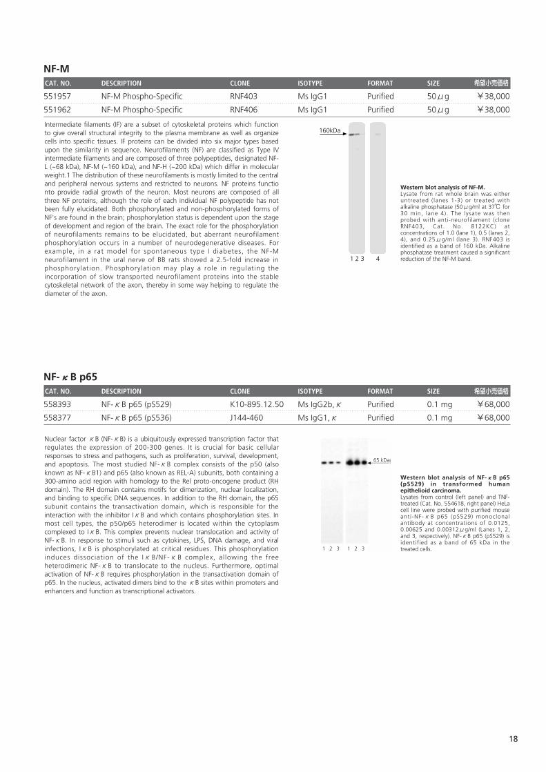
Western blot analysis of NF-κB p65(pS529) in transformed humanepithelioid carcinoma. Lysates from control (left panel) and TNF-treated (Cat. No. 554618, right panel) HeLacell line were probed with purified mouseanti-NF-κB p65 (pS529) monoclonalantibody at concentrations of 0.0125,0.00625 and 0.00312μg/ml (Lanes 1, 2,and 3, respectively). NF-κB p65 (pS529) isidentified as a band of 65 kDa in thetreated cells.
65 kDa
1 2 3 1 2 3
Nuclear factor κB (NF-κB) is a ubiquitously expressed transcription factor thatregulates the expression of 200-300 genes. It is crucial for basic cellularresponses to stress and pathogens, such as proliferation, survival, development,and apoptosis. The most studied NF-κB complex consists of the p50 (alsoknown as NF-κB1) and p65 (also known as REL-A) subunits, both containing a300-amino acid region with homology to the Rel proto-oncogene product (RHdomain). The RH domain contains motifs for dimerization, nuclear localization,and binding to specific DNA sequences. In addition to the RH domain, the p65subunit contains the transactivation domain, which is responsible for theinteraction with the inhibitor IκB and which contains phosphorylation sites. Inmost cell types, the p50/p65 heterodimer is located within the cytoplasmcomplexed to IκB. This complex prevents nuclear translocation and activity ofNF-κB. In response to stimuli such as cytokines, LPS, DNA damage, and viralinfections, IκB is phosphorylated at critical residues. This phosphorylationinduces dissociation of the IκB/NF-κB complex, al lowing the freeheterodimeric NF-κB to translocate to the nucleus. Furthermore, optimalactivation of NF-κB requires phosphorylation in the transactivation domain ofp65. In the nucleus, activated dimers bind to the κB sites within promoters andenhancers and function as transcriptional activators.
Western blot analysis of NF-M. Lysate from rat whole brain was eitheruntreated (lanes 1-3) or treated withalkaline phosphatase (50μg/ml at 37℃ for30 min, lane 4). The lysate was thenprobed with anti-neurofilament (cloneRNF403, Cat. No. 8122KC) atconcentrations of 1.0 (lane 1), 0.5 (lanes 2,4), and 0.25μg/ml (lane 3). RNF403 isidentified as a band of 160 kDa. Alkalinephosphatase treatment caused a significantreduction of the NF-M band.41 2 3
160kDaIntermediate filaments (IF) are a subset of cytoskeletal proteins which functionto give overall structural integrity to the plasma membrane as well as organizecells into specific tissues. IF proteins can be divided into six major types basedupon the similarity in sequence. Neurofilaments (NF) are classified as Type IVintermediate filaments and are composed of three polypeptides, designated NF-L (~68 kDa), NF-M (~160 kDa), and NF-H (~200 kDa) which differ in molecularweight.1 The distribution of these neurofilaments is mostly limited to the centraland peripheral nervous systems and restricted to neurons. NF proteins functionto provide radial growth of the neuron. Most neurons are composed of allthree NF proteins, although the role of each individual NF polypeptide has notbeen fully elucidated. Both phosphorylated and non-phosphorylated forms ofNF's are found in the brain; phosphorylation status is dependent upon the stageof development and region of the brain. The exact role for the phosphorylationof neurofilaments remains to be elucidated, but aberrant neurofilamentphosphorylation occurs in a number of neurodegenerative diseases. Forexample, in a rat model for spontaneous type I diabetes, the NF-Mneurofilament in the ural nerve of BB rats showed a 2.5-fold increase inphosphorylation. Phosphorylation may play a role in regulating theincorporation of slow transported neurofilament proteins into the stablecytoskeletal network of the axon, thereby in some way helping to regulate thediameter of the axon.
18
p38 MAPKCAT. NO. DESCRIPTION CLONE ISOTYPE FORMAT SIZE 希望小売価格
612552 p38 MAPK (pT180/pY182) 36 Ms IgG1 HRPO 50μg ¥35,000
612553 p38 MAPK (pT180/pY182) 36 Ms IgG1 HRPO 150μg ¥66,000
612288 p38 MAPK (pT180/pY182) 36 Ms IgG1 Purified 50μg ¥31,000
612289 p38 MAPK (pT180/pY182) 36 Ms IgG1 Purified 150μg ¥53,000
612280 p38 MAPK (pT180/pY182) 30 Ms IgG1 Purified 50μg ¥31,000
612281 p38 MAPK (pT180/pY182) 30 Ms IgG1 Purified 150μg ¥53,000
p53CAT. NO. DESCRIPTION CLONE ISOTYPE FORMAT SIZE 希望小売価格
558369 p53 (pS37) J159-641.15.4 Ms IgG1,κ Purified 0.1 mg ¥68,000
p90 RSK1CAT. NO. DESCRIPTION CLONE ISOTYPE FORMAT SIZE 希望小売価格
612692 p90 RSK1 (pS380) 20a Ms IgG1 Purified 50μg ¥31,000
612693 p90 RSK1 (pS380) 20a Ms IgG1 Purified 150μg ¥53,000
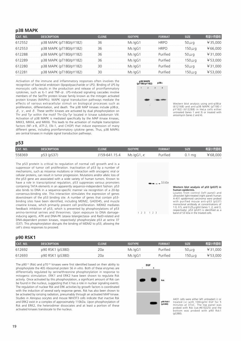
A431 cells were either left untreated (-) ortreated (+) with 100ng/ml EGF for 5minutes at 37oC. The top panel wasprobed with Rsk (cat.#610225) and thebottom was probed with p90 Rsk1(pS380).
EGF- +
Rsk
p90 Rsk1(pS380)
The p90 rsk (Rsk) and p70 s6k kinases were first identified based on their ability tophosphorylate the 40S ribosomal protein S6 in vitro. Both of these enzymes aredifferentially regulated by serine/threonine phosphorylation in response tomitogenic stimulation. ERK1 and ERK2 have been shown to regulate Rskactivity. Once activated by this phosphorylation, a significant amount of Rsk canbe found in the nucleus, suggesting that it has a role in nuclear signaling events.The regulation of nuclear Rsk and ERK activities by growth factors is coordinatedwith the induction of several early response genes. Rsk has also been shown tobe activated by ionizing radiation, presumably through an activated MAP kinase.Studies in Xenopus oocytes and mouse NIH/3T3 cells indicate that inactive Rskand ERK2 exist in a complex of approximately 110kDa. Upon phosphorylation ofRsk and ERK2, the heterodimer dissociates and at least a portion of theseactivated kinases translocate to the nucleus.
Western blot analysis of p53 (pS37) inhuman epidermis. Lysates from control ( left panel) andultraviolet light-treated (right panel) humanA-431 epidermoid carcinoma were probedwith purif ied mouse anti-p53 (pS37)monoclonal antibody at concentrations of1.0, 0.5, and 0.25μg/ml (lanes 1, 2, and 3,respectively). p53 (pS37) is identified as aband of 53 kDa in the treated cells.1 2 3 1 2 3
53 kDa
The p53 protein is critical to regulation of normal cell growth and is asuppressor of tumor cell proliferation. Inactivation of p53 by a number ofmechanisms, such as missense mutations or interaction with oncogenic viral orcellular proteins, can result in tumor progression. Mutations and/or allelic loss ofthe p53 gene are associated with a wide variety of human tumors. Known tohave a role in transcriptional regulation, p53 suppresses various promoterscontaining TATA elements in an apparently sequence-independent fashion. p53also binds to DNA in a sequence-specific manner via recognition of a 20-bpconsensus-binding site. This interaction stimulates the expression of genesdownstream of the p53 binding site. A number of genes that contain p53-binding sites have been identified, including MDM2, GADD45, and musclecreatine kinase, which primarily prevent cell proliferation. MDM2 mediatesfeedback inhibition of p53, which is prevented by phosphorylations of p53amino-terminal serines and threonines. Upon exposure to DNA damage-inducing agents, ATR and DNA-PK (ataxia telangiectasia- and Rad3-related andDNA-dependent protein kinases, respectively) phosphorylate p53 at serine 37(S37). This phosphorylation disrupts the binding of MDM2 to p53, allowing thecell's stress responses to proceed.
Western blot analysis using anti-p38α(612168) and anti-p38 MAPK (pT180 /pY182) (612288) in HeLa cells eitheruntreated (lanes 1 and 3) or treated withanisomycin (lanes 2 and 4).
p38 MAPK(pT180/pY182) p38�
1 2 3 4
Activation of the immune and inflammatory responses often involves therecognition of bacterial endotoxin (lipopolysaccharide or LPS). Binding of LPS bymonocytic cells results in the production and release of proinflammatorycytokines, such as IL-1 and TNF-α. LPS-induced signaling cascades involvemembers of the Ser/Thr protein kinase family known as the mitogen activatedprotein kinases (MAPKs). MAPK signal transduction pathways mediate theeffects of various extracellular stimuli on biological processes such asproliferation, differentiation, and death. The p38 MAP kinases include p38α,β, γ, and δ. These ser/thr kinases are activated by dual phosphorylation onThr and Tyr within the motif Thr-Gly-Tyr located in kinase subdomain VIII.Activation of p38 MAPK is mediated specifically by the MAP kinase kinases,MKK3, MKK4, and MKK6. This leads to the activation of multiple transcriptionfactors (NF-κB, ATF-2, Elk-1, and CHOP) that induce expression of manydifferent genes, including proinflammatory cytokine genes. Thus, p38 MAPKsare central kinases in muliple signal transduction pathways.
19
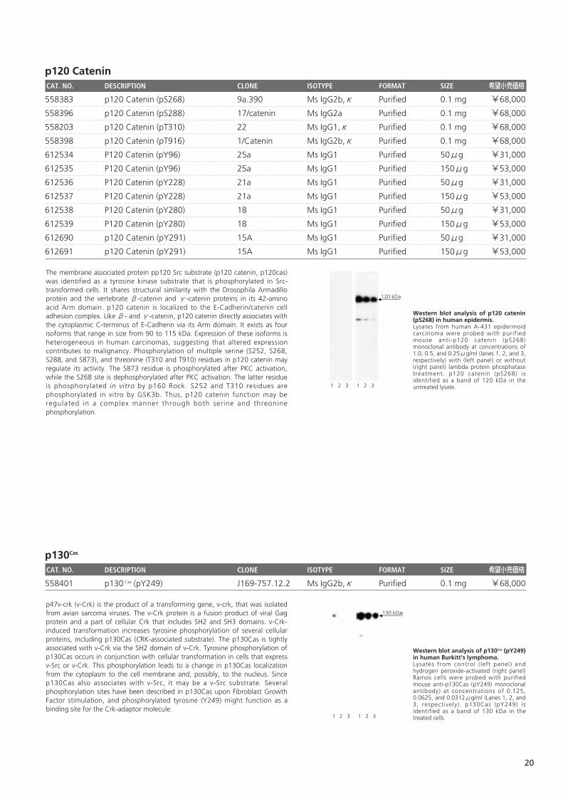
p120 CateninCAT. NO. DESCRIPTION CLONE ISOTYPE FORMAT SIZE 希望小売価格
558383 p120 Catenin (pS268) 9a.390 Ms IgG2b,κ Purified 0.1 mg ¥68,000
558396 p120 Catenin (pS288) 17/catenin Ms IgG2a Purified 0.1 mg ¥68,000
558203 p120 Catenin (pT310) 22 Ms IgG1,κ Purified 0.1 mg ¥68,000
558398 p120 Catenin (pT916) 1/Catenin Ms IgG2b,κ Purified 0.1 mg ¥68,000
612534 P120 Catenin (pY96) 25a Ms IgG1 Purified 50μg ¥31,000
612535 P120 Catenin (pY96) 25a Ms IgG1 Purified 150μg ¥53,000
612536 P120 Catenin (pY228) 21a Ms IgG1 Purified 50μg ¥31,000
612537 P120 Catenin (pY228) 21a Ms IgG1 Purified 150μg ¥53,000
612538 P120 Catenin (pY280) 18 Ms IgG1 Purified 50μg ¥31,000
612539 P120 Catenin (pY280) 18 Ms IgG1 Purified 150μg ¥53,000
612690 p120 Catenin (pY291) 15A Ms IgG1 Purified 50μg ¥31,000
612691 p120 Catenin (pY291) 15A Ms IgG1 Purified 150μg ¥53,000
p130Cas
CAT. NO. DESCRIPTION CLONE ISOTYPE FORMAT SIZE 希望小売価格
558401 p130 Cas (pY249) J169-757.12.2 Ms IgG2b,κ Purified 0.1 mg ¥68,000
Western blot analysis of p130Cas (pY249)in human Burkitt's lymphoma. Lysates from control ( left panel) andhydrogen peroxide-activated (right panel)Ramos cells were probed with purifiedmouse anti-p130Cas (pY249) monoclonalantibody) at concentrations of 0.125,0.0625, and 0.0312μg/ml (Lanes 1, 2, and3, respectively). p130Cas (pY249) isidentified as a band of 130 kDa in thetreated cells.
130 kDa
1 2 3 1 2 3
p47v-crk (v-Crk) is the product of a transforming gene, v-crk, that was isolatedfrom avian sarcoma viruses. The v-Crk protein is a fusion product of viral Gagprotein and a part of cellular Crk that includes SH2 and SH3 domains. v-Crk-induced transformation increases tyrosine phosphorylation of several cellularproteins, including p130Cas (CRK-associated substrate). The p130Cas is tightlyassociated with v-Crk via the SH2 domain of v-Crk. Tyrosine phosphorylation ofp130Cas occurs in conjunction with cellular transformation in cells that expressv-Src or v-Crk. This phosphorylation leads to a change in p130Cas localizationfrom the cytoplasm to the cell membrane and, possibly, to the nucleus. Sincep130Cas also associates with v-Src, it may be a v-Src substrate. Severalphosphorylation sites have been described in p130Cas upon Fibroblast GrowthFactor stimulation, and phosphorylated tyrosine (Y249) might function as abinding site for the Crk-adaptor molecule.
Western blot analysis of p120 catenin(pS268) in human epidermis. Lysates from human A-431 epidermoidcarcinoma were probed with purifiedmouse anti-p120 catenin (pS268)monoclonal antibody at concentrations of1.0, 0.5, and 0.25μg/ml (lanes 1, 2, and 3,respectively) with (left panel) or without(right panel) lambda protein phosphatasetreatment. p120 catenin (pS268) isidentified as a band of 120 kDa in theuntreated lysate.
120 kDa
1 2 3 1 2 3
The membrane associated protein pp120 Src substrate (p120 catenin, p120cas)was identified as a tyrosine kinase substrate that is phosphorylated in Src-transformed cells. It shares structural similarity with the Drosophila Armadilloprotein and the vertebrate β-catenin and γ-catenin proteins in its 42-aminoacid Arm domain. p120 catenin is localized to the E-Cadherin/catenin celladhesion complex. Like β- and γ-catenin, p120 catenin directly associates withthe cytoplasmic C-terminus of E-Cadherin via its Arm domain. It exists as fourisoforms that range in size from 90 to 115 kDa. Expression of these isoforms isheterogeneous in human carcinomas, suggesting that altered expressioncontributes to malignancy. Phosphorylation of multiple serine (S252, S268,S288, and S873), and threonine (T310 and T910) residues in p120 catenin mayregulate its activity. The S873 residue is phosphorylated after PKC activation,while the S268 site is dephosphorylated after PKC activation. The latter residueis phosphorylated in vitro by p160 Rock. S252 and T310 residues arephosphorylated in vitro by GSK3b. Thus, p120 catenin function may beregulated in a complex manner through both serine and threoninephosphorylation.
20
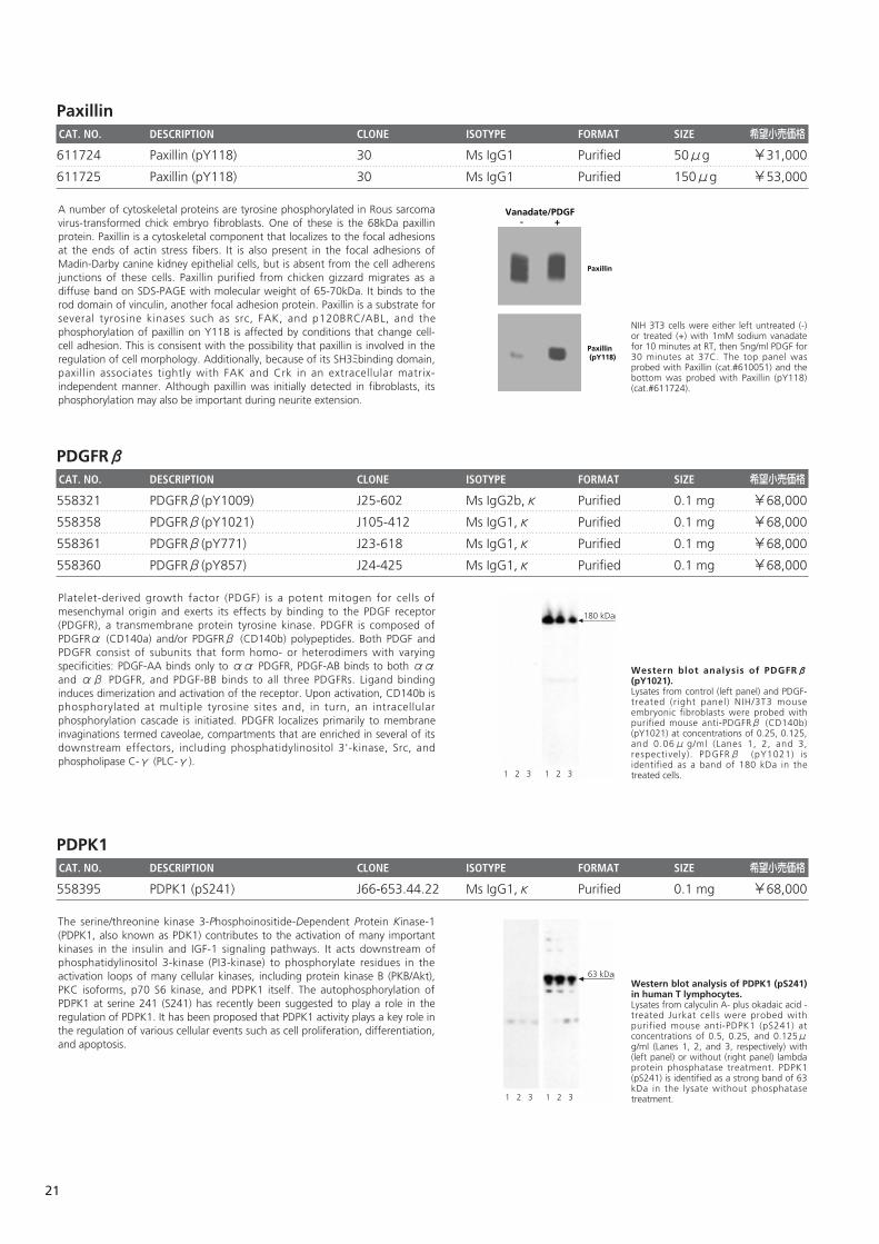
PaxillinCAT. NO. DESCRIPTION CLONE ISOTYPE FORMAT SIZE 希望小売価格
611724 Paxillin (pY118) 30 Ms IgG1 Purified 50μg ¥31,000
611725 Paxillin (pY118) 30 Ms IgG1 Purified 150μg ¥53,000
PDGFRβCAT. NO. DESCRIPTION CLONE ISOTYPE FORMAT SIZE 希望小売価格
558321 PDGFRβ(pY1009) J25-602 Ms IgG2b,κ Purified 0.1 mg ¥68,000
558358 PDGFRβ(pY1021) J105-412 Ms IgG1,κ Purified 0.1 mg ¥68,000
558361 PDGFRβ(pY771) J23-618 Ms IgG1,κ Purified 0.1 mg ¥68,000
558360 PDGFRβ(pY857) J24-425 Ms IgG1,κ Purified 0.1 mg ¥68,000
PDPK1CAT. NO. DESCRIPTION CLONE ISOTYPE FORMAT SIZE 希望小売価格
558395 PDPK1 (pS241) J66-653.44.22 Ms IgG1,κ Purified 0.1 mg ¥68,000
Western blot analysis of PDPK1 (pS241)in human T lymphocytes. Lysates from calyculin A- plus okadaic acid -treated Jurkat cells were probed withpurified mouse anti-PDPK1 (pS241) atconcentrations of 0.5, 0.25, and 0.125μg/ml (Lanes 1, 2, and 3, respectively) with(left panel) or without (right panel) lambdaprotein phosphatase treatment. PDPK1(pS241) is identified as a strong band of 63kDa in the lysate without phosphatasetreatment.
63 kDa
1 2 3 1 2 3
The serine/threonine kinase 3-Phosphoinositide-Dependent Protein Kinase-1(PDPK1, also known as PDK1) contributes to the activation of many importantkinases in the insulin and IGF-1 signaling pathways. It acts downstream ofphosphatidylinositol 3-kinase (PI3-kinase) to phosphorylate residues in theactivation loops of many cellular kinases, including protein kinase B (PKB/Akt),PKC isoforms, p70 S6 kinase, and PDPK1 itself. The autophosphorylation ofPDPK1 at serine 241 (S241) has recently been suggested to play a role in theregulation of PDPK1. It has been proposed that PDPK1 activity plays a key role inthe regulation of various cellular events such as cell proliferation, differentiation,and apoptosis.
Western blot analysis of PDGFRβ(pY1021). Lysates from control (left panel) and PDGF-treated (right panel) NIH/3T3 mouseembryonic fibroblasts were probed withpurified mouse anti-PDGFRβ (CD140b)(pY1021) at concentrations of 0.25, 0.125,and 0.06μ g/ml (Lanes 1, 2, and 3,respectively). PDGFRβ (pY1021) isidentified as a band of 180 kDa in thetreated cells.
180 kDa
1 2 3 1 2 3
Platelet-derived growth factor (PDGF) is a potent mitogen for cells ofmesenchymal origin and exerts its effects by binding to the PDGF receptor(PDGFR), a transmembrane protein tyrosine kinase. PDGFR is composed ofPDGFRα (CD140a) and/or PDGFRβ (CD140b) polypeptides. Both PDGF andPDGFR consist of subunits that form homo- or heterodimers with varyingspecificities: PDGF-AA binds only to αα PDGFR, PDGF-AB binds to both ααand αβ PDGFR, and PDGF-BB binds to all three PDGFRs. Ligand bindinginduces dimerization and activation of the receptor. Upon activation, CD140b isphosphorylated at multiple tyrosine sites and, in turn, an intracellularphosphorylation cascade is initiated. PDGFR localizes primarily to membraneinvaginations termed caveolae, compartments that are enriched in several of itsdownstream effectors, including phosphatidylinositol 3'-kinase, Src, andphospholipase C-γ (PLC-γ).
NIH 3T3 cells were either left untreated (-)or treated (+) with 1mM sodium vanadatefor 10 minutes at RT, then 5ng/ml PDGF for30 minutes at 37C. The top panel wasprobed with Paxillin (cat.#610051) and thebottom was probed with Paxillin (pY118)(cat.#611724).
Vanadate/PDGF- +
Paxillin
Paxillin (pY118)
A number of cytoskeletal proteins are tyrosine phosphorylated in Rous sarcomavirus-transformed chick embryo fibroblasts. One of these is the 68kDa paxillinprotein. Paxillin is a cytoskeletal component that localizes to the focal adhesionsat the ends of actin stress fibers. It is also present in the focal adhesions ofMadin-Darby canine kidney epithelial cells, but is absent from the cell adherensjunctions of these cells. Paxillin purified from chicken gizzard migrates as adiffuse band on SDS-PAGE with molecular weight of 65-70kDa. It binds to therod domain of vinculin, another focal adhesion protein. Paxillin is a substrate forseveral tyrosine kinases such as src, FAK, and p120BRC/ABL, and thephosphorylation of paxillin on Y118 is affected by conditions that change cell-cell adhesion. This is consisent with the possibility that paxillin is involved in theregulation of cell morphology. Additionally, because of its SH3ミbinding domain,paxillin associates tightly with FAK and Crk in an extracellular matrix-independent manner. Although paxillin was initially detected in fibroblasts, itsphosphorylation may also be important during neurite extension.
21
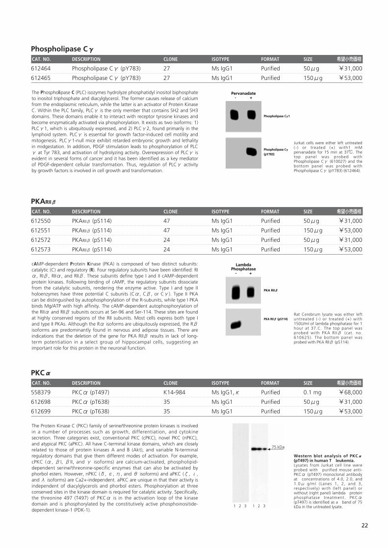
Phospholipase CγCAT. NO. DESCRIPTION CLONE ISOTYPE FORMAT SIZE 希望小売価格
612464 Phospholipase Cγ (pY783) 27 Ms IgG1 Purified 50μg ¥31,000
612465 Phospholipase Cγ (pY783) 27 Ms IgG1 Purified 150μg ¥53,000
PKARIIβ
CAT. NO. DESCRIPTION CLONE ISOTYPE FORMAT SIZE 希望小売価格
612550 PKARIIβ (pS114) 47 Ms IgG1 Purified 50μg ¥31,000
612551 PKARIIβ (pS114) 47 Ms IgG1 Purified 150μg ¥53,000
612572 PKARIIβ (pS114) 24 Ms IgG1 Purified 50μg ¥31,000
612573 PKARIIβ (pS114) 24 Ms IgG1 Purified 150μg ¥53,000
PKCαCAT. NO. DESCRIPTION CLONE ISOTYPE FORMAT SIZE 希望小売価格
558379 PKCα (pT497) K14-984 Ms IgG1,κ Purified 0.1 mg ¥68,000
612698 PKCα (pT638) 35 Ms IgG1 Purified 50μg ¥31,000
612699 PKCα (pT638) 35 Ms IgG1 Purified 150μg ¥53,000
Western blot analysis of PKCα(pT497) in human T leukemia. Lysates from Jurkat cell line wereprobed with purified mouse anti-PKCα (pT497) monoclonal antibodyat concentrations of 4.0, 2.0, and1.0μ g/ml (Lanes 1, 2, and 3,respectively) with ( left panel) orwithout (right panel) lambda proteinphosphatase treatment. PKCα(pT497) is identified as a band of 75kDa in the untreated lysate.
75 kDa
1 2 3 1 2 3
The Protein Kinase C (PKC) family of serine/threonine protein kinases is involvedin a number of processes such as growth, differentiation, and cytokinesecretion. Three categories exist, conventional PKC (cPKC), novel PKC (nPKC),and atypical PKC (aPKC). All have C-terminal kinase domains, which are closelyrelated to those of protein kinases A and B (Akt), and variable N-terminalregulatory domains that give them different modes of activation. For example,cPKC (α, βI, βII, and γ isoforms) are calcium-activated, phospholipid-dependent serine/threonine-specific enzymes that can also be activated byphorbol esters. However, nPKC (δ, ε, η, and θ isoforms) and aPKC (ζ, ι,and λ isoforms) are Ca2+-independent. aPKC are unique in that their activity isindependent of diacylglycerols and phorbol esters. Phosphorylation at threeconserved sites in the kinase domain is required for catalytic activity. Specifically,the threonine 497 (T497) of PKCα is in the activation loop of the kinasedomain and is phosphorylated by the constitutively active phosphoinositide-dependent kinase-1 (PDK-1).
Rat Cerebrum lysate was either leftuntreated (-) or treated (+) with150U/ml of lambda phosphatase for 1hour at 37.C. The top panel wasprobed with PKA RIIβ (cat. no.610625). The bottom panel wasprobed with PKA RIIβ (pS114).
PKA RIIβ
PKA RIIβ (pS114)
LambdaPhosphatase
- +
cAMP-dependent Protein Kinase (PKA) is composed of two distinct subunits:catalytic (C) and regulatory (R). Four regulatory subunits have been identified: RIα, RIβ, RIIα, and RIIβ. These subunits define type I and II cAMP-dependentprotein kinases. Following binding of cAMP, the regulatory subunits dissociatefrom the catalytic subunits, rendering the enzyme active. Type I and type IIholoenzymes have three potential C subunits (Cα, Cβ, or Cγ). Type II PKAcan be distinguished by autophosphorylation of the R-subunits, while type I PKAbinds Mg/ATP with high affinity. The cAMP-dependent autophosphorylation ofthe RIIα and RIIβ subunits occurs at Ser-96 and Ser-114. These sites are foundat highly conserved regions of the RII subunits. Most cells express both type Iand type II PKAs. Although the Rα isoforms are ubiquitously expressed, the Rβisoforms are predominantly found in nervous and adipose tissues. There areindications that the deletion of the gene for PKA RIIβ results in lack of long-term potentiation in a select group of hippocampal cells, suggesting animportant role for this protein in the neuronal function.
Jurkat cells were either left untreated(-) or treated (+) with1 mMpervanadate for 15 min at 37℃. Thetop panel was probed withPhospholipase Cγ (610027) and thebottom panel was probed withPhospholipase Cγ (pY783) (612464).
Phospholipase Cγ1
Phospholipase Cγ(pY783)
Pervanadate- +
The Phospholipase C (PLC) isozymes hydrolyze phosphatidyl inositol biphosphateto inositol triphosphate and diacylglycerol. The former causes release of calciumfrom the endoplasmic reticulum, while the latter is an activator of Protein KinaseC. Within the PLC family, PLCγ is the only member that contains SH2 and SH3domains. These domains enable it to interact with receptor tyrosine kinases andbecome enzymatically activated via phosphorylation. It exists as two isoforms: 1)PLCγ1, which is ubiquitously expressed, and 2) PLCγ2, found primarily in thelymphoid system. PLCγ is essential for growth factor-induced cell motility andmitogenesis. PLCγ1-null mice exhibit retarded embryonic growth and lethalityin midgestation. In addition, PDGF stimulation leads to phosphorylation of PLCγ at Tyr 783, and activation of hydrolyzing activity. Overexpression of PLCγ isevident in several forms of cancer and it has been identified as a key mediatorof PDGF-dependent cellular transformation. Thus, regulation of PLCγ activityby growth factors is involved in cell growth and transformation.
22
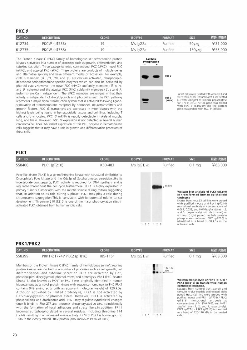
PKCθCAT. NO. DESCRIPTION CLONE ISOTYPE FORMAT SIZE 希望小売価格
612734 PKCθ (pT538) 19 Ms IgG2a Purified 50μg ¥31,000
612735 PKCθ (pT538) 19 Ms IgG2a Purified 150μg ¥53,000
PLK1 CAT. NO. DESCRIPTION CLONE ISOTYPE FORMAT SIZE 希望小売価格
558400 PLK1 (pT210) K50-483 Ms IgG1,κ Purified 0.1 mg ¥68,000
PRK1/PRK2CAT. NO. DESCRIPTION CLONE ISOTYPE FORMAT SIZE 希望小売価格
558399 PRK1 (pT774)/ PRK2 (pT816) I85-1151 Ms IgG1,κ Purified 0.1 mg ¥68,000
Western blot analysis of PRK1 (pT774) /PRK2 (pT816) in transformed humanepithelioid carcinoma.Lysates from control ( left panel) andcalyculin A-plus-okadaic acid-treated (rightpanel) HeLa cell line were probed withpurified mouse anti-PRK1 (pT774) / PRK2(pT816) monoclonal antibody atconcentrations of 0.125,0.0625, and 0.031μg/ml (lanes 1, 2, and 3, respectively).PRK1 (pT774) / PRK2 (pT816) is identifiedas a band of 120-140 kDa in the treatedcells.
120-140 kDa
1 2 3 1 2 3
Members of the Protein Kinase C (PKC) family of homologous serine/threonineprotein kinases are involved in a number of processes such as cell growth, celldifferentiation, and cytokine secretion.PKCs are activated by Ca2+,phospholipids, diacylglycerol, phorbol esters, and proteolysis. PRK1 (PKC-RelatedKinase 1, also known as PKN1 or PKL1) was originally identified in humanhippocampus as a novel protein kinase with sequence homology to PKC.PRK1contains 942 amino acids with an apparent molecular weight of 120 kDa.Although activated by l imited proteolysis, PRK1 is not activated byCa2+/diacylglycerol or phorbol esters. However, PRK1 is activated byphospholipids and arachidonic acid. PRK1 may regulate cytoskeletal changessince it binds to Rho-GTP and becomes phosphorylated in vivo, coincidentallywith the formation of focal adhesions and stress fibers.In addition, PRK1becomes autophosphorylated in several residues, including threonine 774(T774), resulting in an increased kinase activity. T774 of PRK1 is homologous toT816 in the closely related PRK2 protein (also known as PKN2 or PKL2).
Western blot analysis of PLK1 (pT210)in transformed human epithelioidcarcinomaLysates from HeLa S3 cell line were probedwith purified mouse anti-PLK1 (pT210)monoclonal antibody at concentrations of0.063, 0.032, and 0.016μg/ml (Lanes 1,2,and 3, respectively) with (left panel) orwithout (r ight panel) lambda proteinphosphatase treatment. PLK1 (pT210) isidentified as a band of 68 kDa in theuntreated cells.
68 kDa
1 2 3 1 2 3
Polo-like kinase (PLK1) is a serine/threonine kinase with structural similarities toDrosophila's Polo kinase and the Cdc5p of Saccharomyces cerevisiae.Like itsinvertebrate counterparts, PLK1 activity is required for DNA synthesis and isregulated throughout the cell cycle.Furthermore, PLK1 is highly expressed inprimary tumors.It associates with the mitotic spindle during mitosis suggestingthat, in addition to its role during S phase, PLK1 may play a role duringchromosome segregation.This is consistent with its potential role in cancerdevelopment. Threonine 210 (T210) is one of the major phoshorylation sites inactivated PLK1 obtained from human mitotic cells.
Jurkat cells were treated with Anti-CD3 andwere then either left untreated (-)or treated(+) with 200U/ml of lambda phosphatasefor 1 hr at 37℃.The top panel was probedwith PKC θ (610089) and the bottompanel was probed with PKC θ (pT538).
LambdaPhosphatase- +
PKC θ
PKC θ (pT538)
The Protein Kinase C (PKC) family of homologous serine/threonine proteinkinases is involved in a number of processes such as growth, differentiation, andcytokine secretion. Three categories exist, conventional PKC (cPKC), novel PKC(nPKC), and atypical PKC (aPKC). These proteins are products of multiple genesand alternative splicing and have different modes of activation. For example,cPKC's members (α, βl, βll, and γ) are calcium activated, phospholipid-dependent serine/threonine specific enzymes which can also be activated byphorbol esters.However, the novel PKC (nPKC) subfamily members (δ,ε,η,and θ isoforms) and the atypical PKC (PKC) subfamily members (ζ,ι,and λisoforms) are Ca 2+ independent. The aPKC members are unique in that theiractivity is independent of diacylglycerols and phorbol esters. The PKC pathwayrepresents a major signal transduction system that is activated following ligand-stimulation of transmembrane receptors by hormones, neurotransmitters andgrowth factors. PKC θ transcripts are expressed in most tissues with thehighest levels being found in hematopoietic tissues and cell lines, including Tcells and thymocytes. PKC θ mRNA is readily detectable in skeletal muscle,lung, and brain. However, PKC θ expression is not detected in several humancarcinoma cell lines. Abundant expression of this PKC isozyme in hematopoieticcells suggests that it may have a role in growth and differentiation processes ofthese cells.
23
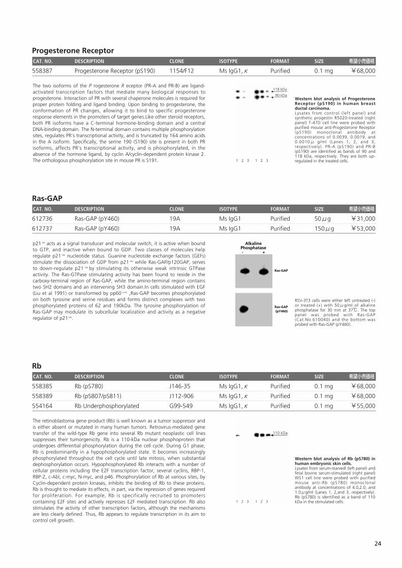
Progesterone ReceptorCAT. NO. DESCRIPTION CLONE ISOTYPE FORMAT SIZE 希望小売価格
558387 Progesterone Receptor (pS190) 1154/F12 Ms IgG1,κ Purified 0.1 mg ¥68,000
Ras-GAPCAT. NO. DESCRIPTION CLONE ISOTYPE FORMAT SIZE 希望小売価格
612736 Ras-GAP (pY460) 19A Ms IgG1 Purified 50μg ¥31,000
612737 Ras-GAP (pY460) 19A Ms IgG1 Purified 150μg ¥53,000
RbCAT. NO. DESCRIPTION CLONE ISOTYPE FORMAT SIZE 希望小売価格
558385 Rb (pS780) J146-35 Ms IgG1,κ Purified 0.1 mg ¥68,000
558389 Rb (pS807/pS811) J112-906 Ms IgG1,κ Purified 0.1 mg ¥68,000
554164 Rb Underphosphorylated G99-549 Ms IgG1,κ Purified 0.1 mg ¥55,000
Western blot analysis of Rb (pS780) inhuman embryonic skin cells.Lysates from serum-starvedl (left panel) andfetal bovine serum-stimulated (right panel)WS1 cell line were probed with purifiedmouse anti-Rb (pS780) monoclonalantibody at concentrations of 4.0,2.0, and1.0μg/ml (Lanes 1, 2,and 3, respectively).Rb (pS780) is identified as a band of 110kDa in the stimulated cells.
110 kDa
1 2 3 1 2 3
The retinoblastoma gene product (Rb) is well known as a tumor suppressor andis either absent or mutated in many human tumors. Retrovirus-mediated genetransfer of the wild-type Rb gene into several Rb mutant neoplastic cell linessuppresses their tumorgenicity. Rb is a 110-kDa nuclear phosphoprotein thatundergoes differential phosphorylation during the cell cycle. During G1 phase,Rb is predominantly in a hypophosphorylated state. It becomes increasinglyphosphorylated throughout the cell cycle until late mitosis, when substantialdephosphorylation occurs. Hypophosphorylated Rb interacts with a number ofcellular proteins including the E2F transcription factor, several cyclins, RBP-1,RBP-2, c-Abl, c-myc, N-myc, and p46. Phosphorylation of Rb at various sites, byCyclin-dependent protein kinases, inhibits the binding of Rb to these proteins.Rb is thought to mediate its effects, in part, via the repression of genes requiredfor proliferation. For example, Rb is specifically recruited to promoterscontaining E2F sites and actively represses E2F mediated transcription. Rb alsostimulates the activity of other transcription factors, although the mechanismsare less clearly defined. Thus, Rb appears to regulate transcription in its aim tocontrol cell growth.
RSV-3T3 cells were either left untreated (-)or treated (+) with 50μg/ml of alkalinephosphatase for 30 min at 37℃. The toppanel was probed with Ras-GAP(Cat.No.610040) and the bottom wasprobed with Ras-GAP (pY460).
AlkalinePhosphatase- +
Ras-GAP
Ras-GAP (pY460)
p21 ras acts as a signal transducer and molecular switch, it is active when boundto GTP, and inactive when bound to GDP. Two classes of molecules helpregulate p21 ras nucleotide status. Guanine nucleotide exchange factors (GEFs)stimulate the dissociation of GDP from p21 ras while Ras-GAP/p120GAP, servesto down-regulate p21 ras by stimulating its otherwise weak intrinsic GTPaseactivity. The Ras-GTPase stimulating activity has been found to reside in thecarboxy-terminal region of Ras-GAP, while the amino-terminal region containstwo SH2 domains and an intervening SH3 domain.In cells stimulated with EGF(Liu et al 1991) or transformed by pp60 v-src ,Ras-GAP becomes phosphorylatedon both tyrosine and serine residues and forms distinct complexes with twophosphorylated proteins of 62 and 190kDa. The tyrosine phosphorylation ofRas-GAP may modulate its subcellular localization and activity as a negativeregulator of p21 ras.
Western blot analysis of ProgesteroneReceptor (pS190) in human breastductal carcinoma.Lysates from control ( left panel) andsynthetic progestin R5020-treated (rightpanel) T-47D cell line were probed withpurified mouse anti-Progesterone Receptor(pS190) monoclonal antibody atconcentrations of 0.0039, 0.0019, and0.0010μ g/ml (Lanes 1, 2, and 3,respectively). PR-A (pS190) and PR-B(pS190) are identified as bands of 90 and118 kDa, respectively. They are both up-regulated in the treated cells.
118 kDa
90 kDa
1 2 3 1 2 3
The two isoforms of the P rogesterone R eceptor (PR-A and PR-B) are ligand-activated transcription factors that mediate many biological responses toprogesterone. Interaction of PR with several chaperone molecules is required forproper protein folding and ligand binding. Upon binding to progesterone, theconformation of PR changes, allowing it to bind to specific progesteroneresponse elements in the promoters of target genes.Like other steroid receptors,both PR isoforms have a C-terminal hormone-binding domain and a centralDNA-binding domain. The N-terminal domain contains multiple phosphorylationsites, regulates PR's transcriptional activity, and is truncated by 164 amino acidsin the A isoform. Specifically, the serine 190 (S190) site is present in both PRisoforms, affects PR's transcriptional activity, and is phosphorylated, in theabsence of the hormone ligand, by cyclin A/cyclin-dependent protein kinase 2.The orthologous phosphorylation site in mouse PR is S191.
24
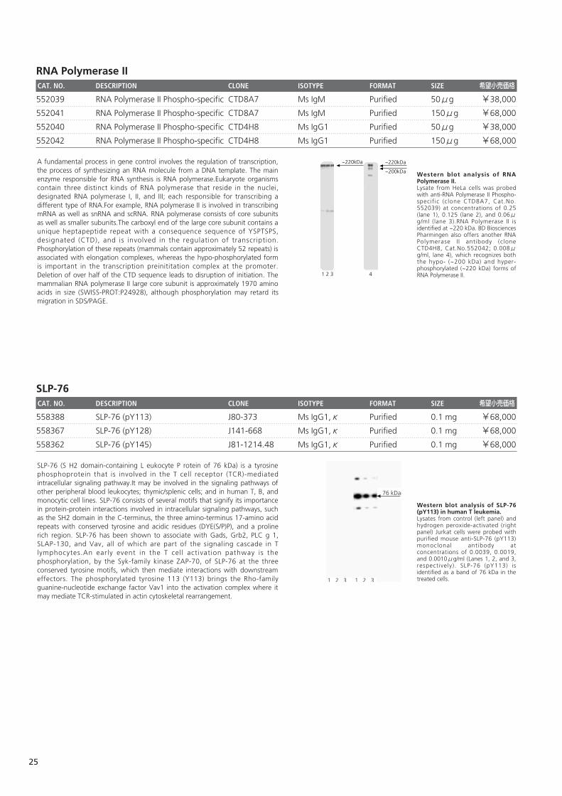
RNA Polymerase IICAT. NO. DESCRIPTION CLONE ISOTYPE FORMAT SIZE 希望小売価格
552039 RNA Polymerase II Phospho-specific CTD8A7 Ms IgM Purified 50μg ¥38,000
552041 RNA Polymerase II Phospho-specific CTD8A7 Ms IgM Purified 150μg ¥68,000
552040 RNA Polymerase II Phospho-specific CTD4H8 Ms IgG1 Purified 50μg ¥38,000
552042 RNA Polymerase II Phospho-specific CTD4H8 Ms IgG1 Purified 150μg ¥68,000
SLP-76CAT. NO. DESCRIPTION CLONE ISOTYPE FORMAT SIZE 希望小売価格
558388 SLP-76 (pY113) J80-373 Ms IgG1,κ Purified 0.1 mg ¥68,000
558367 SLP-76 (pY128) J141-668 Ms IgG1,κ Purified 0.1 mg ¥68,000
558362 SLP-76 (pY145) J81-1214.48 Ms IgG1,κ Purified 0.1 mg ¥68,000
Western blot analysis of SLP-76(pY113) in human T leukemia.Lysates from control (left panel) andhydrogen peroxide-activated (rightpanel) Jurkat cells were probed withpurified mouse anti-SLP-76 (pY113)monoclonal antibody atconcentrations of 0.0039, 0.0019,and 0.0010μg/ml (Lanes 1, 2, and 3,respectively). SLP-76 (pY113) isidentified as a band of 76 kDa in thetreated cells.1 2 3 1 2 3
76 kDa
SLP-76 (S H2 domain-containing L eukocyte P rotein of 76 kDa) is a tyrosinephosphoprotein that is involved in the T cell receptor (TCR)-mediatedintracellular signaling pathway.It may be involved in the signaling pathways ofother peripheral blood leukocytes; thymic/splenic cells; and in human T, B, andmonocytic cell lines. SLP-76 consists of several motifs that signify its importancein protein-protein interactions involved in intracellular signaling pathways, suchas the SH2 domain in the C-terminus, the three amino-terminus 17-amino acidrepeats with conserved tyrosine and acidic residues (DYE(S/P)P), and a prolinerich region. SLP-76 has been shown to associate with Gads, Grb2, PLC g 1,SLAP-130, and Vav, all of which are part of the signaling cascade in Tlymphocytes.An early event in the T cell activation pathway is thephosphorylation, by the Syk-family kinase ZAP-70, of SLP-76 at the threeconserved tyrosine motifs, which then mediate interactions with downstreameffectors. The phosphorylated tyrosine 113 (Y113) brings the Rho-familyguanine-nucleotide exchange factor Vav1 into the activation complex where itmay mediate TCR-stimulated in actin cytoskeletal rearrangement.
Western blot analysis of RNAPolymerase II.Lysate from HeLa cells was probedwith anti-RNA Polymerase II Phospho-specific (clone CTD8A7, Cat.No.552039) at concentrations of 0.25(lane 1), 0.125 (lane 2), and 0.06μg/ml (lane 3).RNA Polymerase II isidentified at ~220 kDa. BD BiosciencesPharmingen also offers another RNAPolymerase I I antibody (cloneCTD4H8, Cat.No.552042; 0.008μg/ml, lane 4), which recognizes boththe hypo- (~200 kDa) and hyper-phosphorylated (~220 kDa) forms ofRNA Polymerase II.
~220kDa
1 2 3
~220kDa
~200kDa
4
A fundamental process in gene control involves the regulation of transcription,the process of synthesizing an RNA molecule from a DNA template. The mainenzyme responsible for RNA synthesis is RNA polymerase.Eukaryote organismscontain three distinct kinds of RNA polymerase that reside in the nuclei,designated RNA polymerase I, II, and III; each responsible for transcribing adifferent type of RNA.For example, RNA polymerase II is involved in transcribingmRNA as well as snRNA and scRNA. RNA polymerase consists of core subunitsas well as smaller subunits.The carboxyl end of the large core subunit contains aunique heptapeptide repeat with a consequence sequence of YSPTSPS,designated (CTD), and is involved in the regulation of transcription.Phosphorylation of these repeats (mammals contain approximately 52 repeats) isassociated with elongation complexes, whereas the hypo-phosphorylated formis important in the transcription preinititation complex at the promoter.Deletion of over half of the CTD sequence leads to disruption of initiation. Themammalian RNA polymerase II large core subunit is approximately 1970 aminoacids in size (SWISS-PROT:P24928), although phosphorylation may retard itsmigration in SDS/PAGE.
25
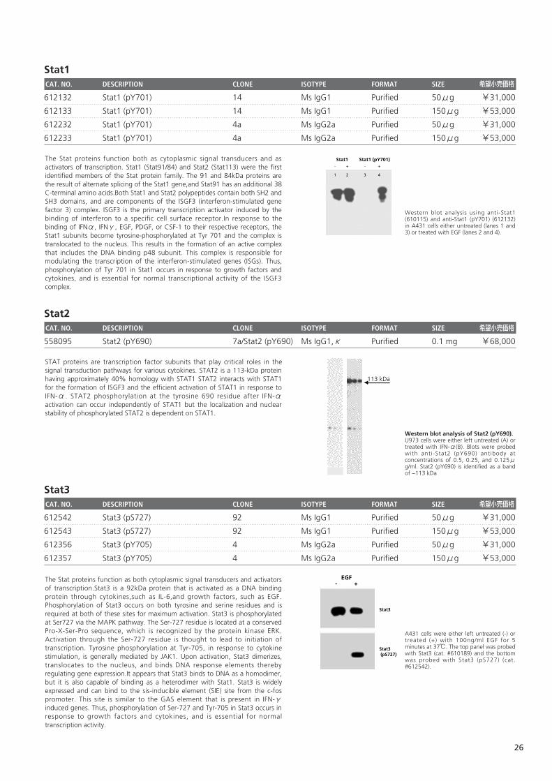
Stat1CAT. NO. DESCRIPTION CLONE ISOTYPE FORMAT SIZE 希望小売価格
612132 Stat1 (pY701) 14 Ms IgG1 Purified 50μg ¥31,000
612133 Stat1 (pY701) 14 Ms IgG1 Purified 150μg ¥53,000
612232 Stat1 (pY701) 4a Ms IgG2a Purified 50μg ¥31,000
612233 Stat1 (pY701) 4a Ms IgG2a Purified 150μg ¥53,000
Stat2CAT. NO. DESCRIPTION CLONE ISOTYPE FORMAT SIZE 希望小売価格
558095 Stat2 (pY690) 7a/Stat2 (pY690) Ms IgG1,κ Purified 0.1 mg ¥68,000
Stat3CAT. NO. DESCRIPTION CLONE ISOTYPE FORMAT SIZE 希望小売価格
612542 Stat3 (pS727) 92 Ms IgG1 Purified 50μg ¥31,000
612543 Stat3 (pS727) 92 Ms IgG1 Purified 150μg ¥53,000
612356 Stat3 (pY705) 4 Ms IgG2a Purified 50μg ¥31,000
612357 Stat3 (pY705) 4 Ms IgG2a Purified 150μg ¥53,000
A431 cells were either left untreated (-) ortreated (+) with 100ng/ml EGF for 5minutes at 37℃. The top panel was probedwith Stat3 (cat. #610189) and the bottomwas probed with Stat3 (pS727) (cat.#612542).
EGF- +
Stat3
Stat3 (pS727)
The Stat proteins function as both cytoplasmic signal transducers and activatorsof transcription.Stat3 is a 92kDa protein that is activated as a DNA bindingprotein through cytokines,such as IL-6,and growth factors, such as EGF.Phosphorylation of Stat3 occurs on both tyrosine and serine residues and isrequired at both of these sites for maximum activation. Stat3 is phosphorylatedat Ser727 via the MAPK pathway. The Ser-727 residue is located at a conservedPro-X-Ser-Pro sequence, which is recognized by the protein kinase ERK.Activation through the Ser-727 residue is thought to lead to initiation oftranscription. Tyrosine phosphorylation at Tyr-705, in response to cytokinestimulation, is generally mediated by JAK1. Upon activation, Stat3 dimerizes,translocates to the nucleus, and binds DNA response elements therebyregulating gene expression.It appears that Stat3 binds to DNA as a homodimer,but it is also capable of binding as a heterodimer with Stat1. Stat3 is widelyexpressed and can bind to the sis-inducible element (SIE) site from the c-fospromoter. This site is similar to the GAS element that is present in IFN-γinduced genes. Thus, phosphorylation of Ser-727 and Tyr-705 in Stat3 occurs inresponse to growth factors and cytokines, and is essential for normaltranscription activity.
Western blot analysis of Stat2 (pY690). U973 cells were either left untreated (A) ortreated with IFN-α(B). Blots were probedwith anti-Stat2 (pY690) antibody atconcentrations of 0.5, 0.25, and 0.125μg/ml. Stat2 (pY690) is identified as a bandof ~113 kDa
113 kDa
STAT proteins are transcription factor subunits that play critical roles in thesignal transduction pathways for various cytokines. STAT2 is a 113-kDa proteinhaving approximately 40% homology with STAT1 STAT2 interacts with STAT1for the formation of ISGF3 and the efficient activation of STAT1 in response toIFN-α. STAT2 phosphorylation at the tyrosine 690 residue after IFN-αactivation can occur independently of STAT1 but the localization and nuclearstability of phosphorylated STAT2 is dependent on STAT1.
Western blot analysis using anti-Stat1(610115) and anti-Stat1 (pY701) (612132)in A431 cells either untreated (lanes 1 and3) or treated with EGF (lanes 2 and 4).
Stat1 (pY701)Stat1- + - +
1 2 3 4
The Stat proteins function both as cytoplasmic signal transducers and asactivators of transcription. Stat1 (Stat91/84) and Stat2 (Stat113) were the firstidentified members of the Stat protein family. The 91 and 84kDa proteins arethe result of alternate splicing of the Stat1 gene,and Stat91 has an additional 38C-terminal amino acids.Both Stat1 and Stat2 polypeptides contain both SH2 andSH3 domains, and are components of the ISGF3 (interferon-stimulated genefactor 3) complex. ISGF3 is the primary transcription activator induced by thebinding of interferon to a specific cell surface receptor.In response to thebinding of IFNα, IFNγ, EGF, PDGF, or CSF-1 to their respective receptors, theStat1 subunits become tyrosine-phosphorylated at Tyr 701 and the complex istranslocated to the nucleus. This results in the formation of an active complexthat includes the DNA binding p48 subunit. This complex is responsible formodulating the transcription of the interferon-stimulated genes (ISGs). Thus,phosphorylation of Tyr 701 in Stat1 occurs in response to growth factors andcytokines, and is essential for normal transcriptional activity of the ISGF3complex.
26
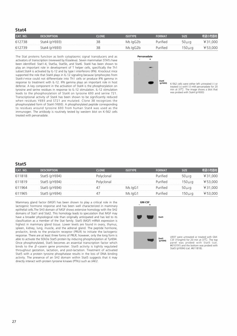
Stat4CAT. NO. DESCRIPTION CLONE ISOTYPE FORMAT SIZE 希望小売価格
612738 Stat4 (pY693) 38 Ms IgG2b Purified 50μg ¥31,000
612739 Stat4 (pY693) 38 Ms IgG2b Purified 150μg ¥53,000
Stat5CAT. NO. DESCRIPTION CLONE ISOTYPE FORMAT SIZE 希望小売価格
611818 Stat5 (pY694) Polyclonal Purified 50μg ¥31,000
611819 Stat5 (pY694) Polyclonal Purified 150μg ¥53,000
611964 Stat5 (pY694) 47 Ms IgG1 Purified 50μg ¥31,000
611965 Stat5 (pY694) 47 Ms IgG1 Purified 150μg ¥53,000
U937 were untreated or treated with GM-CSF (15ng/ml) for 20 min at 37℃. The toppanel was probed with Stat5 (cat.#610191) and the bottom was probed withStat5 (pY694) (cat. #611818).
GM-CSF- +
Stat5
Stat5 (pY694)
Mammary gland factor (MGF) has been shown to play a critical role in thelactogenic hormone response and has been well characterized in mammaryepithelial cells.The SH3 domain of MGF shows extensive homology with the SH2domains of Stat1 and Stat2. This homology leads to speculation that MGF mayhave a broader physiological role than originally anticipated and has led to itsclassification as a member of the Stat family. Stat5 (MGF) mRNA expression ishighest in mammary gland tissue. Lower levels are found in ovary, thymus,spleen, kidney, lung, muscle, and the adrenal gland. The peptide hormone,prolactin, binds to the prolactin receptor (PRLR) to initiate the lactogenicresponse. There are at least three forms of PRLR; however, only the long form isable to activate the 92kDa Stat5 protein by inducing phosphorylation at Tyr694.Once phosphorylated, Stat5 becomes an essential transcription factor whichbinds to the β-casein gene promoter. Stat5 activity is tightly regulatedthroughout gestation, lactation, and post-lactation. Treatment of activatedStat5 with a protein tyrosine phosphatase results in the loss of DNA bindingactivity. The presence of an SH2 domain within Stat5 suggests that it maydirectly interact with protein tyrosine kinases (PTKs) such as JAK2.
K-562 cells were either left untreated (-) ortreated (+) with1.0 mM pervanadate for 20min at 37℃. The image shows a blot thatwas probed with Stat4 (pY693)
Pervanadate- +
Stat4 (pY693)
The Stat proteins function as both cytoplasmic signal transducers and asactivators of transcription (reviewed by Kisseleva). Seven mammalian STATs havebeen identified: Stat1-4, Stat5a, Stat5b, and Stat6. Stat4 has been shown toplay an important role in development of T helper cells, specifically the Th1subset.Stat4 is activated by IL-12 and by type I interferons (IFN). Knockout micesupported the role that Stat4 plays in IL-12 signaling because lymphocytes fromStat4-/-mice could not differentiate into Th1 cells or produce IFN gamma inresponse to treatment with IL-12. IFN gamma plays an important role in hostdefense. A key component in the activation of Stat4 is the phosphorylation ontyrosine and serine residues in response to IL-12 stimulation. IL-12 stimulationleads to the phosphorylation of Stat4 on tyrosine 693 and serine 721.Transcriptional activity of Stat4 has been shown to be significantly reducedwhen residues Y693 and S721 are mutated. Clone 38 recognizes thephosphorylated form of Stat4 (Y693). A phosphorylated peptide correspondingto residues around tyrosine 693 from human Stat4 was used as theimmunogen. The antibody is routinely tested by western blot on K-562 cellstreated with pervanadate.
27
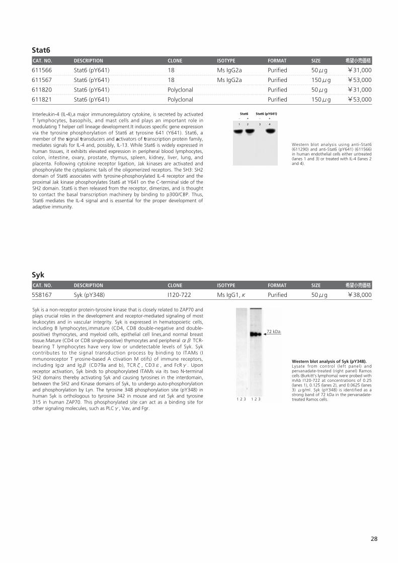
Stat6CAT. NO. DESCRIPTION CLONE ISOTYPE FORMAT SIZE 希望小売価格
611566 Stat6 (pY641) 18 Ms IgG2a Purified 50μg ¥31,000
611567 Stat6 (pY641) 18 Ms IgG2a Purified 150μg ¥53,000
611820 Stat6 (pY641) Polyclonal Purified 50μg ¥31,000
611821 Stat6 (pY641) Polyclonal Purified 150μg ¥53,000
SykCAT. NO. DESCRIPTION CLONE ISOTYPE FORMAT SIZE 希望小売価格
558167 Syk (pY348) I120-722 Ms IgG1,κ Purified 50μg ¥38,000
Western blot analysis of Syk (pY348).Lysate from control ( left panel) andpervanadate-treated (right panel) Ramoscells (Burkitt's lymphoma) were probed withmAb I120-722 at concentrations of 0.25(lanes 1), 0.125 (lanes 2), and 0.0625 (lanes3) μg/ml. Syk (pY348) is identified as astrong band of 72 kDa in the pervanadate-treated Ramos cells.1 2 3 1 2 3
72 kDa
Syk is a non-receptor protein-tyrosine kinase that is closely related to ZAP70 andplays crucial roles in the development and receptor-mediated signaling of mostleukocytes and in vascular integrity. Syk is expressed in hematopoietic cells,including B lymphocytes,immature (CD4, CD8 double-negative and double-positive) thymocytes, and myeloid cells, epithelial cell lines,and normal breasttissue.Mature (CD4 or CD8 single-positive) thymocytes and peripheral αβ TCR-bearing T lymphocytes have very low or undetectable levels of Syk. Sykcontributes to the signal transduction process by binding to ITAMs (Immunoreceptor T yrosine-based A ctivation M otifs) of immune receptors,including Igα and Igβ (CD79a and b), TCRζ, CD3ε, and FcRγ. Uponreceptor activation, Syk binds to phosphorylated ITAMs via its two N-terminalSH2 domains thereby activating Syk and causing tyrosines in the interdomain,between the SH2 and Kinase domains of Syk, to undergo auto-phosphorylationand phosphorylation by Lyn. The tyrosine 348 phosphorylation site (pY348) inhuman Syk is orthologous to tyrosine 342 in mouse and rat Syk and tyrosine315 in human ZAP70. This phosphorylated site can act as a binding site forother signaling molecules, such as PLCγ, Vav, and Fgr.
Western blot analysis using anti-Stat6(611290) and anti-Stat6 (pY641) (611566)in human endothelial cells either untreated(lanes 1 and 3) or treated with IL-4 (lanes 2and 4).
Stat6 (pY641)Stat6- + - +
1 2 3 4
Interleukin-4 (IL-4),a major immunoregulatory cytokine, is secreted by activatedT lymphocytes, basophils, and mast cells and plays an important role inmodulating T helper cell lineage development.It induces specific gene expressionvia the tyrosine phosphorylation of Stat6 at tyrosine 641 (Y641). Stat6, amember of the signal transducers and activators of transcription protein family,mediates signals for IL-4 and, possibly, IL-13. While Stat6 is widely expressed inhuman tissues, it exhibits elevated expression in peripheral blood lymphocytes,colon, intestine, ovary, prostate, thymus, spleen, kidney, liver, lung, andplacenta. Following cytokine receptor ligation, Jak kinases are activated andphosphorylate the cytoplasmic tails of the oligomerized receptors. The SH3: SH2domain of Stat6 associates with tyrosine-phosphorylated IL-4 receptor and theproximal Jak kinase phosphorylates Stat6 at Y641 on the C-terminal side of theSH2 domain. Stat6 is then released from the receptor, dimerizes, and is thoughtto contact the basal transcription machinery by binding to p300/CBP. Thus,Stat6 mediates the IL-4 signal and is essential for the proper development ofadaptive immunity.
28
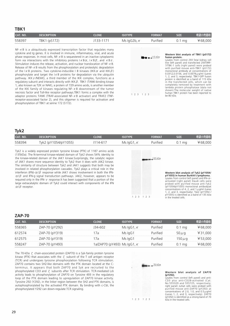
TBK1CAT. NO. DESCRIPTION CLONE ISOTYPE FORMAT SIZE 希望小売価格
558397 TBK1 (pS172) J133-1171 Ms IgG2b,κ Purified 0.1 mg ¥68,000
Tyk2CAT. NO. DESCRIPTION CLONE ISOTYPE FORMAT SIZE 希望小売価格
558394 Tyk2 (pY1054/pY1055) I114-617 Ms IgG1,κ Purified 0.1 mg ¥68,000
ZAP-70CAT. NO. DESCRIPTION CLONE ISOTYPE FORMAT SIZE 希望小売価格
558365 ZAP-70 (pY292) J34-602 Ms IgG1,κ Purified 0.1 mg ¥68,000
612574 ZAP-70 (pY319) 17a Ms IgG1 Purified 50μg ¥31,000
612575 ZAP-70 (pY319) 17a Ms IgG1 Purified 150μg ¥53,000
558247 ZAP-70 (pY493) 1a/ZAP70 (pY493) Ms IgG1,κ Purified 0.1 mg ¥68,000
Western blot analysis of ZAP70(pY292).Lysates from control (left panel) and anti-CD3 plus anti-CD28-activated (Cat.No.555329 and 555725, respectively;right panel) Jurkat cells were probed withpurified mouse anti-ZAP70 (pY292) atconcentrations of 2.0, 1.0, and 0.5μg/ml(Lanes 1, 2, and 3, respectively). ZAP70(pY292) is identified as a strong band of 70kDa in the treated cells.1 2 3 1 2 3
70 kDa
The 70-kDa ζ chain-associated protein (ZAP70) is a Syk-family protein tyrosinekinase (PTK) that associates with the ζ subunit of the T cell antigen receptor(TCR) and undergoes tyrosine phosphorylation following TCR stimulation.ZAP70 contains two SH2-like domains with the PTK domain located at the C-terminus. It appears that both ZAP70 and Syk are recruited to thephosphorylated CD3 and ζ subunits after TCR stimulation. TCR-mediated Lckactivity leads to phosphorylation of ZAP70 on Tyrosine 493 in the regulatoryloop of the PTK domain leading to upregulation of ZAP70 kinase activity.Tyrosine 292 (Y292), in the linker region between the SH2 and PTK domains, isautophosphorylated by the activated PTK domain. By binding with c-Cbl, thephosphorylated Y292 can down-regulate TCR signaling.
Western blot analysis of Tyk2 (pY1054/pY1055) in human Burkitt's lymphoma.Lysates from control (left panel) and IFN-α-activated (right panel) Daudi cells wereprobed with purified mouse anti-Tyk2(pY1054/pY1055) monoclonal antibody)atconcentrations of 4, 2, and 1μg/ml (Lanes1, 2, and 3, respectively). Tyk2 (pY1054 /pY1055) is identified as a band of 135 kDain the treated cells.
135 kDa
1 2 3 1 2 3
Tyk2 is a widely expressed protein tyrosine kinase (PTK) of 1187 amino acids(135kDa). The N-terminal kinase-related domain of Tyk2 shows 54% identity tothe kinase-related domain of the JAK1 kinase.Surprisingly, the catalytic regionof JAK1 shares more sequence identity to Tyk2 than it does with JAK2 kinase.The similarity of structure between Tyk2 and JAK1 suggests that both may beinvolved in related phosphorylation cascades. Tyk2 plays a critical role in theinterferon (IFN)-α/β response while JAK1 shows involvement in both the IFN-α/β and IFN-g signal transduction pathways. JAK2, however, appears to berequired only in the IFN-γ response.It has been suggested that a portion of thelarge extracatalytic domain of Tyk2 could interact with components of the IFNα/β receptor.
Western blot analysis of TBK1 (pS172)fusion protein.Lysates from control 293 fetal kidney cellline (left panel) and transfected 293T/IRF-7/TBK-1 cells (right panel) were probedwith purified mouse anti-TBK1 (pS172)monoclonal antibody at concentrations of0.0312,0.0156, and 0.0078μg/ml (Lanes1, 2, and 3, respectively). TBK1-GFP fusionprotein is identified as a band of 115 kDain the transfected cells, which can becompletely removed by treatment withlambda protein phosphatase (data notshown).The molecular weight of nativehuman TBK1 protein has been reported tobe 80 kDa.1 2 3 1 2 3
115 kDa
NF-κB is a ubiquitously expressed transcription factor that regulates manycytokine and Ig genes. It is involved in immune, inflammatory, viral, and acutephase responses. In most cells, NF-κB is sequestered in an inactive cytoplasmicform via interactions with the inhibitory proteins IκBα, IκBβ, and κBε.Stimulation induces the release, activation, and nuclear translocation of NF-κB.Release of NF-κB results from the phosphorylation and proteolytic degradationof the IκB proteins. Two cytokine-inducible I B kinases (IKKα and IKKβ)phosphorylate and target the IκB proteins for degradation via the ubiquitinpathway. IKKγ/NEMO, a third member of the IKK complex, functions as aregulatory subunit and interacts directly with IKKβ. TBK1 (TANK-binding kinase1, also known as T2K or NAK), a protein of 729 amino acids, is another memberof the IKK family of kinases regulating NF-κB downstream of the tumornecrosis factor and Toll-like receptor pathways.TBK1 forms a complex with theadaptor proteins TANK (TRAF-associated NF-κB activator) and TRAF2 (TNF-receptor-associated factor 2), and this oligomer is required for activation andphosphorylation of TBK1 at serine 172 (S172).
29
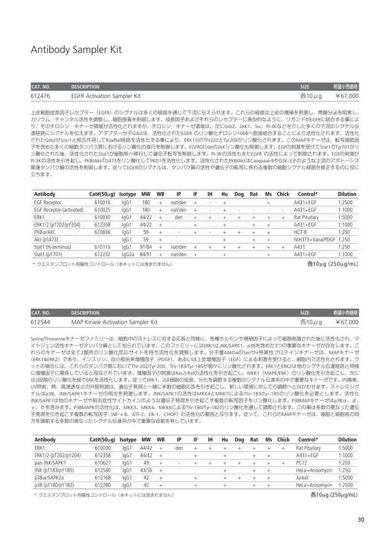
Antibody Sampler Kit
CAT. NO. DESCRIPTION SIZE 希望小売価格
612476 EGFR Activation Sampler Kit 各10μg ¥67,000
上皮細胞成長因子レセプター(EGFR)のシグナルは多くの経路を通じて下流に伝えられます。これらの経路は上皮の増殖を刺激し、胃酸分泌を阻害し、カリウム・チャンネル活性を調整し、細胞接着を制御します。成長因子およびそれらのレセプターに典型的なように、リガンドがEGFRに結合する事により、そのチロシン・キナーゼ領域が活性化されますが、チロシン・キナーゼ領域は、次にGrb2、JAK1、Src、PI-3Kなどを介した多くの下流のシグナル伝達経路にシグナルを伝えます。アダプター分子Grb2は、活性化されたEGFR のリン酸化チロシン1068へ直接結合することにより活性化されます。活性化されたGrb2がSos-1と相互作用してRsa/Raf経路を活性化する事により、ERK1/2のThr202とTyr204がリン酸化されます。このMAPキナーゼは、転写調節因子を含めた多くの細胞タンパク質におけるリン酸化の進行を制御します。EGFRはStatのJAKリン酸化も制御します。EGFの刺激を受けてStat1のTyr701がリン酸化された後、活性化されたStat1が細胞核へ移行して遺伝子転写を制御します。PI-3Kの活性もまたEGFR の活性によって制御されます。EGFの刺激がPI-3Kの活性を引き起し、PKB/AktのS473をリン酸化してPKD1を活性化します。活性化されたPKB/AktはCasepase-9やGSK-3βのような上流のアポトーシス関連タンパク質の活性を制御します。従ってEGFRのシグナルは、タンパク質の活性や遺伝子の転写に係わる複数の細胞シグナル経路を修正するのに役に立ちます。
CAT. NO. DESCRIPTION SIZE 希望小売価格
612544 MAP Kinase Activation Sampler Kit 各10μg ¥67,000
Serine/Threonineキナーゼファミリーは、細胞中のストレスに対する応答と同様に、各種ホルモンや増殖因子によって細胞刺激された後に活性化され、マイトジェン活性キナーゼタンパク質として知られています。このファミリーにはERK1/2,JNK/SAPK1、p38を含めた3つの重要なキナーゼが存在します。これらのキナーゼは全て2箇所のリン酸化反応サイトを持ち活性化を調整します。分子量44kDaのSer/Thr特異性プロテインキナーゼは、MAPキナーゼ(ERK1&ERK2)であり、インスリン、血小板由来増殖因子(PDGF)、あるいは上皮増殖因子(EGF)による刺激を受けると、細胞内で活性化されます。ラットの場合には、これらのタンパク質においてThr-202/Tyr-204、Thr-183/Tyr-185が個々にリン酸化されます。ERK1とERK2は他のシグナル伝達経路と同様に増殖因子に関係していると見なされています。増殖因子の刺激はRasとRafの活性化を引き起こし、MEK1(MAPK/ERK)のリン酸化を引き起こし、次には2段階のリン酸化を経てERKを活性化します。従ってERK1、2は細胞の成長、分化を調節する複数のシグナル伝達系の中で重要なキナーゼです。内毒素、UV照射、熱、高浸透などの外部刺激は、遺伝子発現と一緒に多数の細胞応答を引き起こし、新しい環境に対しての調節へと向かわせます。ストレスシグナルはp38、JNK/SAPK1キナーゼの両方を刺激します。JNK/SAPK1の活性はMKK4とMKK7によるThr-183/Tyr-185のリン酸化を必要とします。活性化JNK/SAPK1は他のキナーゼや前炎症性サイトカインのような遺伝子発現を引き起こす複数の転写因子をリン酸化します。P38MAPキナーゼはp38α、β、γ、δを含みます。P38MAPKの活性化は、MKK3、MKK4、MKK6によるThr-180/Tyr-182のリン酸化を通して調節されます。この事は多数の異なった遺伝子発現を引き起こす複数の転写因子(NF-κB、ATF-2、Elk-1、CHOP)の活性化の要因となります。従って、これらのMAPキナーゼは、細胞と細胞核の両方を調節する多数の異なったシグナル伝達系の中で重要な役割を有しています。
α
α
30
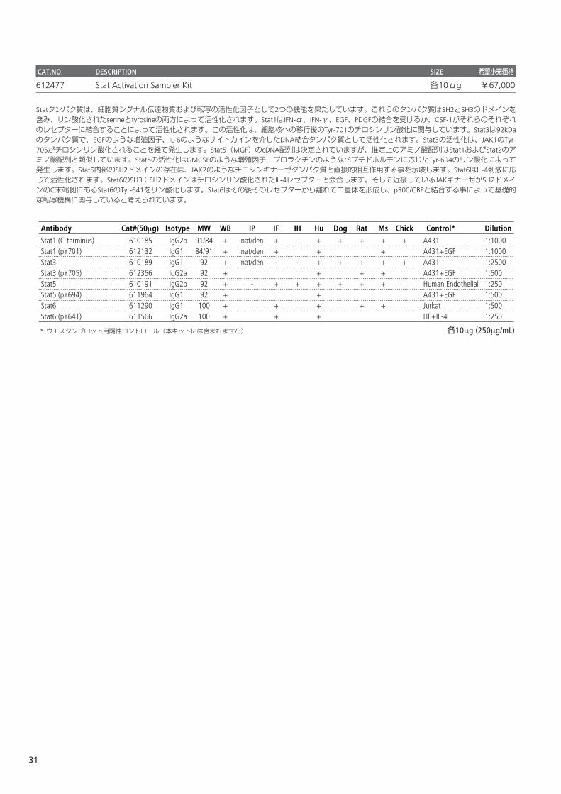
CAT.NO. DESCRIPTION SIZE 希望小売価格
612477 Stat Activation Sampler Kit 各10μg ¥67,000
Statタンパク質は、細胞質シグナル伝達物質および転写の活性化因子として2つの機能を果たしています。これらのタンパク質はSH2とSH3のドメインを含み、リン酸化されたserineとtyrosineの両方によって活性化されます。Stat1はIFN-α、IFN-γ、EGF、PDGFの結合を受けるか、CSF-1がそれらのそれぞれのレセプターに結合することによって活性化されます。この活性化は、細胞核への移行後のTyr-701のチロシンリン酸化に関与しています。Stat3は92kDaのタンパク質で、EGFのような増殖因子、IL-6のようなサイトカインを介したDNA結合タンパク質として活性化されます。Stat3の活性化は、JAK1のTyr-705がチロシンリン酸化されることを経て発生します。Stat5(MGF)のcDNA配列は決定されていますが、推定上のアミノ酸配列はStat1およびStat2のアミノ酸配列と類似しています。Stat5の活性化はGMCSFのような増殖因子、プロラクチンのようなペプチドホルモンに応じたTyr-694のリン酸化によって発生します。Stat5内部のSH2ドメインの存在は、JAK2のようなチロシンキナーゼタンパク質と直接的相互作用する事を示唆します。Stat6はIL-4刺激に応じて活性化されます。Stat6のSH3:SH2ドメインはチロシンリン酸化されたIL-4レセプターと会合します。そして近接しているJAKキナーゼがSH2ドメインのC末端側にあるStat6のTyr-641をリン酸化します。Stat6はその後そのレセプターから離れて二量体を形成し、p300/CBPと結合する事によって基礎的な転写機構に関与していると考えられています。
31
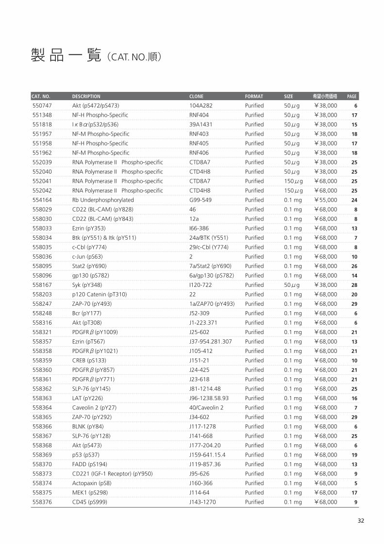
製品一覧(CAT. NO.順)
CAT. NO. DESCRIPTION CLONE FORMAT SIZE 希望小売価格 PAGE
550747 Akt (pS472/pS473) 104A282 Purified 50μg ¥38,000
551348 NF-H Phospho-Specific RNF404 Purified 50μg ¥38,000
551818 IκBα(pS32/pS36) 39A1431 Purified 50μg ¥38,000
551957 NF-M Phospho-Specific RNF403 Purified 50μg ¥38,000
551958 NF-H Phospho-Specific RNF405 Purified 50μg ¥38,000
551962 NF-M Phospho-Specific RNF406 Purified 50μg ¥38,000
552039 RNA Polymerase II Phospho-specific CTD8A7 Purified 50μg ¥38,000
552040 RNA Polymerase II Phospho-specific CTD4H8 Purified 50μg ¥38,000
552041 RNA Polymerase II Phospho-specific CTD8A7 Purified 150μg ¥68,000
552042 RNA Polymerase II Phospho-specific CTD4H8 Purified 150μg ¥68,000
554164 Rb Underphosphorylated G99-549 Purified 0.1 mg ¥55,000
558029 CD22 (BL-CAM) (pY828) 46 Purified 0.1 mg ¥68,000
558030 CD22 (BL-CAM) (pY843) 12a Purified 0.1 mg ¥68,000
558033 Ezrin (pY353) I66-386 Purified 0.1 mg ¥68,000
558034 Btk (pY551) & Itk (pY511) 24a/BTK (Y551) Purified 0.1 mg ¥68,000
558035 c-Cbl (pY774) 29/c-Cbl (Y774) Purified 0.1 mg ¥68,000
558036 c-Jun (pS63) 2 Purified 0.1 mg ¥68,000
558095 Stat2 (pY690) 7a/Stat2 (pY690) Purified 0.1 mg ¥68,000
558096 gp130 (pS782) 6a/gp130 (pS782) Purified 0.1 mg ¥68,000
558167 Syk (pY348) I120-722 Purified 50μg ¥38,000
558203 p120 Catenin (pT310) 22 Purified 0.1 mg ¥68,000
558247 ZAP-70 (pY493) 1a/ZAP70 (pY493) Purified 0.1 mg ¥68,000
558248 Bcr (pY177) J52-309 Purified 0.1 mg ¥68,000
558316 Akt (pT308) J1-223.371 Purified 0.1 mg ¥68,000
558321 PDGFRβ(pY1009) J25-602 Purified 0.1 mg ¥68,000
558357 Ezrin (pT567) J37-954.281.307 Purified 0.1 mg ¥68,000
558358 PDGFRβ(pY1021) J105-412 Purified 0.1 mg ¥68,000
558359 CREB (pS133) J151-21 Purified 0.1 mg ¥68,000
558360 PDGFRβ(pY857) J24-425 Purified 0.1 mg ¥68,000
558361 PDGFRβ(pY771) J23-618 Purified 0.1 mg ¥68,000
558362 SLP-76 (pY145) J81-1214.48 Purified 0.1 mg ¥68,000
558363 LAT (pY226) J96-1238.58.93 Purified 0.1 mg ¥68,000
558364 Caveolin 2 (pY27) 40/Caveolin 2 Purified 0.1 mg ¥68,000
558365 ZAP-70 (pY292) J34-602 Purified 0.1 mg ¥68,000
558366 BLNK (pY84) J117-1278 Purified 0.1 mg ¥68,000
558367 SLP-76 (pY128) J141-668 Purified 0.1 mg ¥68,000
558368 Akt (pS473) J177-204.20 Purified 0.1 mg ¥68,000
558369 p53 (pS37) J159-641.15.4 Purified 0.1 mg ¥68,000
558370 FADD (pS194) J119-857.36 Purified 0.1 mg ¥68,000
558373 CD221 (IGF-1 Receptor) (pY950) J95-626 Purified 0.1 mg ¥68,000
558374 Actopaxin (pS8) J160-366 Purified 0.1 mg ¥68,000
558375 MEK1 (pS298) J114-64 Purified 0.1 mg ¥68,000
558376 CD45 (pS999) J143-1270 Purified 0.1 mg ¥68,000
32
6
17
15
18
17
18
25
25
25
25
24
8
8
13
7
8
10
26
14
28
20
29
6
6
21
13
21
10
21
21
25
16
7
29
6
25
6
19
13
9
5
17
9
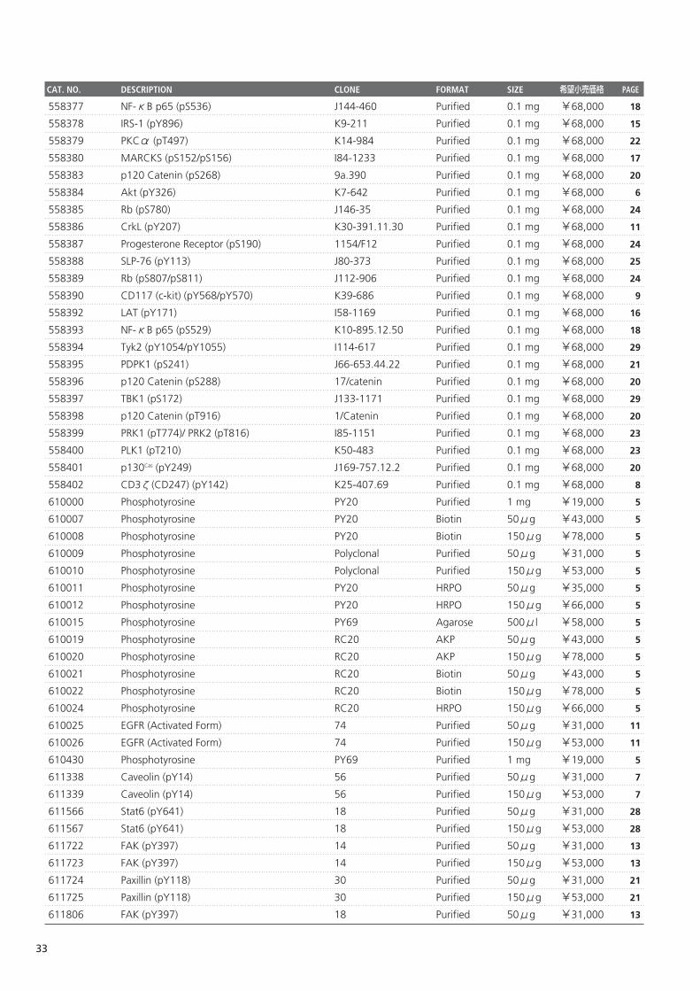
558377 NF-κB p65 (pS536) J144-460 Purified 0.1 mg ¥68,000
558378 IRS-1 (pY896) K9-211 Purified 0.1 mg ¥68,000
558379 PKCα (pT497) K14-984 Purified 0.1 mg ¥68,000
558380 MARCKS (pS152/pS156) I84-1233 Purified 0.1 mg ¥68,000
558383 p120 Catenin (pS268) 9a.390 Purified 0.1 mg ¥68,000
558384 Akt (pY326) K7-642 Purified 0.1 mg ¥68,000
558385 Rb (pS780) J146-35 Purified 0.1 mg ¥68,000
558386 CrkL (pY207) K30-391.11.30 Purified 0.1 mg ¥68,000
558387 Progesterone Receptor (pS190) 1154/F12 Purified 0.1 mg ¥68,000
558388 SLP-76 (pY113) J80-373 Purified 0.1 mg ¥68,000
558389 Rb (pS807/pS811) J112-906 Purified 0.1 mg ¥68,000
558390 CD117 (c-kit) (pY568/pY570) K39-686 Purified 0.1 mg ¥68,000
558392 LAT (pY171) I58-1169 Purified 0.1 mg ¥68,000
558393 NF-κB p65 (pS529) K10-895.12.50 Purified 0.1 mg ¥68,000
558394 Tyk2 (pY1054/pY1055) I114-617 Purified 0.1 mg ¥68,000
558395 PDPK1 (pS241) J66-653.44.22 Purified 0.1 mg ¥68,000
558396 p120 Catenin (pS288) 17/catenin Purified 0.1 mg ¥68,000
558397 TBK1 (pS172) J133-1171 Purified 0.1 mg ¥68,000
558398 p120 Catenin (pT916) 1/Catenin Purified 0.1 mg ¥68,000
558399 PRK1 (pT774)/ PRK2 (pT816) I85-1151 Purified 0.1 mg ¥68,000
558400 PLK1 (pT210) K50-483 Purified 0.1 mg ¥68,000
558401 p130Cas (pY249) J169-757.12.2 Purified 0.1 mg ¥68,000
558402 CD3ζ(CD247) (pY142) K25-407.69 Purified 0.1 mg ¥68,000
610000 Phosphotyrosine PY20 Purified 1 mg ¥19,000
610007 Phosphotyrosine PY20 Biotin 50μg ¥43,000
610008 Phosphotyrosine PY20 Biotin 150μg ¥78,000
610009 Phosphotyrosine Polyclonal Purified 50μg ¥31,000
610010 Phosphotyrosine Polyclonal Purified 150μg ¥53,000
610011 Phosphotyrosine PY20 HRPO 50μg ¥35,000
610012 Phosphotyrosine PY20 HRPO 150μg ¥66,000
610015 Phosphotyrosine PY69 Agarose 500μl ¥58,000
610019 Phosphotyrosine RC20 AKP 50μg ¥43,000
610020 Phosphotyrosine RC20 AKP 150μg ¥78,000
610021 Phosphotyrosine RC20 Biotin 50μg ¥43,000
610022 Phosphotyrosine RC20 Biotin 150μg ¥78,000
610024 Phosphotyrosine RC20 HRPO 150μg ¥66,000
610025 EGFR (Activated Form) 74 Purified 50μg ¥31,000
610026 EGFR (Activated Form) 74 Purified 150μg ¥53,000
610430 Phosphotyrosine PY69 Purified 1 mg ¥19,000
611338 Caveolin (pY14) 56 Purified 50μg ¥31,000
611339 Caveolin (pY14) 56 Purified 150μg ¥53,000
611566 Stat6 (pY641) 18 Purified 50μg ¥31,000
611567 Stat6 (pY641) 18 Purified 150μg ¥53,000
611722 FAK (pY397) 14 Purified 50μg ¥31,000
611723 FAK (pY397) 14 Purified 150μg ¥53,000
611724 Paxillin (pY118) 30 Purified 50μg ¥31,000
611725 Paxillin (pY118) 30 Purified 150μg ¥53,000
611806 FAK (pY397) 18 Purified 50μg ¥31,000
33
CAT. NO. DESCRIPTION CLONE FORMAT SIZE 希望小売価格 PAGE
18
15
22
17
20
6
24
11
24
25
24
9
16
18
29
21
20
29
20
23
23
20
8
5
5
5
5
5
5
5
5
5
5
5
5
5
11
11
5
7
7
28
28
13
13
21
21
13
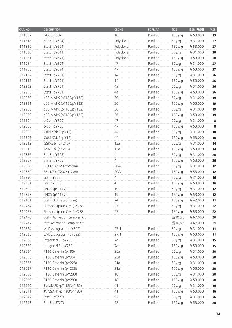
611807 FAK (pY397) 18 Purified 150μg ¥53,000
611818 Stat5 (pY694) Polyclonal Purified 50μg ¥31,000
611819 Stat5 (pY694) Polyclonal Purified 150μg ¥53,000
611820 Stat6 (pY641) Polyclonal Purified 50μg ¥31,000
611821 Stat6 (pY641) Polyclonal Purified 150μg ¥53,000
611964 Stat5 (pY694) 47 Purified 50μg ¥31,000
611965 Stat5 (pY694) 47 Purified 150μg ¥53,000
612132 Stat1 (pY701) 14 Purified 50μg ¥31,000
612133 Stat1 (pY701) 14 Purified 150μg ¥53,000
612232 Stat1 (pY701) 4a Purified 50μg ¥31,000
612233 Stat1 (pY701) 4a Purified 150μg ¥53,000
612280 p38 MAPK (pT180/pY182) 30 Purified 50μg ¥31,000
612281 p38 MAPK (pT180/pY182) 30 Purified 150μg ¥53,000
612288 p38 MAPK (pT180/pY182) 36 Purified 50μg ¥31,000
612289 p38 MAPK (pT180/pY182) 36 Purified 150μg ¥53,000
612304 c-Cbl (pY700) 47 Purified 50μg ¥31,000
612305 c-Cbl (pY700) 47 Purified 150μg ¥53,000
612306 Cdk1/Cdc2 (pY15) 44 Purified 50μg ¥31,000
612307 Cdk1/Cdc2 (pY15) 44 Purified 150μg ¥53,000
612312 GSK-3β (pY216) 13a Purified 50μg ¥31,000
612313 GSK-3β (pY216) 13a Purified 150μg ¥53,000
612356 Stat3 (pY705) 4 Purified 50μg ¥31,000
612357 Stat3 (pY705) 4 Purified 150μg ¥53,000
612358 ERK1/2 (pT202/pY204) 20A Purified 50μg ¥31,000
612359 ERK1/2 (pT202/pY204) 20A Purified 150μg ¥53,000
612390 Lck (pY505) 4 Purified 50μg ¥31,000
612391 Lck (pY505) 4 Purified 150μg ¥53,000
612392 eNOS (pS1177) 19 Purified 50μg ¥31,000
612393 eNOS (pS1177) 19 Purified 150μg ¥53,000
612401 EGFR (Activated Form) 74 Purified 100μg ¥42,000
612464 Phospholipase Cγ (pY783) 27 Purified 50μg ¥31,000
612465 Phospholipase Cγ (pY783) 27 Purified 150μg ¥53,000
612476 EGFR Activation Sampler Kit 各10μg ¥67,000
612477 Stat Activation Sampler Kit 各10μg ¥67,000
612524 β-Dystroglycan (pY892) 27.1 Purified 50μg ¥31,000
612525 β-Dystroglycan (pY892) 27.1 Purified 150μg ¥53,000
612528 Integrinβ3 (pY759) 7a Purified 50μg ¥31,000
612529 Integrinβ3 (pY759) 7a Purified 150μg ¥53,000
612534 P120 Catenin (pY96) 25a Purified 50μg ¥31,000
612535 P120 Catenin (pY96) 25a Purified 150μg ¥53,000
612536 P120 Catenin (pY228) 21a Purified 50μg ¥31,000
612537 P120 Catenin (pY228) 21a Purified 150μg ¥53,000
612538 P120 Catenin (pY280) 18 Purified 50μg ¥31,000
612539 P120 Catenin (pY280) 18 Purified 150μg ¥53,000
612540 JNK/SAPK (pT183/pY185) 41 Purified 50μg ¥31,000
612541 JNK/SAPK (pT183/pY185) 41 Purified 150μg ¥53,000
612542 Stat3 (pS727) 92 Purified 50μg ¥31,000
612543 Stat3 (pS727) 92 Purified 150μg ¥53,000
34
CAT. NO. DESCRIPTION CLONE FORMAT SIZE 希望小売価格 PAGE
13
27
27
28
28
27
27
26
26
26
26
19
19
19
19
8
8
10
10
14
14
26
26
12
12
16
16
12
12
11
22
22
30
31
11
11
15
15
20
20
20
20
20
20
16
16
26
26
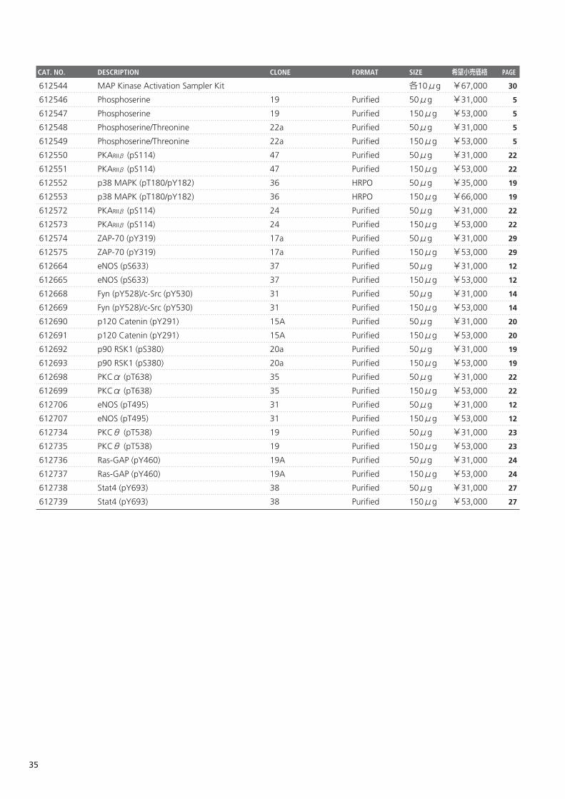
612544 MAP Kinase Activation Sampler Kit 各10μg ¥67,000
612546 Phosphoserine 19 Purified 50μg ¥31,000
612547 Phosphoserine 19 Purified 150μg ¥53,000
612548 Phosphoserine/Threonine 22a Purified 50μg ¥31,000
612549 Phosphoserine/Threonine 22a Purified 150μg ¥53,000
612550 PKARIIβ (pS114) 47 Purified 50μg ¥31,000
612551 PKARIIβ (pS114) 47 Purified 150μg ¥53,000
612552 p38 MAPK (pT180/pY182) 36 HRPO 50μg ¥35,000
612553 p38 MAPK (pT180/pY182) 36 HRPO 150μg ¥66,000
612572 PKARIIβ (pS114) 24 Purified 50μg ¥31,000
612573 PKARIIβ (pS114) 24 Purified 150μg ¥53,000
612574 ZAP-70 (pY319) 17a Purified 50μg ¥31,000
612575 ZAP-70 (pY319) 17a Purified 150μg ¥53,000
612664 eNOS (pS633) 37 Purified 50μg ¥31,000
612665 eNOS (pS633) 37 Purified 150μg ¥53,000
612668 Fyn (pY528)/c-Src (pY530) 31 Purified 50μg ¥31,000
612669 Fyn (pY528)/c-Src (pY530) 31 Purified 150μg ¥53,000
612690 p120 Catenin (pY291) 15A Purified 50μg ¥31,000
612691 p120 Catenin (pY291) 15A Purified 150μg ¥53,000
612692 p90 RSK1 (pS380) 20a Purified 50μg ¥31,000
612693 p90 RSK1 (pS380) 20a Purified 150μg ¥53,000
612698 PKCα (pT638) 35 Purified 50μg ¥31,000
612699 PKCα (pT638) 35 Purified 150μg ¥53,000
612706 eNOS (pT495) 31 Purified 50μg ¥31,000
612707 eNOS (pT495) 31 Purified 150μg ¥53,000
612734 PKCθ (pT538) 19 Purified 50μg ¥31,000
612735 PKCθ (pT538) 19 Purified 150μg ¥53,000
612736 Ras-GAP (pY460) 19A Purified 50μg ¥31,000
612737 Ras-GAP (pY460) 19A Purified 150μg ¥53,000
612738 Stat4 (pY693) 38 Purified 50μg ¥31,000
612739 Stat4 (pY693) 38 Purified 150μg ¥53,000
35
CAT. NO. DESCRIPTION CLONE FORMAT SIZE 希望小売価格 PAGE
30
5
5
5
5
22
22
19
19
22
22
29
29
12
12
14
14
20
20
19
19
22
22
12
12
23
23
24
24
27
27
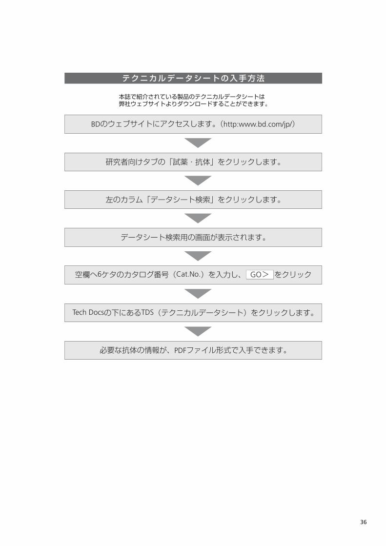
テクニカルデータシートの入手方法
本誌で紹介されている製品のテクニカルデータシートは。弊社ウェブサイトよりダウンロードすることができます。
BDのウェブサイトにアクセスします。(http:www.bd.com/jp/)
研究者向けタブの「試薬・抗体」をクリックします。
左のカラム「データシート検索」をクリックします。
必要な抗体の情報が、PDFファイル形式で入手できます。
データシート検索用の画面が表示されます。
空欄へ6ケタのカタログ番号(Cat.No.)を入力し、 をクリックGO>
Tech Docsの下にあるTDS(テクニカルデータシート)をクリックします。
36
ひょすし 6/21/06 2:19 PM ページ 3

66-031-00R0-0607-004-083


![Layout 1 (Page 1) - Antibodies, Proteins, Kits and … P WB Rb Hu 28225 ADAM17 P WB Rb Hu, Mm, Rt 2051 AKT (phospho S473) [14-6] M ICC, WB Rb Hu, Mm 27773 AKT (phospho T308) P ELISA,](https://static.fdocument.org/doc/165x107/5b0df7317f8b9af65e8e7090/layout-1-page-1-antibodies-proteins-kits-and-p-wb-rb-hu-28225-adam17-p-wb.jpg)