Analysis of the stability and affinity of BlaR-CTD protein ...
17
RESEARCH Open Access Analysis of the stability and affinity of BlaR-CTD protein to β-lactam antibiotics based on docking and mutagenesis studies Jianan Ning 1† , Saeed Ahmed 1† , Guyue Cheng 2* , Ting Chen 1 , Yulian Wang 1 , Dapeng Peng 1 and Zonghui Yuan 1,2* Abstract Owing to the thermal instability and low affinity of BlaR-CTD to some β-lactams, the receptor assay based on BlaR-CTD is limited in the detection of abundant variety of drugs and the result is often unstable. In this study, the three-dimensional structure of BlaR-CTD from Bacillus licheniformis ATCC14580 was constructed by homologous modeling based on the crystal structure of BlaR-CTD from B. licheniformis 749/I, and the binding sites of this protein to 40 β-lactams were also obtained by molecular docking. To improve the stability and affinity of the protein, 23 mutant proteins were designed based on docking and homologous alignment results as well as by inserting disulfide bond and building the salt bridge. The mutation was rationality evaluated by SIFT and PloyPhen2 software. The heterologous expressed and purified mutant proteins were then subjected to the activity and stability assay. It was shown that among all mutant proteins, I188K/S19C/G24C, A138E/R50C/Q147C and S190Y/E183C/I188K respectively exhibited a higher affinity to 33, 22 and 21 β-lactams than the wild-type protein, while I188K/S19C/G24C exhibited the best stability. This may due to that the conformation of the active site in mutant protein I188K/S19C/G24C changed, and the random coli in the surface of protein activity increased. Our study suggests a possible structure-function relationship on the stability and affinity of BlaR-CTD, which provides new insights into protein rational design study and lays a solid foundation for establishing the receptor-based screening assay for the detection of β-lactam residues. Keywords: β-Lactam antibiotics, BlaR-CTD, Docking, Site-directed mutagenesis, Stability, Affinity Introduction The penicillin binding protein (PBP) BlaR is a signal transduction membrane protein which induces the synthesis of β-lactamase. The C-terminal domain of BlaR (BlaR-CTD) protein is located in the extracellular region which acts as a drug binding site [19]. BlaR-CTD, a penicillin-binding protein. BlaR-CTD is firstly the sensor domain of a penicillin receptor that is acylated by peni- cillin. This protein can identify and bind to a variety of β-lactams [8, 12]. In the active site of the protein, STYK, serine was a key amino acid (AA) that can participate in the binding of β-lactams [31]. BlaR-CTD protein has been used to detect the β-lactam residues in the receptor-based screening assay, such as a biological fluid method using colloidal gold labeled receptor protein [7] and an receptor-based enzyme linked immune-sorbent assay developed by our lab in which BlaR-CTD from B. licheniformis ATCC14580 was immobilized on the plate [25]. The limits of detection for 11 β-lactams were lower than the maximum residue limits regulated by European Union. However, the study showed that BlaR-CTD protein had a poor affinity for some β-lactams, e.g. cephalexin and cefadroxil. In addition, the thermal stability of the protein was poor that the protein activity only remained 70% at - 20 °C after 1 month, and lost fast at 4 °C [25]. Presently, site-directed mutagenesis was an effective means for the structural modification of protein [14], such as, improving the enzymatic activity and stability [9, 15]. Contreras-Martel et al. obtained a wild-type and the mutant PBP2b from Streptococcus pneumoniae by © The Author(s). 2019 Open Access This article is distributed under the terms of the Creative Commons Attribution 4.0 International License (http://creativecommons.org/licenses/by/4.0/), which permits unrestricted use, distribution, and reproduction in any medium, provided you give appropriate credit to the original author(s) and the source, provide a link to the Creative Commons license, and indicate if changes were made. The Creative Commons Public Domain Dedication waiver (http://creativecommons.org/publicdomain/zero/1.0/) applies to the data made available in this article, unless otherwise stated. * Correspondence: [email protected]; [email protected] † Jianan Ning and Saeed Ahmed contributed equally to this work. 2 MOA Laboratory for Risk Assessment of Quality and Safety of Livestock and Poultry Products, Huazhong Agricultural University, Wuhan 430070, China 1 National Reference Laboratory of Veterinary Drug Residues (HZAU) and MOA Key Laboratory for the Detection of Veterinary Drug Residues in Foods, Huazhong Agricultural University, Wuhan 430070, China Ning et al. Journal of Biological Engineering (2019) 13:27 https://doi.org/10.1186/s13036-019-0157-4
Transcript of Analysis of the stability and affinity of BlaR-CTD protein ...
RESEARCH Open Access
Analysis of the stability and affinity ofBlaR-CTD protein to β-lactam antibioticsbased on docking and mutagenesis studiesJianan Ning1†, Saeed Ahmed1†, Guyue Cheng2*, Ting Chen1, Yulian Wang1, Dapeng Peng1 and Zonghui Yuan1,2*
Abstract
Owing to the thermal instability and low affinity of BlaR-CTD to some β-lactams, the receptor assay based on BlaR-CTD islimited in the detection of abundant variety of drugs and the result is often unstable. In this study, the three-dimensionalstructure of BlaR-CTD from Bacillus licheniformis ATCC14580 was constructed by homologous modeling based on thecrystal structure of BlaR-CTD from B. licheniformis 749/I, and the binding sites of this protein to 40 β-lactams were alsoobtained by molecular docking. To improve the stability and affinity of the protein, 23 mutant proteins were designedbased on docking and homologous alignment results as well as by inserting disulfide bond and building the salt bridge.The mutation was rationality evaluated by SIFT and PloyPhen2 software. The heterologous expressed and purifiedmutant proteins were then subjected to the activity and stability assay. It was shown that among all mutant proteins,I188K/S19C/G24C, A138E/R50C/Q147C and S190Y/E183C/I188K respectively exhibited a higher affinity to 33, 22and 21 β-lactams than the wild-type protein, while I188K/S19C/G24C exhibited the best stability. This may due tothat the conformation of the active site in mutant protein I188K/S19C/G24C changed, and the random coli in thesurface of protein activity increased. Our study suggests a possible structure-function relationship on the stabilityand affinity of BlaR-CTD, which provides new insights into protein rational design study and lays a solidfoundation for establishing the receptor-based screening assay for the detection of β-lactam residues.
Keywords: β-Lactam antibiotics, BlaR-CTD, Docking, Site-directed mutagenesis, Stability, Affinity
IntroductionThe penicillin binding protein (PBP) BlaR is a signaltransduction membrane protein which induces thesynthesis of β-lactamase. The C-terminal domain of BlaR(BlaR-CTD) protein is located in the extracellular regionwhich acts as a drug binding site [19]. BlaR-CTD, apenicillin-binding protein. BlaR-CTD is firstly the sensordomain of a penicillin receptor that is acylated by peni-cillin. This protein can identify and bind to a variety ofβ-lactams [8, 12]. In the active site of the protein, STYK,serine was a key amino acid (AA) that can participate inthe binding of β-lactams [31].
BlaR-CTD protein has been used to detect theβ-lactam residues in the receptor-based screening assay,such as a biological fluid method using colloidal goldlabeled receptor protein [7] and an receptor-basedenzyme linked immune-sorbent assay developed by ourlab in which BlaR-CTD from B. licheniformis ATCC14580was immobilized on the plate [25]. The limits of detectionfor 11 β-lactams were lower than the maximum residuelimits regulated by European Union. However, the studyshowed that BlaR-CTD protein had a poor affinity forsome β-lactams, e.g. cephalexin and cefadroxil. Inaddition, the thermal stability of the protein was poor thatthe protein activity only remained 70% at − 20 °C after 1month, and lost fast at 4 °C [25].Presently, site-directed mutagenesis was an effective
means for the structural modification of protein [14],such as, improving the enzymatic activity and stability[9, 15]. Contreras-Martel et al. obtained a wild-type andthe mutant PBP2b from Streptococcus pneumoniae by
© The Author(s). 2019 Open Access This article is distributed under the terms of the Creative Commons Attribution 4.0International License (http://creativecommons.org/licenses/by/4.0/), which permits unrestricted use, distribution, andreproduction in any medium, provided you give appropriate credit to the original author(s) and the source, provide a link tothe Creative Commons license, and indicate if changes were made. The Creative Commons Public Domain Dedication waiver(http://creativecommons.org/publicdomain/zero/1.0/) applies to the data made available in this article, unless otherwise stated.
* Correspondence: [email protected];[email protected]†Jianan Ning and Saeed Ahmed contributed equally to this work.2MOA Laboratory for Risk Assessment of Quality and Safety of Livestock andPoultry Products, Huazhong Agricultural University, Wuhan 430070, China1National Reference Laboratory of Veterinary Drug Residues (HZAU) andMOA Key Laboratory for the Detection of Veterinary Drug Residues in Foods,Huazhong Agricultural University, Wuhan 430070, China
Ning et al. Journal of Biological Engineering (2019) 13:27 https://doi.org/10.1186/s13036-019-0157-4
heterologous expression in E. coli, and the structureanalysis showed that the mutation of AAs around theactive site could affect the distribution of charge andpolarity, preventing the entry of substrate into the activesite, thus results in a bacterial resistance against β-lactamdrugs [4]. Powell et al. mutated the AAs around the activesite of the PBP2 from Neisseria gonorrhoeae, and theresults showed that the acylation efficiency for penicillinG was reduced [26]. Therefore, the mutation site shouldavoid the active site and active site of the protein whenthe structure of BlaR-CTD protein was reconstructed.Molecular docking can quickly simulates the bindingmode between a protein and a ligand, and analyze the keyAAs involved in the binding, such as, AAs forming ahydrogen bond(s) with the drug [33]. Moreover, disulfidebond played an important role in the structural stability ofproteins. It was demonstrated that the protein expressionand enzyme activity of β-mannanase from Aspergillusniger BK01 (ManBK) were reduced when the conservedC171-C174 disulfide bond was missing, indicating thatdisulfide bond has a crucial in the stability of [15].Through the site-directed mutagenesis of PBP3 fromStreptococcus pneumoniae R6, the thermal stability of themutant A353C/E393C was increased while the affinity ofthe mutant did not change [32].In this study, 23 mutant proteins of BlaR-CTD from
Bacillus licheniformis ATCC14580 were designed byhomologous modeling, molecular docking, and predic-tion of mutation site in software, disulfide bond insertingand salt bridge building. The mutant proteins were ob-tained by site-directed mutagenesis and heterologousexpression in E. coli. Based on the results of activity andstability tests, I188K/S19C/G24C showed equal or higheraffinity to 33 β-lactams and better stability as comparedto the wild-type protein. This study for the first timemodified BlaR-CTD protein by rational design and site-directed mutagenesis, and the mutant protein I188K/S19C/G24C exhibited not only the improvement instability but also higher affinity than the wild-type.Current study offers a powerful base for establishing thescreening method for the detection of β-lactam residues.
Materials and methodsChemicalsPenicillin G, nafcillin, dicloxacillin, ceftiofur, cefquinome,cefuroxim, carbenicillin and flucloxacillin were boughtfrom Dr. Ehrenstorfer (Deisenhofen, Germany). Ampicil-lin, amoxicillin, oxacillin, azlocillin, penicillin V, cloxacillin,cefalotin, cefoperazone, cefazolin, cefalexin, ceftriaxone,cefadroxil, cefepime, cefradin, latamoxef, cefixim, ceftazi-dim, cefoxitin, sulbenicillin, ticarcillin and aztreonam weresupplied by the National Institute for the Control ofPharmaceutical and Biological Products (Beijing, China).Piperacillin and cefalonium were from Toronto Research
Chemicals (Toronto, Canada). Cefapirin was fromWitega (Berlin, Germany). Benzylpenicillin was fromEuropean Directorate for the Quality of Medicines &HealthCare (Strasbourg, France). Kanamycin wasobtained from TianYuan (Wuhan, China). 1-ethyl-3-(3-dimethylaminopropyl) carbodiimide hydrochloride(EDC) and N-hydroxysuccinimide (NHS) were purchasedfrom Sigma-Aldrich (St. Louis, MO, USA). Bovine serumalbumin (BSA) was obtained from Bovogen (East Keilor,VIC, Australia). β-isopropyl-D-thiogalactopyranoside (IPTG)and horseradish peroxidase (HRP) were purchased fromSolarbio (Beijing, China). 3,3′,5,5′-tetramethylbenzidine(TMB) and Ni2+ Sepharose Fastflow were from ThermoFisher Scientific (Waltham, MA, USA). Luria-Bertani(LB) broth and agar were purchased form Qingdao HopeBio-Technology (Qingdao, China). All other reagents werepurchased from Sinopharm Chemical Reagent Co., Ltd.(Shanghai, China) and were all of analytical grade.
Bacteria and plasmidE. coli BL21 (DE3) and E. coli DH5α were purchasedfrom TransGen Biotech (Wuhan, China). Recombinantplasmid pET-28a(+)-BlaR-CTD was obtained from theNational Reference Laboratory of Veterinary DrugResidues (HZAU), Wuhan, China [25].
Homologous modeling and molecular dockingBy sequence alignment using NCBI-BLAST (BasicLocal Alignment Search Tool), the BlaR-CTD proteinfrom B. licheniformis 749/I was chosen as a template tobuild the three dimensional (3D) structure model ofBlaR-CTD protein from B. licheniformis ATCC14580(the AA sequence identity is 91%). Using homologousmodelling motif of Sybyl-X2.0 software (Tripos, USA),the 3D structure of BlaR-CTD protein was obtainedbased on the crystal structure of BlaR-CTD proteinfrom B. licheniformis 749/I (PDB ID: 1NRF) [16].Sybyl-X2.0 was also used for molecular docking betweenBlaR-CTD protein and 40 β-lactams, and the key AAsinvolved in the formation of hydrogen bonds between theprotein and each drug were obtained. The AAs within 5 Åof the active pocket were considered as AAs that may beassociated with affinity.The 3D structure of mutant protein was also obtained
by homologous modelling and the key AAs involved inthe formation of hydrogen bonds between mutant proteinand each β-lactam were obtained by molecular dockingusing Sybyl-X2.0 software, as described above. Structuralmodel was visualized by PYMOL (www.pymol.org).
Design of mutational sitesSIFT (https://sift.bii.a-star.edu.sg) was used to predictwhether an AA substitution effects on protein functionbased on the degree of conservation of AA residues in a
Ning et al. Journal of Biological Engineering (2019) 13:27 Page 2 of 17
sequence alignments derived from closely related se-quences [21]. PloyPhen2 (http://genetics.bwh.harvard.edu/pph2/) was used to predict the possible impact ofan AA substitution on the structure and function of aprotein based on a number of features comprising thesequence, phylogenetic and structural information char-acterizing the substitution [1]. The higher score of SIFTand the lower score of PloyPhen2 indicated that the mu-tation was more reasonable and rational [23]. To designthe mutational sites, first, the AAs formed hydrogenbond with β-lactam molecules were removed from thelist of AAs within 5 Å of the active pocket from dockingresults. Then, the mutations of the remaining AAs werepredicted by SIFT software, and the mutational siteswith score of 1.0 were screened out. These sites weretaken into the PloyPhen2 analysis to select the muta-tional sites with low scores. Through the sequencealignment of active sites, the AAs involved in a proteinbinding with drugs also can be applied to mutation.In addition, basic AAs are easy to form salt bridges
with acidic AAs, which can improve the protein stability[5, 20]. Therefore, site-directed mutagenesis can be car-ried out by analyzing the physicochemical properties ofAAs. As BlaR-CTD protein did not contain freecysteine and disulfide bonds, disulfide bond were intro-duced in the flexible region of the protein to improvethe stability by using the software Disulfide by Design 2(http://cptweb.cpt.wayne.edu/DbD2/). The sites wheredisulfide bonds could be inserted were based on thepredicted energy and B-Factors [11]. The ΣB-factor,which is related to the protein stability, indicates thesmearing of atomic electron densities regarding to theirequilibrium positions on account of thermal motionand positional disorder [24]. The mutational sites wereselected according to the principle of small bondingenergy and large ΣB-factor. χ3 torsion angle is defined byrotation of the two Cβ atoms about the Sγ-Sγ bond [6].
Construction of mutantsThe site-directed mutagenesis was used for the structuralmodification of BlaR-CTD. Mutant plasmids were obtainedby one step overlap extension polymerase chain reaction[29], using a recombinant plasmid pET-28a(+)-BlaR-CTDas the template and the primers for the mutation wereshown in Table 1. The obtained 0.7-kb DNA fragmentswere digested with restriction enzymes of Dpn I, andthe mutant plasmids were transformed into E.coliDH5α. After confirming by sequencing analysis, themutant plasmids were transformed into E.coli BL21(DE3) for the recombinant expression.
Preparation of mutant proteinsE. coli BL21 (DE3) harboring mutant plasmid were cul-tured overnight in 1 L of LB broth containing 50 μg·mL− 1
kanamycin at 37 °C in shaker. The culture was diluted100-fold into a fresh LB broth containing 50 μg·mL− 1
kanamycin and incubated with vigorous shaking at 37 °Cuntil the OD600 reached 0.6. Then 1mM IPTG was addedand the culture was incubated at 18 °C for 12 h. Bacteriawere collected by centrifugation at 10,000 rpm for 10min,and were washed 3 times with ice cold phosphate-bufferedsaline (PBS) (8 g NaCl, 2.9 g Na2HPO4·12H2O, 0.2 g KCl,0.2 g KH2PO4 per liter, pH 7.4). Then, 50mL binding buf-fer (7.6 g Na3PO4, 29.22 g NaCl, 0.68 g imidazole perliter, pH 7.4) was added into the precipitate, and ultra-sonic fragmentation was carried out after resuspensionof the precipitate. The suspension was centrifuged at10,000 rpm for 40 min at 4 °C, and the soluble fractionwas loaded onto a 2 mL Ni2+-charged chelating sephar-ose resin column. After washing by different concentra-tions of imidazole eluent, the protein was eluted atimidazole concentration of 60, 100 and 200 mM. Thepurified mutant proteins were dialyzed against PBS,and were confirmed by SDS-PAGE with CoomassieBrilliant Blue staining and western blot. The proteinconcentration was determined by the method of Brad-ford [3].
Activity assayThe activity assay was based on directly competitiveinhibition of binding of horseradish peroxidase-labeledampicillin (HRP-AMP) to the immobilized BlaR-CTD orits mutants by β-lactams. HRP-AMP was preparedaccording to the previous study [25]. PBS was used asthe coating buffer to dilute the wild-type and mutantproteins, and 100 μL protein (1 μg·mL− 1) were coated onthe microtiter plates overnight at 4 °C. The plate waswashed 3 time with 250 μL of PBST (PBS containing0.5% Tween-20), followed by blocking with 1% (w/v)BSA in PBS for 12 h at 4 °C. After washing 3 times withPBST, 50 μl of the standard drug (100 μg·L− 1) and 50 μlof HRP-AMP solution were added successively to eachwell and incubated at 30 °C for 30 min. After washing 3times with PBST, 100 μl of enzyme substrate TMB wasadded and incubated at 37 °C for 15 min, and the re-action was stopped by adding 50 μl of 2M H2SO4. Then,the absorbance values at 450 nm were measured withmicroplate reader (Tecan, Seestrasse, Schweiz). The in-hibition rate was used to indicate the recognition abilityof protein to drugs. The smaller the inhibitions rate thestronger recognition ability.Inhibition rate (%) =OD value(drug)/OD value(no drug) × 100%
Statistical analysesDescriptive statistical parameters such as average(AVG) and standard deviation (SD) were calculated.Statistical analysis of the data was performed usingMicrosoft Excel 2003.
Ning et al. Journal of Biological Engineering (2019) 13:27 Page 3 of 17
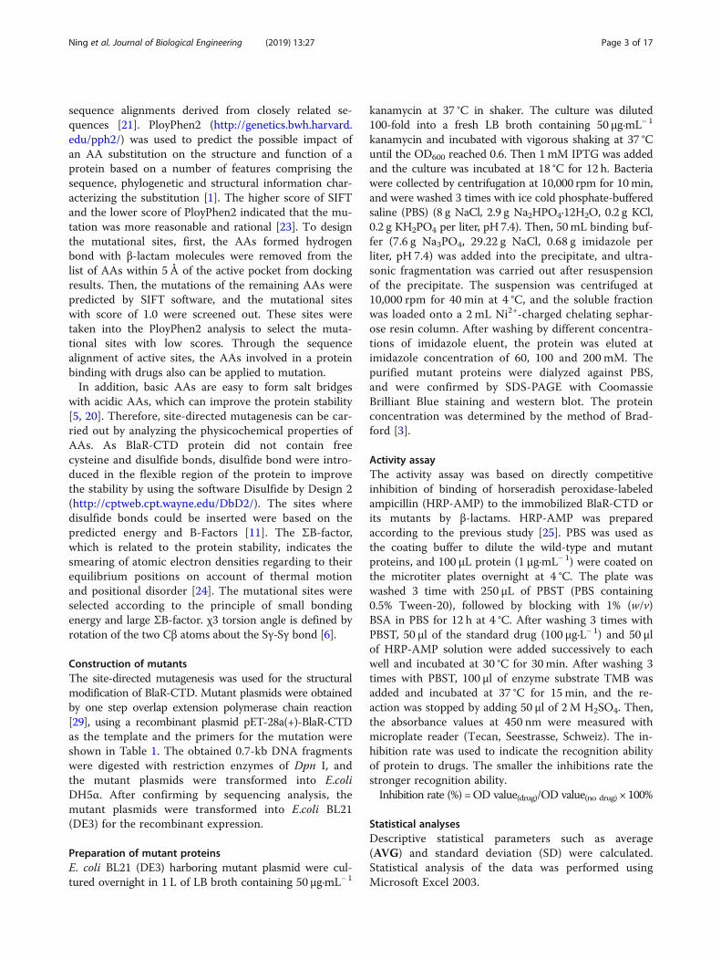
ResultsHomologous model of BlaR-CTD and docking with β-lactamsThe 3D structure of BlaR-CTD protein from B. licheniformisATCC14580 was shown in Fig. 1a. It was mainly com-posed of two domains. One was the α/β domain, whichwas covered by two α helices (α1 in N-terminal andα10 in C-terminal) on one side of a seven parallel βsheets (β1-β7), with the other side covered by α3 and
α8 helix. The other domain was mainly composed offour helices (α5, α6, α7 and α9) encircling the hydro-phobic α4 helix. The active pocket of the BlaR-CTDwas composed of S(55)TYK(58), S(103)AT(105) andK(192)TG(194) [19].The docking result of cefquinome with BlaR-CTD was
shown in Fig. 1b. Nine hydrogen bonds were formedbetween the protein and cefquinome in the active pocket.
Table 1 Primers used for site-directed mutagenesis
Mutation site Primers Sequence 5′-3′
A138E F CATCCAGCCAATAATTCTCCGGACCTGAGAAATCC
R GGATTTCTCAGGTCCGGAGAATTATTGGCTGGATG
Q147K F GAGGGGAAATTTTAAGAGAGCC
R GGCTCTCTTAAAATTTCCCCTC
I188K F GTCCCGGTTTTACCGGATAGTTTTCTGCCATTTGATTCTTCT
R AGAAGAATCAAATGGCAGAAAACTATCCGGTAAAACCGGGAC
S190Y F AAGTCCCGGTTTTACCGTATAG
R TCTATACGGTAAAACCGGGAC
V197D F CTCCGTTGATATCTGAAGTCC
R GGGACTTCAGATATCAACGG
S19C/G24C
S19C F GATGACTGCACCTTTTTTGATGGCT
R GGTGCAGTCATCTTCGTATTCTACA
G24C F TTTGATTGCTTCTCAGGAGGTTTTG
R AGAAGCAATCAAAAAAGGTGCAGTC
R50C/Q147C
R50C F CCGCCTGTTTCGCACCTGCTTCTAC
R GCGAAACAGGCGGTGCTTTCTTTCC
Q147C F TCTCTTTGTATTTCCCCTCTTGAAC
R GAAATACAAAGAGAGCCATCCAGCC
S76C/ L96C
S76C F CAATTGTCAAATGACGTGGGACGGA
R CATTTGACAATTGTTCTTCGTGATGATCCC
L96C F AGGATTGTTTCTCTGCGATGAGCAG
R GCAGAGAAACAATCCTGGTCTTGAT
S135C/S145C
S135C F ATTTCTGTGGTCCGGCGAATTATTG
R CGGACCACAGAAATCCTCATTTCCA
S145C F GATGGCTGTCTTCAAATTTCCCCTC
R TGAAGACAGCCATCCAGCCAATAAT
E183C/ I188C
E183C F TTAGAATGCTCAAATGGCAGAATTC
R TTGAGCATTCTAAACGTATCGAATC
I188C F GGCAGATGTCTATCCGGTAAAACC
R ATAGACATCTGCCATTTGAGCATTC
Notes: The underline shows the bases corresponding to the mutant amino acids
Ning et al. Journal of Biological Engineering (2019) 13:27 Page 4 of 17
Among the hydrogen bond forming AAs, Ser55, Ser103,Thr105 and Thr193 were reported in the previous literature[19], in which one hydrogen bond was respectively formedwith Ser55, Ser103 and Thr105, and three hydrogen bondswere formed with Thr193. In addition, two hydrogen bondsbetween cefquinome and Tyr87, and one hydrogen bondbetween the drug and Thr195 were also formed.
Molecular docking between BlaR-CTD protein andother 39 β-lactams was also carried out by Sybyl-X2.0, and65 key AAs existed within 5 Å around active pocket(Additional file 1: Table S1). The key AAs forming hydro-gen bonds to each β-lactam were shown in Table 2,including Ser55, Ser101, Ser102, Ser103, Thr105, Thr193,Thr195, Try87, Glu89, Arg229 and Ser233. When the
Fig. 1 3D structure of BlaR-CTD protein (a) and interaction with cefquinome (b). a The 3D structure of BlaR-CTD was represented as a cartoon,with α-helices colored in cyan, β-strands in magenta, and loops in salmon. The active sites of Ser55, Ser103, Thr105 and Thr195 were colored inyellow. b The amino acid residues involved in the interaction of BlaR-CTD with cefquinome. The hydrogen bonds were represented as blackdotted lines. The cefquinome molecule was colored in gray. The AAs of the active site were colored in yellow
Ning et al. Journal of Biological Engineering (2019) 13:27 Page 5 of 17
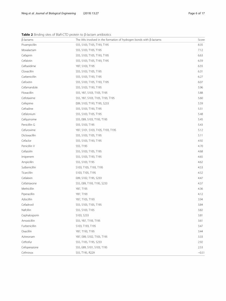
Table 2 Binding sites of BlaR-CTD protein to β-lactam antibiotics
β-lactams The AAs involved in the formation of hydrogen bonds with β-lactams Score
Pivampicillin S55, S103, T105, T193, T195 8.35
Moxalactam S55, S103, T105, T195 7.12
Cefapirin S55, S103, T105, T193, T195 6.63
Cefalotin S55, S103, T105, T193, T195 6.59
Ceftazidime Y87, S103, T195 6.55
Cloxacillin S55, S103, T105, T195 6.31
Carbenicillin S55, S103, T193, T195 6.27
Cefoxitin S55, S103, T105, T193, T195 6.07
Cefamandole S55, S103, T193, T195 5.96
Floxacillin S55, Y87, S103, T105, T195 5.88
Cefotaxime S55, Y87, S103, T105, T193, T195 5.60
Cefepime E89, S103, T193, T195, S233 5.59
Cefradine S55, S103, T193, T195 5.51
Cefalonium S55, S103, T105, T195 5.48
Cefquinome S55, E89, S103, T193, T195 5.45
Penicillin G S55, S103, T195 5.43
Cefuroxime Y87, S101, S103, T105, T193, T195 5.12
Dicloxacillin S55, S103, T105, T195 5.11
Cefaclor S55, S103, T193, T195 4.92
Penicillin V S55, T195 4.70
Cefazolin S55, S103, T105, T195 4.68
Imipenem S55, S103, T193, T195 4.65
Ampicillin S55, S103, T195 4.62
Sulbenicillin S103, T105, T193, T195 4.53
Ticarcillin S103, T105, T195 4.52
Cefalexin E89, S102, T195, S233 4.47
Cefatriaxone S55, E89, T193, T195, S233 4.37
Methicillin Y87, T195 4.36
Piperacillin Y87, T193 4.12
Azlocillin Y87, T105, T193 3.94
Cefadroxil S55, S103, T105, T195 3.84
Nafcillin S55, S103, T105 3.82
Cephalosporin S103, S233 3.81
Amoxicillin S55, Y87, T193, T195 3.61
Furbenicillin S103, T193, T195 3.47
Oxacillin Y87, T193, T195 3.44
Aztreonam Y87, E89, S102, T105, T195 3.33
Ceftiofur S55, T105, T195, S233 2.92
Cefoperazone S55, E89, S101, S103, T195 2.53
Cefminox S55, T195, R229 −0.51
Ning et al. Journal of Biological Engineering (2019) 13:27 Page 6 of 17
score was less than 4, the binding ability between proteinand drug was poor, and when the score was greaterthan 7, the binding ability was stronger.
Determination of mutational sitesPBP3 from Streptococcus pneumoniae R6 had a highbinding ability with cefadroxil, and the docking resultsof PBP3 and cefadroxil showed that D224 in the activesite K(239)TGTTD(244) and cefadroxil formed a hydro-gen bond [32]. The sequence alignment demonstratedthat K(192)TGTSV(197) in BlaR-CTD activity pocketcorresponded to K(239)TGTTD(244) in PBP3. There-fore, the mutation of V197D was selected in order toimprove the affinity.Mutation prediction and evaluation was carried out
according to SIFT and Polyphen 2. The mutational siteswith a SIFT score of 1.0 were obtained as shown inAdditional file 1: Table S2. In order not to affect thestructure and function of the protein, the selection ofmutational sites of protein design should avoid the activesite, especially the hydrogen bond-forming AAs of theprotein [4]. Twelve AAs from the list of AAs in theactive pocket but exclude the active site and hydrogenbond-forming AAs (Additional file 1: Table S1) showeda SIFT score of 1.0 (Additional file 1: Table S2), and thesingle point mutations of A138E, Q147K and S190Ywith the lowest Polyphen scores were screened out(Additional file 1: Table S3).In addition, salt bridge and disulfide bond were inserted
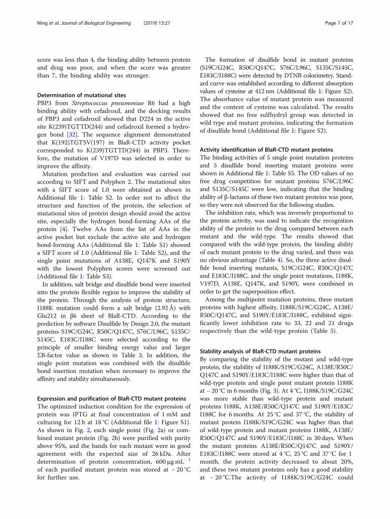
into the protein flexible region to improve the stability ofthe protein. Through the analysis of protein structure,I188K mutation could form a salt bridge (2.92 Å) withGlu212 in β6 sheet of BlaR-CTD. According to theprediction by software Disulfide by Design 2.0, the mutantproteins S19C/G24C, R50C/Q147C, S76C/L96C, S135C/S145C, E183C/I188C were selected according to theprinciple of smaller binding energy value and largerΣB-factor value as shown in Table 3. In addition, thesingle point mutation was combined with the disulfidebond insertion mutation when necessary to improve theaffinity and stability simultaneously.
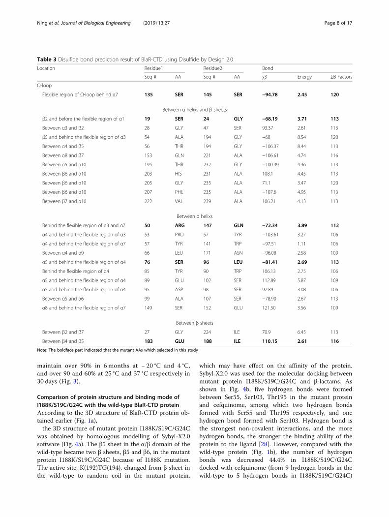
Expression and purification of BlaR-CTD mutant proteinsThe optimized induction condition for the expression ofprotein was IPTG at final concentration of 1 mM andculturing for 12 h at 18 °C (Additional file 1: Figure S1).As shown in Fig. 2, each single point (Fig. 2a) or com-bined mutant protein (Fig. 2b) were purified with purityabove 95%, and the bands for each mutant were in goodagreement with the expected size of 26 kDa. Afterdetermination of protein concentration, 600 μg·mL− 1
of each purified mutant protein was stored at − 20 °Cfor further use.
The formation of disulfide bond in mutant proteins(S19C/G24C, R50C/Q147C, S76C/L96C, S135C/S145C,E183C/I188C) were detected by DTNB colorimetry. Stand-ard curve was established according to different absorptionvalues of cysteine at 412 nm (Additional file 1: Figure S2).The absorbance value of mutant protein was measuredand the content of cysteine was calculated. The resultsshowed that no free sulfhydryl group was detected inwild type and mutant proteins, indicating the formationof disulfide bond (Additional file 1: Figure S2).
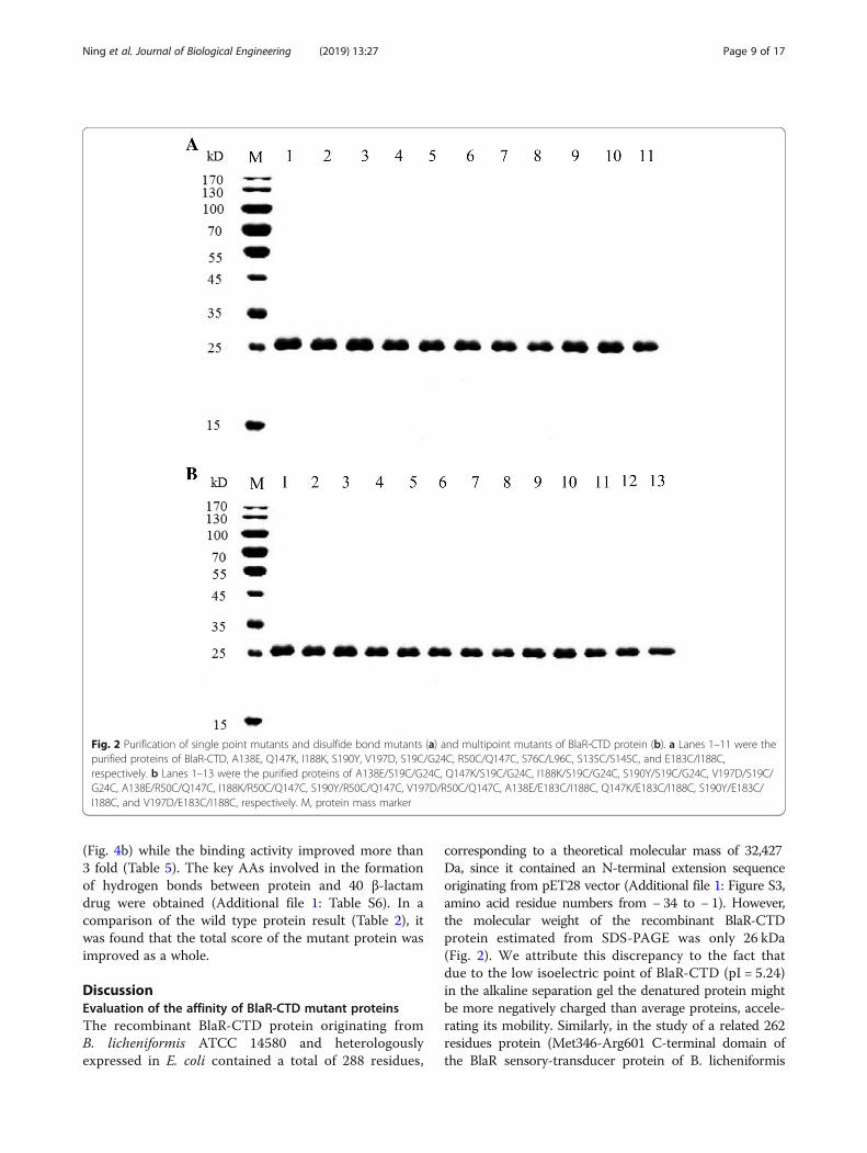
Activity identification of BlaR-CTD mutant proteinsThe binding activities of 5 single point mutation proteinsand 5 disulfide bond inserting mutant proteins wereshown in Additional file 1: Table S5. The OD values of nofree drug competition for mutant proteins S76C/L96Cand S135C/S145C were low, indicating that the bindingability of β-lactams of these two mutant proteins was poor,so they were not observed for the following studies.The inhibition rate, which was inversely proportional to
the protein activity, was used to indicate the recognitionability of the protein to the drug compared between eachmutant and the wild-type. The results showed thatcompared with the wild-type protein, the binding abilityof each mutant protein to the drug varied, and there wasno obvious advantage (Table 4). So, the three active disul-fide bond inserting mutants, S19C/G24C, R50C/Q147Cand E183C/I188C, and the single point mutations, I188K,V197D, A138E, Q147K, and S190Y, were combined inorder to get the superposition effect.Among the multipoint mutation proteins, three mutant
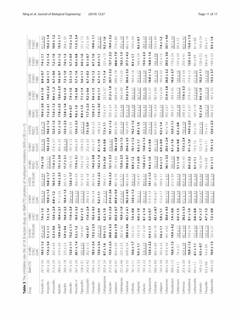
proteins with highest affinity, I188K/S19C/G24C, A138E/R50C/Q147C, and S190Y/E183C/I188C, exhibited signi-ficantly lower inhibition rate to 33, 22 and 21 drugsrespectively than the wild-type protein (Table 5).
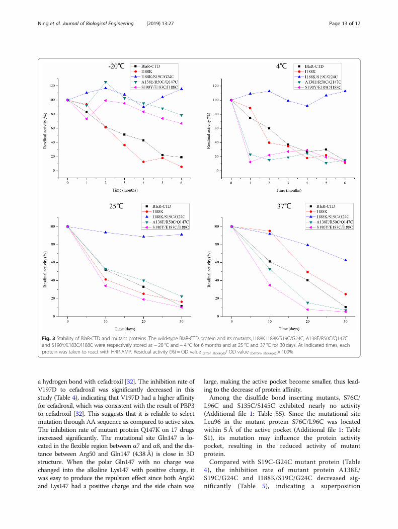
Stability analysis of BlaR-CTD mutant proteinsBy comparing the stability of the mutant and wild-typeprotein, the stability of I188K/S19C/G24C, A138E/R50C/Q147C and S190Y/E183C/I188C were higher than that ofwild-type protein and single point mutant protein I188Kat − 20 °C in 6months (Fig. 3). At 4 °C, I188K/S19C/G24Cwas more stable than wild-type protein and mutantproteins I188K, A138E/R50C/Q147C and S190Y/E183C/I188C for 6months. At 25 °C and 37 °C, the stability ofmutant protein I188K/S19C/G24C was higher than thatof wild-type protein and mutant proteins I188K, A138E/R50C/Q147C and S190Y/E183C/I188C in 30 days. Whenthe mutant proteins A138E/R50C/Q147C and S190Y/E183C/I188C were stored at 4 °C, 25 °C and 37 °C for 1month, the protein activity decreased to about 20%,and these two mutant proteins only has a good stabilityat − 20 °C.The activity of I188K/S19C/G24C could
Ning et al. Journal of Biological Engineering (2019) 13:27 Page 7 of 17
maintain over 90% in 6 months at − 20 °C and 4 °C,and over 90 and 60% at 25 °C and 37 °C respectively in30 days (Fig. 3).
Comparison of protein structure and binding mode ofI188K/S19C/G24C with the wild-type BlaR-CTD proteinAccording to the 3D structure of BlaR-CTD protein ob-tained earlier (Fig. 1a),the 3D structure of mutant protein I188K/S19C/G24C
was obtained by homologous modelling of Sybyl-X2.0software (Fig. 4a). The β5 sheet in the α/β domain of thewild-type became two β sheets, β5 and β6, in the mutantprotein I188K/S19C/G24C because of I188K mutation.The active site, K(192)TG(194), changed from β sheet inthe wild-type to random coil in the mutant protein,
which may have effect on the affinity of the protein.Sybyl-X2.0 was used for the molecular docking betweenmutant protein I188K/S19C/G24C and β-lactams. Asshown in Fig. 4b, five hydrogen bonds were formedbetween Ser55, Ser103, Thr195 in the mutant proteinand cefquinome, among which two hydrogen bondsformed with Ser55 and Thr195 respectively, and onehydrogen bond formed with Ser103. Hydrogen bond isthe strongest non-covalent interactions, and the morehydrogen bonds, the stronger the binding ability of theprotein to the ligand [28]. However, compared with thewild-type protein (Fig. 1b), the number of hydrogenbonds was decreased 44.4% in I188K/S19C/G24Cdocked with cefquinome (from 9 hydrogen bonds in thewild-type to 5 hydrogen bonds in I188K/S19C/G24C)
Table 3 Disulfide bond prediction result of BlaR-CTD using Disulfide by Design 2.0
Location Residue1 Residue2 Bond
Seq # AA Seq # AA χ3 Energy ΣB-Factors
Ω-loop
Flexible region of Ω-loop behind α7 135 SER 145 SER −94.78 2.45 120
Between α helixs and β sheets
β2 and before the flexible region of α1 19 SER 24 GLY −68.19 3.71 113
Between α3 and β2 28 GLY 47 SER 93.37 2.61 113
β5 and behind the flexible region of α3 54 ALA 194 GLY −68 8.54 120
Between α4 and β5 56 THR 194 GLY −106.37 8.44 113
Between α8 and β7 153 GLN 221 ALA −106.61 4.74 116
Between α5 and α10 195 THR 232 GLY −100.49 4.36 113
Between β6 and α10 203 HIS 231 ALA 108.1 4.45 113
Between β6 and α10 205 GLY 235 ALA 71.1 3.47 120
Between β6 and α10 207 PHE 235 ALA −107.6 4.95 113
Between β7 and α10 222 VAL 239 ALA 106.21 4.13 113
Between α helixs
Behind the flexible region of α3 and α7 50 ARG 147 GLN −72.34 3.89 112
α4 and behind the flexible region of α3 53 PRO 57 TYR −103.61 3.27 106
α4 and behind the flexible region of α7 57 TYR 141 TRP −97.51 1.11 106
Between α4 and α9 66 LEU 171 ASN −96.08 2.58 109
α5 and behind the flexible region of α4 76 SER 96 LEU −81.41 2.69 113
Behind the flexible region of α4 85 TYR 90 TRP 106.13 2.75 106
α5 and behind the flexible region of α4 89 GLU 102 SER 112.89 5.87 109
α5 and behind the flexible region of α4 95 ASP 98 SER 92.89 3.08 106
Between α5 and α6 99 ALA 107 SER −78.90 2.67 113
α8 and behind the flexible region of α7 149 SER 152 GLU 121.50 3.56 109
Between β sheets
Between β2 and β7 27 GLY 224 ILE 70.9 6.45 113
Between β4 and β5 183 GLU 188 ILE 110.15 2.61 116
Note: The boldface part indicated that the mutant AAs which selected in this study
Ning et al. Journal of Biological Engineering (2019) 13:27 Page 8 of 17
(Fig. 4b) while the binding activity improved more than3 fold (Table 5). The key AAs involved in the formationof hydrogen bonds between protein and 40 β-lactamdrug were obtained (Additional file 1: Table S6). In acomparison of the wild type protein result (Table 2), itwas found that the total score of the mutant protein wasimproved as a whole.
DiscussionEvaluation of the affinity of BlaR-CTD mutant proteinsThe recombinant BlaR-CTD protein originating fromB. licheniformis ATCC 14580 and heterologouslyexpressed in E. coli contained a total of 288 residues,
corresponding to a theoretical molecular mass of 32,427Da, since it contained an N-terminal extension sequenceoriginating from pET28 vector (Additional file 1: Figure S3,amino acid residue numbers from − 34 to − 1). However,the molecular weight of the recombinant BlaR-CTDprotein estimated from SDS-PAGE was only 26 kDa(Fig. 2). We attribute this discrepancy to the fact thatdue to the low isoelectric point of BlaR-CTD (pI = 5.24)in the alkaline separation gel the denatured protein mightbe more negatively charged than average proteins, accele-rating its mobility. Similarly, in the study of a related 262residues protein (Met346-Arg601 C-terminal domain ofthe BlaR sensory-transducer protein of B. licheniformis
Fig. 2 Purification of single point mutants and disulfide bond mutants (a) and multipoint mutants of BlaR-CTD protein (b). a Lanes 1–11 were thepurified proteins of BlaR-CTD, A138E, Q147K, I188K, S190Y, V197D, S19C/G24C, R50C/Q147C, S76C/L96C, S135C/S145C, and E183C/I188C,respectively. b Lanes 1–13 were the purified proteins of A138E/S19C/G24C, Q147K/S19C/G24C, I188K/S19C/G24C, S190Y/S19C/G24C, V197D/S19C/G24C, A138E/R50C/Q147C, I188K/R50C/Q147C, S190Y/R50C/Q147C, V197D/R50C/Q147C, A138E/E183C/I188C, Q147K/E183C/I188C, S190Y/E183C/I188C, and V197D/E183C/I188C, respectively. M, protein mass marker
Ning et al. Journal of Biological Engineering (2019) 13:27 Page 9 of 17
with a hexapeptide extension), the recombinant proteinwith 29.3 kDa theoretical molecular mass and pI 5.72migrated with 26 kDa on SDS-PAGE [17].The affinity of BlaR-CTD to each β-lactam is different,
due to the different chemical structures of drugs, theinteraction and binding modes of BlaR-CTD protein withdrugs are also different. There are some difficulties inselecting mutation sites because it was uncertain whichamino acids in proteins interact directly with drugs.Molecular docking was used to find the key amino acids
that may interact with each drug (e.g., those amino acidsforming hydrogen bonds with the drug). Through the re-sults of molecular docking between protein and β-lactamantibiotics, the amino acid sites involved in the hydrogenbonding between protein and drug were obtained. Thesekey amino acids should be avoided in the selection ofmutation sites, which provided a reference for theselection of subsequent mutation sites.Mutation of V197D in BlaR-CTD was designed based on
the sequence alignment with PBP3 in which D224 formed
Table 4 The inhibition rates (%) of 33 β-lactam drugs on BlaR-CTD proteins of single point mutation and disulfide bond insertion(AVG ± SD, n = 5)
Drug BlaR-CTD A138E Q147K I188K S190Y V197D S19C-G24C R50C-Q147C E183C-I188C
Penicillin G 26.2 ± 2.1 15.3 ± 1.4 26.7 ± 3.4 25.3 ± 2.8 21.7 ± 1.6 34.2 ± 4.2 17.4 ± 1.5 34.6 ± 2.5 20.5 ± 1.4
Ampicillin 21.2 ± 1.1 21.0 ± 1.8 27.4 ± 2.5 17.4 ± 1.4 24.4 ± 2.4 19.3 ± 1.3 18.4 ± 1.3 17.5 ± 1.3 24.7 ± 2.2
Amoxicillin 19.6 ± 1.0 19.3 ± 1.6 28.9 ± 3.4 40.8 ± 3.7 21.7 ± 1.3 25.6 ± 2.8 22.6 ± 3.4 20.4 ± 3.1 16.8 ± 1.3
Nafcillin 29.0 ± 1.4 32.9 ± 4.8 29.5 ± 3.1 34.3 ± 3.5 24.0 ± 1.9 35.7 ± 3.3 31.4 ± 2.8 35.2 ± 3.8 26.6 ± 3.7
Oxacillin 23.4 ± 2.3 24.0 ± 3.6 29.3 ± 2.5 26.7 ± 2.9 22.3 ± 2.5 25.0 ± 3.4 26.6 ± 2.1 21.5 ± 2.4 23.6 ± 2.4
Azlocillin 15.2 ± 1.1 10.9 ± 1.7 14.7 ± 1.4 10.6 ± 1.3 16.7 ± 1.7 17.0 ± 1.6 14.1 ± 1.3 12.8 ± 1.2 24.9 ± 4.3
Penicillin V 15.5 ± 1.5 12.7 ± 1.2 7.5 ± 1.0 16.7 ± 1.1 10.9 ± 1.2 12.2 ± 1.2 25.9 ± 2.6 35.2 ± 4.6 16.4 ± 2.9
Piperacillin 6.5 ± 0.9 8.0 ± 1.0 12.0 ± 1.6 6.2 ± 0.8 27.3 ± 2.4 20.3 ± 3.6 13.2 ± 1.5 12.2 ± 1.5 10.4 ± 1.1
Dicloxacillin 27.5 ± 2.1 30.3 ± 2.9 23.2 ± 3.4 24.9 ± 2.4 31.7 ± 4.2 25.0 ± 4.5 31.6 ± 5.1 24.2 ± 2.0 26.1 ± 1.3
Cloxacillin 21.1 ± 1.8 25.5 ± 2.4 26.5 ± 3.1 44.4 ± 5.1 22.0 ± 2.5 25.3 ± 3.2 20.2 ± 1.3 31.7 ± 4.7 22.5 ± 3.4
Cefalotin 26.3 ± 2.4 24.6 ± 2.5 27.0 ± 1.9 24.7 ± 2.6 20.9 ± 1.7 34.0 ± 5.1 18.6 ± 1.2 18.4 ± 1.3 16.7 ± 1.2
Cefoperazone 21.5 ± 1.3 18.3 ± 1.7 29.1 ± 4.1 25.8 ± 2.7 30.5 ± 4.2 20.5 ± 3.6 16.2 ± 1.3 25.5 ± 2.1 31.5 ± 4.6
Cefazolin 32.0 ± 2.9 26.1 ± 2.3 30.5 ± 3.2 29.8 ± 3.1 23.4 ± 3.4 26.9 ± 3.4 40.1 ± 2.5 31.5 ± 4.5 40.2 ± 5.1
Cefalexin 88.4 ± 6.8 87.7 ± 9.4 81.9 ± 7.6 69.9 ± 4.5 65.2 ± 7.5 75.2 ± 10.1 63.5 ± 5.4 75.8 ± 9.1 62.5 ± 9.3
Cefatriaxone 25.5 ± 1.5 19.7 ± 1.2 24.8 ± 4.8 23.5 ± 2.4 19.4 ± 1.4 21.2 ± 1.8 17.3 ± 1.2 31.4 ± 2.7 20.5 ± 1.2
Cefadroxil 69.8 ± 4.8 70.6 ± 8.1 65.2 ± 5.6 59.6 ± 6.1 69.9 ± 8.5 56.7 ± 5.2 84.9 ± 10.5 55.2 ± 3.4 71.5 ± 7.9
Ceftiofur 21.4 ± 2.6 16.7 ± 1.5 25.7 ± 2.4 34.6 ± 3.2 28.6 ± 3.4 19.4 ± 1.4 20.6 ± 4.2 22.1 ± 1.5 25.1 ± 3.4
Cefepime 9.4 ± 0.8 17.7 ± 1.2 27.9 ± 2.8 21.7 ± 1.6 17.4 ± 1.8 18.0 ± 1.6 16.9 ± 1.6 14.5 ± 1.1 22.4 ± 2.6
Cefaclor 19.7 ± 1.4 18.5 ± 2.4 31.5 ± 3.4 20.9 ± 1.2 19.6 ± 1.6 17.4 ± 1.2 14.8 ± 2.1 15.8 ± 2.1 20.6 ± 1.3
Cefquinome 19.5 ± 1.2 11.8 ± 1.5 16.9 ± 1.4 23.9 ± 2.5 12.0 ± 1.3 16.6 ± 1.4 11.3 ± 1.2 16.8 ± 1.3 19.9 ± 1.2
Cefradine 61.2 ± 5.7 59.6 ± 6.3 67.3 ± 7.3 58.7 ± 5.6 72.0 ± 5.7 56.1 ± 7.1 49.9 ± 6.1 52.4 ± 4.5 68.4 ± 8.2
Cefapirin 19.6 ± 1.3 14.1 ± 1.2 29.1 ± 1.7 23.3 ± 1.3 10.4 ± 1.0 15.9 ± 1.9 8.3 ± 0.9 18.5 ± 3.1 22.4 ± 3.7
Cefuroxime 45.8 ± 5.3 49.5 ± 5.4 46.2 ± 3.1 52.4 ± 6.1 46.2 ± 6.1 43.6 ± 4.1 46.8 ± 5.2 45.1 ± 4.1 43.5 ± 5.1
Moxalactam 21.4 ± 2.9 19.5 ± 1.7 32.1 ± 4.5 23.4 ± 3.2 13.3 ± 1.2 22.1 ± 2.3 8.2 ± 1.0 20.4 ± 2.5 44.2 ± 3.1
Cefalonium 5.2 ± 0.7 6.2 ± 0.8 11.7 ± 1.6 6.3 ± 0.8 21.5 ± 3.3 32.9 ± 4.3 10.3 ± 1.3 14.2 ± 1.3 15.2 ± 1.5
Cefminox 19.0 ± 1.6 22.9 ± 3.4 18.0 ± 1.2 17.4 ± 1.5 12.8 ± 1.2 20.8 ± 3.4 16.2 ± 2.1 25.9 ± 3.0 18.5 ± 1.1
Ceftazidime 23.1 ± 2.6 19.8 ± 1.5 16.8 ± 2.3 19.6 ± 2.1 21.3 ± 2.3 19.3 ± 1.8 21.1 ± 1.9 18.5 ± 1.2 24.8 ± 3.1
Carbenicillin 6.7 ± 0.8 16.8 ± 1.1 15.2 ± 1.3 11.1 ± 1.0 19.6 ± 1.4 22.5 ± 2.0 15.0 ± 1.2 17.7 ± 1.1 15.9 ± 1.6
Cefoxitin 12.7 ± 2.6 17.0 ± 1.3 19.9 ± 1.2 16.0 ± 1.4 10.5 ± 2.1 11.7 ± 1.8 12.3 ± 2.1 11.5 ± 1.8 15.5 ± 1.0
Floxacillin 6.2 ± 0.8 7.8 ± 1.0 12.3 ± 1.3 8.7 ± 1.1 4.3 ± 0.5 7.2 ± 1.3 10.3 ± 1.3 20.4 ± 3.3 16.7 ± 2.1
Sulbenicillin 8.7 ± 1.1 12.5 ± 1.1 16.6 ± 2.5 10.0 ± 1.0 18.3 ± 1.7 25.5 ± 1.2 7.2 ± 0.9 13.5 ± 2.1 15.2 ± 1.3
Ticarcillin 12.5 ± 1.6 13.8 ± 2.4 15.4 ± 1.4 12.3 ± 2.6 27.9 ± 3.5 24.9 ± 2.1 15.1 ± 1.5 11.3 ± 1.3 11.4 ± 2.0
Aztreonam 93.1 ± 6.7 86.8 ± 5.7 95.4 ± 10.1 90.0 ± 9.4 94.3 ± 10.1 100.9 ± 9.7 106.4 ± 9.1 100.3 ± 9.0 97.5 ± 9.5
Note: The statistical method was used to analyze the significant difference between the wild-type protein and mutant proteins. The underlined number indicatedthat the inhibition rate was significantly increased, and the boldface number showed that the inhibition rate was significantly reduced (p < 0.05)
Ning et al. Journal of Biological Engineering (2019) 13:27 Page 10 of 17
Table
5Theinhibitio
nrate
(%)of
33β-lactam
drug
son
BlaR-CTD
proteins
ofmultip
oint
mutations
(AVG
±SD
,n=5)
Drug
BlaR-CTD
A138E/
S19C
/G24C
Q147K/
S19C
/G24C
I188K/
S19C
/G24C
S190Y/
S19C
/G24C
V197D/
S19C
/G24C
A138E/
R50C
/Q147C
S190Y/
R50C
/Q147C
V197D/
R50C
/Q147C
I188K/
R50C
/Q147C
A138E/
E183C/
I188C
Q147K/
E183C/
I188C
S190Y/
E183C/
I188C
V197D/
E183C/
I188C
PenicillinG
23.7±3.5
18.5
±1.2
45.3±4.5
7.4±1.3
34.0±3.3
32.2±4.4
7.6±1.0
36.5±5.6
67.8±7.9
10.4
±2.0
8.4±1.0
8.9±0.7
7.4±1.1
47.1±4.3
Ampicillin
28.2±2.6
17.6
±3.4
5.1±1.1
7.5±1.4
10.9
±1.3
16.8
±1.7
37.3±5.1
18.3
±2.3
10.6
±1.2
11.2
±2.1
14.0
±2.2
6.8±1.1
7.2±1.4
10.2
±1.4
Amoxicillin
21.2±2.4
20.5±3.4
5.3±0.6
12.6
±2.4
8.8±1.2
16.5
±1.1
11.6
±1.3
21.6±2.7
12.3
±1.5
11.0
±1.3
8.6±1..3
6.7±0.6
7.2±1.0
10.0
±1.2
Nafcillin
28.9±3.7
27.9±4.3
14.9
±0.7
18.4
±2.1
31.1±2.5
14.6
±2.3
13.1
±2.0
26.5±3.5
12.1
±1.2
17.8
±2.5
13.0
±2.5
9.6±0.7
8.3±1.2
15.3
±1.1
Oxacillin
24.0±1.8
22.5±2.3
5.9±0.6
16.6
±2.3
10.4
±1.3
24.5±3.5
11.7
±2.1
28.8±3.4
13.5
±2.3
12.8
±1.6
9.6±1.0
7.2±1.0
7.0±1.3
26.8±3.5
Azlocillin
19.7±2.5
12.0
±1.1
15.2
±1.1
11.6
±2.4
28.6±3.4
12.6
±1.7
19.0±1.3
19.7±1.2
20.5±3.5
8.9±0.7
7.4±1.0
5.5±1.3
5.7±0.9
11.2
±1.2
PenicillinV
24.7±2.6
20.1
±1.4
5.2±1.2
16.3
±2.2
29.4±2.3
26.6±3.4
17.9
±3.1
24.8±4.5
12.6
±2.3
9.6±1.0
7.8±0.8
5.7±1.0
6.0±0.8
11.5
±2.4
Pipe
racillin
12.8±3.1
24.0±4.2
14.4±4.1
9.2±1.2
17.5±2.3
15.1±2.2
14.3±1.3
12.8±2.3
16.3±1.2
8.9±1.3
7.1±1.4
5.6±1.1
6.0±1.0
12.1±1.2
Dicloxacillin
24.3±4.2
26.2±1.3
4.8±0.7
11.6
±2.4
29.0±3.2
26.6±1.8
24.5±5.1
23.2±3.5
7.0±0.9
11.7
±2.3
9.2±0.6
6.7±0.5
8.1±0.7
21.2±3.4
Cloxacillin
23.6±2.4
18.5
±2.4
15.2
±2.3
10.2
±4.2
29.4±2.4
28.3±3.4
14.2
±0.8
24.2±2.7
24.3±2.3
12.0
±2.1
9.6±1.5
7.0±1.2
6.7±1.0
10.9
±1.1
Cefalotin
17.3±1.5
13.0
±3.4
24.8±3.4
8.6±0.9
9.2±1.1
14.3
±2.2
14.2
±1.0
8.1±1.0
14.8±2.2
11.6
±2.0
9.9±1.1
6.7±1.1
6.4±1.1
38.7±4.7
Cefop
erazon
e20.3±2.4
8.7±1.6
9.9±1.1
6.2±1.0
27.7±3.3
18.8±1.1
5.9±0.4
8.6±0.8
7.3±1.1
21.6±3.1
23.2±3.7
7.7±1.4
22.5±1.3
9.2±1.6
Cefazolin
31.3±4.6
15.9
±2.3
20.8
±3.3
10.1
±2.6
21.4
±2.6
19.6
±2.3
12.1
±3.3
47.6±6.7
19.1
±1.2
33.0±2.4
21.2
±1.8
20.7
±2.2
13.7
±2.2
19.4
±2.2
Cefalexin
80.1±9.7
83.3±9.4
65.6
±7.9
63.5
±6.7
65.9
±5.9
80.1±7.9
79.7±6.4
38.4
±5.8
85.0±9.9
91.5±9.4
69.8±5.5
69.0±7.8
66.6
±7.9
77.4±8.9
Cefatriaxone
22.1±4.6
22.5±2.3
26.5±3.2
10.9
±1.0
41.9±6.7
61.7±7.7
15.0
±2.5
12.6
±1.3
29.5±3.4
37.2±4.6
26.6±4.0
17.7±2.3
10.2
±1.5
29.2±3.4
Cefadroxil
70.2±5.5
63.8±5.4
58.8
±4.6
45.1
±6.1
56.3
±4.5
68.3±5.6
59.8
±6.7
29.4
±3.5
76.0±8.9
81.3±7.6
51.0
±7.6
50.4
±5.5
37.7
±4.3
66.2±7.8
Ceftio
fur
16.1±1.1
10.6
±1.7
15.2±2.1
7.6±0.8
12.0
±1.3
26.8±1.5
8.8±1.2
10.1
±1.2
14.2
±1.2
26.3±4.8
24.2±2.1
16.4±1.6
8.3±1.1
15.9±3.4
Cefep
ime
13.2±3.3
9.5±1.1
13.2±1.3
6.0±1.0
18.3±1.1
13.0±4.7
7.1±0.9
8.3±1.1
8.9±1.0
25.2±2.2
26.7±3.7
20.3±2.2
16.6±2.4
8.2±0.7
Cefaclor
19.2±2.2
32.6±2.3
17.1±3.4
8.1±1.4
28.3±2.7
38.4±2.2
13.8
±2.1
13.0
±1.3
26.8±3.4
40.2±4.3
36.8±4.6
31.4±3.4
15.5
±2.3
28.9±2.3
Cefqu
inom
e21.1±1.4
12.6
±2.2
4.4±1.1
6.3±0.7
21.5±1.4
14.1
±1.3
6.4±1.0
6.5±0.8
27.8±2.2
25.3±3.4
23.1±2.1
16.8
±1.2
16.8
±1.6
29.0±4.5
Cefradine
59.6±6.9
66.5±8.9
68.0±7.8
43.1
±8.7
51.8±6.7
58.1±8.9
74.2±5.9
69.1±5.2
65.4±8.3
74.1±6.4
58.7±8.9
76.1±7.6
74.6±8.6
53.3±8.9
Cefapirin
13.5±3.4
10.2±2.3
5.6±1.1
5.8±1.1
11.2±1.7
14.9±1.1
6.4±1.1
6.9±0.9
9.3±1.4
22.2±2.5
15.3±2.4
16.0±3.3
13.2±1.5
7.4±1.2
Cefuroxim
e44.3±5.6
47.4±5.6
49.1±6.5
30.0
±4.5
68.9±5.5
57.1±4.5
36.1
±3.5
20.5
±2.4
46.3±3.4
39.2±6.7
31.4
±3.8
26.0
±2.2
29.5
±3.6
33.1
±4.6
Moxalactam
20.0±1.3
10.6
±1.5
14.9
±4.2
7.4±0.8
29.6±4.3
21.2±3.4
29.0±3.3
8.1±1.0
12.0
±1.1
18.8±2.7
18.4±4.5
16.5
±2.7
17.9±2.4
28.7±3.4
Cefalon
ium
8.9±1.1
7.6±2.1
14.4±1.1
6.5±1.5
30.8±5.4
28.0±2.3
5.1±1.0
6.6±0.8
6.5±0.8
19.8±3.8
19.3±1.2
14.8±2.6
12.3±3.3
9.9±1.5
Cefminox
25.5±3.6
42.4±3.8
48.7±6.9
20.2
±3.3
67.2±7.5
66.9±7.8
24.5±4.5
17.5
±2.4
79.0±5.8
24.8±3.3
17.6
±3.3
22.5±3.4
25.0±4.1
82.0±9.2
Ceftazidime
20.2±4.4
10.8
±1.2
15.9±3.4
8.1±1.0
33.3±3.6
37.2±3.2
8.6±2.2
9.1±1.0
14.9
±2.2
21.7±2.7
21.4±3.2
17.4±1.2
12.0
±2.3
13.4
±1.3
Carbe
nicillin
8.9±1.1
6.1±1.1
15.3±4.2
6.8±0.8
26.6±4.5
30.1±4.4
6.1±1.1
7.4±1.2
9.2±1.6
10.0±1.9
21.3±2.4
21.7±3.3
8.5±0.9
8.1±0.8
Cefoxitin
14.9±2.2
9.3±0.7
22.9±2.2
9.7±1.7
52.4±6.7
40.6±6.6
16.2±3.4
12.2±2.3
25.0±3.5
9.2±2.4
9.6±1.0
10.2
±1.1
12.8±2.2
14.0±2.4
Floxacillin
6.3±0.8
5.4±0.9
15.0±1.3
4.7±1.3
33.8±4.2
26.1±6.3
5.6±0.8
6.6±1.1
7.5±1.1
17.6±1.3
13.1±1.6
12.2±2.6
6.8±1.4
6.9±1.1
Sulben
icillin
18.9±2.3
10.4
±1.3
15.2±2.1
7.2±0.8
32.9±3.7
32.7±2.4
8.1±1.1
7.9±1.2
12.6
±2.3
20.6±3.2
18.7±3.3
13.3
±3.7
14.8±3.3
9.3±1.4
Ning et al. Journal of Biological Engineering (2019) 13:27 Page 11 of 17
Table
5Theinhibitio
nrate
(%)of
33β-lactam
drug
son
BlaR-CTD
proteins
ofmultip
oint
mutations
(AVG
±SD
,n=5)
(Con
tinued)
Drug
BlaR-CTD
A138E/
S19C
/G24C
Q147K/
S19C
/G24C
I188K/
S19C
/G24C
S190Y/
S19C
/G24C
V197D/
S19C
/G24C
A138E/
R50C
/Q147C
S190Y/
R50C
/Q147C
V197D/
R50C
/Q147C
I188K/
R50C
/Q147C
A138E/
E183C/
I188C
Q147K/
E183C/
I188C
S190Y/
E183C/
I188C
V197D/
E183C/
I188C
Ticarcillin
19.7±1.7
10.5
±1.5
17.3±3.4
8.8±1.3
36.2±5.4
30.9±3.2
8.1±1.6
8.0±1.0
15.4
±1.2
16.9±2.7
15.2
±1.2
14.8
±2.2
14.4
±2.7
10.2
±1.0
Aztreon
am102.5±9.1
95.7±10.4
100.1±9.3
90.3
±6.6
62.5
±6.8
70.2
±6.5
105.6±9.1
65.5
±8.2
74.3
±8.2
102.5±9.9
89.6±9.7
98.6±7.8
95.0±9.3
85.0
±5.3
Note:
Thestatistical
metho
dwas
used
toan
alyzethesign
ificant
differen
cebe
tweenthewild
-typ
eproteinan
dmutan
tproteins.The
unde
rline
dnu
mbe
rindicatedthat
theinhibitio
nrate
was
sign
ificantly
increased,
and
thebo
ldface
numbe
rshow
edthat
theinhibitio
nrate
was
sign
ificantly
redu
ced(p
<0.05
Ning et al. Journal of Biological Engineering (2019) 13:27 Page 12 of 17
a hydrogen bond with cefadroxil [32]. The inhibition rate ofV197D to cefadroxil was significantly decreased in thisstudy (Table 4), indicating that V197D had a higher affinityfor cefadroxil, which was consistent with the result of PBP3to cefadroxil [32]. This suggests that it is reliable to selectmutation through AA sequence as compared to active sites.The inhibition rate of mutant protein Q147K on 17 drugsincreased significantly. The mutational site Gln147 is lo-cated in the flexible region between α7 and α8, and the dis-tance between Arg50 and Gln147 (4.38 Å) is close in 3Dstructure. When the polar Gln147 with no charge waschanged into the alkaline Lys147 with positive charge, itwas easy to produce the repulsion effect since both Arg50and Lys147 had a positive charge and the side chain was
large, making the active pocket become smaller, thus lead-ing to the decrease of protein affinity.Among the disulfide bond inserting mutants, S76C/
L96C and S135C/S145C exhibited nearly no activity(Additional file 1: Table S5). Since the mutational siteLeu96 in the mutant protein S76C/L96C was locatedwithin 5 Å of the active pocket (Additional file 1: TableS1), its mutation may influence the protein activitypocket, resulting in the reduced activity of mutantprotein.Compared with S19C-G24C mutant protein (Table
4), the inhibition rate of mutant protein A138E/S19C/G24C and I188K/S19C/G24C decreased sig-nificantly (Table 5), indicating a superposition
Fig. 3 Stability of BlaR-CTD and mutant proteins. The wild-type BlaR-CTD protein and its mutants, I188K I188K/S19C/G24C, A138E/R50C/Q147Cand S190Y/E183C/I188C were respectively stored at − 20 °C and − 4 °C for 6 months and at 25 °C and 37 °C for 30 days. At indicated times, eachprotein was taken to react with HRP-AMP. Residual activity (%) = OD value (after storage)/ OD value (before storage) × 100%
Ning et al. Journal of Biological Engineering (2019) 13:27 Page 13 of 17
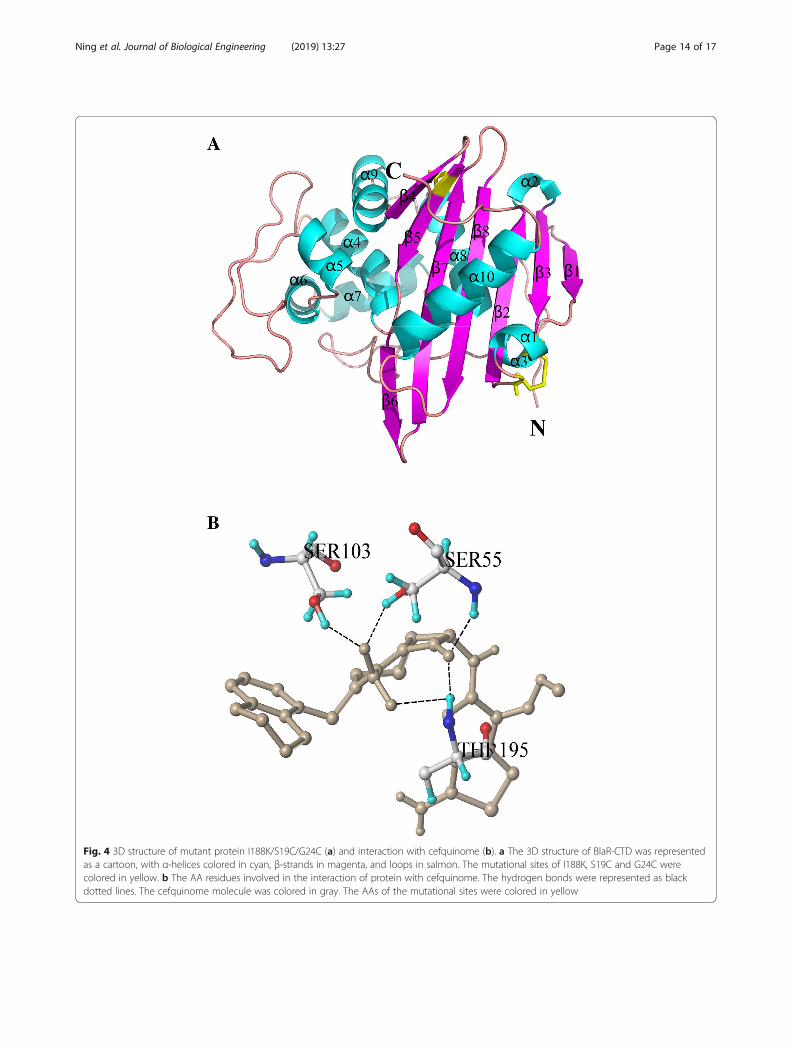
Fig. 4 3D structure of mutant protein I188K/S19C/G24C (a) and interaction with cefquinome (b). a The 3D structure of BlaR-CTD was representedas a cartoon, with α-helices colored in cyan, β-strands in magenta, and loops in salmon. The mutational sites of I188K, S19C and G24C werecolored in yellow. b The AA residues involved in the interaction of protein with cefquinome. The hydrogen bonds were represented as blackdotted lines. The cefquinome molecule was colored in gray. The AAs of the mutational sites were colored in yellow
Ning et al. Journal of Biological Engineering (2019) 13:27 Page 14 of 17
effect. But the inhibition rate of other combinedmutant proteins (Q147K/S19C/G24C, S190Y/S19C/G24C, V197D/S19C/G24C) was not quite differentfrom that of single point mutation. It might be dueto the mutation of hydrophobic AAs to polar AAs,which increased the affinity of the proteins. Themutation of V197D was also from hydrophobic AAto polar AA, but Asp197 was an acid AA, and itwas easy to form a salt bridge with the alkalineresidue Arg229, which was close to it, thus makingthe active pocket smaller and reducing the proteinactivity. Compared with R50C-Q147C (Table 4), theinhibition rate of the A138E/R50C/Q147C to 21drugs significantly decreased (Table 5). It probablydue to Glu138 and Cys147 were located in the flex-ible region of Ω-loop behind α7, and the spatial lo-cations are relatively close (13.35 Å), resulting inthe change in the spatial structure of the proteinand the increase of the affinity. The inhibition ratesof combined mutations of A138E, Q147K, S190Y,and V197D with E183C-I188C to penicillins were signifi-cantly reduced, while the inhibition rates to most of thecephalosporins were significantly increased (Table 5).Cys183 was located on β4, Cys188 was located on β5, andthey were located between adjacent β sheets (the distanceis 4.29 Å), which may lead to a smaller protein activepocket. The β-lactam ring of cephalosporins is larger thanthat of penicillin [27]. When cephalosporins are enteringinto mutant proteins, the binding ability of the proteinsmay be reduced due to the small space of active pocket.The inhibitory rate of the mutant protein S190Y/E183C/I188C on the 21 drugs decreased significantly (Table 5).The spatial distances between Ser190, Glu183 and Ile188were very close. The protein structure changed corres-pondingly after the mutation, since Tyr is larger than Ser,and the affinity of the protein was improved. Moreover, itis interesting to note that Tyr190 and Cys188 are locatedin the β5, implicating that this region is important for thebinding ability of protein to drug.The total score of molecular docking which pre-
dicted by software represented the binding affinity be-tween protein and drug, and the score was expressedas binding constant, pKd [13]. A high value of totalscore indicates a good affinity of the drug with pro-tein. Among the docking results for 40 drugs, thedocking scores of I188K/S19C/G24C with 23 drugswere increased compared with those of the wild-type(Additional file 1: Table S6). In the 33 drugs involvedin the activity test, the docking scores between themutant protein and 18 drugs were increased, but themutant protein showed a higher affinity to all of the33 drugs (Table 5). Since the molecular docking resultis only a software simulation, the binding ability ofprotein to drugs should still confirmed by activity
identification. Nevertheless, our study shows that thesoftware simulation had some reference value, butthere still has some discrepancy.
Evaluation of the stability of BlaR-CTD mutant proteinsIn a comparison of wild-type protein and single pointmutation (I188K), it has been observed that disulfidebond mutation in I188K/S19C/G24C had a better stabi-lity and the activity of protein remained longer (Fig. 3),indicating that the disulfide bond played an importantrole in the stability of protein [10, 18, 22, 30]. I188K/S19C/G24C was more stable than A138E/R50C/Q147Cand S190Y/E183C/I188C at 4 °C, 25 °C and 37 °C (Fig. 3).The disulfide bond of mutant protein I188K/S19C/G24Cwas formed in a flexible region, which is between the lastAA of β2 and before the flexible region of α1 of theprotein. However, the disulfide bonds of mutant pro-teins A138E/R50C/Q147C and S190Y/E183C/I188Cformed in a more rigid region, which was behind theflexible region of α3 and α7, and between β4 and β5,respectively (Table 3). The different positions of disulfidebond may cause a different effect on the protein stability.The disulfide bonds formed in the flexible region of theprotein would prevent the production of extra tension andwas beneficial to the stability of the protein [2].Moreover, when a single point mutation was combined
with the introduction of disulfide bonds, it produced thesuperposition effect in the enhancement of protein affin-ity (Tables 4 and 5) and stability (Fig. 3). In mutant pro-tein I188K/S19C/G24C, the mutational site I188K waslocated on the fragment of β5, which was close to theactive pocket of BlaR-CTD protein. It might affect theaffinity of protein, made the mutation bind to thevarious β-lactams and enhances the binding affinity ofβ-lactams. S19C/G24C, located between α1 helix andthe flexible region in front of the β2 fragment, were faraway from the active site of BlaR-CTD protein andwould not change the structure of the active pocket,thus not affect the affinity of the protein.
ConclusionsTo our best knowledge, our study is a new attempt on thestructural modification of protein which may be used inthe receptor assay for detecting β-lactam antibioticsresidues. In this study, the structure of BlaR-CTD proteinfrom Bacillus licheniformis ATCC14580 was modified bysite-directed mutagenesis based on the combination useof computer simulations, including homologous modeling,molecular docking, disulfide bond inserting and saltbridge building as well as prediction and evaluation ofmutation site by software. Mutant protein I188K/S19C/G24C was screened out from 23 mutant proteins for thebest stability and higher affinity to 33 β-lactams. As
Ning et al. Journal of Biological Engineering (2019) 13:27 Page 15 of 17
expected, the study results of I188K/S19C/G24C showedthat single point mutation combined with disulfide bondinsertions could simultaneously increase the recognitionability and stability of the protein. In subsequent studies,mutant proteins I188K/S19C/G24C can be applied toestablish a fast and effective receptor method for thescreening of β-lactam antibiotic residues.
Additional file
Additional file 1: Table S1. Amino acid residues within 5 Å in theactive pocket. Table S2. The mutational sites with score of 1.0 predictedby SIFT. Table S3. Scoring results by PolyPhen software. Table S4.Determination of free sulfhydryl group in mutant protein. Table S5.Acitivity identification of BlaR-CTD wild-type and mutant protein usingHRP-AMP. Table S6. Binding sites of I188K/S19C/G24C to β-lactam antibi-otics. Figure S1. Expression of BlaR-CTD protein. Figure S2. Standardcurve of cysteine based on DTNB. Figure S3. The DNA and amino acidsequences of recombinant wildtype BlaR-CTD protein. (DOCX 120 kb)
Abbreviations3D: Three-dimensional; AA: Amino acid; BlaR-CTD: The C-terminal domain ofBlaR; BSA: Bovine serum albumin; HRP-AMP: Horseradish peroxidase-labeledampicillin; IPTG: Isopropy-β-d-thiogalactoside; LB: Luria–Bertani; PBP: Penicillinbinding protein; PBS: Phosphate-buffered saline; TMB: 3,3′,5,5′-tetramethylbenzidine
AcknowledgmentsThe authors thank the National Key R&D Program of China for funding thisresearch.
FundingThis work was supported by the National Key R&D Program of China(2018YFD0500300 and 2017YFC1600100), the Natural Science Foundation ofHubei Province (2017CFB445), the National Program for Risk Assessment ofQuality and Safety of Livestock and Poultry Products (GJFP2019007) andNational Program for Risk Assessment of Quality and Safety of Milk(GJFP201800804).
Availability of data and materialsAll data generated or analyzed during this study are included in thispublished article.
Authors’ contributionsZHY and GYC conceived the project, obtained grant support, designedresearch, and analyzed and interpreted data. JNN, SA and TC performed theexperiments, analysis, interpretation, and statistic. JNN and SA wrote thepaper and ZHY, GYC, YLW and DPP revised the manuscript. All authorsedited and approved the manuscript.
Ethics approval and consent to participateNot applicable.
Consent for publicationThis is an open-access article distributed under the terms of the CreativeCommons Attribution Non-Commercial 4.0 International License (http://crea-tivecommons.org/licenses/by-nc/4.0) which permits copy and redistributethe material just in non-commercial usages, provided the original work isproperly cited.
Competing interestsThe authors declare that they have no competing interests.
Publisher’s NoteSpringer Nature remains neutral with regard to jurisdictional claims inpublished maps and institutional affiliations.
Received: 22 October 2018 Accepted: 14 March 2019
References1. Adzhubei IA, Schmidt S, Peshkin L, Ramensky VE, Gerasimova A, Bork P,
Kondrashov AS, Sunyaev SR. A method and server for predicting damagingmissense mutations. Nat Methods. 2010;7(4):248.
2. Bhattacharyya R, Pal D, Chakrabarti P. Disulfide bonds, their stereospecificenvironment and conservation in protein structures. Protein Eng Des Sel.2004;17(11):795–808.
3. Bradford MM. A rapid and sensitive method for the quantitation ofmicrogram quantities of protein utilizing the principle of protein-dyebinding. Anal Biochem. 1976;72(1):248–54.
4. Contreras-Martel C, Dahout-Gonzalez CA, Martins Ados S, Kotnik M, DessenA. PBP active site flexibility as the key mechanism for beta-lactam resistancein pneumococci. J Mol Biol. 2009;387(4):899–909.
5. Coquelle N, Fioravanti E, Weik M, Vellieux F, Madern D. Activity, stability andstructural studies of lactate dehydrogenases adapted to extreme thermalenvironments. J Mol Biol. 2007;374(2):547–62.
6. Craig DB, Dombkowski AA. Disulfide by design 2.0: a web-based tool fordisulfide engineering in proteins. BMC Bioinformatics. 2013;14(1):346.
7. Degelaen J, Granier B, Frere JM, Joris B. Process for determining antibioticscontaining a β-lactam ring in a biological fluid. US, Patent; 2003.
8. Duval V, Swinnen M, Lepage S, Brans A, Granier B, Franssen C, Frere JM,Joris B. The kinetic properties of the carboxy terminal domain of the Bacilluslicheniformis 749/I BlaR penicillin-receptor shed a new light on thederepression of beta-lactamase synthesis. Mol Microbiol. 2003;48(6):1553–64.
9. Gao C, Lan D, Liu L, Zhang H, Yang B, Wang Y. Site-directed mutagenesisstudies of the aromatic residues at the active site of a lipase fromMalassezia globosa. Biochimie. 2014;102(7):29–36.
10. Gasymov OK, Abduragimov AR, Glasgow BJ. Restoration of structuralstability and ligand binding after removal of the conserved disulfide bondin tear lipocalin. Biochem Biophys Res Commun. 2014;452(4):1004–8.
11. Ge L, Li D, Wu T, Zhao L, Ding G, Wang Z, Xiao W. B-factor-saturationmutagenesis as a strategy to increase the thermostability of α-L-rhamnosidase from Aspergillus terreus. J Biotechnol. 2018;275:17–23.
12. Golemi-Kotra D, Cha JY, Meroueh SO, Vakulenko SB, Mobashery S.Resistance to beta-lactam antibiotics and its mediation by the sensordomain of the transmembrane BlaR signaling pathway in Staphylococcusaureus. J Biol Chem. 2003;278(20):18419–25.
13. Gupta SD, Bommaka MK, Mazaira GI, Galigniana MD, Subrahmanyam CVS,Gowrishankar NL, Raghavendra NM. Molecular docking study, synthesis andbiological evaluation of Mannich bases as Hsp90 inhibitors. Int J BiolMacromol. 2015;80:253–9.
14. Hsieh PC, Vaisvila R. Protein engineering: single or multiple site-directedmutagenesis. Methods Mol Biol. 2013;978:173.
15. Huang JW, Chen CC, Huang CH, Huang TY, Wu TH, Cheng YS, Ko TP, Lin CY,Liu JR, Guo RT. Improving the specific activity of β-mannanase fromAspergillus Niger BK01 by structure-based rational design. Biochim BiophysActa. 2014;1844(3):663.
16. Jensen K, Osmani SA, Hamann T, Naur P, Møller BL. Homology modeling ofthe three membrane proteins of the dhurrin metabolon: catalytic sites,membrane surface association and protein-protein interactions.Phytochemistry. 2011;72(17):2113–23.
17. Joris B, Ledent P, Kobayashi T, Lampen JO, Ghuysen JM. Expression inescherichia coli of the carboxy terminal domain of the blar sensory-transducer protein of bacillus licheniformis as a water-soluble mr 26 000penicillin-binding protein. FEMS Microbiol Lett. 1990;70(1):107–13.
18. Kawade R, Akiba H, Entzminger K, Maruyama T, Okumura CJ, Tsumoto K.Roles of the disulfide bond between the variable and the constant domainsof rabbit immunoglobulin kappa chains in thermal stability and affinity.Protein Eng Des Sel. 2018;31:243–7.
19. Kerff F, Charlier P, Colombo ML, Sauvage E, Brans A, Frere JM, Joris B, FonzeE. Crystal structure of the sensor domain of the BlaR penicillin receptor fromBacillus licheniformis. Biochemistry. 2003;42(44):12835–43.
20. Makhatadze GI, Loladze VV, Ermolenko DN, Chen X, Thomas ST.Contribution of surface salt bridges to protein stability: guidelines forprotein engineering. J Mol Biol. 2003;327(5):1135–48.
21. Ng PC, Henikoff S. SIFT: predicting amino acid changes that affect proteinfunction. Nucleic Acids Res. 2003;31(13):3812–4.
Ning et al. Journal of Biological Engineering (2019) 13:27 Page 16 of 17
22. Ning L, Guo J, Jin N, Liu H, Yao X. The role of Cys179-Cys214 disulfide bondin the stability and folding of prion protein: insights from moleculardynamics simulations. J Mol Model. 2014;20(2):2106.
23. Nissen PH, Christensen SE, Ladefoged SA, Brixen K, Heickendorff L,Mosekilde L. Identification of rare and frequent variants of the CASR geneby high-resolution melting. Clin Chim Acta. 2012;413(5):605–11.
24. Parthasarathy S, Murthy MR. Protein thermal stability: insights from atomicdisplacement parameters (B values). Protein Eng. 2000;13(1):9–13.
25. Peng J, Cheng G, Huang L, Wang Y, Hao H, Peng D, Liu Z, Yuan Z.Development of a direct ELISA based on carboxy-terminal of penicillin-binding protein BlaR for the detection of beta-lactam antibiotics in foods.Anal Bioanal Chem. 2013;405(27):8925–33.
26. Powell AJ, Tomberg J, Deacon AM, Nicholas RA, Davies C. Crystal structuresof penicillin-binding protein 2 from penicillin-susceptible and -resistantstrains of Neisseria gonorrhoeae reveal an unexpectedly subtle mechanismfor antibiotic resistance. J Biol Chem. 2009;284(2):1202–12.
27. Tahlan K, Jensen SE. Origins of the β-lactam rings in natural products. JAntibiot. 2013;66:401.
28. Wang J, Papke RL, Stokes C, Horenstein NA. Potential state-selectivehydrogen bond formation can modulate activation anddesensitization of the alpha7 nicotinic acetylcholine receptor. J BiolChem. 2012;287(26):21957–69.
29. Wei H, Hu J, Wang L, Xu F, Wang S. Rapid gene splicing and multi-sitedmutagenesis by one-step overlap extension polymerase chain reaction. AnalBiochem. 2012;429(1):76–8.
30. Zabetakis D, Olson MA, Anderson GP, Legler PM, Goldman ER. Evaluation ofdisulfide bond position to enhance the thermal stability of a highly stablesingle domain antibody. PLoS One. 2014;9(12):e115405.
31. Zapun A, Contrerasmartel C, Vernet T. Penicillin-binding proteins and beta-lactam resistance. FEMS Microbiol Rev. 2008;32(2):361–85.
32. Zhang J, Wang Z, Wen K, Liang X, Shen J. Penicillin-binding protein 3 ofStreptococcus pneumoniae and its application in screening of beta-lactamsin milk. Anal Biochem. 2013;442(2):158–65.
33. Zhou C, Liu L, Zhuang J, Wei J, Zhang T, Gao C, Liu C, Li H, Si H, Sun C. Asystems biology-based approach to uncovering molecular mechanismsunderlying effects of traditional Chinese medicine Qingdai in chronicmyelogenous leukemia, involving integration of network pharmacology andmolecular docking technology. Med Sci Monit. 2018;24:4305–16.
Ning et al. Journal of Biological Engineering (2019) 13:27 Page 17 of 17