![γ λ ω σσά ρι - eclass.unipi.gr¬κρο... · βιώσιμη ανάπτυξη [sustainable develop-ment]: ανάπτυξη που στηρίζεται σε αρ-χές βιωσιμότητας·](https://static.fdocument.org/doc/165x107/5f8931a73a61df27043aeb63/-ff-f-sustainable.jpg)
7aa-Methyl-19-Nortestosterone (MENT):...
85
Steroid Research Laboratory Department of Medical Chemistry Institute of Biomedicine University of Helsinki 7 a -Methyl-19-Nortestosterone (MENT): Pharmacokinetics and Antigonadotropic Effects in Men Janne Suvisaari Academic Dissertation To be presented, with permission of the Medical Faculty of The University of Helsinki, for public examination in The Big Auditorium of the Department of Medical Chemistry, Siltavuorenpenger 10, Helsinki, on December 16, 2000 at 12:00. Helsinki 2000
Transcript of 7aa-Methyl-19-Nortestosterone (MENT):...
Steroid Research Laboratory
Department of Medical Chemistry
Institute of Biomedicine
University of Helsinki
7αα-Methyl-19-Nortestosterone (MENT): Pharmacokinetics and Antigonadotropic Effects in Men
Janne Suvisaari
Academic Dissertation
To be presented, with permission of the Medical Faculty of The
University of Helsinki, for public examination in The Big
Auditorium of the Department of Medical Chemistry,
S i l tavuorenpenger 10 , Hels ink i , on December 16 , 2000 a t 12 :00 .
Helsinki 2000

Supervised by:
Docent Pekka Lähteenmäki, M.D., Ph.D., University of Helsinki, Finland
Reviewed by:
Docent Leo Dunkel, M.D., Ph.D., University of Helsinki, Finland
Docent Pirkko Härkönen, M.D., Ph.D., University of Turku, Finland
Opponent:
Docent Jorma Toppari, M.D., Ph.D., University of Turku, Finland
Portable Document Format (PDF) version
ISBN 952-91-2950-5
Helsinki 2000

3
Contents
List of Original Publications .................................................................................. 7
Abbreviations ......................................................................................................... 8
Abstract .................................................................................................................. 9
Introduction .......................................................................................................... 11
Review of the Literature....................................................................................... 13
Overview of Androgen Physiology ............................................................. 13
Synthesis and Secretion of Androgens ............................................... 13
Effects of Androgens .......................................................................... 14
Effects of Ageing in Testosterone Concentrations ............................. 17
Indications for Androgen Administration ................................................... 17
Beneficial and Adverse Effects of Androgen Administration..................... 21
Beneficial Effects of Androgen Administration................................. 21
Adverse Effects of Androgen Administration .................................... 22
Androgen Administration Methods............................................................. 25
Injectable Androgen Preparations....................................................... 26
Transdermal Androgen Preparations .................................................. 28
Oral Androgen Preparations ............................................................... 29
Androgen Implants ............................................................................. 30
4
Hormonal Male Contraceptives................................................................... 30
7α-Methyl-19-nortestosterone (MENT)...................................................... 35
Aims of the Study................................................................................................. 40
Materials, Methods and Subjects ......................................................................... 41
Trial Sites..................................................................................................... 41
Design of the Trials ..................................................................................... 42
Single Intravenous Injection Trial (Reported in Publication II)......... 42
Single Intramuscular Injection Trial (Reported in Publication I) ....... 42
Six Intramuscular Injections Trial (Reported in Publication I) .......... 42
MENT Implant Trial (Reported in Publications III and IV)............... 42
Subjects........................................................................................................ 43
Injection Preparations and Implants ............................................................ 45
Sample Handling ......................................................................................... 46
Assays.......................................................................................................... 46
Pharmacokinetic Methods ........................................................................... 48
Single Intravenous Injection Trial ...................................................... 48
Single Intramuscular Injection Trial ................................................... 49
Six Intramuscular Injections Trial ...................................................... 49
MENT Implant Trial........................................................................... 50
Statistical Methods ...................................................................................... 50

5
Ethical Issues ............................................................................................... 51
Results .................................................................................................................. 51
Pharmacokinetics of MENT........................................................................ 51
Single Intravenous Injection Trial ...................................................... 51
Single Intramuscular Injection Trial ................................................... 53
MENT Implant Trial........................................................................... 54
Release Rate of MENT Ac Implants .................................................. 56
Effects of MENT on Gonadotropin and Testosterone Concentrations ....... 56
Six Intramuscular Injections Trial ...................................................... 57
MENT Implant Trial........................................................................... 60
Effects of MENT on Other Analytes........................................................... 63
SHBG.................................................................................................. 63
Dihydrotestosterone and Insulin-Like Growth Factor-1..................... 64
Prostate-Specific Antigen ................................................................... 64
Other Clinical Chemistry Variables.................................................... 64
Other Adverse Effects ................................................................................. 65
Discussion ............................................................................................................ 65
The Androgenicity of MENT in Men.......................................................... 65
Pharmacokinetics of MENT........................................................................ 66
MENT Implants........................................................................................... 67

6
Lack of 5α-Reduction ................................................................................. 69
Hormonal Male Contraception .................................................................... 69
Limitations of This Study............................................................................ 71
Conclusions .......................................................................................................... 72
Acknowledgments................................................................................................ 73
References ............................................................................................................ 76

7
List of Original Publications
This thesis is based on the following original publications which are
referred to in the text by the Roman numbers I to IV:
I Janne Suvisaari, Kalyan Sundaram, Gabriela Noé, Narender Kumar, Claude Aguillaume, Yun-Yen Tsong, Pekka Lähteenmäki, C. Wayne Bardin. Pharmacokinetics and pharmacodynamics of 7α-methyl-19-nortestosterone after intramuscular administration in healthy men. Human Reproduction 1997; 12(5):967-973.
II Narender Kumar, Janne Suvisaari, Yun-Yen Tsong, Claude Aguillaume, C. Wayne Bardin, Pekka Lähteenmäki, Kalyan Sundaram. Pharmacokinetics of 7α-Methyl-19-nortestosterone in Men and Cynomolgus Monkeys. Journal of Andrology 1997; 18(4):352-358.
III Gabriela Noé, Janne Suvisaari, Cameron Martin, Alfred Moo-Young, Kalyan Sundaram, Saleh I. Saleh, Eliana Quintero, Horacio B. Croxatto, Pekka Lähteenmäki. Gonadotrophin and testosterone suppression by 7α-methyl-19-nortestosterone acetate administered by subdermal implant to healthy men. Human Reproduction 1999; 14(9):2200-2206.
IV Janne Suvisaari, Alfred Moo-Young, Auni Juhakoski, Kaisa Elomaa, Saleh I. Saleh, Pekka Lähteenmäki. Pharmacokinetics of 7α-Methyl-19-nortestosterone (MENT) Delivery Using Subdermal Implants in Healthy Men. Contraception 1999; 60(5):299-303.
8
Abbreviations
BMI body mass index; weight / height2 (kg/m2)
°C degrees Celsius
FSH follicle-stimulating hormone
GnRH gonadotropin-releasing hormone
i.m. intramuscular
i.v. intravenous
kg kilogram
l liter
LH luteinizing hormone
m2 square meter
MENT 7α-methyl-19-nortestosterone
MENT Ac 7α-methyl-19-nortestosterone acetate
mg milligram
µg microgram
min minute
ml milliliter
mol mole
nmol nanomole
SHBG sex hormone-binding globulin
U international units for gonadotropin concentrations; same as IU
vs. versus
9
Abstract
With the ultimate aim of developing a new hormonal male contraceptive
and a new androgen for replacement therapy, we studied the pharmaco-
kinetics and endocrine effects of 7α-methyl-19-nortestosterone (MENT).
The results of studies carried out in vitro and in animals indicated that
MENT is an aromatizable androgen with progestational effects. It does not
seem to be toxic, its effect on muscle and its feedback effect on the pituitary
are at least 10 times those of testosterone, and it is not activated by 5α-
reduction, its effect on the prostate thus being only 4 times that of
testosterone. Hence, we assumed that a dose of MENT adequate for
androgen substitution and gonadotropin suppression to a level leading to
azoospermia would not overstimulate the prostate. Furthermore, subdermal
implants releasing MENT acetate (MENT Ac), a pro-drug rapidly converted
to MENT in vivo, could be made. We studied the concentrations of MENT,
gonadotropins, and testosterone in the sera of healthy men who had received
single intravenous or intramuscular MENT injections, a series of six daily
intramuscular MENT injections, or who had had MENT Ac implants in-
serted for 4 weeks. We found that MENT has a terminal half-life of about
40 min, a total apparent volume of distribution of about 70 l, and a clear-
ance rate of about 2000 l/day (27 l/kg/day). Intramuscular MENT injections
in doses ranging from 1.0 to 4.0 mg as well as 1, 2, or 4 subdermal implants
releasing MENT acetate at a rate of approximately 0.3 to 1.2 mg per day
caused dose-dependent suppression of gonadotropin concentrations.
Luteinizing hormone (LH) and follicle-stimulating hormone (FSH) concen-
trations in all men with 4 implants decreased to or below 0.6 U/l. These
concentrations are similar to those seen in men rendered azoospermic by
testosterone ester injections in male contraception trials, but due to the short
duration of our studies we could not investigate the potential of MENT
10
administration to cause azoospermia. We conclude that MENT is a very
promising candidate for hormonal male contraception and long-term
androgen replacement therapy.
11
Introduction
Androgen-based hormonal male contraception may become available and
androgen replacement therapy in the elderly may become widespread in the
not so distant future. Whether this will happen or not depends largely on the
availability of suitable androgen administration methods. The development
of transdermal testosterone preparations was an important advance in
androgen replacement therapy but even more convenient administration
methods are needed. In contrast to current androgen users, most of whom
suffer from serious diseases and are thus ready to accept whatever treatment
there is, most of the new potential androgen users are only considering
hormonal male contraception or androgen replacement therapy as an option
and will probably not accept the inconveniences of traditional androgen
administration methods. More user-friendly androgen administration
methods are clearly needed. Furthermore, only minor adverse effects can be
considered acceptable if androgens are to be used in healthy men. (Hayes,
2000)
Hormonal male contraception based on androgen administration has been
shown to be feasible ([Anonymous], 1990). It is not yet available, and one of
the reasons for this situation is the inconvenience of the available methods
of androgen administration. More convenient and safer androgen admini-
stration methods will have to be developed before hormonal male
contraception becomes a viable option (Amory & Bremner, 2000). Hence,
much of the development of new androgen administration methods is taking
place as part of the development of hormonal male contraception. Other
androgen users would, of course, also benefit from the new androgen
administration methods developed to satisfy the demands of contraception.
12
The risks and benefits of long-term androgen administration have not been
studied extensively enough (Bardin et al, 1991). With widespread androgen
use, adverse effects of androgens could become important public health
problems. Hence, the development of more convenient androgen admini-
stration methods may not be enough; it may also be necessary to find
androgens that are safer than testosterone in long-term use, and it would be
even better if androgens with non-contraceptive health benefits could be
found.
The synthetic androgenic anabolic steroid 7α-methyl-19-nortestosterone
(MENT) has properties that could make it a more convenient and safer
alternative to all testosterone-based androgen formulations and an excep-
tionally promising candidate for male hormonal contraception. Previous
experiments carried out in vitro and in animals have revealed that MENT
does not undergo 5α-reduction and therefore it has a relatively low potency
in tissues such as the prostate where the effect of testosterone is amplified
by 5α-reductase enzymes. Hence, it is assumed that MENT is less likely
than testosterone to over-stimulate the prostate. MENT is also several times
more potent than testosterone. This makes it much more suitable for admini-
stration via subdermal implants, a convenient long-term administration
method. It has also been shown that MENT is aromatized to an estrogenic
compound, a metabolic step that is required for many important effects of
androgens. (Agarwal & Monder, 1988; Kumar et al, 1992; Sundaram et al,
1993; LaMorte et al, 1994; Kumar et al, 1999)
Despite its potential advantages, neither the pharmacokinetic properties nor
the effects of MENT in men had been studied previously. Therefore, we in-
vestigated the basic pharmacokinetic properties of MENT administered by
intravenous and intramuscular injections as well as subdermal implants, and
the effects of MENT on gonadotropin and testosterone concentrations.
13
Review of the Literature
Overview of Androgen Physiology
Synthesis and Secretion of Androgens
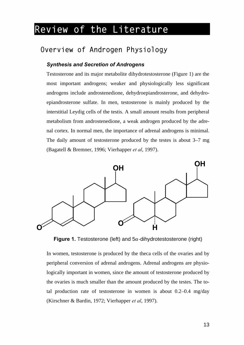
Testosterone and its major metabolite dihydrotestosterone (Figure 1) are the
most important androgens; weaker and physiologically less significant
androgens include androstenedione, dehydroepiandrosterone, and dehydro-
epiandrosterone sulfate. In men, testosterone is mainly produced by the
interstitial Leydig cells of the testis. A small amount results from peripheral
metabolism from androstenedione, a weak androgen produced by the adre-
nal cortex. In normal men, the importance of adrenal androgens is minimal.
The daily amount of testosterone produced by the testes is about 3–7 mg
(Bagatell & Bremner, 1996; Vierhapper et al, 1997).
Figure 1. Testosterone (left) and 5α-dihydrotestosterone (right)
In women, testosterone is produced by the theca cells of the ovaries and by
peripheral conversion of adrenal androgens. Adrenal androgens are physio-
logically important in women, since the amount of testosterone produced by
the ovaries is much smaller than the amount produced by the testes. The to-
tal production rate of testosterone in women is about 0.2–0.4 mg/day
(Kirschner & Bardin, 1972; Vierhapper et al, 1997).
OH
O H
OH
O
14
Testosterone secretion by Leydig cells in men and by theca cells in women
is stimulated by LH, which is produced in the anterior part of the pituitary
gland. LH secretion is stimulated by the pulsatile secretion of GnRH from
the hypothalamus. Androgens, estrogens and progestins exert a negative
feedback effect on the secretion of GnRH and LH by their actions on the
pituitary and the hypothalamus. Most of the negative feedback effect of
androgens is caused by their estrogenic metabolites produced by aromatiza-
tion. 5α-Reduction does not seem to be necessary for the negative feedback
effect of testosterone. (Rittmaster et al, 1992; Kumar et al, 1995a; Hayes et
al, 2000)
Testosterone secretion is pulsatile as a result of the pulsatile secretion of
LH, which results from the pulsatile secretion of GnRH. There is also a
clear circadian variation in testosterone concentrations, which are highest in
the morning. There are probably also seasonal, or circannual variations in
testosterone concentrations. (Veldhuis et al, 1987; Dabbs, 1990; Foresta et
al, 1997)
Effects of Androgens
Testosterone exerts its actions directly through the activation of androgen
receptors, indirectly through its reduction to 5α-dihydrotestosterone which
also acts on androgen receptors, or indirectly through its aromatization to
estradiol and the activation of estrogen receptors. Androgen and estrogen
receptors are ligand-inducible transcription factors. Androgen molecules
enter the nucleus of a target cell and associate with unbound androgen
receptors. Binding of androgen induces a conformational change in the
receptor molecule that leads to its phosphorylation and its dissociation from
so-called heat shock proteins. Next, the receptor dimerizes with another
androgen receptor that is in the same state. The dimer binds to an androgen
response element on an androgen-regulated gene and increases its
15
transcription rate. Some of the effects of androgens and estrogens, called
nongenomic steroid effects, seem to be mediated by different receptors on
the cell surface and signal transduction mechanisms similar to those
involved in the action of peptide hormones. (Revelli et al, 1998; McKenna
et al, 1999; Prins, 2000)
In tissues such as the prostate, seminal vesicles, epididymis, and certain
parts of the skin, testosterone is converted by 5α-reductase enzymes into a
more potent androgen, 5α-dihydrotestosterone. The affinity of 5α-dihydro-
testosterone for the androgen receptor is about five times that of
testosterone (Wilbert et al, 1983). (Russell & Wilson, 1994)
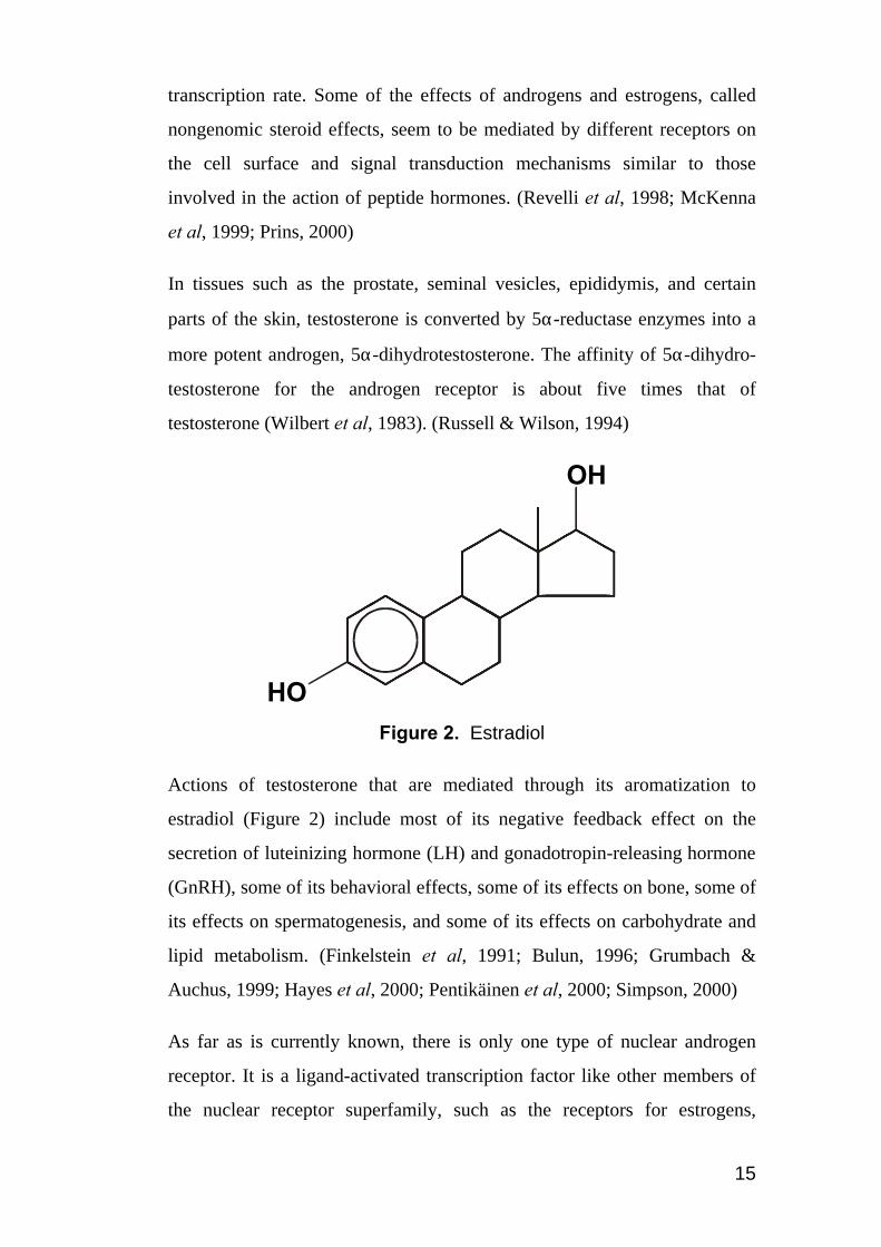
Figure 2. Estradiol
Actions of testosterone that are mediated through its aromatization to
estradiol (Figure 2) include most of its negative feedback effect on the
secretion of luteinizing hormone (LH) and gonadotropin-releasing hormone
(GnRH), some of its behavioral effects, some of its effects on bone, some of
its effects on spermatogenesis, and some of its effects on carbohydrate and
lipid metabolism. (Finkelstein et al, 1991; Bulun, 1996; Grumbach &
Auchus, 1999; Hayes et al, 2000; Pentikäinen et al, 2000; Simpson, 2000)
As far as is currently known, there is only one type of nuclear androgen
receptor. It is a ligand-activated transcription factor like other members of
the nuclear receptor superfamily, such as the receptors for estrogens,
OH
HO
16
progestins, glucocorticoids, mineralocorticoids, thyroid hormones, and
vitamins A and D3. The nuclear receptor superfamily also includes a large
group of proteins referred to as orphan receptors, for which specific ligands
are unknown. There are two types of estrogen receptor, termed alpha and
beta, having clearly different structures, tissue distributions, and effects.
(Gustafsson, 1999; Enmark & Gustafsson, 1999)
The physiological actions of androgens in males include the development
and maintenance of the structure and functions of the reproductive system,
including spermatogenesis, and the development and maintenance of
secondary sexual characteristics. Male secondary sexual characteristics
include a higher bone density and muscle mass, a larger larynx and deeper
voice, and a higher hematocrit than in females, as well as male patterns of
hair growth, male sexual orientation, patterns of behavior, libido and
potency. (Bagatell & Bremner, 1996)
Spermatogenesis is not regulated directly by testosterone or gonadotropins,
but by paracrine substances secreted by the Sertoli cells of the seminiferous
tubules. The function of the Sertoli cells is hormonally regulated by testos-
terone and follicle-stimulating hormone. Recent findings suggest that the
effect of testosterone on spermatogenesis might also be mediated through its
aromatization to estradiol, and estradiol receptors in developing germ cells
(Pentikäinen et al, 2000). An extremely high local testosterone
concentration in the testes, practically achievable only by local testosterone
production by the Leydig cells, is necessary for spermatogenesis. Follicle-
stimulating hormone (FSH) seems to be necessary for quantitatively and
qualitatively normal spermatogenesis and normal fertility, but sperm
production and even fertility appears to be possible in the absence of the
action of FSH. For example, two out of five men with an inactivating FSH
receptor mutation (566CàT, Ala189Val) had fathered children, and none of
17
the five was azoospermic. (Swerdloff et al, 1992; Cummings & Bremner,
1994; Tapanainen et al, 1997; Themmen & Huhtaniemi, 2000)
Effects of Ageing in Testosterone Concentrations
Serum concentrations of testosterone, free testosterone, and non-SHBG-
bound testosterone all decrease with normal ageing in men. Since SHBG
(sex hormone-binding globulin) concentrations tend to increase with age,
non-SHBG-bound testosterone levels tend to decrease more than those of
total testosterone. (Tenover, 1998; Perry, 1999) In women also, testosterone
concentrations decline with increasing age. The decline begins in the
reproductive years and continues after the menopause. (Davis & Burger,
1998)
Indications for Androgen Administration
The most obvious medical indication for androgen administration is male
hypogonadism. Men with hypogonadism are unable to synthesize adequate
quantities of androgens and need long-term androgen replacement to
maintain sexual behavior, androgen-dependent physiological processes,
secondary sexual characteristics, and mental health. The causes of
hypogonadism can be divided into two main groups, primary or
hypergonadotropic hypogonadism, and secondary or hypogonadotropic
hypogonadism. In primary hypogonadism the defect is either in the gonads
or post-gonadal, for example in the conversion of testosterone to 5α-
dihydrotestosterone or at the level of the androgen receptor. In secondary
hypogonadism, the defect is a lack of gonadotropins. The causes of primary
hypogonadism include gonadal defects caused by genetic diseases such as
Klinefelter’s syndrome, anatomical defects, and lesions caused by
infections, toxins or radiation, as well as androgen or LH insensitivity
18
syndromes. The causes of secondary hypogonadism include pituitary and
hypothalamic diseases including panhypopituitarism, hyperprolactinemia,
isolated gonadotropin deficiencies, and various genetic disorders as well as
systemic causes such as chronic disease, starvation, severe obesity, and the
adverse effects of certain drugs such as glucocorticoids. Delayed puberty
can be considered a constitutional cause of secondary hypogonadism.
(Bagatell & Bremner, 1996)
The traditional treatment of choice for male hypogonadism is a testosterone
ester such as testosterone enanthate given intramuscularly (100–300 mg
every 10 to 21 days). Smaller doses are used in adolescent boys with
hypogonadism or boys with a constitutional delay of puberty. Although
constitutional delay of puberty is a variant of normal pubertal maturation,
low-dose androgen replacement therapy is recommended because it results
in psychological and physiological benefits. (Bhasin, 1992; Bagatell &
Bremner, 1996; Houchin & Rogol, 1998)
Androgen replacement therapy is potentially useful in a variety of disorders
where hypogonadism is a consequence of a severe systemic disease such as
chronic renal failure (Handelsman & Liu, 1998) and human immuno-
deficiency virus infection. Hypogonadism occurs in approximately 30% of
men infected with human immunodeficiency virus. Several clinical trials on
the effects of androgen supplementation in men infected with human
immunodeficiency virus have been conducted, and the results are
encouraging. (Dobs, 1998; Bhasin & Javanbakht, 1999).
Androgens may also be useful in the treatment of eugonadal patients with
secondary wasting syndromes. Androgens have been shown to be beneficial
in several groups of chronically ill patients who have lost body mass (Dobs,
1999).
19
The results of several studies indicate that androgen replacement therapy in
older men improves muscle strength and bone mass, decreases fat mass, and
raises the serum hemoglobin concentration. (Perry, 1999) The increase in
hemoglobin and hematocrit may even be excessive (Sih et al, 1997). How-
ever, available data are still insufficient to permit any major conclusions
about the role of androgen replacement in the treatment of age-related
physiological changes (Hayes, 2000).
Androgens may be beneficial in the treatment of skeletal muscle
dysfunction in chronic obstructive pulmonary disease, although very little
experimental evidence exists on this subject. If androgens are administered
to patients with chronic obstructive pulmonary disease, the possibilities that
androgens may increase hematocrit or worsen sleep apnea are of special
concern. (Casaburi, 1998)
Androgens have traditionally been used in the treatment of some forms of
anemia and other hematologic disorders, such as aplastic anemia, Fanconi's
anemia, anemia of chronic renal failure, paroxysmal nocturnal
hemoglobinuria, idiopathic autoimmune hemolytic anemia, agnogenic
myeloid metaplasia, and idiopathic thrombocytopenic purpura. Androgens
have consistent albeit modest efficacy in the treatment of some of these
conditions, such as anemia of chronic renal failure. Androgens are still in
limited use in the treatment of hematologic disorders, but they have been
largely replaced by newer options such as recombinant erythropoietin and
other hematopoietic growth factors, bone-marrow transplantation, anti-
thymocyte globulin, and immunosuppressive drugs such as cyclosporine A.
(Besa, 1994; Handelsman & Liu, 1998)
Hereditary angioedema, a disease caused by deficiency of C1-inhibitor, can
be successfully treated with some synthetic derivatives of testosterone but
not with testosterone itself; it seems that a drug's efficacy in preventing

20
hereditary angioedema attacks is connected to its 17α-alkylation. (Bagatell
& Bremner, 1996)
In women, androgen replacement has been used as a component of hormone
replacement therapy for the restoration of libido and general well-being in
symptomatic testosterone deficiency states following premature ovarian
failure, menopause or iatrogenic ovarian failure. Potential indications also
include the treatment of premenstrual syndrome, the prevention of
glucocorticoid-induced and premenopausal bone loss, loss of libido in
premenopausal women, and the management of wasting syndromes
secondary to human immunodeficiency virus infection and malignancies.
The weak androgen danazol is used in the treatment of endometriosis. As
stated previously, women are much more susceptible to the adverse effects
of androgens. Hence, high doses of androgens should be avoided altogether
in women. Postmenopausal women treated with androgens should usually
receive concurrent estrogen replacement therapy to reduce the risk of
adverse effects. (Davis & Burger, 1998; Friedrich, 2000)
Non-medical use of androgens is widespread in certain parts of the
population in many countries. Athletes have used androgens to increase
muscle mass and improve physical performance at least since the 1940s
(Bagatell & Bremner, 1996). There is a lack of direct and reliable
information on the use of androgens in sports, but, for example, the drastic
decline in Olympic results since the introduction in 1989 of out-of-
competition doping testing is indirect evidence of the widespread use of
androgens and other performance enhancing drugs among top athletes
(Franke & Berendonk, 1997). Until recently, in some countries such as the
German Democratic Republic, androgen use was customary not only among
male athletes but also among female and child athletes (Franke &
Berendonk, 1997). The rate of non-medical androgen use to improve
physical capacities or appearance is also high in some occidental countries.
21
For example, in the United States 12% of high school senior boys reported
having used androgenic anabolic steroids, although the rate in a survey of
the general population was much lower, 0.5% (0.9% for males and 0.1% for
females) (Yesalis et al, 1993).
A new form of non-medical (or more exactly, non-therapeutic) androgen
use would be the use of androgens in male contraception. Hormonal male
contraception is discussed below. It has even been proposed that androgens
could be useful during space travel (Bhasin et al, 1996) — presumably to
prevent the effects of the lack of gravitation on bone and muscle.
Beneficial and Adverse Effects of Androgen Administration
Beneficial Effects of Androgen Administration
The obviously beneficial effects of androgen administration related to the
indication for androgen administration have been discussed above.
Androgens may also have beneficial effects unrelated to the reason for their
administration. For example, long-term androgen use for male contraception
might have non-contraceptive benefits. It is important to consider such
potential benefits in addition to the potential adverse effects of androgens.
A clear increase in muscle size and strength was observed in normal men
who received 600 mg of testosterone enanthate each week for 10 weeks
(Bhasin et al, 1996). This dose is several times higher than those ordinarily
used in androgen replacement therapy (about 100 mg per week) or male
contraception studies (about 200 mg per week). Although indirect evidence
from androgen use in sports (Franke & Berendonk, 1997) also supports the
notion that exogenous androgens increase muscle mass and strength,
previous investigators could not demonstrate this effect, and it seems that a
significant response of muscle to exogenous androgen in healthy men

22
requires a high dose (Bardin, 1996). Hence, this benefit may not be present
or it may be too small to be noticed when androgens are used in androgen
replacement therapy or male contraception.
Androgens are undoubtedly beneficial in the treatment of osteoporosis in
hypogonadal men, and the results of some studies suggest that androgens
may also be beneficial in the treatment of osteoporosis in eugonadal men
(Katznelson, 1998). Hence, it is not unreasonable to assume that long-term
androgen administration for the purposes of contraception might decrease
the risk of osteoporosis later in life.
It has been postulated that a reason for the lower incidence of osteoporosis
and Alzheimer's disease in men could be the fact that men have much higher
testosterone concentrations than women and hence a better supply of
precursors for local estrogen production in estrogen-dependent tissues
(Simpson, 2000).
Adverse Effects of Androgen Administration
The adverse effects of androgen replacement therapy in men with
hypogonadism are mainly due to the androgen administration methods used;
these effects are discussed in the section on androgen administration
methods. Raising testosterone concentrations to the levels seen in normal
healthy men should not cause other effects than those of endogenous
testosterone. There is one exception, however. The effect of exogenously
administered testosterone on the testis itself is different from the effect of
testosterone produced within the testis. This is because, in contrast to
administered testosterone, intratesticular testosterone production leads to the
extremely high intratesticular testosterone concentrations (about 2 nmol/g of
testis tissue, which is approximately 100-fold compared to circulating
testosterone concentrations) (Huhtaniemi et al, 1985). A high intratesticular
testosterone concentration is necessary for spermatogenesis (Wu, 1988).
23
Hence, exogenous testosterone administration leads to infertility and
testicular atrophy, both of which seem to be reversible if testosterone
administration is discontinued (Wu et al, 1996; Rolf & Nieschlag, 1998).
Moderately supraphysiological doses of testosterone, such as those used in
male contraception trials, have caused acne, gynecomastia, increased or
decreased sexual interest, mild behavioral changes, reduction in the
circulating concentrations of high-density lipoprotein cholesterol, prostatic
enlargement, a decrease in testis size and azoospermia. (Anderson et al,
1995; Wu et al, 1996; Rolf & Nieschlag, 1998).
Because androgens control the growth and differentiation of the prostate, a
major cause of concern is the long-term effect of testosterone administration
on the prostate. A small increase in prostate size has been observed in male
contraception studies, but only future longer clinical trials will clarify the
issue of whether very long-term testosterone administration leads to
symptomatic prostatic hyperplasia or an increased risk of cancer of the
prostate. (Wallace et al, 1993; Rolf & Nieschlag, 1998)
The serious adverse hepatic effects associated with prolonged use of some
androgens, such as cholestasis, peliosis hepatis (blood-filled hepatic cysts),
hepatocellular hyperplasia, hepatic adenomas, and hepatocellular carcinoma
seem to be mostly restricted to users of 17α-alkylated derivatives of testos-
terone. (Huhtaniemi, 1994; Bagatell & Bremner, 1996; Rolf & Nieschlag,
1998; Winters, 1999)
The results of several observational studies suggest that some men develop
marked aggression, or hypomanic or manic symptoms when using high
doses of androgenic anabolic steroids (Pope & Katz, 1994). The results of a
recent randomized, placebo-controlled, double-blind clinical trial confirmed
that doses of 600 mg of testosterone cypionate per week caused a significant
increase in measures of manic and aggressive symptoms in a small subset of
24
men, while there were few changes in most. The symptoms were marked in
2, moderate in 6 and minimal in 42 men; the subgroups were otherwise
indistinguishable as regards demographic, psychological, physiological, and
laboratory measures. In the same study, 300 mg per week produced only
slight effects. (Pope et al, 2000) In contrast, no changes in mood or
behavior were observed among 43 men who received 600 mg of
testosterone enanthate weekly for ten weeks (Bhasin et al, 1996). In
conclusion, it seems that a small subset of men is prone to develop adverse
behavioral effects and mood disturbances during high-dose androgen
treatment, but that most men are not vulnerable to such effects.
The question of whether exogenous androgens, at physiological or slightly
supraphysiological doses increase or reduce the risk of cardiovascular
disease remains controversial. Although changes in lipoprotein levels that
are considered atherogenic — mainly a decrease in high-density lipoprotein
cholesterol — have been observed (Anderson et al, 1995), changes in
lipoprotein levels that may be beneficial, such as a decrease in
lipoprotein(a) have also been observed. Lipoprotein(a) concentrations are
increased in some patients with coronary artery disease and cerebrovascular
disease, but the results of prospective studies are conflicting and its role in
atherogenesis remains to be established (Jialal, 1998). The consistent
finding that low testosterone concentrations in men are associated with
common risk factors of coronary artery disease such as a pro-atherogenic
lipid profile, systolic and diastolic hypertension, hyperinsulinemia, android
obesity, and high fibrinogen levels may indirectly suggest that exogenous
testosterone would reduce the risk of cardiovascular disease in some men.
(English et al, 1997)
Overall, it seems that in men, the risks associated with androgen use have
been exaggerated. Really severe side-effects are rare, and many reports on
25
side-effects are case reports and confounding factors have often not been
clarified (Huhtaniemi, 1994; Rolf & Nieschlag, 1998).
The situation is different in women and children. In addition to the adverse
effects androgens can cause in men, women are vulnerable to effects that
are adverse in women but part of normal androgen action in men, such as
hirsutism, temporal balding, and voice deepening, and to adverse effects
that are only possible in women, such as clitorolomegaly. (Davis & Burger,
1998) The possibility of an increase in breast cancer risk is another concern
about testosterone therapy in women. The results of epidemiological studies
have shown both positive and negative associations between androgen
levels and breast cancer risk, and this issue remains unclarified (Friedrich,
2000). In pre-pubertal children of both sexes, androgens can cause
virilization and premature epiphyseal closure (Rolf & Nieschlag, 1998).
Androgen Administration Methods
The traditional androgen administration methods are intramuscular
injections of testosterone 17β-esters and oral administration of testosterone
undecanoate and 17α-alkylated derivatives of testosterone. The alkylated
derivatives are now used less frequently, since they can have serious hepatic
adverse effects. A newer method, transdermal testosterone administration, is
now also in routine use. Lately, the shortcomings of traditional androgen
administration methods have received more attention and many new
androgen administration methods are being developed. (Bhasin, 1992;
Bagatell & Bremner, 1996; McClellan & Goa, 1998; Winters, 1999; Hayes,
2000)

26
Injectable Androgen Preparations
If testosterone is given by intramuscular injection, it is rapidly absorbed
and, having a half-life of only approximately 30 minutes, rapidly degraded.
Hence, testosterone injections cannot be used as such in androgen
replacement. Testosterone esters such as testosterone enanthate (Figure 3)
are absorbed slowly when injected intramuscularly in an oily preparation.
After absorption, these esters are rapidly deesterified and free testosterone is
the active drug. Testosterone enanthate, testosterone cypionate and other
testosterone esters as well as mixtures of testosterone esters are routinely
used for androgen replacement therapy. The recommended dose for
testosterone enanthate or testosterone cypionate is 200 mg every 2 weeks, or
300 mg every 3 weeks. (Bhasin, 1992)
Figure 3. Testosterone enanthate
The drawbacks of conventional testosterone ester regimens include the
inconvenient frequency and painfulness of the intramuscular injections as
well as the wide fluctuation in testosterone concentrations during each
injection interval, concentrations being too high for a few days after the
injection and too low shortly thereafter. The unnecessarily high testosterone
concentrations early in the injection interval may increase the risk of
adverse effects. In addition, the fluctuations in testosterone concentrations
O
O
O

27
lead to fluctuations in psychological androgen effects such as mood and
sexual desire. (Bhasin, 1992; Bagatell & Bremner, 1996; Winters, 1999)
Injectable testosterone undecanoate has a longer duration of action than
testosterone enanthate. A single dose has been shown to maintain normal
serum testosterone levels in hypogonadal men for at least six weeks (Amory
& Bremner, 2000). Injectable testosterone undecanoate was shown to be
very effective in suppressing spermatogenesis in Chinese men when
administered every four weeks (Zhang et al, 1999).
Testosterone buciclate (Figure 4) is a testosterone ester with a benzol ring
incorporated into its side-chain. It has a much longer duration of action than
testosterone enanthate, presumably because of its more hydrophobic side-
chain. It has been developed to solve the problem of short injection
intervals. In hypogonadal men, a single injection of 600 mg of testosterone
buciclate increased circulating testosterone concentrations to the lower
normal range for about 3 months (Behre & Nieschlag, 1992). The potential
of testosterone buciclate in hormonal male contraception has also been
investigated and the first results have been encouraging (Behre et al, 1995).
Figure 4. Testosterone buciclate
O
O
O

28
Another experimental long-acting injectable testosterone formulation is
based on the encapsulation of testosterone in biodegradable microcapsules
made of a lactide/glycolide copolymer, a substance that is also used in
biodegradable sutures. Eugonadal testosterone concentrations could be
maintained for 10–11 weeks in hypogonadal men who received 630 mg of
testosterone in this depot injection preparation. (Bhasin et al, 1992)
Longer-acting injectable testosterone formulations suffer from a common
disadvantage: a large amount of drug needs to be administered and
consequently the injection volume is also large. For example, in the above-
mentioned testosterone microcapsule study, each subject received 5 ml
divided into two injection sites (Bhasin et al, 1992). The need to administer
a large amount of drug is a direct consequence of the potency of
testosterone, and can only be remedied by using a more potent androgen.
Transdermal Androgen Preparations
In suitable formulations, testosterone can be absorbed through the skin.
Because scrotal skin is at least 5 times more permeable to testosterone than
are other skin sites, the first available testosterone transdermal delivery
system was designed for scrotal skin. Systems for the delivery of
testosterone across nonscrotal skin are now also available. Transdermal
testosterone delivery systems provide adequate testosterone replacement for
hypogonadal men and, in addition, produce a testosterone concentration
pattern similar to the normal circadian fluctuation of testosterone
concentrations. (McClellan & Goa, 1998; Winters, 1999)
An obvious inconvenience of both kinds of testosterone patches is the need
to remove the old patch and apply a new one each night. The major
disadvantage of non-scrotal testosterone patches is that skin irritation at the
application site is frequent. Approximately 50% of men who participated in
clinical trials reported transient, mild to moderate erythema at the
29
application site sometime during therapy, and burnlike blister reactions
occurred in 12% of men during the clinical trials. (McClellan & Goa, 1998;
Winters, 1999)
Skin irritation is less frequent for scrotal patches, since these do not require
the enhancers that are required to allow permeation of testosterone through
non-genital skin. Disadvantages of the scrotal patches include the
inconvenience of having to apply the patch to a shaved scrotum, and an
unphysiologically high DHT to testosterone ratio due to the high 5α-
reductase activity in scrotal skin. Gel formulations delivering testosterone
through the skin have also been tested, and some results are encouraging. A
very promising new formulation is a hydroalcoholic gel containing 1%
testosterone. In a recent multicenter study this formulation was compared
with testosterone patches in the treatment of 227 hypogonadal men and
shown to be more or equally effective with a much lower incidence of skin
irritation (6% vs 66%) (Wang et al, 2000). (Bhasin, 1992; Behre et al, 1999;
Winters, 1999)
Oral Androgen Preparations
If administered orally, testosterone cannot reach an effective circulating
concentration because it is extensively degraded by hepatic first-pass
metabolism, as are most testosterone esters. However, there is one long
chain fatty acid testosterone ester, testosterone undecanoate, that can be
administered orally since it is absorbed into the intestinal lymphatic system
whence it passes through the thoracic duct into the systemic circulation, thus
avoiding hepatic first-pass metabolism. However, when given orally it has a
short duration of action and has to be administered thrice daily, making it
somewhat inconvenient for long-term use. It has also been tested as a
possible male contraceptive but sufficient suppression of spermatogenesis
has not been attained. (Nieschlag et al, 1978; Bhasin, 1992)
30
Testosterone from experimental sublingual and buccal formulations is
absorbed through the oral mucosa directly into the systemic circulation thus
avoiding hepatic first-pass metabolism. Sublingual and buccal testosterone
preparations share with testosterone undecanoate the disadvantage that
testosterone concentrations high enough for hormonal male contraception
are difficult to achieve. (Kim et al, 1995; Salehian et al, 1995)
The 17α-alkylated synthetic derivatives of testosterone such as methyl-
testosterone, fluoxymesterone, methandrostenolone, oxandrolone, and
stanozolol are resistant to hepatic first-pass metabolism and are therefore
orally active. However, their use has been associated with serious hepatic
adverse effects such as cholestasis, peliosis hepatis, hepatocellular
hyperplasia, hepatic adenomas, and hepatocellular carcinoma. (Bhasin,
1992; Bagatell & Bremner, 1996; Walter & Mockel, 1997; Winters, 1999)
Androgen Implants
Subdermal testosterone pellets (implants of crystalline testosterone) have
been in clinical use for a long time in some countries. The pellets are
inserted into the lower abdominal wall in a minor surgical operation under
local anesthesia. Testosterone pellets can provide serum testosterone
concentrations in the normal range for several months in hypogonadal men.
The main drawback of this method of androgen administration is that
extrusion of these pellets occurs relatively frequently. The extrusion rate
appears to be approximately once for every 10 implantation procedures.
(Handelsman et al, 1990; Kelleher et al, 1999)
Hormonal Male Contraceptives
Male hormonal contraception has been shown to be both effective and
reversible. All methods that have been found to be effective are based on
31
the same principle: suppression of LH secretion from the pituitary, which
leads to the suppression of testosterone production in the testes, and
ultimately to suppression of spermatogenesis, since spermatogenesis is de-
pendent on a very high intratesticular testosterone concentration. Recent
findings suggest that the effect of testosterone on spermatogenesis might be
mediated through its aromatization to estradiol (Pentikäinen et al, 2000), but
whether testosterone is needed as such or as a precursor for estradiol, a very
high intratesticular testosterone concentration is necessary for
spermatogenesis. The secretion of LH is stimulated by the pulsatile release
of gonadotropin-releasing hormone (GnRH) from the hypothalamus.
Androgens, estrogens and progestagens exert a negative feedback effect on
the secretion of LH by the pituitary and on the secretion of GnRH by the
hypothalamus. Thus, LH secretion can be suppressed with exogenous
androgens, progestins, estrogens, and substances that block the effect of
GnRH. The latter group includes, in addition to GnRH antagonists, GnRH
agonists which have an inhibitory effect on the pituitary when administered
in a continuous, rather than a pulsatile manner. This paradoxical effect is
explained by down-regulation of GnRH receptors in the pituitary. Secretion
of LH can also be suppressed by active immunization against GnRH
through the use of injections of GnRH combined with tetanus toxoid. (Wu,
1988; Swerdloff et al, 1992; Cummings & Bremner, 1994; Amory &
Bremner, 2000)
The hormonal contraceptive methods that inhibit LH secretion also inhibit
the secretion of the other gonadotropin, FSH. This hormone stimulates
spermatogenesis through its effects on Sertoli cells. The suppression of FSH
secretion probably contributes to the anti-fertility effects of male hormonal
contraceptives, but this effect seems to be of less importance than
suppression of LH secretion, since spermatogenesis seems to be possible
without the action of FSH. (Matsumoto & Bremner, 1989; Themmen &
Huhtaniemi, 2000)

32
When azoospermia is achieved by using substances other than androgens,
androgen replacement is necessary. Because all these methods work by
blocking testosterone production in the testes, androgen replacement is
necessary to avoid the symptoms of hypogonadism and to maintain normal
sexual function and secondary sexual characteristics. When azoospermia is
achieved by using androgens alone, two or three times more androgen is
needed than the amount used in androgen replacement therapy to sustain
androgen-dependent physiological functions when endogenous androgen
production is lacking. (Cummings & Bremner, 1997; Amory & Bremner,
1998)
To sum up, hormonal male contraception can be based on the continuous
administration of supraphysiological amounts of androgens or on the
continuous administration of physiological amounts of androgens combined
with either progestins, estrogens, GnRH antagonists or GnRH agonists.
The fact that sperm production can be hormonally suppressed has been
known for decades. A lot of small clinical trials having azoospermia as an
endpoint have been conducted, and a fairly clear picture of the effectiveness
of various methods has emerged (Amory & Bremner, 1998). In addition to
the small clinical trials having azoospermia as an endpoint, two larger
multicenter studies having pregnancy in the partner as an endpoint have also
been conducted. These studies, conducted by the World Health
Organization, showed pregnancy rates of 0.8 and 0.0 per 100 person-years
among the partners of healthy men rendered azoospermic by weekly
testosterone enanthate injections of 200 mg. (In the first study, the partners
of men rendered azoospermic were exposed to the risk of pregnancy for 124
person-years and in the second, for 230 person-years during which weekly
testosterone enanthate injections were the only form of contraception. In the
first study, there was one pregnancy, and in the second, not a single one
among the partners of azoospermic men.) The second study also
33
demonstrated that severe oligozoospermia (sperm counts below 3
million/ml) is associated with a pregnancy rate (8 per 100 person-years)
which is low when compared with those associated with currently available
reversible male contraceptive methods such as condoms, but higher than the
pregnancy rates associated with female hormonal contraceptive methods.
Azoospermia or severe oligozoospermia was achieved in 98% of men.
([Anonymous], 1990; [Anonymous], 1996)
Although only testosterone enanthate injections have been tested with
pregnancy as an endpoint, it seems reasonable to assume that azoospermia
or severe oligozoospermia produced by other hormonal methods would be
equally effective. Combinations of androgens and progestagens can even be
marginally more effective than testosterone alone in suppressing
spermatogenesis, but these regimens appear to decrease high-density
lipoprotein cholesterol concentrations more than testosterone esters alone
(Wu et al, 1999; Amory & Bremner, 2000). In small trials, the combination
of a GnRH antagonist and testosterone enanthate has also been found to be
effective, but not in all men (Tom et al, 1992; Bagatell et al, 1993; Amory
& Bremner, 2000). GnRH antagonists are administered via daily
subcutaneous injections, which is clearly not a feasible mode of
administration for a contraceptive agent. These agents occasionally cause
subcutaneous nodules at the injection site and frequently cause injection site
irritation, which is probably mediated by histamine release. (Cummings &
Bremner, 1997; Amory & Bremner, 2000) Combinations of testosterone and
GnRH agonists have been disappointing (Amory & Bremner, 2000).
For unknown reasons, hormonal contraception is not effective in all men.
The effectiveness of the best methods is strikingly different in different
populations. In non-Asian men, only 40–70% become azoospermic with the
optimal hormonal male contraceptive regimens tested thus far. In contrast,
more than 95% of Asian men become azoospermic with these regimens, and
34
the rest become severely oligospermic. The reasons for this difference as
well as the reasons for variability in the suppression of spermatogenesis
among non-Asian men remain unknown. (Cummings & Bremner, 1994;
Amory & Bremner, 2000)
Despite the fact that male hormonal contraception is feasible and there is a
need for new alternatives in contraception, male hormonal contraception is
not yet available outside experimental settings. There are several reasons —
some of them fairly obvious — for the current situation. One is that the
most extensively studied male hormonal contraceptive method involves
weekly intramuscular injections of testosterone enanthate, a regimen clearly
too inconvenient for widespread use. This problem could be solved by new
androgen administration methods (Amory & Bremner, 2000). A problem
that will not be solved by new androgen administration methods and that is
common to all hormonal male contraceptive methods known today is the
slow onset of action and the slow recovery of fertility after stopping use.
The duration of spermatogenesis and the transit of sperm through the
epididymis and vas deferens is about three months (spermatogenesis lasts
approximately 74 days and passage through the epididymis 12 days (Ross &
Reith, 1985)). (Cummings & Bremner, 1994)
Since hormonal male contraceptive methods seem to exert their effects at
the earliest stages of spermatogenesis (Zhengwei et al, 1998), viable sperm
are present in men using hormonal male contraception for approximately
three months after starting use of the method ([Anonymous], 1996). For the
same reason, it takes three or four months to recover to normal sperm
concentrations after the method is stopped ([Anonymous], 1996). Sperm
counts have to be monitored until spermatogenesis is sufficiently sup-
pressed, and the men in whom spermatogenesis will never be sufficiently
suppressed will only know it after three or four months.

35
The known and unknown adverse effects of male hormonal contraceptives
are of great concern. The adverse effects of androgens have already been
discussed. If another drug is used to suppress LH in combination with an
androgen, a smaller, physiological amount of androgen will be needed and
therefore the risk of androgenic side effects will be lower. However, the
other drug can be a source of other adverse effects, as discussed previously.
Of great concern are the possible effects of male hormonal contraceptives
on future fertility. These and other long-term effects will only be elucidated
when the results of longer clinical trials are available. The studies conducted
thus far show no evidence of irreversible infertility, but spermatogenesis
recovers slowly. As mentioned previously, it takes three or four months to
recover to low normal sperm concentrations (20 million per ml), and it takes
6–8 months to recover to pretreatment baseline levels ([Anonymous], 1996).
7αα-Methyl-19-nortestosterone (MENT)
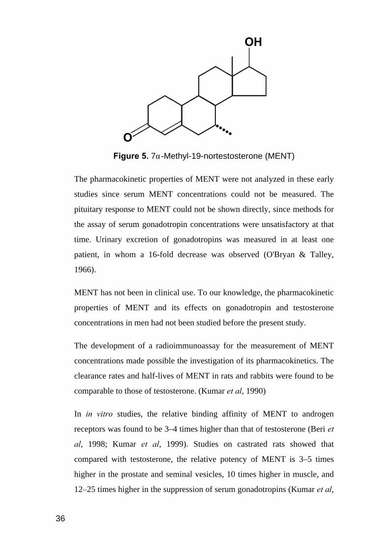
7α-Methyl-19-nortestosterone (MENT) (Figure 5) was synthesized in the
1960s, and studies on animals showed that it has strong androgenic and
anabolic effects (Campbell et al, 1963). It was also evaluated as a palliative
treatment for female breast cancer patients, in whom a virilizing effect was
observed (Segaloff et al, 1964; O'Bryan & Talley, 1966). In addition, it was
administered to three men with carcinoma of the prostate. Although it does
not prove anything, it is interesting to note that one of the patients with
metastatic cancer of the prostate showed a subjective improvement of 6
months duration. (O'Bryan & Talley, 1966)
36
Figure 5. 7α-Methyl-19-nortestosterone (MENT)
The pharmacokinetic properties of MENT were not analyzed in these early
studies since serum MENT concentrations could not be measured. The
pituitary response to MENT could not be shown directly, since methods for
the assay of serum gonadotropin concentrations were unsatisfactory at that
time. Urinary excretion of gonadotropins was measured in at least one
patient, in whom a 16-fold decrease was observed (O'Bryan & Talley,
1966).
MENT has not been in clinical use. To our knowledge, the pharmacokinetic
properties of MENT and its effects on gonadotropin and testosterone
concentrations in men had not been studied before the present study.
The development of a radioimmunoassay for the measurement of MENT
concentrations made possible the investigation of its pharmacokinetics. The
clearance rates and half-lives of MENT in rats and rabbits were found to be
comparable to those of testosterone. (Kumar et al, 1990)
In in vitro studies, the relative binding affinity of MENT to androgen
receptors was found to be 3–4 times higher than that of testosterone (Beri et
al, 1998; Kumar et al, 1999). Studies on castrated rats showed that
compared with testosterone, the relative potency of MENT is 3–5 times
higher in the prostate and seminal vesicles, 10 times higher in muscle, and
12–25 times higher in the suppression of serum gonadotropins (Kumar et al,
OH
O
37
1992; Kumar et al, 1995b; Kumar et al, 1999). The results of a study on
Macaca fascicularis monkeys were very similar regarding the relative
effects of MENT on the prostate, muscle, and gonadotropins. (Cummings et
al, 1998) If the potencies of testosterone and MENT are compared by
measuring the serum concentrations of these hormones required to achieve
complete LH suppression in Macaca fascicularis, instead of comparing the
doses administered, MENT appears to be more than 60 times more potent.
(Cummings et al, 1998)
Unlike testosterone, MENT does not undergo 5α-reduction when incubated
in vitro with homogenized preparations of rat liver, prostate and epididymis
(Agarwal & Monder, 1988). This finding can explain the differences in the
relative potency of MENT compared with testosterone in tissues that
contain or do not contain 5α-reductases. Since MENT is not activated by
5α-reductases, it has a relatively lower potency in tissues that contain 5α-
reductase enzymes. Therefore, when administered at doses that maintain
normal sexual function and secondary sexual characteristics, it should be
less likely to over-stimulate the prostate.
The fact that MENT is not activated by 5α-reductases also raises the
question of whether there are any useful physiological effects of 5α-
dihydrotestosterone in adults that would not be adequately maintained by
MENT. For example, to be an acceptable replacement androgen MENT has
to be able to maintain normal sexual function. The results of some studies
indirectly suggest that 5α-reduction may amplify the effect of testosterone
in maintaining sexual function. In a cross-sectional study in normal men it
was found that higher 5α-dihydrotestosterone concentrations were
associated with a higher frequency of orgasms, whereas testosterone was
unrelated to the frequency of orgasms (Mantzoros et al, 1995). Another
study revealed a slightly higher incidence of decreased libido, impotence,
and ejaculatory disorders among men receiving finasteride, a competitive
38
5α-reductase inhibitor, compared with men receiving a placebo. (Gormley
et al, 1992) However, these pieces of evidence are very indirect and there is
no known unique beneficial role for 5α-dihydrotestosterone in adults
(Cummings et al, 1998). All the available evidence concerning MENT
suggests that it would maintain sexual behavior and sexual function as well
as testosterone. It has been shown that MENT administration can maintain
sexual behavior in castrated male rodents (Morali et al, 1993; Ogawa et al,
1996; Wood et al, 1996). In early studies, MENT increased libido in women
to the extent that some patients discontinued the treatment because of it
(O'Bryan & Talley, 1966). In a recent study in hypogonadal men, MENT
improved several measures of sexual function when compared with the
situation during a treatment-free period. Furthermore, the effect of MENT,
administered by means of subdermal implants releasing MENT acetate, was
similar to that of traditional androgen replacement therapy with
intramuscular testosterone enanthate injections. (Anderson et al, 1999)
As discussed in the Overview of Androgen Physiology, the effects of
testosterone on some tissues are, to a greater or lesser extent, carried out by
estradiol formed by the aromatization of testosterone. The effects of MENT
on these tissues should be physiological, because it has been shown that
MENT is also aromatized to an estrogenic compound (LaMorte et al, 1994).
MENT is also bound to estrogen receptors, but its binding affinity is very
low, about 0.03% of that of estradiol (Beri et al, 1998).
MENT has been shown to have a progestational effect similar to that of
progesterone in rabbits, and the binding affinity of MENT to progesterone
receptors is about 60% of that of progesterone (Beri et al, 1998).
Toxicological studies in animals have not revealed any important adverse
effects. In tolerance studies conducted in the 1960s, oral MENT
administration was associated with changes in liver function test results

39
when doses of 50 or 80 mg per day were administered, but not when the
daily dose was 20 mg. (Unpublished data)
The high potency of MENT makes it potentially suitable for long-term
sustained release administration preparations. In fact, subdermal implants
releasing MENT acetate (MENT Ac) (Figure 6), a pro-drug rapidly
converted to MENT in vivo, can be made. Such implants have been used in
our clinical trials and in another study, in which the effects of MENT Ac
implants and testosterone enanthate injections in hypogonadal men were
compared (Anderson et al, 1999).
Figure 6. 7α-Methyl-19-nortestosterone acetate (MENT Ac)
H CCOO3
O
40
Aims of the Study
The main specific aims of the present study were:
To determine the basic pharmacokinetic parameters of MENT by adminis-
tering MENT by intravenous and intramuscular injections to healthy men
and measuring the resulting serum MENT concentrations.
To determine the pharmacokinetics of MENT during sustained release
administration by measuring serum MENT concentrations in men who had
MENT-releasing implants inserted subdermally.
To determine the effects of MENT administration on the secretion of
gonadotropins and testosterone by measuring serum LH, FSH and
testosterone concentrations before, during, and after MENT administration
in men who received intramuscular MENT injections and in men who had
MENT-releasing implants inserted subdermally.
41
Materials, Methods and Subjects
The present study consists of four phase I clinical trials described in four
original publications. The publications are referred to in this text by the
Roman numbers I – IV according to the order in which they were published
(see the "List of Original Publications"). The trials are discussed in the text
in the order in which they were conducted. The single intravenous injection
trial was described in publication II, the single intramuscular injection trial,
as well as the trial involving six intramuscular injections at daily intervals,
were described in publication I, and the MENT implant trial was described
in publications III and IV.
Trial Sites
The clinical trials were conducted in the following clinics: the Family
Planning Clinic of The Family Federation of Finland (Väestöliitto),
Helsinki, Finland, the Consultorio de Planificación Familiar, Instituto
Chileno de Medicina Reproductiva, Santiago, Chile, and the Centre for
Reproductive Biology, Edinburgh, Great Britain. For brevity, the above-
mentioned trial sites are referred to in this text by the names of the cities in
which they are located.
The single intravenous injection trial was conducted in Helsinki, the single
intramuscular injection trial as well as the trial involving six intramuscular
injections at daily intervals in Helsinki and Santiago, and the MENT
implant trial in all three clinics. The multicenter trials were conducted
according to the same protocol in each clinic.
42
Design of the Trials
Single Intravenous Injection Trial (Reported in Publication II)
Nine men received a single intravenous injection of 0.5 mg MENT. Two
blood samples were collected within 30 minutes before the injection, and
the following samples were collected exactly 3, 6, 10, 15, 30, 45, 60, 90,
120, 150, 180, 210 and 240 minutes after injection.
Single Intramuscular Injection Trial (Reported in Publication I)
A total of 18 men, in two clinics, were randomly allocated to three groups
of equal size (three per group in each clinic). Each subject in the first group
was given 2 mg of MENT intramuscularly. Subjects in the second and third
groups received intramuscular injections of 4 mg and 8 mg of MENT,
respectively. The first blood sample was collected just before injection, the
following ten samples exactly 15, 30, 60, 90, 120, 180, 240, 300, 360 and
480 minutes after injection and the next samples 1, 2, 3, 4 and 9 days after
injection, at the same time in the morning.
Six Intramuscular Injections Trial (Reported in Publication I)
A total of 24 men, in two clinics, were randomly allocated to three groups
of equal size (four per group in each clinic). Each subject in the first group
was given 1 mg of MENT each morning for six consecutive days. Subjects
in the second and third groups received injections of 2 mg and 4 mg
according to the same schedule. Blood samples were drawn before
treatment and each day just before the MENT injection and thereafter at the
same time in the morning, 2, 3, 5, 9, 13 and 24 days after the last injection.
MENT Implant Trial (Reported in Publications III and IV)
A total of 45 men, in three clinics, were randomly allocated into three
groups of equal size (five per group in each clinic). Subjects in each group

43
received either one, two or four MENT acetate-releasing implants. The
implants remained in place for 4 weeks. Blood samples were obtained
before implant insertion and at one-week intervals for 6 weeks thereafter (1,
2, 3 and 4 weeks after insertion and 1 and 2 weeks after removal).
Subjects
The volunteers who participated in the clinical trials were Finnish, Chilean
and Scottish men ranging in age from 19 to 45 years and in weight from 55
to 97 kg. All were judged to be healthy as assessed by medical history,
general physical examination and clinical chemistry measures including
blood count, liver and kidney function tests, lipid profile and assay of
circulating concentrations of prostate-specific antigen. None of the subjects
had any personal or family history of prostate cancer.
Nine Finnish men participated in the single intravenous injection trial,
which was conducted only in Helsinki (Table 1). A total of 18 men, nine in
Helsinki and nine in Santiago, participated in the single-dose intramuscular
MENT trial (Table 2). A total of 24 men, 12 in Helsinki and 12 in Santiago,
participated in the trial of six consecutive daily intramuscular MENT
injections (Table 3). A total of 45 men, 15 in Helsinki, 15 in Santiago and
15 in Edinburgh, participated in the MENT implant trial (Table 4).
44
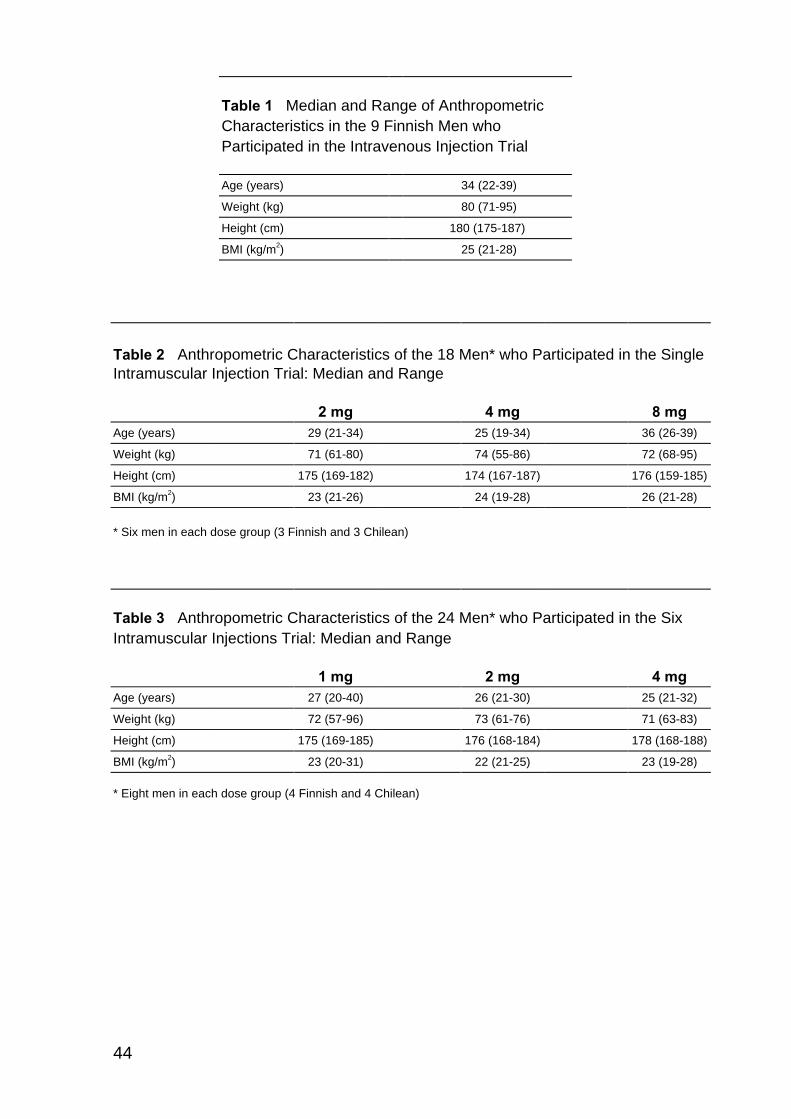
Table 1 Median and Range of Anthropometric Characteristics in the 9 Finnish Men who Participated in the Intravenous Injection Trial
Age (years) 34 (22-39)
Weight (kg) 80 (71-95)
Height (cm) 180 (175-187)
BMI (kg/m2) 25 (21-28)
Table 2 Anthropometric Characteristics of the 18 Men* who Participated in the Single Intramuscular Injection Trial: Median and Range
2 mg 4 mg 8 mg Age (years) 29 (21-34) 25 (19-34) 36 (26-39)
Weight (kg) 71 (61-80) 74 (55-86) 72 (68-95)
Height (cm) 175 (169-182) 174 (167-187) 176 (159-185)
BMI (kg/m2) 23 (21-26) 24 (19-28) 26 (21-28)
* Six men in each dose group (3 Finnish and 3 Chilean)
Table 3 Anthropometric Characteristics of the 24 Men* who Participated in the Six Intramuscular Injections Trial: Median and Range
1 mg 2 mg 4 mg Age (years) 27 (20-40) 26 (21-30) 25 (21-32)
Weight (kg) 72 (57-96) 73 (61-76) 71 (63-83)
Height (cm) 175 (169-185) 176 (168-184) 178 (168-188)
BMI (kg/m2) 23 (20-31) 22 (21-25) 23 (19-28)
* Eight men in each dose group (4 Finnish and 4 Chilean)
45
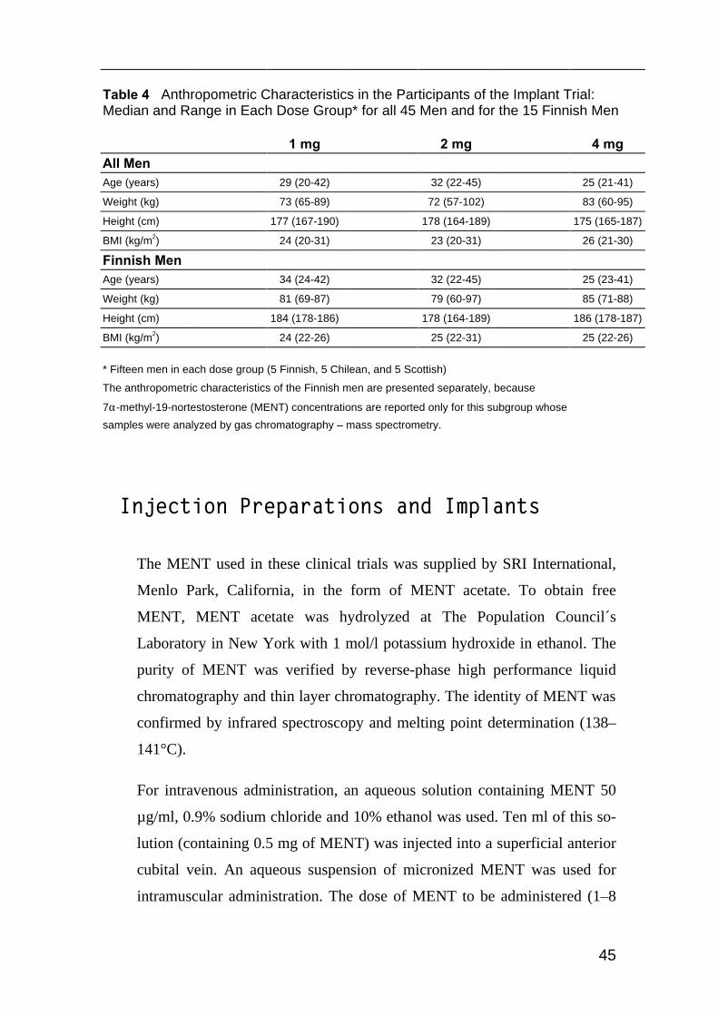
Table 4 Anthropometric Characteristics in the Participants of the Implant Trial: Median and Range in Each Dose Group* for all 45 Men and for the 15 Finnish Men
1 mg 2 mg 4 mg All Men Age (years) 29 (20-42) 32 (22-45) 25 (21-41)
Weight (kg) 73 (65-89) 72 (57-102) 83 (60-95)
Height (cm) 177 (167-190) 178 (164-189) 175 (165-187)
BMI (kg/m2) 24 (20-31) 23 (20-31) 26 (21-30)
Finnish Men
Age (years) 34 (24-42) 32 (22-45) 25 (23-41)
Weight (kg) 81 (69-87) 79 (60-97) 85 (71-88)
Height (cm) 184 (178-186) 178 (164-189) 186 (178-187)
BMI (kg/m2) 24 (22-26) 25 (22-31) 25 (22-26)
* Fifteen men in each dose group (5 Finnish, 5 Chilean, and 5 Scottish)
The anthropometric characteristics of the Finnish men are presented separately, because
7α-methyl-19-nortestosterone (MENT) concentrations are reported only for this subgroup whose
samples were analyzed by gas chromatography – mass spectrometry.
Injection Preparations and Implants
The MENT used in these clinical trials was supplied by SRI International,
Menlo Park, California, in the form of MENT acetate. To obtain free
MENT, MENT acetate was hydrolyzed at The Population Council´s
Laboratory in New York with 1 mol/l potassium hydroxide in ethanol. The
purity of MENT was verified by reverse-phase high performance liquid
chromatography and thin layer chromatography. The identity of MENT was
confirmed by infrared spectroscopy and melting point determination (138–
141°C).
For intravenous administration, an aqueous solution containing MENT 50
µg/ml, 0.9% sodium chloride and 10% ethanol was used. Ten ml of this so-
lution (containing 0.5 mg of MENT) was injected into a superficial anterior
cubital vein. An aqueous suspension of micronized MENT was used for
intramuscular administration. The dose of MENT to be administered (1–8
46
mg) was diluted in 0.5 ml. The injections were given in the deltoid or
gluteus muscles. For the implants, MENT acetate was used instead of free
MENT since it is released more conveniently and it is rapidly hydrolyzed to
MENT in vivo. The implants were manufactured at the Center for
Biomedical Research of The Population Council in New York. Each
implant contained 112 ± 4 mg of MENT acetate in a polyethylene vinyl
acetate copolymer, and the release rate of MENT acetate was estimated to
be about 300 µg per day, based on studies carried out in vitro. The implants
had a diameter of 2.7 mm and a length of 45 mm. The implants were
inserted subdermally in the medial aspect of the upper arm with the aid of a
trocar under local anesthesia and aseptic conditions. The implants were
removed through a short incision under local anesthesia and aseptically.
Sample Handling
Blood samples were collected from the upper limb contralateral to the site
of the injection or implant. When several blood samples had to be taken at
short intervals, an i.v. catheter fitted into a superficial vein on the back of
the hand was used for sampling. When sampling was less frequent, blood
was drawn by antecubital phlebotomy. The blood samples were allowed to
clot and the sera were separated by centrifugation and stored at -20°C in
plastic test tubes until assayed.
Assays
Concentrations of MENT were measured by radioimmunoassay as described
previously. The detection limit of the assay (defined as 2 standard deviations
above the serum blank) was 0.1 nmol/l, its intra-assay coefficient of variation
7.0% and its interassay coefficient of variation 13.8%. The cross-reactivity
of testosterone in the MENT radioimmunoassay was 1.1%. (Kumar et al, 1990)

47
Because of this cross-reactivity, low MENT concentrations cannot be meas-
ured accurately by radioimmunoassay. Hence, we reanalyzed the serum
MENT concentrations of the samples taken in the MENT implant trial in
Helsinki by capillary gas chromatography-mass spectrometry. The detection
limit of this assay was 0.347 nmol/l, the intra-assay coefficient of variation
was 9.8%, and its interassay coefficient of variation was < 10%.
MENT radioimmunoassays of the samples taken in the single intravenous
injection trial and in the single intramuscular injection trial were carried out
in the laboratories of The Population Council in New York, and MENT
radioimmunoassays of the samples taken in the six intramuscular injections
trial and the MENT Ac implant trial were carried out in the Steroid
Research Laboratory, Institute of Biomedicine, University of Helsinki.
MENT concentration measurements by gas chromatography-mass spectro-
metry were carried out in the laboratories of Leiras Oy, Turku, Finland.
Serum testosterone concentrations were determined by conventional
radioimmunoassay according to the standard operating procedure of the
World Health Organization (Sufi et al, 1990). The limit of detection was 0.5
nmol/l, the intra-assay coefficient of variation 6.8% and the interassay
coefficient of variation 13.3%. The cross-reactivity of MENT in the
testosterone radioimmunoassay was 1.2%.
Serum DHT concentrations were determined by radioimmunoassay. The
limit of detection was 0.09 nmol/l, the intra-assay coefficient of variation
5.0% and the interassay coefficient of variation 15.5%. The concentrations
of FSH, LH and SHBG were measured by time-resolved fluoroimmunoas-
says, using commercially available kits (DELFIA®, Wallac Oy, Turku,
Finland). The limit of detection for LH was 0.05 IU/l, the intra-assay coeffi-
cient of variation 5.0% and the interassay coefficient of variation 11.7%.
The limit of detection of FSH was 0.05 IU/l, the intra-assay coefficient of
variation 3.4% and the interassay coefficient of variation 4.9%. For SHBG,
48
the limit of detection was 0.8 nmol/l, the intra-assay coefficient of variation
8.9% and the interassay coefficient of variation 7.0%. The concentrations of
insulin-like growth factor-1 were measured using immunoradiometric kits
(Immunotech, Paris, France). The limit of detection in the insulin-like
growth factor-1 assay was 1.57 nmol/l, the intra-assay coefficient of varia-
tion 7.3% and the interassay coefficient of variation 12.6%.
Concentrations of testosterone, FSH, LH, SHBG, DHT, and insulin-like
growth factor-1 were measured in the Steroid Research Laboratory, Institute
of Biomedicine, University of Helsinki.
Routine clinical chemistry assays used to evaluate the safety of MENT
administration, including blood count, liver and kidney function tests, lipid
profile and assay of prostate-specific antigen, were carried out using
standard methods in local clinical chemistry laboratories.
Pharmacokinetic Methods
To analyze the MENT concentration data obtained in these trials we used
standard pharmacokinetic methods described in standard textbooks of
pharmacokinetics (Gibaldi & Perrier, 1982; Rowland & Tozer, 1994).
Pharmacokinetic data were analyzed with the LAGRAN computer program
(Rocci & Jusko, 1983) and Microsoft Excel for Windows 95 (Microsoft
Corporation, Redmond, Washington; version 7.0 and earlier versions).
Single Intravenous Injection Trial
We determined the pharmacokinetic parameters of MENT by using a two-
compartment model and fitting, by the least squares method, a biexponential
curve to the serum MENT concentration versus time data. The elimination
rate constants and the extrapolated serum MENT concentrations at zero

49
time were estimated for the initial and terminal phases of the concentration
versus time curve. These parameters, as well as the area under the concen-
tration-time curve were used to calculate the other parameters. Half-lives
were calculated by dividing the natural logarithm of 2 by the elimination
rate constants. The initial apparent volume of distribution and the total
apparent volume of distribution were calculated by dividing the dose of
MENT administered by the extrapolated serum MENT concentrations at
zero time estimated for the initial and terminal phases of the concentration
versus time curve. Total clearance was calculated by dividing the MENT
dose by the area under the concentration-time curve.
Single Intramuscular Injection Trial
To estimate the pharmacokinetic parameters of MENT after intramuscular
administration, an exponential curve was fitted to the combined concen-
tration versus time data by least squares fit. The estimated elimination rate
constant and area under the concentration-time curve were used to calculate
the other parameters.
The elimination half-life was calculated by dividing the natural logarithm of
2 by the elimination rate constant. Total clearance was calculated by
dividing the MENT dose by the area under the concentration-time curve.
Pharmacokinetic parameters were calculated for each dose group in each
clinic.
Six Intramuscular Injections Trial
No pharmacokinetic parameters were calculated in this trial, since MENT
concentrations were below the detection limit of the MENT radio-
immunoassay.
50
MENT Implant Trial
For the purposes of pharmacokinetic calculations, the implants were
considered constant-rate release devices. The release rate of MENT from
the implants was estimated by the product of the clearance rate of MENT
(determined in the single intravenous injection trial) and the steady state
concentration of MENT, obtained by averaging the serum MENT
concentrations over the time span from 1 week after insertion to 4 weeks
after insertion in each implant group.
Statistical Methods
Analysis of variance was used to test differences between dose groups and
between clinics. Variations over time in MENT and hormone concentrations
were evaluated by analysis of variance for repeated measures or, for
comparisons involving only two time points, by Student's t-test for paired
data. In the six intramuscular injections trial, the maximum relative effect of
MENT on each hormone was used in statistical tests. The maximum relative
effect was the difference between the most extreme hormone concentration
and the baseline concentration, divided by the baseline concentration. The
purpose of this transformation was to remove the effect of between-subject
variation in baseline hormone levels. In the MENT Ac implant trial,
logarithmic transformation was used to normalize testosterone, FSH, and
SHBG concentrations for analysis of variance. In all statistical tests,
probability values of wrongly rejecting a true null hypothesis (P-values)
were reported two-tailed, and values below 0.05 were considered
statistically significant.
Data were analyzed with Microsoft Excel for Windows 95 (Microsoft
Corporation, Redmond, Washington; version 7.0 and earlier versions) and

51
SPSS for Windows (SPSS Inc., Chicago, Illinois; version 9.0.1. and earlier
versions).
Ethical Issues
The clinical trials were conducted in accordance with good clinical practice
guidelines and the Declaration of Helsinki ([Anonymous], 1997). The Ethics
Committees of each clinic and the Institutional Review Board of The
Population Council approved the trials. All subjects received detailed
information about the trials and gave written informed consent before
enrollment.
Results
Pharmacokinetics of MENT
To study the pharmacokinetics of MENT, we used serum samples taken
after single intravenous and intramuscular injections as well as after the
insertion of MENT-releasing implants. The samples taken in the trial
involving six intramuscular injections at daily intervals could not be used to
study the pharmacokinetics of MENT, since blood samples were taken 24
hours or several days after the injections and no MENT could be detected in
these samples.
Single Intravenous Injection Trial
Peak serum MENT concentrations were measured in the first samples, taken
3 minutes following the intravenous injection of 0.5 mg MENT (Table 5).
Thereafter, MENT concentrations decreased rapidly, reaching the detection
limit of the MENT radioimmunoassay 3–4 hours after injection.
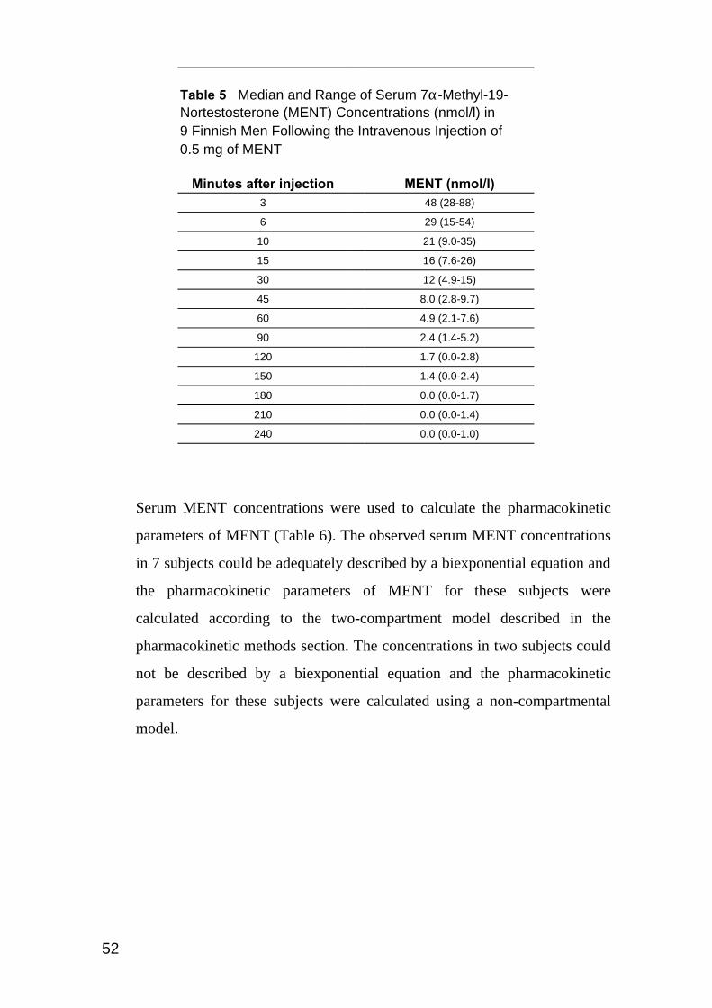
52
Table 5 Median and Range of Serum 7α-Methyl-19- Nortestosterone (MENT) Concentrations (nmol/l) in 9 Finnish Men Following the Intravenous Injection of 0.5 mg of MENT
Minutes after injection MENT (nmol/l)
3 48 (28-88)
6 29 (15-54)
10 21 (9.0-35)
15 16 (7.6-26)
30 12 (4.9-15)
45 8.0 (2.8-9.7)
60 4.9 (2.1-7.6)
90 2.4 (1.4-5.2)
120 1.7 (0.0-2.8)
150 1.4 (0.0-2.4)
180 0.0 (0.0-1.7)
210 0.0 (0.0-1.4)
240 0.0 (0.0-1.0)
Serum MENT concentrations were used to calculate the pharmacokinetic
parameters of MENT (Table 6). The observed serum MENT concentrations
in 7 subjects could be adequately described by a biexponential equation and
the pharmacokinetic parameters of MENT for these subjects were
calculated according to the two-compartment model described in the
pharmacokinetic methods section. The concentrations in two subjects could
not be described by a biexponential equation and the pharmacokinetic
parameters for these subjects were calculated using a non-compartmental
model.

53
Table 6 Median and Range of Pharmacokinetic
Parameters of 7α-Methyl-19-Nortestosterone (MENT) Calculated Using the Serum MENT Concentrations in 8* Finnish Men Following the Intravenous Injection of 0.5 mg of MENT
Volume of distribution 68 (47-94)
Terminal half-life 42 (22-53)
Clearance rate l/day 2000 (1400-2700)
Clearance rate l/kg/day 27 (20-31)
*The obviously erroneous data from one man were omitted
from these summary statistics. (A total of nine men
participated in this study.)
Single Intramuscular Injection Trial
Serum MENT concentrations after single intramuscular injections of 2, 4, or
8 mg of MENT were dose-dependent (Table 7). The differences between
the three dose groups in peak MENT concentrations were statistically
significant (P = 0.028). Over the four-fold dose range tested, each doubling
of the dose of MENT resulted in an increase of approximately 60% in the
peak MENT concentration. The time to reach peak concentrations appeared
to be longer at the higher doses, but for this variable, the differences
between dose groups were not statistically significant. The median and
range of the clearance rates for all subjects were 1600 (720–3200) l/day and
22 (10–49) l/kg. If the probably erroneous clearance rate of one subject, 724
l/day and 10 l/kg, is omitted, the clearance rates for all subjects were 1600
(1100–3200) l/day and 23 (15–49) l/kg. The differences in clearance rates
between dose groups were small and not statistically significant.
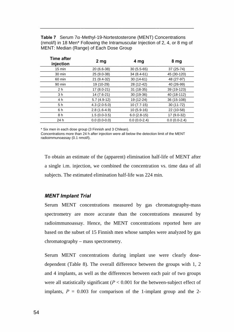
54
Table 7 Serum 7α-Methyl-19-Nortestosterone (MENT) Concentrations (nmol/l) in 18 Men* Following the Intramuscular Injection of 2, 4, or 8 mg of MENT: Median (Range) of Each Dose Group
Time after injection
2 mg 4 mg 8 mg
15 min 20 (6.6-38) 30 (5.5-65) 37 (25-74)
30 min 25 (9.0-38) 34 (8.4-61) 45 (30-120)
60 min 21 (9.4-32) 30 (14-61) 48 (27-97)
90 min 19 (10-29) 28 (12-42) 40 (26-99)
2 h 17 (8.0-21) 31 (18-35) 39 (19-123)
3 h 14 (7.6-21) 30 (19-36) 40 (18-112)
4 h 5.7 (4.9-12) 19 (12-24) 36 (15-108)
5 h 4.3 (2.0-5.0) 10 (7.7-15) 30 (11-72)
6 h 2.8 (1.6-4.9) 10 (5.9-16) 22 (10-58)
8 h 1.5 (0.0-3.5) 6.0 (2.8-15) 17 (9.0-32)
24 h 0.0 (0.0-0.0) 0.0 (0.0-2.4) 0.0 (0.0-2.4)
* Six men in each dose group (3 Finnish and 3 Chilean). Concentrations more than 24 h after injection were all below the detection limit of the MENT radioimmunoassay (0.1 nmol/l).
To obtain an estimate of the (apparent) elimination half-life of MENT after
a single i.m. injection, we combined the concentration vs. time data of all
subjects. The estimated elimination half-life was 224 min.
MENT Implant Trial
Serum MENT concentrations measured by gas chromatography-mass
spectrometry are more accurate than the concentrations measured by
radioimmunoassay. Hence, the MENT concentrations reported here are
based on the subset of 15 Finnish men whose samples were analyzed by gas
chromatography – mass spectrometry.
Serum MENT concentrations during implant use were clearly dose-
dependent (Table 8). The overall difference between the groups with 1, 2
and 4 implants, as well as the differences between each pair of two groups
were all statistically significant (P < 0.001 for the between-subject effect of
implants, P = 0.003 for comparison of the 1-implant group and the 2-

55
implant group, P < 0.001 for comparison of the 1-implant group and the 4-
implant group, and P = 0.003 for comparison of the 2-implant group and the
4-implant group).
Table 8 Serum 7α-Methyl-19-Nortestosterone (MENT) Concentrations (nmol/l) in 15 Men* Before, During, and After the Use of 1, 2 or 4 Subdermal Implants Releasing 7α-Methyl-19-Nortestosterone Acetate (MENT Ac): Median (Range) of Each Dose Group
Weeks after insertion 1 Implant 2 Implants 4 Implants
1 0.5 (0.4-0.8) 1.2 (1.0-1.9) 2.3 (1.6-3.7)
2 0.6 (0.5-1.0) 1.2 (0.9-1.9) 2.3 (1.3-2.8)
3 0.5 (0.4-0.6) 1.4 (1.0-1.7) 2.1 (2.0-3.5)
4 0.6 (0.4-0.7) 1.8 (0.7-1.8) 2.2 (1.3-2.5)
* Five Finnish men in each dose group.
No MENT could be detected in the samples taken 1 and 2 weeks after removal of the implants.
In all dose groups, MENT could only be detected in the samples taken 1, 2,
3 or 4 weeks after insertion of the implants. MENT concentrations in these
samples were relatively constant and there were no statistically significant
differences in MENT concentrations over time within dose groups. Among
the subjects who had one implant inserted, serum MENT concentrations 1–4
weeks after insertion ranged from 0.4 to 1.0 nmol/l. For the subjects who
had two implants inserted, MENT concentrations ranged from 0.7 to 1.9
nmol/l, and for those who had four implants, from 1.3 to 3.7 nmol/l. Since
MENT concentrations did not change significantly over the time span from
1 week after insertion to 4 weeks after insertion, the implants could be
assumed to be constant-rate release devices for the purposes of
pharmacokinetic calculations. The release rate of MENT from the implants
was estimated to be approximately 0.3 mg/day in the 1-implant group, 0.8
mg/day in the 2-implant group, and 1.3 mg/day in the 4-implant group. The

56
corresponding values for MENT acetate were 0.4 mg/day, 0.9 mg/day, and
1.5 mg/day.
Release Rate of MENT Ac Implants
The release rate of 6 unused MENT Ac implants in vitro, measured over a
33-day period was 0.40 mg per day. The release rate of 25 used MENT Ac
implants in vitro, recovered intact after the MENT Ac implant trial, and
measured over a 19-day period, was 0.36 mg per day. Thus, the release rate
of MENT Ac implants used for 4 weeks was 91% of the release rate of new
implants.
The release rate in vivo of 100 MENT Ac implants recovered intact after the
MENT Ac implant trial was measured and found to be 0.48 mg/day in the 1-
implant group, 1.03 mg/day in the 2-implant group, and 1.94 mg/day in the
4-implant group.
The release rates of MENT from the implants calculated from the product of
the clearance rate of MENT, about 2000 l/day, and the mean serum MENT
concentrations in each subject in the samples taken 1, 2, 3, and 4 weeks
after insertion were 0.32 mg/day in the 1-implant group, 0.81 mg/day in the
2-implant group and 1.33 mg/day in the 4-implant group. The corresponding
values for MENT acetate were 0.36 mg/day, 0.93 mg/day and 1.53 mg/day.
Effects of MENT on Gonadotropin and Testosterone Concentrations
To study the effects of MENT on gonadotropin and testosterone
concentrations, we used the serum samples taken during and after the series
of six daily intramuscular injections as well as after the insertion of MENT-
releasing implants. The samples taken in the single intravenous injection
57
trial and the single intramuscular injection trial could not be used to assess
the effect of MENT on hormone concentrations, since blood samples were
taken at different times of the day and the effects of the circadian variation
in hormone concentrations could not be differentiated from the possible
effects of MENT. In contrast, in the trial involving six intramuscular
injections and in the MENT Ac implant trial all samples from each subject
were taken at approximately the same time in the morning.
Six Intramuscular Injections Trial
There were no significant differences in baseline LH concentrations
between clinics. The concentrations of LH decreased in all dose groups after
the i.m. injections (Table 9), and the decreases were significant (P < 0.001).
The mean of the maximal relative decrease in LH concentrations was dose-
dependent: 52 ± 3%, 62 ± 6% and 70 ± 5% in the groups who received 1, 2
or 4 mg doses, respectively. The differences between dose groups were
statistically significant (P = 0.048).

58
Table 9 Serum Luteinizing Hormone Concentrations (U/l) in 24 Men* Before, During
and After a Series of Six Intramuscular Injections of 1, 2 or 4 mg of 7α-Methyl-19- Nortestosterone (MENT) Given at Daily Intervals: Median (Range) of Each Dose Group
1 mg 2 mg 4 mg Before 1st injection 3.5 (2.5-4.7) 4.7 (2.9-9.6) 4.0 (2.5-7.0)
Before 2nd injection 3.8 (1.9-4.8) 3.2 (1.9-6.3) 2.9 (1.5-6.2)
Before 3rd injection 2.3 (1.9-2.8) 2.4 (1.0-8.8) 2.5 (1.0-6.5)
Before 4th injection 2.4 (1.7-3.6) 2.9 (1.2-5.8) 2.0 (0.3-5.5)
Before 5th injection 2.2 (1.3-2.9) 3.1 (1.4-6.4) 1.1 (0.5-3.7)
Before 6th injection 2.6 (1.9-4.5) 2.1 (0.7-3.6) 1.6 (0.5-3.5)
2 Days after last injection 4.2 (1.7-6.2) 3.6 (2.7-5.9) 2.9 (1.8-7.3)
3 Days after last injection 3.4 (2.3-6.0) 5.1 (3.3-8.4) 4.9 (3.8-8.1)
5 Days after last injection 3.4 (1.7-5.3) 4.7 (2.6-7.3) 5.1 (2.9-8.2)
9 Days after last injection 2.9 (2.6-4.4) 4.3 (2.6-5.4) 4.4 (2.9-6.2)
13 Days after last injection 2.7 (2.3-5.2) 4.0 (2.4-7.1) 3.5 (1.3-6.1)
24 Days after last injection 2.8 (2.1-4.2) 4.3 (2.3-5.8) 3.2 (3.0-7.0)
* Eight men in each dose group (4 Finnish and 4 Chilean)
The baseline FSH concentrations in the Finnish subjects were significantly
higher than those of the Chilean subjects (3.9 ± 0.4 vs. 2.4 ± 0.2 U/l, P =
0.009). After the i.m. injections, FSH concentrations decreased in all dose
groups (Table 10), and the decreases were significant (P < 0.001). The
mean of the maximal relative decrease in FSH concentrations was dose-
dependent: 25 ± 5%, 41 ± 5% and 57 ± 4% in the groups who received 1, 2
or 4 mg doses, respectively. The differences between dose groups were
statistically significant (P = 0.001).
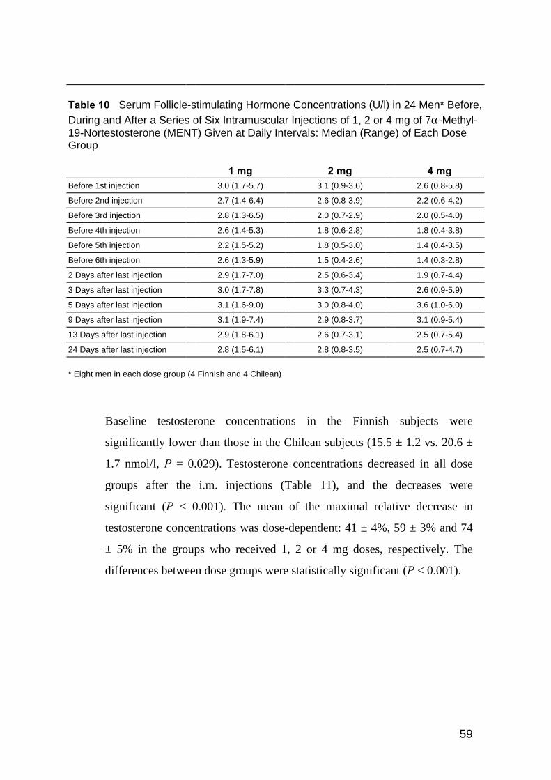
59
Table 10 Serum Follicle-stimulating Hormone Concentrations (U/l) in 24 Men* Before,
During and After a Series of Six Intramuscular Injections of 1, 2 or 4 mg of 7α-Methyl- 19-Nortestosterone (MENT) Given at Daily Intervals: Median (Range) of Each Dose Group
1 mg 2 mg 4 mg Before 1st injection 3.0 (1.7-5.7) 3.1 (0.9-3.6) 2.6 (0.8-5.8)
Before 2nd injection 2.7 (1.4-6.4) 2.6 (0.8-3.9) 2.2 (0.6-4.2)
Before 3rd injection 2.8 (1.3-6.5) 2.0 (0.7-2.9) 2.0 (0.5-4.0)
Before 4th injection 2.6 (1.4-5.3) 1.8 (0.6-2.8) 1.8 (0.4-3.8)
Before 5th injection 2.2 (1.5-5.2) 1.8 (0.5-3.0) 1.4 (0.4-3.5)
Before 6th injection 2.6 (1.3-5.9) 1.5 (0.4-2.6) 1.4 (0.3-2.8)
2 Days after last injection 2.9 (1.7-7.0) 2.5 (0.6-3.4) 1.9 (0.7-4.4)
3 Days after last injection 3.0 (1.7-7.8) 3.3 (0.7-4.3) 2.6 (0.9-5.9)
5 Days after last injection 3.1 (1.6-9.0) 3.0 (0.8-4.0) 3.6 (1.0-6.0)
9 Days after last injection 3.1 (1.9-7.4) 2.9 (0.8-3.7) 3.1 (0.9-5.4)
13 Days after last injection 2.9 (1.8-6.1) 2.6 (0.7-3.1) 2.5 (0.7-5.4)
24 Days after last injection 2.8 (1.5-6.1) 2.8 (0.8-3.5) 2.5 (0.7-4.7)
* Eight men in each dose group (4 Finnish and 4 Chilean)
Baseline testosterone concentrations in the Finnish subjects were
significantly lower than those in the Chilean subjects (15.5 ± 1.2 vs. 20.6 ±
1.7 nmol/l, P = 0.029). Testosterone concentrations decreased in all dose
groups after the i.m. injections (Table 11), and the decreases were
significant (P < 0.001). The mean of the maximal relative decrease in
testosterone concentrations was dose-dependent: 41 ± 4%, 59 ± 3% and 74
± 5% in the groups who received 1, 2 or 4 mg doses, respectively. The
differences between dose groups were statistically significant (P < 0.001).
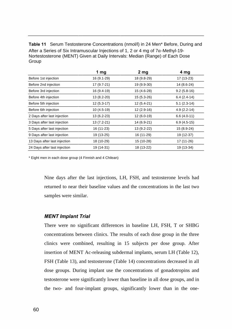
60
Table 11 Serum Testosterone Concentrations (nmol/l) in 24 Men* Before, During and
After a Series of Six Intramuscular Injections of 1, 2 or 4 mg of 7α-Methyl-19- Nortestosterone (MENT) Given at Daily Intervals: Median (Range) of Each Dose Group
1 mg 2 mg 4 mg Before 1st injection 16 (9.1-29) 18 (9.8-29) 17 (13-23)
Before 2nd injection 17 (9.7-21) 19 (9.9-30) 14 (8.6-24)
Before 3rd injection 16 (9.4-19) 15 (4.6-28) 9.2 (5.8-16)
Before 4th injection 13 (8.2-20) 15 (5.3-26) 6.4 (2.4-14)
Before 5th injection 12 (5.3-17) 12 (5.4-21) 5.1 (2.3-14)
Before 6th injection 10 (4.5-19) 12 (2.9-16) 4.9 (2.2-14)
2 Days after last injection 13 (6.2-23) 12 (6.0-19) 6.6 (4.0-11)
3 Days after last injection 13 (7.2-21) 14 (6.9-21) 6.9 (4.5-15)
5 Days after last injection 16 (11-23) 13 (9.2-22) 15 (8.9-24)
9 Days after last injection 19 (13-25) 16 (11-29) 19 (12-37)
13 Days after last injection 18 (10-29) 15 (10-28) 17 (11-26)
24 Days after last injection 19 (14-31) 18 (13-22) 19 (13-34)
* Eight men in each dose group (4 Finnish and 4 Chilean)
Nine days after the last injections, LH, FSH, and testosterone levels had
returned to near their baseline values and the concentrations in the last two
samples were similar.
MENT Implant Trial
There were no significant differences in baseline LH, FSH, T or SHBG
concentrations between clinics. The results of each dose group in the three
clinics were combined, resulting in 15 subjects per dose group. After
insertion of MENT Ac-releasing subdermal implants, serum LH (Table 12),
FSH (Table 13), and testosterone (Table 14) concentrations decreased in all
dose groups. During implant use the concentrations of gonadotropins and
testosterone were significantly lower than baseline in all dose groups, and in
the two- and four-implant groups, significantly lower than in the one-

61
implant group. Serum LH, FSH, and testosterone concentrations in the two-
and four-implant groups were not significantly different. The concentrations
of gonadotropins and testosterone remained at approximately the same level
in all samples taken during implant use.
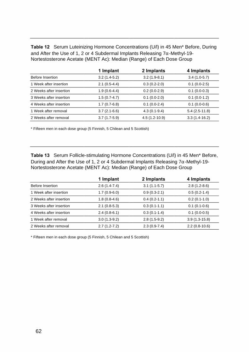
62
Table 12 Serum Luteinizing Hormone Concentrations (U/l) in 45 Men* Before, During and After the Use of 1, 2 or 4 Subdermal Implants Releasing 7α-Methyl-19-Nortestosterone Acetate (MENT Ac): Median (Range) of Each Dose Group
1 Implant 2 Implants 4 Implants Before Insertion 3.2 (1.4-5.2) 3.2 (1.9-8.1) 3.4 (1.0-5.7)
1 Week after insertion 2.1 (0.5-4.4) 0.3 (0.2-2.0) 0.1 (0.0-2.5)
2 Weeks after insertion 1.9 (0.6-4.4) 0.2 (0.0-2.9) 0.1 (0.0-0.3)
3 Weeks after insertion 1.5 (0.7-4.7) 0.1 (0.0-2.0) 0.1 (0.0-1.2)
4 Weeks after insertion 1.7 (0.7-6.8) 0.1 (0.0-2.4) 0.1 (0.0-0.6)
1 Week after removal 3.7 (2.1-6.6) 4.3 (0.1-9.4) 5.4 (2.5-11.8)
2 Weeks after removal 3.7 (1.7-5.9) 4.5 (1.2-10.9) 3.3 (1.4-16.2)
* Fifteen men in each dose group (5 Finnish, 5 Chilean and 5 Scottish)
Table 13 Serum Follicle-stimulating Hormone Concentrations (U/l) in 45 Men* Before, During and After the Use of 1, 2 or 4 Subdermal Implants Releasing 7α-Methyl-19-Nortestosterone Acetate (MENT Ac): Median (Range) of Each Dose Group
1 Implant 2 Implants 4 Implants Before Insertion 2.6 (1.4-7.4) 3.1 (1.1-5.7) 2.8 (1.2-8.6)
1 Week after insertion 1.7 (0.9-6.0) 0.9 (0.3-2.1) 0.5 (0.2-1.4)
2 Weeks after insertion 1.8 (0.8-4.6) 0.4 (0.2-1.1) 0.2 (0.1-1.0)
3 Weeks after insertion 2.1 (0.8-5.3) 0.3 (0.1-1.1) 0.1 (0.1-0.6)
4 Weeks after insertion 2.4 (0.8-6.1) 0.3 (0.1-1.4) 0.1 (0.0-0.5)
1 Week after removal 3.0 (1.3-9.2) 2.8 (1.5-9.2) 3.9 (1.3-15.8)
2 Weeks after removal 2.7 (1.2-7.2) 2.3 (0.9-7.4) 2.2 (0.8-10.6)
* Fifteen men in each dose group (5 Finnish, 5 Chilean and 5 Scottish)

63
Table 14 Serum Testosterone Concentrations (nmol/l) in 45 Men* Before, During and After the Use of 1, 2, or 4 Subdermal Implants Releasing 7α-Methyl-19-Nortestosterone Acetate (MENT Ac): Median (Range) of Each Dose Group
1 Implant 2 Implants 4 Implants Before Insertion 21 (14-28) 17 (7.9-22) 16 (11-49)
1 Week after insertion 8.2 (1.4-19) 1.2 (0.7-5.8) 1.1 (0.7-3.8)
2 Weeks after insertion 6.5 (1.7-20) 1.0 (0.5-2.8) 0.8 (0.6-3.7)
3 Weeks after insertion 7.6 (1.7-17) 0.9 (0.5-3.1) 0.9 (0.4-5.9)
4 Weeks after insertion 7.5 (2.6-23) 1.1 (0.6-3.2) 1.0 (0.5-4.3)
1 Week after removal 17 (12-32) 13 (9.6-18) 11 (7.4-19)
2 Weeks after removal 17 (7.7-30) 19 (8.2-29) 17 (11-45)
* Fifteen men in each dose group (5 Finnish, 5 Chilean and 5 Scottish)
Effects of MENT on Other Analytes
SHBG
In the six intramuscular injections trial, serum SHBG concentrations
decreased in all dose groups. There were no statistically significant differ-
ences either in baseline SHBG concentrations or in the decreases in SHBG
concentrations between clinics or between dose groups, and therefore the
results of all 24 subjects are reported together. The median and range of the
baseline serum SHBG concentrations in all 24 subjects was 30 (15–55)
nmol/l. Two and 24 days after the last injection, the respective values were
24 (13–56) nmol/l and 28(14–56) nmol/l. The difference in SHBG concen-
trations between baseline and two days after the last injection was statisti-
cally significant (P < 0.001).
In the MENT implant trial, SHBG concentrations decreased in all dose
groups, but the decrease was statistically significant only in the four-implant
group (P = 0.011). In the 1-implant group, the median (and range) serum
SHBG concentration before implant insertion was 27 (12–49) nmol/l. Four
64
weeks after insertion the respective value was 22 (14–55) nmol/l, and two
weeks after removal, 25 (14–56) nmol/l. In the 2-implant group, the respec-
tive concentrations were: before insertion 28 (18–59) nmol/l, four weeks
after insertion 27 (17–45) nmol/l, and two weeks after removal 25 (19–57)
nmol/l. In the 4-implant group, the respective concentrations were: before
insertion 28 (16–86) nmol/l, four weeks after insertion 22 (13–56) nmol/l,
and two weeks after removal 28 (15–71) nmol/l.
Dihydrotestosterone and Insulin-Like Growth Factor-1
Dihydrotestosterone concentrations, which were measured only in the
MENT implant trial and only in a subgroup of 23 subjects, decreased by
60% in the 1-implant group, 76% in the 2-implant group, and by 77% in the
4-implant group. Serum insulin-like growth factor-1 concentrations, meas-
ured in the MENT implant trial and only in a subgroup of 10 subjects,
remained constant throughout the trial.
Prostate-Specific Antigen
In all trials, serum concentrations of prostate-specific antigen remained well
below the upper limit of the reference range (4 µg/l).
Other Clinical Chemistry Variables
Although we did not detect any consistent changes in clinical chemistry
variables, small individual increases in total serum cholesterol, low density
lipoprotein cholesterol, triglycerides, aspartate amino transferase, alanine
amino transferase, lactate dehydrogenase and creatinine kinase levels were
observed in some subjects in both intramuscular injection studies as well as
in the MENT implant trial.

65
Other Adverse Effects
In the intravenous and intramuscular injection studies, the subjects did not
report any adverse effects. In the MENT implant trial, 7 subjects experi-
enced pain or other sensations at the site of implant insertion, 7 subjects had
androgenic side-effects such as acne and increased libido, 2 subjects experi-
enced sweating and insomnia and 4 subjects reported mood changes. One of
the cases of mood change was major depression that developed some days
after removal of the implants. The subject required psychiatric treatment.
One subject had local infection at the site of insertion of four implants. The
infection responded to antibiotic treatment and the implants were allowed to
remain in place. In another subject, four implants remained in place for 6
weeks due to problems in removal.
Discussion
The Androgenicity of MENT in Men
This study shows for the first time that MENT is a potent suppressor of LH,
FSH and testosterone secretion in men. Gonadotropin secretion can be sup-
pressed by androgens, estrogens and progestins, which all have a negative
feedback effect on the pituitary and the hypothalamus, and therefore
gonadotropin suppression as such does not prove the androgenicity of
MENT. However, if we take into account what is previously known about
MENT — its virilizing effects on women and its androgenic actions found
in animal studies — it seems almost certain that MENT is also a potent
androgen in men. Further support for this claim is given by the fact that in-
tramuscular MENT injections and MENT Ac implants also caused a de-
crease in SHBG concentrations, a known effect of androgens, and by the
66
fact that some typical androgenic side-effects such as acne and increased li-
bido were observed in the men who took part in the MENT Ac implant trial.
It is important to note that even though we did not have placebo groups, the
changes in serum concentrations of LH, FSH, and testosterone described in
this study cannot plausibly be explained by any other reason than MENT
administration. Since the samples in the trial involving six intramuscular
injections and in the MENT Ac implant trial were all taken at the same time
in the morning, the natural circadian variation in the concentrations of these
substances could not influence these results. (We did not report the hormone
concentrations measured in the single intravenous and single intramuscular
injection trials, because in these trials we could not control the effects of
circadian variation.)
Pharmacokinetics of MENT
The terminal half-life of MENT, about 40 min, is approximately two times
that of testosterone (10–20 minutes). The clearance rate of MENT, about
2000 l/day, is more than 50% higher than the clearance rate of testosterone,
which is about 1200 l/day (Bhasin, 1992). These differences in pharmaco-
kinetics are probably due to differences in metabolism of these two andro-
gens, and to the fact that SHBG binds MENT very weakly, their relative
binding affinity being about 6% of that of SHBG and testosterone (Kumar
et al, 1999). One of the main metabolic pathways of testosterone is 5α-
reduction, and this pathway is not available to MENT. The metabolic fate of
MENT is not exactly known, but it seems that its metabolism is slightly
slower than that of testosterone. This would explain the slightly longer half-
life of MENT. On the other hand, the fact that MENT is very weakly bound
by SHBG should reduce the effect of slower metabolism because a higher
fraction of MENT is free and available for metabolic reactions.

67
The clearance rate after i.m. administration (median 1600, range 720–3200
l/day) was not very different from the clearance rate determined following
i.v. administration, (median 2000, range 1400–2700 l/day) — in fact, the
ranges overlap. In contrast, the half-life estimated from the data of the sin-
gle intramuscular injection trial (224 min) was more than five times longer
than the 42 min elimination half-life determined using a single i.v. dose of
0.5 mg MENT. This difference is almost certainly explained by the slow ab-
sorption of MENT from the i.m. injection site. If absorption is slow enough,
it is rate-limiting in the decay phase of MENT concentrations, and the esti-
mated half-life corresponds to the absorption half-life (Rowland & Tozer,
1994). Since MENT is poorly soluble in water, its absorption from muscle
could be relatively slow. Slow absorption of MENT might help to explain
the fact that in spite of its short half-life, MENT injections given at 24-hour
intervals in the trial involving six intramuscular injections had a cumulative
suppressive effect on serum LH, FSH and testosterone concentrations.
In the trial involving six intramuscular injections, MENT could not be de-
tected in samples taken 24 hours after the injections, and in the implant trial,
MENT could not be detected in the samples taken 1 and 2 weeks after
removal of the implants. This is consistent with the short half-life of MENT.
Thus, it can be concluded that there is no detectable accumulation of MENT
in the body with either intramuscular MENT injections or MENT Ac
implants.
MENT Implants
The results of this study also show that MENT acetate implants function
properly (for at least four weeks) and can be considered constant-rate
release devices. In the 4 samples taken when the implants had been in place
from 1 to 4 weeks, MENT levels were similar, showing some fluctuations,
68
but rising or decreasing trends were not detected and there were no statisti-
cally significant changes over time.
The differences between the release rates calculated from the amount of
MENT Ac actually lost from the implants and the release rates calculated
using MENT concentrations and MENT clearance obtained from the results
of the intravenous injection trial were most likely due to a higher release
rate during the first few days after insertion. As the samples used in the
calculation of release rates were obtained from 1 to 4 weeks after insertion,
and each sample reflects the release rate at the time the sample was drawn,
the calculated release rate does not include information on the release rate
during the first few days. Furthermore, subdermal implants usually show a
relatively high release rate for a few days after insertion. The fact that the
clearance rate used in the calculations was obtained from the intravenous
injection trial and was not the actual clearance rate in the men who took part
in the implant trial, could also account for part of the differences. In spite of
the small differences, the clearance rates calculated from the amount of
MENT Ac lost and the release rates calculated using MENT concentrations
are similar. This indicates that the bioavailability of MENT from the
implants is good.
The type of implant used in this study contains about 112 mg of MENT Ac.
Hence, it can be roughly estimated, using the release rate calculated from
the amount of MENT Ac lost, that the implants would last about 112
mg/0.48 mg/day = 233 days. Of course, this is an oversimplification, since
the release rate will fall as the amount of MENT Ac in the implants
decreases, and after some time the release rate will no longer be sufficient.
Based on all the information available, it seems reasonable to propose that
the effective life of this type of implant would be about 6 months.
69
Lack of 5αα-Reduction
MENT is not activated by 5α-reduction in animals, and probably not in men
either, although this has not been shown. The consequences of this property
can only be guessed. It is hoped that MENT would be a safer androgen
because of this property, since it implies that it should not over-stimulate the
prostate. This property should also reduce the incidence of some other
adverse effects of androgens, such as acne and male pattern baldness. If
used in women, MENT could be less prone to cause hirsutism.
It is also possible that not being activated by 5α-reductases could also be a
handicap. We do not yet know whether MENT can support all androgen-
dependent bodily functions and properties. Probably it will, because even
though 5α-reduction is essential for male sexual development during fetal
life, there are currently no known beneficial effects of 5α-reduction in
adults. Furthermore, though MENT does not undergo 5α-reduction it is not
without effect in 5α-dihydrotestosterone-dependent tissues. The effects of
5α-dihydrotestosterone are mediated through androgen receptors, and
MENT binds more tightly to these receptors than 5α-dihydrotestosterone
itself. The differences in the actions of MENT in different androgen-sensi-
tive tissues are quantitative rather than qualitative.
Hormonal Male Contraception
Our ultimate aim is the development of a hormonal male contraceptive
based on MENT. The contraceptive would consist of MENT Ac-releasing
implants, either alone or in combination with another inhibitor of gonad-
otropin secretion such as a GnRH antagonist or a progestin. Preferably, if
another inhibitor of gonadotropin secretion needs to be used, it should be a
70
substance that can also be administered by subdermal implants, so that it
would not make the method less convenient.
We do not yet know whether MENT will suppress spermatogenesis suffi-
ciently to achieve reliable contraception. Semen analysis has not been
included in our trials because they have all been of short duration. The
MENT Ac implants were in place for only four weeks, and it is known from
a multitude of previous male contraception studies that about four months
are required for the complete suppression of spermatogenesis by androgens
(Cummings & Bremner, 1994). However, we could have detected a smaller
decrease in sperm concentrations. In the first large testosterone enanthate
contraception study of the World Health Organization, sperm concentrations
were already suppressed to 65% of baseline at 1 month of treatment in those
157 men who later became azoospermic, and to 84% in those 68 who did
not (Handelsman et al, 1995).
Assuming that the mechanism of contraceptive action of MENT is the same
as that of testosterone, namely, suppression of gonadotropin secretion, the
results of our study make it reasonable to predict that MENT will be no less
efficient than testosterone enanthate in the suppression of sperm counts.
Two or four MENT Ac implants suppressed LH concentrations to levels
similar to those seen in subjects rendered azoospermic by testosterone
enanthate injections.
A longer trial is about to begin, in which healthy men will use 1, 2 or 4
MENT Ac implants for 6 months. In this trial, sperm counts will be
measured. The results will be very interesting, not only because of the
information obtained on the effect of MENT on male fertility but also
because of the information obtained about the safety of MENT. Since
MENT is not activated by 5α-reductase enzymes, the trial may also reveal
something interesting about the physiological significance, if any, of 5α-
reduction in healthy adult men.
71
Limitations of This Study
Our clinical trials were neither blinded nor placebo-controlled. The number
of subjects enrolled in these trials was small, and even the longest treatment
lasted for only four weeks. Hence, no reliable conclusions can be drawn
about the safety of MENT or the possible significance of the few adverse
events that were reported.
The possible effects of single intravenous and single intramuscular MENT
injections on hormone concentrations could not be determined, because in
the absence of placebo groups, we could not control the confounding effect
of circadian variation in testosterone and gonadotropin concentrations.
The short duration of our studies precluded investigation of the effects of
MENT on sperm counts and therefore the conclusion we have drawn about
the possible contraceptive efficacy of MENT is an extrapolation derived
from the effects of MENT on LH and FSH concentrations.

72
Conclusions
The main conclusions that can be drawn from the results of this study are as
follows:
MENT has pharmacokinetic properties that make it suitable for long-term
androgen administration via subdermal implants releasing MENT acetate, a
MENT ester that is rapidly converted to MENT in vivo.
MENT has strong suppressive effects on serum LH, FSH, and testosterone
secretion.
The results of this study are consistent with the results of previous in vitro
and animal studies. All available data on MENT suggest that it is a potent
androgen and a very promising candidate for hormonal male contraception
and long-term androgen replacement therapy.

73
Acknowledgments
This study is part of multicenter studies sponsored and organized by The
Population Council. The Population Council is gratefully acknowledged for
making this study possible.
This study was carried out in the Steroid Research Laboratory of the Insti-
tute of Biomedicine of the University of Helsinki, the Family Planning
Clinic of The Family Federation of Finland (Väestöliitto), Helsinki, Finland,
the Consultorio de Planificación Familiar, Instituto Chileno de Medicina
Reproductiva, Santiago, Chile, the Centre for Reproductive Biology, Edin-
burgh, Great Britain, the laboratories of Leiras Oy, Turku, Finland, and the
laboratories of The Population Council in New York. I gratefully acknowl-
edge these institutions and I would like to thank all the staff of these insti-
tutions who contributed to this study.
I am most grateful to my supervisor, docent Pekka Lähteenmäki, for intro-
ducing me to the field of biomedical research, and for the guidance, support
and advice he gave me during this study. It has been a pleasure to work with
him in this study as well as several other research projects. Docent Pekka
Lähteenmäki was also the head of our laboratory as well as chief physician
of the Family Planning Clinic of The Family Federation of Finland during
most of the duration of this study. Hence, I am also grateful to him for over-
seeing the arrangements that made possible this study.
I am very grateful for the official reviewers of my dissertation, docent
Pirkko Härkönen and docent Leo Dunkel, for their constructive criticism,
useful comments and advice.
I wish to thank professor Tapani Luukkainen, the former head and founder
of our laboratory and docent Oskari Heikinheimo, head of our laboratory, as
74
well as professor Olli Jänne, head of the Institute of Biomedicine, and pro-
fessor Paavo Kinnunen, head of the Department of medical chemistry, for
creating and maintaining an excellent research environment. A great part of
that excellent environment is due to the staff of our Laboratory who have
given me invaluable help in conducting this study, Sirpa Ranta, M.Sc.,
Marjatta Tevilin, Eeva Harju, Anne Hyvönen, and Agnes Viherä as well as
to my colleagues and friends who worked, like me, as part-time researchers
in our laboratory, docent Juhani Toivonen, Päivi Pakarinen, M.D., Raimo
Kekkonen, M.D., and Satu Suhonen, M.D.
I would also like to thank the staff of the Family Planning Clinic of The
Family Federation of Finland, including Pia Brandt, Siru Salli, and Dr.
Kaisa Elomaa, who took care of inserting and removing the MENT
implants, and many other medical responsibilities during this study.
I wish to thank Auni Juhakoski, M.Sc., for developing the capillary gas
chromatography-mass spectrometry method used in the MENT assays, and
performing those MENT assays that were done with her method. Without
her method an adequate analysis of the pharmacokinetics of MENT admin-
istered by subdermal implants would not have been possible.
I also would like to thank all my other coauthors whose name I have not yet
mentioned: Gabriela Noé, Narender Kumar, Claude Aguillaume, C. Wayne
Bardin, Horacio B. Croxatto, Cameron Martin, Alfred Moo-Young, Eliana
Quintero, Saleh I. Saleh, Kalyan Sundaram, and Yun-Yen Tsong.
I wish to thank Nicholas Bolton, Ph.D., for revising the language and
improving the style and logical structure of this dissertation and all the
original publications
I am also grateful to all the volunteers who participated in these studies: the
Finnish, Chilean and Scottish men who received the MENT injections and
75
implants and underwent repeated phlebotomies, just for the sake of
advancing contraceptive research.
I also want to thank Leiras Oy for financial support in publishing this
dissertation, and my current employer, Helsinki University Central Hospital,
for allowing me to take the vacations that were necessary to finish writing
this dissertation.
I am extremely grateful to my wife Jaana first of all for making my life
happy with her enormous love, and also for the motivation, support and
invaluable advice she has given me in many problems related to this study.
Her comments regarding the contents, structure, statistical methods and
language of my original publications and this dissertation have contributed
greatly to their content. I also would like to thank my son Severi for his
patience during the last part of this research project when I have not been
with him as much as I should, and for bringing a lot of happiness in my life.
I would also like to thank my family and friends, especially my parents
Kirsti and Jukka Suvisaari, my sister Laila, and my brothers Sampo and
Osmo for the encouragement, love, friendship, advice and support they have
given me over the decades.

76
References
[Anonymous]. Contraceptive efficacy of testosterone-induced azoospermia in normal men. World Health Organization Task Force on methods for the regulation of male fertility. Lancet 1990 Oct 20; 336(8721): 955–9.
[Anonymous]. Contraceptive efficacy of testosterone-induced azoospermia and oligozoospermia in normal men. Fertil Steril 1996 Apr; 65(4): 821–9.
[Anonymous]. World Medical Association declaration of Helsinki. Recom-mendations guiding physicians in biomedical research involving human subjects. JAMA 1997 Mar 19; 277(11): 925–6.
Agarwal AK, Monder C. In vitro metabolism of 7 alpha-methyl-19-nortes-tosterone by rat liver, prostate, and epididymis. Endocrinology 1988 Nov; 123(5): 2187–93.
Amory JK, Bremner WJ. The use of testosterone as a male contraceptive. Baillieres Clin Endocrinol Metab 1998 Oct; 12(3): 471–84.
Amory JK, Bremner WJ. Newer Agents for Hormonal Contraception in the Male. Trends Endocrinol Metab 2000 Mar; 11(2): 61–66.
Anderson RA, Wallace EM, Wu FC. Effect of testosterone enanthate on serum lipoproteins in man. Contraception 1995 Aug; 52(2): 115–9.
Anderson RA, Martin CW, Kung AW, Everington D, Pun TC, Tan KC, Bancroft J, Sundaram K, Moo-Young AJ, Baird DT. 7 Alpha-methyl-19-nortestosterone maintains sexual behavior and mood in hypogonadal men. J Clin Endocrinol Metab 1999 Oct; 84(10): 3556–62.
Bagatell CJ, Matsumoto AM, Christensen RB, Rivier JE, Bremner WJ. Comparison of a gonadotropin releasing-hormone antagonist plus testosterone (T) versus T alone as potential male contra-ceptive regimens. J Clin Endocrinol Metab 1993 Aug; 77(2): 427–32.
Bagatell CJ, Bremner WJ. Androgens in men — uses and abuses. N Engl J Med 1996 Mar 14; 334(11): 707–14.

77
Bardin CW, Swerdloff RS, Santen RJ. Androgens: risks and benefits. J Clin Endocrinol Metab 1991 Jul; 73(1): 4–7.
Bardin CW. The anabolic action of testosterone. N Engl J Med 1996 Jul 4; 335(1): 52–3.
Behre HM, Nieschlag E. Testosterone buciclate (20 Aet-1) in hypogonadal men: pharmacokinetics and pharmacodynamics of the new long-acting androgen ester. J Clin Endocrinol Metab 1992 Nov; 75(5): 1204–10.
Behre HM, Baus S, Kliesch S, Keck C, Simoni M, Nieschlag E. Potential of testosterone buciclate for male contraception: endocrine differences between responders and nonresponders. J Clin Endocrinol Metab 1995 Aug; 80(8): 2394–403.
Behre HM, von Eckardstein S, Kliesch S, Nieschlag E. Long-term substitution therapy of hypogonadal men with transscrotal testosterone over 7–10 years. Clin Endocrinol (Oxf) 1999 May; 50(5): 629–35.
Beri R, Kumar N, Savage T, Benalcazar L, Sundaram K. Estrogenic and progestational activity of 7 alpha-methyl-19-nortestosterone, a synthetic androgen. J Steroid Biochem Mol Biol 1998 Nov; 67(3): 275–83.
Besa EC. Hematologic effects of androgens revisited: an alternative therapy in various hematologic conditions. Semin Hematol 1994 Apr; 31(2): 134–45.
Bhasin S, Swerdloff RS, Steiner B, Peterson MA, Meridores T, Galmirini M, Pandian MR, Goldberg R, Berman N. A biodegradable testosterone microcapsule formulation provides uniform eugonadal levels of testosterone for 10–11 weeks in hypogo-nadal men. J Clin Endocrinol Metab 1992 Jan; 74(1): 75–83.
Bhasin S. Clinical review 34: Androgen treatment of hypogonadal men. J Clin Endocrinol Metab 1992 Jun; 74(6): 1221–5.
Bhasin S, Storer TW, Berman N, Callegari C, Clevenger B, Phillips J, Bunnell TJ, Tricker R, Shirazi A, Casaburi R. The effects of supraphysiologic doses of testosterone on muscle size and strength in normal men. N Engl J Med 1996 Jul 4; 335(1):1–7.

78
Bhasin S, Javanbakht M. Can androgen therapy replete lean body mass and improve muscle function in wasting associated with human immunodeficiency virus infection. JPEN J Parenter Enteral Nutr 1999 Nov–Dec; 23(6 Suppl): S195–201.
Bulun SE. Clinical review 78: Aromatase deficiency in women and men: would you have predicted the phenotypes. J Clin Endocrinol Metab 1996 Mar; 81(3): 867–71.
Campbell JA, Lyster SC, Duncan GW, Babcock JC. 7 alpha-methyl-18-nor-steroids: a new class of potent anabolic and androgenic hormones. Steroids 1963; 1: 317–332.
Casaburi R. Rationale for anabolic therapy to facilitate rehabilitation in chronic obstructive pulmonary disease. Baillieres Clin Endo-crinol Metab 1998 Oct; 12(3): 407–18.
Cummings DE, Bremner WJ. Prospects for new hormonal male contracep-tives. Endocrinol Metab Clin North Am 1994 Dec; 23(4): 893–922.
Cummings DE, Bremner WJ. Male contraception: ideas for the future. Curr Ther Endocrinol Metab 1997; 6: 300–4.
Cummings DE, Kumar N, Bardin CW, Sundaram K, Bremner WJ. Prostate-sparing effects in primates of the potent androgen 7 alpha-methyl-19-nortestosterone: a potential alternative to testosterone for androgen replacement and male contraception. J Clin Endocrinol Metab 1998 Dec; 83(12): 4212–9.
Dabbs JM Jr. Age and seasonal variation in serum testosterone concentration among men. Chronobiol Int 1990; 7(3): 245–9.
Davis SR, Burger HG. The rationale for physiological testosterone replace-ment in women. Baillieres Clin Endocrinol Metab 1998 Oct; 12(3): 391–405.
Dobs AS. Androgen therapy in AIDS wasting. Baillieres Clin Endocrinol Metab 1998 Oct; 12(3): 379–90.
Dobs AS. Is there a role for androgenic anabolic steroids in medical prac-tice?. JAMA 1999 Apr 14; 281(14): 1326–7.
English KM, Steeds R, Jones TH, Channer KS. Testosterone and coronary heart disease: is there a link. QJM 1997 Dec; 90(12): 787–91.
Enmark E, Gustafsson JA. Oestrogen receptors – an overview. J Intern Med 1999 Aug; 246(2): 133–8.

79
Finkelstein JS, Whitcomb RW, O'Dea LS, Longcope C, Schoenfeld DA, Crowley WF Jr. Sex steroid control of gonadotropin secretion in the human male. I. Effects of testosterone administration in normal and gonadotropin-releasing hormone-deficient men. J Clin Endocrinol Metab 1991 Sep; 73(3): 609–20.
Foresta C, Bordon P, Rossato M, Mioni R, Veldhuis JD. Specific linkages among luteinizing hormone, follicle-stimulating hormone, and testosterone release in the peripheral blood and human sper-matic vein: evidence for both positive (feed-forward) and negative (feedback) within-axis regulation. J Clin Endocrinol Metab 1997 Sep; 82(9): 3040–6.
Franke WW, Berendonk B. Hormonal doping and androgenization of athletes: a secret program of the German Democratic Republic government. Clin Chem 1997 Jul; 43(7): 1262–79.
Friedrich MJ. Can male hormones really help women? JAMA 2000 May 24–31; 283(20): 2643–4.
Gibaldi M, Perrier D. Pharmacokinetics. Second edition. Marcel Dekker, New York, 1982.
Gormley GJ, Stoner E, Bruskewitz RC, Imperato-McGinley J, Walsh PC, McConnell JD, Andriole GL, Geller J, Bracken BR, Tenover JS, et al. The effect of finasteride in men with benign prostatic hyperplasia. The Finasteride Study Group. N Engl J Med 1992 Oct 22; 327(17): 1185–91.
Grumbach MM, Auchus RJ. Estrogen: consequences and implications of human mutations in synthesis and action. J Clin Endocrinol Metab 1999 Dec; 84(12): 4677–94.
Gustafsson JA. Estrogen receptor beta — a new dimension in estrogen mechanism of action. J Endocrinol 1999 Dec; 163(3): 379–83.
Handelsman DJ, Conway AJ, Boylan LM. Pharmacokinetics and pharmaco-dynamics of testosterone pellets in man. J Clin Endocrinol Metab 1990 Jul; 71(1): 216–22.
Handelsman DJ, Farley TM, Peregoudov A, Waites GM. Factors in nonuni-form induction of azoospermia by testosterone enanthate in normal men. World Health Organization Task Force on Methods for the Regulation of Male Fertility. Fertil Steril 1995 Jan; 63(1): 125–33.
80
Handelsman DJ, Liu PY. Androgen therapy in chronic renal failure. Baillieres Clin Endocrinol Metab 1998 Oct; 12(3): 485–500.
Hayes FJ. Testosterone — fountain of youth or drug of abuse? J Clin Endo-crinol Metab 2000 Sep; 85(9): 3020–3.
Hayes FJ, Seminara SB, Decruz S, Boepple PA, Crowley WF Jr. Aromatase inhibition in the human male reveals a hypothalamic site of estrogen feedback. J Clin Endocrinol Metab 2000 Sep; 85(9): 3027–35.
Houchin LD, Rogol AD. Androgen replacement in children with constitu-tional delay of puberty: the case for aggressive therapy. Baillieres Clin Endocrinol Metab 1998 Oct; 12(3): 427–40.
Huhtaniemi I, Nikula H, Rannikko S. Treatment of prostatic cancer with a gonadotropin-releasing hormone agonist analog: acute and long term effects on endocrine functions of testis tissue. J Clin Endocrinol Metab 1985 Oct; 61(4): 698–704.
Huhtaniemi I. Anabolic-androgenic steroids — a double-edged sword. Int J Androl 1994 Apr; 17(2): 57–62.
Jialal I. Evolving lipoprotein risk factors: lipoprotein(a) and oxidized low-density lipoprotein. Clin Chem 1998 Aug; 44(8 Pt 2): 1827–32.
Katznelson L. Therapeutic role of androgens in the treatment of osteoporosis in men. Baillieres Clin Endocrinol Metab 1998 Oct; 12(3): 453–70.
Kelleher S, Turner L, Howe C, Conway AJ, Handelsman DJ. Extrusion of testosterone pellets: a randomized controlled clinical study. Clin Endocrinol (Oxf) 1999 Oct; 51(4): 469–71.
Kim S, Snipes W, Hodgen GD, Anderson F. Pharmacokinetics of a single dose of buccal testosterone. Contraception 1995 Nov; 52(5): 313–6.
Kirschner MA, Bardin CW. Androgen production and metabolism in normal and virilized women. Metabolism 1972 Jul; 21(7): 667–88.
81
Kumar N, Didolkar AK, Ladd A, Thau R, Monder C, Bardin CW, Sundaram K. Radioimmunoassay of 7 alpha-methyl-19-nortestosterone and investigation of its pharmacokinetics in animals. J Steroid Biochem Mol Biol 1990 Nov 30; 37(4): 587–91.
Kumar N, Didolkar AK, Monder C, Bardin CW, Sundaram K. The biological activity of 7 alpha-methyl-19-nortestosterone is not amplified in male reproductive tract as is that of testosterone. Endocrinology 1992 Jun; 130(6): 3677–83.
Kumar N, Sundaram K, Bardin CW. Feedback regulation of gonadotropins by androgens in rats: is 5 alpha-reduction involved. J Steroid Biochem Mol Biol 1995 Jan; 52(1): 105–12.
Kumar N, Shan LX, Hardy MP, Bardin CW, Sundaram K. Mechanism of androgen-induced thymolysis in rats. Endocrinology 1995 Nov; 136(11): 4887–93.
Kumar N, Crozat A, Li F, Catterall JF, Bardin CW, Sundaram K. 7 alpha-methyl-19-nortestosterone, a synthetic androgen with high potency: structure-activity comparisons with other androgens. J Steroid Biochem Mol Biol 1999 Dec 31; 71(5–6): 213–22.
LaMorte A, Kumar N, Bardin CW, Sundaram K. Aromatization of 7 alpha-methyl-19-nortestosterone by human placental microsomes in vitro. J Steroid Biochem Mol Biol 1994 Feb; 48(2–3): 297–304.
Mantzoros CS, Georgiadis EI, Trichopoulos D. Contribution of dihydrotes-tosterone to male sexual behaviour. BMJ 1995 May 20; 310(6990): 1289–91.
Matsumoto AM, Bremner WJ. Endocrine control of human spermatogene-sis. J Steroid Biochem 1989 Oct; 33(4B): 789–90.
McClellan KJ, Goa KL. Transdermal testosterone. Drugs 1998 Feb; 55(2): 253–8; discussion 259.
McKenna NJ, Lanz RB, O'Malley BW. Nuclear receptor coregulators: cellular and molecular biology. Endocr Rev 1999 Jun; 20(3): 321–44.
82
Morali G, Lemus AE, Munguia R, Arteaga M, Perez-Palacios G, Sundaram K, Kumar N, Bardin CW. Induction of male sexual behavior in the rat by 7 alpha-methyl-19-nortestosterone, an androgen that does not undergo 5 alpha-reduction. Biol Reprod 1993 Sep; 49(3): 577–81.
Nieschlag E, Hoogen H, Bolk M, Schuster H, Wickings EJ. Clinical trial with testosterone undecanoate for male fertility control. Contraception 1978 Dec; 18(6): 607–14.
O'Bryan RM, Talley RW. Preliminary pharmacologic dose-response studies of 7 alpha-methyl-19-nortestosterone (NSC-69948) in patients. Cancer Chemother Rep 1966; 50: 335–338.
Ogawa S, Robbins A, Kumar N, Pfaff DW, Sundaram K, Bardin CW. Effects of testosterone and 7 alpha-methyl-19-nortestosterone (MENT) on sexual and aggressive behaviors in two inbred strains of male mice. Horm Behav 1996 Mar; 30(1): 74–84.
Pentikäinen V, Erkkilä K, Suomalainen L, Parvinen M, Dunkel L. Estradiol acts as a germ cell survival factor in the human testis in vitro. J Clin Endocrinol Metab 2000 May; 85(5): 2057–67.
Perry HM 3rd. The endocrinology of aging. Clin Chem 1999 Aug; 45(8 Pt 2): 1369–76.
Pope HG Jr, Katz DL. Psychiatric and medical effects of anabolic-andro-genic steroid use. A controlled study of 160 athletes. Arch Gen Psychiatry 1994 May; 51(5): 375–82.
Pope HG Jr, Kouri EM, Hudson JI. Effects of supraphysiologic doses of testosterone on mood and aggression in normal men: a ran-domized controlled trial. Arch Gen Psychiatry 2000 Feb; 57(2): 133–40; discussion 155–6.
Prins GS. Molecular biology of the androgen receptor. Mayo Clin Proc 2000 Jan; 75 Suppl: S32–5.
Revelli A, Massobrio M, Tesarik J. Nongenomic actions of steroid hormones in reproductive tissues. Endocr Rev 1998 Feb; 19(1): 3–17.
Rittmaster RS, Lemay A, Zwicker H, Capizzi TP, Winch S, Moore E, Gormley GJ. Effect of finasteride, a 5 alpha-reductase inhibi-tor, on serum gonadotropins in normal men. J Clin Endocrinol Metab 1992 Aug; 75(2): 484–8.
83
Rocci ML Jr, Jusko WJ. LAGRAN program for area and moments in pharmacokinetic analysis. Comput Programs Biomed 1983 Jun; 16(3): 203–16.
Rolf C, Nieschlag E. Potential adverse effects of long-term testosterone therapy. Baillieres Clin Endocrinol Metab 1998 Oct; 12(3): 521–34.
Ross MH, Reith EJ. Histology: a text and atlas. Harper and Row, Publish-ers, Inc., New York, NY, 1985.
Rowland M, Tozer TN. Clinical Pharmacokinetics. Concepts and Applica-tions. Third edition. Media, Philadelphia: Williams & Wilkins; 1994.
Russell DW, Wilson JD. Steroid 5 alpha-reductase: two genes/two enzymes. Annu Rev Biochem 1994; 63: 25–61.
Salehian B, Wang C, Alexander G, Davidson T, McDonald V, Berman N, Dudley RE, Ziel F, Swerdloff RS. Pharmacokinetics, bioeffi-cacy, and safety of sublingual testosterone cyclodextrin in hypogonadal men: comparison to testosterone enanthate — a clinical research center study. J Clin Endocrinol Metab 1995 Dec; 80(12): 3567–75.
Segaloff A, Weeth JB, Cuningham M, Meyer KK. Hormonal therapy in cancer of the breast. XXIII. Effect of 7 alpha-methyl-19-nortestosterone acetate and testosterone propionate on clinical course and hormonal excretion. Cancer 1964; 17: 1248–1253.
Sih R, Morley JE, Kaiser FE, Perry HM 3rd, Patrick P, Ross C. Testos-terone replacement in older hypogonadal men: a 12-month randomized controlled trial. J Clin Endocrinol Metab 1997 Jun; 82(6): 1661–7.
Simpson ER. Role of aromatase in sex steroid action. J Mol Endocrinol 2000 Oct; 25(2): 149–56.
Sufi SB, Donaldson A, Jeffcoate SL. WHO Matched Reagent Programme Method Manual. 14th edition. World Health Organization, London, 1990.
Sundaram K, Kumar N, Bardin CW. 7 alpha-methyl-nortestosterone (MENT): the optimal androgen for male contraception. Ann Med 1993 Apr; 25(2): 199–205.
84
Swerdloff RS, Wang C, Bhasin S. Developments in the control of testicular function. Baillieres Clin Endocrinol Metab 1992 Apr; 6(2): 451–83.
Tapanainen JS, Aittomaki K, Min J, Vaskivuo T, Huhtaniemi IT. Men homozygous for an inactivating mutation of the follicle-stimulating hormone (FSH) receptor gene present variable suppression of spermatogenesis and fertility. Nat Genet 1997 Feb; 15(2): 205–6.
Tenover JS. Androgen replacement therapy to reverse and/or prevent age-associated sarcopenia in men. Baillieres Clin Endocrinol Metab 1998 Oct; 12(3): 419-25.
Themmen APN, Huhtaniemi IT. Mutations of gonadotropins and gonad-otropin receptors: elucidating the physiology and pathophysi-ology of pituitary-gonadal function. Endocr Rev 2000 Oct; 21(5): 551–83.
Tom L, Bhasin S, Salameh W, Steiner B, Peterson M, Sokol RZ, Rivier J, Vale W, Swerdloff RS. Induction of azoospermia in normal men with combined Nal-Glu gonadotropin-releasing hormone antagonist and testosterone enanthate. J Clin Endocrinol Metab 1992 Aug; 75(2): 476–83.
Wallace EM, Pye SD, Wild SR, Wu FC. Prostate-specific antigen and prostate gland size in men receiving exogenous testosterone for male contraception. Int J Androl 1993 Feb; 16(1): 35–40.
Walter E, Mockel J. Images in clinical medicine. Peliosis hepatis. N Engl J Med 1997 Nov 27; 337(22): 1603.
Wang C, Swedloff RS, Iranmanesh A, Dobs A, Snyder PJ, Cunningham G, Matsumoto AM, Weber T, Berman N. Transdermal testos-terone gel improves sexual function, mood, muscle strength, and body composition parameters in hypogonadal men. Testosterone Gel Study Group. J Clin Endocrinol Metab 2000 Aug; 85(8): 2839–53.
Veldhuis JD, King JC, Urban RJ, Rogol AD, Evans WS, Kolp LA, Johnson ML. Operating characteristics of the male hypothalamo-pituitary-gonadal axis: pulsatile release of testosterone and follicle-stimulating hormone and their temporal coupling with luteinizing hormone. J Clin Endocrinol Metab 1987 Nov; 65(5): 929–41.
85
Vierhapper H, Nowotny P, Waldhausl W. Determination of testosterone production rates in men and women using stable isotope/dilution and mass spectrometry. J Clin Endocrinol Metab 1997 May; 82(5): 1492–6.
Wilbert DM, Griffin JE, Wilson JD. Characterization of the cytosol androgen receptor of the human prostate. J Clin Endocrinol Metab 1983 Jan; 56(1): 113–20.
Winters SJ. Current status of testosterone replacement therapy in men. Arch Fam Med 1999 May–Jun; 8(3): 257–63.
Wood RI, Bean AR, Sundaram K, Kumar N, Bardin CW. 7 alpha-methyl-19-nortestosterone facilitates sexual behavior in the male Syrian hamster. Horm Behav 1996 Jun; 30(2): 131–7.
Wu FC. Male contraception: current status and future prospects. Clin Endocrinol (Oxf) 1988 Oct; 29(4): 443–65.
Wu FC, Farley TM, Peregoudov A, Waites GM. Effects of testosterone enanthate in normal men: experience from a multicenter contraceptive efficacy study. World Health Organization Task Force on Methods for the Regulation of Male Fertility. Fertil Steril 1996 Mar; 65(3): 626–36.
Wu FC, Balasubramanian R, Mulders TM, Coelingh-Bennink HJ. Oral progestogen combined with testosterone as a potential male contraceptive: additive effects between desogestrel and testosterone enanthate in suppression of spermatogenesis, pituitary-testicular axis, and lipid metabolism. J Clin Endocrinol Metab 1999 Jan; 84(1): 112–22.
Yesalis CE, Kennedy NJ, Kopstein AN, Bahrke MS. Anabolic-androgenic steroid use in the United States. JAMA 1993 Sep 8; 270(10): 1217–21.
Zhang GY, Gu YQ, Wang XH, Cui YG, Bremner WJ. A clinical trial of injectable testosterone undecanoate as a potential male contraceptive in normal Chinese men. J Clin Endocrinol Metab 1999 Oct; 84(10): 3642–7.
Zhengwei Y, Wreford NG, Bremner WJ, Matsumoto AM, Anawalt BA, McLachlan RI. Immature spermatids are not prevalent in semen from men who are receiving androgen-based contra-ceptive regimens. Fertil Steril 1998 Jan; 69(1): 89–95.


![Ball−McCulloch−Frantz− © The McGraw−Hill Competitive ...1].pdf · ment professors, “business strategy is now the single most im-portant management issue and will remain](https://static.fdocument.org/doc/165x107/5f2d53eba8a6e10b782099f0/ballamccullochafrantza-the-mcgrawahill-competitive-1pdf-ment.jpg)


![The Reaction of [Cp*IrCl2 with 2-Methyl-1-butene-3-yne ... 2013 739 52.pdf · The Reaction of [Cp*IrCl 2] 2 with 2-Methyl-1-butene-3-yne: Formation of a 3-Tetraenyl Transition Metal](https://static.fdocument.org/doc/165x107/604c1ef60917f868c951ca39/the-reaction-of-cpircl2-with-2-methyl-1-butene-3-yne-2013-739-52pdf-the.jpg)


![Mechanical Engineering Research Journalconvection heat transfer of Al2O3 nanoparticle enhanced N-butyl-N-methyl pyrrolidinium bis{trifluoromethyl)sulfonyl} imide ([C4mpyrr][NTf2])](https://static.fdocument.org/doc/165x107/60180d6c8ee8432e99113cbb/mechanical-engineering-research-convection-heat-transfer-of-al2o3-nanoparticle-enhanced.jpg)