π-Stacking between Casiopeinas® and DNA bases
6
14510 Phys. Chem. Chem. Phys., 2011, 13, 14510–14515 This journal is c the Owner Societies 2011 Cite this: Phys. Chem. Chem. Phys., 2011, 13, 14510–14515 p-Stacking between Casiopeinas s and DNA basesw Rodrigo Galindo-Murillo, a Joseelyne Hernandez-Lima, a Mayra Gonza´lez-Rendo´n, a Fernando Corte´s-Guzma´ n,* ab Lena Ruı´z-Azuara c and Rafael Moreno-Esparza c Received 20th January 2011, Accepted 19th May 2011 DOI: 10.1039/c1cp20183b Casiopeı´nas s are copper complexes with the general formula [Cu(N–N)(N–O)]NO 3 and [Cu(N–N)(O–O)]NO 3 where N–N denotes a substituted bipyridine or phenanthroline, N–O indicates a-aminoacidate or peptide and O–O represents acetylacetonate or salicylaldehyde. This family of compounds has been evaluated in vitro and in vivo showing cytotoxic, genotoxic, and antineoplastic activity. The action mechanism is still not completely elucidated, but the possibility exists that these compounds interact with DNA by intercalation due to the aromatic moiety. In this work we found, using the properties of the electron density of a p-complex model base–Casiopeı´na s –base, that the stacking mechanism between Casiopeı´nas s and DNA bases is due to an electron density deficiency of the ligand of the Casiopeı´na s which is compensated for by an electron transfer from adenines by a p–p interaction. Introduction In the last few decades, metal complexes have gained a growing interest as diagnostic agents and chemotherapeutic drugs. One of the main tasks is to develop molecules with less toxicity and more activity compared with known anticancer compounds. Several complexes with a metal center different from platinum have been proved. 1,2 Copper is an essential trace element, important for the function of several enzymes involved in energy metabolism, respiration and DNA synthesis in the cell. 3 The major functions of biologically active copper compounds involve redox reactions in which copper reacts directly with molecular oxygen or hydrogen peroxide to produce free radicals. 4 Copper toxicity comes from its ability to produce reactive oxygen species (ROS), to displace other metal ions, peroxidase lipids and directly cleave DNA and RNA. 5 All these properties were developed in the design of copper(II) coordination compounds recorded and patented under the name Casiopeı´nas s . 6 The general formulae of these complexes are [Cu(N–N)(N–O)]NO 3 and [Cu(N–N)(O–O)]NO 3 where N–N denotes a substituted bipyridine or phenanthroline, N–O indicates a-aminoacidate or peptide and O–O represents acetylacetonate or salicyl- aldehyde. Two compounds of this family [Cu(4,4 0 -dimethyl- 2,2 0 -bipyridine)(acetylacetonato)]NO 3 (Casiopeı´na s III-ia) and [Cu(4,7-dimethyl-1,10-phenanthroline)(glycinato)]NO 3 (Casiopeı´ na s II-gly) have been evaluated in vitro and in vivo showing cytotoxic, 3 genotoxic, 7 and antineoplastic 8 activity. The action mechanism is still not completely elucidated. However, there is evidence that supports the idea that these compounds are able to inhibit cell proliferation and produce cell dose-dependent death by apoptosis through mechanisms dependent and independent of caspase activation. 9 The apoptosis observed might be the result of several processes like generation of ROS 5 or mitochondrial toxicity, 10 that can act alone or in concomitance. Furthermore, experimental data shows that these compounds interact with DNA. Chikira et al. found, using EPR studies, that mono(1,10-phenanthroline)-copper(II) and ternary complexes with amino acids bind to DNA with several different binding modes. 11 Rivero-Mu¨ller et al. found that Casiopeı´nas s bind to DNA and degrade DNA and RNA in the presence of reducing agents. 4 It is possible that the Casiopeı´na s interacts with DNA by stacking due to the planar moiety corresponding to the diimine or the acetylacetonate moiety. The aim of this paper is to study the stacking mechanism between Casiopeı´nas s and DNA bases, based on the properties of the electron density of a p-complex model, base–Casiopeı´na s –base. The p–p interactions are important to understand the behaviour of several chemical and biochemical systems. 12 Most of the theoretical work about the p–p interaction has developed around the benzene dimer, which presents three arrangements: T-shaped; displaced parallel and face to face forms. 13,14 In the displaced parallel form, a region of negative electrostatic potential on the face of one ring is oriented towards a positive region of the other ring. 15 It is generally accepted that the stabilization of the benzene dimer is due to electrostatic, exchange and dispersion contributions. 16 a Instituto de Quı´mica, Universidad Nacional Auto ´noma de Me ´xico, Me ´xico DF 04510, Me ´xico. E-mail: [email protected] b Centro Conjunto de Investigacio ´n en Quı´mica Sustentable UAEMex-UNAM, Carretera Toluca-Atlacomulco km 14.5, Toluca, Me ´xico, 50200 c Facultad de Quı´mica, Universidad Nacional Auto ´noma de Me ´xico, Me ´xico DF 04510, Me ´xico w Electronic supplementary information (ESI) available: See DOI: 10.1039/c1cp20183b PCCP Dynamic Article Links www.rsc.org/pccp PAPER Published on 12 July 2011. Downloaded by Universidade Federal de Sao Paulo on 25/08/2013 23:58:40. View Article Online / Journal Homepage / Table of Contents for this issue
Transcript of π-Stacking between Casiopeinas® and DNA bases
14510 Phys. Chem. Chem. Phys., 2011, 13, 14510–14515 This journal is c the Owner Societies 2011
Cite this: Phys. Chem. Chem. Phys., 2011, 13, 14510–14515
p-Stacking between Casiopeinass and DNA basesw
Rodrigo Galindo-Murillo,aJoseelyne Hernandez-Lima,
aMayra Gonzalez-Rendon,
a
Fernando Cortes-Guzman,*ab
Lena Ruız-Azuaracand Rafael Moreno-Esparza
c
Received 20th January 2011, Accepted 19th May 2011
DOI: 10.1039/c1cp20183b
Casiopeınass are copper complexes with the general formula [Cu(N–N)(N–O)]NO3 and
[Cu(N–N)(O–O)]NO3 where N–N denotes a substituted bipyridine or phenanthroline, N–O
indicates a-aminoacidate or peptide and O–O represents acetylacetonate or salicylaldehyde.
This family of compounds has been evaluated in vitro and in vivo showing cytotoxic, genotoxic,
and antineoplastic activity. The action mechanism is still not completely elucidated, but the
possibility exists that these compounds interact with DNA by intercalation due to the aromatic
moiety. In this work we found, using the properties of the electron density of a p-complex model
base–Casiopeınas–base, that the stacking mechanism between Casiopeınass and DNA bases is
due to an electron density deficiency of the ligand of the Casiopeınas which is compensated for
by an electron transfer from adenines by a p–p interaction.
Introduction
In the last few decades, metal complexes have gained a growing
interest as diagnostic agents and chemotherapeutic drugs. One
of the main tasks is to develop molecules with less toxicity and
more activity compared with known anticancer compounds.
Several complexes with a metal center different from platinum
have been proved.1,2
Copper is an essential trace element, important for the
function of several enzymes involved in energy metabolism,
respiration and DNA synthesis in the cell.3 The major functions
of biologically active copper compounds involve redox reactions
in which copper reacts directly with molecular oxygen or
hydrogen peroxide to produce free radicals.4 Copper toxicity
comes from its ability to produce reactive oxygen species
(ROS), to displace other metal ions, peroxidase lipids and
directly cleave DNA and RNA.5 All these properties were
developed in the design of copper(II) coordination compounds
recorded and patented under the name Casiopeınass.6 The
general formulae of these complexes are [Cu(N–N)(N–O)]NO3
and [Cu(N–N)(O–O)]NO3 where N–N denotes a substituted
bipyridine or phenanthroline, N–O indicates a-aminoacidate
or peptide and O–O represents acetylacetonate or salicyl-
aldehyde. Two compounds of this family [Cu(4,40-dimethyl-
2,20-bipyridine)(acetylacetonato)]NO3 (Casiopeınas III-ia) and
[Cu(4,7-dimethyl-1,10-phenanthroline)(glycinato)]NO3 (Casiopeınas
II-gly) have been evaluated in vitro and in vivo showing
cytotoxic,3 genotoxic,7 and antineoplastic8 activity.
The action mechanism is still not completely elucidated.
However, there is evidence that supports the idea that these
compounds are able to inhibit cell proliferation and produce
cell dose-dependent death by apoptosis through mechanisms
dependent and independent of caspase activation.9 The apoptosis
observed might be the result of several processes like generation
of ROS5 or mitochondrial toxicity,10 that can act alone or in
concomitance. Furthermore, experimental data shows that
these compounds interact with DNA. Chikira et al. found,
using EPR studies, that mono(1,10-phenanthroline)-copper(II)
and ternary complexes with amino acids bind to DNA with
several different binding modes.11 Rivero-Muller et al. found
that Casiopeınass bind to DNA and degrade DNA and RNA
in the presence of reducing agents.4 It is possible that the
Casiopeınas interacts with DNA by stacking due to the planar
moiety corresponding to the diimine or the acetylacetonate
moiety. The aim of this paper is to study the stacking
mechanism between Casiopeınass and DNA bases, based
on the properties of the electron density of a p-complex model,
base–Casiopeınas–base.
The p–p interactions are important to understand the
behaviour of several chemical and biochemical systems.12
Most of the theoretical work about the p–p interaction has
developed around the benzene dimer, which presents three
arrangements: T-shaped; displaced parallel and face to face
forms.13,14 In the displaced parallel form, a region of negative
electrostatic potential on the face of one ring is oriented
towards a positive region of the other ring.15 It is generally
accepted that the stabilization of the benzene dimer is
due to electrostatic, exchange and dispersion contributions.16
a Instituto de Quımica, Universidad Nacional Autonoma de Mexico,Mexico DF 04510, Mexico. E-mail: [email protected]
bCentro Conjunto de Investigacion en Quımica SustentableUAEMex-UNAM, Carretera Toluca-Atlacomulco km 14.5, Toluca,Mexico, 50200
c Facultad de Quımica, Universidad Nacional Autonoma de Mexico,Mexico DF 04510, Mexicow Electronic supplementary information (ESI) available: See DOI:10.1039/c1cp20183b
PCCP Dynamic Article Links
www.rsc.org/pccp PAPER
Publ
ishe
d on
12
July
201
1. D
ownl
oade
d by
Uni
vers
idad
e Fe
dera
l de
Sao
Paul
o on
25/
08/2
013
23:5
8:40
. View Article Online / Journal Homepage / Table of Contents for this issue
This journal is c the Owner Societies 2011 Phys. Chem. Chem. Phys., 2011, 13, 14510–14515 14511
CCSD(T) is the best ab initio method to reproduce the potential
energy surfaces of complexes stabilized by p–p interactions.16
DFT methods are generally deficient in the calculation of the
interaction energy of these kinds of complexes,17 but recently new
functionals have been developed which give good results in the
description of stacking interactions.18
To find the origin of stabilization of p–p complexes some
studies of local and integrated properties of electron density,
r(r), have been performed.19–21 Zhikol et al. studied the
topology of electron density, of ten conformers of the benzene
dimer, finding bond paths (BP) between the rings. The number
of BPs changes from 12 to 1 depending on the dimmer
conformation.22 In all cases, cage critical points (CCP) are
presented in the molecular graph (MG) of the dimer due to the
interaction of the ring critical point (RCP) of each benzene
ring. Zhikol et al. obtained empirical relationships between
interaction energies and the CCP properties, as r(r) and its
laplacian, r2r(r). Mosquera et al.23 studied the properties of
electron density of the benzoquinone–hydroquinone complex,
which presents four BPs and an electron transfer of 0.046 e
from the hydroquinone to quinone. Alkorta et al. evaluated
the p–p stacking by presence of bond critical points between
the atoms of the aromatic rings in the syn conformed
substituted 3,3-dimethyl-1,10-diphenyl-5,50-bis-1H-pyrazoles.24
In the case of stacking DNA interactions, the Hobza group
has carried out several important studies about the stability of
the p complexes.25 Matta et al.26 studied the properties of
electron density of a CGCGAA TTCGCG dodecamer, from
crystallographic geometries. They showed that p–p stacking
between bases is the result of a set of closed shell weak
interactions between neighbouring bases, where the DNA
polymer can be visualised as a cylinder of electron density.
Platts et al.27 performed an ONIOM calculation of trinucleotides
to study the stacking between bases, classifying p–p inter-
actions and the hydrogen bonds for each base in DNA.
Computational methods
The geometries of the base–Casiopeınas–base complexes were
optimized at the M05-2x/LANL2dZ level of theory.18 The
M05-2x functional has been tested several times proving its
reliability and significant improvements over traditional density
functionals for the stacking interactions between biological
and non-biological molecules.28,29 Recently Jeanvoine and
Spezia found that the role of non-local Hartree–Fock exchange
is crucial to describe Cu2+ complexes and M05-2x is the
functional with better agreement with experiments and
CCSD(T) calculations.30 The functional proves to be reliable
when describing stacking interactions and copper interactions.
This was verified by comparing the geometries of the crystal
and calculated structures. The stacking mean difference was
0.10 A whereas the copper–ligand mean difference was 0.03 A.
The largest difference for stacking distances was 0.16 A and
the smallest was 0.02 A, for metal–ligand distances it was
0.03 A and 0.02 A respectively. For electronic density analysis,
a single point calculation with an all electron basis set was
made using the optimized geometry of each Casiopeınas,
performed at the M05-2x/6-311++G(2d,2p) level of theory
to obtain the respective wave function. All the calculations
were made using the Gaussian 09 program.31 The resulting
wave functions were used to calculate local and integrated
properties of electron density using the AIMAll program.32
The structures of 21 Casiopeinass studied in this work are
presented in Table 1.
Results and discussion
Local and integrated properties of the electron density of
Casiopeınass
The Casiopeınass are square planar complexes, which present
bond distances around 1.9515 A between the copper cation and
the ligands. The shortest and largest distances are presented
in the complex 01, Cu–O (1.908 A) and Cu–N (1.998 A)
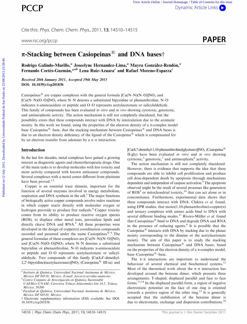
respectively. Fig. 1 shows the MG of Casiopeınas 01 and 13,
where it is possible to observe two BPs between Cu+2 and the
nitrogen atoms of the aromatic ligands, and two more BPs
between Cu+2 and the oxygen atoms of the acetylacetonate
or oxygen and nitrogen atoms of glycine. The four BPs are
characterized by their respective critical points (CP).33 The CPs
around the copper atom have donor–acceptor features, typical of
metal–ligand interactions: small r(r), r2r(r) 40, G(r)/r(r) E 1
and H(r)/r(r) o 0.34 In complex 01, the properties of the N–Cu
CP are r(r) = 0.087 a.u.,r2r(r) = 0.367 a.u., e=0.018 a.u. and
H(r) = �0.022 a.u., whereas the properties of O–Cu CP are
r(r) = 0.095 a.u., r2r(r) = 0.489 a.u., e = 0.032 a.u. and
H(r) = �0.020 a.u.
Table 1 Casiopeinass studied in this work
Family Number Ligandsa,b,c,d Substituents
1 01 acac/bip H02 acac/bip 4,40-diMe
2 03 acac/phen H04 acac/phen 4-Me05 acac/phen 5-Me06 acac/phen 4,7-diMe07 acac/phen 5,6-diMe08 acac/phen 3,4,7,8-tetraMe09 acac/phen 5-phenyl10 acac/phen 4,7-diphenyl11 acac/phen 5-Cl12 acac/phen 5-NO2
3 13 gly/phen H14 gly/phen 4-Me15 gly/phen 5-Me16 gly/phen 4,7-diMe17 gly/phen 5,6-diMe18 gly/phen 3,4,7,8-tetraMe19 gly/phen 4,7-diphenyl20 gly/phen 5-Cl21 gly/phen 5-NO2
a bip = bipyridine. b acac = acetylacetonate. c phen= phenanthroline.d gly = glycine.
Publ
ishe
d on
12
July
201
1. D
ownl
oade
d by
Uni
vers
idad
e Fe
dera
l de
Sao
Paul
o on
25/
08/2
013
23:5
8:40
.
View Article Online
14512 Phys. Chem. Chem. Phys., 2011, 13, 14510–14515 This journal is c the Owner Societies 2011
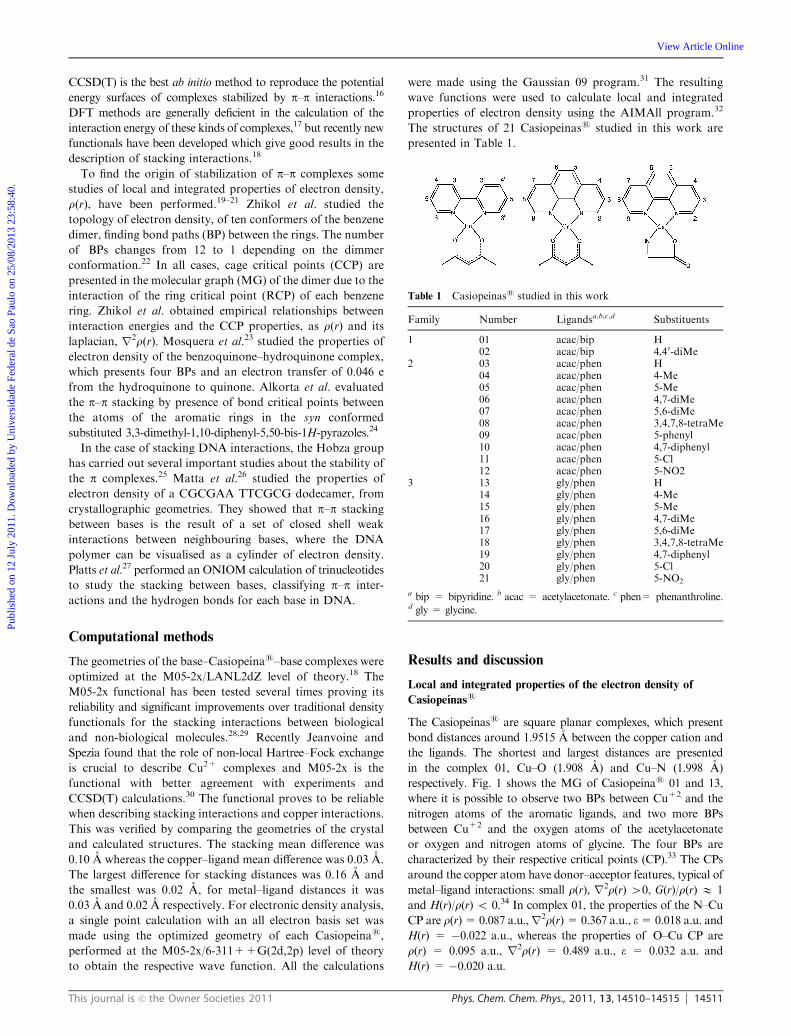
The donor–acceptor interactions can be also observed in the
laplacian of electron density, r2r(r), of Casiopeınass as
shown in Fig. 2. It is possible to observe four charge concen-
tration of the ligand heteroatoms oriented toward the equatorial
charge depletion of the metal, which also has two more charge
depletion at the axial positions, which allow two more
coordination sites in square pyramidal and octahedral geometries.
It is possible to define an atom using the properties of the
gradient of electron density of a molecule.19 The atomic
properties can be obtained by integration in the atomic region,
where the sum of the properties of certain groups of atoms in a
molecule, gives the group properties.35 The atomic electronic
populations of each group (N) in the complex were compared
with those of the isolated molecules as shown in eqn (1).
DN(group) = N(group@complex) � N(isolated group) (1)
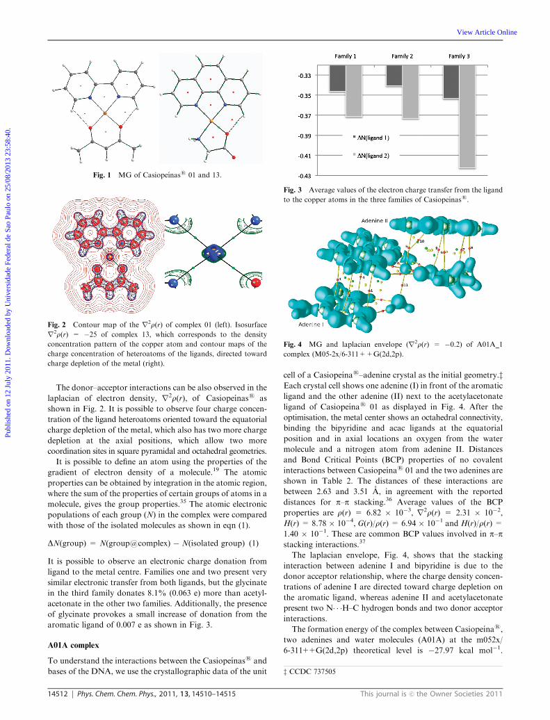
It is possible to observe an electronic charge donation from
ligand to the metal centre. Families one and two present very
similar electronic transfer from both ligands, but the glycinate
in the third family donates 8.1% (0.063 e) more than acetyl-
acetonate in the other two families. Additionally, the presence
of glycinate provokes a small increase of donation from the
aromatic ligand of 0.007 e as shown in Fig. 3.
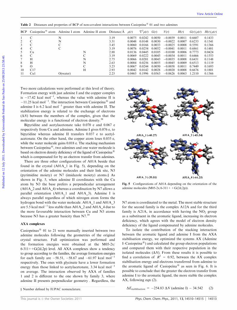
A01A complex
To understand the interactions between the Casiopeınass and
bases of the DNA, we use the crystallographic data of the unit
cell of a Casiopeınas–adenine crystal as the initial geometry.zEach crystal cell shows one adenine (I) in front of the aromatic
ligand and the other adenine (II) next to the acetylacetonate
ligand of Casiopeınas 01 as displayed in Fig. 4. After the
optimisation, the metal center shows an octahedral connectivity,
binding the bipyridine and acac ligands at the equatorial
position and in axial locations an oxygen from the water
molecule and a nitrogen atom from adenine II. Distances
and Bond Critical Points (BCP) properties of no covalent
interactions between Casiopeınas 01 and the two adenines are
shown in Table 2. The distances of these interactions are
between 2.63 and 3.51 A, in agreement with the reported
distances for p–p stacking.36 Average values of the BCP
properties are r(r) = 6.82 � 10�3, r2r(r) = 2.31 � 10�2,
H(r) = 8.78 � 10�4, G(r)/r(r) = 6.94 � 10�1 and H(r)/r(r) =1.40 � 10�1. These are common BCP values involved in p–pstacking interactions.37
The laplacian envelope, Fig. 4, shows that the stacking
interaction between adenine I and bipyridine is due to the
donor acceptor relationship, where the charge density concen-
trations of adenine I are directed toward charge depletion on
the aromatic ligand, whereas adenine II and acetylacetonate
present two N� � �H–C hydrogen bonds and two donor acceptor
interactions.
The formation energy of the complex between Casiopeınas,
two adenines and water molecules (A01A) at the m052x/
6-311++G(2d,2p) theoretical level is �27.97 kcal mol�1.
Fig. 1 MG of Casiopeınass 01 and 13.
Fig. 2 Contour map of the r2r(r) of complex 01 (left). Isosurface
r2r(r) = �25 of complex 13, which corresponds to the density
concentration pattern of the copper atom and contour maps of the
charge concentration of heteroatoms of the ligands, directed toward
charge depletion of the metal (right).
Fig. 3 Average values of the electron charge transfer from the ligand
to the copper atoms in the three families of Casiopeınass.
Fig. 4 MG and laplacian envelope (r2r(r) = �0.2) of A01A_1
complex (M05-2x/6-311++G(2d,2p).
z CCDC 737505
Publ
ishe
d on
12
July
201
1. D
ownl
oade
d by
Uni
vers
idad
e Fe
dera
l de
Sao
Paul
o on
25/
08/2
013
23:5
8:40
.
View Article Online
This journal is c the Owner Societies 2011 Phys. Chem. Chem. Phys., 2011, 13, 14510–14515 14513
Two more calculations were performed at this level of theory.
Formation energy with just adenine I and the copper complex
is �17.42 kcal mol�1, whereas the value with adenine II is
�11.25 kcal mol�1. The interaction between Casiopeınas and
adenine I is 6.2 kcal mol�1 greater than with adenine II. The
stabilization energy is related to the exchange of electrons
(DN) between the members of the complex, given that the
molecular energy is a functional of electron density.38
Bipyridine and acetylacetonate take 0.070 e and 0.067 e
respectively from Cu and adenines. Adenine I gives 0.076 e, to
bipyridine whereas adenine II transfers 0.037 e to acetyl-
acetonate. On the other hand, the copper atom loses 0.035 e,
while the water molecule gains 0.018 e. The stacking mechanism
between Casiopeınas, two adenines and one water molecule is
based on electron density deficiency of the ligand of Casiopeınas
which is compensated for by an electron transfer from adenines.
There are three other configurations of A01A beside that
found in the crystal (A01A_1 in Fig. 5), depending on the
orientation of the adenine molecules and their link site, N3
(pyrimidine moiety) or N7 (imidazole moiety) atoms.y As
shown in Fig. 5, when adenine II coordinates with the Cu
atom by N3 the base prefers a perpendicular arrangement
(A01A_2 and A01A_4) whereas a coordination by N7 allows a
parallel orientation (A01A_1 and A01A_3). Adenine I is
always parallel regardless of which nitrogen atom forms the
hydrogen bond with the water molecule. A01A_1 and A01A_3
are 5.5 kcal mol�1 less stable than A01A_2 and A01A_4 due to
the more favourable interaction between Cu and N3 atoms
because N3 has a greater basicity than N7.39
AXA complexes
Casiopeınass 01 to 21 were manually inserted between two
adenine molecules following the geometries of the original
crystal structure. Full optimization was performed and
the formation energies were obtained at the M05-2x/
6-311++G(2d,2p) level. All AXA complexes show a tendency
to group according to the families, the average formation energies
for each family are �58.51, �58.67 and �61.97 kcal mol�1
respectively. The ones with glycinate have a lower formation
energy than those linked to acetylacetonate, 3.34 kcal mol�1
on average. The interaction observed by AXA of families
1 and 2 is different to the one shown by family 3, where
adenine II presents perpendicular geometry . Regardless, the
N7 atom is coordinated to the metal. The most stable structure
for the second family is the complex A12A and for the third
family is A21A, in accordance with having the NO2 group
as a substituent in the aromatic ligand, increasing its electron
deficiency, which agrees with the model of electron density
deficiency of the ligand compensated by adenine molecules.
To isolate the contribution of the stacking interaction
between the aromatic ligand and adenine I from the AXA
stabilisation energy, we optimized the systems AX (Adenine
I–Casiopeınas) and calculated the group electron populations
and compared them with their respective population in the
isolated molecules (DN). From these results it is possible to
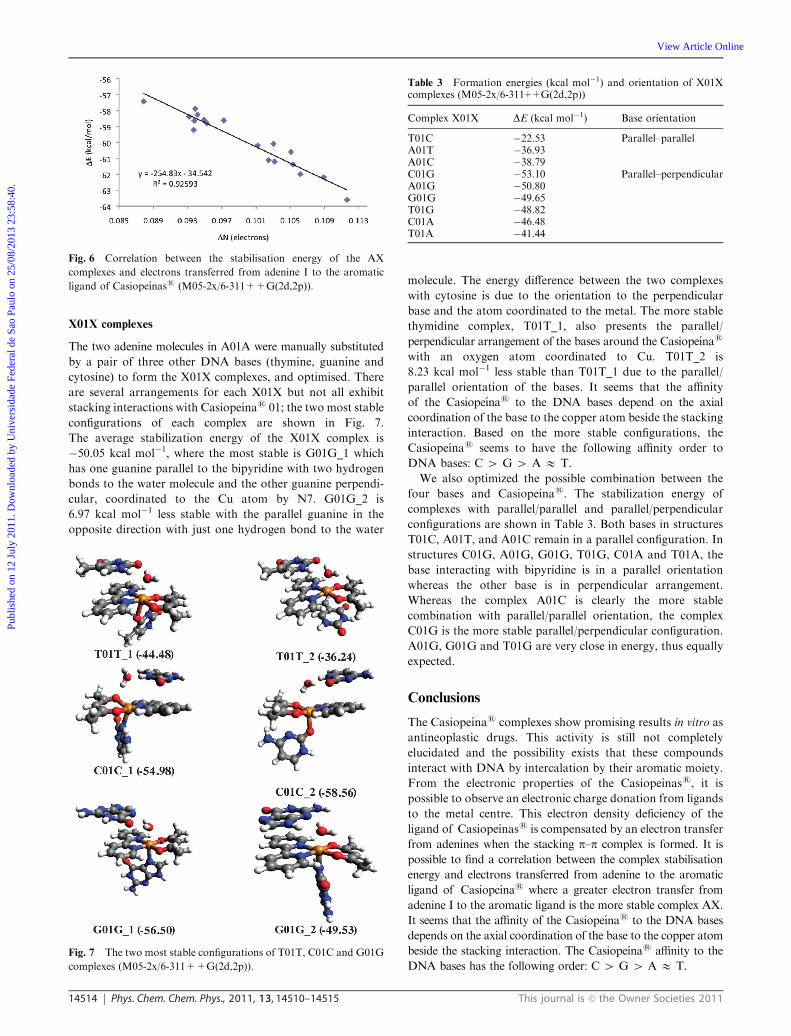
find a correlation of R2 = 0.92, between the AX complex
stabilisation energy and electrons transferred from adenine to
the aromatic ligand of Casiopeınas as seen in Fig. 6. It is
possible to conclude that the greater the electron transfer from
adenine I to the aromatic ligand, the more stable the complex
AX, following eqn (2).
DEstabilisation = �254.83 DN (adenine I) � 34.542 (2)
Table 2 Distances and properties of BCP of non-covalent interactions between Casiopeınas 01 and two adenines
BCP Casiopeınas atom Adenine I atom Adenine II atom Distance/A r(r) r2r(r) G(r) V(r) H(r) G(r)/r(r) H(r)/r(r)
1 C N 3.19 0.0075 0.0242 0.0050 �0.0039 0.0011 0.6607 0.14332 C N 3.51 0.0048 0.0148 0.0030 �0.0022 0.0007 0.6233 0.15413 C C 3.45 0.0060 0.0166 0.0033 �0.0025 0.0008 0.5591 0.13664 C C 3.19 0.0076 0.0254 0.0052 �0.0041 0.0011 0.6861 0.14815 Cu N 2.88 0.0136 0.0445 0.0105 �0.0100 0.0006 0.7773 0.04246 C C 3.19 0.0069 0.0222 0.0045 �0.0034 0.0011 0.6466 0.15537 H N 2.75 0.0066 0.0201 0.0043 �0.0035 0.0008 0.6431 0.11488 H N 2.63 0.0084 0.0256 0.0055 �0.0045 0.0009 0.6513 0.11199 O C 3.23 0.0067 0.0244 0.0050 �0.0038 0.0011 0.7449 0.169310 C17 H 2.93 0.0042 0.0142 0.0028 �0.0020 0.0008 0.6676 0.180111 Cu1 O(water) 2.23 0.0465 0.1996 0.0563 �0.0626 0.0063 1.2110 0.1366
Fig. 5 Configurations of A01A depending on the orientation of the
adenine molecules (M05-2x/6-311++G(2d,2p)).
y Number defined by IUPAC nomenclature.
Publ
ishe
d on
12
July
201
1. D
ownl
oade
d by
Uni
vers
idad
e Fe
dera
l de
Sao
Paul
o on
25/
08/2
013
23:5
8:40
.
View Article Online
14514 Phys. Chem. Chem. Phys., 2011, 13, 14510–14515 This journal is c the Owner Societies 2011
X01X complexes
The two adenine molecules in A01A were manually substituted
by a pair of three other DNA bases (thymine, guanine and
cytosine) to form the X01X complexes, and optimised. There
are several arrangements for each X01X but not all exhibit
stacking interactions with Casiopeınas 01; the two most stable
configurations of each complex are shown in Fig. 7.
The average stabilization energy of the X01X complex is
�50.05 kcal mol�1, where the most stable is G01G_1 which
has one guanine parallel to the bipyridine with two hydrogen
bonds to the water molecule and the other guanine perpendi-
cular, coordinated to the Cu atom by N7. G01G_2 is
6.97 kcal mol�1 less stable with the parallel guanine in the
opposite direction with just one hydrogen bond to the water
molecule. The energy difference between the two complexes
with cytosine is due to the orientation to the perpendicular
base and the atom coordinated to the metal. The more stable
thymidine complex, T01T_1, also presents the parallel/
perpendicular arrangement of the bases around the Casiopeınas
with an oxygen atom coordinated to Cu. T01T_2 is
8.23 kcal mol�1 less stable than T01T_1 due to the parallel/
parallel orientation of the bases. It seems that the affinity
of the Casiopeınas to the DNA bases depend on the axial
coordination of the base to the copper atom beside the stacking
interaction. Based on the more stable configurations, the
Casiopeınas seems to have the following affinity order to
DNA bases: C 4 G 4 A E T.
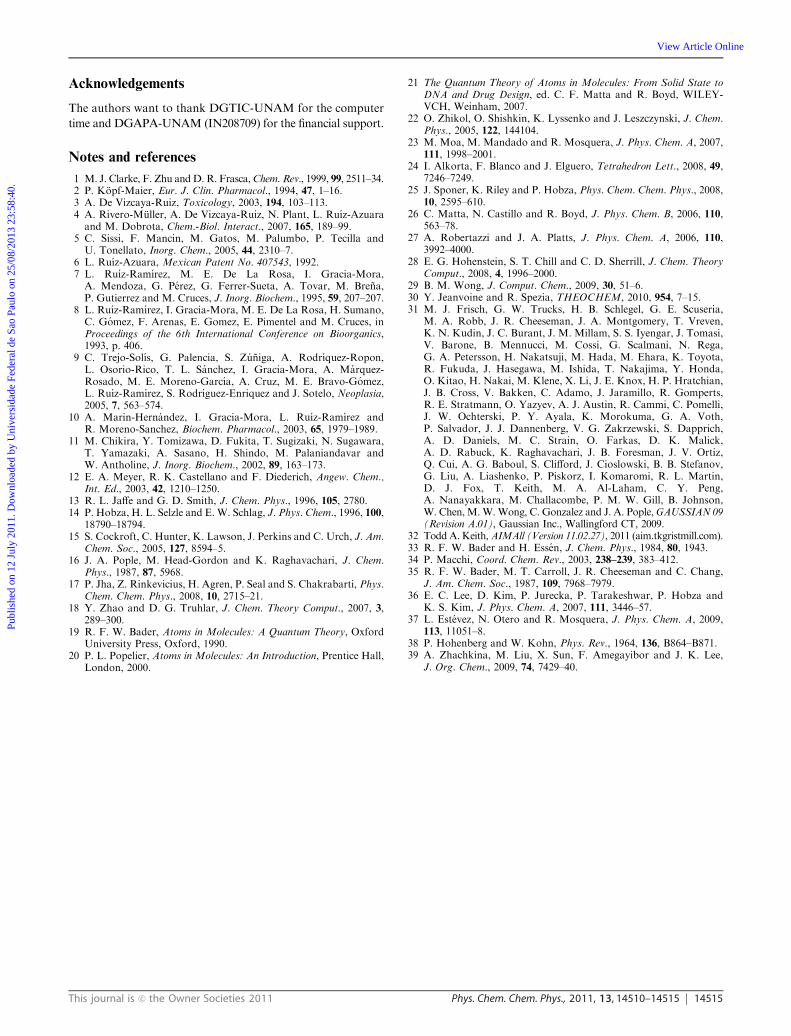
We also optimized the possible combination between the
four bases and Casiopeınas. The stabilization energy of
complexes with parallel/parallel and parallel/perpendicular
configurations are shown in Table 3. Both bases in structures
T01C, A01T, and A01C remain in a parallel configuration. In
structures C01G, A01G, G01G, T01G, C01A and T01A, the
base interacting with bipyridine is in a parallel orientation
whereas the other base is in perpendicular arrangement.
Whereas the complex A01C is clearly the more stable
combination with parallel/parallel orientation, the complex
C01G is the more stable parallel/perpendicular configuration.
A01G, G01G and T01G are very close in energy, thus equally
expected.
Conclusions
The Casiopeınas complexes show promising results in vitro as
antineoplastic drugs. This activity is still not completely
elucidated and the possibility exists that these compounds
interact with DNA by intercalation by their aromatic moiety.
From the electronic properties of the Casiopeınass, it is
possible to observe an electronic charge donation from ligands
to the metal centre. This electron density deficiency of the
ligand of Casiopeınass is compensated by an electron transfer
from adenines when the stacking p–p complex is formed. It is
possible to find a correlation between the complex stabilisation
energy and electrons transferred from adenine to the aromatic
ligand of Casiopeınas where a greater electron transfer from
adenine I to the aromatic ligand is the more stable complex AX.
It seems that the affinity of the Casiopeınas to the DNA bases
depends on the axial coordination of the base to the copper atom
beside the stacking interaction. The Casiopeınas affinity to the
DNA bases has the following order: C 4 G 4 A E T.
Fig. 6 Correlation between the stabilisation energy of the AX
complexes and electrons transferred from adenine I to the aromatic
ligand of Casiopeınass (M05-2x/6-311++G(2d,2p)).
Fig. 7 The two most stable configurations of T01T, C01C and G01G
complexes (M05-2x/6-311++G(2d,2p)).
Table 3 Formation energies (kcal mol�1) and orientation of X01Xcomplexes (M05-2x/6-311++G(2d,2p))
Complex X01X DE (kcal mol�1) Base orientation
T01C �22.53 Parallel–parallelA01T �36.93A01C �38.79C01G �53.10 Parallel–perpendicularA01G �50.80G01G �49.65T01G �48.82C01A �46.48T01A �41.44
Publ
ishe
d on
12
July
201
1. D
ownl
oade
d by
Uni
vers
idad
e Fe
dera
l de
Sao
Paul
o on
25/
08/2
013
23:5
8:40
.
View Article Online
This journal is c the Owner Societies 2011 Phys. Chem. Chem. Phys., 2011, 13, 14510–14515 14515
Acknowledgements
The authors want to thank DGTIC-UNAM for the computer
time and DGAPA-UNAM (IN208709) for the financial support.
Notes and references
1 M. J. Clarke, F. Zhu andD. R. Frasca,Chem. Rev., 1999, 99, 2511–34.2 P. Kopf-Maier, Eur. J. Clin. Pharmacol., 1994, 47, 1–16.3 A. De Vizcaya-Ruiz, Toxicology, 2003, 194, 103–113.4 A. Rivero-Muller, A. De Vizcaya-Ruiz, N. Plant, L. Ruız-Azuaraand M. Dobrota, Chem.-Biol. Interact., 2007, 165, 189–99.
5 C. Sissi, F. Mancin, M. Gatos, M. Palumbo, P. Tecilla andU. Tonellato, Inorg. Chem., 2005, 44, 2310–7.
6 L. Ruız-Azuara, Mexican Patent No. 407543, 1992.7 L. Ruız-Ramırez, M. E. De La Rosa, I. Gracia-Mora,A. Mendoza, G. Perez, G. Ferrer-Sueta, A. Tovar, M. Brena,P. Gutierrez and M. Cruces, J. Inorg. Biochem., 1995, 59, 207–207.
8 L. Ruız-Ramırez, I. Gracia-Mora, M. E. De La Rosa, H. Sumano,C. Gomez, F. Arenas, E. Gomez, E. Pimentel and M. Cruces, inProceedings of the 6th International Conference on Bioorganics,1993, p. 406.
9 C. Trejo-Solıs, G. Palencia, S. Zuniga, A. Rodrıquez-Ropon,L. Osorio-Rico, T. L. Sanchez, I. Gracia-Mora, A. Marquez-Rosado, M. E. Moreno-Garcıa, A. Cruz, M. E. Bravo-Gomez,L. Ruız-Ramırez, S. Rodrıguez-Enriquez and J. Sotelo, Neoplasia,2005, 7, 563–574.
10 A. Marin-Hernandez, I. Gracia-Mora, L. Ruız-Ramırez andR. Moreno-Sanchez, Biochem. Pharmacol., 2003, 65, 1979–1989.
11 M. Chikira, Y. Tomizawa, D. Fukita, T. Sugizaki, N. Sugawara,T. Yamazaki, A. Sasano, H. Shindo, M. Palaniandavar andW. Antholine, J. Inorg. Biochem., 2002, 89, 163–173.
12 E. A. Meyer, R. K. Castellano and F. Diederich, Angew. Chem.,Int. Ed., 2003, 42, 1210–1250.
13 R. L. Jaffe and G. D. Smith, J. Chem. Phys., 1996, 105, 2780.14 P. Hobza, H. L. Selzle and E. W. Schlag, J. Phys. Chem., 1996, 100,
18790–18794.15 S. Cockroft, C. Hunter, K. Lawson, J. Perkins and C. Urch, J. Am.
Chem. Soc., 2005, 127, 8594–5.16 J. A. Pople, M. Head-Gordon and K. Raghavachari, J. Chem.
Phys., 1987, 87, 5968.17 P. Jha, Z. Rinkevicius, H. Agren, P. Seal and S. Chakrabarti, Phys.
Chem. Chem. Phys., 2008, 10, 2715–21.18 Y. Zhao and D. G. Truhlar, J. Chem. Theory Comput., 2007, 3,
289–300.19 R. F. W. Bader, Atoms in Molecules: A Quantum Theory, Oxford
University Press, Oxford, 1990.20 P. L. Popelier, Atoms in Molecules: An Introduction, Prentice Hall,
London, 2000.
21 The Quantum Theory of Atoms in Molecules: From Solid State toDNA and Drug Design, ed. C. F. Matta and R. Boyd, WILEY-VCH, Weinham, 2007.
22 O. Zhikol, O. Shishkin, K. Lyssenko and J. Leszczynski, J. Chem.Phys., 2005, 122, 144104.
23 M. Moa, M. Mandado and R. Mosquera, J. Phys. Chem. A, 2007,111, 1998–2001.
24 I. Alkorta, F. Blanco and J. Elguero, Tetrahedron Lett., 2008, 49,7246–7249.
25 J. Sponer, K. Riley and P. Hobza, Phys. Chem. Chem. Phys., 2008,10, 2595–610.
26 C. Matta, N. Castillo and R. Boyd, J. Phys. Chem. B, 2006, 110,563–78.
27 A. Robertazzi and J. A. Platts, J. Phys. Chem. A, 2006, 110,3992–4000.
28 E. G. Hohenstein, S. T. Chill and C. D. Sherrill, J. Chem. TheoryComput., 2008, 4, 1996–2000.
29 B. M. Wong, J. Comput. Chem., 2009, 30, 51–6.30 Y. Jeanvoine and R. Spezia, THEOCHEM, 2010, 954, 7–15.31 M. J. Frisch, G. W. Trucks, H. B. Schlegel, G. E. Scuseria,
M. A. Robb, J. R. Cheeseman, J. A. Montgomery, T. Vreven,K. N. Kudin, J. C. Burant, J. M. Millam, S. S. Iyengar, J. Tomasi,V. Barone, B. Mennucci, M. Cossi, G. Scalmani, N. Rega,G. A. Petersson, H. Nakatsuji, M. Hada, M. Ehara, K. Toyota,R. Fukuda, J. Hasegawa, M. Ishida, T. Nakajima, Y. Honda,O. Kitao, H. Nakai, M. Klene, X. Li, J. E. Knox, H. P. Hratchian,J. B. Cross, V. Bakken, C. Adamo, J. Jaramillo, R. Gomperts,R. E. Stratmann, O. Yazyev, A. J. Austin, R. Cammi, C. Pomelli,J. W. Ochterski, P. Y. Ayala, K. Morokuma, G. A. Voth,P. Salvador, J. J. Dannenberg, V. G. Zakrzewski, S. Dapprich,A. D. Daniels, M. C. Strain, O. Farkas, D. K. Malick,A. D. Rabuck, K. Raghavachari, J. B. Foresman, J. V. Ortiz,Q. Cui, A. G. Baboul, S. Clifford, J. Cioslowski, B. B. Stefanov,G. Liu, A. Liashenko, P. Piskorz, I. Komaromi, R. L. Martin,D. J. Fox, T. Keith, M. A. Al-Laham, C. Y. Peng,A. Nanayakkara, M. Challacombe, P. M. W. Gill, B. Johnson,W. Chen,M.W.Wong, C. Gonzalez and J. A. Pople,GAUSSIAN 09(Revision A.01), Gaussian Inc., Wallingford CT, 2009.
32 ToddA.Keith,AIMAll (Version 11.02.27), 2011 (aim.tkgristmill.com).33 R. F. W. Bader and H. Essen, J. Chem. Phys., 1984, 80, 1943.34 P. Macchi, Coord. Chem. Rev., 2003, 238–239, 383–412.35 R. F. W. Bader, M. T. Carroll, J. R. Cheeseman and C. Chang,
J. Am. Chem. Soc., 1987, 109, 7968–7979.36 E. C. Lee, D. Kim, P. Jurecka, P. Tarakeshwar, P. Hobza and
K. S. Kim, J. Phys. Chem. A, 2007, 111, 3446–57.37 L. Estevez, N. Otero and R. Mosquera, J. Phys. Chem. A, 2009,
113, 11051–8.38 P. Hohenberg and W. Kohn, Phys. Rev., 1964, 136, B864–B871.39 A. Zhachkina, M. Liu, X. Sun, F. Amegayibor and J. K. Lee,
J. Org. Chem., 2009, 74, 7429–40.
Publ
ishe
d on
12
July
201
1. D
ownl
oade
d by
Uni
vers
idad
e Fe
dera
l de
Sao
Paul
o on
25/
08/2
013
23:5
8:40
.
View Article Online


![π stacking tackled with density functional theory...Sponer, Hobza and co-workers have studied in a ground-breaking series of papers [17–22] the stacking energies of DNA bases and](https://static.fdocument.org/doc/165x107/60732d783e8ccf056a3ee66a/-stacking-tackled-with-density-functional-theory-sponer-hobza-and-co-workers.jpg)