A role for DNA primase in coupling DNA replication to DNA damage...
12
The EMBO Journal Vol.16 No.3 pp.639–650, 1997 A role for DNA primase in coupling DNA replication to DNA damage response entry into mitosis through a surveillance mechanism, Federica Marini, Achille Pellicioli, which, in S.cerevisiae, involves the RAD9, RAD17, Vera Paciotti, Giovanna Lucchini, RAD24, RAD53, MEC1 and MEC3 gene products (Allen Paolo Plevani, David F.Stern 1 and et al., 1994; Weinert et al., 1994). Finally, the budding Marco Foiani 2 yeast RAD53, MEC1 and POL2 genes are required to Dipartimento di Genetica e di Biologia dei Microrganismi, Universita ` prevent entry into mitosis when DNA replication is blocked ˆ degli Studi di Milano, Via Celoria 26, 20133 Milano, Italy and (Allen et al., 1994; Weinert et al., 1994; Navas et al., 1995). 1 Department of Pathology, Yale University School of Medicine, Although it has been demonstrated that the cell cycle New Haven, CT 06520-8023, USA checkpoints are genetically controlled, the roles of the 2 Corresponding author different checkpoint proteins and the final targets of the signal transduction pathways leading to cell cycle delay The temperature-sensitive yeast DNA primase mutant as a consequence of DNA damage or replication block pri1-M4 fails to execute an early step of DNA replication are still unknown. The target of the checkpoint responding and exhibits a dominant, allele-specific sensitivity to to DNA damage in G 2 may be factors controlling execution DNA-damaging agents. pri1-M4 is defective in slowing of mitosis, while components of the replication apparatus down the rate of S phase progression and partially may act as sensors of DNA damage and stalled replication delaying the G 1 –S transition in response to DNA forks, and/or as targets of the checkpoint mechanisms damage. Conversely, the G 2 DNA damage response controlling entry and progression through S phase. The and the S–M checkpoint coupling completion of DNA involvement of replication proteins in cell cycle check- replication to mitosis are unaffected. The signal trans- points is supported by the finding that, in fission yeast, duction pathway leading to Rad53p phosphorylation the cdc18 , cut5 and cdt1 genes are required not induced by DNA damage is proficient in pri1-M4, and only for initiation of DNA synthesis, but also for the cell cycle delay caused by Rad53p overexpression is surveillance mechanisms preventing cells from entering counteracted by the pri1-M4 mutation. Altogether, our mitosis when either arrested or delayed in S phase (Kelly results suggest that DNA primase plays an essential et al., 1993; Hofmann and Beach, 1994; Saka et al., 1994). role in a subset of the Rad53p-dependent checkpoint Moreover, fission yeast DNA polymerases α and δ and pathways controlling cell cycle progression in response budding yeast DNA polymerase ε recently have been to DNA damage. implicated in the same mechanism (Araki et al., 1995; Keywords: budding yeast/cell cycle/checkpoints/DNA D’Urso et al., 1995; Francesconi et al., 1995; Navas et al., damage/DNA primase 1995). Finally, the Schizosaccharomyces pombe cds1 gene, the homolog of RAD53, has been identified as a multicopy suppressor of a temperature-sensitive (ts) mutant in the DNA polymerase α gene (Murakami and Introduction Okayama, 1995). The highly conserved DNA polymerase α–primase (pol Eukaryotic cells have developed a network of highly α–primase) complex is required for both the initiation and conserved surveillance mechanisms (checkpoints), ensur- elongation steps of DNA replication and is the target of ing that damaged chromosomes are repaired before being different regulatory mechanisms during the cell cycle replicated or segregated. These mechanisms are essential (Johnston and Lowndes, 1992; Campbell, 1993; Muzi for maintaining genome integrity and cell viability by Falconi et al., 1993; Foiani et al., 1995; Ferrari et al., delaying cell cycle progression in response to DNA 1996). The genes encoding the four subunits of the budding damage, and several studies have linked the damage yeast pol α–primase complex have been cloned, and response pathways to cell cycle events (for reviews, see several mutants have been produced and characterized Hartwell and Weinert, 1989; Hartwell and Kastan, 1994; (Lucchini et al., 1987, 1990; Francesconi et al., 1991; Murray, 1994; Nurse, 1994; Carr and Hoekstra, 1995; Longhese et al., 1993; Foiani et al., 1994). None of them Humphrey and Enoch, 1995; Lydall and Weinert, 1996). showed any sensitivity to DNA-damaging agents. Entry into S phase is delayed when DNA damage is Here, we describe the production of several new induced in G 1 and, in Saccharomyces cerevisiae, this mutations in the PRI1 gene encoding the catalytic primase control is dependent on the RAD9, RAD53/MEC2/SAD1/ subunit of the budding yeast pol α–primase complex, and SPK1 and RAD24 genes (Siede et al., 1993, 1994; Allen the characterization of the cell cycle defects associated et al., 1994). RAD53, together with the MEC1/ESR1 gene, with the ts pri1-M4 mutation. The pri1-M4 mutant is is also required for the checkpoint which slows down the defective in responding to DNA damage in G 1 /S and rate of DNA synthesis when DNA is damaged during S during S phase, and a role for DNA primase in the phase (Paulovich and Hartwell, 1995). Furthermore, when DNA damage is induced in G 2 , cells are able to delay surveillance mechanisms controlling the rate of progres- © Oxford University Press 639
Transcript of A role for DNA primase in coupling DNA replication to DNA damage...
The EMBO Journal Vol.16 No.3 pp.639–650, 1997
A role for DNA primase in coupling DNA replicationto DNA damage response
entry into mitosis through a surveillance mechanism,Federica Marini, Achille Pellicioli,which, in S.cerevisiae, involves the RAD9, RAD17,Vera Paciotti, Giovanna Lucchini,RAD24, RAD53, MEC1 and MEC3 gene products (AllenPaolo Plevani, David F.Stern1 andet al., 1994; Weinert et al., 1994). Finally, the buddingMarco Foiani2yeast RAD53, MEC1 and POL2 genes are required to
Dipartimento di Genetica e di Biologia dei Microrganismi, Universita prevent entry into mitosis when DNA replication is blockedˆdegli Studi di Milano, Via Celoria 26, 20133 Milano, Italy and (Allen et al., 1994; Weinert et al., 1994; Navas et al., 1995).
1Department of Pathology, Yale University School of Medicine, Although it has been demonstrated that the cell cycleNew Haven, CT 06520-8023, USA
checkpoints are genetically controlled, the roles of the2Corresponding author different checkpoint proteins and the final targets of the
signal transduction pathways leading to cell cycle delayThe temperature-sensitive yeast DNA primase mutant as a consequence of DNA damage or replication blockpri1-M4 fails to execute an early step of DNA replication are still unknown. The target of the checkpoint respondingand exhibits a dominant, allele-specific sensitivity to to DNA damage in G2 may be factors controlling executionDNA-damaging agents. pri1-M4 is defective in slowing of mitosis, while components of the replication apparatusdown the rate of S phase progression and partially may act as sensors of DNA damage and stalled replicationdelaying the G1–S transition in response to DNA forks, and/or as targets of the checkpoint mechanismsdamage. Conversely, the G2 DNA damage response controlling entry and progression through S phase. Theand the S–M checkpoint coupling completion of DNA involvement of replication proteins in cell cycle check-replication to mitosis are unaffected. The signal trans- points is supported by the finding that, in fission yeast,duction pathway leading to Rad53p phosphorylation the cdc18�, cut5� and cdt1� genes are required notinduced by DNA damage is proficient in pri1-M4, and
only for initiation of DNA synthesis, but also for thecell cycle delay caused by Rad53p overexpression is
surveillance mechanisms preventing cells from enteringcounteracted by the pri1-M4 mutation. Altogether, our
mitosis when either arrested or delayed in S phase (Kellyresults suggest that DNA primase plays an essential et al., 1993; Hofmann and Beach, 1994; Saka et al., 1994).role in a subset of the Rad53p-dependent checkpoint
Moreover, fission yeast DNA polymerases α and δ andpathways controlling cell cycle progression in response
budding yeast DNA polymerase ε recently have beento DNA damage.
implicated in the same mechanism (Araki et al., 1995;Keywords: budding yeast/cell cycle/checkpoints/DNA
D’Urso et al., 1995; Francesconi et al., 1995; Navas et al.,damage/DNA primase
1995). Finally, the Schizosaccharomyces pombe cds1�
gene, the homolog of RAD53, has been identified as amulticopy suppressor of a temperature-sensitive (ts)mutant in the DNA polymerase α gene (Murakami and
Introduction Okayama, 1995).The highly conserved DNA polymerase α–primase (polEukaryotic cells have developed a network of highly
α–primase) complex is required for both the initiation andconserved surveillance mechanisms (checkpoints), ensur-elongation steps of DNA replication and is the target ofing that damaged chromosomes are repaired before beingdifferent regulatory mechanisms during the cell cyclereplicated or segregated. These mechanisms are essential(Johnston and Lowndes, 1992; Campbell, 1993; Muzifor maintaining genome integrity and cell viability byFalconi et al., 1993; Foiani et al., 1995; Ferrari et al.,delaying cell cycle progression in response to DNA1996). The genes encoding the four subunits of the buddingdamage, and several studies have linked the damageyeast pol α–primase complex have been cloned, andresponse pathways to cell cycle events (for reviews, seeseveral mutants have been produced and characterizedHartwell and Weinert, 1989; Hartwell and Kastan, 1994;(Lucchini et al., 1987, 1990; Francesconi et al., 1991;Murray, 1994; Nurse, 1994; Carr and Hoekstra, 1995;Longhese et al., 1993; Foiani et al., 1994). None of themHumphrey and Enoch, 1995; Lydall and Weinert, 1996).showed any sensitivity to DNA-damaging agents.Entry into S phase is delayed when DNA damage is
Here, we describe the production of several newinduced in G1 and, in Saccharomyces cerevisiae, thismutations in the PRI1 gene encoding the catalytic primasecontrol is dependent on the RAD9, RAD53/MEC2/SAD1/subunit of the budding yeast pol α–primase complex, andSPK1 and RAD24 genes (Siede et al., 1993, 1994; Allenthe characterization of the cell cycle defects associatedet al., 1994). RAD53, together with the MEC1/ESR1 gene,with the ts pri1-M4 mutation. The pri1-M4 mutant isis also required for the checkpoint which slows down thedefective in responding to DNA damage in G1/S andrate of DNA synthesis when DNA is damaged during Sduring S phase, and a role for DNA primase in thephase (Paulovich and Hartwell, 1995). Furthermore, when
DNA damage is induced in G2, cells are able to delay surveillance mechanisms controlling the rate of progres-
© Oxford University Press 639
F.Marini et al.
showed an accumulation of S phase cells (Figure 2A).Moreover, when pri1-M4 cells were arrested in G1 withα-factor, and then released from the α-factor block atpermissive temperature, they were delayed in reachingG2, although FACS analysis did not allow us to distinguishbetween a defect in entering S phase and a slowerprogression through S phase (Figure 2A).
The pri1-M4 mutation caused a tight ts phenotype,since pri1-M4 cells released from the α-factor block atthe restrictive temperature (36°C) arrested as large-buddedcells, with a single nucleus, short spindle and a 1C DNAcontent (Figure 2A and data not shown), suggesting thatthey failed to execute an early step of DNA synthesis.Finally, pri1-M4 cells showed first cell cycle arrest(Hereford and Hartwell, 1974; Hartwell 1976), either whenblocked at 36°C and then released at the permissivetemperature in the presence of hydroxyurea (HU), or whenfirst arrested in HU at 25°C and then released from theHU block at 37°C (see Materials and methods). Thisfinding indicates that DNA primase is required for ongoingDNA synthesis, although it does not exclude the possibilitythat DNA primase may also play an essential function ininitiation of DNA synthesis, as suggested by the resultsshown in Figure 2A.
The ts phenotype associated with the pri1-M4 mutationis recessive, since both the growth rate and FACS profile of
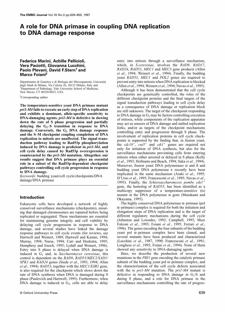
Fig. 1. Primase stability in pri1 mutants. (A) A bar schematicallyPRI1/pri1-M4 heterozygous and PRI1/PRI1 homozygousrepresents the p48 polypeptide. Shaded boxes within the bar indicatediploid strains were indistinguishable from each otherconserved amino acid regions. Arrows indicate the position of the two
amino acid insertions described in Materials and methods. (B) Twenty at 36°C.five μg of protein extracts prepared from the indicated strains wereanalyzed by Western blotting as described in Materials and methods. A dominant and allele-specific DNA damage(C) A total of 3.5 mg of protein extracts from the indicated strains sensitivity is associated with the pri1-M4 mutationwere immunoprecipitated with the anti-p180 y48 monoclonal antibody
The pri1-M4 mutant is significantly sensitive to DNA-(Ferrari et al., 1996), and analyzed by SDS–PAGE and Westerndamaging agents at the permissive temperature. In fact,blotting with specific antibodies against the pol α–primase subunits.
when pri1-M4 cultures were UV irradiated or treated withthe alkylating agent methyl methanesulfonate (MMS), thepercentage of viable cells decreased compared with thesion through S phase in response to DNA damage will beisogenic wild-type (Figure 2B and C). Moreover, the pri1-discussed.M4 allele caused increased sensitivity to the radiomimeticdrug bleomycin (data not shown).
Results DNA damage sensitivity was specific for the pri1-M4mutant, since neither different pri1 alleles, nor mutations
Mutagenesis of the PRI1 genein the genes encoding the other subunits of the pol α–We have mutagenized the PRI1 gene carried on a centro-primase complex, which severely affect DNA synthesis,meric plasmid by using the two-codon insertion techniquewere more sensitive than wild-type to UV, MMS and(Barany, 1988). Among the obtained mutations (Figure 1A,bleomycin (data not shown).
Materials and methods), the pri1-M4 and pri1-T1 allelesThe sensitivity to UV and MMS treatments associated
caused a ts phenotype, pri1-M2 and pri1-M3 were lethal,with the pri1-M4 mutation is dominant, since the pri1-
while the other mutations did not result in any detectableM4/PRI1 heterozygous diploid strain showed a DNA
phenotype (data not shown).damage sensitivity comparable with that of the pri1-M4/
As shown in Figure 1B, the level of the p48 primasepri1-M4 homozygous strain (Figure 2D and E). However,
polypeptide was reduced dramatically in pri1-H2 and pri1-DNA damage sensitivity of pri1-M4/PRI1 heterozygoussH2 protein extracts, while the amount of p48 onlycells was similar to that of PRI1/PRI1 homozygous cells
partially decreased in pri1-M4 extracts and pol α–primasewhen tested at 37°C (Figure 2F and G), probably due to
complex formation and stability were not affectedinactivation of the pri1-M4 gene product. Therefore, while
(Figure 1B and C). Shift to the restrictive temperature ofthe ts phenotype associated with the pri1-M4 mutation is
pri1-M4 mutant cells did not influence either the p48 levelrecessive, DNA damage sensitivity at the permissive
or the stability of the pol α–primase complex.temperature is allele specific and dominant.
The pri1-M4 mutant is defective in DNA synthesis Cell cycle delay in response to DNA damageThe pri1-M4 mutant is partially defective in DNA synthesis during G1 or S phase is reduced in the pri1-M4already at the permissive temperature. In fact, pri1-M4 mutantcells, exponentially growing at 25°C, were mostly budded, Genetically distinguishable surveillance mechanisms are
employed to delay cell cycle progression in response toand fluorescence-activated cell sorter (FACS) analysis
640

DNA primase and checkpoints
Fig. 2. pri1-M4 is defective in an early step of DNA synthesis and is sensitive to DNA-damaging agents. (A) Cultures of strains K699 (wild-type)and CY387 (pri1-M4) logarithmically growing at 25°C (log) were synchronized by α-factor treatment (2 μg/ml) and shifted either to 25 or to 36°Cat time zero after α-factor release. Samples were taken at the indicated times and analyzed by FACS. (B–G) One hundred and 1000 cells fromovernight saturated YPD cultures of strains K699 (PRI1), CY387 (pri1-M4), CYd438 (PRI1/PRI1), CYd439 (PRI1/pri1-M4) and CYd524 (pri1-M4/pri1-M4) were either plated on YPD medium containing the indicated MMS concentrations (B, D and F) or UV irradiated on YPD plates at theindicated dosages (C, E and G). Plates were incubated at 25°C (B–E) or at 37°C (F and G) and colonies were counted after 3–4 days. StrainCyd524 did not give rise to any colony when incubated at 37°C (F and G). Standard deviations were calculated using two to three samples. Theexperiments in (B–G) were performed two to four times with similar results.
DNA damage (Weinert et al., 1994; Carr and Hoekstra, paradoxically, this mutant allele, which is defective inDNA synthesis, replicates DNA faster than wild-type1995; Friedberg et al., 1995; Lydall and Weinert, 1996).
Wild-type cells, UV irradiated in G1, delay the G1–S under these conditions. pri1-M4 cells held in α-factorthroughout the MMS treatment mantained a 1C DNAtransition, probably to allow DNA repair (Figure 3A),
while mutant strains defective in this checkpoint mechan- content and did not lose cell viability (Figure 4B), sug-gesting that increased cell lethality induced by DNAism replicate DNA prematurely and lose cell viability
(Siede et al., 1993; Allen et al., 1994). damage in the pri1-M4 mutant strain is related to its fasterprogression through S phase.In pri1-M4 cultures released from a G1 block after
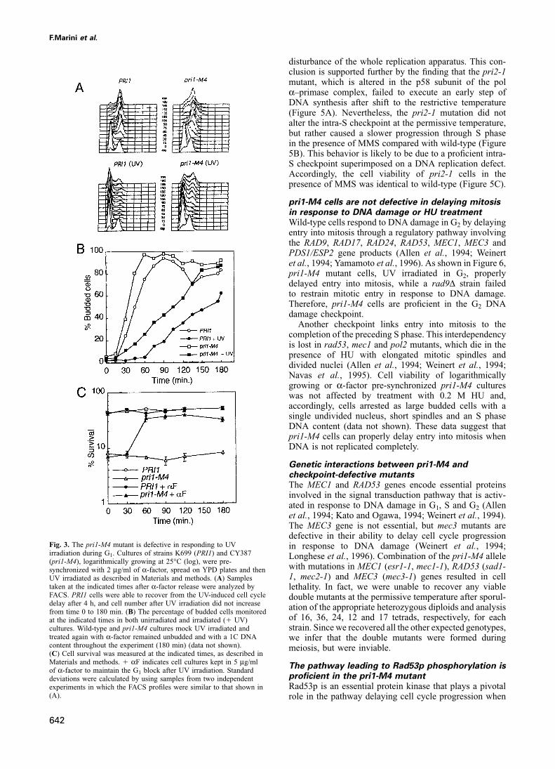
UV treatment, both bud emergence (Figure 3B) and the In order to correlate pri1-M4 DNA damage sensitivityto its intra-S checkpoint defect, we tested whether fasterappearance of cells with a 2C DNA content (Figure 3A)
occurred earlier when compared with the wild-type strain S phase progression of pri1-M4 cells in the presence ofMMS was also dominant. We found that pri1-M4 [pFE139]in the same conditions, suggesting that pri1-M4 cells
are partially defective in properly delaying cell cycle cells, containing a centromeric plasmid carrying the wild-type PRI1 gene, failed to properly delay cell cycle progres-progression in response to UV irradiation during G1. This
phenotype was associated with increased cell lethality, sion in the presence of MMS, similarly to what wasobserved in pri1-M4 [pFE202] cells, carrying the pri1-which was almost completely prevented by holding the
cells in α-factor for at least 60 min after UV irradiation M4 allele on the same vector (Figure 4C). As expected,the checkpoint defect of pri1-M4 [pFE139] cells caused(Figure 3C).
Another genetically controlled regulatory mechanism, an increase in cell lethality which was prevented byα-factor treatment (Figure 4D). However, both the tsrequiring the RAD53 and MEC1 genes, slows the rate of
S phase progression, when DNA damage occurs during phenotype (data not shown) and the mitotic cell cycledelay in the absence of MMS treatment observed inDNA replication (Paulovich and Hartwell, 1995). When
pri1-M4 cultures were released from α-factor block in the pri1-M4 cells were abolished in pri1-M4 [pFE139] cells(Figure 4C). Since the pri1-M4 intra-S checkpoint defectpresence of MMS, progression through S phase was more
rapid than in wild-type (Figure 4A), and cell viability is dominant and can be distinguished genetically from therecessive DNA synthesis defect, the inability to delaywas strongly reduced (Figure 4B). Hence, the rate of
progression through S phase in pri1-M4 shows only partial, properly the rate of S phase progression in response toDNA damage is unlikely to be related to a generalif any, reduction in response to MMS treatment and,
641
F.Marini et al.
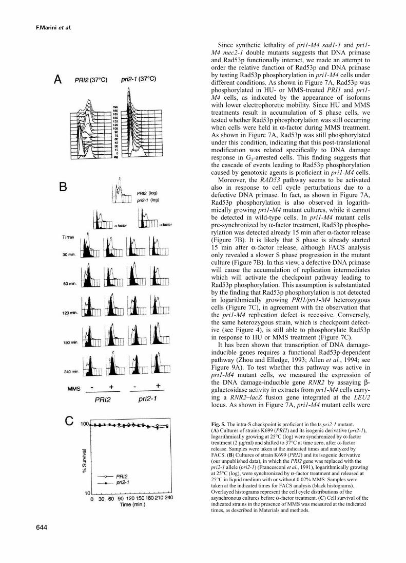
disturbance of the whole replication apparatus. This con-clusion is supported further by the finding that the pri2-1mutant, which is altered in the p58 subunit of the polα–primase complex, failed to execute an early step ofDNA synthesis after shift to the restrictive temperature(Figure 5A). Nevertheless, the pri2-1 mutation did notalter the intra-S checkpoint at the permissive temperature,but rather caused a slower progression through S phasein the presence of MMS compared with wild-type (Figure5B). This behavior is likely to be due to a proficient intra-S checkpoint superimposed on a DNA replication defect.Accordingly, the cell viability of pri2-1 cells in thepresence of MMS was identical to wild-type (Figure 5C).
pri1-M4 cells are not defective in delaying mitosisin response to DNA damage or HU treatmentWild-type cells respond to DNA damage in G2 by delayingentry into mitosis through a regulatory pathway involvingthe RAD9, RAD17, RAD24, RAD53, MEC1, MEC3 andPDS1/ESP2 gene products (Allen et al., 1994; Weinertet al., 1994; Yamamoto et al., 1996). As shown in Figure 6,pri1-M4 mutant cells, UV irradiated in G2, properlydelayed entry into mitosis, while a rad9Δ strain failedto restrain mitotic entry in response to DNA damage.Therefore, pri1-M4 cells are proficient in the G2 DNAdamage checkpoint.
Another checkpoint links entry into mitosis to thecompletion of the preceding S phase. This interdependencyis lost in rad53, mec1 and pol2 mutants, which die in thepresence of HU with elongated mitotic spindles anddivided nuclei (Allen et al., 1994; Weinert et al., 1994;Navas et al., 1995). Cell viability of logarithmicallygrowing or α-factor pre-synchronized pri1-M4 cultureswas not affected by treatment with 0.2 M HU and,accordingly, cells arrested as large budded cells with asingle undivided nucleus, short spindles and an S phaseDNA content (data not shown). These data suggest thatpri1-M4 cells can properly delay entry into mitosis whenDNA is not replicated completely.
Genetic interactions between pri1-M4 andcheckpoint-defective mutantsThe MEC1 and RAD53 genes encode essential proteinsinvolved in the signal transduction pathway that is activ-ated in response to DNA damage in G1, S and G2 (Allenet al., 1994; Kato and Ogawa, 1994; Weinert et al., 1994).The MEC3 gene is not essential, but mec3 mutants aredefective in their ability to delay cell cycle progression
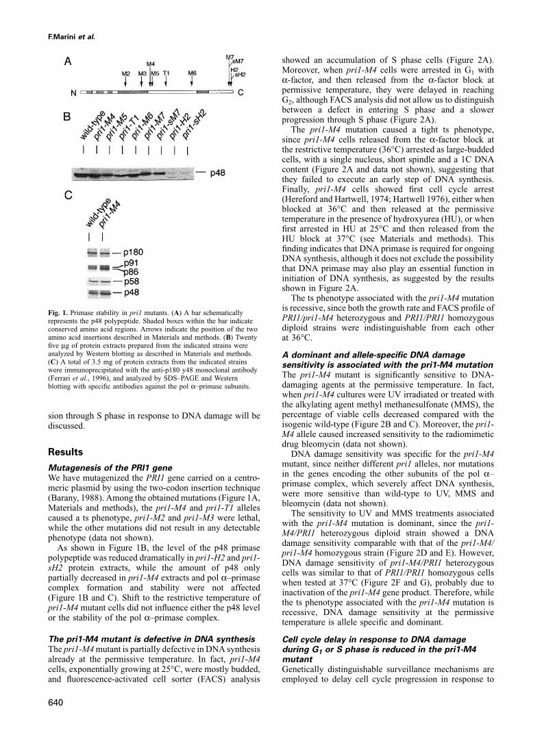
Fig. 3. The pri1-M4 mutant is defective in responding to UV in response to DNA damage (Weinert et al., 1994;irradiation during G1. Cultures of strains K699 (PRI1) and CY387 Longhese et al., 1996). Combination of the pri1-M4 allele(pri1-M4), logarithmically growing at 25°C (log), were pre-
with mutations in MEC1 (esr1-1, mec1-1), RAD53 (sad1-synchronized with 2 μg/ml of α-factor, spread on YPD plates and then1, mec2-1) and MEC3 (mec3-1) genes resulted in cellUV irradiated as described in Materials and methods. (A) Samples
taken at the indicated times after α-factor release were analyzed by lethality. In fact, we were unable to recover any viableFACS. PRI1 cells were able to recover from the UV-induced cell cycle double mutants at the permissive temperature after sporul-delay after 4 h, and cell number after UV irradiation did not increase ation of the appropriate heterozygous diploids and analysisfrom time 0 to 180 min. (B) The percentage of budded cells monitored
of 16, 36, 24, 12 and 17 tetrads, respectively, for eachat the indicated times in both unirradiated and irradiated (� UV)strain. Since we recovered all the other expected genotypes,cultures. Wild-type and pri1-M4 cultures mock UV irradiated and
treated again with α-factor remained unbudded and with a 1C DNA we infer that the double mutants were formed duringcontent throughout the experiment (180 min) (data not shown). meiosis, but were inviable.(C) Cell survival was measured at the indicated times, as described inMaterials and methods. � αF indicates cell cultures kept in 5 μg/ml
The pathway leading to Rad53p phosphorylation isof α-factor to maintain the G1 block after UV irradiation. Standard
proficient in the pri1-M4 mutantdeviations were calculated by using samples from two independentRad53p is an essential protein kinase that plays a pivotalexperiments in which the FACS profiles were similar to that shown in
(A). role in the pathway delaying cell cycle progression when
642
DNA primase and checkpoints
Fig. 4. The pri1-M4 mutant is defective in slowing down S phase progression in the presence of DNA damage. (A) and (C) Log-phase (log) culturesof strains K699 (PRI1), CY387 (pri1-M4), CY387 transformed with the pFE139 centromeric plasmid carrying the wild-type PRI1 gene (pri1-M4[pFE139]) and CY387 transformed with the pFE202 centromeric plasmid carrying the pri1-M4 allele (pri1-M4[pFE202]) were pre-synchronizedby α-factor treatment and released in YPD with or without 0.02% MMS. Samples were taken at the indicated times for FACS analysis (blackhistograms). Overlayed histograms represent the cell cycle distributions of the asynchronous cultures before α-factor treatment. The experimentsshown in (A) and (C) were performed three times and twice, respectively, with similar results. FACS profiles of strain K699 transformed with thecentromeric plasmid pFE139 were indistinguishable from that shown for the non-transformed K699 strain. (B) and (D) Cell survival was measured atthe indicated times, as described in Materials and methods. � αF indicates cell cultures kept in 5 μg/ml of α-factor throughout the MMS treatmentto maintain the G1 block. Standard deviations were calculated by using samples from two independent experiments in which the FACS profiles weresimilar to that shown in (A) and (C).
DNA is damaged or replication is not complete (Zheng causes Rad53pphosphorylation during S phaseunder condi-tions perturbing proper cell cycle progression (Sun et al.,et al., 1993; Allen et al., 1994; Weinert et al., 1994). Rad53p
is phosphorylated in trans by a Mec1p- and Mec3p-depend- 1996), but this is not observed in logarithmically growingwild-type cells or during S phase of α-factor pre-synchron-ent mechanism in response to DNA damage (Sanchez et al.,
1996; Sun et al., 1996). A Mec1p-dependent mechanism ized cell cultures (Figure 7B, and Sun et al., 1996).
643
F.Marini et al.
Since synthetic lethality of pri1-M4 sad1-1 and pri1-M4 mec2-1 double mutants suggests that DNA primaseand Rad53p functionally interact, we made an attempt toorder the relative function of Rad53p and DNA primaseby testing Rad53p phosphorylation in pri1-M4 cells underdifferent conditions. As shown in Figure 7A, Rad53p wasphosphorylated in HU- or MMS-treated PRI1 and pri1-M4 cells, as indicated by the appearance of isoformswith lower electrophoretic mobility. Since HU and MMStreatments result in accumulation of S phase cells, wetested whether Rad53p phosphorylation was still occurringwhen cells were held in α-factor during MMS treatment.As shown in Figure 7A, Rad53p was still phosphorylatedunder this condition, indicating that this post-translationalmodification was related specifically to DNA damageresponse in G1-arrested cells. This finding suggests thatthe cascade of events leading to Rad53p phosphorylationcaused by genotoxic agents is proficient in pri1-M4 cells.
Moreover, the RAD53 pathway seems to be activatedalso in response to cell cycle perturbations due to adefective DNA primase. In fact, as shown in Figure 7A,Rad53p phosphorylation is also observed in logarith-mically growing pri1-M4 mutant cultures, while it cannotbe detected in wild-type cells. In pri1-M4 mutant cellspre-synchronized by α-factor treatment, Rad53p phospho-rylation was detected already 15 min after α-factor release(Figure 7B). It is likely that S phase is already started15 min after α-factor release, although FACS analysisonly revealed a slower S phase progression in the mutantculture (Figure 7B). In this view, a defective DNA primasewill cause the accumulation of replication intermediateswhich will activate the checkpoint pathway leading toRad53p phosphorylation. This assumption is substantiatedby the finding that Rad53p phosphorylation is not detectedin logarithmically growing PRI1/pri1-M4 heterozygouscells (Figure 7C), in agreement with the observation thatthe pri1-M4 replication defect is recessive. Conversely,the same heterozygous strain, which is checkpoint defect-ive (see Figure 4), is still able to phosphorylate Rad53pin response to HU or MMS treatment (Figure 7C).
It has been shown that transcription of DNA damage-inducible genes requires a functional Rad53p-dependentpathway (Zhou and Elledge, 1993; Allen et al., 1994; seeFigure 9A). To test whether this pathway was active inpri1-M4 mutant cells, we measured the expression ofthe DNA damage-inducible gene RNR2 by assaying β-galactosidase activity in extracts from pri1-M4 cells carry-ing a RNR2–lacZ fusion gene integrated at the LEU2locus. As shown in Figure 7A, pri1-M4 mutant cells were
Fig. 5. The intra-S checkpoint is proficient in the ts pri2-1 mutant.(A) Cultures of strains K699 (PRI2) and its isogenic derivative (pri2-1),logarithmically growing at 25°C (log) were synchronized by α-factortreatment (2 μg/ml) and shifted to 37°C at time zero, after α-factorrelease. Samples were taken at the indicated times and analyzed byFACS. (B) Cultures of strain K699 (PRI2) and its isogenic derivative(our unpublished data), in which the PRI2 gene was replaced with thepri2-1 allele (pri2-1) (Francesconi et al., 1991), logarithmically growingat 25°C (log), were synchronized by α-factor treatment and released at25°C in liquid medium with or without 0.02% MMS. Samples weretaken at the indicated times for FACS analysis (black histograms).Overlayed histograms represent the cell cycle distributions of theasynchronous cultures before α-factor treatment. (C) Cell survival of theindicated strains in the presence of MMS was measured at the indicatedtimes, as described in Materials and methods.
644
DNA primase and checkpoints
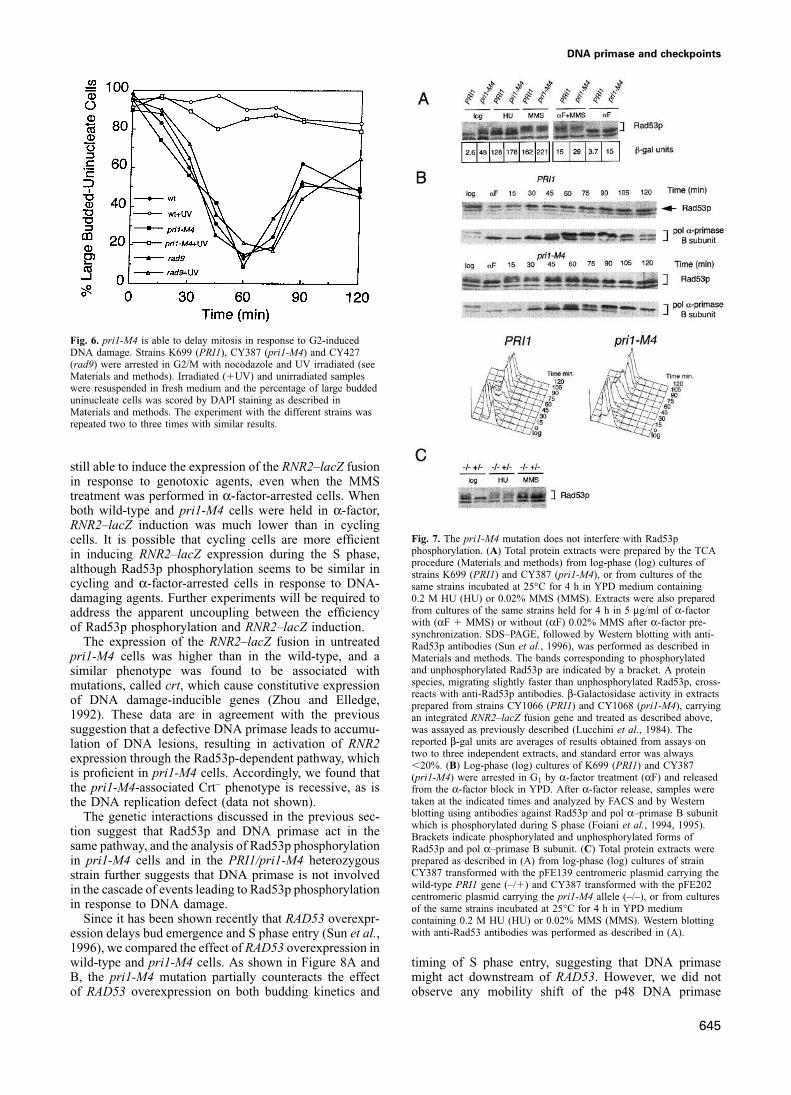
Fig. 6. pri1-M4 is able to delay mitosis in response to G2-inducedDNA damage. Strains K699 (PRI1), CY387 (pri1-M4) and CY427(rad9) were arrested in G2/M with nocodazole and UV irradiated (seeMaterials and methods). Irradiated (�UV) and unirradiated sampleswere resuspended in fresh medium and the percentage of large buddeduninucleate cells was scored by DAPI staining as described inMaterials and methods. The experiment with the different strains wasrepeated two to three times with similar results.
still able to induce the expression of the RNR2–lacZ fusionin response to genotoxic agents, even when the MMStreatment was performed in α-factor-arrested cells. Whenboth wild-type and pri1-M4 cells were held in α-factor,RNR2–lacZ induction was much lower than in cycling
Fig. 7. The pri1-M4 mutation does not interfere with Rad53pcells. It is possible that cycling cells are more efficientphosphorylation. (A) Total protein extracts were prepared by the TCAin inducing RNR2–lacZ expression during the S phase,procedure (Materials and methods) from log-phase (log) cultures of
although Rad53p phosphorylation seems to be similar in strains K699 (PRI1) and CY387 (pri1-M4), or from cultures of thecycling and α-factor-arrested cells in response to DNA- same strains incubated at 25°C for 4 h in YPD medium containing
0.2 M HU (HU) or 0.02% MMS (MMS). Extracts were also prepareddamaging agents. Further experiments will be required tofrom cultures of the same strains held for 4 h in 5 μg/ml of α-factoraddress the apparent uncoupling between the efficiencywith (αF � MMS) or without (αF) 0.02% MMS after α-factor pre-of Rad53p phosphorylation and RNR2–lacZ induction.synchronization. SDS–PAGE, followed by Western blotting with anti-
The expression of the RNR2–lacZ fusion in untreated Rad53p antibodies (Sun et al., 1996), was performed as described inpri1-M4 cells was higher than in the wild-type, and a Materials and methods. The bands corresponding to phosphorylated
and unphosphorylated Rad53p are indicated by a bracket. A proteinsimilar phenotype was found to be associated withspecies, migrating slightly faster than unphosphorylated Rad53p, cross-mutations, called crt, which cause constitutive expressionreacts with anti-Rad53p antibodies. β-Galactosidase activity in extracts
of DNA damage-inducible genes (Zhou and Elledge, prepared from strains CY1066 (PRI1) and CY1068 (pri1-M4), carrying1992). These data are in agreement with the previous an integrated RNR2–lacZ fusion gene and treated as described above,
was assayed as previously described (Lucchini et al., 1984). Thesuggestion that a defective DNA primase leads to accumu-reported β-gal units are averages of results obtained from assays onlation of DNA lesions, resulting in activation of RNR2two to three independent extracts, and standard error was alwaysexpression through the Rad53p-dependent pathway, which�20%. (B) Log-phase (log) cultures of K699 (PRI1) and CY387
is proficient in pri1-M4 cells. Accordingly, we found that (pri1-M4) were arrested in G1 by α-factor treatment (αF) and releasedthe pri1-M4-associated Crt– phenotype is recessive, as is from the α-factor block in YPD. After α-factor release, samples were
taken at the indicated times and analyzed by FACS and by Westernthe DNA replication defect (data not shown).blotting using antibodies against Rad53p and pol α–primase B subunitThe genetic interactions discussed in the previous sec-which is phosphorylated during S phase (Foiani et al., 1994, 1995).
tion suggest that Rad53p and DNA primase act in the Brackets indicate phosphorylated and unphosphorylated forms ofsame pathway, and the analysis of Rad53p phosphorylation Rad53p and pol α–primase B subunit. (C) Total protein extracts were
prepared as described in (A) from log-phase (log) cultures of strainin pri1-M4 cells and in the PRI1/pri1-M4 heterozygousCY387 transformed with the pFE139 centromeric plasmid carrying thestrain further suggests that DNA primase is not involvedwild-type PRI1 gene (–/�) and CY387 transformed with the pFE202in the cascade of events leading to Rad53p phosphorylationcentromeric plasmid carrying the pri1-M4 allele (–/–), or from cultures
in response to DNA damage. of the same strains incubated at 25°C for 4 h in YPD mediumSince it has been shown recently that RAD53 overexpr- containing 0.2 M HU (HU) or 0.02% MMS (MMS). Western blotting
with anti-Rad53 antibodies was performed as described in (A).ession delays bud emergence and S phase entry (Sun et al.,1996), we compared the effect of RAD53 overexpression inwild-type and pri1-M4 cells. As shown in Figure 8A and timing of S phase entry, suggesting that DNA primase
might act downstream of RAD53. However, we did notB, the pri1-M4 mutation partially counteracts the effectof RAD53 overexpression on both budding kinetics and observe any mobility shift of the p48 DNA primase
645

F.Marini et al.
Fig. 8. The pri1-M4 mutation counteracts the cell cycle delay caused by Rad53p overexpression. (A) Strains K699 (PRI1) and CY387 (pri1-M4)were transformed with plasmids pNB187 (Vector) or pNB187-SPK1 (GAL1-RAD53). Transformants were grown in SD medium containing 2%raffinose (log), synchronized by α-factor treatment and released from the α-factor block at 25°C in SD medium containing 2% raffinose and 2%galactose. Samples for FACS analysis were taken at the indicated times after α-factor release (time 0). (B) The percentage of budded cells wasmonitored, at the indicated times, in the same strains described in (A). (C) Total protein extracts were prepared by the TCA procedure (Materialsand methods) from cultures of strains Y300 (wild-type) and Y301 (sad1-1) logarithmically growing in YPD (log), growing for 4 h in YPDcontaining 0.02% MMS (MMS), or held in G1 for 4 h by α-factor treatment in the presence of 0.02 MMS (αF � MMS). Proteins were separated onlow cross-linking SDS–polyacrylamide gels as described in Materials and methods, and immunoreactive polypeptides were visualized on Westernblots with anti-B subunit and anti-p48 antibodies. (D) In vitro phosphorylation of histone H1 and of a GST–p48 fusion protein was carried out asdescribed in Sun et al. (1996). The GST–p48 fusion protein was purified by affinity chromatography (Mitchell et al., 1993) from yeast extractsprepared from a strain expressing a GST–p48 fusion protein.
subunit in wild-type and in rad53 mutant (sad1-1) cell Discussionextracts prepared from untreated or MMS-treated cells, Role of DNA primase in DNA synthesiseven in conditions which magnify the difference in electro- The p48 subunit of the budding yeast pol α–primasephoretic mobility between the unphosphorylated p86 and complex is sufficient for DNA primase activity in vitrothe hyperphosphorylated p91 isoforms of the pol α– (Santocanale et al., 1993), and previous characterizationprimase B subunit (Figure 8C). Moreover, immunoprecipi- of pri1 mutants established that p48 is essential for celltated Rad53p is not able to phosphorylate a GST–p48 viability and DNA replication in vivo (Francesconi et al.,fusion in vitro, while Rad53p is able to phosphorylate 1991; Longhese et al., 1993).histone H1 under the same conditions (Figure 8D). There- Production of new pri1 mutants has allowed the identi-fore, although DNA primase might act downstream of fication of the tight ts pri1-M4 allele. Reciprocal shiftRad53p, it does not seem to be a direct substrate of experiments and FACS analysis performed on pri1-M4
cells showed that p48 is required for ongoing DNARad53p kinase.
646
DNA primase and checkpoints
DNA damage sensitivity of the pri1-M4 mutantThe previously characterized pri1 mutants did not showany increased sensitivity to DNA-damaging agents com-pared with wild-type, although they were defective inDNA synthesis and caused enhanced rates of mitoticintrachromosomal recombination and mutation, probablydue to the accumulation of DNA lesions (Francesconiet al., 1991; Longhese et al., 1993). The pri1-M4 mutant,besides being ts and defective in DNA synthesis, is alsosensitive to UV radiation and MMS treatment. The tsphenotype is recessive, while DNA damage sensitivity isdominant and they are, therefore, genetically distin-guishable.
DNA damage sensitivity can be due to different causes,such as defective DNA repair, inability to induce transcrip-tion of DNA damage-inducible genes or defective check-point mechanisms. It is unlikely that DNA primase playsany direct role in DNA repair since, with the exceptionof the pri1-M4 allele, none of the alleles so far identifiedin the PRI1 gene or in the genes encoding the othersubunits of the pol α–primase complex exhibits DNAdamage sensitivity (Lucchini et al., 1990; Francesconiet al., 1991; Longhese et al., 1993; Foiani et al., 1994).Furthermore, gap filling repair synthesis does not requireRNA primer synthesis.
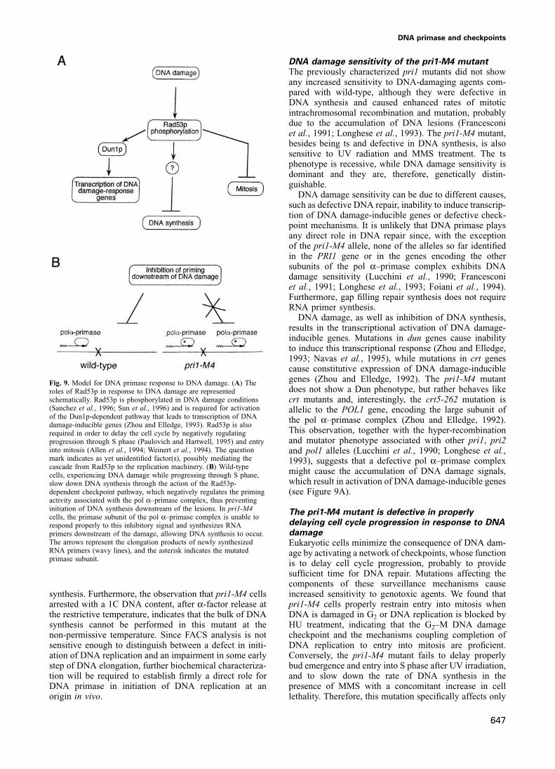
DNA damage, as well as inhibition of DNA synthesis,results in the transcriptional activation of DNA damage-inducible genes. Mutations in dun genes cause inabilityto induce this transcriptional response (Zhou and Elledge,1993; Navas et al., 1995), while mutations in crt genescause constitutive expression of DNA damage-induciblegenes (Zhou and Elledge, 1992). The pri1-M4 mutantFig. 9. Model for DNA primase response to DNA damage. (A) Thedoes not show a Dun phenotype, but rather behaves likeroles of Rad53p in response to DNA damage are represented
schematically. Rad53p is phosphorylated in DNA damage conditions crt mutants and, interestingly, the crt5-262 mutation is(Sanchez et al., 1996; Sun et al., 1996) and is required for activation allelic to the POL1 gene, encoding the large subunit ofof the Dun1p-dependent pathway that leads to transcription of DNA the pol α–primase complex (Zhou and Elledge, 1992).damage-inducible genes (Zhou and Elledge, 1993). Rad53p is also
This observation, together with the hyper-recombinationrequired in order to delay the cell cycle by negatively regulatingand mutator phenotype associated with other pri1, pri2progression through S phase (Paulovich and Hartwell, 1995) and entry
into mitosis (Allen et al., 1994; Weinert et al., 1994). The question and pol1 alleles (Lucchini et al., 1990; Longhese et al.,mark indicates as yet unidentified factor(s), possibly mediating the 1993), suggests that a defective pol α–primase complexcascade from Rad53p to the replication machinery. (B) Wild-type
might cause the accumulation of DNA damage signals,cells, experiencing DNA damage while progressing through S phase,which result in activation of DNA damage-inducible genesslow down DNA synthesis through the action of the Rad53p-
dependent checkpoint pathway, which negatively regulates the priming (see Figure 9A).activity associated with the pol α–primase complex, thus preventinginitiation of DNA synthesis downstream of the lesions. In pri1-M4 The pri1-M4 mutant is defective in properlycells, the primase subunit of the pol α–primase complex is unable to
delaying cell cycle progression in response to DNArespond properly to this inhibitory signal and synthesizes RNAdamageprimers downstream of the damage, allowing DNA synthesis to occur.
The arrows represent the elongation products of newly synthesized Eukaryotic cells minimize the consequence of DNA dam-RNA primers (wavy lines), and the asterisk indicates the mutated age by activating a network of checkpoints, whose functionprimase subunit.
is to delay cell cycle progression, probably to providesufficient time for DNA repair. Mutations affecting thecomponents of these surveillance mechanisms causeincreased sensitivity to genotoxic agents. We found thatsynthesis. Furthermore, the observation that pri1-M4 cells
arrested with a 1C DNA content, after α-factor release at pri1-M4 cells properly restrain entry into mitosis whenDNA is damaged in G2 or DNA replication is blocked bythe restrictive temperature, indicates that the bulk of DNA
synthesis cannot be performed in this mutant at the HU treatment, indicating that the G2–M DNA damagecheckpoint and the mechanisms coupling completion ofnon-permissive temperature. Since FACS analysis is not
sensitive enough to distinguish between a defect in initi- DNA replication to entry into mitosis are proficient.Conversely, the pri1-M4 mutant fails to delay properlyation of DNA replication and an impairment in some early
step of DNA elongation, further biochemical characteriza- bud emergence and entry into S phase after UV irradiation,and to slow down the rate of DNA synthesis in thetion will be required to establish firmly a direct role for
DNA primase in initiation of DNA replication at an presence of MMS with a concomitant increase in celllethality. Therefore, this mutation specifically affects onlyorigin in vivo.
647

F.Marini et al.
a subset of the checkpoint pathways, and DNA damage stream of the damage (Figure 9B). This model is consistentwith the biochemical properties of DNA primase, whichsensitivity of the pri1-M4 mutant may be related to its
failure to delay properly S phase entry and progression in is a highly distributive enzyme with the unique propertyof providing the RNA primers required to initiate DNAresponse to genotoxic agents. This is an apparent paradox:
in fact, pri1-M4 is defective in DNA synthesis at the synthesis (Kornberg and Baker, 1992). Moreover, it iswell known that, although many lesions block DNApermissive temperature in the absence of DNA-damaging
agents, while the same mutant proceeds faster than wild- polymerases in vitro, cells are still able to synthesize DNA(reviewed by Naegeli, 1994). Therefore, DNA primasetype through S phase in the presence of MMS. Therefore,
at the permissive temperature, the partially defective pri1- activity might be required to bypass a DNA lesion inorder to resume DNA synthesis downstream of the damageM4 gene product is still capable of carrying out DNA
synthesis and probably fails to respond properly to a (Figure 9B). A mechanism analogous to that required tobypass a DNA lesion occurs in Escherichia coli toregulatory mechanism which is required to inhibit G1–S
transition and S phase progression in the presence of DNA reconstitute rolling circle synthesis, and depends on prim-ing proteins (Allen et al., 1993). The observation thatdamage. While a role for DNA primase in connecting
DNA damage response to DNA replication is reasonable, the pri1-M4 DNA damage sensitivity and the intra-Scheckpoint defect are dominant further supports the hypo-a direct involvement of DNA primase in the budding
pathway can hardly be envisaged. It is more likely thesis that the pri1-M4 mutant primase fails to sense theinhibitory signal and resumes DNA synthesis downstreamthat the failure of pri1-M4 cells to delay properly bud
emergenge in response to UV irradiation in G1 is a of the lesion. A similar mechanism might also explain theG1–S checkpoint defect of pri1-M4 cells. In fact, since allconsequence of premature entry into S phase, which then
results in the activation of the budding pathway. the genes controlling the intra-S checkpoint analyzed sofar are also required to delay G1–S transition in responseto DNA damage (Lydall and Weinert, 1996; LongheseA possible role for DNA primase in linking DNA
damage response to DNA replication et al., 1996), and since primer formation is essential toinitiate DNA synthesis, it is tempting to speculate that aDifferent types of DNA damage are likely to be detected
by several sensors, and the generated signal is then failure of DNA primase to respond to checkpoint inhibitorysignals might also lead to premature entry into S phase.transduced through the Rad53p-dependent pathway in
order to delay cell cycle progression and to activatetranscription of DNA damage-inducible genes (Allen et al.,
Materials and methods1994; Weinert et al., 1994; Navas et al., 1995; Paulovichand Hartwell, 1995; Sanchez et al., 1996; Sun et al., 1996) Plasmids
Plasmid pFE139 contains the 2449 bp PstI–SacI PRI1 genomic fragment(Figure 9A). It has been suggested recently that thecloned in plasmid YCplac22 (Gietz and Sugino, 1988). Plasmid pFE5slowing down of S phase in the presence of DNA damage,contains the blunted 1920 bp NruI–SacI PRI1 genomic fragment cloned
which is genetically controlled at least by the MEC1 and into the NruI site of plasmid YCp50 (Rose et al., 1990). pLAN2 is aRAD53 genes, must target some component(s) of the DNA ARS1 TRP1 CEN6 plasmid carrying the PRI1 gene (Francesconi et al.,
1991). Plasmids pFE202 and pFE299 contain the 2449 bp PstI–SacIreplication machinery (Paulovich and Hartwell, 1995).genomic fragment carrying the pri1-M4 mutation, cloned, respectively,The Rad53p protein kinase is phosphorylated in trans byin plasmids YCplac22 and YIplac211 (Gietz and Sugino, 1988). Thea Mec3p- and Mec1p-dependent mechanism in responseBglII-linearized pFE299 plasmid has been used to replace the chromo-
to DNA damage (Sanchez et al., 1996; Sun et al., 1996), somal copy of PRI1 in different genetic backgrounds by the two-stepsuggesting that Rad53p is an intermediate component of procedure (Rothstein, 1991). Plasmid pNB187-SPK1 (Sun et al., 1996)
is a pNB187 derivative plasmid in which RAD53/SPK1 expression isthe signal transduction pathway coupling DNA damagedriven by a GAL1-inducible promoter. Plasmid p0-1Kpn, carrying theto cell cycle arrest (Figure 9A).RNR2–lacZ fusion cloned into the pSZX vector (Hurd and Roberts,
Although further studies will be required to establish 1989), was provided by M.Fasullo (Loyola University, Chicago). Plasmidfirmly the order of relative functions of RAD53 and PRI1, pRR330 carrying the rad9Δ cassette was provided by L.Prakash
(University of Texas, Dallas). Plasmid pFE302 is a pEG(KT) derivativethe following observations suggest that DNA primase acts(Mitchell et al., 1993) in which the PRI1 coding region has been fuseddownstream of RAD53: (i) the pathways leading to Rad53pto an inducible GAL1–GST gene, and this plasmid is able to complementphosphorylation as a consequence of HU and MMSa lethal disruption of the PRI1 gene.
treatment are proficient in both pri1-M4 and PRI1/pri1-M4 cells; (ii) the pri1-M4 mutation counteracts the cell Yeast strains
Strains K699, MATa ade2-1 trp1-1 can1-100 leu2-3,112 his3-11,15 ura3cycle delay caused by RAD53 overexpression; andand K700, MATα ade2-1 trp1-1 can1-100 leu2-3,112 his3-11,15 ura3(iii) both the DUN1-dependent pathway and the checkpointare isogenic and were provided by K.Nasmyth (I.M.P., Vienna). Unless
preventing entry into mitosis in response to DNA damageotherwise stated, all yeast strains used in this work are isogenic to K699.
are functional in pri1-M4 cells. However, the results Strain CG378, MATa ade5-7 leu2-3,112 ura3-52 trp1-289 can1 wasfrom L.H.Johnston (NIMR, London). Strain YLAN MATa Δpri1 ura3-presented in Figure 8 indicate that DNA primase is not a52 trp1 [pLAN2] is a L1156 derivative carrying a deletion of the PRI1direct substrate of Rad53p kinase and, therefore, we mustchromosomal locus complemented by the TRP1 CEN6 pLAN2 plasmidassume that, if DNA primase acts downstream of RAD53,(Francesconi et al., 1991). Strain CY124 is a YLAN derivative where
then other factor(s) might mediate the inhibitory signal the pLAN2 plasmid has been substituted by the URA3 CEN4 pFE5from Rad53p to DNA primase in response to DNA damage. plasmid. The PRI1 chromosomal copy has been replaced with the pri1-
M4 allele in strains K699, K700 and CG378 to originate, respectively,Since the intra-S checkpoint (Paulovich and Hartwell,strains CY387, CY522 and CY399. Strains CY1066 and CY1068 are,1995) is dependent on the MEC1, RAD53 and PRI1 genes,respectively, K699 and CY387 derivatives containing one copy of the
and DNA primase seems to act downstream of Mec1pp0-1Kpn plasmid integrated at the LEU2 locus, and were obtained as
and Rad53p, we propose that this pathway might lead to described in Hurd and Roberts (1989). Strain CY427 is a K699 derivativecontaining the rad9::URA3 disruption, obtained by transformation withinhibition of DNA primase, preventing priming down-
648
DNA primase and checkpoints
the 7100 bp SalI–EcoRI fragment of plasmid pRR330 (Schiestl et al., were collected for FACS analyisis and measurement of cell survival, asdescribed above. To analyze cell cycle delay at the G2–M boundary in1989). In all cases, correct replacements and plasmid integrations were
verified by Southern blotting. Strain K700 was crossed to strains CG378 response to UV treatment, log-phase cultures were first blocked for110 min in G2/M by nocodazole (5 μg/ml) and dimethylsulfoxide (1%)and CY399 to obtain, respectively, diploid strains CYd438 and CYd439,
while strain CY522 was crossed to strain CY399 to give rise to diploid and then plated on YPD and UV irradiated with 45 J/m2. Cells werewashed from the plates, rinsed to remove nocodazole, and resuspendedstrain CYd524. Strain Y301, MATa sad1-1 can1-100 ade2-1 his3-11,15
leu2-3,112 trp1-1 ura3-1 and its isogenic RAD53/SAD1 strain (Y300) in fresh YPD at 25°C. At timed intervals, cells were collected, and thepercentage of uni- and bi-nucleate cells was scored microscopically afterwere provided by S.J.Elledge (Baylor College, Houston). Strains
TWY308, MATα mec1-1 ura3 trp1, TWY312, MATa mec2-1 ura3 his7 staining with 4�,6-diamidino-2-phenylindole (DAPI).trp1 and TWY316, MATa mec3-1 ura3 his3 trp1 were provided byT.Weinert (University of Arizona, Tucson). Strain PK110A-15, MATα FACS analysisesr1-1 leu2-1 his4 can1 ura3 cyh2 ade6 ade2 was provided by H.Ogawa Cells were grown in the appropriate media, sonicated for 15 s, collected(Osaka University). Genetic methods and yeast growth media were by centrifugation and suspended in 70% ethanol for 16 h. Cells wereaccording to Rose et al. (1990). then washed in 0.25 M Tris–HCl (pH 7.5), and suspended in the same
buffer containing 2 mg/ml of RNase A. Samples were incubated forMutagenesis of the PRI1 gene 12 h at 37°C, collected by centrifugation and the pellet was resuspendedThe procedure to generate two-codon insertions has already been in 0.5 M pepsin freshly dissolved in 55 mM HCl. Cells were thendescribed (Barany, 1988; Foiani et al., 1994). Briefly, pri1-M2 (T122- washed in 180 mM Tris–HCl (pH 7.5), 190 mM NaCl, 70 mM MgCl2DP-C123), pri1-M3 (R164-RI-R165), pri1-M4 (N186-GS-V187), pri1- and stained in the same buffer containing 50 μg/ml of propidium iodide.M5 (N192-GS-V193), pri1-T1 (L229-GS-E230), pri1-M6 (L297-RI- Samples were then diluted 10-fold in 50 mM Tris–HCl (pH 7.8) andR298), pri1-M7 (E397-RI-R398) and pri1-H2 (E403-DP-P404) have analyzed by using a Becton Dickinson FACScan.been produced by using plasmid pFE139 and the TAB-linker 5�-CGGATC-3�. The pri1-sM7 (E397-RA-R398) and pri1-sH2 (E403-SS-
AcknowledgementsP404) were generated in plasmid pFE5 by using the TAB-linker 5�-CGAGCT-3�. The two amino acid in-frame insertions obtained (bold
We wish to thank T.Kelly, M.Muzi-Falconi (Johns Hopkins University)letters, one letter code) within the PRI1 open reading frame are indicated
P.Nurse (ICRF, London), K.Nasmyth, S.Piatti (IMP, Wien) and allin parentheses next to each pri1 allele, together with their positions in
the members of our laboratory for useful suggestions and criticisms.the p48 amino acid sequence. The pFE139 and pFE5 derivative plasmids
M.Fasullo, S.Elledge, T.Weinert, H.Ogawa, L.Prakash, K.Nasmyth,carrying the two-codon insertions listed above were used to transform
L.Johnston, R.J.Deschenes kindly provided strains or plasmids. We arestrains CY124 and YLAN, respectively, and transformants were tested
particularly grateful to Z.Sun (Yale University) for advice on performingat different temperatures for their ability to lose the wild-type PRI1 copy
the Rad53p in vitro phosphorylation experiments. This work wascarried, respectively, on the pFE5 and pFE139 plasmids.
supported partially by Progetto Bilaterale 95.00840.CT04 and by ProgettoStrategico Ciclo Cellulare e Apoptosi, Centro Nazionale Ricerche, Italy,
Mapping the pri1-M4-dependent step within the cell cycleby a grant from Associazione Italiana per la Ricerca sul Cancro and by
by reciprocal shift experimentscontract CHRX-CT93-0248 from the European Union. D.F.S. was
The rationale of reciprocal shifts to map the order of events during thesupported by grant VM-70 from the American Cancer Society.
yeast cell cycle has been described previously (Hereford and Hartwell,1974; Hartwell, 1976). The conditions used to map the pri1-M4-dependent step were similar to those previously described to map the Referencespol12-T9-dependent step (Foiani et al., 1994). Briefly, α-factor pre-synchronized cells were arrested at 36°C for 2 h and then plated on Allen,G.C.,Jr, Dixon,N.E. and Kornberg,A (1993) Strand switching of a
replicative DNA helicase promoted by the E.coli primosome. Cell,YPD at the permissive temperature in the presence of 0.3 M HU.Alternatively, cells were held in 0.1 M HU for 2 h at the permissive 74, 713–722.
Allen,J.B., Zhou,Z., Siede,W., Friedberg,E.C. and Elledge,S.J. (1994)temperature and then plated on YPD at 37°C in the absence of HU. Thepercentage of one large budded cell or of two adjacent budded cells The SAD1/RAD53 protein kinase controls multiple checkpoints and
DNA damage-induced transcription in yeast. Genes Dev., 8, 2416–after the second incubation on solid medium was scored microscopically(Foiani et al., 1994). Since in both shifts �90% of the cells failed to 2428.
Araki,H., Leem,S.-H., Phongdara,A. and Sugino,A. (1995) Dpb11, whichdivide (one large budded cell), we conclude that the pri1-M4-dependentstep and the HU-dependent step are interdependent (Hereford and interacts with DNA polymerase II(ε) in Saccharomyces cerevisiae,
has a dual role in S-phase progression and at a cell cycle checkpoint.Hartwell, 1974; Hartwell, 1976).Proc. Natl Acad. Sci. USA, 92, 11791–11795.
Barany,F. (1988) Procedures for linker insertion mutagenesis and use ofPreparation of yeast extracts, Western blot analysis andimmunoprecipitation procedures new kanamycin resistance cassettes. DNA Protein Engng Tech., 1,
29–44.The preparation of total protein extract from trichloroacetic acid (TCA)-treated cells and the procedure of Western blot analysis have been Campbell,J.L. (1993) Yeast DNA replication. J. Biol. Chem., 268,
25261–25264.described already (Foiani et al., 1994), as well as the preparation ofnon-denaturing protein extracts and immunoprecipitation of the pol α– Carr,A. and Hoekstra,M.F. (1995) The cellular responses to DNA
damage. Trends Cell Biol., 5, 32–40.primase complex (Ferrari et al., 1996). Immunological reagents againstthe different subunits of the pol α–primase complex and against Rad53p D’Urso,G., Grallert,B. and Nurse,P. (1995) DNA polymerase alpha, a
component of the replication initiation complex, is essential for thehave been also described (Foiani et al., 1995; Sun et al., 1996). Tobetter resolve differences in electrophoretic mobility due to protein checkpoint coupling S phase to mitosis in fission yeast. J. Cell. Sci.,
108, 3109–3118.phosphorylation (Figure 7B), protein extracts were separated on 30 cmSDS–polyacrylamide gels containing 17% acrylamide and 0.072% bis- Ferrari,M., Lucchini,G., Plevani,P. and Foiani,M. (1996) Phosphorylation
of the DNA polymerase α–primase B subunit is dependent on itsacrylamide at 150 V for 20 h.association with the p180 polypeptide. J. Biol. Chem., 271, 8661–8666.
Foiani,M., Marini,F., Gamba,D., Lucchini,G. and Plevani,P. (1994)Determination of DNA damage-induced cell cycle delayTo measure cell cycle delay at the G1–S boundary in response to UV The B subunit of the DNA polymerase α–primase complex in
Saccharomyces cerevisiae executes an essential function at the initialtreatment, log-phase cultures were blocked in G1 with 2 μg/ml of α-factor as previously described (Foiani et al., 1994), plated in YPD and stages of DNA replication. Mol. Cell. Biol., 14, 923–933.
Foiani,M., Liberi,G., Lucchini,G. and Plevani,P. (1995) Cell cycle-UV irradiated with 45 J/m2. Cells were then washed from plates, rinsedto remove pheromone and resuspended in fresh YPD at 25°C. At timed dependent phosphorylation and dephosphorylation of the yeast DNA
polymerase α–primase complex B subunit. Mol. Cell. Biol., 15,intervals, samples were removed for FACS analysis, and ~400 cells wereplated in triplicate onto YPD plates to measure cell survival after 3 days 883–891.
Francesconi,S., Longhese,M.P., Piseri,A., Santocanale,C., Lucchini,G.of incubation at 25°C. Cell cycle delay during S phase was analyzed byusing the procedure described by Paulovich and Hartwell (1995). Briefly, and Plevani,P. (1991) Mutations in conserved yeast DNA primase
domains impair DNA replication in vivo. Proc. Natl Acad. Sci. USA,α-factor pre-synchronized cells were released from the α-factor blockin YPD medium containing 0.02% MMS. At timed intervals, samples 88, 3877–3881.
649
F.Marini et al.
Francesconi,S., De Recondo,A.M. and Baldacci,G. (1995) DNA Paulovich,A.G. and Hartwell,L.H. (1995) A checkpoint regulates therate of progression through S phase in S.cerevisiae in response topolymerase δ is required for the replication feedback control of cell
cycle progression in Schizosaccharomyces pombe. Mol. Gen. Genet., DNA damage. Cell, 82, 841–847.Rose,M.D., Winston,F. and Hieter,P. (1990) Methods in Yeast Genetics.246, 561–569.
Friedberg,E.C., Walker,G.C. and Siede,W.D. (1995) DNA Repair and Cold Spring Harbor Laboratory Press. Cold Spring Harbor, NY.Rothstein,R. (1991) Targeting, disruption, replacement, and allele rescue:Mutagenesis. ASM Press, Washington, DC.
Gietz,R.D. and Sugino,A. (1988) New yeast–Escherichia coli shuttle integrative transformation in yeast. Methods Enzymol., 194, 281–301.Saka,Y., Fantes,P., Sutani,T., McInerny,C., Creanor,J. and Yanagida,M.vectors constructed with in vitro mutagenized yeast genes lacking six
base-pair restriction sites. Gene, 74, 527–534. (1994) Fission yeast cut5 links nuclear chromatin and M phaseregulator in the replication checkpoint control. EMBO. J., 13, 5319–Hartwell,L.H. (1976) Sequential function of gene products relative to
DNA synthesis in the yeast cell cycle. J. Mol. Biol., 104, 803–817. 5329.Sanchez,Y., Desany,B.A., Jones,W.J., Liu,Q., Wang,B. and Elledge,S.J.Hartwell,L.H. and Kastan,M.B. (1994) Cell cycle control and cancer.
Science, 266, 1821–1828. (1996) Regulation of RAD53 by the ATM-like kinases MEC1 andTEL1 in yeast cell cycle checkpoint pathways. Science, 271, 357–360.Hartwell,L.H. and Weinert,T. (1989) Checkpoints: controls that ensure
the order of cell cycle events. Science, 246, 629–634. Santocanale,C., Foiani,M., Lucchini,G. and Plevani,P. (1993) The isolated48 000 dalton subunit of yeast DNA primase is sufficient for RNAHereford,L.M. and Hartwell,L.H. (1974) Sequential gene function in the
initiation of Saccharomyces cerevisiae DNA synthesis. J. Mol. Biol., primer synthesis. J. Biol. Chem., 268, 1343–1348.Schiestl,R.H., Reynolds,P., Prakash,S. and Prakash,L. (1989) Cloning84, 445–461.
Hofmann,J.F.X. and Beach,D. (1994) cdt1 is an essential target of the and sequence analysis of the Saccharomyces cerevisiae RAD9 geneand further evidence that its product is required for cell cycle arrestCdc10/Sct1 transcription factor: requirement for DNA replication and
inhibition of mitosis. EMBO J., 13, 425–434. induced by DNA damage. Mol. Cell. Biol., 9, 1882–1896.Siede,W., Friedberg,A.S. and Friedberg,E.C. (1993) RAD9-dependent G1Humphrey,T. and Enoch,T. (1995) Keeping mitosis in check. Curr. Biol.,
5, 376–379. arrest defines a second checkpoint for damaged DNA in the cellcycle of Saccharomyces cerevisiae. Proc. Natl Acad. Sci. USA, 90,Hurd,H.K. and Roberts,J.W. (1989) Upstream regulatory sequences of
the yeast RNR2 gene include a repression sequence and an activation 7985–7989.Siede,W., Friedberg,A.S., Dianova,I. and Friedberg,E.C. (1994)site that binds the Rap1 protein. Mol. Cell. Biol., 9, 5359–5372.
Johnston,L.H. and Lowndes,N.F. (1992) Cell cycle control of DNA Characterization of G1 checkpoint control in the yeast Saccharomycescerevisiae following exposure to DNA damaging agents. Genetics,synthesis in budding yeast. Nucleic Acids Res., 20, 2403–2410.
Kato,R. and Ogawa,H. (1994) An essential gene, ESR1, is required 138, 271–281.Sun,Z., Fay,D.S., Marini,F., Foiani,M. and Stern,D.F. (1996) Spk1/Rad53for mitotic cell growth, DNA repair and meiotic recombination in
Saccharomyces cerevisiae. Nucleic Acids Res., 22, 3104–3112. is regulated by Mec1-dependent protein phosphorylation in DNAreplication and DNA damage checkpoint pathway. Genes Dev., 10,Kelly,T.J., Martin,G.S., Forsburg,S.L., Stephen,R.J., Russo,A. and
Nurse,P. (1993) The fission yeast cdc18� gene product couples S 395–406.Weinert,T.A., Kiser,G.L. and Hartwell,L.H. (1994) Mitotic checkpointsphase to start and mitosis. Cell, 74, 371–382.
Kornberg,A. and Baker,T. (1992) DNA Replication. 2nd edn. in budding yeast and the dependence of mitosis on DNA replicationand repair. Genes Dev., 8, 652–665.W.H.Freeman and Co., San Francisco.
Longhese,M.P., Jovine,L., Plevani,P. and Lucchini,G. (1993) Conditional Yamamoto,A., Guacci,B. and Koshland,D. (1996) Pds1p, an inhibitor ofanaphase in budding yeast plays a critical role in the APC andmutations in the yeast DNA primase genes affect different aspects of
DNA metabolism and interactions in the DNA polymerase α–primase checkpoint pathway(s). J. Cell Biol., 133, 99–110.Zheng,P., Fay,D.S., Burton,J., Xiao,H., Pinkham,J.L. and Stern,D.F.complex. Genetics, 133, 183–191.
Longhese,M.P., Fraschini,R., Plevani,P. and Lucchini,G. (1996) Yeast (1993) SPK1 is an essential S-phase-specific gene of Saccharomycescerevisiae that encodes a nuclear serine/threonine/tyrosine kinase.pip3/mec3 mutants fail to delay entry into S phase and to slow down
DNA replication in response to DNA damage, and they define a Mol. Cell. Biol., 13, 5829–5842.Zhou,Z. and Elledge,S.J. (1992) Isolation of crt mutants constitutivefunctional link between Mec3 and DNA primase. Mol. Cell. Biol., 16,
3235–3244. for transcription of the DNA damage inducible gene RNR3 inSaccharomyces cerevisiae. Genetics, 131, 851–866.Lucchini,G., Hinnebusch,A.G., Chen,C. and Fink,G.R. (1984) Positive
regulatory interactions of the HIS4 gene of Saccharomyces cerevisiae. Zhou,Z. and Elledge,S.J. (1993) DUN1 encodes a protein kinase thatcontrols the DNA damage response in yeast. Cell, 75, 1119–1127.Mol. Cell. Biol., 4, 1326–1333.
Lucchini,G., Francesconi,S., Foiani,M., Badaracco,G. and Plevani,P.Received on June 13, 1996; revised on October 31, 1996(1987) The yeast DNA polymerase–primase complex: cloning of
PRI1, a single essential gene related to DNA primase activity. EMBOJ., 6, 737–742.
Lucchini,G., Muzi Falconi,M., Pizzagalli,A., Aguilera,A., Klein,A. andPlevani,P. (1990) Nucleotide sequence and characterization oftemperature sensitive pol1 mutants of Saccharomyces cerevisiae.Gene, 90, 99–104.
Lydall,D. and Weinert,T. (1996) From DNA damage to cell cycle arrestand suicide: a budding yeast perspective. Curr. Opin. Genet. Dev., 6,4–11.
Mitchell,D.A., Marshall,T.K. and Deschenes,R.J. (1993) Vectors for theinducible overexpression of glutathione S-transferase fusion proteinsin yeast. Yeast, 9, 715–723.
Murakami,H. and Okayama,H. (1995) A kinase from fission yeastresponsible for blocking mitosis in S phase. Nature, 374, 817–819.
Murray,A. (1994) Cell cycle checkpoints. Curr. Opin. Cell Biol., 6,872–876.
Muzi Falconi,M., Piseri,A., Ferrari,M., Lucchini,G., Plevani,P. andFoiani,M. (1993) De novo synthesis of budding yeast DNA polymeraseα and POL1 transcription at the G1/S boundary are not required forentrance into S phase. Proc. Natl Acad. Sci. USA, 90, 10519–10523.
Naegeli,H. (1994) Roadblocks and detours during DNA replication:mechanisms of mutagenesis in mammalian cells. BioEssays, 16,557–564.
Navas,T.A., Zhou,Z. and Elledge,S. (1995) DNA polymerase ε links theDNA replication machinery to the S phase checkpoint. Cell, 80, 29–39.
Nurse,P. (1994) Ordering S phase and M phase in the cell cycle. Cell,79, 547–550.
650