
γλώσσες
Σελίδες
Νομικός


Platelet Disorders
Lalitha Nayak, M.D.
Division of Hematology and Oncology
INTRODUCTION
• QUANTITATIVE DISORDERS
Thrombocytopenia
Thrombocytosis
• QUALITATIVE DISORDERS
THROMBOCYTOPENIA
• Thrombocytopenia is defined as a count below 150,000/μL.
• Platelet-type bleeding typically involves skin or mucous membranes, including petechiae, purpura, ecchymosis, epistaxis, menorrhagia, and GI hemorrhage.
• Deep muscle hematomas and hemarthrosis are typically seen with defects in fluid hemostatic system
• Clinical bleeding varies
THROMBOCYTOPENIA
• Symptoms depend on the degree of thrombocytopenia
• At counts above 50,000/µL there are usually NO Symptoms
• At counts of 20,000 to 50,000/µL the patient may report EASY BRUISABILITY but no spontaneous bleeding is seen
• At counts <20,000/µL patients are AT HIGH RISK FOR SPONTANEOUS BLEEDING (GI bleeds, Mucous Membranes, Petechiae)
Thrombocytopenia: CAUSE?
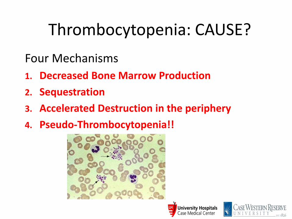
Four Mechanisms
1. Decreased Bone Marrow Production
2. Sequestration
3. Accelerated Destruction in the periphery
4. Pseudo-Thrombocytopenia!!
DRUGS AND PLATELETS
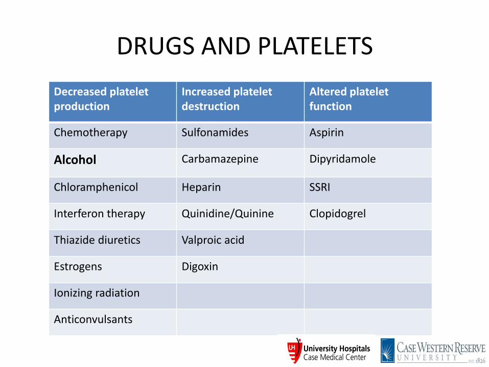
Decreased platelet production
Increased platelet destruction
Altered platelet function
Chemotherapy Sulfonamides Aspirin
Alcohol Carbamazepine Dipyridamole
Chloramphenicol Heparin SSRI
Interferon therapy Quinidine/Quinine Clopidogrel
Thiazide diuretics Valproic acid
Estrogens Digoxin
Ionizing radiation
Anticonvulsants
SEQUESTRATION
• Kasabach-Merritt syndrome – giant cavernous hemangioma and consumptive coagulopathy
• Hypersplenism (including liver disease) – Splenomegaly
– Peripheral blood shows anemia, leukopenia, thrombocytopenia
– Normo- or hypercellular bone marrow
– Counts normalize after splenectomy
ACCELERATED DESTRUCTION
• IMMUNE Neonatal alloimmune thrombocytopenia (NAIT) Posttransfusion purpura (PTP) Immune Thrombocytopenia (ITP) and neonatal
autoimmune thrombocytopenia Drugs HIV Sepsis
• NONIMMUNE (mechanical damage or consumption)
TTP/HUS
DIC
ITP (Immune Thrombocytopenia)
• One of the most common acquired bleeding disorders encountered by the Hematologist
• Also the most common autoimmune disorder affecting a blood element
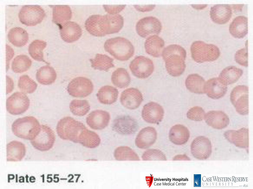
DIAGNOSIS
• Diagnosis of exclusion
• Antecedent infectious illness ~ 60%
• Physical exam remarkable only for purpura
• Negative family history
• Peripheral blood smear reveals thrombocytopenia and normal to large platelets
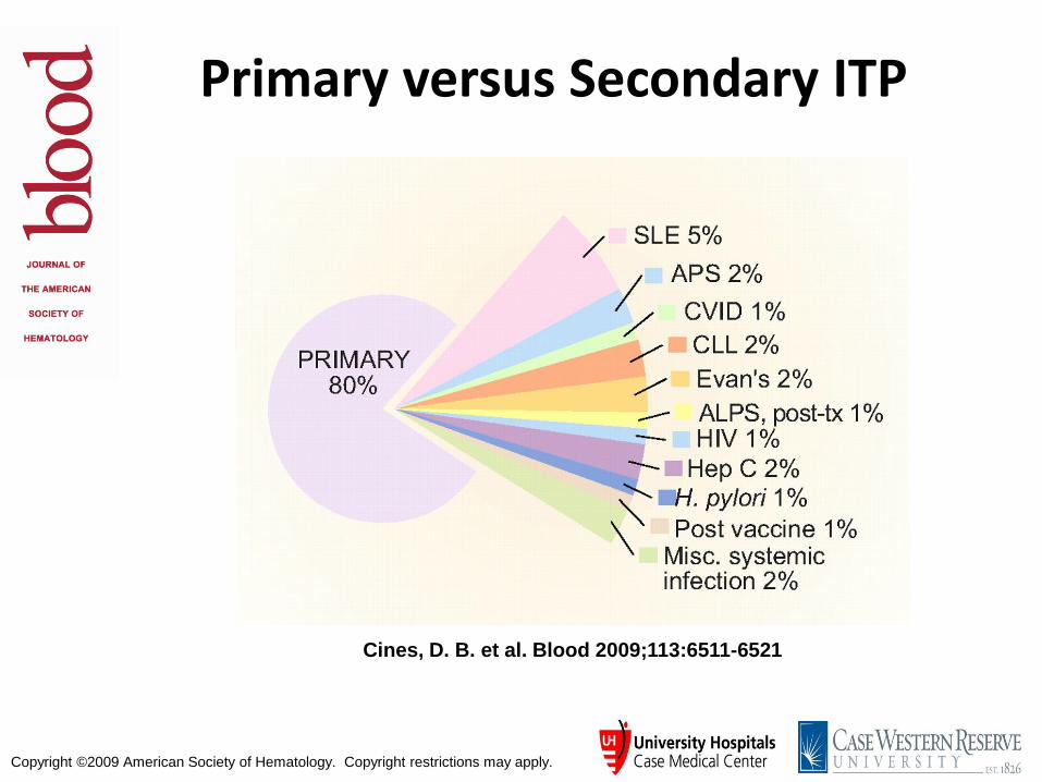
Copyright ©2009 American Society of Hematology. Copyright restrictions may apply.
Cines, D. B. et al. Blood 2009;113:6511-6521
Primary versus Secondary ITP

ITP Diagnosis
• Thrombocytopenia without
obvious etiology • Exclude: HIV, HepC, HepB, H. pylori, Lymphoma, common variable hypo- gammaglobulinemia • Bone marrow shows
megakaryocyte production • May have increased IgG/IgM
on platelets
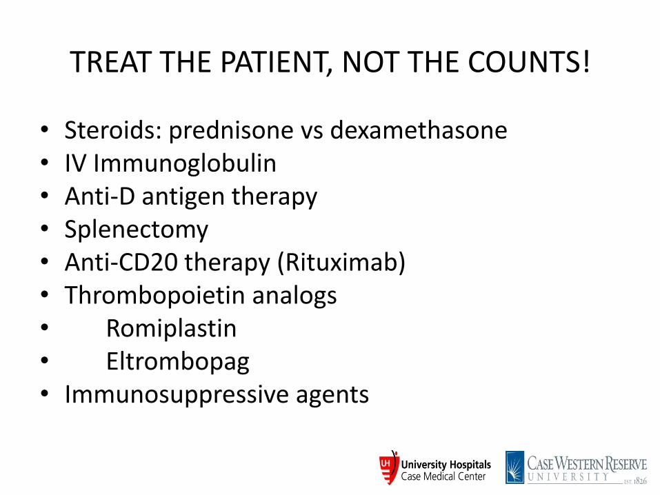
TREAT THE PATIENT, NOT THE COUNTS!
• Steroids: prednisone vs dexamethasone • IV Immunoglobulin • Anti-D antigen therapy • Splenectomy • Anti-CD20 therapy (Rituximab) • Thrombopoietin analogs • Romiplastin • Eltrombopag • Immunosuppressive agents
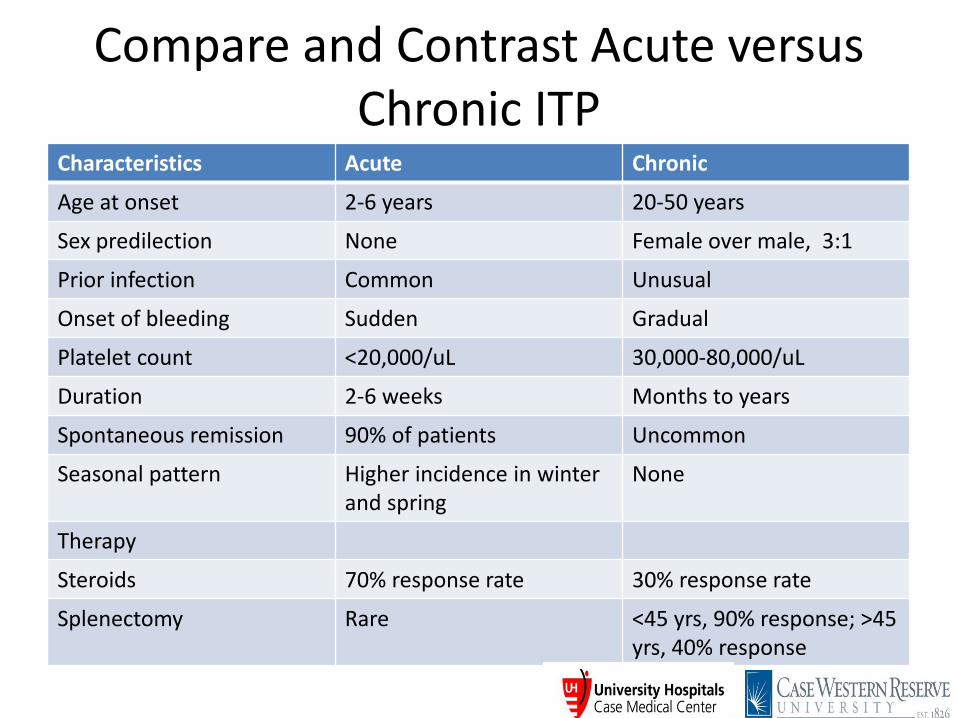
Compare and Contrast Acute versus Chronic ITP
Characteristics Acute Chronic
Age at onset 2-6 years 20-50 years
Sex predilection None Female over male, 3:1
Prior infection Common Unusual
Onset of bleeding Sudden Gradual
Platelet count <20,000/uL 30,000-80,000/uL
Duration 2-6 weeks Months to years
Spontaneous remission 90% of patients Uncommon
Seasonal pattern Higher incidence in winter and spring
None
Therapy
Steroids 70% response rate 30% response rate
Splenectomy Rare <45 yrs, 90% response; >45 yrs, 40% response
Heparin-Induced Thrombocytopenia &
Thrombosis Syndrome (HITTS)
• Occurs in 1-2% of patients getting unfractionated heparin and 0.5-1% patients getting low molecular weight heparin.
• Defined as a 50% drop in platelet count anywhere from 3-30 days after heparin administration
• First manifestation is thrombocytopenia; 70% or more patients get venous or arterial thrombosis;
• Most are subclinical and thus need to be searched for by objective testing like venous doppler studies

Pathogenesis of HITTS
Clinical-pathologic Criteria for Diagnosis of HIT
Clinical (1)One or more of the following: Thrombocytopenia Thrombosis (venous, arterial, or microvascular) Necrotizing skin lesions at injection sites Acute anaphylactoid reactions (2) Appropriate timing of heparin exposure (3) Absence of a more compelling explanation
Pathologic (1) Positive platelet activation assay (2) Positive anti-PF4/heparin IgG assay
Warkentin TE Hematol Oncol Clin North Am. 2010 Aug;24(4):755-75 Agents for the treatment of heparin-induced thrombocytopenia.
Diagnosis & Management of HIT/HITTS
• Diagnosis - High index of clinical suspicion - Elisa for PF4 antibodies with heparin suppression - Platelet serotonin release assay with patient serum
• Management - Stop heparin, low molecular weight heparin - Warfarin is CONTRAINDICATED! - Anticoagulate with a direct thrombin inhibitor - Determine if occult thrombosis to ascertain duration of anticoagulation
• TTP and HUS (hemolytic uremic syndrome) are both acute syndromes with abnormalities in multiple organ systems
• Evidence of MAHA and thrombocytopenia
• Presenting features are essentially the same in most adult patients
• Pathologic changes and Initial treatment is same
Thrombotic Thrombocytopenia Purpura (TTP) (Moschcowitz Syndrome)
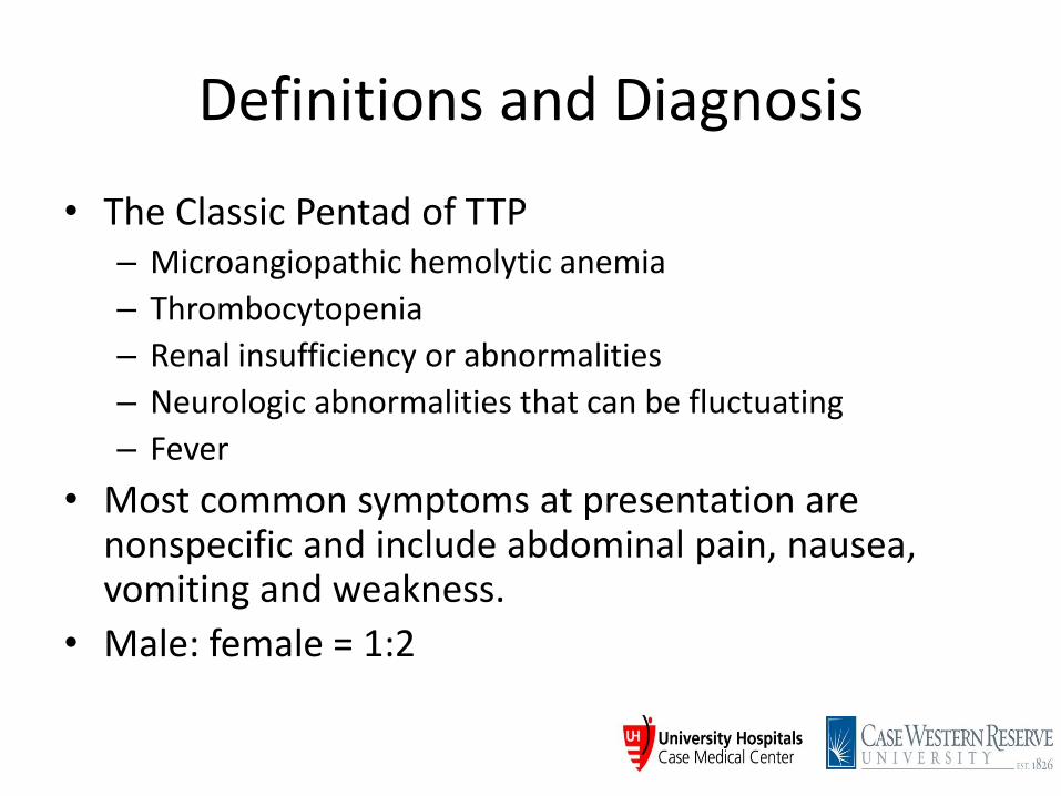
Definitions and Diagnosis
• The Classic Pentad of TTP – Microangiopathic hemolytic anemia
– Thrombocytopenia
– Renal insufficiency or abnormalities
– Neurologic abnormalities that can be fluctuating
– Fever
• Most common symptoms at presentation are nonspecific and include abdominal pain, nausea, vomiting and weakness.
• Male: female = 1:2
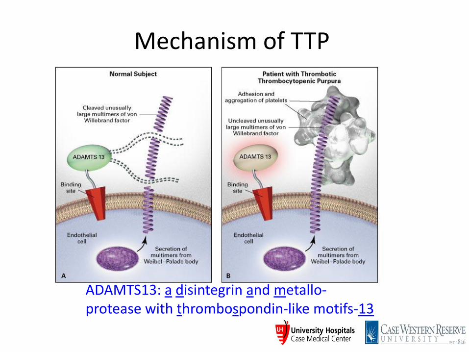
Mechanism of TTP
ADAMTS13: a disintegrin and metallo- protease with thrombospondin-like motifs-13
Copyright ©2006 American Society of Hematology. Copyright restrictions may apply.
Sadler, J. E. Hematology 2006;2006:415-420
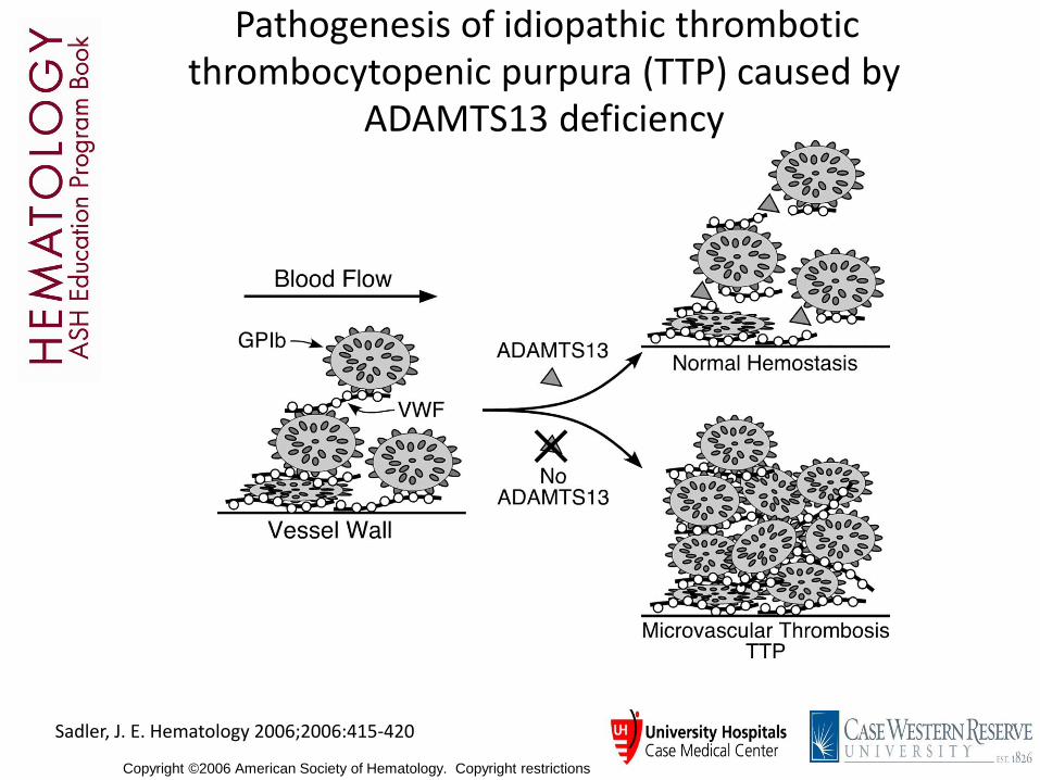
Pathogenesis of idiopathic thrombotic thrombocytopenic purpura (TTP) caused by
ADAMTS13 deficiency
• Clinical Presentations
• Congenital (Upshaw-Schulman Syndrome)
• Idiopathic acquired
Associated with medication such as clopidogrel, ticlopidine, quinine, cyclosporine, gemcitabine, mitomycin C
• BM transplant-associated (TMA-thrombocytopenic microangiopathy)
Thrombotic Thrombocytopenia Purpura (TTP) (Moschcowitz Syndrome)
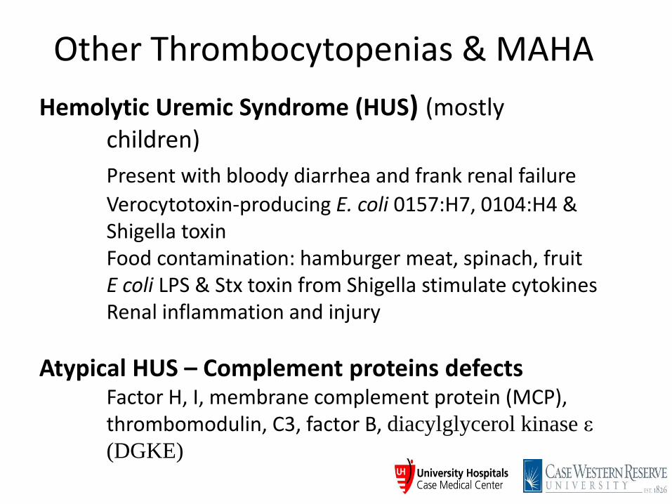
Other Thrombocytopenias & MAHA
Hemolytic Uremic Syndrome (HUS) (mostly children)
Present with bloody diarrhea and frank renal failure
Verocytotoxin-producing E. coli 0157:H7, 0104:H4 & Shigella toxin Food contamination: hamburger meat, spinach, fruit E coli LPS & Stx toxin from Shigella stimulate cytokines Renal inflammation and injury
Atypical HUS – Complement proteins defects Factor H, I, membrane complement protein (MCP), thrombomodulin, C3, factor B, diacylglycerol kinase ε
(DGKE)
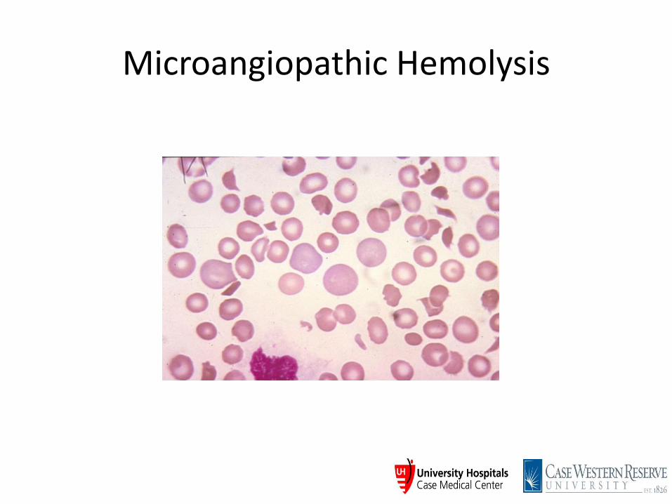
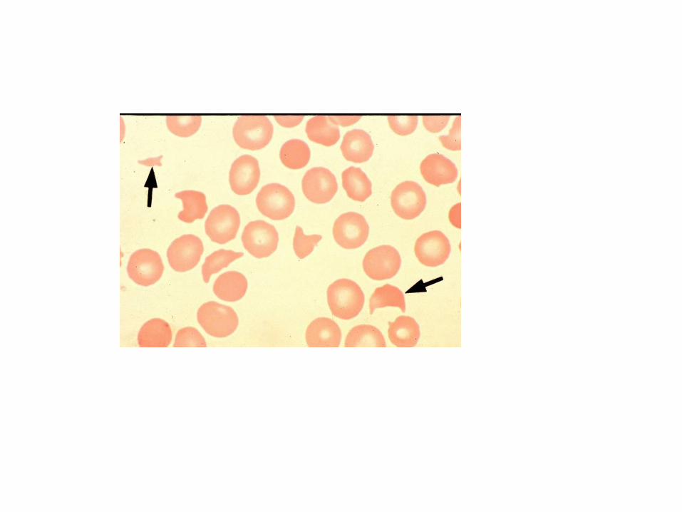
Microangiopathic Hemolysis
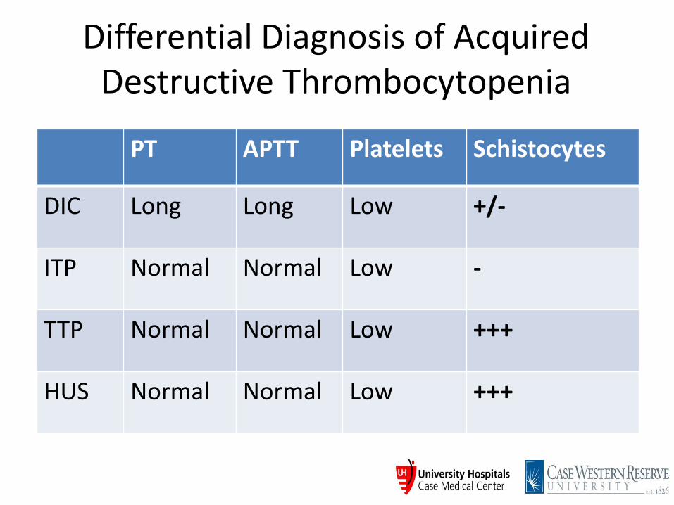
Differential Diagnosis of Acquired Destructive Thrombocytopenia
PT APTT Platelets Schistocytes
DIC Long Long Low +/-
ITP Normal Normal Low -
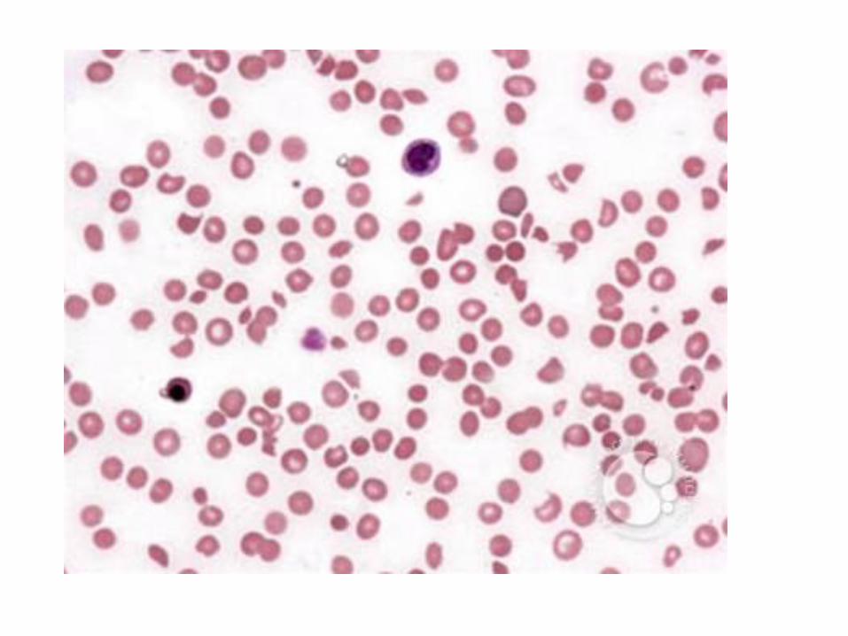
TTP Normal Normal Low +++
HUS Normal Normal Low +++
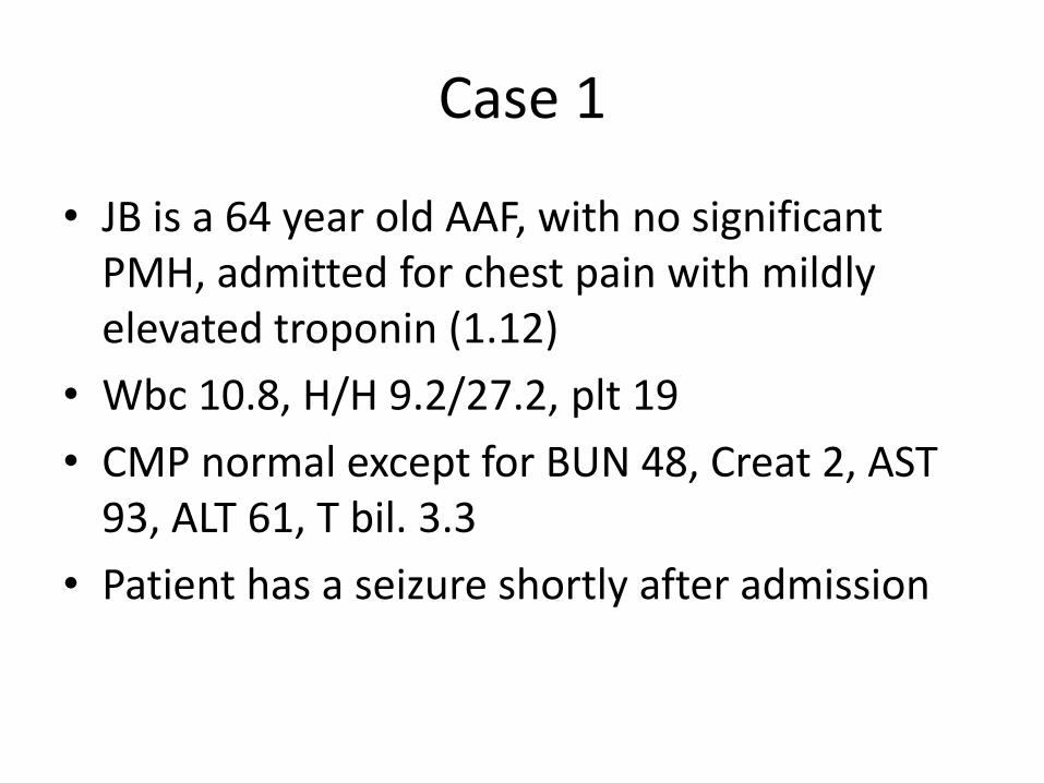
Case 1
• JB is a 64 year old AAF, with no significant PMH, admitted for chest pain with mildly elevated troponin (1.12)
• Wbc 10.8, H/H 9.2/27.2, plt 19
• CMP normal except for BUN 48, Creat 2, AST 93, ALT 61, T bil. 3.3
• Patient has a seizure shortly after admission
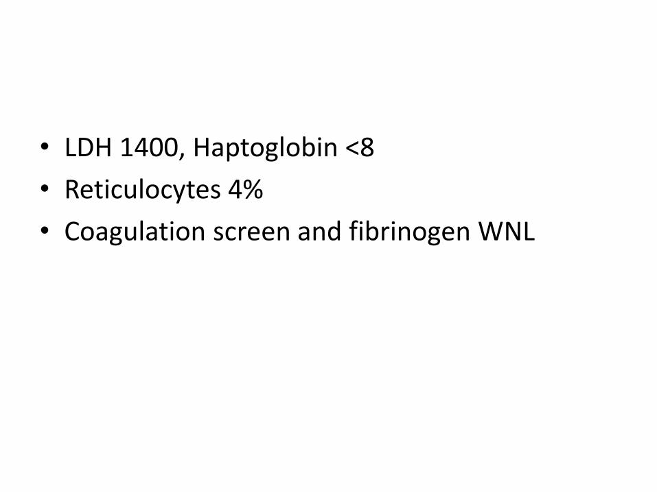
• LDH 1400, Haptoglobin <8
• Reticulocytes 4%
• Coagulation screen and fibrinogen WNL
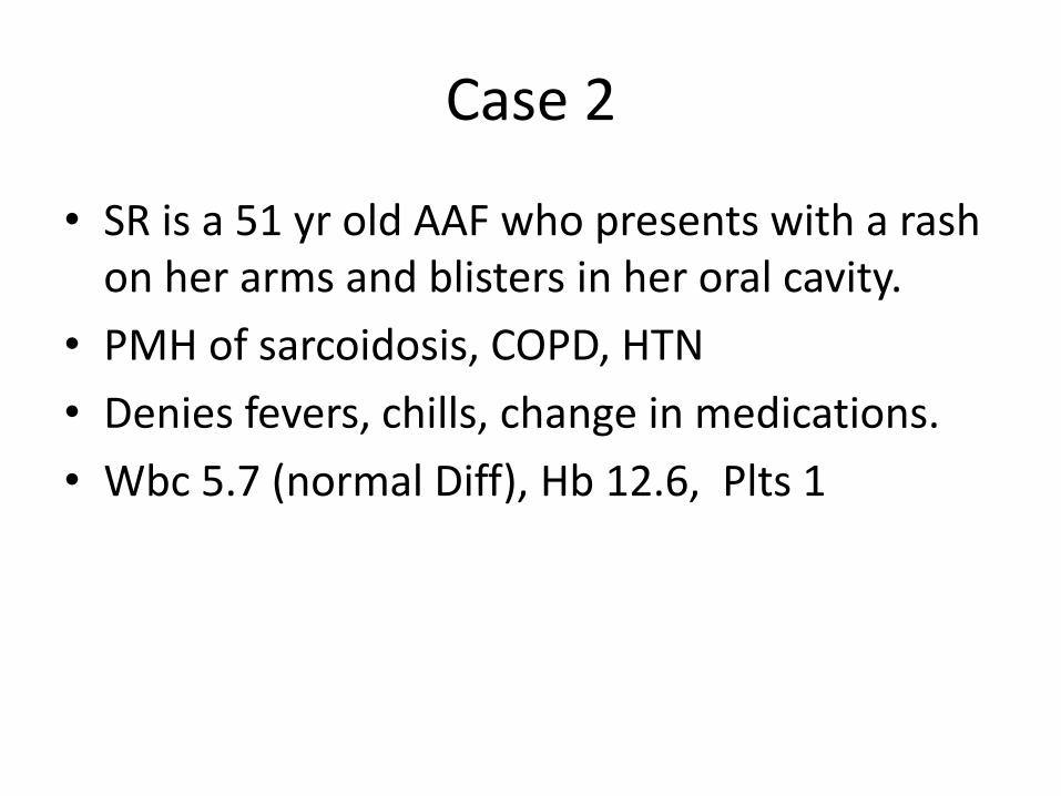
Case 2
• SR is a 51 yr old AAF who presents with a rash on her arms and blisters in her oral cavity.
• PMH of sarcoidosis, COPD, HTN
• Denies fevers, chills, change in medications.
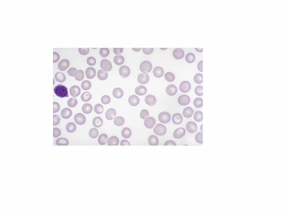
• Wbc 5.7 (normal Diff), Hb 12.6, Plts 1
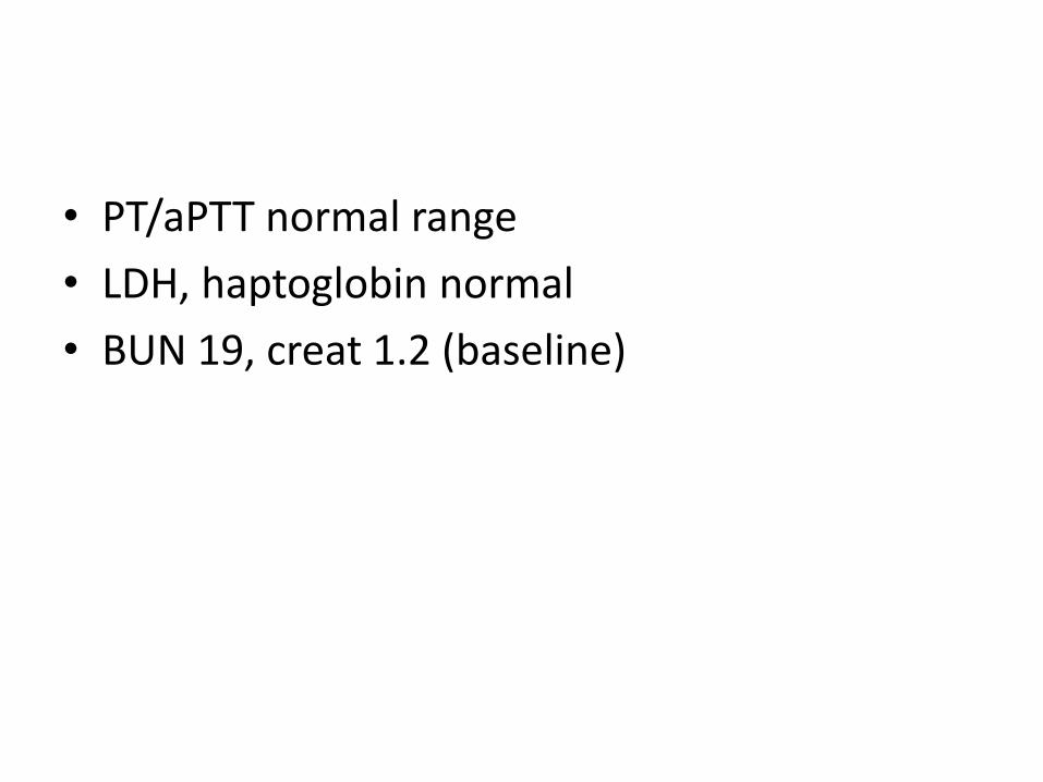
• PT/aPTT normal range
• LDH, haptoglobin normal
• BUN 19, creat 1.2 (baseline)
Case 3
• CF is a 28 yr male, with not significant PMH.
• CBC on routine annual examination reveals platelet count of 70K, rest cbc within normal limits.
• No bleeding history
• No FH of blood disorders
• No medications
• P/E within normal limits
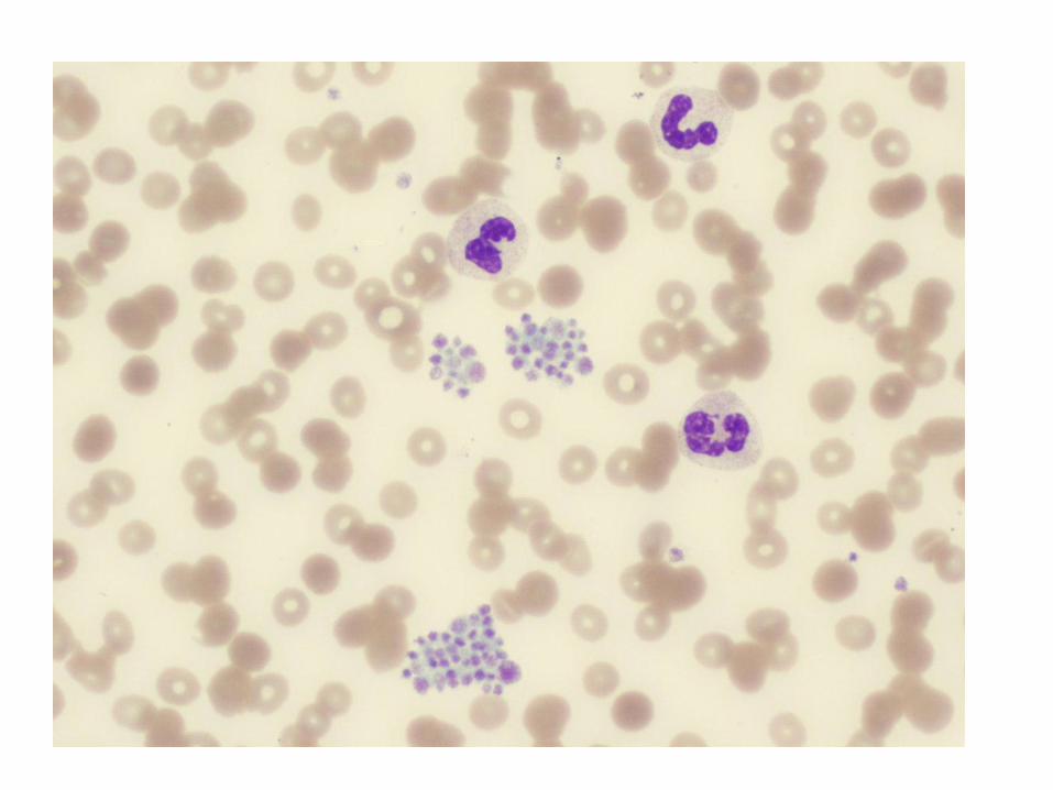
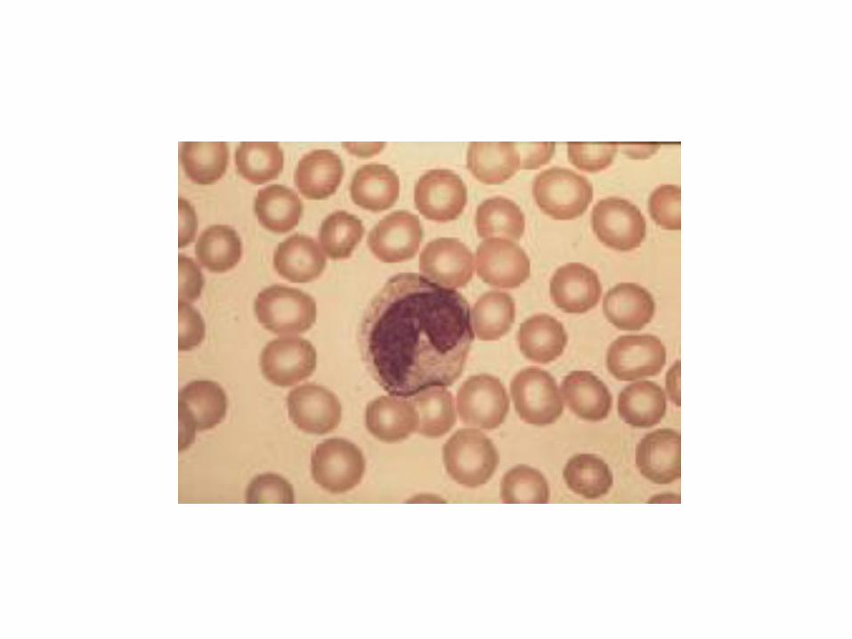
Case 4
• AE is a 66 yr old male with a neuroendocrine duodenal mass. Underwent Whipple procedure.
• 8 days post op, wbc 12.2, H/H 9.1/27.7, plts 27 (250-309 previously)
• BUN 24, creat 3.4 (previously normal)
• Slightly drowsy
• Routine CT abd day prior show non-occlusive splanchnic vein thrombus
• AST 792, ALT 253, Bil normal
• LDH 981, Hapto 166
• Coags: PT 17.6, aPTT normal
• Argatroban initiated for suspected HIT
• 4 limb usg - right femoral vein clot
• Repeat abd imaging – hepatic vein thrombus
• PF4 Ab strongly positive
• SRA confirmed diagnosis
Top Related