Lalitha Nayak, M.D. Division of Hematology and...
41
Platelet Disorders Lalitha Nayak, M.D. Division of Hematology and Oncology
Transcript of Lalitha Nayak, M.D. Division of Hematology and...

Platelet Disorders
Lalitha Nayak, M.D.
Division of Hematology and Oncology
INTRODUCTION
• QUANTITATIVE DISORDERS
Thrombocytopenia
Thrombocytosis
• QUALITATIVE DISORDERS
THROMBOCYTOPENIA
• Thrombocytopenia is defined as a count below 150,000/μL.
• Platelet-type bleeding typically involves skin or mucous membranes, including petechiae, purpura, ecchymosis, epistaxis, menorrhagia, and GI hemorrhage.
• Deep muscle hematomas and hemarthrosis are typically seen with defects in fluid hemostatic system
• Clinical bleeding varies
THROMBOCYTOPENIA
• Symptoms depend on the degree of thrombocytopenia
• At counts above 50,000/µL there are usually NO Symptoms
• At counts of 20,000 to 50,000/µL the patient may report EASY BRUISABILITY but no spontaneous bleeding is seen
• At counts <20,000/µL patients are AT HIGH RISK FOR SPONTANEOUS BLEEDING (GI bleeds, Mucous Membranes, Petechiae)
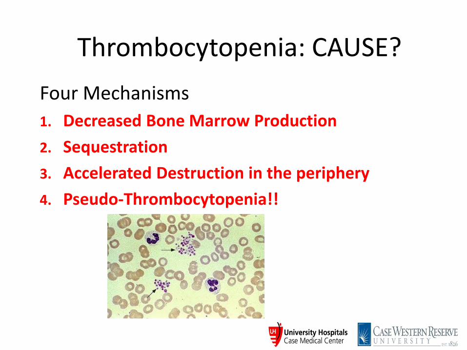
Thrombocytopenia: CAUSE?
Four Mechanisms
1. Decreased Bone Marrow Production
2. Sequestration
3. Accelerated Destruction in the periphery
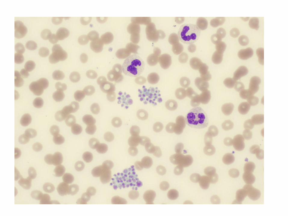
4. Pseudo-Thrombocytopenia!!
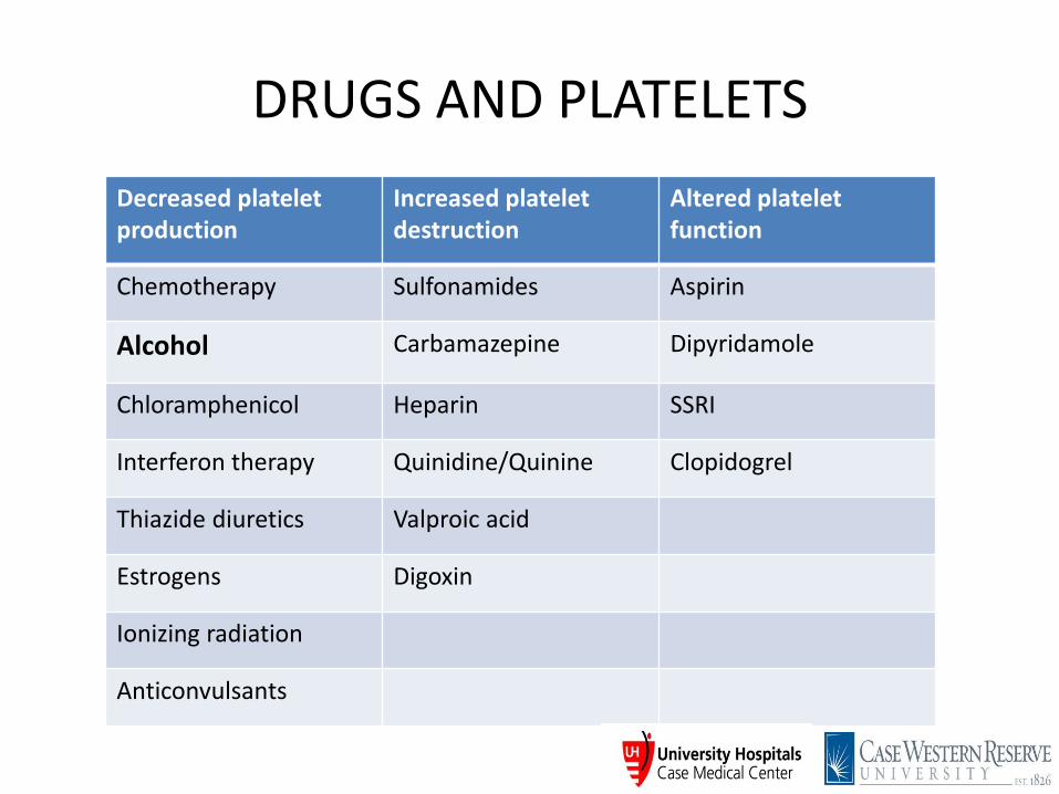
DRUGS AND PLATELETS
Decreased platelet production
Increased platelet destruction
Altered platelet function
Chemotherapy Sulfonamides Aspirin
Alcohol Carbamazepine Dipyridamole
Chloramphenicol Heparin SSRI
Interferon therapy Quinidine/Quinine Clopidogrel
Thiazide diuretics Valproic acid
Estrogens Digoxin
Ionizing radiation
Anticonvulsants
SEQUESTRATION
• Kasabach-Merritt syndrome – giant cavernous hemangioma and consumptive coagulopathy
• Hypersplenism (including liver disease) – Splenomegaly
– Peripheral blood shows anemia, leukopenia, thrombocytopenia
– Normo- or hypercellular bone marrow
– Counts normalize after splenectomy
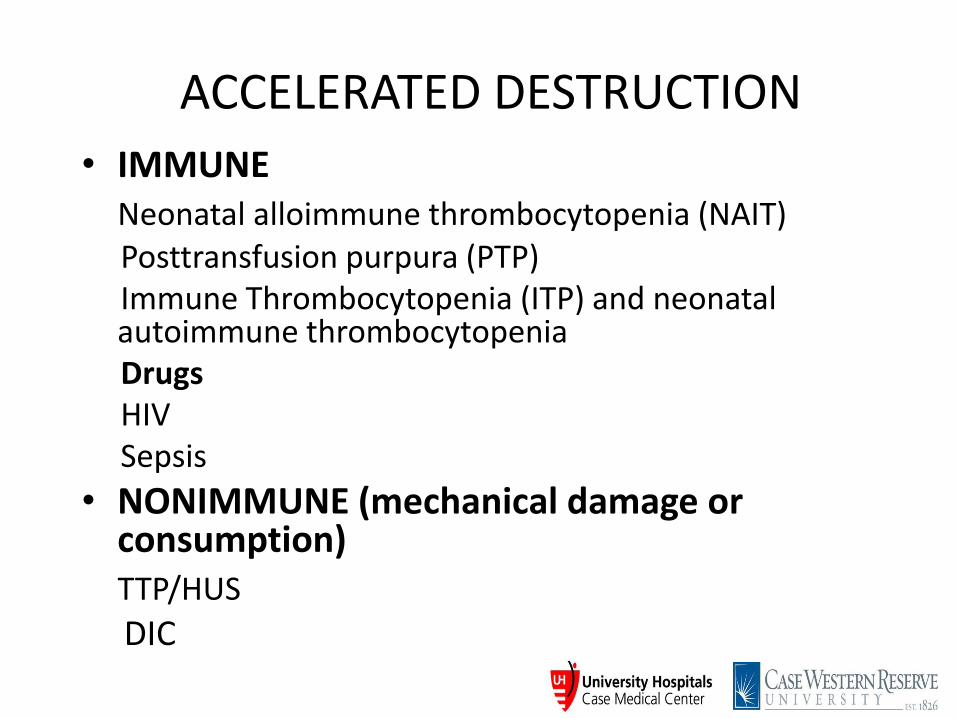
ACCELERATED DESTRUCTION
• IMMUNE Neonatal alloimmune thrombocytopenia (NAIT) Posttransfusion purpura (PTP) Immune Thrombocytopenia (ITP) and neonatal
autoimmune thrombocytopenia Drugs HIV Sepsis
• NONIMMUNE (mechanical damage or consumption)
TTP/HUS
DIC
ITP (Immune Thrombocytopenia)
• One of the most common acquired bleeding disorders encountered by the Hematologist
• Also the most common autoimmune disorder affecting a blood element
DIAGNOSIS
• Diagnosis of exclusion
• Antecedent infectious illness ~ 60%
• Physical exam remarkable only for purpura
• Negative family history
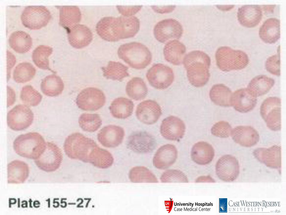
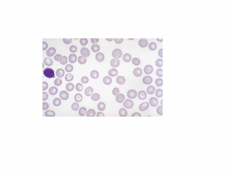
• Peripheral blood smear reveals thrombocytopenia and normal to large platelets
Copyright ©2009 American Society of Hematology. Copyright restrictions may apply.
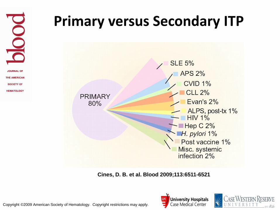
Cines, D. B. et al. Blood 2009;113:6511-6521
Primary versus Secondary ITP

ITP Diagnosis
• Thrombocytopenia without
obvious etiology • Exclude: HIV, HepC, HepB, H. pylori, Lymphoma, common variable hypo- gammaglobulinemia • Bone marrow shows
megakaryocyte production • May have increased IgG/IgM
on platelets
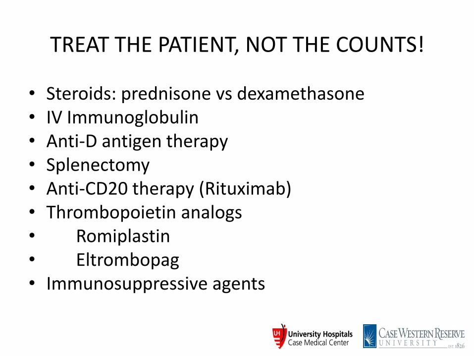
TREAT THE PATIENT, NOT THE COUNTS!
• Steroids: prednisone vs dexamethasone • IV Immunoglobulin • Anti-D antigen therapy • Splenectomy • Anti-CD20 therapy (Rituximab) • Thrombopoietin analogs • Romiplastin • Eltrombopag • Immunosuppressive agents
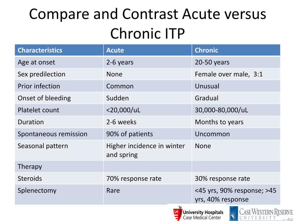
Compare and Contrast Acute versus Chronic ITP
Characteristics Acute Chronic
Age at onset 2-6 years 20-50 years
Sex predilection None Female over male, 3:1
Prior infection Common Unusual
Onset of bleeding Sudden Gradual
Platelet count <20,000/uL 30,000-80,000/uL
Duration 2-6 weeks Months to years
Spontaneous remission 90% of patients Uncommon
Seasonal pattern Higher incidence in winter and spring
None
Therapy
Steroids 70% response rate 30% response rate
Splenectomy Rare <45 yrs, 90% response; >45 yrs, 40% response
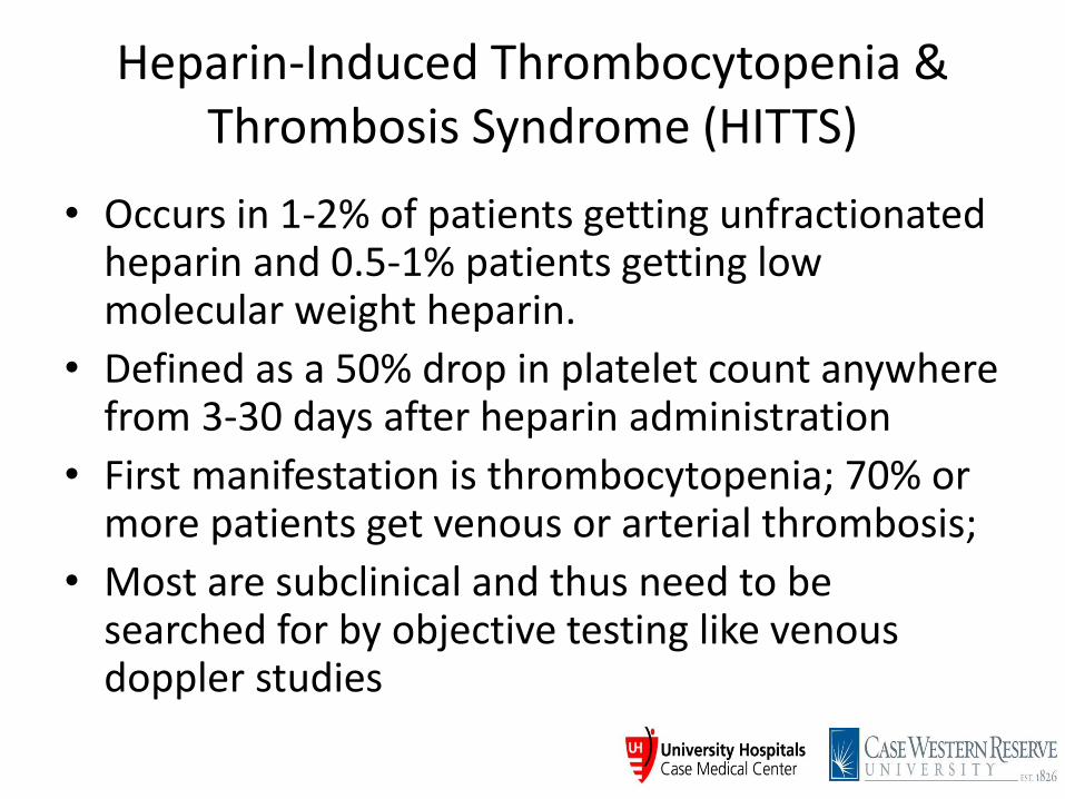
Heparin-Induced Thrombocytopenia &
Thrombosis Syndrome (HITTS)
• Occurs in 1-2% of patients getting unfractionated heparin and 0.5-1% patients getting low molecular weight heparin.
• Defined as a 50% drop in platelet count anywhere from 3-30 days after heparin administration
• First manifestation is thrombocytopenia; 70% or more patients get venous or arterial thrombosis;
• Most are subclinical and thus need to be searched for by objective testing like venous doppler studies

Pathogenesis of HITTS
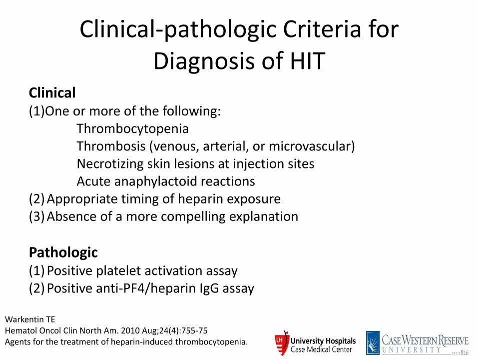
Clinical-pathologic Criteria for Diagnosis of HIT
Clinical (1)One or more of the following: Thrombocytopenia Thrombosis (venous, arterial, or microvascular) Necrotizing skin lesions at injection sites Acute anaphylactoid reactions (2) Appropriate timing of heparin exposure (3) Absence of a more compelling explanation
Pathologic (1) Positive platelet activation assay (2) Positive anti-PF4/heparin IgG assay
Warkentin TE Hematol Oncol Clin North Am. 2010 Aug;24(4):755-75 Agents for the treatment of heparin-induced thrombocytopenia.
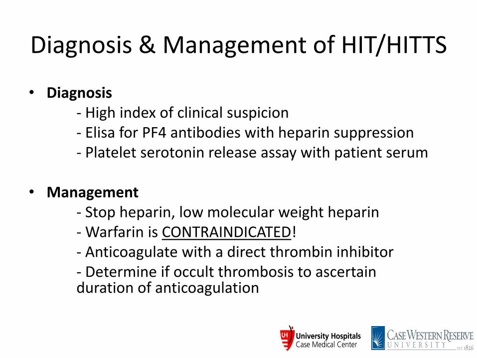
Diagnosis & Management of HIT/HITTS
• Diagnosis - High index of clinical suspicion - Elisa for PF4 antibodies with heparin suppression - Platelet serotonin release assay with patient serum
• Management - Stop heparin, low molecular weight heparin - Warfarin is CONTRAINDICATED! - Anticoagulate with a direct thrombin inhibitor - Determine if occult thrombosis to ascertain duration of anticoagulation
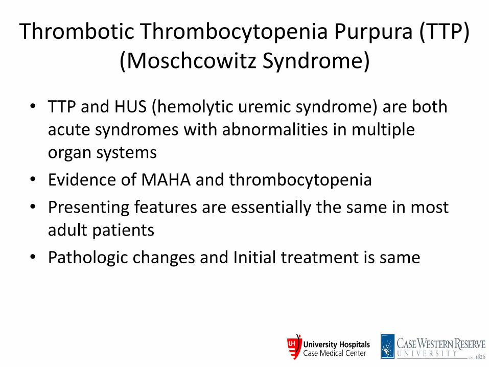
• TTP and HUS (hemolytic uremic syndrome) are both acute syndromes with abnormalities in multiple organ systems
• Evidence of MAHA and thrombocytopenia
• Presenting features are essentially the same in most adult patients
• Pathologic changes and Initial treatment is same
Thrombotic Thrombocytopenia Purpura (TTP) (Moschcowitz Syndrome)
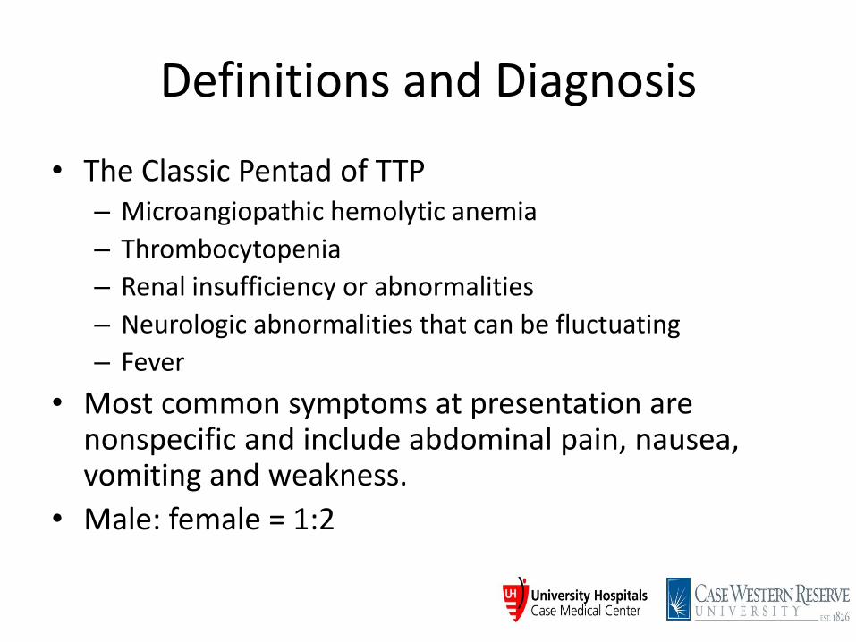
Definitions and Diagnosis
• The Classic Pentad of TTP – Microangiopathic hemolytic anemia
– Thrombocytopenia
– Renal insufficiency or abnormalities
– Neurologic abnormalities that can be fluctuating
– Fever
• Most common symptoms at presentation are nonspecific and include abdominal pain, nausea, vomiting and weakness.
• Male: female = 1:2
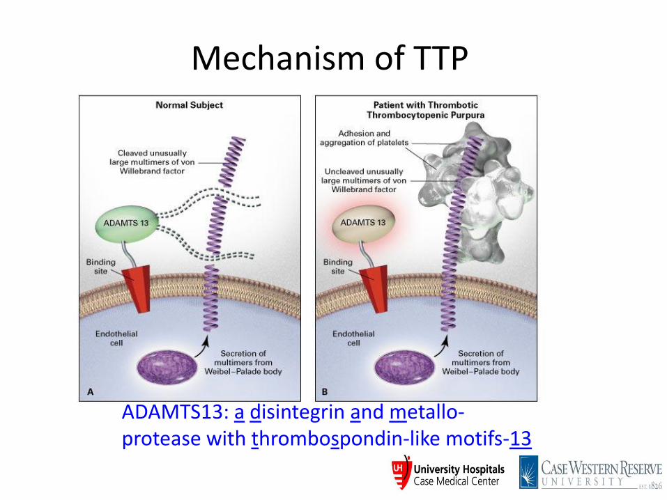
Mechanism of TTP
ADAMTS13: a disintegrin and metallo- protease with thrombospondin-like motifs-13
Copyright ©2006 American Society of Hematology. Copyright restrictions may apply.
Sadler, J. E. Hematology 2006;2006:415-420
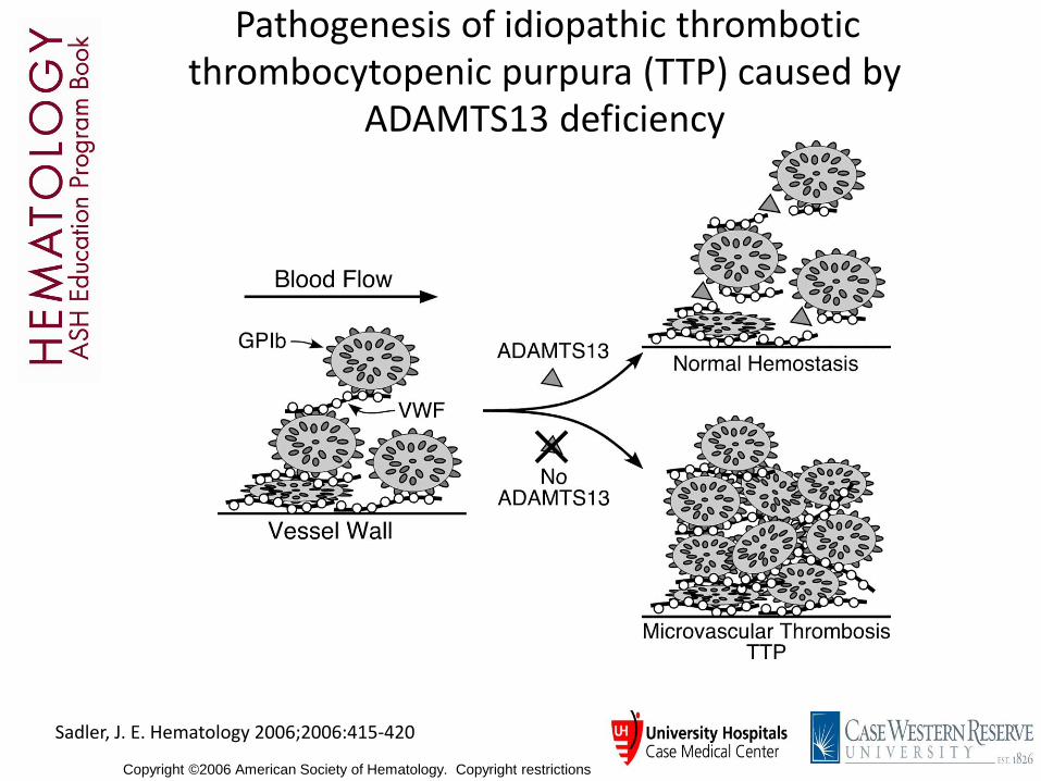
Pathogenesis of idiopathic thrombotic thrombocytopenic purpura (TTP) caused by
ADAMTS13 deficiency
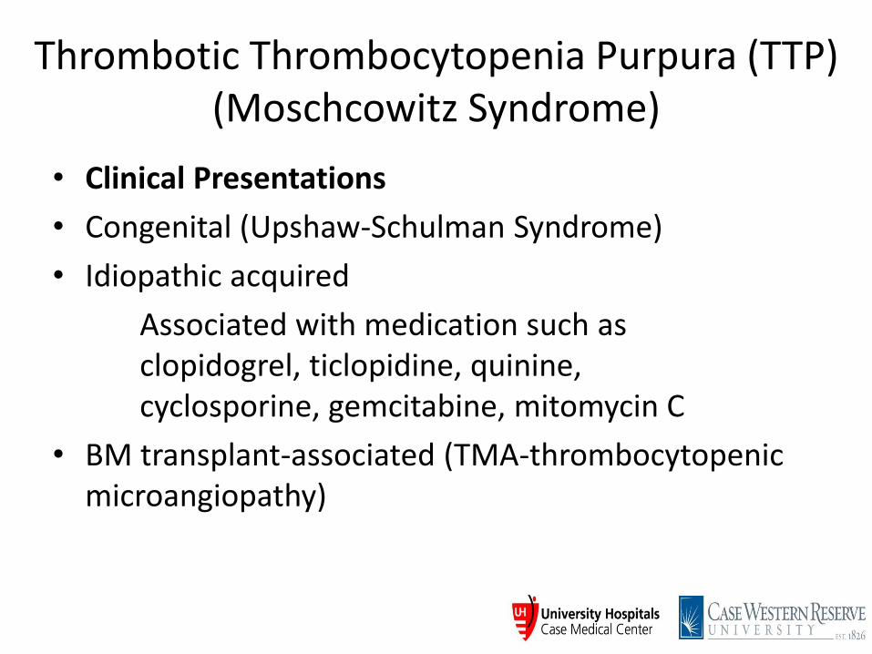
• Clinical Presentations
• Congenital (Upshaw-Schulman Syndrome)
• Idiopathic acquired
Associated with medication such as clopidogrel, ticlopidine, quinine, cyclosporine, gemcitabine, mitomycin C
• BM transplant-associated (TMA-thrombocytopenic microangiopathy)
Thrombotic Thrombocytopenia Purpura (TTP) (Moschcowitz Syndrome)
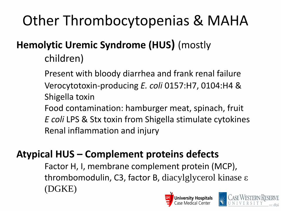
Other Thrombocytopenias & MAHA
Hemolytic Uremic Syndrome (HUS) (mostly children)
Present with bloody diarrhea and frank renal failure
Verocytotoxin-producing E. coli 0157:H7, 0104:H4 & Shigella toxin Food contamination: hamburger meat, spinach, fruit E coli LPS & Stx toxin from Shigella stimulate cytokines Renal inflammation and injury
Atypical HUS – Complement proteins defects Factor H, I, membrane complement protein (MCP), thrombomodulin, C3, factor B, diacylglycerol kinase ε
(DGKE)
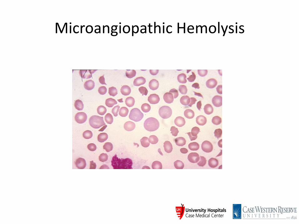
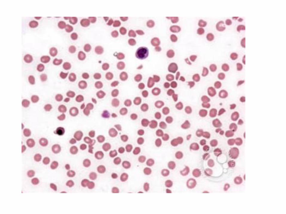
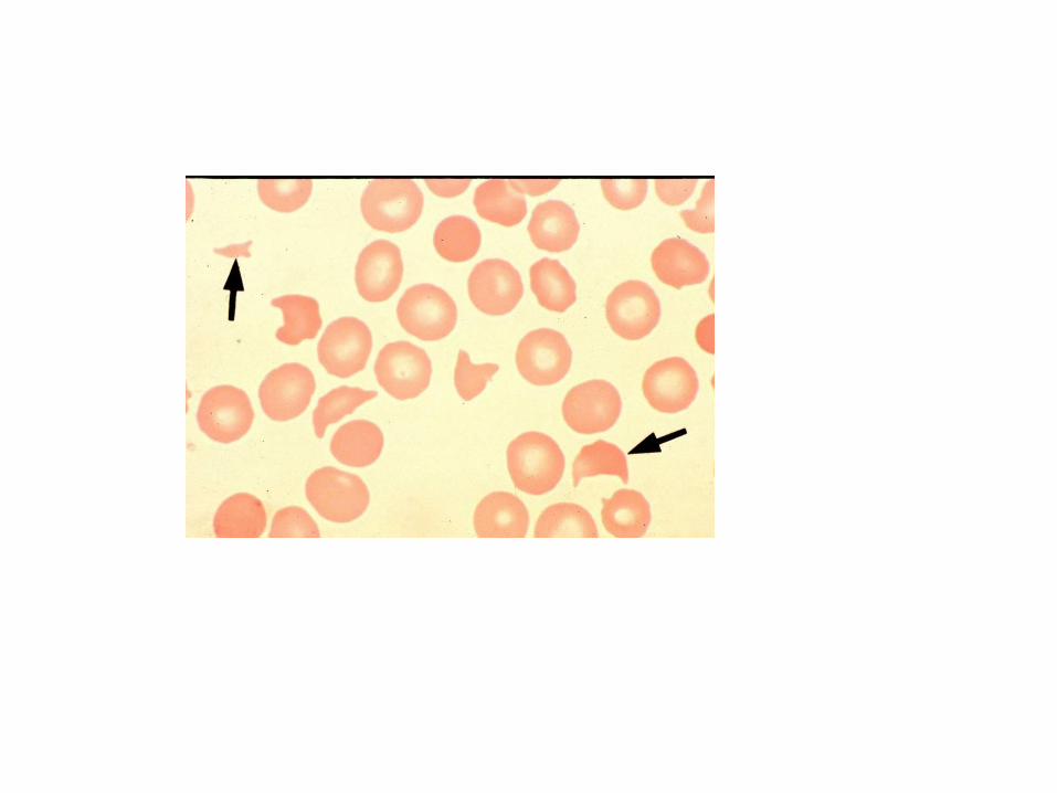
Microangiopathic Hemolysis
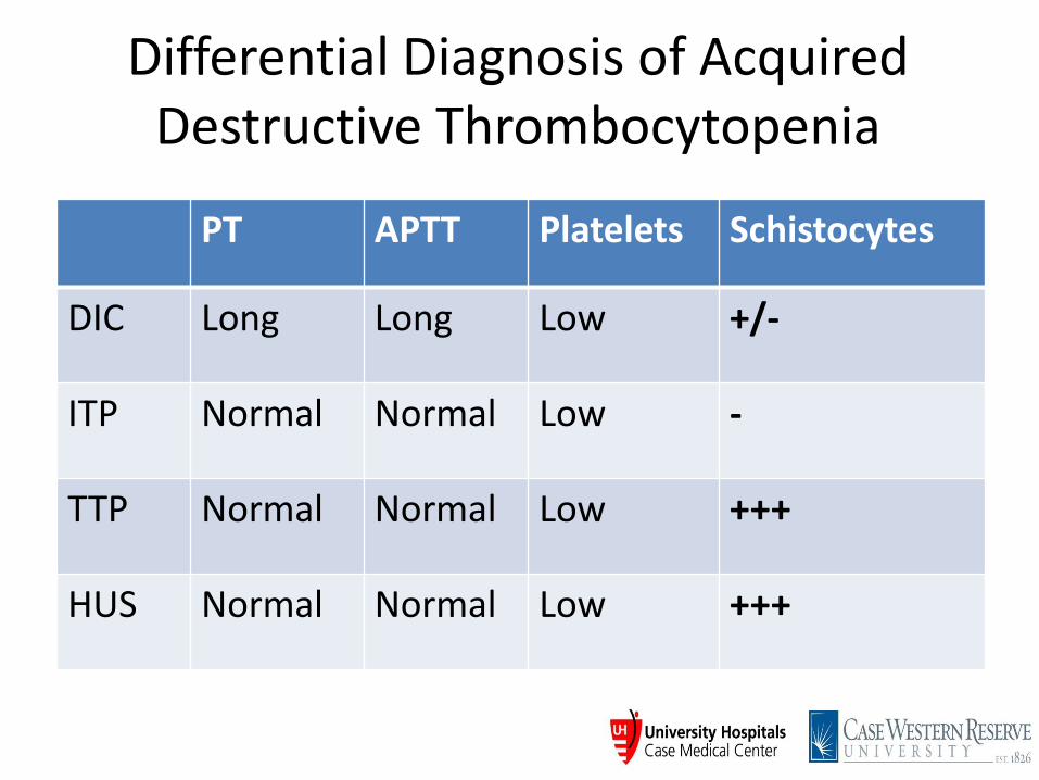
Differential Diagnosis of Acquired Destructive Thrombocytopenia
PT APTT Platelets Schistocytes
DIC Long Long Low +/-
ITP Normal Normal Low -
TTP Normal Normal Low +++
HUS Normal Normal Low +++
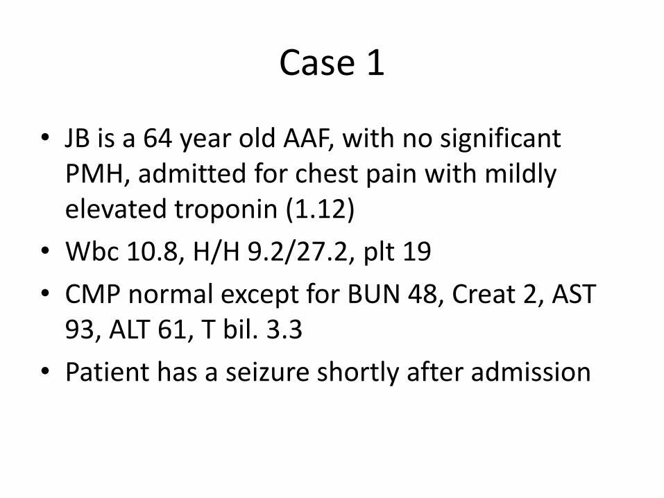
Case 1
• JB is a 64 year old AAF, with no significant PMH, admitted for chest pain with mildly elevated troponin (1.12)
• Wbc 10.8, H/H 9.2/27.2, plt 19
• CMP normal except for BUN 48, Creat 2, AST 93, ALT 61, T bil. 3.3
• Patient has a seizure shortly after admission
• LDH 1400, Haptoglobin <8
• Reticulocytes 4%
• Coagulation screen and fibrinogen WNL
Case 2
• SR is a 51 yr old AAF who presents with a rash on her arms and blisters in her oral cavity.
• PMH of sarcoidosis, COPD, HTN
• Denies fevers, chills, change in medications.
• Wbc 5.7 (normal Diff), Hb 12.6, Plts 1
• PT/aPTT normal range
• LDH, haptoglobin normal
• BUN 19, creat 1.2 (baseline)
Case 3
• CF is a 28 yr male, with not significant PMH.
• CBC on routine annual examination reveals platelet count of 70K, rest cbc within normal limits.
• No bleeding history
• No FH of blood disorders
• No medications
• P/E within normal limits
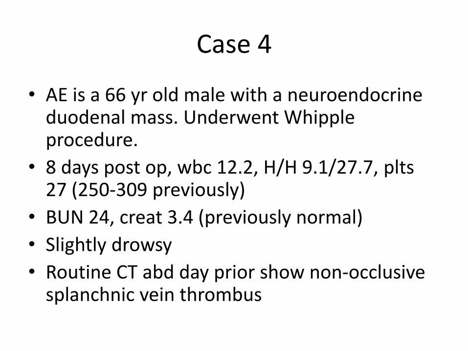
Case 4
• AE is a 66 yr old male with a neuroendocrine duodenal mass. Underwent Whipple procedure.
• 8 days post op, wbc 12.2, H/H 9.1/27.7, plts 27 (250-309 previously)
• BUN 24, creat 3.4 (previously normal)
• Slightly drowsy
• Routine CT abd day prior show non-occlusive splanchnic vein thrombus
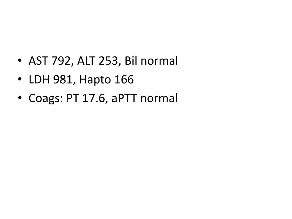
• AST 792, ALT 253, Bil normal
• LDH 981, Hapto 166
• Coags: PT 17.6, aPTT normal
• Argatroban initiated for suspected HIT
• 4 limb usg - right femoral vein clot
• Repeat abd imaging – hepatic vein thrombus
• PF4 Ab strongly positive
• SRA confirmed diagnosis